

Volume 8 Issue 3 - Australasian Society for Ultrasound in Medicine
Volume 8 Issue 3 - Australasian Society for Ultrasound in Medicine
Volume 8 Issue 3 - Australasian Society for Ultrasound in Medicine

You also want an ePaper? Increase the reach of your titles
YUMPU automatically turns print PDFs into web optimized ePapers that Google loves.
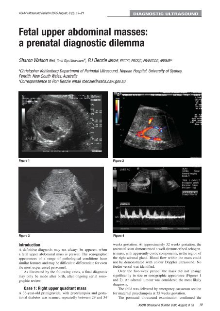
ASUM <strong>Ultrasound</strong> Bullet<strong>in</strong> 2005 August; 8 (3): 19–21DIAGNOSTIC ULTRASOUNDFetal upper abdom<strong>in</strong>al masses:a prenatal diagnostic dilemmaSharon Watson BHA, Grad Dip <strong>Ultrasound</strong> A , RJ Benzie MBChB, FRCOG, FRCS(C) FRANZCOG, ARDMS ABAChristopher Kohlenberg Department of Per<strong>in</strong>atal <strong>Ultrasound</strong>, Nepean Hospital, University of Sydney,Penrith, New South Wales, AustraliaBCorrespondence to Ron Benzie email rbenzie@wahs.nsw.gov.auFigure 1 Figure 2Figure 3 Figure 4IntroductionA def<strong>in</strong>itive diagnosis may not always be apparent whena fetal upper abdom<strong>in</strong>al mass is present. The sonographicappearances of a range of pathological conditions havesimilar features and may be difficult to differentiate <strong>for</strong> eventhe most experienced personnel.As illustrated by the follow<strong>in</strong>g cases, a f<strong>in</strong>al diagnosismay only be made after birth, after ongo<strong>in</strong>g serial sonographicreview.Case 1: Right upper quadrant massA 36-year-old primigravida, with preeclampsia and gestationaldiabetes was scanned repeatedly between 29 and 34weeks gestation. At approximately 32 weeks gestation, theantenatal scan demonstrated a well circumscribed echogenicmass, with apparently cystic components, <strong>in</strong> the region ofthe right adrenal gland. Blood flow with<strong>in</strong> the mass couldnot be demonstrated with colour Doppler ultrasound. Nofeeder vessel was identified.Over the five-week period, the mass did not changesignificantly <strong>in</strong> size or sonographic appearance (Figures 1and 2). An adrenal tumour was considered the most likelydiagnosis.The child was delivered by emergency caesarean section<strong>for</strong> maternal preeclampsia at 35 weeks gestation.The postnatal ultrasound exam<strong>in</strong>ation confirmed theASUM <strong>Ultrasound</strong> Bullet<strong>in</strong> 2005 August; 8 (3)19


