
You also want an ePaper? Increase the reach of your titles
YUMPU automatically turns print PDFs into web optimized ePapers that Google loves.
Electron Microscopy<br />
Members of the electron microscopy<br />
group have experience in various<br />
microscopic techniques ranging from<br />
light microscopy to high resolution<br />
electron microscopy. Special<br />
importance is given to the application<br />
and improvement of immunohistoand<br />
immunocytochemical methods.<br />
Recently, methods for correlative<br />
immunofluorescence and<br />
immunoelectron microscopy have<br />
been introduced as well as marked<br />
improvements in the preparation of<br />
ultrathin cryosections, the most<br />
sensitive target for high resolution<br />
immunodetection of antigens<br />
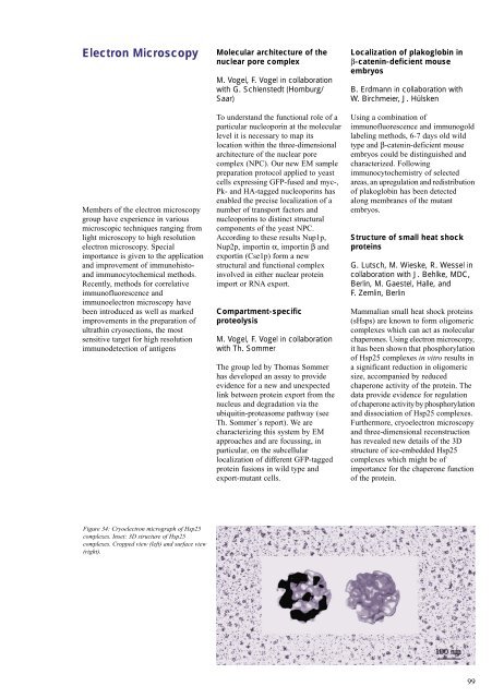
Figure 34: Cryoelectron micrograph of Hsp25<br />
complexes. Inset: 3D structure of Hsp25<br />
complexes. Cropped view (left) and surface view<br />
(right).<br />
Molecular architecture of the<br />
nuclear pore complex<br />
M. Vogel, F. Vogel in collaboration<br />
with G. Schlenstedt (Homburg/<br />
Saar)<br />
To understand the functional role of a<br />
particular nucleoporin at the molecular<br />
level it is necessary to map its<br />
location within the three-dimensional<br />
architecture of the nuclear pore<br />
complex (NPC). Our new EM sample<br />
preparation protocol applied to yeast<br />
cells expressing GFP-fused and myc-,<br />
Pk- and HA-tagged nucleoporins has<br />
enabled the precise localization of a<br />
number of transport factors and<br />
nucleoporins to distinct structural<br />
components of the yeast NPC.<br />
According to these results Nup1p,<br />
Nup2p, importin α, importin β and<br />
exportin (Cse1p) form a new<br />
structural and functional complex<br />
involved in either nuclear protein<br />
import or RNA export.<br />
Compartment-specific<br />
proteolysis<br />
M. Vogel, F. Vogel in collaboration<br />
with Th. Sommer<br />
The group led by Thomas Sommer<br />
has developed an assay to provide<br />
evidence for a new and unexpected<br />
link between protein export from the<br />
nucleus and degradation via the<br />
ubiquitin-proteasome pathway (see<br />
Th. Sommer´s report). We are<br />
characterizing this system by EM<br />
approaches and are focussing, in<br />
particular, on the subcellular<br />
localization of different GFP-tagged<br />
protein fusions in wild type and<br />
export-mutant cells.<br />
Localization of plakoglobin in<br />
β-catenin-deficient mouse<br />
embryos<br />
B. Erdmann in collaboration with<br />
W. Birchmeier, J. Hülsken<br />
Using a combination of<br />
immunofluorescence and immunogold<br />
labeling methods, 6-7 days old wild<br />
type and β-catenin-deficient mouse<br />
embryos could be distinguished and<br />
characterized. Following<br />
immunocytochemistry of selected<br />
areas, an upregulation and redistribution<br />
of plakoglobin has been detected<br />
along membranes of the mutant<br />
embryos.<br />
Structure of small heat shock<br />
proteins<br />
G. Lutsch, M. Wieske, R. Wessel in<br />
collaboration with J. Behlke, <strong>MDC</strong>,<br />
Berlin, M. Gaestel, Halle, and<br />
F. Zemlin, Berlin<br />
Mammalian small heat shock proteins<br />
(sHsps) are known to form oligomeric<br />
complexes which can act as molecular<br />
chaperones. Using electron microscopy,<br />
it has been shown that phosphorylation<br />
of Hsp25 complexes in vitro results in<br />
a significant reduction in oligomeric<br />
size, accompanied by reduced<br />
chaperone activity of the protein. The<br />
data provide evidence for regulation<br />
of chaperone activity by phosphorylation<br />
and dissociation of Hsp25 complexes.<br />
Furthermore, cryoelectron microscopy<br />
and three-dimensional reconstruction<br />
has revealed new details of the 3D<br />
structure of ice-embedded Hsp25<br />
complexes which might be of<br />
importance for the chaperone function<br />
of the protein.<br />
99