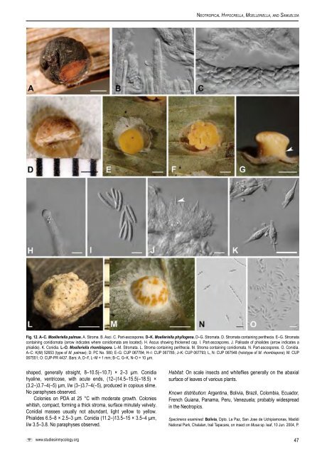
Ne o t r o p i c a l Hy p o c r e l l a, Mo e l l e r i e l l a, a n d Sa m u e l s i aFig. 12. A–C. <strong>Moelleriella</strong> palmae. A. Stroma. B. Asci. C. Part-ascospores. D–K. <strong>Moelleriella</strong> phyllogena. D–G. Stromata. D. Stromata containing perithecia. E–G. Stromatacontaining conidiomata (arrow indicates where conidiomata are located). H. Ascus showing thickened cap. I. Part-ascospores. J. Palisade of phialides (arrow indicates aphialide). K. Conidia. L–O. <strong>Moelleriella</strong> rhombispora. L–M. Stromata. L. Stroma containing perithecia. M. Stroma containing conidiomata. N. Part-ascospores. O. Conidia.A–C: K(M) 52653 (type of M. palmae); D: PC No. 580; E–G: CUP 067784; H–I: CUP 067785; J–K: CUP 067793; L, N: CUP 067548 (holotype of M. rhombispora); M: CUP067551; O: CUP-PR 4437. Bars: A, D–F, L–M = 1 mm; B–C, G–K, N–O = 10 µm.shaped, generally straight, 8–10.5(–10.7) × 2–3 µm. Conidiahyaline, ventricose, with acute ends, (12–)14.5–15.5(–18.5) ×(3.2–)3.7–4(–5) µm, l/w (3–)3.7–4(–5), produced in copious slime.No paraphyses observed.Colonies on PDA at 25 °C with moderate growth. Colonieswhitish, compact, forming a thick stroma, surface minutely velvety.Conidial masses usually not abundant, light yellow to yellow.Phialides 6.5–8 × 2.5–3 µm. Conidia (11.2–)13.5–15 × 3.5–4 µm,l/w 3.5–3.8. No paraphyses observed.Habitat: On scale insects <strong>and</strong> whiteflies generally on the abaxialsurface of leaves of various plants.Known distribution: Argentina, Bolivia, Brazil, Colombia, Ecuador,French Guiana, Panama, Peru, Venezuela; probably widespreadin the Neotropics.Specimens examined: Bolivia, Dpto. La Paz, San Jose de Uchipiamonas, MadidiNational Park, Chalalan, trail Tapacare, on insect on Musa sp. leaf, 10 Jun. 2004, P.www.studiesinmycology.org47
Ch av e r r i e t a l.Chaverri (P.C. 554, P.C. 555), D. Quintana, M. Sogonov, A. Alvarez (CUP 067784;CUP 067785). Brazil, On leaves of Lauraceae, Aug. 1895, Edwalli (holotype of H.edwallii, S-F10585); Itaimbezinho, on leaf, 22 May 1994, H.C. Evans (I94-927) (CUP067786); Rio de Janeiro, Tijuca National Park, on leaf, 15 Jun. 1989, H.C. Evans(neotype of M. sulphurea P.C. 756 = CUP 067787). Colombia, Choco, North of RioJobi-Coqui, forest, on leaf, 29–31 May 1993, H.C. Evans (I93-856 = CUP 067788;P.C. 753 = CUP 067789). Ecuador, Manabi. Y de la Laguna, Reserva Bilsa, primaryforest, 00°24’N, 79° 49’E, elev. 500–600 m, 2 May 2004, on insect on leaf, G.J.Samuels (GS 9513, GS 9508, GS 9516), H.C. Evans, M.C. Aime (P.C. 628 = CUP067790; CUP 067791; CUP 067792). French Guiana, Cayenne, on insect on leaf,Mar. 1839, Leprieur 580 (lectotype of H. phyllogena PC!) Panama, Fortuna, onleaf of Costa, 14 Jul. 2002, J. F. Bischoff (J.B. 130 = P.C. 738) (CUP 067793).Venezuela, Bolivar, Canaima, on leaf, 23 Jan. 1994, H.C. Evans (I94-909) (CUP067794).Notes: <strong>Moelleriella</strong> phyllogena is similar to M. basicystis, M.umbospora, <strong>and</strong> M. disjuncta. The differences are in the size<strong>and</strong> shape of the part-spores <strong>and</strong> conidia, <strong>and</strong> the geographicaldistribution. The type of “A.” juruensis was deposited in herbariumB <strong>and</strong> was lost in WW II. According to Petch (1921), the type of A.juruensis contained only one stroma that was poorly developed <strong>and</strong>in bad condition. The type of A. lauricola in LPS does not includeany stromata. However, based on a drawing on the envelope <strong>and</strong>the original description of the species, this species appears to bea synonym of M. phyllogena. The types of H. weberbauri <strong>and</strong> M.sulphurea in herbarium B were also lost in WWII. According toPetch’s (1921) measurements of these specimens, they seem tobe synonyms of M. phyllogena. <strong>Moelleriella</strong> sulphurea is neotypifiedherein with a specimen from Brazil. Cultures did not survive storageat 8 °C. <strong>Moelleriella</strong> phyllogena belongs in the Effuse clade.18. <strong>Moelleriella</strong> rhombispora (M. Liu & K.T. Hodge) M. Liu& Chaverri, comb. nov. MycoBank MB511378. Fig. 12L–O.≡ Hypocrella rhombispora M. Liu & K.T. Hodge, Mycol. Res. 110: 551.2006.Anamorph: aschersonia-like.Stromata pale yellow to pale orange, pulvinate <strong>and</strong> slightlytuberculate, 2–2.5 mm diam, slightly constricted at base, sometimessurrounded by hypothallus. Stromatal tissue dense texturaintricata. If present, hypothallus narrow, 0.6 mm wide, <strong>and</strong> minutelytomentose. Perithecia densely arranged in stroma, embedded,ostioles not projecting, brownish yellow; 300–450 × 210–300µm. Asci cylindrical, 148–296 × 6–14 µm, caps 5–8 µm thick.Ascospores initially filiform, dividing into part-spores. Part sporesfusoid, acute at both ends, 10–14 × 2–3 µm, or others cylindricalwith blunt ends, usually swollen at midpoint, 7–12 × 1.5–2.5 µm.Anamorphic stromata white, thin pulvinate, hemi-globose orscutate with a hemispheric central region abruptly attenuating<strong>and</strong> towards edge, 1–3 mm diam; surface minutely pruinose.Hypothallus, if present, 0.2–0.8 mm wide. Conidiomata >4, arrangedconcentrically, scattered, or forming a reticulum on conical part ofstroma. Conidial masses pale yellow, not confluent. ConidiomataU-shaped or convolute in section, hymenium lining inner surface ofconidioma. Phialides flask-shaped, slender, tapering near truncateapices, 8–12 × 1.5–2 µm. Conidia 9–14 × 2.5–3 µm, inflated atmidpoint <strong>and</strong> tapering at both ends, l/w ca. 3.5–4.5. Paraphysesabsent.Colonies on PDA at 25 °C slow-growing, thick pulvinate,moderately compact, firm <strong>and</strong> leathery, greyish white to yellowishwhite, surface minutely tomentose, smooth to radially wrinkled,covered with deep yellow conidial masses. No discrete conidiomataformed, conidial masses directly produced from surface of colony.Phialides 8–12(–15) × 2–2.5 µm. Conidia markedly inflated at themidpoint <strong>and</strong> tapering at both ends, 8.5–12(–17) × 2–3 µm, l/w ca.4–4.5. No paraphyses observed.Habitat: On scale insects or whiteflies on leaves of Cyclanthus,Guarea, <strong>and</strong> unidentified.Distribution: Costa Rica, Guatemala, Honduras, Mexico, <strong>and</strong>Puerto Rico.Specimens examined: Costa Rica, Heredia, La Selva Biological Station, CaminoCantarrana, on Cyclanthus bipartitus, 19 Jun. 2002, M. Liu (CR 07) (CUP 67296 =culture ARSEF 7511); 5 Jan. 2004, P. Chaverri, (P.C. 466, P.C. 467) (CUP 067537,CUP 067538); beside entrance to Plantation RCC, 20 Jun. 2002, M. Liu (ML44-3),(culture CR32 = CUP 067346); Puntarenas, Las Cruces Biological Reserve, WilsonBotanical Garden, large loop of jungle trail, on Guarea rhopalocaipa, 4 Jul. 2002,M. Liu (ML64) (culture CR34 = CUP 67369). Guatemala, Tikal National Park, onleaf, 26 Jun. 2004, M.G. Milgroom (CUP 067494). Honduras, Yojoa, Los Pinos,Parque Nacional Cerro Azul-Meambar, 850 m elev., 3 Sep. 2004, P. Chaverri (P.C.691, P.C. 693, P.C. 696, P.C. 698), P.A. Sheikh, (holotype CUP 067548; CUP067549, CUP 067550; CUP 067551); Copan, Santa Rita, Reserva Peña Quemada,9 Sep. 2004, P. Chaverri (P.C. 675), P. A. Sheikh (CUP 067547). Mexico, Veracruz,Amayaga, Catemaco, 500 m elev., 14 Dec. 2003, P. Chaverri (P.C. 458, P.C. 460),J. García-Alvarado (CUP 067795, CUP 067534). Puerto Rico, between Mayaguez<strong>and</strong> Maricao, beside road 105, 15 Dec. 2003, M. Liu & Z.D. Wang, ML164 (CUP-PR4406; ex-type culture ML164 = ARSEF 7390); Guajataca Forest, trail no. 9, on fern,18 Dec. 2003, M. Liu (ML201-1, ML201-3, ML201-5a), Z.D. Wang (CUP-PR 4437 =ARSEF 7395, ARSEF 7399, ARSEF 7400).Notes: The most distinctive characters of this species are the shapeof the part-spores <strong>and</strong> conidia, both of which are distinctly inflatedin the middle. These characters are shared by M. phyllogena, M.basicystis, <strong>and</strong> M. umbospora. <strong>Moelleriella</strong> rhombispora belongs inthe Effuse clade.Additional illustrations: figs 4J–L, 8A–M, in Liu et al. (2006).19. <strong>Moelleriella</strong> sloaneae (Pat.) Chaverri & K.T. Hodge,comb. nov. MycoBank MB511379. Fig. 13A–I.≡ Hypocrella sloaneae Pat., Enum. Champ. Guadeloupe, p. 80. 1903.= Hypocrella amazonica Henn., Hedwigia 43: 246. 1904.Anamorph: aschersonia-likeStromata containing teleomorph, (1.5–)1.7–2(–2.2) mm diam,whitish, greyish yellow, pale orange, thin-pulvinate to pulvinate withwide base, composed of few to numerous gregarious tuberclesarising from a pulvinate to hemispherical base; when present,conidiomata scattered throughout; surface of tubercles <strong>and</strong> basepruinose due to loosely woven, thick-walled hyphae that formstroma. Tubercles projecting <strong>and</strong> aggregated, hemispherical,cylindrical or slightly narrowing apically; ostioles brownish yellow tobrownish orange. Perithecia embedded in stroma, one peritheciumper tubercle, perithecia nearly flask-shaped to ovoid, 400–500 ×250–300 µm. Asci cylindrical, (163–)175–240(–248) × 7.2–9.7(–10.5) µm, caps thick, (3.3–)4.7–5.5(–6.3) µm. Ascospores filiform,disarticulating into cylindrical part-spores, with somewhat roundedends, (9–)13.5–15(–18.3) × (2–)2.8–3(–4) µm.Teleomorph <strong>and</strong> anamorph sometimes in same stromata.Exclusively anamorphic stromata when young, effuse, thin, whitish,tomentose, with irregular <strong>and</strong> shallow conidiomata that may besomewhat circular or irregular <strong>and</strong> confluent, sometimes formingsterile cylindrical finger-like projections. Conidiomata when maturegenerally scattered, 2 to numerous per stroma, appearing as simpleshallow depressions of stromatic surface without a differentiated48