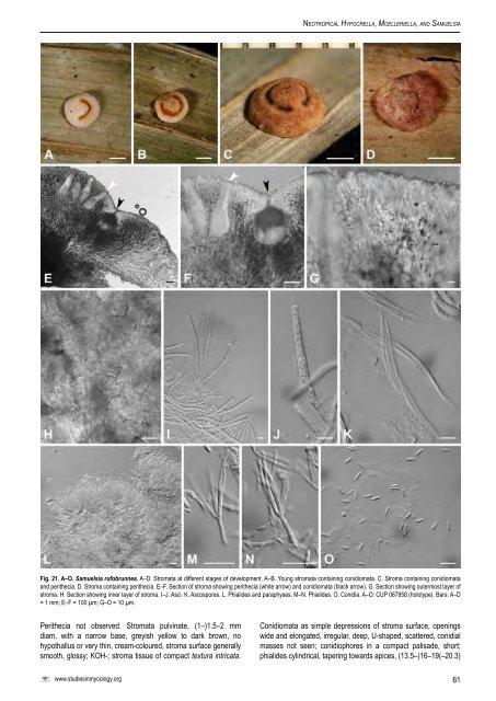
Ne o t r o p i c a l Hy p o c r e l l a, Mo e l l e r i e l l a, a n d Sa m u e l s i aFig. 21. A–O. <strong>Samuelsia</strong> rufobrunnea. A–D. Stromata at different stages of development. A–B. Young stromata containing conidiomata. C. Stroma containing conidiomata<strong>and</strong> perithecia. D. Stroma containing perithecia. E–F. Section of stroma showing perithecia (white arrow) <strong>and</strong> conidiomata (black arrow). G. Section showing outermost layer ofstroma. H. Section showing inner layer of stroma. I–J. Asci. K. Ascospores. L. Phialides <strong>and</strong> paraphyses. M–N. Phialides. O. Conidia. A–O: CUP 067858 (holotype). Bars: A–D= 1 mm; E–F = 100 µm; G–O = 10 µm.Perithecia not observed. Stromata pulvinate, (1–)1.5–2 mmdiam, with a narrow base, greyish yellow to dark brown, nohypothallus or very thin, cream-coloured, stroma surface generallysmooth, glossy; KOH-; stroma tissue of compact textura intricata.www.studiesinmycology.orgConidiomata as simple depressions of stroma surface, openingswide <strong>and</strong> elongated, irregular, deep, U-shaped, scattered, conidialmasses not seen; conidiophores in a compact palisade, short;phialides cylindrical, tapering towards apices, (13.5–)16–19(–20.3)61
Ch av e r r i e t a l.Fig. 22. A–J. <strong>Samuelsia</strong> sheikhii. A–C. Stromata. B–C. Stromata after adding 3 % KOH. D–E. Section of stroma showing conidiomata. F. Section showing outermost layer ofstroma. G. Section showing inner layer of stroma. H. Phialides <strong>and</strong> paraphyses. I. Phialides. J. Conidia. A–J: CUP 067859 (holotype). Bars: A–C = 1 mm; D–E = 100 µm; F–J= 10 µm.× (1.3–)1.7–2(–2.2) µm; conidia small, hyaline, unicellular, smooth,allantoid, (5.5–)6–6.5(–7.7) × (1.8–)2(–2.5) µm, l/w (2.5–)3(–3.8).Paraphyses present, circinate.Habitat: On scale-insects on leaves.Known distribution: Chile (type locality).Specimens examined: Chile, Corral, on scale-insects on leaves, Dec. 1905, R.Thaxter (lectotype of A. intermedia designated here, <strong>and</strong> holotype of S. intermediaFH 3991!); Jan. 1906, R. Thaxter (FH 3990!).Notes: Micromorphologically, S. intermedia is indistinguishablefrom S. rufobrunnea. The species are distinguished by the distinctmacromorphologies of the stromata. In addition, S. rufobrunnea istropical (Bolivia <strong>and</strong> Peru) while S. intermedia is subtropical (Chile).There are several specimens in FH labeled as “A. intermedia”(Petch 1921), but only one reads “co-type.” Therefore, this speciesis lectotypified here with FH 3991. Although there are no DNAsequences of S. intermedia, the micro <strong>and</strong> macro characteristics ofthe anamorph place this species within <strong>Samuelsia</strong>.4. <strong>Samuelsia</strong> rufobrunnea Chaverri & K.T. Hodge, sp. nov.MycoBank MB511389. Fig. 21A–O.Anamorph: aschersonia-likeTeleomorphosis: Stromatibus pulvinatus, (2–)2.2–3.3(–4) mm diam, sub-brunneusvel brunneoluteus; ascosporae multicellulares, macrofusiformes, (42–)48–55(–57)× 1.5–2.5 µm. Anamorphosis: Stromatibus pulvinatus, pycnidium; phialides (14.5–)15.3–19.2(–22.5) × 1.3–1.5 µm; conidii allantoideus vel subelliposideus, (5.5–)6–6.2(–6.7) × (1.3–)1.7–2 µm, longitudo/crassitudo (3–)3.5–3.7(–4.5); paraphysisprescens, circinatus. Holotypus: CUP 067858.Stromata pulvinate, (2–)2.2–3.3(–4) mm diam, with a wide base,hard, pale yellow when young, then brownish yellow with a reddishtinge when perithecia are fully mature, no hypothallus, stromasurface generally smooth <strong>and</strong> glossy, KOH+, ostioles brownishorange; stroma tissue of compact textura intricata. Peritheciacompletely embedded in stroma, scattered. Perithecia, subglobose,320–380 × 110–120 µm. Asci mostly cylindrical to clavate,(98–)100–118(–120) × 5–6.5(–7) µm, caps 2–3 µm. Ascosporeshyaline, multiseptate, smooth, long fusiform to filiform, (42–)48–55(–57) × 1.5–2.5 µm.Conidiomata as deep depressions of stromal surface, from topview elongated <strong>and</strong> generally fusing with neighboring conidiomata,62