Materials Science and Engineering Laboratory FY 2004 ... - NIST
Materials Science and Engineering Laboratory FY 2004 ... - NIST
Materials Science and Engineering Laboratory FY 2004 ... - NIST

You also want an ePaper? Increase the reach of your titles
YUMPU automatically turns print PDFs into web optimized ePapers that Google loves.
Multi-Modal Imaging <strong>and</strong> Quantitative Data Reduction<br />
Methods for Regenerative Medicine<br />
This project develops methods for determining<br />
the viability of tissue engineered medical<br />
products (TEMPs) through the use of in-vitro<br />
imaging coupled with data reduction techniques.<br />
These techniques are vital for distilling the<br />
voluminous amount of imaging data down to<br />
selected metrics of interest relating to TEMP<br />
viability. We illustrate this approach by using<br />
various imaging techniques, along with 3D image<br />
quantitation, to establish relationships between<br />
cell proliferation <strong>and</strong> scaffold microstructure.<br />
Joy P. Dunkers <strong>and</strong> Forrest A. L<strong>and</strong>is<br />
TEMP Imaging: Collinear Optical Coherence/<br />
Confocal Fluorescence Microscopies<br />
We use collinear optical coherence microscopy<br />
(OCM) in conjunction with one photon confocal<br />
fluorescence microscopy (CFM) as a multifunctional<br />
technique for characterization of TEMPs. OCM with<br />
its unparalleled combination of resolution (≈ 1 µm) <strong>and</strong><br />
sensitivity (> 100 dB) is well-suited for imaging TEMPs.<br />
CFM has proven to be an extremely powerful technique<br />
for underst<strong>and</strong>ing cell viability, differentiation, <strong>and</strong><br />
protein expression in tissue engineering <strong>and</strong> provides<br />
complementary information to the structural<br />
characterization provided by OCM.<br />
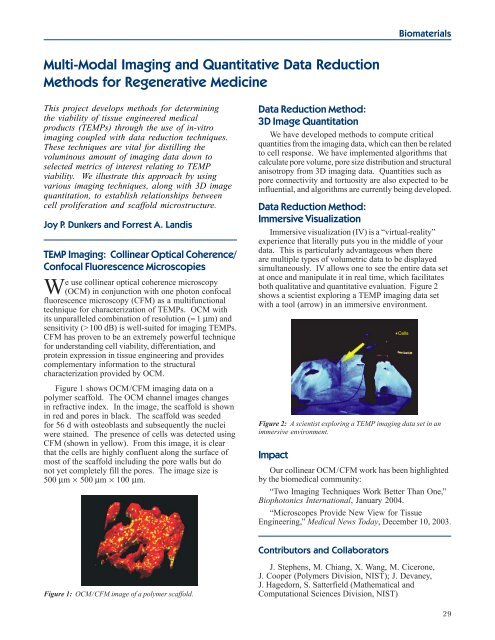
Figure 1 shows OCM/CFM imaging data on a<br />
polymer scaffold. The OCM channel images changes<br />
in refractive index. In the image, the scaffold is shown<br />
in red <strong>and</strong> pores in black. The scaffold was seeded<br />
for 56 d with osteoblasts <strong>and</strong> subsequently the nuclei<br />
were stained. The presence of cells was detected using<br />
CFM (shown in yellow). From this image, it is clear<br />
that the cells are highly confluent along the surface of<br />
most of the scaffold including the pore walls but do<br />
not yet completely fill the pores. The image size is<br />
500 µm × 500 µm × 100 µm.<br />
Figure 1: OCM/CFM image of a polymer scaffold.<br />
Biomaterials<br />
Data Reduction Method:<br />
3D Image Quantitation<br />
We have developed methods to compute critical<br />
quantities from the imaging data, which can then be related<br />
to cell response. We have implemented algorithms that<br />
calculate pore volume, pore size distribution <strong>and</strong> structural<br />
anisotropy from 3D imaging data. Quantities such as<br />
pore connectivity <strong>and</strong> tortuosity are also expected to be<br />
influential, <strong>and</strong> algorithms are currently being developed.<br />
Data Reduction Method:<br />
Immersive Visualization<br />
Immersive visualization (IV) is a “virtual-reality”<br />
experience that literally puts you in the middle of your<br />
data. This is particularly advantageous when there<br />
are multiple types of volumetric data to be displayed<br />
simultaneously. IV allows one to see the entire data set<br />
at once <strong>and</strong> manipulate it in real time, which facilitates<br />
both qualitative <strong>and</strong> quantitative evaluation. Figure 2<br />
shows a scientist exploring a TEMP imaging data set<br />
with a tool (arrow) in an immersive environment.<br />
Figure 2: A scientist exploring a TEMP imaging data set in an<br />
immersive environment.<br />
Impact<br />
Our collinear OCM/CFM work has been highlighted<br />
by the biomedical community:<br />
“Two Imaging Techniques Work Better Than One,”<br />
Biophotonics International, January <strong>2004</strong>.<br />
“Microscopes Provide New View for Tissue<br />
<strong>Engineering</strong>,” Medical News Today, December 10, 2003.<br />
Contributors <strong>and</strong> Collaborators<br />
J. Stephens, M. Chiang, X. Wang, M. Cicerone,<br />
J. Cooper (Polymers Division, <strong>NIST</strong>); J. Devaney,<br />
J. Hagedorn, S. Satterfield (Mathematical <strong>and</strong><br />
Computational <strong>Science</strong>s Division, <strong>NIST</strong>)<br />
29