Materials Science and Engineering Laboratory FY 2004 ... - NIST
Materials Science and Engineering Laboratory FY 2004 ... - NIST
Materials Science and Engineering Laboratory FY 2004 ... - NIST

You also want an ePaper? Increase the reach of your titles
YUMPU automatically turns print PDFs into web optimized ePapers that Google loves.
Technical Highlights<br />
Coherent Anti-Stokes Raman Scattering (CARS) for<br />
In-Situ Chemical Imaging of Tissue <strong>Engineering</strong> Constructs<br />
Design issues relating to bioactive devices for<br />
regenerative medicine reflect the spatial <strong>and</strong><br />
chemical complexity of biological <strong>and</strong> materials<br />
issues <strong>and</strong> their interactions. Tools developed<br />
for the purpose of aiding underst<strong>and</strong>ing of these<br />
systems must have sufficient discriminatory power<br />
to sort out this complexity. We have introduced<br />
a relatively simple broadb<strong>and</strong> spectroscopic<br />
microscopy based on CARS that can be used to<br />
rapidly acquire volumetric, chemically-specific<br />
images with submicrometer resolution.<br />
Marcus T. Cicerone<br />
Biological research in general could be significantly<br />
aided by a high-resolution, chemically sensitive<br />
volumetric imaging method that allows rapid, non-invasive<br />
study of processes on the tissue, cellular <strong>and</strong> sub-cellular<br />
levels. The need for such a method is particularly<br />
acute in the field of tissue engineering, where cycles<br />
of cell-seeding <strong>and</strong> analysis can take months. St<strong>and</strong>ard<br />
analysis methods are labor intensive <strong>and</strong> destructive,<br />
so multiple endpoint studies must be performed <strong>and</strong><br />
temporal correlation assumed. Non-invasive microspectroscopic<br />
imaging could alleviate many problems<br />
associated with the evaluation of tissue scaffolds both<br />
by allowing continuity of analysis on a single scaffold<br />
construct, <strong>and</strong> by maintaining spatio-temporal information.<br />
Our vision is the creation of a non-invasive microscopic<br />
imaging technique that rapidly discriminates between an<br />
arbitrary number of chemically distinct structures or<br />
species in a spatially resolved way within a biological<br />
system. We envision that this would be done without<br />
staining or otherwise intrusively manipulating the system.<br />
We expect that such a microscopy tool would have a<br />
significant impact on the way in-vitro tissue-engineering<br />
studies are carried out.<br />
While our vision may sound fanciful, the basis<br />
for such powerful <strong>and</strong> generally applicable chemical<br />
discrimination was established many decades ago via<br />
vibrational spectroscopy. Our contribution has been<br />
to marry broadb<strong>and</strong> vibrational spectroscopy, with<br />
its inherent chemical resolving power, to a nonlinear,<br />
multiphoton microscopy (CARS microscopy), which<br />
has already been demonstrated to have significant utility<br />
for non-invasively imaging biological systems, even in<br />
the absence of significant chemical resolving power.<br />
Microscopic coherent anti-Stokes Raman<br />
scattering (µCARS) can answer important imaging<br />
2<br />
needs of the biological research community, including<br />
relative non-invasiveness, rapidity, <strong>and</strong> chemical specificity.<br />
CARS provides chemical specificity through its intrinsic<br />
sensitivity to molecular vibrational transitions. CARS is<br />
sensitive to the third order polarizability, χ (3) , which has<br />
nonresonant <strong>and</strong> resonant components, the latter being<br />
related to the Raman scattering cross section. Therefore,<br />
µCARS uses Raman (vibrational) susceptibility as a<br />
contrast mechanism, but it is approximately 10 3 more<br />
efficient than spontaneous Raman scattering.<br />
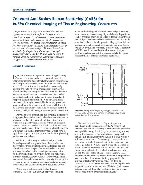
Figure 1: Energy level diagram for single frequency CARS<br />
process (solid vertical arrows) <strong>and</strong> broadb<strong>and</strong> CARS (solid<br />
<strong>and</strong> dashed vertical arrows).<br />
The solid vertical lines of Figure 1 represent<br />
transitions made by molecules in the “classic” CARS<br />
scheme. Molecules in a sample of interest are promoted<br />
to a specific energy E = h (wp – wS), where ωp <strong>and</strong> ωS are the frequencies of the near infrared pump <strong>and</strong><br />
Stokes light pulses, respectively, <strong>and</strong> h is h/2π where h<br />
is Planck’s constant. If that energy corresponds to a<br />
particular excited vibrational state in the molecule, that<br />
state is populated. A subsequent probe photon (also ωp) promotes the vibrationally excited molecule to another<br />
(higher) virtual state, from which it relaxes <strong>and</strong> emits<br />
an anti-Stokes photon at a specific frequency, ωaS. The flux of anti-Stokes photons detected is proportional<br />
to the number of molecules initially prepared in the<br />
targeted vibrationally excited state. To date, this<br />
“classic” µCARS scheme has been used to acquire only<br />
single-frequency or narrowb<strong>and</strong> (200 cm –1 –<br />
–<br />
) vibrational<br />
information, <strong>and</strong> thus, chemical specificity is limited<br />
to only one or two species simultaneously.