

Official Proceedings - AIUM
Official Proceedings - AIUM
Official Proceedings - AIUM

You also want an ePaper? Increase the reach of your titles
YUMPU automatically turns print PDFs into web optimized ePapers that Google loves.
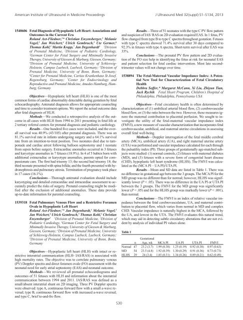
American Institute of Ultrasound in Medicine <strong>Proceedings</strong> J Ultrasound Med 32(suppl):S1–S134, 20131540606 Fetal Diagnosis of Hypoplastic Left Heart: Associations andOutcomes in the Current EraRoland Axt-Fliedner, 1 * Christian Enzensberger, 1 MelanieVogel, 2 Jan Weichert, 4 Ulrich Gembruch, 5 Ute Germer, 6Thomas Kohl, 3 Martin Krapp, 7 Jan Degenhardt 1 1 Divisionof Prenatal Medicine, 2 Division of Pediatric Cardiology,3German Center for Fetal Surgery and Minimally InvasiveTherapy, University of Giessen & Marburg, Giessen, Germany;4Division of Prenatal Medicine, University of Schleswig-Holstein, Campus Luebeck, Luebeck, Germany; 5 Division ofPrenatal Medicine, University of Bonn, Bonn, Germany;6Center for Prenatal Medicine, Caritas Krankenhaus St Josef,Regensburg, Germany; 7 Center for Endocrinology andReproductive and Prenatal Medicine, Amedes Hamburg, Hamburg,GermanyObjectives—Hypoplastic left heart (HLH) is one of the mostcommon forms of cardiac abnormality detectable during gestation by fetalechocardiography. Antenatal diagnosis allows for appropriate counselingand time to consider treatment options. We report the actual outcome dataafter fetal diagnosis of HLH.Methods—We conducted a retrospective analysis of the outcomein all cases with HLH from 1994 to 2011 presenting in fetal life at2 tertiary referral centers for prenatal diagnosis and pediatric cardiology.Results—One hundred five cases were included, and the overallsurvival was 40.9% (43/105) after prenatal diagnosis. There was an81.1% survival rate in infants undergoing surgery and a 64.1% survivalrate from an intention-to-treat position. Two neonates died due to tamponadeand cardiac arrest following balloon septostomy and 1 neonatefrom sepsis before surgery. Extracardiac anomalies occurred in 3 fetusesand karyotype anomalies in 7 fetuses (18.9%). In 4 of 5 babies born withadditional extracradiac or karyotype anomalies, parents opted for compassionatecare. The first had trisomy 13; the second had trisomy 18; thethird neonate presented with spina bifida; and the fourth presented with hydronephrosisand pulmonary atresia. Termination of pregnancy took placein 17 cases (16.1%).Conclusions—Thorough antenatal evaluation should includekaryotyping and detailed extracardiac and intracardiac assessment to accuratelypredict the risks of surgery. Prenatal counseling might be modifiedafter the exclusion of additional anomalies. These data provideup-to-date information for parental counseling.1539318 Fetal Pulmonary Venous Flow and a Restrictive ForamenOvale in Hypoplastic Left HeartRoland Axt-Fliedner, 1 * Jan Degenhardt, 1 Melanie Vogel, 2Jan Weichert, 4 Ulrich Gembruch, 5 Thomas Kohl, 3 ChristianEnzensberger 1 1 Division of Prenatal Medicine, 2 Division ofPediatric Cardiology, 3 German Center for Fetal Surgery andMinimally Invasive Therapy, University of Giessen & Marburg,Giessen, Germany; 4 Division of Prenatal Medicine, Universityof Schleswig-Holstein, Campus Luebeck, Luebeck, Germany;5Division of Prenatal Medicine, University of Bonn, Bonn,GermanyObjectives—Hypoplastic left heart (HLH) with intact or restrictiveinteratrial communication (HLH- IAS/RAS) is associated withhigh mortality rates. The objective was to correlate pulmonary venous(PV) Doppler spectra and direct foramen ovale (FO) assessment with theneonatal need for early atrial septostomy (EAS) and neonatal outcome.Methods—We reviewed all prenatal echocardiograms andoutcomes of 51 fetuses with HLH and information about the interatrialcommunication between 1994 and 2011. IAS/RAS was defined as asmall/absent interatrial shunt on 2D imaging. Three PV Doppler spectrawere observed: type A, continuous forward flow with a small a-wave reversal;type B, continuous forward flow with increased a-wave reversal;and type C, brief to-and-fro flow.S30Results—Three of 51 neonates with the type C PV flow patternand suspicion of IAS/ RAS on 2D evaluation required EAS. In 1 fetus, PVflow changed from type B to type C spectra throughout gestation. Fetuseswith type C spectra showed 71.4% survival after 30 days compared to92.3% in fetuses with type A spectra. Short-term survival after EAS was33%.Conclusions—The prenatal PV flow pattern and 2D evaluationof the FO size help in identifying the fetus at risk for neonatal EASand patient selection for fetal cardiac intervention. Most late secondtrimestervalues will not change over time.1538894 The Fetal-Maternal Vascular Impedance Index: A PotentialNew Tool for Characterization of Fetal CirculatoryHealthDebbra Soffer,* Margaret McCann, Xi Liu, Zhiyun Tian,Jack Rychik Fetal Heart Program, Children’s Hospital ofPhiladelphia, Philadelphia, Pennsylvania USAObjectives—Fetal circulatory health is often determined bycharacterization of (1) umbilical arterial blood flow, (2) cerebrovascularblood flow, or (3) the ratio between the two. However, these measures ignorethe maternal contribution to placental perfusion. We sought to investigatethe utility of the fetal-maternal vascular impedance index(FMVI), a new measure of vascular characterization that incorporates fetalcerebrovascular, umbilical, and maternal uterine circulations in assessingoverall fetal well-being.Methods—Doppler interrogation of the fetal middle cerebralartery (MCA), umbilical artery (UA), and right maternal uterine artery(UTA) was performed and vascular impedance calculated for each throughthe pulsatility index (PI). Three groups of gestationally age-matched subjectswere studied: (1) normal controls, (2) fetuses with maternal diabetes(MD), and (3) fetuses with a severe form of congenital heart disease(CHD), hypoplastic left heart syndrome (HLHS). The FMVI was calculatedas the (MCA PI – UA PI)/UTA PI.Results—Mean and SD data are listed in Table 1. There wasno difference in gestational age between the 3 groups. The MCA PI for theMD group was no different than for normal; however, HLHS was significantlylower (P < .05). There was no difference in the UA PI or UTA PIbetween the 3 groups. The FMVI for the MD group was significantlylower (P < .05) and for the HLHS group was markedly lower (P < .001),than normal.Conclusions—The FMVI is an index of relative vascular impedancebetween the fetal cerebrovasculature, UA, and maternal contributionto placental flow, which varies from normal in MD and complexCHD. Vascular impedance is naturally highest in the MCA, followed bythe UA, and lowest in the UTA. The FMVI evaluates this natural trend,which may aid in detecting subtle circulatory alterations that are not evidentby analysis of individual PI values alone.Table 1Gestationaln Age, wk MCA PI UA PI UTA PI FMVINormal 47 23.2 (3.7) 1.99 (0.38) 1.25 (0.19) 0.92 (0.38) 0.97 (0.63)MD 54 23.5 (4.8) 1.92 (0.39) 1.30 (0.29) 0.91 (0.36) 0.73 (0.73)HLHS 29 24 (3.4) 1.85 (0.31) 1.34 (0.26) 0.89 (0.21) 0.62 (0.49)


