

Official Proceedings - AIUM
Official Proceedings - AIUM
Official Proceedings - AIUM

You also want an ePaper? Increase the reach of your titles
YUMPU automatically turns print PDFs into web optimized ePapers that Google loves.
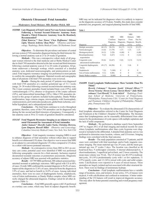
American Institute of Ultrasound in Medicine <strong>Proceedings</strong> J Ultrasound Med 32(suppl):S1–S134, 2013Obstetric Ultrasound: Fetal AnomaliesModerators: Israel Meizner, MD, Heather Welch, MDMRI may not be indicated for diagnoses where it is unlikely to improveon the diagnostic accuracy of US alone. Notably, this study does considerpotential cost, prognostic, and surgical-planning benefits of fetal MRI.1525585 Late Diagnosis of Fetal Central Nervous System AnomaliesFollowing a Normal Second-Trimester Anatomy Scan:Should a Third-Trimester Anatomy Scan Be RoutinelyRecommended?Eldad Katorza, 1 * Yoav Yinon, 1 Chen Hoffmann, 2 ShlomoLipitz, 1 Reuven Achiron, 1 Boaz Weisz 1 1 Obstetrics and Gynecology,2 Radiology, Sheba Medical Center, Tel Hashomer, IsraelObjectives—To determine the prevalence and nature of centralnervous system (CNS) anomalies diagnosed during the third trimester followinga normal anatomy scan at 21 to 24 weeks of gestation.Methods—This was a retrospective cohort study of all pregnantwomen referred to the fetal medicine unit at Sheba Medical Centerdue to fetal CNS anomalies detected at the late second and third trimestersfollowing a normal anatomy scan at 21 to 24 weeks of gestation. All patientsunderwent a thorough workup, which consisted of a detailedanatomy scan, dedicated neurosonography, and amniocentesis as indicated.Fetal magnetic resonance imaging was performed in most patientsto confirm the sonographic diagnosis. Maternal records and sonographicdata of all patients with fetal CNS anomalies were reviewed.Results—During the study period, 47 patients were diagnosedwith fetal CNS anomalies at a median gestational age of 31.1 weeks(range, 24–38 weeks) following a normal second-trimester anatomy scan.The 4 most common anomalies found included brain cysts (19%), mildventriculomegaly (15%), absence or dysgenesis of the corpus callosum(10%), and intracerebral hemorrhage (10%). Other CNS anomalies detectedin this group of patients included hydrocephalus, Dandy-Walkermalformation, large cisterna magna, microcephalus with lissencephaly,craniosynestosis, periventricular pseudocysts, global brain ischemia, cerebellarhypoplasia, and a subependymal nodule.Conclusions—The fetal brain continues to evolve throughoutgestation; therefore, some of the CNS anomalies can be diagnosed onlyduring the late second and third trimesters of pregnancy. Consequently, alate anatomy scan at 30 to 32 weeks of gestation should be considered.1531347 Fetal Magnetic Resonance Imaging as an Adjunct to AntenatalUltrasound for Assessment of Fetal AnomaliesAmber Samuel,* Sherelle Laifer-Narin, Christina Herrera,Lynn Simpson, Russell Miller Obstetrics and Gynecology,Columbia University Medical Center, New York, New York USAObjectives—Fetal magnetic resonance imaging (MRI) is usedto enhance diagnosis of fetal anomalies without robust data to supportbenefit over ultrasound (US) alone. Our objective was to assess fetal MRIas an adjunct to conventional diagnostic US when compared to US alonein a cohort with known postnatal outcomes.Methods—In a retrospective review from 2003 to 2011 at a tertiarycare center, potential cases were identified if MRI was performedfollowing sonographic concern for a fetal anomaly. Inclusion requireddocumented neonatal outcomes or postmortem assessments. Diagnosticaccuracy of adjunct MRI was assessed with qualitative statistics.Results—Of 799 MRIs performed, 406 subjects possessed documentedneonatal or pathologic outcomes. MRI agreed with US in 68%of cases. Overall, MRI confirmed the neonatal diagnosis in 56.4% of cases,improved the diagnosis in 12.8% of cases, detracted from the diagnosis in5.9% of cases, and had no benefit in 24.9% of cases. Among individualanomalies, there were no cases of diaphragmatic hernia, omphalocele,vein of Galen malformation, or Dandy-Walker complex where MRI correctlychanged the ultrasound diagnosis. Findings varied for all other diagnoses(Table 1).Conclusions—Fetal MRI generally agrees with US performedat a tertiary care center, which may limit its adjunct diagnostic benefit.S53Table 1MRIMRI MRI Detracts MRIConfirms Improves From Has NoDiagnosis, Diagnosis, Diagnosis, Benefit,Diagnosis n (%) n (%) n (%) n (%) n (%)Multiple anomalies 60 (15) 20 (33) 10 (17) 8 (13) 22 (37)Ventriculomegaly 14 (4) 11 (79) 1 (7) 1 (7) 1 (7)Meningomyelocele 10 (3) 2 (20) 4 (40) 0 4 (40)Bronchopulmonary 9 (2) 6 (67) 1 (11) 0 2 (22)sequestrationCongenital cystic 24 (6) 16 (67) 2 (8) 3 (13) 3 (13)adenomatoid malformationCongenital diaphragmatic 69 (17) 66 (96) 0 3 (4) 0herniaOmphalocele 7 (2) 7 (100) 0 0 0Vein of Galen malformation 5 (1) 4 (80) 0 0 1 (20)Dandy-Walker complex 5 (1) 5(100) 0 0 01541099 Fetal Lymphatic Malformations: More Variable Than WeThink?Beverly Coleman, 1,2 Suzanne Iyoob, 2 Edward Oliver, 1,2 *Teresa Victoria, 2 Devon Looney, 2 Steven Horii, 1,2 Julie Moldenhauer,2 Lori Howell, 2 N. Scott Adzick 2 1 Radiology, PerelmanSchool of Medicine, University of Pennsylvania,Philadelphia, Pennsylvania USA; 2 Center for Fetal Diagnosisand Treatment, Children’s Hospital of Philadelphia, Philadelphia,Pennsylvania USAObjectives—To evaluate the ultrasound (US) characteristics offetal lymphatic abnormalities referred to the Center for Fetal Diagnosisand Treatment at the Children’s Hospital of Philadelphia. The literaturestates that lymphangiomas can be reasonably differentiated from othermasses by the predominance of cystic spaces with multiple septations andthe lack of solid components.Methods—We performed a database search from September1997 to August 2012 of all fetal imaging and medical records for caseswhere lymphatic malformations other than cystic hygroma were diagnosedor included in the differential. A detailed fetal anatomic survey wasperformed to determine mass location, volume, and US texture. Imagingfindings were correlated with the final outcome.Results—The study population consisted of 73 patients, and 68cases were correlated with fetal neurologic and/or body magnetic resonanceimaging. The mean maternal age was 29 years, and the mean gestationalage was 27 weeks 2 days. The location was classified as 46head/neck/face, 9 axilla/upper extremity, 8 internal abdomen/pelvis, 5chest/mediastinum, 3 superficial pelvis/back, and 2 lower extremity. Themean mass volume was 70 mL. The US texture was 40 (55%) multiseptate/cystic,10 (14%) predominantly cystic with 1 or few septations, 11(15%) purely cystic, and 12 (16%) mixed with cystic and solid components.Calcifications were reported in 4 cases of mixed masses. Theseanomalies are largely isolated and not associated with other structural defects.There were no findings of nonimmune hydrops.Conclusions—Fetal lymphatic malformations have a variablerange of locations, sizes, and textures. In our series, 31% of masses wereatypical, 4 with calcifications and confused as teratomas. A better understandingof the US features will result in improved diagnostic accuracy.This may allow for better parental counseling and overall pregnancy management.


