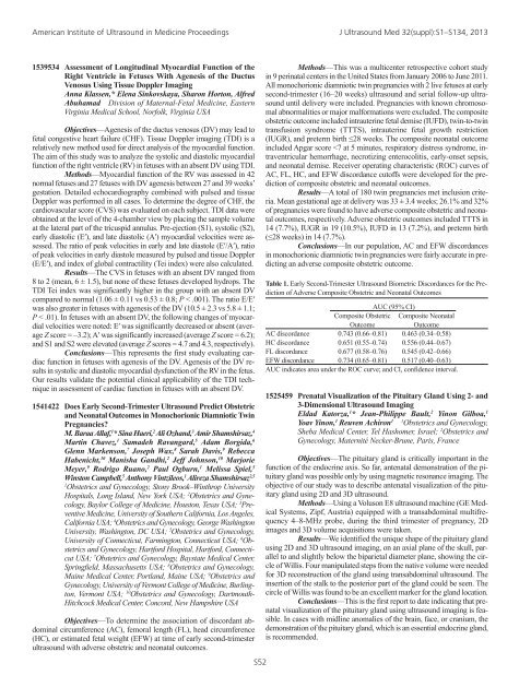
American Institute of Ultrasound in Medicine <strong>Proceedings</strong> J Ultrasound Med 32(suppl):S1–S134, 20131540161 Heterogeneity Assessment of Tumor Perfusion Using HighresolutionDynamic Contrast-Enhanced Ultrasound andDynamic Contrast-Enhanced Magnetic Resonance ImagingSong-Ee Baek,* Patrick Pan, Ergys Subashi, Cäcilia Reiner,Daniele Marin, Allan Johnson, Rendon Nelson Radiology,Duke University Hospital, Durham, North Carolina USAObjectives—To determine the reproducibility of measurementsof tumor perfusion heterogeneity using high-resolution contrast-enhancedultrasound compared to high-resolution dynamic contrast-enhanced (DCE)magnetic resonance imaging (MRI) in murine colorectal cancer. We anticipateusing this technique to predict and monitor treatment response to anantiangiogenesis agent.Methods—Experiments were approved by the local AnimalCare Committee. Five CD-1 nu/nu athymic female mice with subcutaneousmurine colorectal carcinomas (mean tumor height/width, 0.66/1.21cm) were injected with SonoVue (Bracco Diagnostic, Inc) via a tail vein.At first, we determined reproducibility of tumor perfusion measurementwith DCE-US using a GE LOGIQ E9 with an ML6-15-D transducer (4–13 MHz). Three separate injections by 2 radiologists were performed, andmaximum peak intensity (in video intensity) of all pixels within the regionsof interest (ROIs) and coefficients of the enhancement for wash-inand wash-out (25%–75% of the peak enhancement) slopes were calculated.Quantitative measurements were performed by positioning of ROIsin the frame displaying maximum contrast enhancement of the tumor. Acoefficient of variation was used to compare the variability for each parameter.Second, perfusion heterogeneity according to tumor region wasperformed with DCE-US and DCE-MRI, and the 2 results were compared(perfusion graph: wash-in and wash-out slopes) in 1 mouse.Results—The average coefficients of variation for repeated injectionsin the 5 mice were 3% (range, 1%–4%) for peak enhancement,12% (range, 3%–25%) for slope of the wash-in phase, and 12% (range,3%–19%) for slope of the wash-out phase. Perfusion measurement withDCE US showed reproducible results. Perfusion graphs showed a differentpattern by regions presenting tumor heterogeneity. DCE-US and DCE-MRI wash-in and wash-out slopes are well correlated.Conclusions—We obtained reproducible measurements of heterogeneityof tumor perfusion with DCE-US. These results also showedcompatible perfusion patterns with DCE-MRI. As a result of this information,we will pursue a further experiment design to determine the abilityof this technique to predict treatment response.1541104 Subharmonic Imaging of Angiogenesis in a Murine BreastCancer ModelAndrew Marshall, 1,3 * Valgerdur Halldorsdottir, 1,3 JaydevDave, 1 Anya Forsberg, 1,4 Manasi Dahibawkar, 1,3 Traci Fox, 2Ji-Bin Liu, 1 Xiangdong Hu, 1,5 Yu He, 1,6 Flemming Forsberg 11Radiology, 2 Radiological Sciences, Thomas Jefferson University,Philadelphia, Pennsylvania USA; 3 School of BiomedicalEngineering, Sciences, and Health Systems, Drexel University,Philadelphia, Pennsylvania USA; 4 Plymouth Whitemarsh HighSchool, Plymouth Meeting, Pennsylvania USA; 5 Ultrasonography,Beijing Friendship Hospital, Beijing, China; 6 Ultrasound,First Hospital of Jilin University, Jilin, ChinaObjectives—To compare contrast-enhanced subharmonic ultrasoundimaging (SHI) of breast tumor neovascularity to 3 immunohistochemicalmarkers of angiogenesis in nude rats.Methods—Twenty-five athymic nude female rats were implantedwith 5 × 10 6 breast cancer cells (MDA MB 231) in the mammaryfat pad. The contrast agent Definity (Lantheus Medical Imaging, NorthBillerica, MA) was injected in a tail vein (dose, 200 µL/kg), and fundamentalultrasound imaging as well as pulse-inversion SHI were performedwith a modified Sonix RP scanner (Ultrasonix Imaging, Richmond, BritishColumbia, Canada) using a L9-4 linear array (transmitting at 8 MHz andreceiving at 4 MHz in SHI mode). After the experiments, specimens werestained for endothelial cells (CD31), vascular endothelial growth factor,and cyclooxygenase-2. Fractional tumor vascularity was calculated fromdigital images as contrast-enhanced pixels over tumor area (for SHI) andstaining over tumor area (for specimens). Results were compared using alinear regression analysis.Results—Of the 25 rats implanted 16 (64 %) exhibited tumorgrowth, and 13 were successfully imaged. SHI depicted the tortuous morphologyof tumor neovessels and delineated areas of necrosis better thanfundamental ultrasound imaging, due to the marked suppression of tissuesignals. The strongest correlation determined by linear regression in thisbreast cancer model was between SHI and percent area stained with CD31(r = 0.42).Conclusions—Quantitative contrast-enhanced SHI measuresof tumor neovascularity in a breast cancer xenograft model appear to providea noninvasive marker for angiogenesis corresponding to the expressionof CD31, albeit based on a limited sample size. (Supported by USArmy Medical Research Material Command grant W81XWH-08-1-0503and Lantheus Medical Imaging.)1539281 A Sequential Stepwise Algorithm Helps Improve Detectionof Fetal Venous AnomaliesElena Sinkovskaya,* Anna Klassen, Sharon Horton, AlfredAbuhamad Division of Maternal-Fetal Medicine, EasternVirginia Medical School, Norfolk, Virginia USAObjectives—The assessment of the fetal venous system is anessential component to fetal echocardiography as it adds significantly tothe complete diagnosis of heart defects. The purpose of this study was todevelop a method to standardize and simplify comprehensive examinationof the fetal venous system.Methods—Eight hundred thirty-four fetal congenital cardiovascularanomalies (CVAs) were detected between January 2005 andDecember 2010 in the Division of Maternal-Fetal Medicine at EasternVirginia Medical School. Fetal echocardiograms, which incorporated theassessment of anatomic components of the fetal venous system, were performedbetween 16 and 39 weeks’ gestation. Since 2008, the stepwiseapproach, which included evaluation of 5 transverse planes, was used: (1)view of the upper abdomen; (2) coronary sinus view; (3) 4-chamber view;(4) Three-vessel trachea view; and (5) view of the left brachiocephalicvein. Color and pulsed Doppler was used to detect blood flow patterns.Prenatal diagnosis was confirmed in most cases by postnatal echocardiography,angiography, operative findings, or autopsy.Results—Of 834 cases of CVAs, 333 (39.9%) were detectedbetween years 2005 and 2007 and 501 (59.1%) between years 2008 and2010. Since 2008, the detection of fetal isolated systemic and pulmonaryvein anomalies significantly increased (Table 1), while the distribution ofcongenital heart defects (CHDs) with and without venous malformationsstayed the same. This increased identification of fetal venous system abnormalitiesmay be related to the adoption of the new stepwise approach.Conclusions—Our results demonstrated that the sequentialanalysis of 5 transverse views helps significantly improve the detectionof isolated fetal anomalies of systemic and pulmonary veins.Table 1. Detection of Fetal Cardiovascular AnomaliesIsolated Venous CHDs With CHDs WithObservation Anomalies, Venous System Normal VenousPeriod N n (%) Anomalies, n (%) System, n (%)2005–2007 333 32/333 (9.6) 33/333 (10) 268/333 (80.4)2008–2010 501 137/501 (27.3) a 40/501 (8) 324/501 (64.7)aSignificant difference, P < .05.S51
American Institute of Ultrasound in Medicine <strong>Proceedings</strong> J Ultrasound Med 32(suppl):S1–S134, 20131539534 Assessment of Longitudinal Myocardial Function of theRight Ventricle in Fetuses With Agenesis of the DuctusVenosus Using Tissue Doppler ImagingAnna Klassen,* Elena Sinkovskaya, Sharon Horton, AlfredAbuhamad Division of Maternal-Fetal Medicine, EasternVirginia Medical School, Norfolk, Virginia USAObjectives—Agenesis of the ductus venosus (DV) may lead tofetal congestive heart failure (CHF). Tissue Doppler imaging (TDI) is arelatively new method used for direct analysis of the myocardial function.The aim of this study was to analyze the systolic and diastolic myocardialfunction of the right ventricle (RV) in fetuses with an absent DV using TDI.Methods—Myocardial function of the RV was assessed in 42normal fetuses and 27 fetuses with DV agenesis between 27 and 39 weeks’gestation. Detailed echocardiography combined with pulsed and tissueDoppler was performed in all cases. To determine the degree of CHF, thecardiovascular score (CVS) was evaluated on each subject. TDI data wereobtained at the level of the 4-chamber view by placing the sample volumeat the lateral part of the tricuspid annulus. Pre-ejection (S1), systolic (S2),early diastolic (E′), and late diastolic (A′) myocardial velocities were assessed.The ratio of peak velocities in early and late diastole (E′/A′), ratioof peak velocities in early diastole measured by pulsed and tissue Doppler(E/E′), and index of global contractility (Tei index) were also calculated.Results—The CVS in fetuses with an absent DV ranged from8 to 2 (mean, 6 ± 1.5), but none of these fetuses developed hydrops. TheTDI Tei index was significantly higher in the group with an absent DVcompared to normal (1.06 ± 0.11 vs 0.53 ± 0.8; P < .001). The ratio E/E′was also greater in fetuses with agenesis of the DV (10.5 ± 2.3 vs 5.8 ± 1.1;P < .01). In fetuses with an absent DV, the following changes of myocardialvelocities were noted: E′ was significantly decreased or absent (averageZ score = –3.2); A′ was significantly increased (average Z score = 6.2);and S1 and S2 were elevated (average Z scores = 4.7 and 4.3, respectively).Conclusions—This represents the first study evaluating cardiacfunction in fetuses with agenesis of the DV. Agenesis of the DV resultsin systolic and diastolic myocardial dysfunction of the RV in the fetus.Our results validate the potential clinical applicability of the TDI techniquein assessment of cardiac function in fetuses with an absent DV.1541422 Does Early Second-Trimester Ultrasound Predict Obstetricand Neonatal Outcomes in Monochorionic Diamniotic TwinPregnancies?M. Baraa Allaf, 1 * Sina Haeri, 2 Ali Ozhand, 3 Amir Shamshirsaz, 4Martin Chavez, 1 Samadeh Ravangard, 5 Adam Borgida, 6Glenn Markenson, 7 Joseph Wax, 8 Sarah Davis, 9 RebeccaHabenicht, 10 Manisha Gandhi, 2 Jeff Johnson, 10 MarjorieMeyer, 9 Rodrigo Ruano, 2 Paul Ogburn, 1 Melissa Spiel, 5Winston Campbell, 5 Anthony Vintzileos, 1 Alireza Shamshirsaz 2,51Obstetrics and Gynecology, Stony Brook–Winthrop UniversityHospitals, Long Island, New York USA; 2 Obstetrics and Gynecology,Baylor College of Medicine, Houston, Texas USA; 3 PreventiveMedicine, University of Southern California, Los Angeles,California USA; 4 Obstetrics and Gynecology, George WashingtonUniversity, Washington, DC USA; 5 Obstetrics and Gynecology,University of Connecticut, Farmington, Connecticut USA; 6 Obstetricsand Gynecology, Hartford Hospital, Hartford, ConnecticutUSA; 7 Obstetrics and Gynecology, Baystate Medical Center,Springfield, Massachusetts USA; 8 Obstetrics and Gynecology,Maine Medical Center, Portland, Maine USA; 9 Obstetrics andGynecology, University of Vermont College of Medicine, Burlington,Vermont USA; 10 Obstetrics and Gynecology, Dartmouth-Hitchcock Medical Center, Concord, New Hampshire USAObjectives—To determine the association of discordant abdominalcircumference (AC), femoral length (FL), head circumference(HC), or estimated fetal weight (EFW) at time of early second-trimesterultrasound with adverse obstetric and neonatal outcomes.S52Methods—This was a multicenter retrospective cohort studyin 9 perinatal centers in the United States from January 2006 to June 2011.All monochorionic diamniotic twin pregnancies with 2 live fetuses at earlysecond-trimester (16–20 weeks) ultrasound and serial follow-up ultrasounduntil delivery were included. Pregnancies with known chromosomalabnormalities or major malformations were excluded. The compositeobstetric outcome included intrauterine fetal demise (IUFD), twin-to-twintransfusion syndrome (TTTS), intrauterine fetal growth restriction(IUGR), and preterm birth ≤28 weeks. The composite neonatal outcomeincluded Apgar score