SIM0216

Create successful ePaper yourself
Turn your PDF publications into a flip-book with our unique Google optimized e-Paper software.
COVER STORY<br />
Fig. 3: (Top) Montage of image thumbnails of HCT116 spheroids in a 96 well<br />
plate treated with compounds and imaged with a 10X Plan Fluor objective.<br />
Hoechst stained nuclei (blue) are overlaid with CellEvent Caspase 3/7 apoptosis<br />
marker (green). Untreated controls are in column 4 and a Caspase 3/7<br />
response is evident in columns 5–7 where Paclitaxel was serially diluted 1:3<br />
from 1 µM in Row A (replicates of 3 across). (Left) Eleven Z planes were<br />
combined into a 2D Maximum Projection image and analyzed with a simple<br />
custom module. Raw images showing low and high degree of apoptosis<br />
with their corresponding segmentation masks are shown (royal blue =<br />
nuclei, pink = apoptotic cells). (Right) By normalizing the amount of apoptosis<br />
as compared to untreated spheroids and plotting on a graph, it can be<br />
seen that Paclitaxel (green line) induces apoptosis at a much lower concentration<br />
than either Mitomycin C or Etoposide.<br />
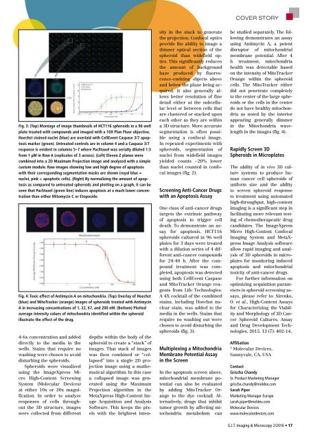
Fig. 4: Toxic effect of Antimycin A on mitochondria. (Top) Overlay of Hoechst<br />
(blue) and MitoTracker (orange) images of spheroids treated with Antimycin<br />
A in increasing concentrations of 1, 22, 67, and 200 nM. (Bottom) Plotted<br />
average intensity values of mitochondria identified within the spheroid<br />
illustrate the effect of the drug.<br />
4-6x concentration and added<br />
directly to the media in the<br />
wells. Stains that require no<br />
washing were chosen to avoid<br />
disturbing the spheroids.<br />
Spheroids were visualized<br />
using the ImageXpress Micro<br />
High-Content Screening<br />
System (Molecular Devices)<br />
at either 10x or 20x magnification.<br />
In order to analyze<br />
responses of cells throughout<br />
the 3D structure, images<br />
were collected from different<br />
sity in the stack to generate<br />
the projection. Confocal optics<br />
provide the ability to image a<br />
thinner optical section of the<br />
spheroid than widefield optics.<br />
This significantly reduces<br />
the amount of background<br />
haze produced by fluorescence-emitting<br />
objects above<br />
and below the plane being acquired.<br />
It also generally allows<br />
better resolution of fine<br />
detail either at the subcellular<br />
level or between cells that<br />
are clustered or stacked upon<br />
each other as they are within<br />
a 3D structure. More accurate<br />
segmentation is often possible<br />
using a confocal image.<br />
In repeated experiments with<br />
spheroids, segmentation of<br />
nuclei from widefield images<br />
yielded counts ~20% lower<br />
than nuclei counted in confocal<br />
images (fig. 2).<br />
Screening Anti-Cancer Drugs<br />
with an Apoptosis Assay<br />
One class of anti-cancer drugs<br />
targets the extrinsic pathway<br />
of apoptosis to trigger cell<br />
death. To demonstrate an assay<br />
for apoptosis, HCT116<br />
spheroids cultured in 96 well<br />
plates for 3 days were treated<br />
with a dilution series of 4 different<br />
anti-cancer compounds<br />
for 24-48 h. After the compound<br />
treatment was completed,<br />
apoptosis was detected<br />
using both CellEvent Caspase<br />
and MitoTracker Orange reagents<br />
from Life Technologies.<br />
A 4X cocktail of the combined<br />
stains, including Hoechst nuclear<br />
stain, was added to the<br />
media in the wells. Stains that<br />
require no washing out were<br />
chosen to avoid disturbing the<br />
spheroids (fig. 3).<br />
Multiplexing a Mitochondria<br />
Membrane Potential Assay<br />
in the Screen<br />
In the apoptosis screen above,<br />
mitochondrial membrane potential<br />
can also be evaluated<br />
by adding MitoTracker Orange<br />
to the dye cocktail. Alternatively,<br />
drugs that inhibit<br />
tumor growth by affecting mitochondria<br />
metabolism can<br />
depths within the body of the<br />
spheroid to create a “stack” of<br />
images. That stack of images<br />
was then combined or “collapsed”<br />
into a single 2D projection<br />
image using a mathematical<br />
algorithm. In this case<br />
a collapsed image was generated<br />
using the Maximum<br />
Projection algorithm in the<br />
MetaXpress High-Content Image<br />
Acquisition and Analysis<br />
Software. This keeps the pixels<br />
with the brightest intenbe<br />
studied separately. The following<br />
demonstrates an assay<br />
using Antimycin A, a potent<br />
disruptor of mitochondrial<br />
membrane potential. After 4<br />
h treatment, mitochondria<br />
health was detectable based<br />
on the intensity of MitoTracker<br />
Orange within the spheroid<br />
cells. The MitoTracker either<br />
did not penetrate completely<br />
to the center of the large spheroids<br />
or the cells in the center<br />
do not have healthy mitochondria<br />
as noted by the interior<br />
appearing generally dimmer<br />
in the Mitochondria wavelength<br />
in the images (fig. 4).<br />
Rapidly Screen 3D<br />
Spheroids in Microplates<br />
The ability of in vivo 3D culture<br />
systems to produce human<br />
cancer cell spheroids of<br />
uniform size and the ability<br />
to screen spheroid response<br />
to treatment using automated<br />
high-throughput, high-content<br />
imaging is a significant step in<br />
facilitating more relevant testing<br />
of chemotherapeutic drug<br />
candidates. The ImageXpress<br />
Micro High-Content Confocal<br />
Imaging System and MetaXpress<br />
Image Analysis software<br />
allow rapid imaging and analysis<br />
of 3D spheroids in microplates<br />
for monitoring induced<br />
apoptosis and mitochondrial<br />
toxicity of anti-cancer drugs.<br />
For further information on<br />
optimizing acquisition parameters<br />
in spheroid screening assays,<br />
please refer to: Sirenko,<br />
O. et al., High-Content Assays<br />
for Characterizing the Viability<br />
and Morphology of 3D Cancer<br />
Spheroid Cultures. Assay<br />
and Drug Development Technologies,<br />
2015. 13 (7): 402-14.<br />
Affiliation<br />
1<br />
Molecular Devices,<br />
Sunnyvale, CA, USA<br />
Contact<br />
Grischa Chandy<br />
Sr. Product Marketing Manager<br />
grischa.chandy@moldev.com<br />
Sarah Piper<br />
Marketing Manager Europe<br />
sarah.piper@moldev.com<br />
Molecular Devices<br />
www.moleculardevices.com<br />
G.I.T. Imaging & Microscopy 2/2016 • 17