SIM0216

You also want an ePaper? Increase the reach of your titles
YUMPU automatically turns print PDFs into web optimized ePapers that Google loves.
Products<br />
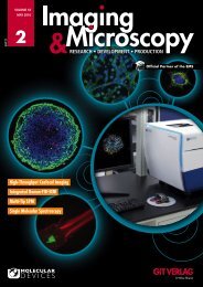
Compact Linear Positioning Stage<br />
The compact linear stage L-509<br />
from PI (Physik Instrumente)<br />
is providing high precision linear<br />
motion with minimized<br />
runout. It is designed for research<br />
and industrial applications,<br />
including beamline systems, microscopy, semiconductor<br />
manufacturing, and photonics instrumentation. Despite its compact<br />
dimensions, the stage can handle payloads of more than<br />
20 lbs. Available with travel ranges of 26mm (1”), 52mm (2”),<br />
and 102mm (4”) and velocity to 20mm/sec, the stage delivers<br />
high accuracy and smooth motion with unidirectional repeatability<br />
down to 0.1 microns. Long service life and very high guiding<br />
accuracy with minimum backlash are ensured by the precision<br />
crossed roller bearings with anti-creep cage assist. For<br />
very high precision requirements, a linear encoder with 1 nanometer<br />
resolution is available.<br />
Confocal Laser Scanning<br />
Microscope<br />
Olympus’ Fluoview FV3000 confocal<br />
laser scanning microscope<br />
combines high-performance imaging<br />
capabilities with ease of use so researchers in such fields<br />
as cell biology, cancer research and stem cell research can collect<br />
relevant imaging data quickly and easily. Built for fast, stable<br />
and accurate measurements of biological reactions within<br />
living cells and tissues, the microscope offers flexibility for all<br />
live-cell imaging applications, providing high-resolution images<br />
of structures and dynamic intracellular processes. It is controlled<br />
by an intuitive software interface so even novice users<br />
can generate high-quality data and images. The instrument’s<br />
optical design offers macro to micro imaging capabilities with<br />
objectives ranging from 1.25X to 150X magnification. One version<br />
comes with galvanometer scanner, and the FV3000RS is<br />
equipped with a hybrid resonant / galvanometer scanner.<br />
Physik Instrumente<br />
www.physikinstrumente.com<br />
Olympuswww.olympus-lifescience.com<br />
Dichroics for Super-Resolution Microscopy<br />
AHF analysentechnik offers<br />
now the new Semrock<br />
beamsplitter series<br />
additionally to their existing<br />
superflat dichroic<br />
program for super-resolution<br />
microscopy: λ /10 P-V<br />
per inch flatness on 3 mm<br />
thick dichroics and improved<br />
λ /2 P-V per inch<br />
flatness on improved 1<br />
mm dichroics are now available. There will be no compromise<br />
regarding guaranteed steepest edges, short wavelength<br />
reflectivity down to 350 nm, and long wavelength transmission<br />
optimized out to 1200 nm or 1600 nm. Super-resolution<br />
imaging systems are highly sensitive to optical wavefront distortion<br />
and demand the highest quality components. Laser<br />
dichroic beamsplitters with λ /10 flatness minimize the reflected<br />
wavefront distortion, thereby maximizing both the<br />
signal and the signal-to-noise ratio in super-resolution microscopes.<br />
1 mm thick laser dichroic beamsplitters have been<br />
significantly improved to λ /2 flatness (~255 m radius of curvature).<br />
They will fit into microscopy filter cubes and improve<br />
the performance of laser based confocal and TIRF illumination<br />
systems. They are also ideal for reflection of imaging<br />
beams in conventional structured-illumination techniques as<br />
well as patterned illumination systems for localized photoactivation.<br />
These dichroic beamsplitters allow the use of<br />
much larger diameter illumination beams, offering researchers<br />
and instrument developers more flexibility in system design<br />
with no compromise to overall performance. Please ask<br />
AHF for a demo system.<br />
WEBINAR<br />
FLUORESCENCE LIFETIME IMAGING<br />
USING FLUORESCENT DECAY RATES<br />
TO IDENTIFY INDIVIDUAL FLUOROPHORES<br />
JUNE 28, 2016 AT 2PM<br />
TO REGISTER FOR THIS FREE<br />
WEBINAR PLEASE GO TO<br />
bit.ly/Webinar-PCO<br />
AHF analysentechnik <br />
www.ahf.de<br />
G.I.T. Imaging & Microscopy 2/2016 • 49