SIM0216

Create successful ePaper yourself
Turn your PDF publications into a flip-book with our unique Google optimized e-Paper software.
LIGHT MICROSCOPY<br />
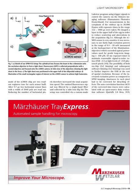
Fig. 1: a) Sketch of our SPIM-FCS-Setup: The cylindrical lens focuses the beam in the x-dimension onto<br />
the excitation objective to form a light sheet. Fluorescence (GFP) is collected perpendicular with a<br />
second objective and focused onto the sCMOS camera. b) Side-view of the objectives showing the light<br />
sheet in the focus. c) The light sheet was positioned in the upper half of the ellipsoidal embryo. d)<br />
Illustration of the small rectangular region of interest on the sCMOS sensor to achieve high framerates.<br />
mode of the sCMOS chip are two readout<br />
registers (one for each sensor half).<br />
After 9.7 µs two horizontal sensor lines<br />
with a width of 2048 pxls are read out.<br />
Reducing the number of horizontal pixels<br />
therefore increased the total acquisition<br />
speed. The emitted fluorescence signal<br />
was filtered by a single-band filter<br />
and collected by a tube lens (fig.1b). The<br />
setup was controlled via a custom-made<br />
Labview program using trigger signals to<br />
control the camera via the Hokawo imaging<br />
software (Hamamatsu Photonics<br />
Deutschland). For measurements in the<br />
cytoplasm of the embryo up to 20,000<br />
frames with exposure times in the range<br />
152 − 1004 µs were taken. We imaged a<br />
layer in the upper half of the egg in order<br />
to reduce scattering and aberrations in<br />
the acquisition (fig.1c). Although the sC-<br />
MOS sensor is very sensitive it was necessary<br />
to use fairly high excitation powers<br />
in the range of 0.8 – 20 mW (measured<br />
at the backaperture of the illuminationobjective)<br />
which exceeded typical powervalues<br />
used for gentle long-term imaging<br />
(∼ 100 μW, 50 ms exposure-time) to<br />
maintain reasonable signal-to-noise ratios<br />
(SNR ~2.8 at light levels of ~210 photons/4<br />
pixels [10]). The possibility of both<br />
on-chip (2x2 binning) and subsequent<br />
software-binning (3x3 binning) was used<br />
to further improve the SNR at the cost<br />
of spatial resolution. Because of the increased<br />
excitation power as compared to<br />
SPIM imaging, timetraces in each pixel<br />
had to be corrected for bleaching effects.<br />
The auto-correlation function (ACF)<br />
of the corrected time traces were calculated<br />
with an open-source data evaluation<br />
software (Quickfit 3.0 Beta, SVN:<br />
Märzhäuser TrayExpress.<br />
Automated sample handling for microscopy.<br />
www.marzhauser.com<br />
Improve Your Microscope.<br />
G.I.T. Imaging & Microscopy 2/2016 • 19