SIM0216

You also want an ePaper? Increase the reach of your titles
YUMPU automatically turns print PDFs into web optimized ePapers that Google loves.
liGHT MICROSCOPY<br />
Single Molecular Spectroscopy<br />
Parallel Lifetime and Imaging of Single Molecules<br />
Adrian Mantsch 1 and Ashley Cadby 1<br />
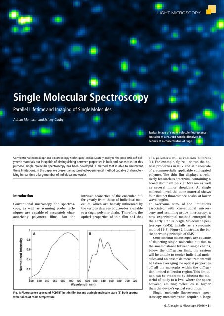
Typical image of single molecule fluorescence<br />
emission of a PCDTBT sample dissolved in<br />
Zeonex at a concentration of 5ng/l.<br />
Conventional microscopy and spectroscopy techniques can accurately analyze the properties of polymeric<br />
materials but incapable of distinguishing between properties in bulk and nanoscale. For this<br />
purpose, single molecular spectroscopy has been developed, a method that is able to circumvent<br />
these limitations. In this paper we present an automated experimental method capable of characterizing<br />
in real time a large number of individual molecules.<br />
Introduction<br />
Conventional microscopy and spectroscopy,<br />
as well as scanning probe techniques<br />
are capable of accurately characterizing<br />
polymeric films. But the<br />
intrinsic properties of the ensemble differ<br />
greatly from those of individual molecules,<br />
which are heavily influenced by<br />
the various degrees of disorder available<br />
to a single polymer chain. Therefore, the<br />
optical properties of thin film and that<br />
Fig. 1: Fluorescence spectra of PCDTBT in thin film (A) and at single molecule scale (B) both spectra<br />
were taken at room temperature.<br />
of a polymer’s will be radically different<br />
[1]. For example, figure 1 shows the optical<br />
properties in bulk and at nanoscale<br />
of a commercially applicable conjugated<br />
polymer. The thin film displays a relatively<br />
featureless spectrum, containing a<br />
broad dominant peak at 680 nm as well<br />
as several minor shoulders. At single<br />
molecule level, the same material shows<br />
four distinct fluorescence peaks, at lower<br />
wavelengths.<br />
To overcome some of the limitations<br />
associated with conventional microscopy<br />
and scanning probe microscopy, a<br />
new experimental method emerged in<br />
the early 1990’s: Single Molecular Spectroscopy<br />
(SMS), initially as a cryogenic<br />
method [1-3]. Figure 2 illustrates the basic<br />
operating principle of SMS.<br />
Conventional microscopes are capable<br />
of detecting single molecules but due to<br />
the small distance between single chains,<br />
below the diffraction limit, the system<br />
will be unable to resolve individual molecules<br />
and an ensemble measurement will<br />
be taken averaging the optical properties<br />
off all the molecules within the diffraction<br />
limited collection region. This limitation<br />
can be overcome by diluting the material<br />
of study to a level where the space<br />
between emitting molecules is higher<br />
than the device’s optical resolution.<br />
Single molecule fluorescence spectroscopy<br />
measurements require a large<br />
G.I.T. Imaging & Microscopy 2/2016 • 21