Procedure - INOVA Diagnostics
Procedure - INOVA Diagnostics
Procedure - INOVA Diagnostics
You also want an ePaper? Increase the reach of your titles
YUMPU automatically turns print PDFs into web optimized ePapers that Google loves.
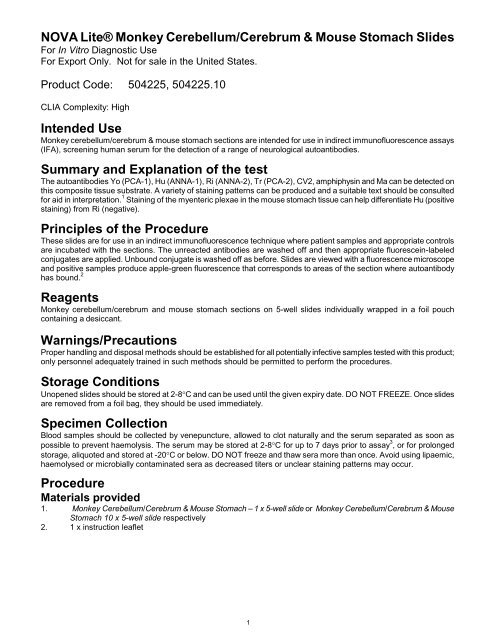
N O V A L i ® t Monkey e Cerebellum/Cerebrum & Mouse Stomach Slides<br />
For In Vitro Diagnostic Use<br />
For Export Only. Not for sale in the United States.<br />
Product Code: 504225, 504225.10<br />
CLIA Complexity: High<br />
Intended Use<br />
Monkey cerebellum/cerebrum & mouse stomach sections are intended for use in indirect immunofluorescence assays<br />
(IFA), screening human serum for the detection of a range of neurological autoantibodies.<br />
Summary and Explanation of the test<br />
The autoantibodies Yo (PCA-1), Hu (ANNA-1), Ri (ANNA-2), Tr (PCA-2), CV2, amphiphysin and Ma can be detected on<br />
this composite tissue substrate. A variety of staining patterns can be produced and a suitable text should be consulted<br />
for aid in interpretation. 1 Staining of the myenteric plexae in the mouse stomach tissue can help differentiate Hu (positive<br />
staining) from Ri (negative).<br />
Principles of the <strong>Procedure</strong><br />
These slides are for use in an indirect immunofluorescence technique where patient samples and appropriate controls<br />
are incubated with the sections. The unreacted antibodies are washed off and then appropriate fluorescein-labeled<br />
conjugates are applied. Unbound conjugate is washed off as before. Slides are viewed with a fluorescence microscope<br />
and positive samples produce apple-green fluorescence that corresponds to areas of the section where autoantibody<br />
has bound. 2<br />
Reagents<br />
Monkey cerebellum/cerebrum and mouse stomach sections on 5-well slides individually wrapped in a foil pouch<br />
containing a desiccant.<br />
Warnings/Precautions<br />
Proper handling and disposal methods should be established for all potentially infective samples tested with this product;<br />
only personnel adequately trained in such methods should be permitted to perform the procedures.<br />
Storage Conditions<br />
Unopened slides should be stored at 2-8°C and can be used until the given expiry date. DO NOT FREEZE. Once slides<br />
are removed from a foil bag, they should be used immediately.<br />
Specimen Collection<br />
Blood samples should be collected by venepuncture, allowed to clot naturally and the serum separated as soon as<br />
possible to prevent haemolysis. The serum may be stored at 2-8°C for up to 7 days prior to assay 3 , or for prolonged<br />
storage, aliquoted and stored at -20°C or below. DO NOT freeze and thaw sera more than once. Avoid using lipaemic,<br />
haemolysed or microbially contaminated sera as decreased titers or unclear staining patterns may occur.<br />
<strong>Procedure</strong><br />
Materials provided<br />
1. Monkey Cerebellum/Cerebrum & Mouse Stomach – 1 5-well x slide or Monkey Cerebellum/Cerebrum & Mouse<br />
Stomach 10 x 5-well slide respectively<br />
2. 1 x instruction leaflet<br />
1
Additional Materials Required But Not Provided<br />
1. PBS for sample dilution and washes<br />
2. Container for PBS buffer<br />
3. Micropipettes and disposable tips to apply patient samples<br />
4. Humid chamber for incubation steps<br />
5. Fluorescence microscope with 495nm exciter filter and 515nm barrier filter<br />
6. Plastic squeeze bottle for initial wash in PBS<br />
Additional components may be obtained from <strong>INOVA</strong> <strong>Diagnostics</strong>: PBS (508002), IFA System Negative Control<br />
(508186), ANNA-1 positive control (504002), PCA-1 positive control (504009), FITC IgG (H&L), Monkey Adsorbed<br />
Conjugate (504011, 504071), 1% Evans Blue (504049) and PVA mounting medium (504046, 504047).<br />
Test <strong>Procedure</strong><br />
Quality control<br />
Positive and negative controls should be used every time samples are tested.<br />
1. Mounting medium: Remove the mounting medium from the fridge to allow it to reach room temperature (18-<br />
28°C) before it is needed.<br />
2. Dilute patient samples.<br />
Screening: Dilute patient samples 1/50 and 1/500 by adding 10µL of serum to 490µL of PBS buffer and 5µL of<br />
serum to 2495µL of PBS buffer respectively.<br />
Titration: Make serial dilutions of positive screened samples with PBS buffer (e.g. 1/50, 1/100, 1/200, 1/400<br />
etc).<br />
For example: Take 100µL of the 1/50 dilution, mix with 100µL PBS to give a 1/100 dilution (repeat for further<br />
dilutions).<br />
3. Substrate slides. Allow substrate slide(s) to reach room temperature (18-28°C) prior to removal from pouch(es).<br />
Label slides appropriately, place in the humid chamber and add positive and negative controls to appropriate<br />
wells. Add 50-100µL of diluted patient samples to the remaining wells.<br />
4. Slide incubation. Incubate slides for 30 minutes in a humid chamber at room temperature (18-28°C).<br />
5. PBS wash. Remove slides from humid chamber and rinse briefly with PBS squeeze bottle. Do not squirt directly<br />
on to the wells. Place slides in a rack and immerse in PBS and agitate or stir for 5-10 minutes.<br />
6. Addition of fluorescent conjugate. Shake off excess PBS and blot around wells. Return slides to humid chamber<br />
and immediately cover each well with a drop of appropriately diluted fluorescent conjugate. DO NOT LEAVE<br />
WELLS UNCOVERED FOR LONGER THAN 15 SECONDS. Drying out of the substrate seriously affects the<br />
results. The use of a monkey adsorbed conjugate will greatly enhance results (e.g. 504011).<br />
7. Slide incubation. Incubate slides for 30 minutes in humid chamber at room temperature (18-28°C), in the dark.<br />
8. PBS wash. Wash again as described in step 5. OPTIONAL COUNTERSTAIN. Add 2-3 drops of 1% Evans Blue<br />
per 100mL of PBS prior to slide immersion.<br />
9. Mounting with coverslip. Remove one slide at a time from PBS wash. Quickly dry around the wells and add a<br />
drop of mounting medium to each well. Carefully lower the slide onto the coverslip, avoiding air bubbles but, if<br />
present, do not attempt to remove. Wipe excess medium from around edge of coverslip.<br />
10. View slides under fluorescence microscope. Slides may be stored for up to 3 days at 2-8°C, in the dark, without<br />
significant loss of fluorescence.<br />
Results<br />
Quality Control<br />
A serum sample containing Purkinje cell auto-antibodies should give bright apple-green fluorescent staining of Purkinje<br />
cell cytoplasm.<br />
A serum sample containing ANNA-1 auto-antibodies should give apple green granular fluorescent staining of most<br />
neuronal nuclei in the cerebellum, including Purkinje cells.<br />
A negative control should show dull green staining in all the tissue, with no discernible fluorescence.<br />
If controls do not appear as described, the test is invalid and should be repeated.<br />
2
Interpretation of Results<br />
See reference 1 for color photographic examples of this pattern. Results are reported as positive or negative.<br />
N.B. Each laboratory should establish at which point a positive result is considered clinically significant.<br />
Limitations of the <strong>Procedure</strong><br />
1. The light source, filters and optics of different makes of fluorescence microscopes will influence the sensitivity of<br />
the kit. The performance of the microscope is significantly influenced by correct maintenance especially<br />
centering of the mercury vapour lamp and changing of the lamp after the recommended period of time.<br />
2. Suitability for use with other manufacturers' IFA reagents has not been assessed but use with such reagents<br />
should not necessarily be excluded.<br />
3. This test alone should not be considered diagnostic. All other factors including the clinical history of the patients<br />
and other serological or biopsy results must also be taken into account.<br />
Summary of the <strong>Procedure</strong><br />
1. Equilibrate mounting medium to room temperature.<br />
2. Dilute PBS with distilled water.<br />
3. Dilute patient sera 1/50 and 1/500 with PBS.<br />
4. Equilibrate substrate slides to room temperature (18-28°C).<br />
5. Apply 50-100µL positive and negative controls and diluted patient sera to appropriate wells.<br />
6. Incubate in a humid chamber for 30 minutes.<br />
7. Wash for 5-10 minutes in PBS.<br />
8. Blot around each well and immediately cover each well with a drop of conjugate.<br />
9. Incubate as in step 6.<br />
10. Wash as step 7.<br />
11. Mount.<br />
12. View slide under fluorescence microscope.<br />
References<br />
1. Bradwell A.R. et al (2008). Atlas of Tissue Autoantibodies (Third Edition). Publ. The Binding Site Ltd.,<br />
Birmingham, UK<br />
2. Weller T.H., Coons A.H. (1954). Fluorescent antibody studies with agent of Varicella and Herpes Zoster<br />
propagated in vitro. Proc. Soc. Exp. Biol. Med. 86: 789-794.<br />
3. Protein Reference Handbook of Autoimmunity (3 rd Edition) 2004. Ed. A Milford Ward, GD Wild. Publ. PRU<br />
Publications, Sheffield. 14.<br />
Manufactured By:<br />
<strong>INOVA</strong> <strong>Diagnostics</strong>, Inc.<br />
9900 Old Grove Road<br />
San Diego, CA 92131<br />
United States of America<br />
Technical Service (Outside the U.S.) : 00+ 1 858-805-7950<br />
info@inovadx.com<br />
Authorized Representative in the EU:<br />
Medical Technology Promedt Consulting GmbH<br />
Altenhofstrasse 80<br />
D-66386 St. Ingbert, Germany<br />
Tel.: +49-6894-581020<br />
Fax.: +49-6894-581021<br />
www.mt-procons.com<br />
624225ENG May 2010<br />
Revision 0<br />
3


