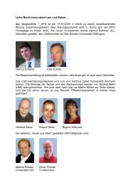
Deutsche Tagung f ¨ur Forschung mit ... - SNI-Portal
Deutsche Tagung f ¨ur Forschung mit ... - SNI-Portal
Deutsche Tagung f ¨ur Forschung mit ... - SNI-Portal

Create successful ePaper yourself
Turn your PDF publications into a flip-book with our unique Google optimized e-Paper software.
<strong>Deutsche</strong> <strong>Tagung</strong> für <strong>Forschung</strong> <strong>mit</strong><br />
Synchrotronstrahlung, Neutronen und<br />
Ionenstrahlen an Großgeräten 2006<br />
Universität Hamburg,<br />
04.-06. Oktober 2006<br />
Programm und Abstracts
<strong>Deutsche</strong> <strong>Tagung</strong> für <strong>Forschung</strong> <strong>mit</strong><br />
Synchrotronstrahlung, Neutronen und<br />
Ionenstrahlen an Großgeräten 2006<br />
Universität Hamburg,<br />
04.-06. Oktober 2006<br />
Programm und Abstracts<br />
Hrsg.: A. Schreyer, R. Willumeit, R. Röhlsberger,<br />
L. Incoccia-Hermes, K. Griewatsch<br />
Ko<strong>mit</strong>ee<br />
<strong>Forschung</strong><br />
<strong>mit</strong><br />
Neutronen<br />
Ko<strong>mit</strong>ee Erforschung<br />
kondensierter Materie<br />
<strong>mit</strong> nuklearen Sonden<br />
KFS KOMITEE <strong>Forschung</strong> <strong>mit</strong> Synchrotronstrahlung<br />
und Ionenstrahlen
Impressum:<br />
Herausgeber: A. Schreyer, R. Willumeit, R. Röhlsberger,<br />
L. Incoccia-Hermes, K. Griewatsch<br />
<strong>Deutsche</strong> <strong>Tagung</strong> für <strong>Forschung</strong> <strong>mit</strong> Synchrotronstrahlung,<br />
Neutronen und Ionenstrahlen an Großgeräten 2006<br />
Programm und Abstracts<br />
Universität Hamburg<br />
Oktober 2006<br />
Veranstalter: <strong>Deutsche</strong>s Elektronen-Synchrotron DESY und<br />
GKSS <strong>Forschung</strong>szentrum Geesthacht GmbH<br />
Programmko<strong>mit</strong>ee: H. Schober, O. Pietsch, H. Hofsäss, A. Schreyer,<br />
R. Röhlsberger<br />
c○ GKSS <strong>Forschung</strong>szentrum Geesthacht GmbH, 2006<br />
Alle Rechte vorbehalten.<br />
Mit freundlicher Unterstützung durch<br />
ESRF
Vorwort<br />
Die Nutzung von Großgeräten zur Erforschung der kondensierten Materie hat in den<br />
letzten Jahren zu einer Vielzahl von herausragenden <strong>Forschung</strong>sergebnissen geführt.<br />
Experimente <strong>mit</strong> Neutronen, Synchrotron- und Ionenstrahlung bieten komplementäre<br />
Möglichkeiten, die Eigenschaften der Materie in all ihren Facetten auszuleuchten. Die<br />
<strong>SNI</strong>2006 stellt ein gemeinsames Forum für die Präsentation neuester Erkenntnisse,<br />
für den Ideenaustausch und die weitere Vernetzung der Methoden dar. Eine spezielle<br />
Veranstaltung widmet sich der allgemeinverständlichen Darstellung der <strong>Forschung</strong> für<br />
die Öffentlichkeit.<br />
Die <strong>Tagung</strong> findet auf Initiative der Kommission ” Erforschung kondensierter Materie<br />
<strong>mit</strong> Großgeräten“ (KEKM), der Dachorganisation der Ko<strong>mit</strong>ees für <strong>Forschung</strong><br />
<strong>mit</strong> Neutronen (KFN), Synchrotronstrahlung (KFS) und nuklearen Sonden und Ionenstrahlen<br />
(KFSI) statt. Sie knüpft an frühere BMBF-Statusseminare (Helgoland 1986,<br />
Kloster Seeon 1997) an und soll die Komplementarität der Methoden deutlich machen<br />
und den wissenschaftlichen Austausch beflügeln. Die <strong>SNI</strong>2006 bietet ein Forum für ein<br />
breites Spektrum von Disziplinen und deren Vernetzung. Folgende Themenbereiche<br />
werden abgedeckt:<br />
Weiche Materie Magnetismus<br />
Nanstrukturen und Grenzflächen Biologische Systeme und Medizin<br />
Mikroskopie und Tomographie Materialien/Werkstoffe<br />
Struktur und Dynamik Methoden und Instrumentierung<br />
Chemische Prozesse und Phasenübergänge Materie unter extremen Bedingungen<br />
Teilchen und Kerne<br />
Ein Höhepunkt der <strong>Tagung</strong> wird ein Senatsempfang am Abend des 05.10.2006 im<br />
Hamburger Rathaus sein, bei dem der Hamburger Senator für Wissenschaft und Gesundheit,<br />
Jörg Dräger und Schleswig-Holsteins Minister für Wissenschaft, Wirtschaft<br />
und Verkehr Dietrich Austermann sowie eine/e VertreterIn des BMBF Grußworte sprechen<br />
werden. Die wichtige Rolle der <strong>Forschung</strong> <strong>mit</strong> Synchrotronstrahlung, Neutronen<br />
und Ionenstrahlen an Großgeräten wird anschließend in einem populärwissenschaftlichen<br />
Vortrag beispielhaft von Helmut Dosch aufgezeigt werden. Im Anschluss daran<br />
laden der Senat der Stadt Hamburg und die <strong>SNI</strong>2006 zu einem Buffet und geselligem<br />
Zusammensein ein.<br />
Wie der Zufall will, findet die <strong>SNI</strong>2006 exakt 20 Jahre nach dem ersten nationalen<br />
Treffen der Pioniere auf Helgoland statt. Mit rund 500 erwarteten Teilnehmern wird<br />
die <strong>SNI</strong>2006 die bisher wohl größte nationale wissenschaftliche <strong>Tagung</strong> dieses wachsenden<br />
<strong>Forschung</strong>sgebiets werden. Die Organisatoren begrüßen Sie hier in Hamburg<br />
und wünschen Ihnen aufregenden Diskussionen, viele neuen Ideen, interessante neuen<br />
Kollaborationspartner und vor allem viel Spaß.<br />
Willkommen in Hamburg!<br />
1
8:00<br />
9:30<br />
10:00<br />
10:30<br />
11:00<br />
12:30<br />
14:00<br />
16:30<br />
17:30<br />
Mittwoch, 4. Oktober 2006 Donnerstag, 5. Oktober 2006 Freitag, 6. Oktober 2006<br />
Registrierung<br />
8:30<br />
8:30<br />
Plenarvorträge:<br />
Plenarvorträge:<br />
Butz, Pyzalla Müller,Braun<br />
9:40<br />
9:40<br />
Begrüßung<br />
Parallelsitzungen: Instrumentierung,<br />
Parallelsitzungen: Biologie, Magnetismus,<br />
Plenarvortrag: Zabel<br />
10:40<br />
Weiche Materie, Materialien<br />
10:40<br />
Materialien, Weiche Materie<br />
Kaffeepause<br />
11:10<br />
Kaffeepause<br />
11:10<br />
Kaffeepause<br />
Parallelsitzungen:<br />
Parallelsitzungen:<br />
Parallelsitzungen:<br />
Mikroskopie/Tomographie, Dynamik, Methoden und Instrumentierung Biologie, Magnetismus,<br />
Nanostrukturen und Grenzflächen S, N, <strong>SNI</strong> Chemische Prozesse<br />
Mittagspause<br />
Postersitzung A<br />
Instrumentierung, Biologie,<br />
Struktur/Dynamik, Chem. Prozesse,<br />
Mikroskopie/Tomographie<br />
Plenarvorträge:<br />
Meyer,Fink<br />
Parallelsitzungen:<br />
Magnetismus, Struktur,<br />
Nanostrukturen und Grenzflächen<br />
12:30<br />
13:00<br />
15:30<br />
17:00<br />
18:00<br />
19:00<br />
19:30<br />
Verleihung Wolfram-Prandl-Preis<br />
19:30<br />
Mittagspause<br />
Postersitzung B<br />
Magnetismus, Nano/Grenzflächen,<br />
Weiche Materie, extreme Bedingungen,<br />
Materialien, Teilchen und Kerne<br />
<strong>SNI</strong> für Neugierige<br />
Salditt, Kuhs,<br />
Dollinger<br />
Bustransfer ins Rathaus<br />
Senatsempfang im Rathaus<br />
Grußworte: Dräger/Austermann<br />
Vortrag: Dosch<br />
Buffet<br />
2<br />
12:30<br />
13:35<br />
14:55<br />
15:30<br />
16:00<br />
19:00<br />
Mittagspause<br />
Plenarvorträge: Feldhaus,<br />
Neuhaus, Altarelli, Trautmann<br />
Schlusswort<br />
Bustransfer zu DESY und GKSS<br />
Besichtigungen von DESY und GKSS<br />
Rückkehr/Ankunft am Bahnhof bzw. Flughafen
8:00 Öffnung des <strong>Tagung</strong>sbüros, Registrierung<br />
Plenarsitzung, Hörsaal ESAA<br />
9:30 Begrüßung<br />
10:00 Hartmut Zabel (Ruhr-Universität Bochum)<br />
Magnetische Schichtsysteme: Tiefe Einblicke <strong>mit</strong> polarisierter Neutronenreflexion<br />
und resonanter magnetischer Röntgenstreuung M-PV1<br />
10:30 Kaffeepause<br />
Parallelsitzungen<br />
Mittwoch, 4. Oktober 2006, vor<strong>mit</strong>tags<br />
Mikroskopie und Tomographie ESA A Dynamik ESA B Nanostrukturen und Grenzflächen ESA J<br />
11:00 Michael Schulz (TU München) Michael Krisch (ESRF) Wolfgang Bolse (Universität Stuttgart)<br />
Neutron Imaging Methods at FRM-II<br />
Inelastic x-ray scattering from phonons: status Instabilität und Selbstorganisation bei der Be-<br />
M-V1 and perspectives M-V5 strahlung von Schichtpaketen <strong>mit</strong> hochenergetischen<br />
Ionen M-V9<br />
11:30 Gerd Schneider (BESSY) Philippe Wernet (BESSY) Daniel Schwen (Universität Göttingen)<br />
Nano-Tomography and Spectromicroscopy Wasser in neuem Licht – Röntgenspektrosko- Electronic properties of graphite-like ion<br />
with the new BESSY X-Ray Microscope pie liefert neue Erkenntnisse zur Nahordnung tracks in insulating tetrahedral amorphous<br />
M-V2 in Wasser M-V6 carbon M-V10<br />
11:50 Th. Schmidt (Universität Würzburg) Walter Schirmacher (TU München) Olaf Magnussen (Universität Kiel)<br />
SMART - an aberration corrected spectromi- Collective excitations in a molten transition Transmission surface x-ray diffraction studies<br />
croscope for surface characterization with high metal<br />
of solid-liquid and liquid-liquid interfaces<br />
resolution M-V3<br />
M-V7<br />
M-V11<br />
12:10 Christoph Greubel (Univ.d.Bundeswehr) Peter Fouquet (ILL) Jochen Stahn (ETH Zürich)<br />
Untersuchung der Dynamik von DNA Repa- Combining neutron and helium spin echo: A Antiphase magnetic proxi<strong>mit</strong>y effect in perovraturfaktoren<br />
in lebenden Zellen am Rasterio- powerful tool to clarify surface effects in conskite superconductor / ferromagnet multinenmikroskop<br />
SNAKE M-V4 fined systems M-V8 layers M-V12<br />
12:30 Mittagspause (bis 14:00)<br />
3
Postersitzung A<br />
Mittwoch, 4. Oktober 2006, nach<strong>mit</strong>tags<br />
14:00 ESA Westflügel (Foyer und Raum 221) : Instrumentierung und Methoden, Mikroskopie und Tomographie<br />
ESA Ostflügel (Foyer und Raum 221) : Struktur und Dynamik, Chemische Prozesse und Phasenübergänge, Biologische Strukturen und Medizin<br />
Foyer vor Hörsaal ESA B : Großgeräte stellen sich vor<br />
Plenarsitzung, Hörsaal ESAA<br />
16:30 Andreas Meyer (DLR Köln): Structure and dynamics of undercooled metallic melts M-PV2<br />
17:00 Rainer Fink (Universität Erlangen): Zone-plate based microspectroscopy with soft x-rays M-PV3<br />
Parallelsitzungen<br />
Magnetismus ESA A Struktur ESA B Nanostrukturen und Grenzflächen ESA J<br />
17:40 Wolfgang Kuch (FU Berlin) Susan Schorr (HMI) Gernot Buth (FZ Karlsruhe)<br />
Time, layer, and spatially resolved magnetic Neutron diffraction study of the multinary Structural Aspects of Porphyrins as Biomime-<br />
domain imaging of layered magnetic structures<br />
by x-ray magnetic circular dichroism pho-<br />
chalcogenides CuFe1−xZnxSnS4 - a potential<br />
photovoltaic material<br />
tic Antennas<br />
M-V21<br />
toelectron emission microscopy M-V13<br />
M-V17<br />
18:00 Christian Stamm (BESSY) Stefan Kowarik (Universität Tübingen) Dietmar Schwahn (FZ Jülich)<br />
Ultraschnelle Magnetisierungsdynamik unter- Real-time observation of structural and orien- Inhibition of Calcium Phosphate Formation in<br />
sucht <strong>mit</strong> Femtosekunden-Röntgenpulsen tational transitions in organic semiconductor the Presence of the Protein Fetuin-A<br />
M-V14 growth M-V18<br />
M-V22<br />
18:20 Klaus Habicht (HMI) Manfred Deicher (Univ. des Saarlandes) Jörg Zegenhagen (ESRF)<br />
Time-resolved SANS studies of field induced Electrical and structural properties of DX de- Photoelektronen Spektroskopie <strong>mit</strong> harter<br />
ordering in Ferrofluids<br />
fects in CdTe<br />
Röntgenstrahlung für chemisch sensitive<br />
M-V15<br />
M-V19 Strukturanalyse und Valenzbandspektroskopie<br />
M-V23<br />
18:40 A. Michels (Universität des Saarlandes) Kurt Walther (GFZ Potsdam) Reinhard Neder (Universität Würzburg)<br />
Dipolar correlations in nanocomposites Neutronographische Texturanalyse zur Structure determination of nanoparticles<br />
M-V16 Abschätzung statischer bzw. dynamischer using the pair distribution function<br />
Deformationsanteile in Carrara-Marmor<br />
M-V24<br />
(Appenin) M-V20<br />
Plenarsitzung, Hörsaal ESAA<br />
19:00 Verleihung des Wolfram-Prandl Preises, Vortrag des Preisträgers (bis 19:30)<br />
4
Plenarsitzung, Hörsaal ESAA<br />
Donnerstag, 5. Oktober 2006, vor<strong>mit</strong>tags<br />
8:30 Tilman Butz (Universität Leipzig): Nukleare Sonden und Ionenstrahlen für die Zukunft D-PV4<br />
9:00 Anke Pyzalla (MPI für Eisenforschung): Applications of Synchrotron Radiation, Neutrons and Ions<br />
in Engineering Material Science D-PV5<br />
Parallelsitzungen<br />
Instrumentierung <strong>SNI</strong> ESA A Weiche Materie ESA B Materialien und Werkstoffe ESA J<br />
9:40 Hermann Franz (DESY) Maikel C. Rheinstädter (ILL) Heinz-Günter Brokmeier (TU Clausthal)<br />
PETRA III: a new high brilliance synchrotron Using Neutron Spectroscopy to Study Collec- Kristallographische Textur industrierelevanter<br />
radiation source at DESY<br />
tive Dynamics of Biological and Model Mem- Komponenten<br />
D-V25 brane Systems D-V28<br />
D-V31<br />
10:00 Reinhard Neumann (GSI) Roland Steitz (HMI) Bernd Hasse (TU Berlin)<br />
Materialforschung <strong>mit</strong> energiereichen Schwe- Swelling kinetics and structural changes of Bestimmung von Spannungsfeldern <strong>mit</strong> hoher<br />
rionen bei der GSI<br />
polyelectrolyte multilayers in contact with Ortsauflösung<br />
D-V26 aqueous solution and water vapor D-V29<br />
D-V32<br />
10:20 Alan Tennant (HMI) Christian Gutt (DESY) Ulrich A. Glasmacher (Univ. Heidelberg)<br />
New High Field Magnet for Neutron Scatte- Dynamics of soft matter surfaces investigated Phase Transitions in Solids Stimulated by Siring<br />
at Hahn-Meitner Institute<br />
with X-ray photon correlation spectroscopy multaneous Exposure to High Pressure and<br />
D-V27<br />
D-V30 Relativistic Heavy Ions D-V33<br />
10:40 Kaffeepause<br />
Instrumentierung S ESA A Instrumentierung N ESA B Instrumentierung <strong>SNI</strong> ESA J<br />
11:10 Lothar Strüder (MPI München) Wolfgang Treimer (HMI) Christoph Hugenschmidt (TU München)<br />
High Speed Semiconductor Detectors for Ex- Neutron Tomography: Status Quo and Future Positron Experiments and Instrumentation at<br />
periments at LCLS and XFEL D-V34 Developments D-V38 the FRM-II Positron Source D-V42<br />
11:30 Christian Schroer (TU Dresden) Christian Grünzweig (PSI) Elke Plönjes (DESY)<br />
Hard X-Ray Microscopy based on Refractive Neutron phase contrast imaging using a gra- Wavefront Studies at the Free-Electron Laser<br />
X-Ray Lenses D-V35 ting interferometer D-V39 FLASH D-V43<br />
11:50 Tilo Baumbach (ANKA) Adrian Rühm (MPI Stuttgart/FRM-II) Yuri Shvyd’ko (APS)<br />
Synchrotron-Radiation Computed Laminogra- N-REX<br />
phy - A New Method for the Tree-Dimensional<br />
Imaging of Flat Objects D-V36<br />
+ - The New Neutron / X-Ray Reflec- Progress in the Development of New Optics for<br />
tometer for Materials Science at FRM II Very High Resolution Inelastic X-Ray Scatte-<br />
D-V40 ring Spectroscopy D-V44<br />
12:10 Axel Bernhard (Universität Karsruhe) Ulf Garbe (GKSS) Jochen Krempel (ILL)<br />
Supraleitende Undulatoren an ANKA<br />
High Gain Focusing Optics for Stress and Lo- Von Einstein zum Kilogramm D-V45<br />
D-V37 cal Texture Measurement at the FRM-II Materials<br />
Science Diffractometer D-V41<br />
12:30 Mittagspause vor Ort (bis 13:00)<br />
5
Postersitzung B<br />
Donnerstag, 5. Oktober 2006, nach<strong>mit</strong>tags<br />
13:00 ESA Westflügel (Foyer und Raum 221) : Magnetismus, Nanostrukturen und Grenzflächen<br />
ESA Ostflügel (Foyer und Raum 221) : Weiche Materie, Werkstoffe und Materialien, Materie unter extremen Bedingungen, Teilchen und Kerne<br />
Foyer vor Hörsaal ESA B : Großgeräte stellen sich vor<br />
Plenarsitzung (öffentlich): <strong>SNI</strong> für Neugierige, Hörsaal ESAA<br />
15:30 Tim Salditt (Institut für Röntgenphysik, Universität Göttingen)<br />
Neues Licht für mehr Sicht: Röntgenblick im Nanokosmos D-NV1<br />
16:00 Werner F. Kuhs (Abteilung Kristallographie, Universität Göttingen)<br />
Kristalle aus Gas und Wasser: Gashydrate als Herausforderung für Wissenschaft und Technik D-NV2<br />
16:30 Günther Dollinger (Institut für Angewandte Physik und Messtechnik, Universität der Bundeswehr München)<br />
Mirakel der Mikroskopie <strong>mit</strong> Antimaterie und hochenergetischen Protonen D-NV3<br />
Senatsempfang im Hamburger Rathaus<br />
17:00 Bustransfer ins Rathaus<br />
18:00 Grußworte<br />
Jörg Dräger, Ph.D., Senator für Wissenschaft und Gesundheit der Freien und Hansestadt Hamburg<br />
Dietrich Austermann, Minister für Wissenschaft, Wirtschaft und Verkehr des Landes Schleswig-Holstein<br />
Vortrag<br />
19:30 Buffet<br />
Prof. Dr. Helmut Dosch, Direktor am Max-Planck Institut für Metallforschung, Stuttgart<br />
Die Eroberung des Nanokosmos - Von der Grundlagenforschung zu neuen Technologien D-AV1<br />
6
Plenarsitzung, Hörsaal ESAA<br />
Freitag, 6. Oktober 2006, vor<strong>mit</strong>tags<br />
8:30 Martin Müller (Universität Kiel): Struktur und Dynamik biologischer Materialien F-PV6<br />
9:00 Wolfgang Braun (Paul-Drude Institut): In-situ Röntgenbeugungsuntersuchungen epitaktischer Kristallwachstumsprozesse F-PV7<br />
Parallelsitzungen<br />
Biologische Systeme ESA A Magnetismus ESA B Materialien ESA C Weiche Materie ESA J<br />
09:40 L. T. Wasserthal (UniErlangen) K. Theis-Bröhl (Univ. Bochum) P. Klaus Pranzas (GKSS) W. Ensinger (Univ. Marburg)<br />
Synchroton videography and - Exchange bias instability in a bi- Untersuchung von nanokristal- Degradation of polyimide (Kap-<br />
tomography combined with phylayer with an ion beam imprinted linen Metallhydrid - Wasserstoffton) induced by irradiation with<br />
siological measurements for ana- stripe pattern of FM/AFM interspeichermaterialien <strong>mit</strong> Hilfe der swift heavy ions<br />
lysis of circulation and respiration faces<br />
Neutronen- und Röntgenklein-<br />
F-V46<br />
dynamics in insects F-V55<br />
F-V52 winkelstreuung F-V49<br />
10:00 S. Fiedler (EMBL Hamburg) C. M. Schneider (FZ Jülich) K. Nikolowski (TU Darmstadt) A. Radulescu (FZ Jülich)<br />
Concepts and highlights in struc- A View on Fast Magnetization Combined neutron and syn- Multilevel structures formed by<br />
tural biology at EMBL-Hamburg: Dynamics: Studies by XPEEM chrotron diffraction study of partially crystalline polymers in<br />
Towards future applications at<br />
F-V53 Li(Ni,Co)O2 in Li-ion batteries solution: from fundamentals to<br />
PETRA-III F-V56<br />
at different charging states F-V50 applications F-V47<br />
10:20 A. Marx (MPG Hamburg) Thomas Diederich (DESY) Bernd Leiss (Univ. Göttingen) Marion Kuhlmann (DESY)<br />
Structure of the Protein Kina- Antiferromagnetic coupling bet- Neutron Texture Analyses of Tomographic small-angle x-ray<br />
se MARK/Par-1 : Catalytic and ween the oxide layers in Fe/Fe- Rocks - Recent Applications and scattering of nanostructured soft-<br />
UBA Domain F-V57 oxide superlattices F-V54 Perspectives F-V51 matter materials F-V48<br />
10:40 Kaffeepause<br />
Biologische Systeme ESA A Magnetismus ESA B Chemische Prozesse ESA J<br />
11:10 Thomas Nawroth (Universität Mainz) Arno Hiess (ILL) Götz Eckold (Universität Göttingen)<br />
Indirect Radiation Therapy of Cancer by Neutrons<br />
and Synchrotron Radiation<br />
F-V58<br />
Die magnetischen und supraleitenden Eigenschaften<br />
von UPd2Al3 - mikroskopische Einblicke<br />
durch Streumethoden F-V66<br />
Zeitaufgelöste Phononenspektroskopie<br />
F-V62<br />
11:30 Himadri Shikhar Gupta (MPI Golm) Di<strong>mit</strong>ri Argyriou (HMI) Jan-Dierk Grunwaldt (ETH Zürich)<br />
Nanostructure and mechanics in hierarchical<br />
biocomposites<br />
F-V59<br />
Melting of the magneto-electric state in<br />
TbMnO3<br />
F-V67<br />
Time-resolved and operando XAS studies<br />
on heterogeneous catalysts in liquid phase<br />
and in supercritical fluids F-V63<br />
11:50 Volker Schünemann (TU Kaiserslautern) Peter Wochner (MPI für Metallforschung) Reinhard Denecke (Universität Erlangen)<br />
Inelastic Nuclear Resonant Scattering as a Local<br />
Probe for the Dynamics of Iron-Sulfur Proteins<br />
F-V60<br />
Orbital Polaron Lattice Formation in Lightly<br />
Doped La1−xSrxMnO3<br />
F-V68<br />
Surface reactions studied by in-situ x-ray<br />
photoelectron spectroscopy<br />
F-V65<br />
12:10 Ingo Köper (MPI für Polymerforschung) Hans-Henning Klauss (TU Braunschweig) Tobias Panzner (Universität Siegen)<br />
Festkörperunterstützte Modellmembranen Magnetism and Superconductivity in Electron Coherence experiments with white synchro-<br />
F-V61 Doped Cuprates: A Muon Spin Relaxation Study tron radiation<br />
on Thin Films of La2−xCexCuO4 F-V69<br />
F-V64<br />
7
12:30<br />
-<br />
13:35<br />
Mittagspause<br />
Plenarsitzung, Hörsaal ESAA<br />
13:35 Josef Feldhaus (DESY)<br />
Current research at FLASH F-PV8<br />
14:00 Jürgen Neuhaus (FRM-II)<br />
<strong>Forschung</strong>sneutronenquelle Heinz Maier-Leibnitz (FRM-II) F-PV9<br />
14:25 Massimo Altarelli (European XFEL Project Team, DESY)<br />
The European X-Ray Free-Electron Laser Facility in Hamburg F-PV10<br />
14:40 Christina Trautmann (GSI)<br />
The Future FAIR accelerator facility for Antiprotons and Ion Research F-PV11<br />
14:55 Verleihung der Posterpreise und Schlusswort (Andreas Schreyer, GKSS)<br />
15:30 Bustransfer zu DESY und GKSS (Besichtigungen)<br />
16:00 Besichtigungen von DESY und GKSS<br />
19:00 Rückkehr/Ankunft am Bahnhof bzw. Flughafen<br />
8<br />
Freitag, 6. Oktober 2006, nach<strong>mit</strong>tags
Programm der Postersitzungen<br />
Postersitzung A: Mittwoch, d. 4. 10. 2006, 14:00 - 16:30<br />
Flügelbau ESA - West<br />
Methoden und Instrumentierung M-P1 – M-P96<br />
Mikroskopie und Tomographie M-P97 – M-P114<br />
Flügelbau ESA - Ost<br />
Struktur und Dynamik M-P115 – M-P167<br />
Chemische Prozesse und Phasenübergänge M-P168 – M-P180<br />
Biologische Systeme und Medizin M-P181 – M-P213<br />
Postersitzung B: Donnerstag, d. 5. 10. 2006, 13:00 - 15:30<br />
Flügelbau ESA - West<br />
Magnetismus D-P214 – D-P266<br />
Nanostrukturen und Grenzflächen D-P267 – D-P320<br />
Weiche Materie D-P321 – D-P360<br />
Flügelbau ESA - Ost<br />
Materie unter extremen Bedingungen D-P361 – D-P368<br />
Materialien und Werkstoffe D-P369 – D-P411<br />
Teilchen und Kerne D-P412 – D-P415<br />
9
Allgemeine Hinweise<br />
<strong>Tagung</strong>sort<br />
Die <strong>Tagung</strong> findet im Hauptgebäude der Universität Hamburg statt. Das Gebäude liegt<br />
zentral, gegenüber dem Bahnhof Dammtor.<br />
Adresse: Edmund-Siemers-Allee 1, D-20146 Hamburg.<br />
Ein Stadtplan der näheren Umgebung sowie ein Netzplan der Hamburger U- und S-<br />
Bahnen befindet sich in dem ” Eventkalender“ in Ihrer Konferenztasche.<br />
<strong>Tagung</strong>sbüro<br />
Das <strong>Tagung</strong>sbüro (tagungsbuero@sni2006.de) befindet sich im Hauptgebäude der Universität<br />
und ist täglich geöffnet:<br />
am Mi., den 04.10. von 8:00 bis 19:30 Uhr<br />
am Do., den 05.10. von 8:00 bis 15:30 Uhr<br />
am Fr., den 06.10. von 8:00 bis 15:30 Uhr<br />
Mittagessen<br />
Die Teilnehmer haben die Möglichkeit, das Mittagessen in den nahegelegenen Mensen<br />
einzunehmen, siehe Umgebungsplan auf Seite 12. Am Donnerstag wird am <strong>Tagung</strong>sort<br />
für die Konferenzteilnehmer ein Imbiss angeboten.<br />
Vorträge<br />
In den Hörsälen steht je ein Computer <strong>mit</strong> angeschlossenem Beamer und ein Overhead-<br />
Projektor zu Ihrer Verfügung. Eigene Laptops können angeschlossen werden. Bitte<br />
überprüfen Sie die Wiedergabe Ihres Vortrages rechtzeitig vor der Sitzung.<br />
10
Öffentliche Veranstaltung und Rahmenprogramm<br />
<strong>SNI</strong> für Neugierige<br />
Am Nach<strong>mit</strong>tag des 05.10.2006 wird eine öffentliche Sitzung <strong>mit</strong> allgemeinverständlichen<br />
Vorträgen zu den Themen der <strong>SNI</strong> stattfinden. Alle Interessierten sind eingeladen!<br />
Termin: 05.10.2006, 15:30 Uhr, Hauptgebäude der Universität Hamburg<br />
15:30 Tim Salditt (Institut für Röntgenphysik, Universität Göttingen):<br />
Neues Licht für mehr Sicht: Röntgenblick im Nanokosmos<br />
16:00 Werner F. Kuhs (GZG, Abteilung Kristallographie, Universität Göttingen):<br />
Kristalle aus Gas und Wasser: Gashydrate als Herausforderung für Wissenschaft<br />
und Technik<br />
16:30 Günther Dollinger (Institut für Angewandte Physik und Messtechnik, Universität<br />
der Bundeswehr München):<br />
Mirakel der Mikroskopie <strong>mit</strong> Antimaterie und hochenergetischen Protonen<br />
Senatsempfang im Hamburger Rathaus<br />
Ein Höhepunkt der <strong>Tagung</strong> wird der Senatsempfang im Hamburger Rathaus sein, bei<br />
dem Jörg Dräger, Ph.D., Senator für Wissenschaft und Gesundheit der Freien und Hansestadt<br />
Hamburg, und Dietrich Austermann, Minister für Wissenschaft, Wirtschaft und<br />
Verkehr des Landes Schleswig-Holstein sowie eine/e VertreterIn des BMBF Grußworte<br />
sprechen werden. Die Bedeutung der <strong>Forschung</strong> <strong>mit</strong> Synchrotronstrahlung, Neutronen<br />
und Ionenstrahlen wird in einem populärwissenschaftlichen Vortrag verdeutlicht werden.<br />
Im Anschluss daran laden der Senat der Stadt Hamburg und die <strong>SNI</strong>2006 zu einem<br />
Buffet und geselligem Zusammensein ein.<br />
Besichtigung der Helmholtz-Zentren DESY (Hamburg) oder<br />
GKSS (Geesthacht)<br />
Die Konferenzteilnehmer und ihre Begleitung haben die Möglichkeit, im Anschluss an<br />
die Konferenz wahlweise DESY (Hamburg) oder GKSS (Geesthacht) zu besichtigen.<br />
Alle Kosten dafür sind im Konferenzbeitrag enthalten. Eine Anmeldung bis spätestens<br />
04.10.2006 ist erforderlich. Die Anzahl der Teilnehmer ist aus organisatorischen<br />
Gründen begrenzt.<br />
Termin: 06.10.2006, 15:30-19:00 Uhr<br />
Abfahrt um 15:30 Uhr vom <strong>Tagung</strong>sort<br />
Rückfahrt zum Hauptbahnhof oder Flughafen <strong>mit</strong> Ankunft um 19:00 Uhr<br />
11
Lageplan<br />
Mensa Studierendenhaus (Hauptmensa)<br />
Mensa Campus<br />
Mensa Philosophenturm<br />
12
Abstracts: Plenarvorträge
Plenarvortrag Mi., 10:00–10:30 M-PV1<br />
Magnetische Schichtsysteme: tiefe Einblicke <strong>mit</strong> polarisierter Neutronenreflexion<br />
und resonanter magnetischer Röntgenstreuung<br />
Hartmut Zabel 1<br />
1 Lehrstuhl für Experimentalphysik/Festkörperphysik, Ruhr-Universität Bochum, D-<br />
44780 Bochum<br />
Künstliche magnetische Stapelschichten sowie laterale magnetische Anordnungen haben<br />
in den letzten Jahren völlig neue Möglichkeiten eröffnet, Spinstrukturen und Spintransport<br />
auf der Nanometerskala zu untersuchen, zu manipulieren und für gezielte<br />
Anwendungen Maß zu schneidern. Dabei spielt die Grenzfläche zwischen ferromagnetischen<br />
und paramagnetischen, antiferromagnetischen, ferroelektrischen, halbleitenden<br />
oder supraleitenden Schichten eine herausragende Rolle. Beispielhaft ist die Austauschasymmetrie,<br />
die beim Wachstum von ferromagnetischen Schichten auf antiferromagnetischen<br />
Substraten beobachtet wird. Hier führt die Austauschwechselwirkung<br />
an der Grenzfläche sowohl zu einer Verschiebung als auch zu einer Verbreiterung der<br />
ferromagnetischen Hysterese. Bei Reduzierung der lateralen Dimension durch lithographische<br />
Methoden kann man zusätzlich auf die ferromagnetischen und antiferromagnetischen<br />
Domänengrößen Einfluss zu nehmen. Mit polarisierter Neutronenreflexion im<br />
diffusen nicht-spekulären Bereich kann die Spinunordnung an den inneren Grenzflächen<br />
und die Domänenbildung während der Ummagnetisierung beobachtet werden, während<br />
resonante magnetische Röntgenstreuung elementspezifisch die Bestimmung der magnetischen<br />
Hysterese sowohl in der ferro- wie auch in der antiferromagnetischen Schicht erlaubt.<br />
Die Kombination dieser beiden Methoden hat inzwischen zu einer weitgehenden<br />
Aufklärung der Grenzflächenprozesse geführt, die fast fünfzig Jahre lang die Magnetiker<br />
plagte. Ähnlich kompliziert ist die Situation der Heusler-Legierungsschichten, die<br />
wegen ihrer theoretisch vorausgesagten hundertprozentigen Spinpolarisation als ferromagnetische<br />
Elektrode in Spinventilen besonders geeignet wären. Jedoch ist die hohe<br />
Polarisation an die geordnete L21-Struktur geknüpft, die in Grenzflächennähe zu Tunnelbarrieren<br />
nur schwer realisiert werden kann. Zur Optimierung der Polarisation muss<br />
unabhängig das chemische, strukturelle und magnetische Profil der Heuslerschichten als<br />
Funktion von Wachstumsparametern und Schichtdicken charakterisiert werden. Dies<br />
gelingt nur durch die Kombination von polarisierter Neutronenreflexion und resonanter<br />
magnetischer Röntgenstreuung. In diesem Vortrag werden die Streumethoden kurz<br />
vorgestellt und an Hand von aktuellen Beispielen zu magnetischen Heteroschichten<br />
diskutiert.<br />
Diese Arbeiten werden dankenswerterweise durch die DFG (SFB 491) and das BMBF<br />
(05KS4PCA and 03ZA6BC1) unterstützt.
Plenarvortrag Mi., 16:30–17:00 M-PV2<br />
Structure and Dynamics of Undercooled Metallic Melts<br />
Andreas Meyer 1<br />
1 <strong>Deutsche</strong>s Zentrum für Luft und Raumfahrt, Institut für Raumsimulation, 51170 Köln<br />
We investigate the atomic motion in multicomponent melts on a Ni-, Zr-, Ti- and Albasis<br />
with quasielastic neutron scattering in order to clarify the microscopic transport<br />
mechanisms. The high-resolution energy and momentum information emerging from<br />
quasielastic neutron scattering experiments allows to study the interplay between structure,<br />
viscous flow and atomic diffusion. Our results reveal non-trivial mechanisms of<br />
mass transport in these systems. However, in spite of a pronounced chemical short<br />
range order on intermediate length scales, the mass transport in these systems is dominated<br />
by packing effects like in other hard-sphere like liquids.<br />
In a recent experiment we succeeded to process liquid droplets of 6-8 mm in diameter<br />
in an electromagnetic levitation device on the neutron time-of-flight spectrometer<br />
ToF-ToF of the FRM-II [1]. This containerless processing of the samples not only<br />
gives access to experiments on high temperature and chemically reactive liquids, but<br />
also allows for undercooling the samples several 100 K below their liquidus. The undercooling<br />
and the corresponding slowing down of dynamics allows for a stringent test<br />
of theoretical descriptions of dynamics in liquid matter.<br />
[1] A. Meyer, S. Stüber, D. Holland-Moritz, O. Heinen, T. Unruh, to be published
Plenarvortrag Mi., 17:00–17:30 M-PV3<br />
Zone-plate based Microspectroscopy with soft x-rays<br />
Rainer Fink 1<br />
1 Physikalische Chemie II, Univ. Erlangen, Egerlandstraße 3, 91058 Erlangen<br />
With the advance in the fabrication of micro zone plates focusing of x-rays down to<br />
about 20 nm has been demonstrated [1]. Taking advantage of the high brilliance of<br />
3 rd generation synchrotron sources, high-resolution spectroscopy can be combined with<br />
high spatial resolution. Scanning-transmission microspectroscopy offers many different<br />
applications with lateral resolutions well below 50 nm routinely in an easy-to use experiment.<br />
The operation of the microscope in either He atmosphere or in vacuum allows<br />
the investigation of thin solid films and liquid films in wet cells. The BMBF funded<br />
PolLux project, which is presently under commissioning at the Swiss Light Source<br />
(SLS, Paul Scherrer Institut, Villigen) will be a microspectroscopy facility which offers<br />
a wide variety of experimental possibilities in the photon energy range from 260 to<br />
1100 eV. PolLux is an interferometrically controlled STXM based on the ALS polymer<br />
STXM design [2]. We make particular use of the high stability of the stored electron<br />
beam at the SLS, which is a prerequisite for high spatial resolution. Steering of the<br />
beam will even allow one to perform XMCD experiments with fast switching of the<br />
helicity or time-resolved experiments (although the instrument is installed at a bending<br />
magnet beamline). Several applications of STXM shall be discussed in this contribution.<br />
Emphasis will be lying on the investigation of so-called soft matter samples,<br />
which were prepared as thin films either by dip- or spin coating. The materials used<br />
range from functionalized π-conjugated molecules, molecular magnets, liquid crystal<br />
films, and block copolymers. The near-edge x-ray absorption fine structure (NEXAFS)<br />
is used to explore the structure-dependent electronic structure of nanostructured selforganized<br />
films, which in some cases form small nanosized crystals. Other examples to<br />
be discussed will be biologically relevant samples (e.g. human hair, insect eyes), which<br />
are sometimes difficult to image in routinely used transmission electron microscopy.<br />
Present STXM highlights concern the investigation of the magnetization dynamics in<br />
magnetic nanostructures using time-resolved NEXAFS. In these systems, STXM is superior<br />
compared to commonly used XPEEM studies [3]. This project is funded by the<br />
BMBF under contract 05 KS4WE1/6.<br />
[1] Weilun Chao, B.D. Harteneck1, J.A. Liddle, E.H. Anderson, and D.T. Attwood,<br />
Nature 435 (2005) 1210<br />
[2] A.L.D. Kilcoyne, T. Tyliszczak et al., J. Synchrotr. Rad. 10(2003) 125<br />
[3] A. Puzic A, B. Van Waeyenberge, K.W. Chou et al., J. Appl. Phys. 97(10) (2005)<br />
10E704
Plenarvortrag Do., 08:30–09:00 D-PV4<br />
Nukleare Sonden und Ionenstrahlen für die Zukunft<br />
Tilman Butz 1<br />
1 Universität Leipzig, Fakultät für Physik und Geowissenschaften, Linnéstr.5, 04103<br />
Leipzig<br />
Nukleare Sonden, das sind instabile Kerne und Teilchen, werden für die Erforschung<br />
der Struktur und Dynamik in kondensierter Materie gerne dann eingesetzt, wenn es<br />
um höchste Empfindlichkeit geht. Der Einsatz basiert auf der Detektion der e<strong>mit</strong>tierten<br />
Teilchen und γ-Strahlung. Strahlen von stabilen Ionen werden sowohl für die Analytik<br />
als auch für die Materialbearbeitung und auch für strahlenbiologische Fragestellungen<br />
eingesetzt. Daneben gibt es auch Einrichtungen wie ISOLDE/CERN, die radioaktive<br />
Ionenstrahlen (RIB) erzeugen.<br />
In dem vorliegenden Beitrag wird über neueste Entwicklungen auf den folgenden Teilgebieten<br />
berichtet:<br />
a) neue Quellen: RIB am CERN für gestörte Winkelkorrelation (PAC), Emissions-<br />
Channeling, Photolumineszenz, Deep Level Transient Spectroscopy, Tracerdiffusion;<br />
Synchrotron-basierte PAC (SRPAC)unter Ausnutzung der Polarisation; gepulste Positronenquellen;<br />
low-energy Myonen am PSI;<br />
b) neue Detektoren: Szintillatoren <strong>mit</strong> exzellenter Energie- und Zeitauflösung; Szintillatoren<br />
für SRPAC bei 60 KeV; ortsauflösende Detektoren für Emissions-Channeling;<br />
c) neue Spektrometer: voll-digitale PAC-Spectrometer, geeignet unter anderem für<br />
” gestreckte Kaskaden“ <strong>mit</strong> mehreren prompten γ-Quanten;<br />
d) neue Datenanalyseverfahren.
Plenarvortrag Do., 13:00–15:30 D-PV5<br />
Applications of Synchrotron Radiation, Neutrons and Ions in Engineering<br />
Material Science<br />
Anke Rita Pyzalla 1<br />
1 Max-Planck-Institut für Eisenforschung GmbH, Max-Planck-Strasse 1, 40237 Düssel-<br />
dorf, Germany<br />
Neutron diffraction techniques have become an established technique in engineering<br />
material science. Neutron small angle scattering and neutron diffraction are valuable<br />
tools for material characterization and the development of new materials. In addition<br />
neutron diffraction, in particular residual stress analyses and neutron tomography<br />
reveal unique information due to allowing a non-destructive testing of components.<br />
Neutron diffraction and tomography thus can play an important role for the optimization<br />
of manufacturing processes.<br />
The availability of synchrotron radiation has strongly expanded the spectrum of techniques<br />
available for investigating material microstructures, their development and also<br />
their degradation under thermal and mechanical loading. A multitude of different<br />
approaches e.g. using diffraction methods for the analyses of residual stresses give<br />
complementary information to those obtained by neutron diffraction. The high brilliance<br />
at modern synchrotron radiation sources further provides new insight in-situ<br />
into very localized and time-dependent phenomena, e.g. into the growth of individual<br />
crystallites in a polycrystalline material and the nucleation and into the development<br />
of voids in materials subjected to loading at high temperatures.<br />
In addition to several examples showing the impact of investigations using neutrons<br />
and synchrotron radiation on material and component development in engineering<br />
material science, an example will be presented which highlights the complementarity<br />
of the three probes (neutrons, synchrotron radiation and ions) in an investigation of<br />
an ancient nanomaterial.
Plenarvortrag Fr., 08:30–09:00 F-PV6<br />
Struktur und Dynamik biologischer Materialien<br />
Martin Müller 1<br />
1 Institut für Experimentelle und Angewandte Physik, Universität Kiel, 24098 Kiel<br />
Ein Merkmal nahezu aller biologischen Materialien ist ihre hierarchische Strukturierung<br />
über viele Längenskalen. Auf mesoskopischer Ebene findet sich fast immer eine<br />
Verbundstruktur <strong>mit</strong> kristallinen Regionen, die in eine weichere, ungeordnete Matrix<br />
eingebettet sind. Diese Matrix wiederum ist typischerweise für Wasser zugänglich. Der<br />
Wassergehalt beeinflußt erheblich die mechanischen Eigenschaften von Biomaterialien;<br />
im speziellen Fall von Holz ist das von erheblicher technischer Bedeutung.<br />
Wir untersuchen die strukturellen Veränderungen biologischer Materialien wie Zellulosefasern,<br />
Seide und Holz in situ unter mechanischer Belastung <strong>mit</strong> Mikro–Röntgenstrahlen<br />
an Synchrotronstrahlungsquellen. Mit einer neuartigen Probenumgebung<br />
können wir den Wassergehalt der Proben variieren. Unsere Ergebnisse erlauben es,<br />
erstmals mikroskopische Modelle für die unterschiedlichen Verformungsmechanismen<br />
trockener und feuchter Biomaterialien zu entwickeln. Hierbei spielt die weiche Matrix<br />
eine entscheidende Rolle.<br />
Um lokale Information über Moleküle in ungeordneten Bereichen zu gewinnen, wenden<br />
wir Neutronenspektroskopie an. Da<strong>mit</strong> messen wir u. a. Anregungen adsorbierten<br />
Wassers in orientierten Zellulosefasern, die Informationenen über die Anisotropie ihrer<br />
Umgebung liefern. Experimente an selektiv deuterierten Seidenfasern wurden erfolgreich<br />
<strong>mit</strong> Zugversuchen kombiniert. Die erhaltenen Phononenspektren bei verschiedenen<br />
Dehnungen geben einen direkten Hinweis darauf, daß die ungeordneten Moleküle<br />
einen großen Teil der makroskopischen Dehnung ermöglichen.<br />
Die vorgestellten Ergebnisse an Biomaterialien sind ein Beispiel dafür, wie durch die<br />
Komplementarität verschiedener Sonden ein umfassendes Modell der Struktur und Eigenschaften<br />
komplexer Proben erhalten werden kann.
Plenarvortrag Fr., 09:00–09:30 F-PV7<br />
In-situ-Röntgenbeugungsuntersuchungen epitaktischer Kristallwachstumsprozesse<br />
Wolfgang Braun 1 , Klaus H. Ploog 1<br />
1 Paul-Drude-Institut für Festkörperelektronik, Berlin<br />
Die Röntgenbeugung ist ein nahezu ideales Werkzeug, um Mechanismen des Kristallwachstums<br />
zu studieren. Dabei spielt die geringe Wechselwirkung des Röntgenstrahls<br />
<strong>mit</strong> Materie eine entscheidende Rolle, erlaubt sie doch einerseits eine zerstörungsfreie<br />
Messung, andererseits oft die Vernachlässigung von Mehrfachstreuung in der<br />
mathematischen Analyse. Zur Untersuchung dünner Schichten und Oberflächen sind<br />
wir jedoch auf Synchrotronstrahlung angewiesen, um trotz der geringen Wechselwirkung<br />
ausreichende Signalstärken zu erreichen. Die Kombination einer Molekularstrahl-<br />
Epitaxie(MBE-)anlage <strong>mit</strong> einem Diffraktometer ist besonders viel versprechend, da<br />
in der MBE Schichten auf einkristallinen Substraten direkt aus den konstituierenden<br />
Elementen aufgebaut werden. Dadurch wird die Physik des Kristallwachstumsprozesses<br />
so einfach wie möglich gehalten.<br />
Durch die Variation des Einfallswinkels der Röntgenstrahlung kann die Wechselwirkungstiefe<br />
von einigen ˚Angström bis zu vielen Mikrometern verändert werden, was einerseits<br />
extrem oberflächenempfindliche Messungen erlaubt, andererseits eine Analyse<br />
dicker Schichtstapel ermöglicht. Die Röntgenbeugung arbeitet dabei als Frequenzfilter<br />
im Realraum, wodurch Strukturen <strong>mit</strong> unterschiedlichen Korrelationslängen separiert<br />
werden können.<br />
Das Paul-Drude-Institut betreibt ein eigenes Strahlrohr bei BESSY, an dem wechselweise<br />
eine von drei MBE-Anlagen in einem Sechskreis-Diffraktometer betrieben werden<br />
kann. Die Energie des monochromatischen Primärstrahls kann zwischen 6 und 12 keV<br />
variiert werden. Dadurch ergibt sich eine breit angelegte Palette sowohl in der möglichen<br />
Wahl der untersuchten Materialsysteme als auch der anwendbaren Beugungsmethoden.<br />
Wir stellen verschiedene Beispiele vor, so unter anderem die Wachstumskinetik verschiedener<br />
III-V-Halbleiteroberflächen, das Wachstum ferromagnetischen Manganarsenids<br />
auf Galliumarsenid und die Untersuchung des Ordnungszustandes von epitaktischem<br />
Eisensilizid auf GaAs.
Plenarvortrag Fr., 13:35–14:00 F-PV8<br />
Current research at FLASH<br />
Josef Feldhaus 1<br />
1 DESY, Notkestraße 85, D-22607 Hamburg<br />
The free-electron laser at DESY in Hamburg (FLASH) is the first free-electron laser<br />
(FEL) built for the vacuum-ultraviolet (VUV) and soft X-ray region. In the present<br />
configuration the FEL can be tuned to any wavelength between approximately 50 nm<br />
and 13 nm by changing the electron beam energy from approximately 350 MeV to<br />
700 MeV. The FEL has been operated at various wavelengths, the radiation pulses<br />
were characterised in terms of pulse energy, spectral distribution and coherence, and<br />
they have been used for a variety of experiments. Saturated intensities in the 10 -<br />
100 µJ range have been reached with pulse durations of 10 - 50 fs. At these intensities<br />
strong second and third harmonic radiation with some 0.5 % of the main peak has been<br />
observed.<br />
FLASH has started regular user operation in summer 2005. Currently 16 science<br />
projects involving approximately 200 scientists from 11 countries are sharing 20 weeks<br />
of beamtime per year. The remaining time is used for work on the accelerator to improve<br />
and extend the operation of the FEL. In order to make efficient use of the FEL<br />
beam, it can be switched between four experimental stations by movable mirrors. A<br />
synchronised optical laser system is available for pump-probe experiments. Diagnostics<br />
has been implemented to monitor the pulse energy and its timing with respect to the<br />
optical laser. The science projects focus currently on four different areas:<br />
(i) interaction of the ultra-intense FEL pulses with matter, including multiphoton excitation<br />
of atoms, molecules and clusters, creation and characterisaton of dense plasmas,<br />
and imaging of small objects;<br />
(ii) femtosecond time-resolved experiments;<br />
(iii) investigation of extremely dilute samples such as mass selected clusters and highly<br />
charged ions;<br />
(iv) investigation of solids and surfaces.<br />
The current status of the facility is reviewed and examples of first user experiments<br />
are presented.
Plenarvortrag Fr., 14:00–14:25 F-PV9<br />
<strong>Forschung</strong>sneutronenquelle Heinz Maier-Leibnitz (FRM II)<br />
Jürgen Neuhaus 1<br />
1 <strong>Forschung</strong>sneutronenquelle Heinz Maier-Leibnitz, TU München, D-85747 Garching<br />
Starting in May 2005 the FRM II has conducted its first year of routine operation.<br />
Right from the beginning a comprehensive number of beam tube instruments were<br />
available for external users. These instruments are exclusively operated by external<br />
collaboration groups from universities, Max-Planck institutes and large scale facilities.<br />
From the available beam time 2/3 is distributed via a biannual proposal selection<br />
procedure (http://user.frm2.tum.de). Access for European users is provided through<br />
the NMI3 consortium in the FP6 frame work program.<br />
Four different sources (cold, thermal, hot, fast) provide intense neutron beams via<br />
10 horizontal beam tubes to the experimental and neutron guide hall. In addition,<br />
the inclined beam tube SR11 serves as a high intensity positron source with a flux of<br />
moderated positrons up to 5 × 10 8 e + /s.<br />
Today 15 instruments are operational, two more will follow by the end of 2006. With<br />
the installation of the Jülich Center of Neutron Science JCNS the suite of first generation<br />
instruments at the FRM II will be completed especially for applications of SANS,<br />
high resolution spectroscopy and reflectometry.<br />
A major effort is put to improve the user service for sample environment, especially for<br />
extreme conditions. Already available are cooling liquid free cryostats (top loader and<br />
cooling head), high temperature furnaces (2000 ◦ C) and high magnetic fields (15 T at<br />
50 mK on the cold three-axis spectrometer Panda). A cooling liquid free magnet up<br />
to 7.5 T with flexible core (room temperature, 100 mm) is in the commissioning phase.<br />
First high pressure experiments (Paris-Edinburgh cell) could be performed successfully.<br />
Gas pressure cells are under construction. In house developments focus on the very low<br />
temperature equipment in the mK range. First experiments with a 3 He cooling system<br />
have been performed, the de-mixing chamber is in the commissioning phase.
Plenarvortrag Fr., 14:25–14:40 F-PV10<br />
The European X-ray Free-Electron Laser Facility in Hamburg<br />
Massimo Altarelli 1<br />
1 European XFEL Project Team - DESY, Notkestr. 85, 22607 Hamburg<br />
In the quest for more brilliant x-ray sources, a number of projects worldwide are<br />
pursuing the realization of a source of extremely brilliant (peak brilliance ∼ 10 33<br />
photons/s/mm 2 /mrad 2 /0.1 %BW), ultra-short (∼ 100 fs) pulses of spatially coherent<br />
x-rays with wavelengths down to 0.1 nm, and to exploit them for revolutionary scientific<br />
experiments in a variety of disciplines spanning physics, chemistry, materials<br />
science and biology. In the US and in Japan, Free-Electron Lasers (FEL) are being<br />
developed based on room-temperature linear accelerators (Linacs). In Europe, the<br />
superconducting linear accelerator technology developed within the TESLA collaboration,<br />
and successfully applied to produce laser-like radiation in the UV and soft x-ray<br />
range at the FLASH facility at DESY, is adopted for the international facility to be<br />
built in Hamburg. The most important advantage of the superconducting technology<br />
is the possibility to produce up to 30000 pulses per second, instead of 60 or 120 foreseen<br />
in the Japanese and American projects, respectively. In the European facility,<br />
it is foreseen that electron bunches, accelerated to 17.5 GeV in a ∼ 1.7 km long linac,<br />
pass through long (up to 200 m) undulators, where they generate bursts of coherent<br />
x-rays via the process known as SASE (Self-Amplified Spontaneous Emission). Commissioning<br />
with first beam of the facility is expected to take place in 2013. An initial<br />
contingent of 5 photon beamlines with 10 experimental stations is foreseen, where experiments<br />
exploiting the high intensity, the coherence and the time structure of the<br />
new source are going to be performed. A brief illustration of some of the potential experiments<br />
is presented. The possibility to investigate the structure of macromolecules<br />
with atomic resolution without the need for crystallization; the study of molecular<br />
configuration rearrangements during chemical reactions down to the sub-ps scale; the<br />
dynamics of fluctuations on unprecedented time and length scales; the experimental<br />
access to regions of the phase diagram of materials so far only found in astrophysical<br />
environments or under conditions unfavourable for accurate experiments are some of<br />
the examples discussed.
Plenarvortrag Fr., 14:40–14:55 F-PV11<br />
The future FAIR accelerator facility for Antiprotons and Ion Research<br />
Christina Trautmann 1<br />
1 Gesellschaft für Schwerionenforschung, Planckstr.1, 64291 Darmstadt<br />
The future international accelerator Facility for Antiproton and Ion Research (FAIR)<br />
will deliver relativistic beams of both stable and unstable heavy nuclei of highest intensities.<br />
The central part of FAIR consists of two superconducting synchrotrons which<br />
will accelerate heavy ion beams up to uranium for experiments at energies up to 35<br />
GeV per nucleon, and for the production of radioactive ion beams and antiprotons.<br />
The existing GSI accelerators UNILAC and SIS will serve as injector. A complex system<br />
of storage rings is equipped with beam cooling, internal targets, and deceleration<br />
facilities. The presentation provides information about the current concept and design<br />
status with special focus on future installations and possibilities for research activities<br />
in atomic physics, material science, biophysics and other interdisciplinary fields.<br />
Fig. 1: Scheme of the<br />
planned FAIR facility
Abstracts: <strong>SNI</strong> für Neugierige
<strong>SNI</strong> für Neugierige Do., 15:30–16:00 D-NV1<br />
Neues Licht für mehr Sicht: Röntgenblick im Nanokosmos<br />
Tim Salditt 1 , Christian Fuhse 1 , Christoph Ollinger 1<br />
1 Inst. für Röntgenphysik, Friedrich-Hund-Platz 1, 37077 Göttingen<br />
Unsere Kenntnis der Nanostruktur biologischer und unbelebter Materie hängt entscheidend<br />
von den experimentellen Methoden ab, die uns zur Untersuchung zur Verfügung<br />
stehen. Die Beobachtung funktioneller Mechanismen auf molekularer Ebene erfordert<br />
Methoden <strong>mit</strong> einer räumlichen und zeitlichen Auflösung, die häufig über die heute<br />
vorhandenen Möglichkeiten weit hinausgehen. Unsere Bilder der molekularen Welt<br />
entsprechen daher streckenweise noch eher einem Cartoon.<br />
Die unterschiedlichen Beschränkungen der Sonden hoher räumlicher Auflösung (optisches<br />
Licht, Elektronen, Röntgen, Neutronen, Rasterkraftmikroskopie, Kernspinresonanz)<br />
werden sich auch in Zukunft nicht durch einfache experimentelle oder theoretische<br />
Ansätze aufheben lassen. So kann niemand erwarten, dass Methoden, die z. B.<br />
zur Strukturuntersuchung von Kristallen geeignet sind, sich morgen auf Strukturuntersuchung<br />
biologischer Zellen anwenden lassen. Dennoch haben Entwicklungen gerade<br />
in der Optik gezeigt, dass lange als unvermeidbar geltende Beschränkungen durchaus<br />
durch neue experimentelle Ansätze umgangen werden können.<br />
Dieser Vortrag behandelt die Weiterentwicklung von Quellen, Optik und Untersuchungsmethoden,<br />
um <strong>mit</strong> Röntgenstrahlung zu Abbildungen des Nanokosmos zu gelangen.<br />
Die Vision ” Molekularer Filme“ wird besonders auch durch das XFEL-Projekt<br />
greifbar, <strong>mit</strong> dem in wenigen Jahren in Hamburg Laserstrahlung im Bereich harter<br />
Röntgenstrahlung realisiert werden soll.
<strong>SNI</strong> für Neugierige Do., 16:00–16:30 D-NV2<br />
Kristalle aus Gas und Wasser: Gashydrate als Herausforderung für Wissenschaft<br />
und Technik<br />
Werner F. Kuhs 1<br />
1 GZG Abt. Kristallographie, Universität Göttingen<br />
Gashydrate sind kristalline Verbindungen aus Wasser und Gas. Riesige Mengen solcher<br />
eisähnlichen methanhaltigen Substanzen lagern im Meeresboden und im arktischen<br />
Permafrost. Ihr geologisches Risikopotenzial bei einer Zersetzung ist erheblich;<br />
vor dem Hintergrund knapper Energievorräte stellen sie aber auch eine wichtige<br />
Kohlenwasserstoff-Ressource dar. Ob und wie Gashydrate als Energiequelle genutzt<br />
werden können steht allerdings noch dahin. Im technischen Bereich sind sie ebenfalls<br />
von großer Bedeutung; viele hundert Millionen Euro werden jedes Jahr ausgegeben, um<br />
die Bildung von Gashydraten in Erdöl- und Erdgas-Pipelines zu verhindern. Eine Reduzierung<br />
der genannten Unsicherheiten, Risiken und Kosten kann letztendlich nur über<br />
ein besseres Verständnis des molekularen Geschehens gelingen. Obschon Gashydrate<br />
seid fast 200 Jahren bekannt sind, hat sich ein breites und vertieftes wissenschaftliches<br />
Interesse erst in den letzten 20 Jahren gebildet; von einem wirklichen Verständnis<br />
dieser Substanzen sind wir allerdings noch weit entfernt. Der Vortrag soll wichtige<br />
Ergebnisse aktueller Untersuchungen an Gashydraten greifbar machen und die Bedeutung<br />
der Großforschungseinrichtungen bei der Beantwortung der zahlreichen offenen<br />
Fragen beleuchten. Insbesondere Neutronenstreuexperimente haben hier den Wissenschaftlern<br />
entscheidende Einblicke in die Struktur und Dynamik sowie die Bildungsund<br />
Zersetzungsvorgänge von Gashydraten gewährt.
<strong>SNI</strong> für Neugierige Do., 16:30–17:00 D-NV3<br />
Mirakel der Mikroskopie <strong>mit</strong> Antimaterie und hochenergetischen Protonen<br />
Günther Dollinger 1 , Christoph Dollinger 2<br />
1 Universität der Bundeswehr München, 85577 Neubiberg – 2 Technische Universität<br />
München, 85748 Garching<br />
” Mikroskopie“ bedeutet nichts anderes als etwas kleines Betrachten“. Ursprünglich<br />
”<br />
war <strong>mit</strong> Mikroskopie“ die optische Abbildung kleiner Gegenstände gemeint, die <strong>mit</strong><br />
”<br />
bloßem Auge nicht beobachtbar sind. Inzwischen wurden aber eine ganze Reihe von<br />
Mikroskopen entwickelt, die Eigenschaften von kleinen Objekten sichtbar machen, die<br />
der optischen Mikroskopie verborgen bleiben. Ziel des Vortrages ist es, zwei neuartige<br />
Mikroskope zu beschreiben, die <strong>mit</strong> hochenergetischen Protonen beziehungsweise <strong>mit</strong><br />
Positronen arbeiten. Protonen sind ionisierte Wasserstoffatome, die <strong>mit</strong> großen Teilchenbeschleunigern<br />
auf hohe Energien beschleunigt werden und die wir zur Abbildung<br />
der Verteilung von Wasserstoff in Mikrostrukturen verwenden. Positronen sind Teilchen,<br />
die wir in unserer Welt selten antreffen. Positronen sind ein Teil dessen, was man<br />
als Antimaterie“ bezeichnet; sie sind die Antiteilchen der Elektronen. Das hat kata-<br />
”<br />
strophale Konsequenzen für die Positronen, wenn sie auf normale Materie geschossen<br />
werden: Wenn ein Positron <strong>mit</strong> den Elektronen der Materie in Kontakt tritt, vernichten<br />
sich das Positron und eines der Elektronen in einem Röntgenblitz innerhalb sehr<br />
kurzer Zeiten. Das interessante ist, dass uns dieser Vernichtungsprozess über atomare<br />
Leerstellen in der Materie berichtet. Ein Positronen-Mikroskop erlaubt es uns also, die<br />
Verteilung von atomaren Leerstellen abzubilden.<br />
Wir wollen versuchen, diese wunderbare Welt der neuartigen Mikroskope näher zu<br />
beleuchten: Wie erhält man <strong>mit</strong> solchen exotischen Teilchen mikroskopische Abbildungen?<br />
Aus welchen Teilen bestehen diese neuartigen Mikroskope? Was ” sieht“ man <strong>mit</strong><br />
diesen Mikroskopen?
Abstracts: Abendvortrag
Senatsempfang Do., nach 18:00 D-AV1<br />
Die Eroberung des Nanokosmos: Von der Grundlagenforschung zu neuen<br />
Technologien<br />
Helmut Dosch 1<br />
1 Max-Planck-Institut für Metallforschung, Stuttgart<br />
Ohne raffinierte High-Tech Materialien geht in den modernen Industriestaaten heute<br />
nichts mehr. Maßgeschneiderte Materialsysteme sind die Grundbausteine für alle modernen<br />
Technologien, angefangen von Information und Kommunikation, Medizin und<br />
Gesundheitswesen, Energie- und Umwelt bis hin zu Mobilität und Transport. Im Alltag<br />
bedienen wir uns von morgens bis abends Spitzenprodukten aus den Denkstuben der<br />
Festkörperforscher und Materialwissenschaftler. Und der Ideenvorrat der Grundlagenforscher<br />
für die Zukunft ist nicht schlecht, er reicht von Einzelelektronen-Transistoren,<br />
organischen Lasern, Quanten- und Lichtcomputern, magnetischer Elektronik, die <strong>mit</strong><br />
dem Elektronenspin arbeitet, Datenspeichermedien in Stecknadelkopf-Größe, staubkorn-kleinen<br />
Chips ( ” smart dust“) bis hin zu neuen superharten Materialien, die Stahl<br />
weich wie Butter aussehen lassen.<br />
Diese Zukunftsvisionen lassen sich nur verwirklichen, wenn wir die analytischen Technologien<br />
bereitstellen, <strong>mit</strong> denen man die neuen Strukturen, Phänomene und Funktionen<br />
im Nanokosmos <strong>mit</strong> hoher Präzision ertasten kann. Den Synchotronstrahlungs-,<br />
Neutronen- und Ionenquellen kommt hier eine Schlüsselrolle zu, da sie eine zerstörungsfreie<br />
dreidimensionale Analyse der Nanostrukturen unter realistischen Umwelt- und<br />
Technologiebedingungen bzw. hochpräzise Nanostrukturierung ermöglichen. Revolutionäre<br />
neue Einblicke in den Nanokosmos lassen neue Röntgen- und Neutronenquellen<br />
erwarten, welche von Linearbeschleunigern gespeist werden. Mit dem Europäischen<br />
Röntgenlaser X-FEL wird es bespielsweise erstmals möglich werden, direkt zu beobachten,<br />
wie chemische Bindungen entstehen und brechen, wie Medikamente wirken und<br />
Nanomaschinen funktionieren.<br />
Der Vortrag entführt Sie in einer allgemeinverständlichen Sprache in eine bislang<br />
verborgene, unsichtbare Dimension des Nanokosmos.
Abstracts: Vorträge
Mikroskopie und Tomographie Vortrag: Mi., 11:00–11:30 M-V1<br />
Neutron Imaging Methods at FRM-II<br />
Michael Schulz 1 , Thomas Bücherl 2 , Elbio Calzada 1 , Klaus Lorenz 1 , Martin<br />
Mühlbauer 1 , Burkhard Schillinger 1<br />
1 FRM-II, Munich and Physics E21, Munich – 2 Institut für Radiochemie, München<br />
The research reactor FRM-II hosts two facilities for neutron imaging offering several<br />
different spectra and radiations for different imaging methods.<br />
The beam tube of the ANTARES facility faces the cold source and offers standard neutron<br />
radiography as well as computed tomography with very high spatial resolution.<br />
Using a Cd filter, the epithermal part of the spectrum can be used for better penetration.<br />
A B4C filter blocks nearly all cold to epithermal neutrons. With a gamma scintillation<br />
screen, the gamma radiation e<strong>mit</strong>ted from the reactor vessel and beam nozzle can be<br />
used for high-energy gamma imaging. Several pin hole apertures can be used for phase<br />
contrast imaging. Additional switchable lead filters and a monocrystalline Bismuth<br />
filter serve as gamma filters to suppress the remaining gamma background that is still<br />
detected by the neutron scintillation screen, a polycrytalline Bismuth filter can shift<br />
the spectrum to cold neutrons around 6˚A.<br />
When the neutron shutter is closed, a 300 kV X-ray tube can be rotated into the beam<br />
close to the neurton shutter, rendering an X-ray beam with virtually the same geometry<br />
as the neutron beam. X-ray and neutron images can be superimposed without a<br />
registering process.<br />
The NECTAR facility is the only neutron imaging facility world-wide situated at a<br />
beam tube with a uranium converter plate inside the reactor vessel delivering an unmoderated<br />
fission spectrum of fast neutrons. Thermal neutrons are suppressed with Cd<br />
filters, fast neutron radiography and tomography can be performed on thick samples.<br />
The talk will describe the setup of the facilities and show examples for each imaging<br />
method available.<br />
Fig. 1: Cold neutron<br />
radiography of<br />
an oil-filled pump<br />
Fig. 2: The same<br />
pump seen with reactor<br />
gammas
Mikroskopie und Tomographie Vortrag: Mi., 11:30–11:50 M-V2<br />
Nano-Tomography and Spectromicroscopy with the new BESSY X-Ray Microscope<br />
Gerd Schneider 1 , Peter Guttmann 1 , Stefan Rehbein 1 , Stefan Heim 1 , Diane<br />
Eichert 1<br />
1 BESSY m.b.H., Albert-Einstein-Str. 15<br />
X-ray microscopy is a powerful imaging technique with many applications in materials,<br />
environmental and life sciences. Among many scientific questions in life sciences, the<br />
cell nucleus which is a vital and complex organelle is still a mystery. How the DNA<br />
it contains and its associated proteins are arranged and packaged to fit within this<br />
approx. 10 micron diameter organelle is unknown. The normal cellular contingent of<br />
DNA is 105-fold longer than the nuclear diameter. Some DNA compaction (6-fold)<br />
can be accounted for by its wrapping around nucleosomes. The nucleosomal fiber<br />
is itself probably folded to yield a thicker fiber of 30 nm thickness, providing 7-fold<br />
more compaction. How this “30 nm fiber” is folded to achieve the further necessary<br />
compaction is unknown. Other questions of packaging concern how much “free” space<br />
for diffusion is available in the nucleus.<br />
The interaction of x-rays is element specific, therefore, x-ray nano-tomography can be<br />
used to quantify the packing density of organic material. However, different proteins<br />
or molecular structures cannot be distinguished directly in x-ray microscope images.<br />
This problem is solved by the availability of specific fluorescent probes detectable by<br />
fluorescence microscopy. Thus the two imaging modalities are complementary. Since<br />
fluorescence and x-ray microscopy per<strong>mit</strong> analysis of whole cells, it is possible to investigate<br />
the same cell in both microscopes. These correlative studies are ideally suited<br />
to x-ray microscopy because of its ability to image cells in 3D. We expect to develop a<br />
widely applicable technique that, as applied to nuclear structure, will yield significant<br />
new insights.<br />
3D x-ray microscopy - pioneered at BESSY has found numerous applications worldwide.<br />
To further improve 3D x-ray imaging towards sub-10 nm spatial resolution and<br />
to increase the usable photon energy range by phase contrast methods, progress has to<br />
be made in x-ray optics, instrumentation and theory. In the talk, the current status<br />
and future aspects of x-ray microscopy at 3rd generation electron storage rings and<br />
the upcoming Free Electron Lasers with their fs-pulses will be discussed.
Mikroskopie und Tomographie Vortrag: Mi., 11:50–12:10 M-V3<br />
SMART - an aberration corrected spectromicroscope for surface characterization<br />
with high resolution<br />
Th. Schmidt 1 , F. Maier 1 , U. Groh 1 , E. Umbach 1 , H. Marchetto 2 , P.<br />
Lévesque 2 , T. Skàla 2 , H.-J. Freund 2 , R. Fink 3 , SMART Collaboration 4<br />
1 Exp. Physik 2, Uni Würzburg – 2 Fritz-Haber-Institut, Berlin – 3 Phys. Chemie 2, Uni<br />
Erlangen – 4 U Wü, FHI, U Erl., TU Darmstadt, TU Clausthal, Carl Zeiss NTS GmbH<br />
With the availability of high flux beamlines at third-generation synchrotron radiation<br />
sources spectromicroscopy has developed into one of the most promising techniques.<br />
Combining high-brilliance synchrotron radiation with a parallel imaging LEEM (low<br />
energy electron microscope) or PEEM (photoemission electron microscope) allows a<br />
comprehensive characterization of surfaces, adsorbates, and ultrathin films. One of the<br />
most challenging projects in this field is the SMART [1] (Spectro-Microscope with<br />
Aberration correction for Resolution and Transmission enhancement), aiming at a lateral<br />
resolution of 2 nm and an energy resolution of 100 meV which can only be achieved<br />
by aberration correction and energy filtering. Regarding aberration correction pioneering<br />
work has been done: the tetrode mirror [2] is the first and only working aberration<br />
corrector which simultaneously compensates for both, the spherical and chromatic aberrations<br />
of the electron lens system. The quality of our magnetic OMEGA filter - a<br />
second-order aberration-corrected energy filter - is demonstrated by the absolute energy<br />
resolution of 100 meV at a pass energy of 15 keV, yielding in an unusual resolving<br />
power of 150.000. Utilizing different sources (linearly or circularly polarized x-rays,<br />
UV-light, electron gun, etc.) the SMART excels as a versatile instrument with a variety<br />
of contrast mechanisms by imaging photoe<strong>mit</strong>ted (XPEEM, UV-PEEM) and reflected<br />
electrons (LEEM, MEM). Thus it enables the spatially resolved study of morphology,<br />
chemical distribution, electronic state, molecular orientation, magnetization, work<br />
function, structural properties, atomic steps, etc. Within seconds the instrument can be<br />
switched from microscopy to two further methods [3]: (a) laterally resolved spectroscopy<br />
from small object areas with the size of the lateral resolution (nano-XPS, nano-AES,<br />
nano-NEXAFS, etc.) and (b) laterally resolved and energy filtered imaging of angular<br />
distributions: nano-PED (Photoelectron diffraction), Fermi surface/valence band<br />
mapping, LEED (low energy electron diffraction), etc. This variety of complementary<br />
probing tools enables a comprehensive characterization of, e.g., deposited nano-objects,<br />
nano-structured surfaces, and due to the possibility of real-time observation of processes<br />
like crystal growth, chemical surface reactions, and surface phase transitions.<br />
First experiments on the growth properties of organic thin films, their dependence on<br />
the substrate, and the internal structure of microcrystallites of organic molecules show<br />
the potential of the instrument and will be briefly presented in the talk.<br />
Project funded by the BMBF, contract 05 KS4 WWB/4; [1] R. Fink et al., J. Electr.<br />
Spectrosc. 84 (1997) 231; [2] D. Preikszas, H. Rose, J. Electr. Micr. 1 (1997) 1; [3] Th.<br />
Schmidt et al., Surf. Rev. Lett. 9 (2002) 223.
Mikroskopie und Tomographie Vortrag: Mi., 12:10–12:30 M-V4<br />
Untersuchung der Dynamik von DNA Reparaturfaktoren in lebenden Zellen<br />
am Rasterionenmikroskop SNAKE<br />
Christoph Greubel 1 , Volker Hable 1 , Günther Dollinger 1 , Andreas<br />
Hauptner 2 , Reiner Krücken 2 , Hilmar Strickfaden 3 , Steffen Dietzel 3 , Thomas<br />
Cremer 3 , Guido Drexler 4 , Anna Friedl 4<br />
1 LRT2, Universität der Bundeswehr, 85577 Neubiberg – 2 Physik Department E12,<br />
TU München, 85748 Garching – 3 Biologie Department II, LMU München, München –<br />
4 Radiobiologisches Institut, LMU München, München<br />
Mit dem Rasterionenmikroskop SNAKE (Supraleitendes Nanoskop für angewandte<br />
kernphysikalische Experimente) am Münchener 14 MV Tandembeschleuniger können<br />
Ionenstrahlen auf einen Durchmesser von weniger als einem Mikrometer fokussiert werden.<br />
Mikroschlitze beschneiden den Strahl und präparieren so ein Objekt. Dieses wird<br />
<strong>mit</strong>tels einer supraleitenden Multipollinse verkleinert in die Fokalebene abgebildet. Die<br />
Strahlposition dort kann <strong>mit</strong>tels einer elektrostatischen Ablenkeinheit variiert werden.<br />
Da für Zellbestrahlungsexperimente die Kontrolle der applizierten Energiedosis essentiell<br />
ist, wurde hierzu eine Einzelionenpräparation realisiert. Mittels eines Choppers<br />
wird der Strahl elektrostatisch so weit abgelenkt, dass kein Ion mehr die Fokalebene<br />
und so<strong>mit</strong> die Zellprobe erreicht, sobald diese <strong>mit</strong> der gewünschten Anzahl von Ionen<br />
bestrahlt wurde. Der hierfür nötige Ionennachweis geschieht in Transmissionsgeometrie<br />
hinter der senkrecht montierten Zellprobe <strong>mit</strong> einem Szintilationsdetektor. Durch<br />
die Kopplung der Strahlablenkung und der Einzelionenpräparation ist es möglich die<br />
Zellprobe in beliebigen geometrischen Mustern zu bestrahlen. Ebenso ist die gezielte<br />
Bestrahlung einzelner Zellen oder Zellkerne möglich.<br />
Der derzeitige Gegenstand biologischer Studien ist die Dynamik von <strong>mit</strong>telbar oder<br />
un<strong>mit</strong>telbar an der DNA Reparatur beteiligten Proteinen oder Proteinmodifikationen.<br />
Zellproben wurden zu verschiedenen Zeitpunkten nach der Bestrahlung fixiert und<br />
der Doppelstrangbruchmarker γ–H2AX sowie das Protein 53BP1 <strong>mit</strong>tels Immunofluoreszenztechniken<br />
angefärbt. Als Antwort auf die durch Ionenbestrahlung induzierten<br />
Doppelstrangbrüche akkumulieren diese beiden Faktoren an den Schadensorten, sie<br />
bilden sogenante Foci. Die Abweichung dieser Foci vom bestrahlten Muster korrelliert<br />
<strong>mit</strong> der Bewegung des geschädigten Chromatins. Unsere Daten sind <strong>mit</strong> einer<br />
Diffusionsbewegung verträglich. Weitergehende Studien zur Abhängigkeit der Diffusionskonstante<br />
von der Schädigungsdichte und des aktuellen Zellzyklus der Zellen sind in<br />
Bearbeitung. Die zeitliche Änderung der Focigröße der betrachteten Reparaturfaktoren<br />
zeigt ein komplexes Verhalten. Einem anfänglichen Anstieg der Focigröße bis etwa zwei<br />
Stunden nach Bestrahlung folgt ein steiler Abfall <strong>mit</strong> anschließendem Plateau.
Dynamik Vortrag: Mi., 11:00–11:30 M-V5<br />
Inelastic X-ray scattering from phonons: status and perspectives<br />
Michael Krisch 1<br />
1 European Synchrotron Radiation Facility, B.P. 220, F-38043 Grenoble Cedex, France<br />
Inelastic x-ray scattering (IXS) with meV energy resolution has become a powerful<br />
spectroscopic tool in the study of phonon dispersion in condensed matter. The most<br />
important aspects distinguishing IXS from the well-established coherent inelastic neutron<br />
scattering (INS) techniques are the absence of kinematic li<strong>mit</strong>ations and the possibility<br />
to study very small sample quantities (down to 10 −5 mm 3 ). This has opened<br />
up new possibilities in research fields ranging from biology to geophysics. The aim of<br />
the contribution is to illustrate the present capabilities of IXS and to discuss future<br />
perspectives.
Dynamik Vortrag: Mi., 11:30–11:50 M-V6<br />
Wasser in neuem Licht — Röntgenspektroskopie liefert neue Erkenntnisse<br />
zur Nahordnung in Wasser<br />
Philippe Wernet 1<br />
1 BESSY, Albert-Einstein-Strasse 15, D-12489 Berlin<br />
So gewöhnlich flüssiges Wasser auch erscheinen mag so außergewöhnlich sind doch seine<br />
Eigenschaften. Ein fluktuierendes Netzwerk aus Wasserstoffbrückenbindungen (WBB)<br />
verknüpft die außergewöhnlich mobilen Wassermoleküle und bestimmt seine Struktur<br />
und seine Eigenschaften. Da WBB aber permanent brechen und neu geformt werden,<br />
ist die Struktur flüssigen Wassers äußerst unzugänglich und bis heute umstritten.<br />
Uns gelang kürzlich ein neuer Einblick in die Nahordnung von Wasser <strong>mit</strong> Hilfe Röntgenspektroskopischer<br />
Verfahren [1-4]. Die Anwendung von Synchrotronstrahlung <strong>mit</strong><br />
Energien zwischen 500 und 10000 eV und der Nachweis von Elektronen bzw. Photonen<br />
ermöglichte es, Wasser <strong>mit</strong> Informationstiefen zwischen 1 ˚A und 1 mm zu untersuchen.<br />
Einblicke in die Struktur von Wasser wurden aus Absorptionsspektroskopie <strong>mit</strong> weicher<br />
[1] und Raman Streuung <strong>mit</strong> harter Röntgenstrahlung [3] gewonnen. Diese elementspezifischen,<br />
symmetrie-sensitiven, lokalen und ultra-schnellen Methoden geben Aufschluss<br />
über die Stärke und die Anzahl der WBB eines Moleküls zu seinen nächsten Nachbarn.<br />
So konnten wir die Nahordnung in flüssigem Wasser aus Vergleichen <strong>mit</strong> bekannten<br />
Modellsystemen nämlich der Oberfläche und dem Innern von Eis er<strong>mit</strong>teln [1]. Danach<br />
bildet die Mehrzahl der Moleküle lediglich 2 starke WBB aus, eine auf der Sauerstoffund<br />
eine auf der Wasserstoffseite. Die Moleküle in flüssigem Wasser ordnen sich also in<br />
Ringen und Ketten und nicht, wie bisher angenommen und vergleichbar dem Innern von<br />
Eis, tetraedrisch an. Unser asymmetrisches Modell steht in krassem Widerspruch zum<br />
allgemein akzeptierten Wissen. Bei genauerer Betrachtung stellt sich jedoch heraus,<br />
dass dieses Wissen auf molekulardynamischen Simulationen basiert und dass unsere<br />
neuen Erkenntnisse im Einklang <strong>mit</strong> etablierten experimentellen Ergebnissen aus der<br />
Neutronen- und Röntgenstreuung sind [5].<br />
In diesem Beitrag werden die Methoden eingeführt, die Erkenntnisse werden dargestellt<br />
und <strong>mit</strong> Ergebnissen aus der Neutronen- und Röntgenstreuung und <strong>mit</strong> molekulardynamischen<br />
Simulationen verglichen. Untersuchungen zu Eis, zu flüssigem Wasser bei<br />
Raumtemperatur [1], nahe des Siedepunktes [2] und bis in den überkritischen Bereich<br />
[4] werden vorgestellt. Ein Ausblick führt zu neuesten Anstrengungen zur Untersuchung<br />
der Dynamik in Wasser wobei ultra-kurze Laserpulse verwendet werden, um spezifische<br />
Anregungen zu erzeugen deren Auswirkungen auf die Struktur dann stroboskopisch <strong>mit</strong><br />
ultra-kurzen Röntgenpulsen abgefragt werden.<br />
[1] Ph. Wernet et al., Science 304 (2004) 995. [2] A. Nilsson et al., Science 308 (2005)<br />
793a. [3] U. Bergmann et al., Phys. Rev. B 66 (2002) 092107. [4] Ph. Wernet et al., J.<br />
Chem. Phys. 123 (2005) 154503. [5] A. K. Soper, J. Phys.: Condens. Matter 17 (2005)<br />
S3273.
Dynamik Vortrag: Mi., 11:50–12:10 M-V7<br />
Collective excitations in a molten transition metal<br />
Walter Schirmacher 1,2 , Harald Sinn 2 , Ayman Said 2<br />
1 Phys. Dept. E13, TU München. D-85747 Garching, Germnay – 2 Argonne National<br />
Laboratory, 9700 S. Cass Avenue, Argonne, IL 60439,<br />
We report inelastic X-ray scattering measurements of the atomic collective excitations<br />
of an early transition metal, namely liquid titanium at T=2020 K. The data show well<br />
defined sound excitations with a low damping constant comparable to the damping observed<br />
in liquid alkaline metals. Calculations of the spectrum of density fluctuations as<br />
a function of the excitation wave number based on Götzes self-consistent mode coupling<br />
approach were carried out. The calculations were performed on the knowledge of the<br />
static structure factor and number density of liquid titanium only, no adjustable parameters<br />
were used. A remarkable agreement between the prediction of mode-coupling<br />
theory and experiment is observed.
Dynamik Vortrag: Mi., 12:10–12:30 M-V8<br />
Combining neutron and helium spin echo: A powerful tool to clarify surface<br />
effects in confined systems<br />
Peter Fouquet 1 , Andrew P. Jardine 2 , Holly Hedgeland 2 , Gil<br />
Alexandrowicz 2 , John Ellis 2 , William Allison 2<br />
1 Institut Laue-Langevin, BP 156, 38042 Grenoble Cedex 9, France – 2 Cavendish Laboratory,<br />
Madingley Road, Cambridge CB3 0HE, U.K.<br />
We present a new approach to gain a microscopic understanding of surface contributions<br />
to the dynamics of molecules confined in mesoporous materials. In mesoporous<br />
confinements a large fraction of molecules interacts with the wall of the matrix changing<br />
the overall dynamics significantly [1]. At present, however, the understanding of the<br />
surface contribution is very li<strong>mit</strong>ed and almost exclusively based on volume measurements.<br />
In our approach we combine neutron spin-echo spectroscopy (NSE) to measure<br />
the diffusion of molecules confined in mesoporous matrices with increasing pore size<br />
and helium spin-echo spectroscopy (HeSE) [2,3] to measure the diffusion of molecules<br />
adsorbed on surfaces which are structurally similar to the walls of the confining matrices.<br />
Both techniques work at typical diffusional ps to ns time scales and on molecular<br />
lenth scales. Using additionally state-of-the-art molecular dynamics (MD) simulations<br />
we get a detailed microscopic picture of the surface diffusion.<br />
As a prototype study for our approach we have measured the diffusion of benzene<br />
molecules on HOPG and exfoliated graphite surfaces as well as in activated carbon<br />
fibers. The dynamics of the benzene molecules on the graphite surfaces differ strongly<br />
from the widely assumed jump diffusion model (Fig. 1). With the aid of MD calculations<br />
we were able to interpret the dynamics in terms of a 2d liquid with rather low<br />
friction and we could clarify the important role of rotational activation.<br />
[1] C. Alba-Simionesco et al., Eur. Phys. J. E 12 (2003) 19. [2] A.P. Jardine et al.,<br />
Science 304 (2004) 1790. [3] P. Fouquet et al., Rev. Sci. Instrum. 76 (2005) 053109.
Nanostrukturen und Grenzflächen Vortrag: Mi., 11:00–11:30 M-V9<br />
Instabilität und Selbstorganisation bei der Bestrahlung von Schichtpaketen<br />
<strong>mit</strong> hochenergetischen Ionen<br />
Wolfgang Bolse 1<br />
1 Institut für Strahlenphysik, Universität Stuttgart<br />
Energetische Ionen sind ein Werkzeug <strong>mit</strong> einzigartigen Eigenschaften zur Erzeugung<br />
von Nano- und Submikrometerstrukturen auf oder in der Nähe der Oberfläche von<br />
Festkörpern. Bei seiner Abbremsung wird ein großer Teil der kinetischen Energie des<br />
Ions nahezu instantan in ein hochlokalisiertes Volumen von nm-Dimensionen (zumindest<br />
in lateraler Richtung) im Festkörper deponiert. Die dabei erreichte Energiedichte<br />
übersteigt bei weitem die Bindungsenergien im Festkörper und resultiert in einer<br />
nano-skaligen Zone <strong>mit</strong> extremen Nicht-Gleichgewichtsbedingungen. Da dies in einer<br />
kalten Umgebung stattfindet, kühlt die angeregte Zone innerhalb weniger 10 bis 100 ps<br />
wieder ab. Derart intensive, hochlokalisierte und extrem kurze Festkörperanregungen<br />
werden von keiner anderen Methode zur Materialmodifikation erreicht. Einzelne Ionen<br />
können daher dazu verwendet werden, Nicht-Gleichgewichtsstrukturen <strong>mit</strong> neuartigen<br />
physikalischen Eigenschaften direkt auf einer nm-Skala zu erzeugen, die dann als Ausgangsbasis<br />
für funktionelle Nano-Strukturen dienen. Einzelionenbestrahlung ist aber<br />
nicht der einzige Weg zur Erzeugung von von Nano- bzw. Submikrometerstrukturen<br />
<strong>mit</strong> energetischen Ionen. Erhöht man die Ionenfluenz und da<strong>mit</strong> die Dichte der durch<br />
Ioneneinschläge modifizierten Bereiche, so führt die Wechselwirkung und der Überlapp<br />
dieser Zonen oft zu Oberflächeninstabilitäten und Selbstorganisationseffekten, in deren<br />
Folge großflächige und oft periodische, nanometrische Muster und Strukturen entstehen,<br />
deren Eigenschaften weit über eine einfache Superposition der Einzelioneneffekte<br />
hinaus gehen. In diesem Beitrag sollen einige kürzlich erzielte Ergebnisse zu Instabilitäten<br />
und Selbstorganisationsprozessen in Dünnschichtsystem unter Bestrahlung <strong>mit</strong><br />
hochenergetischen Ionen (MeV/amu) und ihre potentielle Anwendung zur Herstellung<br />
großflächiger Anordnungen von maßgeschneiderten Nano- und Submikrometerstrukturen<br />
diskutiert werden.
Nanostrukturen und Grenzflächen Vortrag: Mi., 11:30–11:50 M-V10<br />
Electronic properties of graphite-like ion tracks in insulating tetrahedral<br />
amorphous carbon<br />
Daniel Schwen 1 , Anne-Katrin Nix 1 , Carsten Ronning 1 , Johann Krauser 2 ,<br />
Christina Trautmann 3 , Hans Hofsäss 1<br />
1 II. Physikalisches Institut, Universität Göttingen, Germany – 2 Hochschule Harz,<br />
Wernigerode, Germany – 3 Gesellschaft für Schwerionenforschung, Darmstadt, Germany<br />
We investigated the formation of quasi one-dimensional conducting filaments in diamond<br />
like carbon (DLC) films by swift heavy ion irradiation. Various DLC films with<br />
thicknesses of several 100 nm were grown using mass separated ion beam deposition<br />
on highly conducting Si, Fe and Ni substrates. After deposition the films were irradiated<br />
with 1 GeV 238 U ions with fluences between 10 9 and 10 11 ions/cm 2 . Due to their<br />
high electronic energy loss of about 30 keV/nm the swift heavy ions graphitize the predominantly<br />
(80 %) sp 3 -bound carbon film along their trajectories yielding conducting<br />
nanowires embedded in an insulating matrix. Using atomic force microscopy (AFM)<br />
with conducting cantilevers and applied bias voltage the presence of conducting tracks<br />
was confirmed and their conductivities were determined to be several orders of magnitude<br />
higher than of the host matrix. Temperature dependent electrical measurements<br />
were performed on the irradiated samples at 300 K - 10 K with fields up to 5 V/µm. We<br />
will discuss the results with respect to contact resistances and possible one-dimensional<br />
conduction mechanisms within the tracks.
Nanostrukturen und Grenzflächen Vortrag: Mi., 11:50–12:10 M-V11<br />
Transmission surface x-ray diffraction studies of solid-liquid and liquidliquid<br />
interfaces<br />
Olaf Magnussen 1<br />
1 Institut für Experimentelle und Angewandte Physik, Universität Kiel, Olshausenstr.<br />
40, 24098 Kiel<br />
Deeply buried interfaces between condensed phases, such as solid-liquid and liquidliquid<br />
interfaces, are ubiquitous in nature as well as of great importance in many<br />
technological processes, ranging from fuel cells to plating for microelectronics applications.<br />
The high brilliance of modern synchrotron sources allows studies of the atomicscale<br />
interface structure in-situ under realistic conditions. This will be illustrated<br />
by studies of Au(111) and liquid Hg electrodes in aqueous electrolytes. Specifically,<br />
grazing-incidence diffraction studies during homoexitaxial gold electrodeposition will<br />
be presented that reveal a noticeable more compressed reconstructed Au surface layer<br />
during Au deposition as compared to Au-free electrolyte [1]. Contrary to the Au-free<br />
case, the surface compression increases with decreasing potential in Au-containing solution.<br />
Second, it will be shown by in-situ x-ray reflectivity measurements that liquid<br />
Hg electrodes exhibit stratification into atomic layers at the interface to simple salt solutions<br />
[2]. This surface layering is even more pronounced than that found in previous<br />
studies at the liquid Hg / vapour interface [3].<br />
[1] Ayad, J. Stettner, O.M. Magnussen, Phys. Rev. Lett. 94 (2005) 066106.<br />
[2] O. M. Magnussen, B. M. Ocko, I. Sloutkin, J. Baumert, I. Kuzmenko, and M.<br />
Deutsch, in preparation.<br />
[3] O. M. Magnussen, B. M. Ocko, M. J. Regan, K. Penanen, P. S. Pershan, and M.<br />
Deutsch, Phys.Rev.Lett. 74 (1995) 4444.
Nanostrukturen und Grenzflächen Vortrag: Mi., 12:10–12:30 M-V12<br />
Antiphase magnetic proxi<strong>mit</strong>y effect in perovskite superconductor / ferromagnet<br />
multilayers<br />
Jochen Stahn 1 , Justin Hoppler 2 , Christof Niedermayer 1 , Jacques<br />
Chakhalian 3 , Georg Cristiani 3 , Hans-Ulrich Habermeier 3 , Bernhard<br />
Keimer 3 , Christian Bernhard 2<br />
1 Laboratory for Neutron Scattering, ETH Zürich – 2 Physics department, Université<br />
de Fribourg, Switzerland – 3 Max Planck Institute for Solid State Research, Stuttgart,<br />
Germany<br />
Materials with strongly correlated charge carriers are promising candidates for new<br />
electronic devices because their charge carrier mobility is linked to its spin degree of<br />
freedom and its exchange coupling to magnetic ions. Superconducting materials are<br />
also very attractive since their charge carriers are condensed in a macroscopic quantum<br />
state, the phase of which can be easily manipulated, for example, by an externally<br />
applied magnetic field. Based upon these considerations, heterostructures made from<br />
perovskite-oxides, such as cuprate high Tc superconductors and ferromagnetic manganite<br />
compounds that exhibit the so-called colossal magneto resistance effect seem to<br />
be ideal candidates for novel devices.<br />
Our neutron reflectometry measurements on YBa2Cu3O7/La2/3Ca1/3MnO3 multilayers<br />
have revealed detailed, microscopic information about the magnetization profile as<br />
a function of in-plane and out-of-plane wave vectors. [1] We observed characteristic<br />
differences between the nuclear and the magnetic scattering profiles which in combination<br />
with XMCD [2] results allowed us to identify an antiphase magnetic proxi<strong>mit</strong>y<br />
coupling where FM moment is induced in YBCO that is oriented antiparallel to the one<br />
in LCMO. We also observed an anomalous enhancement of the off-specular reflection<br />
in the SC state which suggests a strong mutual interaction between the SC and the<br />
FM order parameters.<br />
[1] J. Stahn et.al., Phys. Rev. B 71 (2005) 140509.<br />
[2] J. Chakhalian et.al., Nature Physics 2 (2006) 244.
Magnetismus Vortrag: Mi., 17:40–18:00 M-V13<br />
Time, layer, and spatially resolved magnetic domain imaging of layered<br />
magnetic structures by x-ray magnetic circular dichroism photoelectron<br />
emission microscopy<br />
Wolfgang Kuch 1 , Keiki Fukumoto 1 , Jan Vogel 2 , Julio Camarero 3 , Fabien<br />
Romanens 2 , Stefania Pizzini 2 , Marlio Bonfim 2 , Jürgen Kirschner 4<br />
1 Freie Universität Berlin, Institut für Experimentalphysik, Arnimallee 14, D-14195<br />
Berlin – 2 Laboratoire Louis Neel, CNRS, 25 avenue des Martyrs, F-38042 Grenoble,<br />
France – 3 Universidad Autónoma de Madrid, E-28049 Madrid, Spain – 4 Max-Planck-<br />
Institut für Mikrostrukturphysik, Weinberg 2, D-06120 Halle<br />
Magnetic structures consisting of ferromagnetic layers separated by ultra-thin nonmagnetic<br />
spacer layers exhibit many new and interesting effects. The fundamental<br />
investigation of the magnetization dynamics of such layered magnetic structures calls<br />
for a method capable of delivering microscopic magnetic information about each of<br />
the magnetic layers separately. X-ray magnetic circular dichroism photoelectron emission<br />
microscopy (XMCD-PEEM) is such a technique. It combines the layer-resolved<br />
visualization of magnetic domain patterns at surfaces and in buried layers with a timeresolved<br />
stroboscopic measurement of the magnetization reversal dynamics, taking advantage<br />
of the time structure of the synchrotron radiation. Using this time-resolved,<br />
layer-selective microscopic technique we observed evidence for the importance of a local<br />
magnetic interlayer coupling mediated by magnetostatic stray fields emanating from<br />
domain walls. During the fast magnetization reversal, stray fields from domain walls<br />
in the hard magnetic layer can lead to a locally enhanced domain nucleation and thus<br />
to a faster switching of the soft magnetic layer. The images show furthermore that<br />
the speed of domain wall motion is influenced by the domain wall energy, leading to<br />
a lower velocity when domains are small. This becomes apparent as a delay in the<br />
nucleation of reversed domains.
Magnetismus Vortrag: Mi., 18:00–18:20 M-V14<br />
Ultraschnelle Magnetisierungsdynamik untersucht <strong>mit</strong> Femtosekunden-Röntgenpulsen<br />
Christian Stamm 1 , Niko Pontius 1 , Torsten Kachel 1 , Hermann Dürr 1 , Wolfgang<br />
Eberhardt 1<br />
1 BESSY GmbH, Albert-Einstein-Str. 15, 12489 Berlin<br />
Die Entmagnetisierung eines dünnen ferromagnetischen Films auf der fs-Zeitskala ist<br />
ein momentan noch nicht ausreichend verstandenes, grundlegendes Problem. Bisherige<br />
Experimente verwendeten fs-Pulse eines Lasers als Anregung wie auch als Sonde zur<br />
Messung der Magnetisierung. Diese ergaben eine Zeitkonstante von weniger als 1 ps für<br />
die Entmagnetisierung [1]. Fragen nach dem Transfer von Drehmoment aus dem magnetischen<br />
System an das Gitter blieben jedoch offen: Die Spin-Gitter Relaxationszeit<br />
von 50-100 ps [2] ist zu lange, um eine Änderung der Magnetisierung auf der sub-ps<br />
Zeitskala zu erklären.<br />
Wir verwenden Synchrotronstrahlung für einen neuen, alternativen Ansatz zur Untersuchung<br />
der Magnetisierungsdynamik. Die Röntgen-Absorptionsspektroskopie erlaubt<br />
uns, element-spezifische Messungen an dünnen Filmen durchzuführen. Messungen des<br />
Zirkular-Dichroismus geben Aufschluss über die magnetischen Momente der Probe.<br />
Über die Summenregeln ist es sogar möglich, die beiden Beiträge zur Magnetisierung<br />
in Festkörpern — das Orbital- und das Spinmoment der Elektronen — getrennt zu<br />
bestimmen. Dies ermöglicht einen Einblick in den Transfer von Drehmoment zwischen<br />
den einzelnen Freiheitsgraden während der fs Entmagnetisierung.<br />
Unser Experiment verwendet einen sogenannten ” laser pump – x-ray probe“ Aufbau.<br />
Der magnetische Film wird <strong>mit</strong> einem fs-Laserpuls angeregt, und danach durch<br />
einen Röntgenpuls <strong>mit</strong> variabler Zeitverzögerung gemessen. Die Zeitauflösung ist durch<br />
die zeitliche Struktur der verwendeten Synchrotronstrahlung gegeben. Die Dauer eines<br />
einzelnen Röntgenpulses liegt normalerweise bei etwa 50 ps, in speziellen Betriebsschichten<br />
(low-alpha [3]) kann sie bis auf wenige ps verkürzt werden. Noch kürzere<br />
Röntgenpulse von maximal 150 fs Dauer werden durch die Methode des Femtoslicings<br />
bei BESSY erzeugt [4]. Diese Zeitstruktur zusammen <strong>mit</strong> den Eigenschaften der<br />
Synchrotronstrahlung (einstellbare Photonenenergie und variable Polarisation) sind ein<br />
ideales Werkzeug für unsere Problemstellung. Dadurch konnten wir zum ersten Mal die<br />
Entmagnetisierung eines 30 nm dicken Ni-Films innerhalb von 300 fs <strong>mit</strong>tels Röntgen-<br />
Zirkulardichroismus nachweisen.<br />
[1] E. Beaurepaire et al., Phys. Rev. Lett. 76 (1996) 4250. [2] A. Vaterlaus et al.,<br />
Phys. Rev. Lett. 67 (1991) 3314. [3] J. Feikes et al., Proceedings of EPAC Lucerne<br />
(2004) 1954. [4] K. Holldack et al., Phys. Rev. ST Accel. Beams 8 (2005) 040704.
Magnetismus Vortrag: Mi., 18:20–18:40 M-V15<br />
Time-resolved SANS studies of field induced ordering in Ferrofluids<br />
Albrecht Wiedenmann 1 , Uwe Keiderling 1 , Klaus Habicht 1 , Margerita<br />
Russina 1 , Roland Gähler 2<br />
1 Hahn- Meitner Institut , Glienickerstr. 100, 14109 Berlin, Germany – 2 Institut Laue-<br />
Langevin, BP 85X, F-38042 Grenoble Cedex<br />
We report on real-time stroboscopic investigations of the kinetics of field induced ordering<br />
processes in concentrated Co-ferrofluids by means of SANS. Ordering of the<br />
magnetic particles was forced by an alternating magnetic field of frequency f(s) with<br />
a maximum amplitude of Hmax=±200 Gs. We applied for the first time the TISANE<br />
method where a fast chopper of frequency f(e) was synchronized to f(s) and adjusted<br />
to a data acquisition frequency f(d) of the detector.A considerable gain in intensity<br />
and resolution was obtained by this technique where a large frame overlap and high<br />
repetition rates could be achieved. Up to about f(s)=1300 Hz the SANS scattering<br />
patterns oscillated as a function of time from isotropic at H=0 to strongly anisotropic<br />
at Hmax. The observed threshold frequency is far below the resolution li<strong>mit</strong> of the<br />
TISANE technique which must therefore result from the characteristic time needed for<br />
re-orientation of the magnetic particle moments. In a conventional stroboscopic SANS<br />
experiment when the data acquisition was triggered by a signal from the ac-field the<br />
periodic response was detected only up to about f(s)=600 Hz. With increasing frequency<br />
the oscillations are smeared out by the different flight times corresponding to<br />
the wavelength resolution of the velocity selector of 0.1 [2].<br />
[1] R. Gähler, R. Golub ILL Scientific council April (1999) SC 99-1, page 73<br />
[2] A. Wiedenmann, U. Keiderling, K. Habicht, M. Russina, R. Gähler sbm. PRL
Magnetismus Vortrag: Mi., 18:40–19:00 M-V16<br />
Dipolar correlations in nanocomposites<br />
A. Michels 1 , C. Vecchini 2 , O. Moze 2 , K. Suzuki 3 , P. K. Pranzas 4 , J.<br />
Kohlbrecher 5 , J. Weissmüller 1,6<br />
1 Technische Physik, Universität des Saarlandes, Saarbrücken, Germany – 2 CNR-INFM<br />
S3 National Research Center, Physics Department, University of Modena and Reggio<br />
Emilia, Italy – 3 Department of Materials Engineering, Monash University, Melbourne,<br />
Australia – 4 GKSS Research Center, Geesthacht, Germany – 5 Paul Scherrer Institute,<br />
CH-5232 Villigen PSI, Switzerland – 6 Institut für Nanotechnologie, <strong>Forschung</strong>szentrum<br />
Karlsruhe, Karlsruhe, Germany<br />
We present results for the magnetic-field, temperature,<br />
and neutron-polarization dependence of the recently observed<br />
dipole-field-induced spin disorder in the soft magnetic<br />
Fe-based nanocomposite Nanoperm (Fe89Zr7B3Cu1).<br />
The mismatch of the saturation-magnetization values<br />
between the nanosized Fe particles (particle size: 12 nm)<br />
and the amorphous magnetic matrix gives rise to a dipolar<br />
stray field around each crystallite which induces spin<br />
disorder at the nanoscale and manifests itself as a pronounced<br />
clover-leaf-shaped angular anisotropy in the<br />
magnetic small-angle neutron scattering cross section<br />
(see figure below). We provide an analysis of the spindependent<br />
magnetic neutron scattering cross section of<br />
a multicomponent and multiphase nanocrystalline fer-<br />
Fig. 1: (a) Field dependence<br />
of difference-intensity data of<br />
Nanoperm at T = 298 K.<br />
romagnet which entails anisotropic contributions that<br />
give rise to enhanced magnetic scattering at angles θ =<br />
The total SANS cross section<br />
dΣ/dΩ at µ0H = 1.5 T has<br />
been subtracted from the respective<br />
dΣ/dΩ at the lower field.<br />
45<br />
(b) Temperature dependence of<br />
difference-intensity data.<br />
◦ relative to the direction of an applied magnetic<br />
field. The clover-leaf-shaped anisotropy is observed in<br />
the scattering patterns over a wide range of applied<br />
magnetic fields (∼ 30 − 290 mT) and momentum transfers<br />
(0.08 nm −1 ≤ q ≤ 0.6 nm −1 ) and persists up to<br />
T = 693 K, i.e., several hundred degrees Kelvin higher than the Curie transition temperature<br />
of the amorphous matrix phase (T am<br />
C ∼ = 345 K). Analysis of the radiallyaveraged<br />
spin-misalignment scattering cross section suggests that the characteristic<br />
wavelength of the dipole-field-induced spin disorder is temperature independent and of<br />
the order of at least 80 nm, in other words, dipolar correlations evolve on a length scale<br />
larger than the mean particle size. Measurements with a polarized incident neutron<br />
beam indicate that the dipole-field-associated spin-misalignment scattering contributes<br />
only very little to the polarization dependence.
Struktur Vortrag: Mi., 17:40–18:00 M-V17<br />
Neutron diffraction study of the multinary chalcogenides CuFe1−xZnxSnS4<br />
- a potential photovoltaic material<br />
Susan Schorr 1 , Hans-Joachim Hoebler 2 , Michael Tovar 3<br />
1 Hahn-Meitner-Institute Berlin, Dep. Solar Energy Research, Glienicker Str. 100,<br />
14109 Berlin – 2 Institute of Mineralogy, Crystallography and Materials Science, University<br />
Leipzig, Scharnhorststr. 20, 04275 Leipzig – 3 Hahn-Meitner-Institute Berlin,<br />
Dep. Structural Research, Glienicker Str. 100, 14109 Berlin<br />
The multinary chalcogenides Cu2FeSnS4 (stannite) and Cu2ZnSnS4 (kesterite) have<br />
newly attracted attention as possible photovoltaic materials [1] since the availability of<br />
indium is an object of discussion regarding the large-scale production of CuInSe2 solar<br />
cells. Their both structures are are topologically identical, but assigned to different<br />
space groups due to a different distribution of the cations Cu + , Zn 2+ and Fe 2+ . The<br />
stannite structure is consistent with the I-42m symmetry, with Fe located at at the<br />
origin (2a) and Cu at 4d (0, 1 1<br />
, ) [2]. In the kesterite structure, one Cu atom occupies<br />
2 4<br />
1<br />
, ) and 2d<br />
4<br />
3<br />
, ) respectively, leading to the spacegroup I-4 [2]. In both structures Sn is located<br />
4<br />
). The elements Cu and Zn are neighbours in the periodic table, Cu+<br />
the 2a (0,0,0) position, with Zn and the remaining Cu ordered at 2c (0, 1<br />
2<br />
(0, 1<br />
4<br />
at 2b (0,0, 1<br />
2<br />
and Zn 2+ have the same number of electrons, i. e. they have equal atomic form factors<br />
(f (Cu + )=f (Zn 2+ )). Hence these both cations are not distinguishable by conventional<br />
X-ray diffraction. The problem can be solved using neutron diffraction, because of<br />
the different neutron scattering lengths of Cu and Zn (bCu=7.718 fm, bZn=5.67 fm).<br />
Thus the investigation of the restructure process of the cation substructure in the<br />
stannite-kesterite join, CuFe1−xZnxSnS4, is a prime example to demonstrate the power<br />
of neutron diffraction in structural research of complex materials.<br />
Neutron powder diffraction experiments were performed at the Hahn-Meitner-Institute<br />
Berlin at the high resolution powder diffractometer E9 (λ=1.79 ˚A). Structural parameters<br />
and cation site occupancies were determined by Rietveld analysis of the data. From<br />
the latter the average neutron scattering length (bav(exp)) of the cation sites was obtained,<br />
providing the information about the cation restructure process. Based on a<br />
cation distribution model a theoretical average neutron scattering lengths (bav(calc))<br />
was calculated. The condition bav(exp)=bav(calc) could be fullfilled by modelling the<br />
cation distribution. Surprisingly the evaluated Cu + and Zn 2+ distribution in kesterite<br />
(Cu2ZnSnS4) is random on the sites 2c and 2d, which is in disagreement to literature<br />
[2, 3]. This may be caused by the Cu-Zn differentiation problem in X-ray diffraction<br />
as described above. The crossover from stannite (x=0) to kesterite (x=1) in<br />
CuFe1−xZnxSnS4 was found as a three-stage process of cation restructure involving<br />
Cu + , Zn 2+ and Fe 2+ . The Sn 4+ cation does not take part in this process. The knowledge<br />
about the metal distribution in CuFe1−xZnxSnS4 multinary compounds provides<br />
a basis for further studies, i. e. about defect compounds.<br />
[1] H. Katagiri, Thin Sol. Films 480-481 (2005) 426. [2] Hall et al., Can. Mineral. 16<br />
(1978) 131. [3] Bonazzi et al., Can. Mineral. 41 (2003) 639 and references within.
Struktur Vortrag: Mi., 18:00–18:20 M-V18<br />
Real-time observation of structural and orientational transitions in organic<br />
semiconductor growth<br />
Stefan Kowarik 1,2 , Alexander Gerlach 1,2 , Stefan Sellner 1 , Leide<br />
Cavalcanti 3 , Oleg Konovalov 3 , Frank Schreiber 1<br />
1 Tübingen University, Institute for Applied Physics, 72076 Tübingen, Germany –<br />
2 Oxford University, Physical and Theoretical Chemistry, Oxford OX1 3QZ, UK –<br />
3 ESRF, 38053 Grenoble, France<br />
We use in-situ and real-time X-ray scattering during growth of the organic semiconductor<br />
diindenoperylene to study the kinetically controlled evolution of the film structure<br />
with time. We manage to produce movies of the changes in reflectivity and grazing<br />
incidence diffraction in a relatively broad q range, i.e. beyond simple anti-Bragg-point<br />
time scans during organic molecular beam deposition [1]. These measurements yield<br />
structural and morphological information for a range of film thicknesses. We identify<br />
a transition from layer by layer growth to rapid roughening [2] after 9 MLs for<br />
high temperature growth, subtle changes of the molecular tilt angle, a dynamic change<br />
of the in-plane unit cell dimensions, and competition between standing up and lying<br />
down phases for low temperature growth. Importantly the real-time measurements also<br />
reveal dynamic growth phenomena such as transient structures during growth which<br />
would be missed in simple post-growth measurements.<br />
Fig. 1: Real-time evolution of<br />
the specular reflectivity during<br />
diindenoperylene growth.
Struktur Vortrag: Mi., 18:20–18:40 M-V19<br />
Electrical and structural properties of DX defects in CdTe<br />
Muhammed Türker 1 , Jörg Kronenberg 1 , Manfred Deicher 1 , Herbert<br />
Wolf 1 , Karl Johnston 1 , Thomas Wichert 1 , ISOLDE Collaboration 2<br />
1 Technische Physik, Universität des Saarlandes, D-66041 Saarbrücken, Germany –<br />
2 CERN, CH-1211 Geneva 23, Switzerland<br />
In some semiconductors the achievable doping levels are li<strong>mit</strong>ed by the formation of<br />
intrinsic defects which leads to the effect of electrical “self-compensation” of dopants.<br />
In AlGaAs and CdTe, donors are electrically compensated by the formation of so-called<br />
DX centers. In the case of In donors in CdTe and In concentrations exceeding 10 18<br />
cm −3 , these DX centers are created. A characteristic feature of DX centers is the<br />
so-called “persistent photoconductivity (PPC)”, a metastable state of the DX-center,<br />
that is formed by illumination at low temperatures thereby increasing the carrier concentration.<br />
The DX-center and the PPC effect are theoretically explained by the “large<br />
lattice relaxation model” [1]. In this model, the DX center is formed by the relaxation<br />
of the In donor towards an interstitial lattice site thereby generating a neighboring Cd<br />
vacancy. Then, the PPC effect consists in a relaxation of the In donor back to a substitutional<br />
lattice site resulting in an electrically reactivation of the donor. In this model,<br />
the lattice relaxation involves changes of the In-Te distance of more then 1 ˚A thereby<br />
locally breaking the cubic symmetry of the CdTe lattice. Such a lattice perturbation<br />
should create an electric field gradient (EFG) observable by perturbed angular correlation<br />
spectroscopy (PAC). Using the probe 111 In/ 111 Cd, an EFG assigned to the DX<br />
defect has been reported characterized by νQ = 21 MHz and η = 0 [2]. This assignment<br />
is based on theoretical calculations of the respective EFG. At the same time, these calculations<br />
predict a short lived stability of the DX center even after the decay of 111 In to<br />
111 Cd, the isotope at which the actual PAC measurements take place. We performed<br />
PAC measurements using 111 In/ 111 Cd and simultaneously measured the conductivity<br />
of the same samples as a function of temperature with and without illumination. Below<br />
150 K, the samples showed a PPC effect that was accompanied by an increase of about<br />
20 % of the carrier concentration. But, this effect is not accompanied by any changes<br />
of the observed EFG. Possible explanations of the observed EFG, originally assigned to<br />
the DX center will be discussed. We will also present first results on the experimental<br />
detection of DX centers in CdTe by PAC using the radioactive isotope 117 Cd decaying<br />
to 117 In; this isotope is both a donor in CdTe and a PAC probe atom. In this type of<br />
experiment, the DX centers including In donors are formed after the decay of 117 Cd<br />
and are still present during the PAC measurement performed at an excited nuclear<br />
state of 117 In.<br />
This work has been supported by the Bundesministerium für Bildung und <strong>Forschung</strong><br />
(BMBF) under Contract No. 05 KK1TSB/5.<br />
[1] C.H. Park and D.J. Chadi, Phys. Rev. B 52 (1995) 11884.<br />
[2] S. Lany, H. Wolf, and Th. Wichert, Phys. Rev. Lett. 92 (2004) 225504.
Struktur Vortrag: Mi., 18:40–19:00 M-V20<br />
Neutronographische Texturanalyse zur Abschätzung statischer bzw. dynamischer<br />
Deformationsanteile in Carrara-Marmor (Appenin)<br />
Kurt Walther 1 , Alexander Frischbutter 1 , Christian Scheffzük 1,2<br />
1 Geo<strong>Forschung</strong>sZentrum Potsdam, Section 5.3, Telegrafenberg, 14473 Potsdam, Germany<br />
– 2 Frank Laboratory of Neutron Physics, JINR Dubna, 141980 Dubna, Russia<br />
Die vorgestellte Texturmessung erfolgte am Neutronendiffraktometer SKAT des <strong>Forschung</strong>sreaktors<br />
IBR-2 in Dubna (RUS) <strong>mit</strong> der Flugzeitmethode.<br />
Im westlichen Teil der Toskana sind im Bereich des Apennin mächtige Kalkformationen<br />
triasischen bis jurasischen Alters am S-Rand des Alpenbogens deformiert worden.<br />
Teil dieser mächtigen Flyschsedimente, abgelagert in der Tethys, sind auch die tiefjurassischen<br />
(200 Mill. Jahre) Ausgangsgesteine der Carrara-Marmore. Die bereits vor<br />
2000 Jahren zu Zeiten des Augustus intensiv abgebauten Gesteine streichen in einem<br />
weit nach Osten auslaufenden Gebirgsbogen des Apennin (Apuanische Alpen) in einem<br />
geologischen Fenster umgeben von tertiären Folgen aus. Ihre Deformation erfolgte im<br />
wesentlichen zu tertiärer Zeit (Eozän 40 Mill. Jahre) im Zusammenhang <strong>mit</strong> der Auffaltung<br />
von nach Osten jünger werdenden Faltenbögen. Den stärksten Hebungsphasen<br />
im Miozän vor etwa 15 Mill. Jahren folgt im Pleistozän eine magmatisch-intrusive und<br />
vulkanische Entwicklung im südlich an das Apennin grenzende Latium.<br />
Die Struktur der Carrara-Marmore wird in der Fachliteratur einer kontaktmetamorphen<br />
Überprägung im Bereich dieser granitischen Intrusionen (um 2 Mill. Jahre und<br />
jünger), das heißt einer im wesentlichen durch statische Rekristallisation geprägten<br />
Formung zugeschrieben. Die Ergebnisse der durchgeführten Texturanalyse (Fasertextur<br />
<strong>mit</strong> Gürteltendenz, Abb. 1) belegen jedoch zusätzlich eine deutliche kristallographische<br />
Vorzugsorientierung der Kalzitminerale, - auch für eine Probe von einer makroskopisch<br />
massigen Gesteinsvariante. Da<strong>mit</strong> kann zusätzlich auch eine dynamische Deformationskomponente<br />
belegt werden, wie sie in Anbetracht der in den alpiden Faltenbau<br />
einbezogenen Formation auch zu erwarten war. Vor allem diese Deformationsanteile<br />
beeinflussen letztlich nicht unwesentlich die Brauchbarkeit des Gesteins als Werkstoff.<br />
Abb. 1: Experimentelle Kalzit-(0006)-Polfigur
Nanostrukturen und Grenzflächen Vortrag: Mi., 17:40–18:00 M-V21<br />
Structural Aspects of Porphyrins as Biomimetic Antennas<br />
Gernot Buth 1 , Myriam Linke-Schaetzel 2 , Christopher E. Anson 3 , Teodor<br />
Silviu Balaban 2<br />
1 <strong>Forschung</strong>szentrum Karlsruhe, Institut für Synchrotronstrahlung (ISS), Hermannvon-Helmholtz-Platz<br />
1, D-76344 Eggenstein-Leopoldshafen – 2 <strong>Forschung</strong>szentrum<br />
Karlsruhe, Institut für Nanotechnologie (INT) – 3 Universität Karlsruhe (TH), Institut<br />
für Anorganische Chemie, Engesserstr. 15, D-76131 Karlsruhe<br />
Light-harvesting occurs in some photosynthetic bacteria with great efficiency, sometimes<br />
even at 100 m under the water surface. These photon sponges have organelles<br />
called chlorosomes, which act as light-harvesters and which agglomerate bacteriochlorophyll<br />
c as the main pigment molecule [1]. The advent of the ANKA Synchrotron source<br />
[2] made possible collection of X-ray diffraction data on quite small crystals of several<br />
self-assembling porphyrins which are equipped with the same recognition groups as<br />
the natural bacteriochlorophyll. The solved crystal structures allow an unprecedented<br />
insight into the supramolecular organization of such antenna systems [3]. Below is<br />
shown a stack of a porphyrin which crystallizes into two different modifications and<br />
which can pack in different ways [3]. In another example, a conformational switching<br />
between energy and electron transfer will be presented [4]. X-ray crystallography allows<br />
to understand the structural implications and thus programming of the self-assemblies<br />
for a certain function, as for instance light-harvesting. It is hoped that this knowledge<br />
will bear fruit in the construction of efficient biomimetic solar cells.<br />
[1] T. S. Balaban, Acc. Chem. Res. 38 (2005) 612.<br />
[2] H. O. Moser, Journal of Alloys and Compounds 328 (2001) 42.<br />
[3] T. S. Balaban et al., Chem. Eur. J. 11 (2005) 2267.<br />
[4] M. Linke Schaetzel et al., Chem Eur J. 12 (2006) 1931.
Nanostrukturen und Grenzflächen Vortrag: Mi., 18:00–18:20 M-V22<br />
Inhibition of Calcium Phosphate Formation in the Presence of the Protein<br />
Fetuin-A<br />
Alexander Heiss 2 , Hitoshi Endo 1 , Willi Jahnen-Dechent 2 ,<br />
Dietmar Schwahn 1<br />
1 IFF - FZ Jülich, 52425 Jülich – 2 IZKF BioMAT, Universitätsklinik, Pauwelsstrasse<br />
30, 52074 Aachen<br />
Fetuin-A / α2-HS glycoprotein is a liver derived serum protein [1]. It strongly accumulates<br />
in bone due to its affinity for the mineral phase[2] and a direct impact on<br />
bone metabolism was reported [3]. However, the prime physiological role of fetuin-A<br />
apparently is the systemic inhibition of calcium phosphate mineral deposition in extracellular<br />
fluids [4]. Clinical studies conducted in humans demonstrated that reduced<br />
fetuin-A serum levels in dialysis patients correlate with the extent of soft tissue calcification<br />
and mortality [5]. Thus the lack of fetuin-A has serious pathobiochemical<br />
consequences. SANS is a rather new experimental technique in this field of biomineralization,<br />
it delivers detailed structural information about mineral-protein composites<br />
by applying contrast variation method which is done by proper varying the H2O/D2O<br />
composition of the aqueous solution [6].<br />
The formation and maturation of the mineral particles in the presence of fetuin proceeds<br />
in a two-stage process. During the first stage particles of about 250 ˚A radius of<br />
gyration were observed for about 5 hours immediately after mixing. Between 5 and<br />
10 hours the particles slowly increased their size to the order of 500 ˚A representing the<br />
second stage. From 10 to 24 hours (the experimental endpoint) we observed stable<br />
particles.<br />
The structure of the mineral-organic particles of the second stage was further analyzed<br />
by the method of contrast variation [6]. The partial scattering functions were<br />
derived by the “singular value decomposition” method. The self-term PP,P represents<br />
a bimodal distribution of the fetuin-A attributed to the mineralized particles (at low<br />
Q) and the fetuin-A monomers (at high Q). All self-terms show Porods Q −4 power law<br />
indicating compact particles with a sharp interphase. The positive cross-term PP,M<br />
suggests a mineral core covered by a protein layer. We found an octacalcium phosphate<br />
core and a dense protein monolayer, which protects the mineral from further growth<br />
until sedimentation takes place. So, inhibition of mineral deposition is based on the<br />
stabilization of 1000 ˚A sized colloidal particles.<br />
We thank German Science Foundation for financial support within the priority program<br />
“Principles of Biomineralization”.<br />
[1] K.O. Pedersen, Nature 3914 (1944) 575. [2] J.T. Triffitt et al., Nature 262 (1976)<br />
226. [3] M. Szweras et al., J. Biol. Chem. 277 (2002) 1999. [4] C. Schäfer et al., J. Clin.<br />
Invest. 112 (2003) 357. [5] M. Ketteler et al., Lancet 361 (2003) 828. [6] H. Endo, D.<br />
Schwahn , H. Cölfen, J. Chem. Phys. 120 (2004) 9410.
Nanostrukturen und Grenzflächen Vortrag: Mi., 18:20–18:40 M-V23<br />
Photoelektronen Spektroskopie <strong>mit</strong> harter Röntgenstrahlung für chemisch<br />
sensitive Strukturanalyse und Valenzbandspektroskopie<br />
Jörg Zegenhagen 1 , Tien-Lin Lee 1 , Sebastian Thieß 1 , Christof Kunz 2 , Robert<br />
L. Johnson 2<br />
1 European Synchrotron Radiation Facility, Grenoble, France – 2 Institut für Experi-<br />
mentalphysik, Universität Hamburg<br />
Das Gebiet der Photoelektronen Spektroskopie (PS) <strong>mit</strong> harter Röntgenstrahlung (Hard<br />
X-Ray Photoelectron Spektroscopie, HAXPES) ist durch die Verfügbarkeit von Synchrotronstrahlungsquellen<br />
der dritten Generation zugänglich geworden. Man benötigt<br />
eine brilliante Anregungsquelle, da die Wirkungsquerschnitte für die Elektronenanregung<br />
annähernd <strong>mit</strong> der dritten Potenz der Photonenenergie abfallen. Ein zentraler<br />
Vorteil der HAXPES Technik ist die große Ausdringtiefe von Elektronen hoher kinetischer<br />
Energie. Mit Photoelektronen im 10 keV Energiebereich werden Informationstiefen<br />
im 10 nm Bereich erreicht, ideal um die Stärken der PS auf heutzutage interessante<br />
Nano-Strukturen anzuwenden. Zudem kann <strong>mit</strong> Röntgenmonochromatoren im<br />
harten Röntgenbereich eine exzellente Energieauflösung erzielt werden (ca. 1 meV bei<br />
15 keV). Jedoch ist es zur Zeit noch nicht erwiesen, dass eine entsprechende Auflösung<br />
vom Elektronenspektrometer erzielt werden kann. (Kommerzielle Spektrometer erreichen<br />
zur Zeit maximal 10 keV und eine Auflösung von 50-100 meV). Ein weiterer<br />
Vorteil ergibt sich durch die Kombination von HAXPES <strong>mit</strong> Röntgeninterfrenzfeldern<br />
(X-ray Standing Waves, XSW) wodurch atomare Strukturen element-spezifisch und<br />
auch chemisch spezifisch (d.h. gemäß des Bindungszustandes der Atome eines Elementes)<br />
abgebildet werden können. Der Stand des Gebietes ist zur Zeit am besten in Ref.<br />
1 dokumentiert, den Proceedings des ersten HAXPES Workshops in Grenoble 2003.<br />
Wir werden in unserem Beitrag über die Messung von Wirkungsquerschnitten von<br />
schwach gebundenen Elektronenniveaus im harten Röntgenbereich bis ca. 15 keV berichten,<br />
die wir <strong>mit</strong> einem kommerziellen Elektronenspektrometer (PHI 10-360) und<br />
einer speziellen von uns konstruierten Retardierungslinse analysiert haben. Wir werden<br />
weiterhin schildern wie die Kombination von HAXPES und XSW es erlaubt nachzuweisen,<br />
dass ultra-dünne Schichten des Supraleiters YBa2Cu3O7 auf SrTiO3 in Form<br />
von perowskitischen Cu(Y,Ba)-oxid Einheitszellen wachsen und welchen Atomen in der<br />
SrTiO3 Einheitszelle die verschiedenen Regionen des SrTiO3 Valenzbandes zugeordnet<br />
werden können.<br />
Im Rahmen der laufenden BMBF Förderperiode ist inzwischen zusammen <strong>mit</strong> der<br />
Industrie ein Spektrometer im Bau, das für die Spektroskopie im harten Röntgenbereich<br />
optimiert ist.<br />
[1] HAXPES 2003, proceedings of the workshop on Hard X-ray Photoelectron Spectroscopy,<br />
ESRF, Grenoble September 11-12, 2003, Editors: Jörg Zegenhagen and Christof<br />
Kunz; Nuclear Instruments and Methods in Physics Research, section A, Vol. 547, No.<br />
1 (2005).
Nanostrukturen und Grenzflächen Vortrag: Mi., 18:40–19:00 M-V24<br />
Structure determination of nanoparticles using the pair distribution function<br />
Reinhard Neder 1 , Vladimir Korsunkiy 1<br />
1 Institut für Mineralogie, Am Hubland 97074 Würzburg<br />
PDFs of interatomic distances G(R) are obtained by Fourier transformation of powder<br />
diffraction pattern [1]. The PDF peak positions correspond to interatomic distances<br />
and the peak intensity is proportional to the number of corresponding distances. This<br />
allows the modelling of the real atomic mutual arrangement in extremely small (1-<br />
3 nm) nanoparticles [1,2], whose structure cannot be investigated with the standard<br />
methods like Rietveld fitting of powder diffraction intensities. By the use of high intensity<br />
high energy synchrotron radiation (λ=0.1 ˚A, E=100-120keV), one can reliably<br />
obtain highly resolved PDFs. This is due to the possibility to precisely measure the<br />
very weak scattering up to a very high diffraction vector qmax=4πsinΘ/λ=30 ˚A −1 .<br />
Our measurements were carried out at the beam line BW5 at HASYLAB DESY in<br />
Hamburg [1]. The PDFs of both, polycrystalline and nanocrystalline ZnSe show sharp<br />
peaks, characteristic of interatomic distances in a crystal. The PDF of crystalline ZnSe<br />
does show these up to very large R, while in the PDF of nanocrystalline ZnSe, equivalent<br />
peaks have already disappeared at R>30 ˚A due to the finite nanoparticle size. This<br />
allows to estimate the particle size as being ca. 30 ˚A. The modelling of the PDF shows<br />
that the nanoparticles have the zincblende structure but with several defects. Small<br />
(D=18 ˚A) CdS nanoparticles have a quasi-amorphous structure and the two types of S<br />
atoms the internal inorganic and the external belonging to the organic ligands define<br />
two quite distinct CdSCd angles (109 and 100 ◦ ) between the Cd-S (2.52 ˚A) bonds. In<br />
the RDF, the two peaks produced by distances between heavy Cd atoms (at 3.85 and<br />
4.11 ˚A) correspond to these different bond angles. In the PDF of composite nanoparticles<br />
consisting of a CdSe core and a ZnS shell, interatomic distances characteristic for<br />
both components were observed directly. In the diffraction pattern, however, only very<br />
broad and overlapping peaks are observed due to the small particle size and structural<br />
defects.<br />
[1] V.I. Korsounski, R.B. Neder, at all, J.Appl.Cryst. 36 (2003) 1389.<br />
[2] V.I. Korsunskiy, , R.B. Neder, J.Appl.Cryst. 38 (2005) 1020.
Methoden und Instrumentierung Vortrag: Do., 09:40–10:00 D-V25<br />
PETRA III: a new high brilliance synchrotron radiation source at DESY<br />
Hermann Franz 1 , Ulrich Hahn 1 , Olaf Leupold 1 , Ralf Röhlsberger 1 , Horst<br />
Schulte-Schrepping 1 , Oliver Seeck 1 , Joachim Spengler 1 , Markus Tischer 1 ,<br />
Edgar Weckert 1 , Thomas Wroblewski 1<br />
1 <strong>Deutsche</strong>s Elektronen Synchrotron DESY, Notkestr. 85 22607 Hamburg<br />
In 2003 DESY has decided to reconstruct its 2.3 km long PETRA storage ring into<br />
a third generation synchrotron radiation source called PETRA III from mid 2007 on.<br />
A technical design report has been published in spring 2004 (http://petra3.desy.de).<br />
PETRA III will be operated at a particle energy of 6 GeV and its e<strong>mit</strong>tance will be<br />
1 nmrad which is a world record low value for a high energy storage ring. The storage<br />
ring current will be 100 mA (in top-up mode) in the beginning. Due to these parameters<br />
PETRA III will be the most brilliant storage ring based X-ray source enabling<br />
a wealth of new experimental techniques like sub 100 nm focusing or experiments exploiting<br />
coherent photons. In total 14 insertion device beamlines will be available for<br />
experiments. Planning and R&D for PETRA III is in an advanced stage and the first<br />
components for the new storage ring sections have already been ordered. The scientific<br />
program of the phase I beamlines has been decided on and detailed planning in collaboration<br />
with the user community is ongoing. The presentation will give an overview<br />
about the scientific possibilities of PETRA III and report on the status.
Methoden und Instrumentierung Vortrag: Do., 10:00–10:20 D-V26<br />
Materialforschung <strong>mit</strong> energiereichen Schwerionen bei der GSI<br />
Reinhard Neumann 1<br />
1 Gesellschaft für Schwerionenforschung (GSI), Planckstr. 1, 64291 Darmstadt<br />
Die GSI verfügt <strong>mit</strong> Hochladungsinjektor HLI, Linearbeschleuniger UNILAC und Schwerionensynchrotron<br />
SIS über Großgeräte, <strong>mit</strong> denen sich Ionenstrahlen sämtlicher Elemente<br />
des Periodensystems (am SIS in allen Ladungszuständen bis hin zu nacktem<br />
Uran) erzeugen lassen. Die Energien betragen 1,4 MeV/u am HLI, etwa 4 bis 12 MeV/u<br />
am UNILAC und ca. 50 MeV/u bis 1 GeV/u (für leichtere Ionen bis 2 GeV/u) am SIS.<br />
Am Anfang der Präsentation steht eine kurze Darstellung der an diesen Beschleunigern<br />
betriebenen Strahlplätze für Material- und Festkörper-Untersuchungen, sowie<br />
der in-situ und in Laboren außerhalb der Strahlplätze vorhandenen Analysetechniken.<br />
Es folgt dann ein Überblick der im GSI-Bereich Materialforschung in Kooperation<br />
<strong>mit</strong> zahlreichen in- und ausländischen Partnern bearbeiteten Themenschwerpunkte:<br />
(1) Ein breites grundlagenorientiertes Arbeitsgebiet betrifft Schwerionen-induzierte<br />
Veränderungen wie Punktdefekte oder Phasenübergänge in Festkörpern, zum Beispiel<br />
bei gleichzeitiger Anwendung von Druck oder tiefer Temperatur. So wurden erstmals<br />
in einer Diamantstempelzelle unter hohem Druck befindliche Festkörper betrahlt, wobei<br />
der SIS-Ionenstrahl aufgrund seiner hohen kinetischen Energie mehrere Millimeter<br />
Diamant durchlief. Weitere Arbeitsgebiete sind sowohl grundlagen- als auch in erheblichem<br />
Maß anwendungsorientiert: (2) Energiereiche Schwerionen erzeugen lange sog.<br />
latente Spuren, durch deren Aufätzung Nanoporen entstehen. Durch Bestrahlung einer<br />
Polymerfolie <strong>mit</strong> nur einem Schwerion erhält man auf diese Weise eine Einzelloch-<br />
Membran, die zum Studium von Materialtransport wie z. B. biologischer Moleküle<br />
besonders geeignet ist. (3) Materialabscheidung in solchen Kanälen ermöglicht auch<br />
die Erzeugung von Nanodrähten, die je nach den verwendeten Abscheideparametern<br />
poly- oder einkristallin sein können. Zu den an Nanodrähten untersuchten Eigenschaften<br />
gehören elektrischer Widerstand, thermische Instabilität und mögliche, durch die<br />
eingeschränkte Geometrie bewirkte Quanteneffekte. (4) Die Schwerionen-Mikrosonde<br />
am UNILAC dient der Bestrahlung von Objekten, wie elektronische Schaltkreise oder<br />
biologische Zellen. Diese Sonde zeichnet sich durch die Fähigkeit aus, einzelne Schwerionen<br />
einschließlich Uran auch bei maximaler UNILAC-Energie von 12 MeV/u <strong>mit</strong><br />
einer Präzision von ca. 0,5 Mikrometer zu plazieren. So werden u.a. in Zusammenarbeit<br />
<strong>mit</strong> dem GSI-Bereich Biophysik einzelne Zellkerne in einer Zellkultur bestrahlt<br />
<strong>mit</strong> anschließender Beobachtung, wie sich getroffene und nicht bestrahlte Zellen über<br />
größere Zeitspannen verhalten.
Methoden und Instrumentierung Vortrag: Do., 10:20–10:40 D-V27<br />
New High Field Magnet for Neutron Scattering at Hahn-Meitner Institute<br />
M. Steiner 1 , A. Tennant 1 , F. Mezei 1 , P. Smeibidl 1<br />
1 Hahn-Meitner Institut, Glienicker Str. 100, D-14109 Berlin, Germany<br />
The Berlin Neutron Scattering Center BENSC at the Hahn-Meitner-Institute as a<br />
user facility for structural research with neutrons and synchrotron radiation has a<br />
special emphasis on experiments under high magnetic field. For over 10 years HMI has<br />
developed experience in special neutron sample environment at low temperatures and<br />
high magnetic fields.<br />
Neutron scattering is uniquely suited to study magnetic properties on a microscopic<br />
length scale. This is because neutrons have comparable wavelengths and, due to their<br />
magnetic moment, they interact with the atomic magnetic moments. Magnetic interactions<br />
and magnetic phenomena depend on thermodynamical parameters like magnetic<br />
field, temperature and pressure. Therefore special efforts are made in HMI’s neutron<br />
research to offer outstanding sample environment equipment. We are running eight<br />
different magnet systems up to the highest static, vertical field for neutron scattering<br />
of 17.5 T worldwide. Combined with systems for low and high temperatures as well as<br />
high pressure, these magnets can be used at several neutron instruments.<br />
For the future a dedicated instrument for neutron scattering at extreme fields is under<br />
construction (Extreme Environment Diffractometer, ExED). The TOF technique<br />
per<strong>mit</strong>s the access of a broad range of Q-values or d-spacing domains under scattering<br />
angle access strongly restricted to forward and backward directions by the use of<br />
highest field magnets above 30 T. Both cold and thermal neutrons are delivered to the<br />
instrument by a new multispectral extraction system and a ballistic guide and give rise<br />
to a very broad wavelength spectrum of 0.7 ˚A < λ < 20 ˚A.<br />
For this instrument the existing superconducting magnets as well as a future hybrid<br />
system can be used. The highest fields, above 30 T will be produced by the planned<br />
series-connected hybrid magnet system, designed and constructed in collaboration with<br />
the National High Magnetic Field Laboratory, Tallahassee, FL. This will be a horizontal<br />
magnet system with coned ends at both sides.<br />
Results of present neutron scattering experiments and examples for future research<br />
possibilities at very high fields will be given.
Weiche Materie Vortrag: Do., 09:40–10:00 D-V28<br />
Using Neutron Spectroscopy to Study Collective Dynamics of Biological<br />
and Model Membrane Systems<br />
Maikel C. Rheinstädter 1 , Tilo Seydel 1 , Wolfgang Häussler 2 , Tim Salditt 3<br />
1 Institut Laue-Langevin, B.P. 156, 6 rue Jules Horowitz, 38042 Grenoble, France –<br />
2 FRM-II, Technische Universität München, 85747 Garching, Germany – 3 Institut für<br />
Röntgenphysik, Friedrich-Hund Platz 1, 37077 Göttingen, Germany<br />
While most spectroscopic techniques, as, e.g., nuclear magnetic resonance or dielectric<br />
spectroscopy probe macroscopic responses, neutron and within some restrictions<br />
also x-ray scattering experiments give the unique access to microscopic dynamics at<br />
length scales of intermolecular or atomic distances. Only recently, it has become possible<br />
to study collective dynamics of planar lipid bilayers using neutron spectroscopy<br />
techniques [1]. We determined the dispersion relation of the coherent fast picosecond<br />
density fluctuations on nearest neighbor distances of the phospholipid acyl chains in<br />
the gel and in the fluid phases of a DMPC bilayer and could shed light on the evolution<br />
of structure and dynamics in the range of the gel-fluid main phase transition. By<br />
combining different neutron scattering techniques, namely three-axis, backscattering<br />
and spin-echo spectroscopy, we present measurements of short and long wavelength<br />
collective fluctuations in biomimetic and biological membranes in a large range in momentum<br />
and energy transfer, covering time scales from about 0.1 ps to almost 1 µs and<br />
length scales from 3 ˚A to about 0.1 µm [1-4]. Because of optimized setups and sample<br />
preparation, inelastic neutron scattering experiments supply for the first time sufficiently<br />
strong coherent inelastic signals for quantitative analysis. The measurements<br />
offer a large window of length and time scales to test and refine theoretical models<br />
of dynamics of biomimetic and biological membranes. From a smectic hydrodynamic<br />
theory, the long wavelength dispersion relation give direct access to the elasticity parameters<br />
of the membranes in the fluid phase [3], i.e., the bilayer bending rigidity κ<br />
and the compressional modulus B of the stacks.<br />
[1] M.C. Rheinstädter, C. Ollinger, G. Fragneto, F. Demmel, T. Salditt, Phys. Rev.<br />
Lett. 93, 108107 (2004).<br />
[2] Maikel C. Rheinstädter, Tilo Seydel, Franz Demmel, Tim Salditt, Phys. Rev. E<br />
71, 061908 (2005).<br />
[3] Maikel C. Rheinstädter, Wolfgang Häußler, Tim Salditt, sub<strong>mit</strong>ted.<br />
[4] Maikel C. Rheinstädter, Tilo Seydel, Tim Salditt, sub<strong>mit</strong>ted.
Weiche Materie Vortrag: Do., 10:00–10:20 D-V29<br />
Swelling kinetics and structural changes of polyelectrolyte multilayers in<br />
contact with aqueous solution and water vapor<br />
Roland Steitz 1 , John E. Wong 2 , Regine v. Klitzing 3<br />
1 Hahn-Meitner-Institut, SF1, Glienicker Str. 100, D-14109 Berlin – 2 TU Berlin,<br />
Stranski-Laboratorium, Straße des 17. Juni 112, D-10623 Berlin – 3 Christian-<br />
Albrechts-Universität Kiel, Ludewig-Meyn-Strasse 8, D-24118 Kiel<br />
The sequential layer-by-layer adsorption of polyanions and polycations to build polyelectrolyte<br />
multilayers has triggered enormous interest in their potential uses in a wide<br />
range of fields, from photonic to pharmaceutical applications. This is due to the ease<br />
of preparation, the possibility to control film thickness with Angstrom precision and<br />
because the macroscopic properties can be influenced by the chemical composition of<br />
the polymers [1]. The detailed mechanism of film formation and the manipulation of<br />
the films by external means are current topics of active research. Experimental studies<br />
have shown that the main driving force for the assembly of multilayer films is the<br />
electrostatic attraction between the oppositely charged polyelectrolyte chains and the<br />
conco<strong>mit</strong>ant release of counter ions. We have found that a minimum charge density<br />
of ca. 60 % of the fully charged chains is necessary for obtaining stable films [2]. We<br />
will show that the conformation of the solvent swollen films - prior to drying - is determined<br />
by the initial adsorption conditions [3], but can be altered ex-situ by exposure<br />
to a liquid phase of very high ionic strength. Recently, it has been observed that the<br />
swelling depends on outermost layer [4, 5]. The water uptake of the outer layers is<br />
more pronounced, which leads to the assumption, that they are more loosely packed<br />
than the inner layers near the template [5]. A neutron reflectivity study of this effect<br />
in addition indicated the existence of two kinds of water, bound with different strength<br />
within the films. Beside unexpected two-step kinetics of swelling, the reflectivity curves<br />
of the layers against vacuum before and after re-hydration in D2O vapor did not agree.<br />
It was only after subsequent re-hydration in saturated H2O vapor that the initial and<br />
final reflectivity curves against vacuum superimposed. We will discuss our findings in<br />
the context of polyion complex formation, interdigitation and film imperfections.<br />
[1] G. Decher, Science 277, 1232 (1997)<br />
[2] R. Steitz, W. Jaeger, R. v. Klitzing, Langmuir 17, 4471 (2001)<br />
[3] R. Steitz, V. Leiner, R. Siebrecht, R. v. Klitzing, Colloids Surfaces A 163, 63 (2000)<br />
[4] B. Schwarz, M. Schönhoff, Langmuir 18, 2964 (2002)<br />
[5] J. E. Wong, F. Rehfeldt, P. Hänni, M. Tanaka, and R. v. Klitzing, Macromolecules,<br />
37, 7285 (2004)
Weiche Materie Vortrag: Do., 10:20–10:40 D-V30<br />
Dynamics of soft matter surfaces investigated with X-ray photon correlation<br />
spectroscopy<br />
Christian Gutt 1 , Robert Fendt 2 , Simone Streit 2 , Tuana Ghaderi 2,3 , Anders<br />
Madsen 4 , Metin Tolan 2 , Sunil K. Sinha 3<br />
1 Hasylab at Desy, Notkestrasse 85, 22603 Hamburg – 2 Experimentelle Physik Ia,<br />
Universität Dortmund – 3 University of California at San Diego, CA 92039, USA –<br />
4 European Synchrotron Radiation Facility (ESRF), Grenoble, France<br />
X-ray photon correlation spectroscopy is used to study the dynamics of soft matter<br />
surfaces. We present results of capillary wave dynamics on bulk liquid surfaces, thin<br />
wetting films on solid substrates and the 2-dimensional slow dynamics of gold cluster<br />
on polymer surfaces.<br />
We present a formalism for resolution effects in XPCS experiments based on the theory<br />
presented in reference [1]. This formalism enables us to explain experimentally observed<br />
transitions between homodyne and heterodyne correlation functions and to explain the<br />
excess damping usually found for propagating waves.<br />
XPCS experiments from thin vapour deposited wetting films on solid substrates showed<br />
the absence of capillary wave dynamics on length scales even below the thickness<br />
dependent van der Waals cutoff. Instead of capillary wave dynamics we found a static<br />
surface morphology with a peculiar 1/q 3 power law behaviour. The speckle structure<br />
from the diffuse scattering in plane scattering and the reflectivities depend on the<br />
conditions of sample preparation.<br />
The dynamic structure factor of gold particles moving on the surface of thin polymer<br />
films has been measured. Above the glass transition of the polymer we found<br />
the dynamic structure factor of the gold clusters to follow the peculiar form f(q,t) ∼<br />
exp(-(t/τ) α ) with an exponent α= 3/2 that is a faster than exponential decay of the<br />
correlation function. The relaxation times scales 1/q thus pointing to hyperdiffusive<br />
motion. Moreover aging phenomena are observed.<br />
[1] S. K. Sinha, M. Tolan, A. Gibaud, Phys. Rev. B 57, 2740 (1998)
Materialien/Werkstoffe Vortrag: Do., 09:40–10:00 D-V31<br />
Kristallographische Textur industrierelevanter Komponenten<br />
Heinz-Günter Brokmeier 1<br />
1 IWW - Technische Universität Clausthal / GKSS-<strong>Forschung</strong>szentrum, Max-Planck-<br />
Str, 21502 Geesthacht<br />
In der materialkundlichen Beschreibung von realen Komponenten (Bauteilen oder<br />
Halbzeugen) sind zerstörungsfreie Prüfungen bezüglich Bauteilfehlern, anisotroper Eigenschaften<br />
und Restspannungen von großer Bedeutung für deren Einsatz. Für anisotropes<br />
Verhalten ist in vielen Fällen die Gesamtheit der Mikrostruktur inklusiv<br />
der Vorzugsorientierung, der das Bauteil aufbauenden Kristalle, verantwortlich. Die<br />
Standardverfahren zur Bestimmung kristallographischer Texturen (Texturtyp, Texturschärfe<br />
und Einfluss auf die Anisotropie der Eigenschaften) arbeiten zerstörend <strong>mit</strong>tels<br />
Laborröntgenanlagen [1] und Elektronenstrahlen (EBSD-Verfahren) [2]. Im Falle<br />
von grobkörnigen Materialien, wo große Probenvolumina zur Erreichung der Kornstatistik<br />
benötigt werden, kommt die Neutronenbeugung zum Einsatz [3]. Im Gegensatz<br />
dazu kann die Texturuntersuchung von Bauteilen und Halbzeugen zerstörungsfreies<br />
Messen erforderlich machen, insbesondere wenn Restspannungen an identischen Proben<br />
untersucht werden müssen. Messungen im inneren kompakter Proben erfordert<br />
eine Messsonde <strong>mit</strong> hoher Eindringtiefe. Hierfür sind thermische Neutronen und harten<br />
Röntgenstrahlen <strong>mit</strong> Eindringtiefen im cm-Bereich (z. B. 100 keV-Röntgen: Mg -<br />
3,40 cm, Cu - 0,20 cm; Neutronen: Mg - 6,10 cm, Cu - 0,85 cm).<br />
Die Anforderungen an das Texturexperiment sind je nach Probenmaterial und -<br />
geometrie sehr unterschiedlich. Untersuchungen an Niobrohren unterschiedlicher Qualitäten<br />
wurden durchgeführt um Texturinhomogenitäten über den Umfang von 84mm<br />
dicken Rohren <strong>mit</strong> Wandstärken von 4 mm zu er<strong>mit</strong>teln. Die Bestimmung von kristallographischen<br />
Texturen an Standardzugproben dient der direkten Korrelation von<br />
Materialeigenschaften (Spannungs-Dehnungs-Messungen, Streckgrenze, Bruchdehnung<br />
etc.) <strong>mit</strong> der Textur an identischen Probenbereichen. Messungen an einer Stahlwelle,<br />
an einer Raketendüse und an TiAl-Turbinenschaufeln wurden wahlweise <strong>mit</strong> Neutronen<br />
oder harten Röntgenstrahlen durchgeführt. Die Auswahlkriterien waren die Eindringtiefe<br />
des Strahles, das Streuverhalten des Materials, die Probengeometrie und die<br />
Korngrößenverteilung.<br />
[1] L. Spieß et al., Moderne Röntgenbeugung, Teubner (2005).<br />
[2] H.J. Bunge und R. A. Schwarzer, Advanced Engineering Materials 3 (2001) 25.<br />
[3] V. Jung und H.-G. Brokmeier, Crystal Research and Technology 35 (2000) 321.
Materialien/Werkstoffe Vortrag: Do., 10:00–10:20 D-V32<br />
Bestimmung von Spannungsfeldern <strong>mit</strong> hoher Ortsauflösung<br />
Bernd Hasse 1 , Mustafa Kocak 2 , Walter Reimers 1<br />
1 TU Berlin, Institut für Materialwissenschaften und –technologien, Sekr. BH18, Ernst-<br />
Reuter-Platz 1, 10587 Berlin – 2 GKSS, Max-Planck-Str. 1, 21502 Geesthacht<br />
Am Messplatz G3 am HASYLAB/DESY [1] wurden <strong>mit</strong> dem MAXIM Verfahren Eigenspannungen<br />
am Beispiel von Reibrühr- und Laserschweißnähten ortsaufgelöst unter<br />
Verwendung des sin 2 Ψ-Verfahrens [2] untersucht. Im Rahmen des sin 2 Ψ-Verfahrens<br />
werden Beugungsmessungen im Hinblick auf die Bestimmung von Eigenspannungen<br />
unter verschiedenen Einfallswinkeln Ψ durchgeführt. Ψ ist dabei der Winkel zwischen<br />
Oberflächennormale und Beugungsvektor. Die MAXIM-Methode am Messplatz G3 am<br />
HASYLAB erlaubt die simultane Untersuchung einer Probenoberfläche (bis zu 12 x<br />
4 mm 2 ) <strong>mit</strong> einer Ortsauflösung von bis zu 13 x 13 µm 2 . Das Ergebnis einer solchen<br />
Messung <strong>mit</strong> diesem Aufbau ist dann eine Karte <strong>mit</strong> Eigenspannungswerten als Funktion<br />
der Position im oberflächennahen Probenbereich. Bei einer Röntgenwellenlänge<br />
zur Erfassung von Netzebenenabständen von 1.78 ˚A beträgt die Eindringtiefe in Magnesium<br />
und Aluminium etwa 10 bis 20 µm.<br />
Um Spannungsfelder auch im Innern einer Probe messen zu können, müssen Messungen<br />
<strong>mit</strong> höherenergetischer Röntgenstrahlung durchgeführt werden. Diese steht am<br />
Messplatz HARWI-2 zur Verfügung, wo dann in Verbindung <strong>mit</strong> einem neuen Detektorsystem<br />
ebenfalls ortsaufgelöste Beugungsmessungen möglich sein werden. Der zur<br />
Auswertung erforderliche Algorithmus wird dann entsprechend angepasst.<br />
[1] Th. Wroblewski et al., Nucl. Inst. Met. Phys Res. A 428 (1999) 570.<br />
[2] E. Macherauch et al., Z. f. angew. Physik 13 (1961) 305.
Materialien/Werkstoffe Vortrag: Do., 10:20–10:40 D-V33<br />
Phase Transitions in Solids Stimulated by Simultaneous Exposure to High<br />
Pressure and Relativistic Heavy Ions<br />
Ulrich A. Glasmacher 1 , Maik Lang 2 , Hans Keppler 3 , Falko Langenhorst 4 ,<br />
Reinhard Neumann 2 , Dieter Schardt 2 , Christina Trautmann 2 , Günther A.<br />
Wagner 5<br />
1 Geologisch-Paläontologisches Institut, Ruprecht-Karls Universität Heidelberg, Heidelberg,<br />
Germany – 2 Gesellschaft für Schwerionenforschung (GSI), Darmstadt, Germany<br />
– 3 Bayerisches Geoinstitut, Universität Bayreuth, Bayreuth, Germany – 4 Institut für<br />
Geowissenschaften, Friedrich-Schiller-Universität, Jena, Germany – 5 <strong>Forschung</strong>sstelle<br />
Archäometrie der Heidelberger Akademie der Wissenschaften am Max-Planck-Institut<br />
für Kernphysik, Heidelberg, Germany<br />
In many solids, heavy ions of high kinetic energy (MeV-GeV) produce long cylindrical<br />
damage trails with diameters of order 10 nm (see e. g. [1]). Up to now, almost no<br />
information was available [2] how solids cope with the simultaneous exposure to these<br />
energetic projectiles and to high pressure. We report the first experiments where relativistic<br />
heavy ions from the SIS heavy-ion synchrotron at GSI were injected through<br />
several mm of diamond into solid samples pressurized up to 18 GPa (180 kbar) in a<br />
diamond anvil cell. We used ions with kinetic energies up to 70 GeV which traversed<br />
completely one of the two diamond anvils and penetrated the sample enclosed in the<br />
high-pressure cell with an energy loss value sufficiently high for damage creation [3,4].<br />
In several solids, such as synthetic graphite and natural zircon, the combination of<br />
pressure and ion beams triggered drastic structural changes not caused by the applied<br />
pressure or the ions alone [5]. The modifications comprise long-range amorphization<br />
of graphite rather than individual track formation, and in the case of zircon the decomposition<br />
into nanocrystals and nucleation of the high-pressure phase reidite. In<br />
contrast, for other materials, e. g., dark mica, no significant influence of the pressure<br />
on the ion-track formation could be observed.<br />
[1] Nucl. Instrum. Methods Phys. Res., Sect. B 245 (2006), contain the Proceedings<br />
of the “Sixth International Symposium on Swift Heavy Ions in Matter (SHIM<br />
2005)”.<br />
[2] C. Trautmann, S. Klaumünzer and H. Trinkaus, Phys. Rev. Lett. 85, 36483651<br />
(2000).<br />
[3] M. Lang, U.A Glasmacher, R. Neumann, C. Trautmann, D. Schardt and G.A. Wagner,<br />
Appl. Phys. A 80, 691-694 (2005).<br />
[4] http://www.srim.org/SRIM/SRIM2003.htm<br />
[5] U.A. Glasmacher, M. Lang, H. Keppler, F. Langenhorst, R. Neumann, D. Schardt,<br />
C. Trautmann, G.A. Wagner, Phys. Rev. Lett. (2006), in press.
Methoden und Instrumentierung Vortrag: Do., 11:10–11:30 D-V34<br />
High Speed Semiconductor Detectors for Synchrotron Experiments at LCLS<br />
and XFEL<br />
Lothar Strüder 1<br />
1 MPI Halbleiterlabor, Otto-Hahn-Ring 6, 81739 München<br />
Silicon Drift Detector type detectors (SDDs, pnCCDs, CDDs and active pixel sensors(APS),<br />
DEPFETs) have been developed as high speed spectrometers for energies<br />
from 50 eV up to 50 keV in a single photon counting mode as well in an integration<br />
mode. They show high energy and high position resolution as well as high quantum<br />
efficiency for X-rays. Their full well capacities exceed 106 electrons per pixel. The read<br />
noise close to room temperature is less than 10 electrons (rms) leading to Fano li<strong>mit</strong>ed<br />
energy measurements at readout speeds of 108 pixels per second. Pixel sizes of 20 ×<br />
20 µm 2 have been realized on 500 µm thick fully depleted silicon as well as pixel sizes<br />
up to 1 cm 2 . Typical formats being experimentally evaluated are 256 × 256 or 512 ×<br />
512 with pixel sizes from 35 µm 2 to 75 µm 2 . The minimum sensitive detector thickness<br />
is 50 µm, a maximum detector thickness is technologically li<strong>mit</strong>ed to 1.5 mm.<br />
Special designs have been proposed for synchrotron applications complying with the<br />
time structure (bunch structure) conditions of the recently approved XFEL to be built<br />
at DESY and the LCLS at SLAC. The X-ray imagers are based on the concepts of<br />
(non-controlled) controlled drift detectors (CDD) and fully depleted pnCCDs. They<br />
are dedicated for X-ray imaging from the kHz to the MHz range. A detector system<br />
which satisfies the LCLS (X-FEL at SLAC) specifications is presented on the basis of<br />
fully depleted pnCCDs with a format of 1024 × 1024 combined with a pixel size of<br />
50 µm and a readout rate of 500 Hz. Subsystems are already operational and show the<br />
full agreement with the performance specified and required by the synchrotron light<br />
user.<br />
The presentation will include the discussion of the user requirements and specifications,<br />
a detailed concept of the LCLS detection system and measured properties of such a<br />
system. A conceptual study of the XFEL detector system based on the controlled drift<br />
detectors will be given. The present concept of the XFEL approach uses controlled<br />
drift detectors to be read out with 1 MHz having a drift path of 2.6 cm per subunit.<br />
It includes integrated FETs on the CDD and dedicated preamplifiers, ADCs and data<br />
buffers for each individual channel. First experimental results from the CDD detectors<br />
will be shown.
Methoden und Instrumentierung Vortrag: Do., 11:30–11:50 D-V35<br />
Hard X-Ray Microscopy based on Refractive X-Ray Lenses<br />
Christian Schroer 1 , Jens Patommel 1 , Pit Boye 1 , Jan Feldkamp 1 , Bruno<br />
Lengeler 2 , Manfred Burghammer 3 , Christian Riekel 3<br />
1 Institut f. Strukturphysik, TU Dresden, D-01062 Dresden – 2 II. Physikalisches Institut,<br />
RWTH Aachen, D-52056 Aachen – 3 ESRF, B.P. 220, F-38043 Grenoble Cedex,<br />
France<br />
Fig. 1: (a) nanofocusing refractive x-ray<br />
lenses (NFLs) made of Si. (b) Hard x-ray<br />
nanoprobe based on two crossed NFLs.<br />
At synchrotron radiation sources, parabolic refractive<br />
x-ray lenses allow one to built both full<br />
field and scanning microscopes in the hard xray<br />
range [1]. The latter microscope can be operated<br />
in transmission, fluorescence, and diffraction<br />
mode, giving chemical, elemental, and structural<br />
contrast. For scanning microscopy, a small<br />
and intensive microbeam is required. Parabolic<br />
refractive x-ray lenses with a focal distance in<br />
the centimeter range, so-called nanofocusing lenses<br />
(NFLs, Fig. 1(a)), can generate hard x-ray<br />
nanobeams in the range of 100 nm and below,<br />
even at short distances, i. e., 40 to 70 m from the<br />
source [2,3].<br />
Recently, a 47nm×55nm beam with 1.7·10 8 ph/s<br />
at 21 keV (monochromatic, Si 111) was generated<br />
using silicon NFLs in crossed geometry<br />
(Fig. 1(b)) at a distance of 47 m from an undulator<br />
source (ID13) at the European Synchrotron<br />
Radiation Facility [3]. This beam is not diffrac-<br />
tion li<strong>mit</strong>ed, and smaller beams may become available in the future. Lenses made<br />
of more transparent materials, such as boron or diamond, could yield an increase in<br />
flux of one order of magnitude and have a larger numerical aperture. For these NFLs,<br />
diffraction li<strong>mit</strong>s below 20 nm are conceivable [2]. Using adiabatically focusing lenses<br />
[4], the diffraction li<strong>mit</strong> can in principle be pushed below 5 nm. The fundamental li<strong>mit</strong><br />
to focusing with refractive optics is discussed [4].<br />
[1] B. Lengeler, et al., J. Phys. D: Appl. Phys. 38 (2005) A218.<br />
[2] C. G. Schroer, et al., Appl. Phys. Lett. 82 (2003) 1485.<br />
[3] C. G. Schroer, et al., Appl. Phys. Lett. 87 (2005) 124103.<br />
[4] C. G. Schroer, B. Lengeler, Phys. Rev. Lett. 94 (2005) 054802.
Methoden und Instrumentierung Vortrag: Do., 11:50–12:10 D-V36<br />
Synchrotron-Radiation Computed Laminography - A New Method for the<br />
Tree-Dimensional Imaging of Flat Objects<br />
Lukas Helfen 1,2 , Tilo Baumbach 1 , Petra Pernot 2 , Petr Mikulik 3<br />
1 ANKA / Institute for Synchrotron Radiation, <strong>Forschung</strong>szentrum Karlsruhe GmbH,<br />
P.O. Box 36 40, D-76021 Karlsruhe, Germany – 2 European Synchrotron Radiation<br />
Facility, F-38043 Grenoble – 3 Masaryk University, Kotlarska 2, 61137 Brno, The Czech<br />
Republic<br />
In the paper the methodical development and first instrumental realization of computed<br />
laminography with synchrotron radiation is described. Synchrotron radiation<br />
computed Laminography, in the following called SRCL, combines tomosynthesis principles<br />
with the specific advantages of synchrotron radiation, such as high brilliance<br />
(allowing contrast improvement by working with monochromatic radiation), high partial<br />
coherence (enabling to use phase contrast enhancement due to Fresnel diffraction),<br />
high spatial resolutions with electronic detector systems with fast read-out and high<br />
dynamic range. CT consists in the acquisition of a series of projection radiographs<br />
for different rotation angles around the tomographic axis, which is set perpendicular<br />
to the trans<strong>mit</strong>ted beam direction. By applying suitable reconstruction algorithms, a<br />
3d image of the sample volume can be reconstructed, providing that the entire sample<br />
width was imaged. Since the field of view of digital 2D detectors is li<strong>mit</strong>ed CT<br />
has to accept a compromise between spatial resolution and maximum lateral object<br />
width a li<strong>mit</strong>ation, which often hinders applications of CT for non-destructive testing<br />
of micro systems, e.g. the inspection of the bump bonds of flip-chip bonded micro devices.<br />
Contrary to computed tomography, for flat objects the resolution realizable by<br />
SRCL does not depend on theobject size. By some simple modification of the scanning<br />
geometry and adaptation of tomosynthesis reconstruction algorithms, SRCL enables<br />
non-destructively imaging of the 3D-structure of flat, laterally extended objects such<br />
as devices of micro system technology. In the talk we discuss the novel instrumental<br />
set-up, based on a new concept also compared to laboratory tomosynthesis set-ups. By<br />
a series of first experimental examples we show the feasibility of the method for imaging<br />
with both, white and monochromatic radiation and high spatial resolution down to<br />
submicron scale. The comparison to phantom samples demonstrates the methodical<br />
and instrumental feasibilities, the selected examples illustrate the methods potential<br />
for non-destructive testing and quality assurance.
Methoden und Instrumentierung Vortrag: Do., 12:10–12:30 D-V37<br />
Supraleitende Undulatoren an ANKA<br />
Axel Bernhard 1 , Sara Casalbuoni 2 , Michael Hagelstein 2 , Barbara<br />
Kostka 2 , Robert Rossmanith 2 , Theo Schneider 3 , Matthias Weißer 4 ,<br />
Daniel Wollmann 1 , Erhard Steffens 4 , Tilo Baumbach 1,2<br />
1 LAS, Universität Karsruhe – 2 Institut für Synchrotronstrahlung, <strong>Forschung</strong>szentrum<br />
Karlsruhe, ANKA – 3 Institut für Technische Physik, <strong>Forschung</strong>szentrum Karlsruhe –<br />
4 Physikalisches Institut, Friedrich-Alexander-Universität Erlangen<br />
Supraleitende In-Vakuum-Undulatoren <strong>mit</strong> kleinem Gap und kurzer Periodenlänge bieten<br />
wegen ihres im Vergleich zu Permanentmagnetundulatoren gleicher Periodenlänge<br />
höheren erreichbaren Magnetfeldes die Möglichkeit, an Speicherringen <strong>mit</strong>tlerer Energie<br />
hochbrillante Undulatorstrahlung bis in den harten Röntgenbereich zu erzeugen.<br />
Ihre rein elektrische Durchstimmbarkeit ohne mechanische Verstellung ist ein weiterer<br />
Vorzug.<br />
Seit einem Jahr wird an ANKA der weltweit erste supraleitende Undulator an einem<br />
Speicherring betrieben. Die Installation dieses SCU14 stellt einen wesentlichen Meilenstein<br />
einer seit mehr als zehn Jahren an ANKA verfolgten Entwicklung dar: Erstmals<br />
konnte die Kompatibilität supraleitender In-Vakuum-Undulatoren <strong>mit</strong> dem Speicherringbetrieb<br />
nachgewiesen und eine Reihe von Experimenten zur Charakterisierung der<br />
erzeugten Strahlung und zu Wärmeeinträgen in die Kaltmasse durch den gespeicherten<br />
Elektronenstrahl durchgeführt werden.<br />
Nach diesem erfolgreichen Test wird an ANKA eine zweite Generation supraleitender<br />
Insertion Devices entwickelt. Diese neue Generation umfasst supraleitende Undulatoren<br />
<strong>mit</strong> elektrischer Feldfehlerkompensation (shimming), elektrisch kontrollierbarer Polarisation<br />
sowie supraleitende Hybrid-Undulatoren/Wiggler auf der Basis elektrischer<br />
Vervielfachung der Periodenlänge.<br />
Dieser Beitrag gibt einen Überblick über die Betriebserfahrungen und Experimente<br />
<strong>mit</strong> dem SCU14 sowie über die Entwicklung neuartiger supraleitender Insertion Devices<br />
an ANKA.
Methoden und Instrumentierung Vortrag: Do., 11:10–11:30 D-V38<br />
Neutron Tomography: Status Quo and Future Developments<br />
Wolfgang Treimer 1,2 , Markus Strobl 1,2 , Nikolay Kardjilov 2 , Andre<br />
Hilger 1,2 , Ingo Manke 2,3<br />
1 University of Applied Sciences (TFH), FB II, 13353 Berlin – 2 Hahn Meitner Institut,<br />
SF3, 14106 Berlin – 3 Technical University Berlin, 10623 Berlin<br />
Fig. 1: 3D reconstruction<br />
of a part of<br />
a vent tube with sediments,<br />
monochromatic<br />
neutrons l=0.528nm<br />
In the last decade neutron tomography has become an impor-<br />
tant part in neutron physics due to a number of applications in<br />
material research, non-destructive testing, material design, material<br />
testing, archeology, biological systems, imaging of rapid<br />
processes, etc. The main developing work on this subject was<br />
done by improving geometry, spatial resolution, szintillator materials<br />
and detectors, based on the main imaging contrast of<br />
attenuation of neutrons by materials. Experimental conditions,<br />
small or large samples, low or high absorbing materials, possible<br />
activation of parts of the sample, etc., demand very often<br />
different techniques or imaging signals, which are not based on<br />
attenuation contrast. In the last few years a number new imaging<br />
signals such as time resolved CT, energy dispersive contrast<br />
CT, phase contrast CT, refraction contrast CT (which is topological<br />
also a phase contrast), small angle scattering contrast<br />
CT , CT with monochromatic neutrons were investigated and<br />
carefully proved to be suited for neutron tomography [1] [6].(see<br />
Fig.1). Other, new techniques will be developed and applied<br />
to condensed matter physics. The wave nature and the spin of<br />
the neutron will bring further CT- techniques such as Fourier-<br />
CT and polarized neutron CT , which will enlarge the field of<br />
application of n-Ct [7], [8]. The future of neutron tomography,<br />
however, will belong to such new techniques and therefore interact with neutron scattering<br />
(as well as x-ray scattering ) methods in a very sustainable and fruitful way.<br />
[1] W. Treimer, et al, Appl. Phys. Lett. (2003) 83, 2 [2] M. Strobl, et al, Physica<br />
B (2004) 350, 155-158 [3] M. Strobl, et al, Appl. Phys. Lett. (2004) 85 [4] W.<br />
Treimer, et al, IEEE (2005) 52, 1 [5] N. Kardjilov, et al Nucl. Instr. Meth. A (2005)<br />
542 ,16 [6] M. Strobl et al, Nucl. Instr. Meth. A (2005) 542 383 [7] W. Treimer et<br />
al , Nucl. Instr. Meth. A (2005) 542 367 [8] W. Treimer, et al, Applied Solid State<br />
Physics, (2005) Vol 45, 407
Methoden und Instrumentierung Vortrag: Do., 11:30–11:50 D-V39<br />
Neutron phase contrast imaging using a grating interferometer<br />
Christian Grünzweig 1,2 , Franz Pfeiffer 1 , Oliver Bunk 1 , Ian Johnson 1 ,<br />
Xavier Donath 1 , Gabriel Frei 1 , Eberhard Lehmann 1 , Hennrik Rønnow 1,2 ,<br />
Christian David<br />
1 Paul Scherrer Institut, 5232 Villigen PSI, Switzerland – 2 ETH-Zürich, Switzerland<br />
Today the majority of radiographic imaging applications is based on the attenuation<br />
of the radiation inside the object. However, the imaging of the phase shift induced by<br />
the object can provide substantially new and otherwise not accessible information. In<br />
the case of x-rays, phase contrast imaging yields an increased contrast for biological<br />
samples, and thus is of potential interest for medical applications [1,2]. Neutron phase<br />
measurements, on the other hand, have a long and distinguished history in the exploration<br />
of the fundamental properties of quantum mechanics [3]. Thus, a combination<br />
of phase sensitive measurements with a neutron imaging approach has the potential of<br />
providing two- or even three-dimensionally resolved spatial information on the quantum<br />
mechanical interactions of massive particles with matter.<br />
Here we report how a setup consisting of three transmission gratings can yield quantitative<br />
differential neutron phase contrast images [4]. As opposed to existing techniques,<br />
the method requires only little spatial and chromatic coherence. Since separate phase<br />
and attenuation images are obtained simultaneously, our method provides additional<br />
information through neutron phase-sensitive imaging, but yet is fully compatible with<br />
conventional neutron radiography. The high efficiency of our method, compared to the<br />
other existing methods (pinhole geometry, crystal interferometer and analyzer crystal),<br />
reduces the measurement time for 3D neutron phase contrast tomography by more than<br />
two orders of magnitude and opens up the way for imaging the phase shifts induced<br />
by other than the nuclear interaction as shown in Fig. 1.<br />
[1] R. Fitzgerald, Physics Today 53, 7, 23-27 (2000).<br />
[2] F. Pfeiffer, O. Bunk, T. Weitkamp, and C. David, Nature Physics 2, 258-261 (2006).<br />
[3] H. Rauch and S.A. Werner, Neutron Interferometry, Oxford University Press, Oxford<br />
(2000).<br />
[4] F. Pfeiffer, C. Grünzweig, O. Bunk, G. Frei, E. Lehmann, and C. David, accepted<br />
for publication in Phys. Rev. Lett. (2006).<br />
Fig. 1: Conventional neutron radiograph (a) and retrieved<br />
phase image (b) of Cupper and Titanium metal rods. The<br />
white scale bar corresponds to 5 mm. Due to similar neutron<br />
capture cross sections and incoherent scattering lengths, a<br />
difference in attenuation of the neutron beam in the rods is<br />
hardly recognized (a). In the phase image it is interesting<br />
that Ti has a brighter colour compared to the background<br />
whereas Cu appears darker. This is due to the negative neutron<br />
scattering length density of Ti and consequently a negative<br />
phase shift [π] is measured in the material.
Methoden und Instrumentierung Vortrag: Do., 11:50–12:10 D-V40<br />
N-REX + -<br />
The New Neutron / X-Ray Reflectometer for Materials Science at FRM II<br />
Adrian Rühm 1,2 , Max Nülle 1 , Felix Maye 1,2 , Janos Major 1 , Helmut Dosch 1<br />
1 Max-Planck-Institut für Metallforschung, Stuttgart – 2 <strong>Forschung</strong>sneutronenquelle<br />
Heinz Maier-Leibnitz, FRM II, Garching<br />
In the framework of the Max Planck initiative “Materials and Solid-State Research at<br />
the New Neutron Source FRM II” a novel neutron/X-ray-contrast reflectometer for<br />
materials science, N-REX + , is currently being commissioned at FRM II. It enables<br />
the simultaneous characterization of samples with both neutrons and X-rays, which<br />
is especially interesting for in-situ studies of time-dependent processes. Another focus<br />
lies on the further development of the spin-echo resolved grazing incidence scattering<br />
(SERGIS) technique, as well as its utilization in a routine manner for the study of<br />
in-plane correlations on surfaces, interfaces and thin films. Complementary to conventional<br />
angle-resolved diffraction techniques, this novel spin-encoded real-space method<br />
is well suited for the study of length scales from the nanometer up into the micron<br />
range. No in-plane collimation of the beam is required, so that a remarkably high<br />
neutron flux is available at the detector for all in-plane length-scales to be examined.<br />
Systems to be investigated at N-REX + with this new characterization method<br />
range from self-assembled and artificial nanostructures over rough and modulated surfaces<br />
and interfaces to polymers and biological membranes. First experimental results<br />
demonstrating the diverse capabilities of the instrument will be presented.
Methoden und Instrumentierung Vortrag: Do., 12:10–12:30 D-V41<br />
High Gain Focusing Optics for Stress and Local Texture Measurement at<br />
the Materials Science Diffractometer STRESS-SPEC at FRM-II<br />
Ulf Garbe 1 , Joana Rebelo-Kornmeier 2 , Andreas Ostermann 3 , Michael<br />
Hofmann 3<br />
1 GKSS <strong>Forschung</strong>szentrum, Max-Planck-Str. 1, D-21502 Geesthacht – 2 HMI, BENSC,<br />
Glienickerstr. 100, D-14109 Berlin – 3 FRM-II, TU München, Lichtenbergstr. 1, D-<br />
85747 Garching<br />
In response to the development of new materials and the application of materials and<br />
components in new technologies the direct measurement, calculation and evaluation of<br />
textures and residual stresses has gained worldwide significance in recent years. Nondestructive<br />
analysis for phase specific residual stresses and textures is only possible by<br />
means of diffraction methods. In order to cater for the development of these analytical<br />
techniques the new Materials Science Diffractometer STRESS-SPEC has been installed<br />
recently at the new reactor FRM-II to be equally applied to texture or residual stress<br />
analyses by virtue of its flexible configuration and high neutron flux at the sample<br />
position [1]. The results of our first user experiments show that small gauge volumes<br />
down to 1x1x1 mm 3 can be easily used for residual stress measurements. However,<br />
with measurement points deeply embedded in components the primary slit has to be<br />
positioned far away from the scattering center thus causing a poor definition of the<br />
nominal gauge volume. Here we present the results of simulations where the primary<br />
slit is replaced by a parabolic focussing guide which provides a good definition of a<br />
small gauge volume even deep within components and a substantial flux increase at<br />
the sample position.<br />
In addition the combination of intensity increase provided by the focussing guide<br />
together with the extension of the existing PSD-detector will enable local texture measurements<br />
[3] in a short timescales and also 3-D texture mapping is realistic. Currently<br />
even with the high flux of STRESS-SPEC the measuring time per pole figure amounts<br />
up to 14 hours using a gauge volume of 2x2x2 mm 3 . Simulations are presented to<br />
demonstrate the possible gains using two or three position sensitive detectors, all installed<br />
at the same position in 2θ with different χ angles. In this way more orientations<br />
are detected at the same time. Together with the focussing guide due to the neutron<br />
flux increase and the reduced number of necessary sample orientations local texture<br />
measurements with high spatial resolution will be possible.<br />
[1] M. Hofmann, R. Schneider, G.A. Seidl, J. Rebelo-Kornmeier, R.C. Wimpory, U.<br />
Garbe and H.-G. Brokmeier, Physica B, in press.<br />
[2] C. Schanzer, P. Böni, U. Filges, T. Hils, Nucl. Instr. and Meth. A 529 (2004) 6368.<br />
[3] C.S.Choi, H.J. Prask S.F. Trevino, J. Appl. Cryst. 12, (1979) 327-331.
Methoden und Instrumentierung Vortrag: Do., 11:10–11:30 D-V42<br />
Positron Experiments and Instrumentation at the Positron Source at the<br />
FRM-II<br />
Christoph Hugenschmidt 1,2 , Thomas Brunner 2 , Jakob Mayer 2 , Christian<br />
Piochacz 1 , Martin Stadlbauer 1 , Klaus Schreckenbach 1,2<br />
1 ZWE FRM II, Technische Universität München, Lichtenbergstraße 1, 85747 Garching,<br />
Germany – 2 Physik-Department E21, Technische Universität München, James-Franck-<br />
Strasse, 85748 Garching, Germany<br />
The in-pile positron source NEPOMUC (Neutron Induced Positron Source Munich) of<br />
the Munich research reactor FRM-II delivers a low-energy positron beam of highest<br />
intensity. The primary kinetic energy of the positrons can be varied in the range between<br />
15 eV and 1 keV. The maximum yield of positrons was up to 5·10 8 moderated<br />
positrons per second. New instruments for beam diagnostics have been implemented<br />
for the determination of the positron intensity and for positron beam profile measurements.<br />
In the present arrangement of NEPOMUCs instrumentation the mono-energetic positron<br />
beam is magnetically guided to various experiments: a coincident Doppler broadening<br />
spectrometer (CDBS), a PAES (positron induced Auger electron spectroscopy)<br />
analysis chamber and a multi-purpose beam port. An overview of the current status<br />
of the positron beam facility is given and first experimental results are presented.<br />
Future developments such as beam brightness enhancment and the production of a<br />
Positronium-beam are discussed as well.
Methoden und Instrumentierung Vortrag: Do., 11:30–11:50 D-V43<br />
Wavefront Studies at the Free-Electron Laser FLASH<br />
Elke Plönjes 1 , Marion Kuhlmann 1 , Sven Toleikis 1 , Philippe Zeitoun 2 ,<br />
Julien Gautier 2 , Thierry Lefrou 2 , Denis Douillet 2 , Pascal Mercere 3 , Guillaume<br />
Dovillaire 4 , Marta Fajardo 5<br />
1 HASYLAB at DESY, Notkestr. 85, D-22603 Hamburg, Germany – 2 Laboratoire<br />
d’Optique Appliquee, ENSTA, Chemin de la Huniere, F-91761 Palaiseau cedex, France<br />
– 3 Synchrotron SOLEIL, L’Orme des Merisiers - Saint-Aunin - BP 48, 91192 Gif-sur-<br />
Yvette Cedex, France – 4 Imagine Optic, 18 rue Charles de Gaulle, Orsay France 91400,<br />
France – 5 Centro de Fisica dos Plasmas, Institutio Superior Tecnico, Av. Rovisco Pais,<br />
1049-001 Lisboa, Portugal<br />
The Free-Electron Laser in Hamburg (FLASH) is operated in the “self-amplified spontaneous<br />
emission” (SASE) mode and delivers sub-picosecond radiation pulses, with<br />
gigawatt peak powers. At present, lasing has been observed down to about 13 nm<br />
in the fundamental, the shortest wavelength ever achieved with a free electron laser.<br />
User experiments started in August 2005 and were carried out between 45 nm and 13<br />
nm. For these wavelengths, FEL pulse intensities from typical 5 µJ up to more than<br />
50 µJ have been obtained with pulse lengths between 20 and 50 fs. FEL wavefronts<br />
observations have been recorded using a Hartmann sensor (by Imagine Optic). The<br />
Hartmann principle is based on a pinhole array, which divides the incoming beam into<br />
a large number of sub-rays monitored in intensity and position of individual spots.<br />
The identification of the local slope of the incident wavefront makes the aberrations<br />
from a perfect spherical wavefront visible. Ray tracing backwards accesses the beam<br />
focal point in size and position. The intense and coherent vacuum-ultraviolet FEL<br />
beam of various repetition rates leads to unique requirements for the wavefront sensor<br />
setup. The wavefront studies were carried out in the context of commissioning of<br />
the experimental stations. A further goal is to provide an online diagnostic tool for<br />
experiments which need to determine beam parameters which can vary with the shot<br />
to shot characteristic of the FEL, e.g. the focal spot size. We report measurements of<br />
the metrology of flat and curved mirrors at FLASH beamlines. The effects of solid and<br />
gaseous filters are selectively described in the wavelength regime of 10 nm to 32 nm.<br />
The use of wavefront measurements to provide reliable machine parameter is discussed.<br />
The wavefront sensor proved to be a valuable tool to determine the FEL beam quality<br />
and the performance of optical elements, filters and diagnostic tools.
Methoden und Instrumentierung Vortrag: Do., 11:50–12:10 D-V44<br />
Progress in the Development of New Optics for Very High Resolution Inelastic<br />
X-Ray Scattering Spectroscopy<br />
Yuri Shvyd’ko 1 , Ulrich Kuetgens 2 , Hans Dierk Rüter 3 , Michael Lerche 4,1 ,<br />
Ahmet Alatas 1 , Jiyong Zhao 1<br />
1 Advanced Photon Source, Argonne National Laboratory, Argonne, IL 60439, USA<br />
– 2 Physikalisch-Technische Bundesanstalt (PTB), D-38116 Braunschweig, Germany<br />
– 3 Institut für Experimentalphysik, Universität Hamburg, D-22761 Germany –<br />
4 University of Illinois at Urbana-Champaign, IL 61801, USA<br />
Inelastic x-ray scattering (IXS) spectroscopy with meV resolution is one of the major<br />
techniques for studying vibrational dynamics in solids, liquids, and biological molecules.<br />
X-ray monochromators and analyzers with meV-resolution are the main optical components<br />
of the spectrometers. Single-crystal and multiple-crystal techniques for the meVmonochromatization<br />
and the spectral analysis of x-rays have been developed during<br />
the last decades. These techniques have a basic general property: the higher the required<br />
energy-resolution, the higher indexed Bragg reflections have to be used, and thus<br />
photons of higher energy E are needed. By this reason, photons with E > 20 keV<br />
are typically used in IXS experiments. This tendency is, however, in dissonance with<br />
the increasing demand for more spectral flux in IXS experiments and the fact that undulator<br />
based synchrotron radiation sources generate less photons in the high-energy<br />
spectral range. Using high-energy photons allows IXS spectroscopy only at high-energy<br />
machines like ESRF, APS, and SPring-8, but not at future powerful medium-energy<br />
facilities such as DIAMOND, SOLEIL, NSLS-II, etc., and future X-FELs including the<br />
European X-FEL in Hamburg.<br />
To overcome these problems, and thus to broaden the potential of the IXS spectroscopy,<br />
a concept of an IXS spectrometer with meV and sub-meV resolution has<br />
been proposed for x-rays in the medium-energy spectral range (5-10 keV). It is based<br />
on a principle of monochromatization and spectral analysis, which exploits the effect<br />
of angular dispersion in asymmetric Bragg diffraction. One of its beneficial features<br />
is that higher energy-resolutions are achieved at lower(!) photon energies, typically<br />
E < 10 keV, a tendency, which is consistent with better performance of x-ray undulators<br />
at these energies.<br />
The realization of the monochromatization principle requires an arrangement of three<br />
crystals, playing the role of a collimator (C), a dispersive element (D), and a wavelength<br />
selector (W) in a three bounce CDW monochromator. The CDW monochromator<br />
combined with a paraboloidal mirror could be used as a high-resolution analyzer of<br />
scattered radiation from a sample.<br />
We will present the design of the CDW monochromator, and the results of the first<br />
tests of the CDW monochromator for 9.1 keV x-rays. Two CDW monochromators are<br />
used in the experiments, with the second one as an analyzer, to measure the effect of<br />
monochromatization. A 2.2 meV spectral bandwidth is demonstrated.
Methoden und Instrumentierung Vortrag: Do., 12:10–12:30 D-V45<br />
Von Einstein zum Kilogramm<br />
Jochen Krempel 1,2,3 , Michael Jentschel 1 , Giovanni Mana 4 , Peter Becker 2<br />
1 Institut Laue-Langevin, 38042 Grenoble Cedex 9, France – 2 Physikalisch-<br />
Technische Bundesanstalt, Bundesallee 100, 38116 Braunschweig, Germany – 3 Ludwig-<br />
Maximilians-Universität München, Am Coulombwall 1, 85748 Garching, Germany –<br />
4 Istituto Nazionale di Ricerca Metrologica, str. delle cacce 91, 10135 Torino, Italy<br />
Am Hochflussreaktor des ILL können Proben in un<strong>mit</strong>telbarer Nähe des Reaktorkernes<br />
in einen Neutronenfluss von 5·10 14 n s −1 cm −2 gebracht werden. Die beim Neutroneneinfang<br />
entstehende Gammastrahlung kann <strong>mit</strong> dem GAMS Doppelkristallspektrometer<br />
gemessen werden. Das Messprinzip basiert auf zwei Silizium-Einkristallen deren relativer<br />
Winkel durch optische Interferometrie bestimmt wird. Es können Energien im<br />
Bereich von 20 keV bis 8 MeV <strong>mit</strong> einer relativen Unsicherheit von 10 −7 gemessen<br />
werden. Die Energieauflösung liegt im ppm-Bereich und es besteht die Möglichkeit zur<br />
absoluten Kalibration. Dies erlaubt neben Untersuchungen von Kernübergängen[1] die<br />
Bestimmung der Neutronen-Bindungsenergie. Mit Hilfe von Ionenmassenmessungen<br />
kann so die Neutronenmasse bestimmt werden[2]. Ebenso ist es möglich die Masse-<br />
Energie- Äquivalenz E = mc2 experimentell zu verifizieren[3].<br />
Mit dem derzeitigen Umbau des Instruments wird das Auflösungsvermögen und die Genauigkeit<br />
stark verbessert. Es ist geplant die Molare Planckkonstante <strong>mit</strong> einer Unsicherheit<br />
von 1 · 10 −8 zu bestimmen und so<strong>mit</strong> einen Beitrag zur Neudefinition des<br />
Kilogramms zu leisten.<br />
[1] C. Granja et al., Phys. Rev. C 70, 034316 (2004)<br />
[2] E.G. Kessler et al., Phys. Lett. A255 (1999) 221<br />
[3] S. Rainville et al., Nature, Vol. 438 (2005)
Weiche Materie Vortrag: Fr., 09:40–10:00 F-V46<br />
Degradation of polyimide (Kapton) induced by irradiation with swift heavy<br />
ions<br />
D. Severin 1 , W. Ensinger 1,2 , C. Trautmann 3 , G. Walter 3 , R. Neumann 3<br />
1 Philipps-University Marburg – 2 Darmstadt University of Technology – 3 Gesellschaft<br />
für Schwerionenforschung, Darmstadt<br />
Polyimide (Kapton) is a highly radiation and thermally resistant polymer. For this<br />
reason, it is one of the preferred insulating materials in technical devices exposed to<br />
high-dose radiation. This not only the case for high-energy photons, but also for swift<br />
heavy ions. The latter might be a problem in the heavy ion synchrotron in the future<br />
ion-beam facility FAIR at GSI, with its very high ion fluxes (10E12 ions/pulse) at high<br />
energies (10 GeV/u). In order to simulate long-term use of Kapton in such devices, foils<br />
were exposed to beams of various ions at ∼10 MeV/u [1]. Ion beam induced material<br />
degradation was investigated by means of UV/Vis-spectroscopy for light transmission,<br />
by IR-spectroscopy for structural degradation, by dielectric spectroscopy for changes<br />
in electrical properties, and by mechanical strength measurements.<br />
It turns out that the data obtained by the different techniques can be scaled by the<br />
total absorbed dose. Up to a dose of about 1 MGy, the material modifications are less<br />
pronounced. However, above this threshold the properties decay significantly. Surprisingly,<br />
the dielectric spectroscopy data do not follow this trend. This method seems<br />
to be sensitive for a different degradation process. The figure shows degradation of<br />
Kapton exposed to beams of various ionic species. The change of the different material<br />
properties is normalized to pristine Kapton. The threshold behaviour, shown by the<br />
different characterization techniques, can well be seen. The underlying physical and<br />
chemical processes, such as bond scission and carbonization, are discussed.<br />
[1] D. Severin, W. Ensinger, R. Neumann, C. Trautmann, G. Walter, I. Alig, S. Dudkin,<br />
Nucl. Instr. and Meth. B 236 (2005) 456-460<br />
Fig. 1: Results of dielectric relaxation, IR,<br />
UV/Vis and tensile strength measuremsnts, of<br />
Kapton foil, as a function of radiation dose
Weiche Materie Vortrag: Fr., 10:00–10:20 F-V47<br />
Multilevel structures formed by partially crystalline polymers in solution:<br />
from fundamentals to applications - a combined USANS, focusing-SANS<br />
and conventional SANS study<br />
Aurel Radulescu 1 , Dietmar Schwahn 1 , Michael Monkenbusch 1 , Emmanuel<br />
Kentzinger 1 , Lewis J. Fetters 2 , Dieter Richter 1<br />
1 Institut für Festkörperforschung, <strong>Forschung</strong>szentrum Jülich, 52425 Jülich – 2 School<br />
of Chemical and Biomolecular Engineering, Cornell University, Ithaca NY 14853-1301,<br />
USA<br />
Under wintry conditions, middle distillate fuels can clog the filters by precipitating<br />
large waxy crystals and thereby prevent engine operations. The copolymers consisting<br />
of crystalline and amorphous segments can interact favorably with waxes (long-chain<br />
CnH2n+2 alkanes or simply Cn) and modify the crystal morphology to smaller units.<br />
The wax-copolymer interaction in solution is rather complex and is not yet fully understood.<br />
Because the morphology of the common wax-copolymer aggregates strongly<br />
depends on the precipitation temperatures of both components, a good knowledge of<br />
the copolymer self-assembling behavior in solution is required first of all. Therefore,<br />
these copolymers are also of much interest in basic research. Several polymer architectures<br />
with a combination of crystalline and amorphous sequences have been studied<br />
for determination of their self-assembly and wax crystal modification properties.<br />
We report here the case of the poly(ethylene-butene) random copolymers and syndiotactic-polypropylene<br />
-block-poly(ethylene-co-propylene) diblock copolymers. Their<br />
self-assembling or common aggregation with waxes yields in hydrocarbon solutions<br />
complex morphologies with characteristic lengths covering four orders of magnitude,<br />
from 10 µm to 1 nm. These morphologies evolving either separately or as hierarchical<br />
multilevel structures could be fully analysed by three small-angle neutron scattering<br />
techniques namely, the conventional pin-hole SANS, the focusing-mirror SANS and<br />
the double-crystal diffractometry (USANS) covering a Q range from 3x10 −5 - 0.2 ˚A −1 .<br />
Microscopy techniques have been complementarily used in order to visualize the morphologies<br />
formed.
Weiche Materie Vortrag: Fr., 10:20–10:40 F-V48<br />
Tomographic small-angle x-ray scattering of nanostructured soft-matter<br />
materials<br />
Marion Kuhlmann 1 , Stephan Volker Roth 1 , Rainer Gehrke 1 , Christian<br />
G. Schroer 2 , Ulrich Nöchel 3 , Armando Almendarez-Camarillo 3 , Norbert<br />
Stribeck 3<br />
1 HASYLAB at DESY, Notkestr. 85, D-22603 Hamburg, Germany – 2 Institut für Strukturphysik,<br />
TU Dresden, D-01062 Dresden, Germany – 3 University Hamburg, Institute<br />
TMC, Bundesstr. 45, D-20146 Hamburg, Germany<br />
Scanning microscopy is performed in combination with small angle x-ray scattering<br />
(SAXS), exploiting the SAXS contrast inside the sample. This is especially useful for<br />
samples, where classical absorption tomography fails. The result is an invasive but<br />
non-destructive analytic tool to determine the local microstructure on the nanometre<br />
scale inside a specimen. The tomographic reconstruction yields the SAXS pattern in<br />
the direction of the rotation axis at each location of a virtual slice through a sample.<br />
In case of rotationally symmetric specimen the full reciprocal space information can be<br />
reconstructed [1]. These reconstructed SAXS patterns can be analysed in order to track<br />
the variations of the nanostructure inside the sample. Many nanostructured soft-matter<br />
materials exhibit rotational symmetry, e.g. natural fibres, polymer fibres, wood. The<br />
method is demonstrated by data from polyethylene rods made by injection moulding<br />
[2], a freeze dried arabidopsis flower stalk, and rat tail collagen. The experiments were<br />
performed at the beamline BW4 at the DORIS III storage ring of HASYLAB / DESY.<br />
The monochromatic synchrotron radiation was focused by parabolic refractive lenses<br />
made of beryllium [3]. In case of a polyethylene rod a spatial resolution of ≈80 µm has<br />
been resolved in the reconstructed slice. A 2D detector at distances >1800 mm allowed<br />
to cover a q-range from 0.06 nm −1 to 2 nm −1 . The primary, the directly trans<strong>mit</strong>ted,<br />
and the scattered intensity were measured for projections of slices through the specimen<br />
over a full rotation of the sample to allow for tomographic reconstruction. Currently,<br />
single pattern acquisition times are of 20 s up to 360 s, this leads to days of required<br />
beamtime for one tomogram. Technical developments are discussed to minimize such<br />
li<strong>mit</strong>ations.<br />
[1] C.G. Schroer et al., Appl. Phys. Lett. 88, (2006) 164102<br />
[2] N. Stribeck et al., Macromol. Chem. Phys. 205, (2004) 1445<br />
[3] B. Lengeler et al., J. Phys. D: Appl. Phys. 38, (2005) A218
Materialien/Werkstoffe Vortrag: Fr., 09:40–10:00 F-V49<br />
Untersuchung von nanokristallinen Metallhydrid-Wasserstoffspeichermaterialien<br />
<strong>mit</strong> Hilfe der Neutronen- und Röntgenkleinwinkelstreuung<br />
P. Klaus Pranzas 1 , Martin Dornheim 1 , Ulrike Bösenberg 1 , Günter<br />
Goerigk 2 , Stephan Volkher Roth 3 , Rainer Gehrke 3 , Andreas Schreyer 1<br />
1 Institut für Werkstoffforschung, GKSS <strong>Forschung</strong>szentrum, Max-Planck-Str. 1, 21502<br />
Geesthacht – 2 Institut für Festkörperforschung, <strong>Forschung</strong>szentrum Jülich, Postfach<br />
1913, 52425 Jülich – 3 HASYLAB, Notkestr. 85, 22603 Hamburg<br />
Nanokristalline Leichtmetallhydride werden für die reversible Speicherung von Wasserstoff<br />
eingesetzt. Eines der interessantesten Speichermaterialien ist das Magnesiumhydrid,<br />
das eine Wasserstoffspeicherkapazität von bis zu 7.6 Gewichtsprozenten besitzt.<br />
Die Wasserstoff-Absorptions- und Desorptionskinetik von MgH2 ist bekannt, ebenso<br />
die Zyklier- und thermische Stabilität. Die Sorptionseigenschaften werden durch die<br />
Verwendung von Metalloxid-Katalysatoren, wie Fe3O4, Nb2O3 und TiO2, deutlich verbessert.<br />
Allerdings wurden strukturelle Änderungen während dieser Prozesse sowie die<br />
Funktion und Verteilung verschiedener Additive bisher kaum untersucht.<br />
In dieser Arbeit wurden<br />
Änderungen in der Struktur Hochenergie-gemahlener Me-<br />
tallhydride während der Absorption und Desorption von Wasserstoff ohne und unter<br />
Zusatz von Additiven <strong>mit</strong> Hilfe der Neutronen- (SANS/USANS) und Röntgen-<br />
(ultra)kleinwinkelstreuung (SAXS/USAXS) untersucht. Die SANS/USANS-Experimente<br />
wurden an den Instrumenten SANS-2 und DCD der Geesthachter Neutronenforschungseinrichtung<br />
(GeNF) durchgeführt, SAXS/USAXS Messungen am BW4 Instrument<br />
am Hamburger Synchrotronstrahlungslabor (HASYLAB). Um Informationen über die<br />
Funktion und Verteilung verschiedener Metalloxid-Additive zu erhalten, wurden zusätzlich<br />
Anomale SAXS (ASAXS) Untersuchungen unter Verwendung verschiedener Energien<br />
nahe der Absorptionskanten der Katalysator-Metalle am B1/JUSIFA Instrument<br />
am HASYLAB vorgenommen.<br />
Größenverteilungen von Inhomogenitäten <strong>mit</strong> Dimensionen zwischen 1 nm und bis zu<br />
20 µm wurden als Funktion des Wasserstoffgehalts und der Anzahl der durchgeführten<br />
Zyklen berechnet, um den Einfluss des Zyklierens auf die Mikrostruktur zu bestimmen<br />
und um Informationen über den Hydridbildungsmechanismus zu erhalten.<br />
Die Resultate weisen auf eine Kornvergröberung und das Aufbrechen von Partikeln<br />
<strong>mit</strong> Radien größer 10 µm <strong>mit</strong> fortschreitendem Zyklierprozess hin. Signifikante Effekte<br />
auf die Mikrostruktur wurden ebenfalls bei der Verwendung verschiedener Metalloxid-<br />
Katalysatoren und Mischhydride erhalten, sowie bei der Variation der Mahlparameter.<br />
Unterschiede in den aus den SANS und SAXS Streukurven berechneten Größenverteilungen<br />
lassen auf die Verteilung des Wasserstoffs zurückschließen. Bei den ASAXS-<br />
Untersuchungen wurde für die Additive Fe3O4 and TiO2 eine energieabhängige Streuung<br />
gefunden.<br />
Die Ergebnisse tragen zu einem besseren Verständnis der Sorptionsmechanismen in<br />
nanokristallinen Leichtmetallhydriden bei und liefern nützliche Informationen für die<br />
Entwicklung dieses neuen Typs von Wasserstoffspeichermaterialien.
Materialien/Werkstoffe Vortrag: Fr., 10:00–10:20 F-V50<br />
Combined neutron and synchrotron diffraction study of Li(Ni,Co)O2 in Liion<br />
batteries at different charging states<br />
Kristian Nikolowski 1 , Helmut Ehrenberg 1 , Natascha Bramnik 1 , Anatoliy<br />
Senyshyn 1 , Markus Hölzel 1 , Carsten Baehtz 2<br />
1 Institute for Materials Science, Darmstadt University of Technology, Petersenstr. 23,<br />
D-64287 Darmstadt, Germany – 2 HASYLAB, Notkestr. 85, D-22607 Hamburg, Germany<br />
LiCoO2 with a cation-ordered rocksalt superstructure is the standard cathode material<br />
in Li-ion batteries for high-energy applications. The partial replacement of Co<br />
by other 3d-transitional metals is an intensively studied approach to enhance thermal<br />
stability of this cathode material, to reduce costs or to improve cycle stability. The<br />
effect of annealing temperature on the structure and the electrochemical properties of<br />
Ni-substituted material was previously reported [1]. In addition, a model for the conditioning<br />
of Li(Ni,Co)O2 during the first charge-discharge cycles was proposed based<br />
on in situ synchrotron diffraction at beamline B2, HASYLAB, Germany [2]. The advantages<br />
of this synchrotron study, in situinvestigation and high angular resolution,<br />
are now combined with the benefits of neutron diffraction: Li-ions become visible, the<br />
occupation factor of the oxygen site can be refined and Ni versus Co can be distinguished.<br />
In a first step two series of Li1−x(Ni0.8Co0.2)O2 samples are studied by ex<br />
situ neutron powder diffraction at SPODI, FRM II. One sample has a low degree of<br />
cation disorder and shows good electrochemical properties, another sample has a high<br />
degree of disorder and a poor Li-extraction and Li-reinsertion behaviour. Both samples<br />
are studied as-prepared, charged to x = 0.5 and after one complete charge-discharge<br />
cycle. These experiments contribute to establish in situ neutron diffraction on Li-ion<br />
batteries as a complementary tool to our in situ synchrotron diffraction studies [3].<br />
[1] T. Gross, Th. Buhrmester, K. G. Bramnik, N. N. Bramnik, K. Nikolowski, C. Baehtz,<br />
H. Ehrenberg, H. Fuess, Structure-intercalation relationships in LiNiyCo1−yO2,<br />
Solid State Ionics 176 (2005) 1193-1199.<br />
[2] H. Ehrenberg, K. Nikolowski, N. Bramnik, C. Baehtz, T. Buhrmester, T. Gross,<br />
Conditioning of Li(Ni,Co)O2 cathode materials for rechargeable batteries during the<br />
first charge-discharge cycles, Adv. Eng. Mater. 7 (2005) 932-935.<br />
[3] K. Nikolowski, C. Baehtz, N. N. Bramnik, H. Ehrenberg, A Swagelok-type in situ<br />
cell for battery investigations using synchrotron radiation, J. Appl. Cryst. 38 (2005)<br />
851-853.
Materialien/Werkstoffe Vortrag: Fr., 10:20–10:40 F-V51<br />
Neutron Texture Analyses of Rocks – Recent Applications and Perspectives<br />
Bernd Leiss 1 , Klaus Ullemeyer 2 , Jens Walter 3<br />
1 Geowissenschaftliches Zentrum der Universität Göttingen (GZG), Abteilung Strukturgeologie<br />
und Geodynamik, Goldschmidtstr. 3, 37077 Göttingen – 2 Geologisches<br />
Institut, Universität Freiburg, Albertstr. 23B, 79104 Freiburg – 3 Mineralogisch-<br />
Petrologisches Institut der Universität Bonn, Aussenstelle am <strong>Forschung</strong>szentrum<br />
Jülich, MIN/ZFR, D-52425 Jülich<br />
Analyses of the crystallographic preferred orientations (CPO, textures) of mineral<br />
phases contribute significantly to the quantitative characterisation of the anisotropic<br />
physical rock properties, as well as to the understanding of the mechanisms of fabric<br />
development. Most rocks in the Earths crust and mantle are relatively coarse-grained<br />
and are composites of several mineral phases, usually of low crystal symmetry. In addition,<br />
geological structures are often heterogenous and show complicated structures<br />
due to polyphase deformation. Thus, volume-related texture analyses of rocks require<br />
(a) the measurement of large sample volumes, (b) high d-resolution in the diffraction<br />
patterns, and (c) the measurement of large sample series. Due to low absorption in<br />
matter, neutrons are especially suitable for diffraction experiments of large sample volumes.<br />
Both the SKAT time-of-flight diffractometer at the IBR-2 reactor in Dubna and<br />
the SV-7 instrument at the FRJ-2 reactor in Jülich enabled the measurement of large<br />
sample volumes up to 50x50x50 mm 3 . While the SKAT instrument offers a high resolution<br />
of ∆d/d = 0.5 % to the best, the long measuring periods of the SV-7 instrument<br />
allowed the measurement of large sample series. Many innovating projects could be<br />
realized, e.g: (1) The systematic structure-related analysis of carbonate rocks revealed<br />
new texture types and allowed a differentiated interpretation of the deformation history<br />
and the deformation mechanisms of rocks, as well as the understanding of weathering<br />
processes. (2) First quantitative textures analysis of granites showed CPOs for the<br />
quartz and plagioclase phase. In addition to new ideas on the fabric development in<br />
granites, the results are important for predicting anisotropic crack. development, for<br />
the safety of repositories, and for the initiation of fluid pathways for geothermal power<br />
plants. (3) The texture analysis of rock salt from salt dome structures showed unexpected<br />
results, which will strongly contribute to the understanding of the mechanisms<br />
of salt diapirism and, therefore, for the development of concepts of radioactive waste<br />
repositories in Germany. (4) The quantitative correlation of experimentally determined<br />
and texture-derived anisotropic elastic rock properties is helpful for the understanding<br />
of seismic cross sections. (5) Correlation of shape preferred orientations and CPOs of<br />
lava flows in dependence on viscosity, temperature, chemical composition and degree<br />
of crystallization may contribute to the understanding of emplacement mechanisms<br />
of lava flows. – The above examples demonstrate the importance of neutron texture<br />
analyses for the geoscience community. Due to increasing requests for beam time, it<br />
is essential to provide measuring capabilities in the future, and to refine and further<br />
develop neutron diffraction instruments for rock texture analyses.
Magnetismus Vortrag: Fr., 09:40–10:00 F-V52<br />
Exchange bias instability in a bi-layer with an ion beam imprinted stripe<br />
pattern of FM/AFM interfaces<br />
K. Theis-Bröhl 1 , B.P. Toperverg 1 , M. Wolff 1 , H. Zabel 1 , U. Rücker 2<br />
1 Department of Physics, Ruhr-University Bochum, D-44780 Bochum – 2 Institut für<br />
Festkörperforschung, <strong>Forschung</strong>szentrum Jülich GmbH, D-52425 Jülich<br />
We have investigated the magnetization arrangement in an in-plane stripe pattern with<br />
alternating exchange bias domains. The stripe pattern was produced by ion bombardment<br />
induced magnetic patterning, which changed locally the exchange bias direction<br />
at the ferromagnet/ antiferromagnet interface, but not the ferro- or antiferromagnetic<br />
properties of the Co70Fe30 and Mn83Ir17 layers, respectively. For the analysis of the<br />
magnetic domain structure evolution along the hysteresis loop we used a combination<br />
of experimental techniques: magneto-optical Kerr effect (MOKE), Kerr microscopy,<br />
polarized neutron reflectometry (PNR), and off-specular scattering of polarized neutrons<br />
with polarization analysis. Instead of a perfect antiparallel alignment we found<br />
that the magnetization in neighboring stripes is periodically canted with respect to<br />
the stripe axis so that the net magnetization of the ferromagnetic film turns almost<br />
perpendicular to the stripes. At the same time the projection of the magnetization<br />
vector onto the stripe axis has a periodically alternating sign. In Fig.1 the longitudinal<br />
MOKE hysteresis loop and in Fig. 2 specular PNR curves are displayed. In<br />
this measurements the non-spin-flip-reflectivities are almost degenerate and the spin<br />
asymmetry is SA≈0. The experimental observations are explained and quantitatively<br />
described within the frame of a phenomenological model. The model defines conditions<br />
which can be used for tailoring nano- and micro-patterned EB systems with different<br />
types of magnetic order. We gratefully acknowledge financial support by the DFG via<br />
SFB 491 and BMBF 03ZA6BC1.<br />
Fig. 1: MOKE hysteresis loop (line)<br />
and results of fits to the PNR data (symbols).<br />
Fig. 2: PNR measurements taken in the<br />
antiparallel state of the sample.
Magnetismus Vortrag: Fr., 10:00–10:20 F-V53<br />
A View on Fast Magnetization Dynamics: Studies by XPEEM<br />
C. M. Schneider 1 , M. Bolte 2 , A. Krasyuk 3 , A. Oelsner 3 , S. A. Nepijko 3 , H.-J.<br />
Elmers 3 , G. Schönhense 3<br />
1 IFF, FZ Jülich GmbH, D-52425 Jülich, Germany – 2 Inst. f. Ang. Physik u. Zentrum<br />
f. Mikrostrukturforschung, Univ. Hamburg, D-20355 Hamburg, Germany – 3 Inst. f.<br />
Physik, Univ. Mainz, Staudinger Weg 7, D-55099 Mainz, Germany<br />
Understanding the microscopic mechanisms governing fast magnetic switching processes<br />
is of high fundamental interest as well as of vital technological importance. A<br />
macrospin picture often fails to adequately describe the situation in extended systems.<br />
A detailed study of the magnetization dynamics in complex magnetic materials thus<br />
requires a real-space mapping of the magnetization distribution in the ground state and<br />
of its time evolution. Imaging these transient magnetization distributions on a subnanosecond<br />
time scale is an experimental challenge, which can be successfully addressed<br />
by time-resolved x-ray photoemission microscopy (XPEEM). XPEEM is known as an<br />
extremely versatile tool to image static domain patterns, combining element selectivity<br />
with strong magnetic contrast and high lateral resolution. The choice of different magnetic<br />
contrast modes with circularly or linearly polarized light provides access to both<br />
ferro- and antiferromagnetically ordered structures. The technique can be extended<br />
into the sub-nanosecond time-domain by exploiting the intrinsic time structure of the<br />
synchrotron light [1-3].<br />
We investigated the magnetodynamic behavior of small Permalloy platelets using a<br />
stroboscopic approach with synchronized magnetic field pulses (pump) via coplanar<br />
waveguides. The time resolution is li<strong>mit</strong>ed by the width of the soft x-ray pulse (probe)<br />
and can be varied via the operation mode of the storage ring between 10 and 70<br />
ps. Starting from well-defined Landau ground state domain patterns we observed a<br />
variety of reversal modes and magnetodynamic phenomena, depending on the timescale<br />
and -structure of the magnetic field pulse. For nanosecond field pulses we mainly<br />
found coherent and incoherent rotation events [4, 5]. Incoherent magnetization rotation<br />
occurs, if the exciting pulse field Hp points opposite to the sample magnetization<br />
M. It is associated with sizable magnetic stray fields, proving the importance of the<br />
magnetization torque in these fast processes. The picture changes, if we apply sub-ns<br />
magnetic field pulses. In addition to domain wall motion and rotation events, we find<br />
also collective excitations of the magnetization. These modes have frequencies in the<br />
GHz regime and are determined by the shape of the platelets. Exciting the magnetic<br />
system close to the resonance frequency of one of these modes leads to an interesting<br />
self-trapping process of the modes [6].<br />
[1] A. Krasyuk et al., Appl. Phys. A 76 (2003) 836. [2] J. Vogel et al., Appl. Phys.<br />
Lett. 82 (2003) 2299. [3] S.-B. Choe et al., Science 304 (2004) 420. [4] C. M. Schneider<br />
et al., Appl. Phys. Lett. 85 (2004) 2562. [5] G. Schönhense et al., Adv. Imaging Elec.<br />
Phys. 142 (2006) 157. [6] A. Krasyuk et al., Phys. Rev. Lett. 95 (2005) 207201.
Magnetismus Vortrag: Fr., 10:20–10:40 F-V54<br />
Antiferromagnetic coupling between the oxide layers in Fe/Fe-oxide superlattices<br />
Thomas Diederich 1 , Sebastien Couet 1 , Ralf Röhlsberger 1<br />
1 Hasylab am DESY, Notkestr. 85, 22607 Hamburg<br />
We have studied the magnetic structure of multilayer systems consisting of Fe and<br />
native Fe-oxide. The Fe layers have been produced by magnetron sputtering. Native<br />
oxide layers on the Fe were prepared by subsequent dosage of oxygen into the chamber.<br />
The samples have been analysed in-situ by nuclear resonant scattering (NRS) of<br />
synchrotron radiation. Ultrathin layers of 57 Fe are used to probe the magnetic structure<br />
of the Fe and the Fe-oxide layers with very high depth resolution. Surface oxide<br />
layers coupled to the metallic Fe appeared to be non-magnetic at room temperature.<br />
After deposition of another Fe layer one observes a magnetically ordered component<br />
in the Fe-oxide layer that increases with growing thickness of the iron capping layer.<br />
This results in a relatively high magnetization of these buried oxide layers [1]. In an<br />
Fe/Fe-oxide superlattice we discovered an antiferromagnetic (AFM) alignment of the<br />
oxide layers [2]. This became evident by a strong superstructure (half-order) Bragg<br />
peak in the nuclear resonant reflectivty of a Fe/ 57 Fe-oxide multilayer. If an external<br />
magnetic field of 1 T is applied this peak vanishes. These observations point to a pure<br />
magnetic origin of the superstructure and hence to an antiferromagnetically ordered<br />
spin arrangement within the lattice of the Fe-oxide layers. Quite remarkably, the coupling<br />
angle between the metallic iron and the component in the oxide that participates<br />
in the AFM order is about 90 ◦ . This suggests the existence of a spin-flop phase that is<br />
stabilized by the magnetization of the metallic Fe layers. The spin-flop transition, i. e.,<br />
a sudden reorientation of the layer moments upon application of an external field has<br />
in fact been found. Such an antiferromagnetic coupling has so far only been observed<br />
for multilayers consisiting of ferromagnetic layers separated by non-magnetic spacer<br />
layers that mediated the interaction between the neighbouring ferromagnetic layers.<br />
This is in contrast to our system where the metallic iron layers seem to mediate the<br />
coupling between the oxide layers.<br />
[1] G.S.D. Beach et al. Phys. Rev. Lett. 91 (2003) 267201.<br />
[2] Th. Diederich et al., in preparation
Biologische Systeme und Medizin Vortrag: Fr., 09:40–10:00 F-V55<br />
Synchrotron x-ray-videography and -tomograpy combined with physiological<br />
measurements for analysis of circulation and respiration dynamics in<br />
insects (Drosophila and Calliphora).<br />
Lutz Thilo Wasserthal 1 , Peter Cloetens 3 , Rainer Fink 2<br />
1 Universität Erlangen, Lehrstuhl für Zoologie I, Staudtstr. 5, D-91058 Erlangen –<br />
2 Universität Erlangen, Lehrstuhl für Physikalische Chemie II, Egerlandstr. 3, D-91058<br />
Erlangen – 3 ESRF, 6 rue Jules Horowitz, F-38043 Grenoble<br />
Despite their small size, higher flies (fruitflies and blowflies) have a sophisticated respiratory<br />
gas exchange system based not only on diffusion as generally assumed - but<br />
also on mechanical ventilation of the tracheal system [1]. The mechanism has largely<br />
been ignored, as the ventilatory movements are invisble from outside. The periodic<br />
volume changes of the air sacs and of heart pulses are visualised by x-ray videos in<br />
Drosophila and Calliphora respectively. Intratracheal pressure pulses measured in the<br />
mesothorax coincide with pulsations of the air sacs in the head and with anterograde<br />
pulses of the heart shown in x-ray-videos. Parallel measurements of pressure pulses<br />
with x-ray videos of heart pulsations reveal that the enlarged conical heart chamber in<br />
the anterior abdomen is the main motor of periodic hemolymph shift between anterior<br />
body and abdomen. It works as a pressure pump during forward (anterograde) pulsations<br />
and as a suction pump during backward (retrograde) pulses.<br />
[1] L.T.Wasserthal, Int. J. Insect Morph. and Embryol. 28 (1999) 111-129.
Biologische Systeme und Medizin Vortrag: Fr., 10:00–10:20 F-V56<br />
Concepts and highlights in structural biology at EMBL-Hamburg: towards<br />
future applications at PETRA-III<br />
Stefan Fiedler 1 , Manfred Weiss 1 , Manfred Roessle 1 , D<strong>mit</strong>ri Svergun 1 ,<br />
Christoph Hermes 1 , Matthias Wilmanns 1<br />
1 European Molecular Biology Laboratory, Hamburg Unit c/o DESY, Notkestr. 85,<br />
D-22603 Hamburg, Germany<br />
An important development in structural biology is the investigation of macromolecular<br />
complexes of increasing size such as the molecular architecture of muscle sarcomeres.<br />
At the EMBL Hamburg Unit it has been possible to determine the structure of the<br />
N-terminal assembly complex of titin, the largest gene product of the human genome,<br />
which comprises up to 38,000 residues in its largest isoform.<br />
Structural proteomics projects like the Mtb initiative, coordinated by our institute,<br />
form another trend. This initiative is targeting proteins from the Mycobacterium tuberculosis<br />
and is aiming to identify novel lead compounds to combat tuberculosis. Such<br />
projects call for high-throughput techniques and integrated approaches including X-ray<br />
crystallography and X-ray small angle scattering.<br />
These developments are reflected in our concept for an Integrated Life Science Centre<br />
built and operated by the EMBL at PETRA-III, the planned 3rd generation synchrotron<br />
on the DESY site in Hamburg. The centre will comprise two beamlines for<br />
Macromolecular X-ray crystallography (MX) and one for Small Angle X-ray Scattering<br />
(SAXS) all linked through a joint sample preparation and storage facility.<br />
One of the MX beamlines will be dedicated to experimental phase determination over<br />
a broad energy range, the other optimised for measurements of protein crystals with<br />
micrometer size scattering volume. The main focus of the SAXS beamline will be<br />
the study of protein solutions with ultra-small volume or concentration and kinetic<br />
experiments.<br />
The beamlines will be integrated into a highly automated pipeline ranging from biophysical<br />
and biochemical protein characterisation to high throughput crystallisation,<br />
automated crystal mounting, centering, data collection and data interpretation allowing<br />
large numbers of samples to be investigated. The automation of most of these steps<br />
is being tested and implemented already at the existing facilities at the DORIS storage<br />
ring.<br />
We expect to make the facilities successively available to the international scientific<br />
community with the planned beginning of user operation of PETRA-III in 2009.
Biologische Systeme und Medizin Vortrag: Fr., 10:20–10:40 F-V57<br />
Structure of the Protein Kinase MARK/Par-1: Catalytic and UBA Domain<br />
Saravanan Panneerselvam 1 , Alexander Marx 1 , Eva-Maria Mandelkow 1 ,<br />
Eckhard Mandelkow 1<br />
1 Max-Planck Unit for Structural Molecular Biology, Notkestrasse 85, 22607 Hamburg<br />
MAP/Microtubule affinity regulating kinases (MARKs) are Ser/Thr kinases that phosphorylate<br />
the microtubule-associated proteins tau, MAP2 and MAP4 [1]. Phosphorylation<br />
of the neuronal MAP tau at serine S262 leads to detachment of tau from the surface<br />
of microtubules and to destabilization of microtubules [2]. Phosphorylated tau can aggregate<br />
and form filamentous structures. Formation of paired helical filaments (PHFs)<br />
and neurofibrillary tangles is a hallmark of Alzheimer disease. The closely related kinase<br />
Par-1 is important for the development of cell polarity [3].<br />
The human kinome contains four MARK isoforms. A stable fragment of MARK2,<br />
containing the catalytic and the ubiquitin-associated (UBA) domain, was identified by<br />
li<strong>mit</strong>ed proteolysis. Structures of the wild type construct and of two mutants, K82R and<br />
T208A/S212A, were determined by x-ray crystallography using synchrotron radiation<br />
of DORIS at DESY, Hamburg [4]. Mutation of K82 to arginine inhibits the kinase<br />
activity by interference with nucleotide binding. T208 is the primary phosphorylation<br />
site in the activation loop which controls access of the substrate. S212 was also found to<br />
be phosphorylated in MARK2 from brain. These two residues were mutated to alanine<br />
to mimic the unphosphorylated state.<br />
All three constructs crystallized in space group P61, in two distinct, but similar crystal<br />
forms, which differ by the length of the c-axis: 106.0 ± 0.37 ˚A (sd, n = 15) for the<br />
double mutant, 99.7 ± 0.23 ˚A (n = 8) for the K82R mutant (both forms observed for<br />
the wild type). The catalytic domain exhibits the typical bi-lobed structure with the<br />
cleft wide opened. Two molecules form a symmetric dimer, with the catalytic clefts<br />
facing each other in the center of the dimer. The UBA domain is attached via a taut<br />
linker to the large lobe of the kinase domain and leans against a hydrophobic patch on<br />
the distal side of the small lobe. The UBA structure is unusual in that the orientation<br />
of its third helix is inverted, relative to previous structures.<br />
Data were collected at the X13 Consortium beamline at HASYLAB (DESY, Hamburg).<br />
We thank W.Rypniewski, M.Perbandt, and J. Müller for help with the beamline<br />
facilities and for stimulating discussions.<br />
[1] G. Drewes et al., Cell 89 (1997) 297<br />
[2] J. Biernat et al., Neuron 11 (1993) 153<br />
[3] J. Pellettieri and G. Seydoux, Science 298 (2002) 1946<br />
[4] S. Panneerselvam et al., Structure 14 (2006) 173
Biologische Systeme und Medizin Vortrag: Fr., 11:10–11:30 F-V58<br />
Indirect Radiation Therapy of Cancer by<br />
Neutrons and Synchrotron Radiation<br />
Thomas Nawroth 1a , Heinz Decker 1b , Christian Meesters 1b , Bruno Pairet 1b ,<br />
Christoph Alexiou 2 , Roland P. May 3a , Roland Gähler 3b , Peter Boesecke 4a ,<br />
Alberto Bravin 4b , Géraldine Le Duc 4c<br />
1 Gutenberg-Universität: a) Biochemistry, Becherweg 30, b) Molecular Biophysics,<br />
Welder-Weg 23; D-55099 Mainz, Germany – 2 Klinik für Hals- Nasen-, Ohrenkranke<br />
HNO; Universitätsklinik, Waldstraße 1, D-91054 Erlangen – 3 ILL Institut Lange<br />
Langevin: a) Large Scale Structure group LSS, b) NeutroGraph; BP156, F-38042<br />
Grenoble – 4 ESRF European Synchrotron Radiation Facility: a) ASAXS beamline<br />
ID01, b) Medical beamline ID17, c) BioMedical Facility BMF; BP220, F-38043 Grenoble,<br />
France<br />
Fig. 1: Indirect radiation<br />
therapy inactivates cancer cells by<br />
secondary radiation products of short<br />
range upon specific absorption at the<br />
local target.<br />
The target is a biocompatible DTPAcomplex,<br />
which can be enriched by<br />
magnetic nanoparticles<br />
References:<br />
(1) T. Nawroth, M. Rusp, R.P. May;<br />
Physica B 350(2004), e635-638<br />
(2) T. Nawroth, G. Le Duc, St. Corde,<br />
R.P. May, P. Boesecke, A. Bravin;<br />
ESRF User meeting proceedings (2006,<br />
3 contributions)<br />
(3) WEB: www.mpsd.de/irt<br />
Indirect radiation therapy of cancer IRT inactivates<br />
tumors cells by secondary products evolving<br />
from an incorporated target upon specific<br />
absorption of external radiation (fig.1). We have<br />
developed those methods using biocompatible<br />
heavy metal complexes of Lanthanides, e.g. Gadolinium-<br />
to Lutetium-DTPA. The heavy metals<br />
are applied as key-formulations reversibly braking<br />
the blood-brain barrier (BBB), or in targetnanoparticles,<br />
i.e. magnetic target liposomes (1)<br />
and target-Ferrofluids. As external radiation we<br />
use synchrotron X-ray radiation at the K-edge of<br />
absorption and neutrons, which are completely<br />
absorbed at locally injected 157 Gd (Gd-NCT,<br />
black target).<br />
The long term project is an institutional and<br />
international cooperation (Germany, Spain, France;<br />
at ESRF and ILL). During three years it shall<br />
proceed from the current animal tests and cell<br />
culture experiments to the first human applications,<br />
i.e. a novel adjuvant cancer therapy.
Biologische Systeme und Medizin Vortrag: Fr., 11:30–11:50 F-V59<br />
Nanostructure and mechanics in hierarchical biocomposites: applications<br />
of synchrotron X - ray microfocus scanning and in - situ diffraction and<br />
scattering<br />
Himadri Shikhar Gupta 1 , Oskar Paris 1 , Wolfgang Wagermaier 1 , Markus<br />
Rueggeberg 1 , Chenghao Li 1 , Ingo Burgert 1 , Manfred Burghammer 2 , Sergio<br />
S. Funari 3 , Christian Riekel 2 , Peter Fratzl 1<br />
1 Department of Biomaterials, Max Planck Institute of Colloids and Interfaces, Potsdam,<br />
Germany – 2 ESRF, Grenoble, France – 3 Beamline A2, HASYLAB-DESY,<br />
Notkestrasse 85, Hamburg, Germany<br />
Biological connective tissues like bone or wood are natural composite materials which<br />
are hierarchically structured from the nanometer level to macroscopic length scales<br />
[1, 2]. As a result, spatial variation and optimization of structural and mechanical<br />
properties occurs at all levels, including the length scale of 0.1 µm - 10 µm. To obtain<br />
information at this level on the micrometer level variation in the average nanostructural<br />
properties, structural and chemical techniques like small angle X - ray scattering<br />
(SAXS), wide - angle X - ray diffraction (WAXD) and X - ray fluorescence (XRF) can<br />
be combined with micron sized beams, scanning setups, and thin sample sections to<br />
generate two-dimensional spatial maps of nanostructural variations over sizes up to 50<br />
to 100 µm. Complementary to the high spatial resolution available with a synchrotron<br />
X - ray microbeam, time - resolved in - situ mechanical testing enables us to track<br />
temporal variations at the molecular and supramolecular level induced, for example, by<br />
the application of mechanical stress. In this contribution, we report some recent work<br />
carried out by our group along these two lines. High resolution (1 µm) scanning X -<br />
ray diffraction and scattering enabled us to reconstruct the 3 - dimensional crystallite<br />
orientation in the individual lamellae of compact human osteonal bone, revealing a<br />
right - handed spiralling fiber pattern [3]. Variations in the mineral particle size and<br />
resolution could be studied at the level of individual bone packets. Two - dimensional<br />
maps of the total SAXS intensity provided much higher contrast at the lamellar level<br />
than microradiography measurements. The fibrillar level mechanics of parallel fibered<br />
bone revealed a shearing mechanism at the nanometer level, between stiff fibers and<br />
an intervening extrafibrillar matrix [4]. We highlight SAXS and WAXD measurements<br />
of wood and bone obtained recently at the newly commissioned SAXS/WAXS/XRF<br />
beamline at BESSY, specially designed for scanning microbeam experiments on biological<br />
samples.<br />
[1] O. Paris et al, Cell. Mol. Biol. 46 (2000) 993.<br />
[2] P. Fratzl et al, J. Mater. Chem. 14 (2004) 2115.<br />
[3] W. Wagermaier et al, Biointerphases 1 (2006) 1.<br />
[4] H. S. Gupta et al, Nano Lett. 5 (2005) 2108.
Biologische Systeme und Medizin Vortrag: Fr., 11:50–12:10 F-V60<br />
Inelastic Nuclear Resonant Scattering as a Local Probe for the Dynamics<br />
of Iron-Sulfur Proteins<br />
Volker Schünemann 1<br />
1 TU Kaiserslautern, Fachbereich Physik, Erwin-Schrödinger-Str. 56, 67663 Kaiser-<br />
slautern<br />
Iron-sulfur proteins serve mainly as electron transfer proteins but act sometimes also<br />
as oxygen sensors or as enzymes. In both cases the dynamic properties of the ironsulfur<br />
centers are related to protein function. In electron transfer proteins they tune<br />
electron transfer rates. In sensor proteins as well as in iron enzymes dynamic properties<br />
influence the binding of molecules to the active iron site. The dynamic properties<br />
of these iron sites are conventionally studied by vibrational spectroscopy like Infrared<br />
(IR) or Resonance Raman (RR) spectroscopy. Inelastic nuclear resonant scattering<br />
is complementary to these methods because this technique is site selective and does<br />
not rely on optical selection rules. Therefore iron vibrations can also be detected if<br />
IR or RR fails as in the case of reduced rubredoxins which have a single tetrahedral<br />
sulfur coordinated iron site. The applicability of inelastic nuclear resonant scattering<br />
to reduced rubredoxin has been recently shown independently by two groups [1,2]. In<br />
this contribution inelastic nuclear resonant scattering experiments applied to proteins<br />
with more complicated iron sites like the 4Fe-4S site of ferredoxin will be discussed.<br />
These experiments may put the basis for future studies on the dynamics of even more<br />
complex iron sulfur centers like those in hydrogenases, enzymes which are involved in<br />
biological hydrogen production.<br />
[1] Y. Xiao et al., J. Am. Chem. Soc. 127 (2005) 14596.<br />
[2] A.X. Trautwein et al. Hyperfine Interact accepted.
Biologische Systeme und Medizin Vortrag: Fr., 12:10–12:30 F-V61<br />
Festkörperunterstützte Modellmembranen<br />
Ingo Köper 1<br />
1 Max Planck Institut für Polymerforschung, Ackermannweg 10, 55128 Mainz<br />
Festkörperunterstützte Modellmembranen (tethered membranes, tBLM) stellen ein<br />
wichtiges Modellsystem zur Untersuchung von Membranen und Membranproteinen dar.<br />
Eine tBLM besteht aus einer Lipid Doppelschicht, deren proximale Schicht kovalent an<br />
eine Festkörperoberfläche angebunden ist. Meist wird als Substrat Gold gewählt, was<br />
die Charakterisierung <strong>mit</strong>tels elektrochemische Methoden zulässt. Wir haben in den<br />
letzten Jahren ein Baukastensystem entwickelt, <strong>mit</strong> dessen Hilfe verschiedene Membranarchitekturen<br />
realisiert werden können [1-2]. Die Lipid-Doppelschichten erlauben<br />
den funktionellen Einbau von Membranproteinen [3]. Die Funktion der Eingebauten<br />
Proteine kann so<strong>mit</strong> in einer quasi-natürlichen und sehr stabilen Platform untersucht<br />
werden. Anwendungsmöglichkeiten liegen im Bereich der Biosensorik, wo einzelne modifizierte<br />
Ionenkanäle als Detektionseinheiten <strong>mit</strong> hoher Selektivität verwendet werden<br />
sollen.<br />
Die molekulare Architektur der Membranen konnte <strong>mit</strong> Hilfe von Neutronenreflektivität<br />
aufgeklärt werden. Die am AND/R Spektrometer (NIST, Gaithersburg, MD)<br />
erhaltenen Ergebnisse geben wertvolle Hinweise für mögliche Verbesserungen des Systems.<br />
[1] I. Köper et al., Advances in Planar Lipid Bilayers, Vol. 3, (2006) 37-53.<br />
[2] V. Atanasov et al., Bioconjugated Chemistry (2006, in press).<br />
[3] V. Atanasov et al., Biophysical Journal, 89(3)(2005)1780.<br />
Abb. 1: schematische Darstellung einer<br />
tBLM. Die proximale Schicht ist kovalent<br />
über eine Spacer-Gruppe an ein<br />
Substrat angebunden. Membraneproteine,<br />
z.B. Ionenkanäle können funktionell<br />
eingebaut werden.
Chemische Prozesse und Phasenübergänge Vortrag: Fr., 11:10–11:30 F-V62<br />
Zeitaufgelöste Phononenspektroskopie<br />
Götz Eckold 1 , Friedrich Güthoff 1 , Marcel Petri 1 , Klaudia Hradil 1 , Helmut<br />
Schober 2 , Frederic Descamps 2 , Harald Schneider 1<br />
1 Institut für Physikalische Chemie, Universität Göttingen, D-37077 Göttingen –<br />
2 Institut Laue-Langevin, F-38042 Grenoble<br />
Phononen bilden auf direkte Weise die interatomaren Kräfte in Kristallen ab. Dementsprechend<br />
sind sie besonders geeignet, um Veränderungen innerhalb fester Systeme<br />
aufgrund von chemischen Umwandlungen, Phasenübergängen etc. zu charakterisieren.<br />
Am Beispiel von Silber-Alkalihalogeniden als Modellsystemen wird gezeigt, dass Entmischungsprozesse<br />
und ihre Mechanismen durch die zeitliche Veränderung von Phononenspektren<br />
eindeutig verfolgt werden können, was durch strukturelle Untersuchungen<br />
allein nicht möglich ist. Mit stroboskopischen Methoden können nicht nur zeitabhängige<br />
Aufspaltungen und Frequenzverschiebungen von Phononen aufgrund der Phasentrennung<br />
beobachtet werden, sondern darüber hinaus <strong>mit</strong> dem neuen Dreiachsenspektrometer<br />
PUMA auch erstmals die Linienbreiten und Lebensdauern von Gitterschwingungen,<br />
die sich im Laufe der Entmischung deutlich verändern. Zudem ist es gelungen, auch<br />
am Flugzeitspektrometer IN5 ein stroboskopisches Datenerfassungssystem zu installieren,<br />
das es erlaubt, an Pulverproben die Veränderungen der Phononenzustandsdichte<br />
während der Phasentrennung auf einer Sekunden-Zeitskala zu beobachten. Abbildung<br />
1 zeigt die Veränderung des Flugzeitspektrums einer Ag0.5Na0.5Br-Probe bei zyklischer<br />
Temperaturvariation zwischen der homogenen Phase bei 350 ◦ C und der Entmischungstemperatur<br />
von 100 ◦ C. Deutlich zu erkennen ist die breite Intensitätsverteilung<br />
bei hohen Temperaturen (t< 0 und t > 180 s), während nach dem Abschrecken auf<br />
die Entmischungstemperatur (t=0) innerhalb weniger Sekunden schärfer strukturierte<br />
Spektren entstehen. Die quantitative Auswertung sowohl der Einkristall- als auch der<br />
Pulverdaten erlaubt eine detaillierte Charakterisierung des Entmischungsprozesses auf<br />
atomarer Ebene.<br />
Abb. 1: Zeitliche Entwicklung der Phononenzustandsdichte<br />
(Flugzeitspektren integriert über alle Streuwinkel)<br />
von Ag0.5Na0.5Br während zyklischer Temperaturvariation<br />
zwischen 350 ◦ C und 100 ◦ C
Chemische Prozesse und Phasenübergänge Vortrag: Fr., 11:30–11:50 F-V63<br />
Time-resolved and operando XAS studies on heterogeneous catalysts in<br />
liquid phase and in supercritical fluids up to 200 bar<br />
Jan-Dierk Grunwaldt 1 , Alfons Baiker 1<br />
1 Institute for Chemical and Bioengineering, Department of Chemistry and Applied<br />
Biosciences, ETH Zurich, Hoenggerberg, HCI, CH-8093 Zurich<br />
Catalyst characterization during preparation, activation and under reaction conditions<br />
is important for a targeted catalyst development. X-ray absorption spectroscopy is a<br />
valuable tool for this, since it is element-specific, information both on amorphous and<br />
crystalline materials can be gained and it can be used in situ under reaction conditions.<br />
In order to mimic the conditions of a catalyst under reaction conditions, the in situ<br />
cell should operate as closely as possible to the reaction conditions found in a catalytic<br />
reaction [1].<br />
Mostly, in situ spectroscopic studies on heterogeneous catalysts are reported at normal<br />
pressure and in gas phase. However, many heterogeneously catalysed reactions<br />
are performed in liquids or even at elevated pressure [2]. Although the study of such<br />
reactions is equally important, in situ studies in these areas are only seldomly reported.<br />
The potential of XANES and/or EXAFS are discussed in these areas focusing<br />
on Pd- and Pd-Bi-catalysed selective oxidation of alcohols to the corresponding aldehydes.<br />
Starting with investigations in liquid phase [3], we recently succeeded in deriving<br />
structure-performance relationships even under the demanding conditions of supercritical<br />
fluids [4,5].<br />
[1] J.-D. Grunwaldt, M. Caravati, S. Hannemann, A. Baiker, Phys. Chem. Chem.<br />
Phys. 6 (2004) 3037.<br />
[2] J. D. Grunwaldt and A. Baiker, Phys. Chem. Chem. Phys. 7 (2005) 3526.<br />
[3] C. Keresszegi, J.-D. Grunwaldt, T. Mallat, A. Baiker, J. Catal. 222 (2004) 268<br />
[4] J.-D. Grunwaldt, M. Caravati, M. Ramin, A. Baiker, HASYLAB Highlight Research<br />
Report, Newsletter 08-2004.<br />
[5] J.-D. Grunwaldt, M. Caravati, A. Baiker, J. Phys. Chem. B, in press.
Chemische Prozesse und Phasenübergänge Vortrag: Fr., 11:50–12:10 F-V64<br />
Surface reactions studied by in-situ x-ray photoelectron spectroscopy<br />
Reinhard Denecke 1 , Christian Papp 1 , Barbara Tränkenschuh 1 , Thomas<br />
Fuhrmann 1 , Regine Streber 1 , Hans-Peter Steinrück 1<br />
1 Lehrstuhl für Physikalische Chemie II, Universität Erlangen-Nürnberg, Egerlandstr. 3,<br />
91058 Erlangen<br />
Employing high-flux and high-resolution synchrotron radiation from BESSY II, surface<br />
reactions can be studied in situ using a combination of high-resolution X-ray photoelectron<br />
spectroscopy (XPS) and a supersonic molecular beam set-up [1]. By continuously<br />
measuring C 1s or O 1s core level spectra with total times per spectrum of only a few<br />
seconds, surface reactions such as CO adsorption and CO oxidation with preadsorbed<br />
atomic oxygen as well as dissociative adsorption of methane and its subsequent dehydrogenation<br />
have been studied. From a quantitative analysis it is possible to determine<br />
the time- and coverage-dependent occupation of different adsorption sites or the population<br />
of certain surface species, which can be distinguished by in-situ high-resolution<br />
XPS. This includes the identification of step and terrace species and sites on regularly<br />
stepped surfaces [2] as well as the identification of different hydrocarbons by their specific<br />
vibrational fine structure [3].<br />
The majority of the experiments rely on the use of a supersonic molecular beam for<br />
various reasons. The spatially confined beam allows for a relatively high particle flux<br />
(equivalent to pressure) on the sample, while keeping UHV conditions in the rest of the<br />
chamber. Secondly, the confined beam can be switched on and off precisely by a moveable<br />
flag. Finally, activated processes, like the dissociative adsorption of methane, can<br />
be facilitated by using highly energetic molecules provided by a supersonic molecular<br />
beam [3].<br />
Using all these properties, a variety of surface reactions can be studied. We will present<br />
examples from our recent investigations.<br />
Supported by the DFG (STE 620/4-3).<br />
[1] R. Denecke, Appl. Phys. A 80 (2005) 977.<br />
[2] B. Tränkenschuh, N. Fritsche, T. Fuhrmann, C. Papp, J.F. Zhu, R. Denecke,<br />
H.-P. Steinrück, J. Chem. Phys. 124 (2006) 074712.<br />
[3] T. Fuhrmann, M. Kinne, B. Tränkenschuh, C. Papp, J.F. Zhu, R. Denecke,<br />
H.-P. Steinrück, New J. Phys. 7 (2005) 107.
Chemische Prozesse und Phasenübergänge Vortrag: Fr., 12:10–12:30 F-V65<br />
Coherence experiments with white synchrotron radiation<br />
Tobias Panzner 1 , Wolfram Leitenberger 2 , Gudrun Gleber 1 , Tushar Sant 1 ,<br />
Ullrich Pietsch 1<br />
1 Festkörperphysik, Universität Siegen – 2 Institut für Physik, Universität Potsdam<br />
The third-generation X-ray source BESSYII (Berlin, Germany) provides a certain<br />
portion of coherent x-ray radiation which can be used for static and dynamic speckle<br />
analysis. Since about three years our group follows the concept of using “white”<br />
radiation for coherence experiments. Exploiting the exponentially decaying part of<br />
the BESSYII emission spectrum (critical energy 1 keV) we can use energies between 5<br />
< E < 20 keV with sufficient coherent flux.<br />
Various experiments were performed to test and to measure the coherence properties<br />
at the white-beam bending-magnet beamline (EDR). We measured static speckles<br />
from plane and surface patterend semiconductor and polymer surfaces under grazingincidence<br />
over a wide energy range simultaneous. In this scattering geometry the<br />
Fourier-back transformation is influenced by the incindence spectrum which already<br />
contains features of Fresnel diffraction of the coherent beam at the incident pinhole.<br />
This apparatus function has been measured at the samples site for different incidence<br />
conditions and was simulated in terms of Lommel formalism. Knowing this function<br />
one is able to deconvolute measured reciprocal space maps by the apparatus function<br />
to receive the speckle pattern of the measured surface. On the other hand one can<br />
consider this function as input for reconstruction of the true sample surface.<br />
Using this setup we performed first XCPS studies at polymer surfaces. On time scale<br />
of several 10 seconds we detected the dynamics of molecular movement initiated by<br />
laser light at the surface of light-sensitive azopolymer film. Considering the apparatus<br />
function one can discriminate between static features due to Fresnel scattering from<br />
incident pinhole and dynamic speckles induced by the light. Both show different autocorrelation<br />
function as well. The features can be observed simulataneous over a broad<br />
energy band which help to distinguish between diffusion and not-diffusion controlled<br />
processes.
Magnetismus Vortrag: Fr., 11:10–11:30 F-V66<br />
Die magnetischen und supraleitenden Eigenschaften von UPd2Al3 − mikroskopische<br />
Einblicke durch Streumethoden<br />
Arno Hiess 1<br />
1 Insitut Laue Langevin, BP 156, 38042 Grenoble Cedex 9, France<br />
Information über das Wechselspiel zwischen Supraleitung und Magnetismus ist der<br />
Schlüssel zum Verständnis einer Reihe bedeutsamer Materialien, beispielsweise der<br />
Hochtemperatur- und Schwere-Fermionen-Supraleiter. Ein ausgezeichnetes Beispiel ist<br />
der magnetische Supraleiter UPd2Al3, der bei tiefen Temperaturen die Koexistenz von<br />
Supraleitung und antiferromagnetischer Ordnung zeigt. Diese intermetallische Verbindung<br />
ist - neben Hochtemperatursupraleitern - eine der am besten <strong>mit</strong> Streumethoden<br />
untersuchten supraleitenden Materialien, lassen doch die Beobachtungen einzigartige<br />
Rückschlsse auf beide elektronischen Eigenschaften zu. In meinem Beitrag gebe ich<br />
einen Überblick der experimentellen Untersuchungen der vergangenen Jahre: Magnetische<br />
Röntgenstreuung an dünnen Schichten beleuchtet die räumliche Ausdehnung<br />
der magnetischen Ordnung in Abhängigkeit der Temperatur. Dreiachsenspektroskopie<br />
bei tiefen Temperaturen und in hohen Magnetfeldern zeigt den Einfluss der Supraleitung<br />
auf die impuls- und energieabhängige Magnetisierungsdynamik, Neutronen Spin<br />
Echo Spektroskopie erlaubt Einblicke in die Fermiflächentopologie des supraleitenden<br />
Zustands.<br />
Ich danke meinen vielen in den verschiedenen Veröffentlichungen genannten Kollegen<br />
für die langjährige erfolgreiche Zusammenarbeit.<br />
[1] A. Hiess, N. Bernhoeft, S. Langridge, C. Vettier, M. Huth, M. Jourdan, H. Adrian,<br />
G. H. Lander, Physica B 259-261 (1999) 631-633; N. Bernhoeft, A. Hiess, S. Langridge,<br />
A. Stunault, D. Wemeille, C. Vettier, G. H. Lander, M. Huth, M. Jourdan, H. Adrian,<br />
Physical Review Letters 81 (1998) 3419-3422 [2] for a review see: A. Hiess, N. Bernhoeft,<br />
N. Metoki, G. H. Lander, B. Roessli, N. K. Sato, N. Aso, Y. Haga, Y. Koike, T.<br />
Komatsubara, and Y. Onuki, accepted for publication in J. Phys: Cond. Matt. (topical<br />
review, 2006) and references therein. [3] E. Blackburn, A. Hiess, N. Bernhoeft, G. H.<br />
Lander and N. K. Sato, accepted for publication in Phys. Rev. B [4] E. Blackburn, A.<br />
Hiess, M. C. Rheinstädter, W. Häußler, N. Bernhoeft and G. H. Lander, sub<strong>mit</strong>ted to<br />
Phys. Rev. Lett.<br />
Abb. 1: Magnetisierungsdynamik des magnetischen Supraleiters<br />
UPd2Al3 am antiferromagnetischen Zonenzentrum<br />
Q = (0 0 als Funktion der Temperatur (links)<br />
und des Magnetfeldes (rechts). Neutronenzhlrate entsprechend<br />
der Farbscala oben links. Die superleitende<br />
Tsc = 2 K und magnetische TmN = 14 Kbergangstemperatur<br />
sind durch Pfeile gekennzeichnet. Kleine Punkte<br />
geben die Messpunkte an. Die Messung erfolgte am kalten<br />
Dreiachsenspektrometer IN14 des ILL <strong>mit</strong> kf = 1.15<br />
AA [2,3].
Magnetismus Vortrag: Fr., 11:30–11:50 F-V67<br />
Melting of magneto-electric state in TbMnO3 with c−axis aligned magnetic<br />
field<br />
Di<strong>mit</strong>ri Argyriou 1 , N. Aliouane 1 , J. Strempfer 2 , I. Zegkinoglou 2 , M. v.<br />
Zimmermann 3<br />
1 Hahn-Meitner-Institut, Glienicker Str. 100, Berlin D-14109, Germany – 2 Max-Planck-<br />
Institut für Festkörperforschung, Heisenbergstraße 1, 70569,Stuttgart, Germany –<br />
3 HASYLAB at DESY, Notkestr. 85, 22065 Hamburg, Germany<br />
TbMnO3 is an example of a multiferroic materials where magnetism and ferroelectricity<br />
are strongly coupled. This particular material exhibits a novel flop of the electric<br />
polarization P from from P || c to P || a with magnetic field when field is applied<br />
parallel to the a− or b−axis. Recently it was shown that as TbMnO3 is cooled in<br />
a ∼10 Tesla magnetic field aligned along the c−axis, the ferroelectric phase (P || c)<br />
exhibits a reiterant behavior, with a paraelectric phase appearing at intermediate temperatures.[1]<br />
We have performed in-field neutron and X-ray diffraction measurements<br />
on a TbMnO3 single crystal and find that an applied field parallel to the c−axis destabilizes<br />
the incommensurate magnetic modulation of Mn-spins in preference of a simple<br />
commensurate antiferromagnetic ordering (see fig. 1). The reiterant behavior of the<br />
ferroelectric state arises from the magneto-structural coupling of Tb-spins as they order<br />
incommensurately. [1] T. Kimura et. al., Phys. Rev. B 71, 224425 (2005).<br />
Fig. 1: Scans along (1,k,0)<br />
as a function of temperature<br />
from a TbMnO3 single crystal<br />
cooled in a H || c=12T<br />
magnetic field. The incommensurate<br />
reflections due to<br />
Mn ordering are labeled as<br />
q Mn , while incommensurate<br />
reflections from Tb spin ordering<br />
are labeled as q T b .<br />
The commensurate (010) reflection<br />
is also indicated. The<br />
data shows that the disapperance<br />
of the q Mn reflections<br />
coincides with the melting of<br />
the ferroelectric phase, while<br />
the ordering of Tb spins at<br />
lower temperature with its reiterant<br />
behavior.
Magnetismus Vortrag: Fr., 11:50–12:10 F-V68<br />
Orbital Polaron Lattice Formation in Lightly Doped La1−xSrxMnO3<br />
Jochen Geck 1,2 , Peter Wochner 2 , Sven Kiele 1,3 , Rüdiger Klingeler 1 , Pascal<br />
Reutler 1,4 , Alex Revcolevschi 4 , Bernd Büchner 1<br />
1 Leibniz-Institut für Festkörper- und Werkstoffforschung IFW Dresden, Helmholtzstr.<br />
20, 01069 Dresden – 2 Max-Planck-Institut für Metallforschung, Heisenberg Str. 3,<br />
70569 Stuttgart – 3 Hamburger Synchrotronstrahlungslabor HASYLAB am <strong>Deutsche</strong>n<br />
Elektronen-Synchrotron DESY, Notkestr. 85, 22603 Hamburg – 4 Laboratoire de<br />
Physico-Chimie de l’Etat Solide, Université de Paris-Sud, 91405 Orsay Cedex, France<br />
Fig. 1: Orbital polaron model for the<br />
FMI phase of La 7/8Sr 1/8MnO3. A top<br />
view of the different ab planes along the<br />
c axis is shown. The hatched cross (z=0)<br />
and the gray bars (z=0.5) mark the orbital<br />
polaron and charge stripes in the ab<br />
planes, respectively.<br />
During the last 20 years there have been tremen-<br />
dous efforts worldwide to unravel the physics behind<br />
the doping induced changes in magnetic<br />
transition metal oxides. The physics of doped<br />
manganites has become one of the main foci<br />
in this field. In these materials not only are<br />
the spins and the charges important, but also<br />
the so-called orbital degree of freedom plays a<br />
central role for the physical properties. This<br />
is already documented by the insulating ground<br />
state of undoped LaMnO3, which does not only<br />
display antiferromagnetic (AFM) order but also<br />
antiferro-orbital order, meaning that the spatial<br />
distribution of the 3d electrons alternates from<br />
one Mn site to the next. Doping of this correlated<br />
magnet with holes results in a large number<br />
of intriguing physical phenomena. The most<br />
prominent is the colossal magnetoresistance effect,<br />
i. e., the enormous suppression of the electrical<br />
resistivity in applied magnetic fields. By<br />
resonant x-ray scattering at the Mn K-edge on<br />
La7/8Sr1/8MnO3, we show that an orbital polaron<br />
lattice (OPL) develops at the metal-insulator<br />
transition of this compound [1]. This orbital<br />
reordering explains consistently the unexpected<br />
coexistence of ferromagnetic and insulating properties at low temperatures, the quadrupling<br />
of the lattice structure parallel to the MnO2 planes, and the observed polarization<br />
and azimuthal dependencies. The OPL is a clear manifestation of strong orbital-hole<br />
interactions, which play a crucial role for the colossal magnetoresistance effect and the<br />
doped manganites in general.<br />
[1] J. Geck et al., Phys. Rev. Lett. 95 (2005) 236401.
Magnetismus Vortrag: Fr., 12:10–12:30 F-V69<br />
Magnetism and Superconductivity in Electron Doped Cuprates<br />
A Muon Spin Relaxation Study on Thin Films of La2−xCexCuO4<br />
Hans-Henning Klauss 1 , Hubertus Luetkens 2 , Yoshinari Krockenberger 3 ,<br />
Andreas Winkler 4 , Lambert Alff 4 , Michio Naito 5 , Elvezio Morenzoni 2 ,<br />
Thomas Prokscha 2 , Andreas Suter 2 , Jochen Litterst 1<br />
1 TU Braunschweig, Inst. f. Physik d. Kondensierten Materie – 2 Paul-Scherrer-Institut,<br />
Villigen, Switzerland – 3 MPI f. Festkörperforschung, Stuttgart – 4 TU Darmstadt,<br />
Institut f. Materialwissenschaften – 5 NTT Basic Research Laboratory, Atsugi, Japan<br />
Unconventional superconductivity in strongly correlated electron systems like high temperature<br />
superconductors and heavy fermion systems is always in competion with magnetically<br />
ordered ground states. Muon spin relaxation (µSR) provides an ideal tool to<br />
examine long and short range magnetic order as well as the superconducting order<br />
parameter in these systems. In high-TC-cuprates it is used intensively to study e.g.<br />
the interplay of nanoscale stripe order and superconductivity in hole doped cuprates[1].<br />
Similar studies of electron doped systems like (Nd,Pr)2−xCexCuO4 are disturbed by<br />
the presence of the magnetic rare earth elements.<br />
La2−xCexCuO4, not containing a magnetic rare earth, can only be prepared as thin<br />
films. We studied the magnetic and superconducting properties of this system for different<br />
x ranging from long range antiferromagnetism to the pure superconducting state on<br />
thin films with 60 to 300 nm thickness by means of state-of-the-art low-energy muon<br />
spin rotation (LE-µSR). The results show that optimally doped samples (x ≈ 0.10)<br />
with the highest TC = 28 K still exhibit a nanoscale coexistence of a magnetic surface<br />
layer with bulk superconductivity in the film. In an externally applied magnetic field,<br />
the superconducting films show Meissner screening with a magnetic penetration depth<br />
of the order of 250 to 300 nm. These measurements provide a clear differentiation<br />
between volume and filamentary surface superconductivity. The temperature dependence<br />
of the magnetic penetration depth can be used to characterize the symmetry of<br />
the order parameter. The results on these films will be compared with similar µSR<br />
studies on bulk samples of (Nd,Pr)2−xCexCuO4 [2].<br />
[1] H.-H. Klauss, J. Phys: Cond. Mat. 16 (2004), S4457.<br />
[2] D. Baabe et al., Phys. Rev. B 69 (2004), 134512.<br />
Work supported in part by BMBF under contract 05 KK4MBA/2.
Abstracts: Poster<br />
Postersitzung A: Mittwoch, 4. 10. 2006, 14:00 - 16:30<br />
Flügelbau ESA - West<br />
Methoden und Instrumentierung M-P1 – M-P96<br />
Mikroskopie und Tomographie M-P97 – M-P114<br />
Flügelbau ESA - Ost<br />
Struktur und Dynamik M-P115 – M-P167<br />
Chemische Prozesse und Phasenübergänge M-P168 – M-P180<br />
Biologische Systeme und Medizin M-P181 – M-P213
Methoden und Instrumentierung Poster: Mi., 14:00–16:30 M-P1<br />
Optimization of the Cairo Fourier Diffractometer Facility for the Polarized<br />
Neutron Diffraction Measurements<br />
Ihab Abdel-Latif 1 , Vitaly Trounov 2<br />
1 Reactor Physics Dept., NRC, Atomic Energy Authority, Abou Zabaal – 2 Petersburg<br />
Nuclear Physics Institute, Gatchina, 188350, Petersburg, Russia<br />
The present work deals with the calculations of the polarized neutron diffraction option<br />
for the Cairo Fourier Diffractometer Facility CFDF. The CFDF is based on the reverse<br />
time of flight RTOF diffraction method using Fourier chopper. The Neutron flux<br />
incident on the sample is 10 6 neutrons.cm −2 .s −1 in the case of non-polarized neutron<br />
diffraction, (in the present time). The polarized neutron PN possibility will decrease<br />
the intensity incident on the sample ∼ 2x10 5 neutrons.cm −2 .s −1 with resolution of<br />
5x10 −3 . The PN option enable studying the magnetic structure and spin density of the<br />
magnetic materials besides suppressing the incoherent contributions in the diffraction<br />
pattern.
Methoden und Instrumentierung Poster: Mi., 14:00–16:30 M-P2<br />
A Combined Fabry-Perot Interferometer for Moessbauer Radiation And<br />
Visible Laser Light<br />
Birk Andreas 1 , Yuri Shvyd’ko 2 , Ulrich Kuetgens 1 , Peter Becker 1<br />
1 Physikalisch-Technische Bundesanstalt, Bundesallee 100, D-38116 Braunschweig, Germany<br />
– 2 Argonne National Laboratory, Advanced Photon Source, Argonne, IL 60439,<br />
U.S.A.<br />
By x-ray interferometry the lattice constant of silicon can be used as a length standard<br />
in the subnanometer regime. The relative uncertainties are typically not smaller than<br />
10 −8 [1]. The li<strong>mit</strong>ing factor is the variation of the lattice constant due to wanted or<br />
unwanted dopands and the isotopic mixture ratio.<br />
In order to define a better standard on this length scale we aim to use the wavelength<br />
of 57 Fe Moessbauer radiation λM=0.086 nm (photon energy EM=14.4125 keV) and to<br />
trace it back to the SI system. The relative spectral width of the Moessbauer radiation<br />
and thus the uncertainty in the wavelength is as small as 3×10 −13 . For this purpose<br />
we are developing a Fabry-Perot interferometer (FPI) based on two sapphire crystals<br />
which are cut and tempered properly for backscattering of the Moessbauer radiation<br />
[2,3]. The crystals are also used as cavity of an optical FPI. By scanning the mirror<br />
spacing it is possible to compare the wavelength of an iodine-stabilized laser at 532 nm<br />
with that of the Moessbauer radiation.<br />
The crucial requirements and the current layout of the setup as well as the remaining<br />
obstacles will be discussed in this presentation.<br />
[1] P. Becker and G. Mana, Metrologia 31, 203 (1994).<br />
[2] Yu. V. Shvyd’ko et al., Phys. Rev. Lett. 90, 013904 (2003).<br />
[3] Yu. V. Shvyd’ko, X-Ray Optics: High energy-resolution Applications. Berlin, Heidelberg,<br />
New York: Springer, 2004.
Methoden und Instrumentierung Poster: Mi., 14:00–16:30 M-P3<br />
Soft x-ray dispersive electron spectroscopy and near-edge<br />
absorption finestructure<br />
D.R. Batchelor 1 , Th. Schmidt 1 , Ch. Jung 2 , R. Follath 2 , A. Schöll 1 , R.<br />
Fink 3 , M. Knupfer 4 , E. Umbach 1<br />
1 Exp.Physik II, Univ. Würzburg, D-97074 Würzburg – 2 BESSY Gmbh, Phys, D-<br />
12489, Berlin – 3 Physikalische Chemie II, Universität Erlangen, D-91058 Erlangen –<br />
4 IFW Dresden, D-01171 Dresden<br />
The technique of energy dispersive NEXAFS is well established in the hard x-ray region,<br />
but its application using soft x-rays is much less common [1]. By combining the<br />
photon energy dispersion of a monochromator and utilising the imaging properties of a<br />
hemispherical electron analyser orthogonal to the photon dispersion, energy dispersive<br />
electron spectroscopy is possible. To realise this multiplex technique, a small spot size<br />
of a state-of-art synchrotron beamline and a modern electron energy analyser with<br />
2D imaging detectors are needed. We present the design for such an upgrade of the<br />
existing UFF UE52-PGM beamline and present results of a pilot experiment utilising<br />
the low angular divergence of a PGM which is operated at low Cff values.<br />
Data for various edges will be presented illucidating the complex nature of the electronic<br />
excitations responsible for the near edge fine structure. Besides the common<br />
photoemission and Auger lines and the known participator and spectator decay channels<br />
the extensive, quasi-3-dimensional data sets show that other excitations, such<br />
as ”shake-up”, play an important role in both, the normal and resonant Auger processes.<br />
The detailed analysis also allows the identification of valence band features and<br />
demonstrates that orbitals other than the LUMO have a substantial contribution to<br />
the NEXAFS spectra.<br />
As an example of the method we show data for a C60 film below. The map is built up<br />
from a collage of maps with a narrower energy range taken at discrete photon energies.<br />
The inset displays a NEXAFS spectrum obtained by integrating along the electron<br />
energy axis and is in excellent agreement with published data.<br />
[1] K. Amemiya, H. Kondoh, T. Yokoyama, T. Ohta, J. Electron Spectr. Rel. Phen.124<br />
(2002) 151.<br />
Fig. 1: Resonant excitation<br />
at the carbon edge for a thin<br />
film of C60. The displayed<br />
“map” consists of a collage of<br />
individual maps with a narrower<br />
energy range taken at<br />
discrete photon energies. The<br />
inset shows a NEXAFS spectrum<br />
obtained by integrating<br />
along the kinetic energy<br />
scale.
Methoden und Instrumentierung Poster: Mi., 14:00–16:30 M-P4<br />
Microdiffraction Imaging of Semiconductor Materials<br />
Tilo Baumbach 1 , Daniel Lübbert 1 , Petr Mikulik 3 , Lukas Helfen 2 , Petra<br />
Pernot 2 , Christoph Landesberger 4 , Juergen Schreiber 5 , Stacia Keller 6 ,<br />
Steven P. Den Baars 6 , Jose Baruchel 2<br />
1 ANKA / Institute for Synchrotron Radiation, <strong>Forschung</strong>szentrum Karlsruhe GmbH,<br />
P.O. Box 36 40, D-76021 Karlsruhe, Germany – 2 European Synchrotron Radiation<br />
Facility, F-38043 Grenoble – 3 Masaryk University, Kotlarska 2, 61137 Brno, Czech<br />
Republic – 4 Fraunhofer Institute for Reliability and Microintegration, D-80686 Munich,<br />
Germany – 5 Fraunhofer Institute for Nondestructive Testing, D-01326 Dresden,<br />
Germany – 6 University of California, Santa Barbara, California 93106<br />
We report on a method for microstructural characterization and quality control of<br />
single-crystalline materials and epitaxial devices in microelectronics and micro system<br />
technology.<br />
The parallel-beam X-ray microdiffraction imaging technique realizes spatial resolution<br />
on a µm scale. The method is based on a combination of x-ray rocking curve<br />
analysis and digital X-ray diffraction topography. Back projection and multi peak<br />
analysis of local rocking curves allow full field mapping of microdiffraction properties.<br />
Segmentation procedures and 2D and 3D reconstruction of characteristic parameters<br />
provide convenient means to monitor the local crystalline quality in semiconductor<br />
microstructures.<br />
The development and production of highly perfect semiconductor materials and their<br />
application in components in various branches of microelectronics and micro system<br />
technology require information on their structural perfection. Of crucial importance<br />
are structural properties and their relations to technological steps. Moreover, the investigation<br />
of structure-property relationships for the components, from semiconductor<br />
substrate up to the final component, e. g. the completed laser or integrated circuit,<br />
and beyond to the entire device, e. g. the single photon counting pixel detector arrays,<br />
the finished PCB-unit or hybrid circuit, is needed.<br />
We demonstrate the principles of imaging and reconstruction of the full field microdiffraction<br />
imaging method and its instrumental realization. We illustrate the potential<br />
for technologically relevant applications by examples of non-destructive testing<br />
for Thin Chip technology - Vertical System Integration by vertical interchip bonding<br />
Wafer technology - macrodefects and dislocation density mapping in GaAs and InP<br />
GaN - technology - virtual substrates for blue laser diodes by GaN Epitaxial Lateral<br />
Overgrowth on sapphire and SiC Flip-Chip technology strain and damage induced by<br />
the interconnection technology Direct converting x-ray detectors - strain and defect<br />
analysis and others.
Methoden und Instrumentierung Poster: Mi., 14:00–16:30 M-P5<br />
Synchrotronstrahlung und Untersuchung von Proteinen <strong>mit</strong>tels Zirkulardichroismus-Spektroskopie<br />
im fernen UV<br />
Peter Baumgärtel 1 , Ben Schuler 2 , Gerd Reichardt 3 , Olgica Bakajin 4 , Jan<br />
Lengefeld 1 , Avinash Kane 4 , Armin Hoffmann 2 , Robert Seckler 1<br />
1 Universität Potsdam, Physikalische Biochemie, Karl-Liebknecht-Str. 24, 14476 Golm –<br />
2 Universität Zürich, Biochemisches Institut, Winterthurerstrasse 190, CH-8057 Zürich<br />
– 3 BESSY, Albert-Einstein-Str. 15, 12489 Berlin – 4 Lawrence Livermore National Laboratory,<br />
BioSecurity and Nanosciences Laboratory, Livermore, CA USA<br />
Zirkulardichroismus-Spektroskopie (CD) im fernen UV ist eine etablierte Methode zur<br />
Untersuchung von Proteinen. Aus einem Spektrum lässt sich der Sekundärstrukturgehalt<br />
eines Proteins berechnen. Mit Hilfe der ” stopped-“ oder ” continous-flow“ Technik<br />
kann zudem der Faltungszustand des Proteins zeitaufgelöst abgebildet werden.<br />
Der hierzu benötigte Spektralbereich im fernen UV (300 bis 130 nm) legt es nahe,<br />
Synchrotronstrahlung als Lichtquelle zu nutzen. Bei BESSY II konnte bereits eine<br />
Messkammer in Betrieb genommen werden. Sie dient unter anderem dazu, den Vorteil<br />
der Synchrotronstrahlung für die Methodik zu sondieren und aufzuzeigen. Ein Vorteil<br />
der Synchrotronstrahlung liegt im erweiterten Spektralbereich bis 130 nm – in Laborgeräten<br />
liegt die untere Grenze bei 170 nm. Die Ergebnisse zeigen, dass der erweiterte<br />
Bereich nur durch die Präparation äußerst dünner Schichten oder getrockneter Proben<br />
zugänglich wird. Bei den Techniken für zeitaufgelöste Messungen sind zudem die<br />
geringe Divergenz und der hohe Photonenfluss der Synchrotronstrahlung von Vorteil.<br />
Sie erlauben es die Zeitauflösung zu verbessern und das Probenvolumen wesentlich zu<br />
verkleinern. Dies wird am Beispiel erster erfolgreicher Messungen, <strong>mit</strong> dem von uns<br />
entwickelten Mikromixer, dargestellt [1]. Die Messungen zeigen jedoch auch, dass derzeit<br />
bei BESSY II kein für die Methode optimal geeigneter Experimentierplatz zur<br />
Verfügung steht. Eine Lösung für die Zukunft könnte deshalb ein für CD dediziertes<br />
Strahlrohr bei BESSY II sein.<br />
[1] A. Hoffmann et al., eingereicht bei PNAS, Mai 2006
Methoden und Instrumentierung Poster: Mi., 14:00–16:30 M-P6<br />
Wafer contamination analysis and speciation as well as reference-free<br />
nanolayer characterization employing X-ray spectrometry<br />
Burkhard Beckhoff 1 , Rolf Fliegauf 1 , Michael Kolbe 1 , Matthias Müller 1 ,<br />
Beatrix Pollakowski 1 , Jan Weser 1 , Gerhard Ulm 1<br />
1 Physikalisch-Technische Bundesanstalt, Abbestraße 2-12, 10587 Berlin, Germany<br />
Monochromatized synchrotron radiation has been used for the non-destructive investigation<br />
of wafer surface contamination and nanolayered materials by X-ray spectrometry<br />
in different beam geometries: varying the incident angle from close to zero degrees, i.e.<br />
total-reflection geometry (T), over grazing incidence (GI) to conventional X-ray fluorescence<br />
(XRF). In TXRF geometry only the surface is analyzed, whereas GIXRF and<br />
XRF provide information from subsurface layers respectively the bulk of a sample.<br />
The Physikalisch-Technische Bundesanstalt operates XRF equipment at a plane grating<br />
monochromator beamline for undulator radiation in its laboratory at BESSY II.<br />
The absolute lower levels of detection for TXRF of light elements range between 100 fg<br />
and 1 pg with respect to a measuring time of 1000 s. For the explicit purpose of the<br />
semiconductor industry, PTB can handle 25 mm through 300 mm silicon wafers in its<br />
XRF and TXRF instrumentation [1] ensuring reference-free quantitation by calibrated<br />
components [2].<br />
Near-edge X-ray absorption fine structure (NEXAFS) investigations in conjunction<br />
with TXRF are able to contribute to the speciation of low Z and organic compounds.<br />
TXRF-NEXAFS experiments [1,3] of contaminants have been performed at the K<br />
absorption edges of light elements and the L absorption edges of transition metals.<br />
Reference-free quantification in X-ray fluorescence analysis requires the accurate knowledge<br />
of all experimental values both in the excitation and detection channels as well as<br />
of the fundamental parameters involved. The experimental data are accessible through<br />
calibrated devices such as photodiodes, diaphragms and energy-dispersive detectors.<br />
Besides the instruments’ contributions, the relative uncertainties of the XRF analytical<br />
results are also affected by the tabulated fundamental parameters of the elements,<br />
some of which only have estimated uncertainties. The layer thickness obtained for<br />
transition metals as well as silicon dioxide layers by reference-free XRF has been compared<br />
to the thicknesses determined by X-ray reflectometry. Both methods showed<br />
the same results within the frame of their respective uncertainties [4]. For the speciation<br />
of nanolayer compositions, grazing incidence XRF (GIXRF or GIXF) allows for<br />
different penetration depths by varying the incident angle. At a given incident angle,<br />
GIXRF can be combined with NEXAFS revealing the depth profile of the chemical<br />
layer structure.<br />
[1] B. Beckhoff et.al, ECS Proc. 2003-3 (2003) 120<br />
[2] F. Scholze et al., Microchim. Acta (2006) online<br />
[3] G. Pepponi et al., Spectrochim. Acta B 58 (2003) 2245<br />
[4] M. Kolbe et al., Spectrochim. Acta B 60 (2005) 505
Methoden und Instrumentierung Poster: Mi., 14:00–16:30 M-P7<br />
Die TGM-Beamline an der Synchrotronstrahlungsquelle DELTA<br />
Ulf Berges 1,2 , Sven Döring 1,2 , Carsten Westphal 1,2<br />
1 DELTA, Universität Dortmund, Maria-Goeppert-Mayer Str. 2, 44221 Dortmund –<br />
2 Experimentelle Physik I, Universität Dortmund, Otto-Hahn-Str. 4, 44221 Dortmund<br />
Die ehemalige TGM3-Beamline des BESSYI wurde am 1,5 GeV Elektronenspeicherring<br />
DELTA der Universität Dortmund wieder aufgebaut. Diese Dipolbeamline nutzt<br />
Synchrotronstrahlung zwischen 6 und 200 eV, um Fermi-Flächen zu vermessen und<br />
Photoelektron-Spektroskopie im Valenzband-Bereich zu betreiben. Das Design von<br />
BESSYI wurde beibehalten, insbesondere das optische Layout. Das Vakuum-System<br />
wurde komplett neu konstruiert, nur die Spiegel-, Monochromator- und Spaltkammern<br />
wurden wieder verwendet. Wegen der höheren thermischen Last an DELTA aufgrund<br />
der höheren Elektronenstrahlenergie musste eine Wasserkühlung in die erste Spiegelkammer<br />
integriert werden. Das führte zu einem komplett neuen Design der Vakuumkammer.<br />
An DELTA befindet sich diese Kammer innerhalb der Strahlenschutzmauer,<br />
was eine Motorisierung aller Achsen der Spiegelkammer zusätzlich erfordert. Das<br />
Monochromator-Kontrollsystem inklusive des Motors wurde komplett durch ein neues<br />
System ersetzt, das zu dem neuen Beamlinekontrollsystem an DELTA paßt. Erste<br />
Experimente zur Charakterisierung der Beamline werden zur Zeit durchgeführt. Die<br />
Leistung der Beamline und erste Ergebnisse werden präsentiert.
Methoden und Instrumentierung Poster: Mi., 14:00–16:30 M-P8<br />
Die PGM-Beamline am Undulator U55 bei DELTA<br />
Ulf Berges 1,2 , Sven Döring 1,2 , Carsten Westphal 1,2<br />
1 DELTA, Universität Dortmund, Maria-Goeppert-Mayer Str. 2, 44221 Dortmund –<br />
2 Experimentelle Physik I, Universität Dortmund, Otto-Hahn-Str. 4, 44221 Dortmund<br />
Eine VUV-Beamline für linear polarisierte Synchrotronstrahlung von einem Permanentmagnetundulator<br />
wird am DELTA, der 1.5 GeV Synchrotronstrahlungsquelle der<br />
Universität Dortmund, betrieben. Diese Beamline im PGM-Design deckt einen Energiebereich<br />
von 55 bis 1500 eV ab. Die berechnete Fokusgröße am Experiment beträgt<br />
70 µm (h) x 30 µm (v). Der gemessene Photonenfluss liegt in der Größenordnung von<br />
10 12 Photonen / s / 100 mA / 0.1 % BW bei 400 eV, 200 µm Austrittsspaltbreite und<br />
einem cff-Wert von 2. Die Beamline kann auch in einem hochauflösenden Modus betrieben<br />
werden <strong>mit</strong> einer berechneten Energieauflösung von bis zu 30.000. Verschiedene<br />
Messungen bezüglich der Energieauflösung, des Photonenflusses und der Strahlstabilität,<br />
die zu einer Charakterisierung der Beamline führen, werden präsentiert. Auch die<br />
Instrumentierung der Beamline wird diskutiert.
Methoden und Instrumentierung Poster: Mi., 14:00–16:30 M-P9<br />
Status der Synchrotron-Licht-Quelle DELTA<br />
Ulf Berges 1,2 , Sven Döring 1,2 , Christian Sternemann 1,2 , Metin Tolan 1,2 ,<br />
Thomas Weis 1 , Carsten Westphal 1,2 , Klaus Wille 1<br />
1 DELTA, Universität Dortmund, Maria-Goeppert-Mayer Str. 2, 44221 Dortmund –<br />
2 Experimentelle Physik I, Universität Dortmund, Otto-Hahn-Str. 4, 44221 Dortmund<br />
Der Dortmunder Elektronenbeschleuniger DELTA ist eine 1,5 GeV Synchrotronstrahlungsquelle<br />
an der Universität Dortmund. Sie wird an 3000 Stunden im Jahr betrieben,<br />
2000 Stunden davon als Strahlzeit für Synchrotronstrahlungsnutzer und 1000 Stunden<br />
für die Maschinenphysik, Optimierung und Wartung. Der Status der Synchrotronstrahlungsquelle<br />
wird präsentiert <strong>mit</strong> besonderer Betonung auf den Betrieb, die Kommissionierung<br />
und den Aufbau der Beamlines und Insertion-Devices. Zwei Undulator-<br />
Beamlines für Photonen im VUV-Bereich (von 5 bis 400 eV (U250) und zwischen<br />
55 und 1500 eV (U55)), zwei Dipol-Beamlines (zwischen 6 und 200 eV und für weiße<br />
Synchrotronstrahlung <strong>mit</strong> einer kritischen Energie von 2.2 keV) und einer Wiggler-<br />
Beamline zwischen 4 und 20 keV an einem supraleitenden asymmetrischen Wiggler<br />
(SAW, Feldstärke 5,3 T, kritische Energie 7,9 keV) werden zur Zeit betrieben. Eine<br />
weitere Beamline am SAW wird kommissioniert, die den Energiebereich von 20 keV bis<br />
hinunter zu 2 keV abdecken wird. Eine dritte Beamline am SAW wird zur Zeit aufgebaut.<br />
Verschiedene Dipol-Beamlines sind zur Zeit noch in Planung. Die Beamlines<br />
werden von verschiedenen Universitäten (Dortmund, Wuppertal und Siegen) und vom<br />
<strong>Forschung</strong>szentrum Jülich betrieben.
Methoden und Instrumentierung Poster: Mi., 14:00–16:30 M-P10<br />
ThALES - towards the next generation cold three axis spectrometer at ILL<br />
Martin Boehm 1 , Stephane Roux 1 , Arno Hiess 1 , Jiri Kulda 1<br />
1 Institut Laue Langevin, 6, rue Jules Horowitz, BP 156, 38042 Grenoble, France<br />
Das kalte Neutronen Dreiachsenspektrometer IN14 am Institut Laue Langevin (ILL,<br />
Grenoble, Frankreich) gilt seit über 20 Jahren als weltweit führend zur Untersuchung<br />
magnetischer Wechselwirkungen. Der Energiebereich von etwa 2 bis 15 meV erlaubt<br />
die Untersuchung geordneter Systeme <strong>mit</strong> ihren Spinwellen sowie - <strong>mit</strong> in den letzten<br />
Jahren zunehmender Bedeutung - ” exotischer“ Spinsysteme, die direkten Einblick<br />
in deren Quantenwelt erlauben. Der oft niederdimensionale Charakter der Wechselwirkungen<br />
(1D oder 2D Systeme) bedingt jedoch in vielen Fällen im Impuls- und<br />
Energieraum breite und deswegen intensitätsschwache Kontinua, deren experimentelle<br />
Nachweis schwierig ist. Auch kleine Spinquantenzahlen (Spin 1/2 Systeme) und durch<br />
die Verwendung extremer Probenumgebungen (hohe Drücke, hohe magnetische Felder,<br />
Temperaturen < 1K) reduzierte Probenvolumina führen zu geringen - oftmals zu zu<br />
geringen - Zählraten.<br />
Die kontinuierliche Entwicklung der letzten Jahrzehnte auf dem Gebiet der Neutronenoptik<br />
bringt neue Perspektiven für Dreiachsenspektrometer. Im Rahmen des so genannten<br />
” Millenium Programms“ des Institut Laue Langevin wurden bereits die thermische<br />
Neutronen Dreiachsenspektrometer IN8 und IN20 erneuert. Für das Primärspektrometer<br />
werden nun doppelfokusierende Monochromatoren erfolgreich eingesetzt, für das Sekundärspektrometer<br />
stehen neuartige Mehrfachanalysatorensysteme (FlatCone, IMPS)<br />
zur Verfügung.<br />
Das Projekt ThALES - Three Axis Low Energy Spectroscopy -, zielt darauf ab durch<br />
Übertragung dieser neutronenoptischen Konzepte die nächste Generation kalter Neutronen<br />
Dreiachsenspektrometer am ILL zu implementieren. ThALES verfolgt im besonderen<br />
die folgenden instrumentellen Schwerpunkte: - Die Erhöhung der Zählraten um<br />
eine Größenordnung durch Einbindung modernster Neutronenleitertechnologie (elliptische<br />
m=3 Supermirrors), doppelfokusierende Monochromatoren sowie neuer Mehrfachanalysatorensysteme.<br />
- Eine effiziente Option für polarisierte Neutronen, die in<br />
Verbindung <strong>mit</strong> erhöhten Zählraten dringend benötigte inelastische Experimente <strong>mit</strong><br />
polarisierten kalten Neutronen ermöglicht. - Eine Vergrößerung des dynamischen Bereiches<br />
zu thermischen Energien (E=25 meV) durch Modifikationen des primären Spekrometers.<br />
- Die Optimierung des Spetrometers für extreme Probenumbegung, v. a.<br />
durch die Verwendung nichtmagnetischer Materialien in der Experimentierzone.<br />
In unserem Beitrag werden wir die Erfolge der bisherigen Umbaumaßnahmen der thermische<br />
Neutronen Dreiachsenspektrometer beleuchten, die instrumentellen Aspekte des<br />
Projekts ThALES vorstellen und den Einfluss für die Untersuchung der dynamischen<br />
Eigenschaften magnetischer System erläutern.
Methoden und Instrumentierung Poster: Mi., 14:00–16:30 M-P11<br />
Neues TDC-Modul für das MCA-System des GKSS-<strong>Forschung</strong>szentrums<br />
Geesthacht<br />
Jörg Burmester 1 , Jörn Plewka 1 , Robert Kulcke 1 , Burkhard Wenzel 1 , Oliver<br />
Frank 1<br />
1 GKSS-<strong>Forschung</strong>szentrum Max-Planck-Str.1 21502 Geesthacht<br />
Durch das ständige Streben nach gößeren Zählraten, höheren Auflösungen und kompakterer<br />
Hardware für Detektoren, bei der Elektronikabteilung (TEL) des GKSS-<br />
<strong>Forschung</strong>szentrum Geesthacht, konnte ein neues TDC-Modul entwickelt werden, daß<br />
für zukünftige Meßaufgaben gerüstet ist.<br />
Besondere Highlights:<br />
5 multihitfähige TDC-Kanäle; 1 ADC-Kanal (Abtasten der Pulsform für Analyse);<br />
Peakzählrate bis 200 Mevents/s für 100 Events; kontinuierliche Zählrate<br />
ca. 5 MEvents/s; Zeitliches Auflösungsvermögen ≤ 40 ps; frei über Software einstellbarer<br />
Eingangskomparator; positive wie negative Flankenerkennung; Pulsbreitenerkennung/auswertung<br />
(Gammaunterdrückung); Onchip Pulsanalyse (Pulsformanalyse);<br />
Datenerfassung Listmode (Raid-System); PC-Anbindung über cPCI, Ethernet oder<br />
Glasfaser.<br />
An der Geesthachter Neutron Facility (GeNF) werden mehrere Experimente betrieben,<br />
die alle <strong>mit</strong> einer Experimentsteuerung angesteuert werden, die in LabVIEW<br />
programmiert ist. Hierbei ist hervorzuheben, daß es sich um eine dynamisch ans Experiment<br />
anpassbare Software handelt. Es können Motoren frei definiert werden, die alle<br />
über eine Inidatei initialisiert werden. So können Grenzen, Freigaben etc. vordefiniert<br />
werden, um evtl. Kollisionen sehr einfach und effizient zu verhindern. Über eine graphische<br />
Oberfläche können Position etc. angezeigt werden. So erhält man sehr schnell<br />
einen Gesamtüberblick über die Stellung aller Achsen und Schalter etc. Die Experimentablaufsteuerung<br />
erfolgt über eine Scriptsprache, welche größtmögliche Flexibilität<br />
bietet.<br />
Rückfragen bitte an<br />
Dipl. Ing. Jörg Burmester<br />
GKSS-<strong>Forschung</strong>szentrum Geesthacht<br />
Abteilung TEL<br />
Max-Planck Str. 1<br />
21502 Geesthacht<br />
Tel.: +49(0)4152/872733<br />
Fax: +49(0)4152/872727<br />
email: joerg.burmester@gkss.de
Methoden und Instrumentierung Poster: Mi., 14:00–16:30 M-P12<br />
Resonant Inelastic X-ray Scattering of Transition Metals and Transition<br />
Metal Compounds<br />
Wolfgang Caliebe 1 , Edmund Welter 1 , Pavel Machek 2<br />
1 Hasylab am Desy, Notkestraße 85, 22607 Hamburg – 2 Institute of Organic and Ana-<br />
lytical Chemistry, Justus Liebig Universität<br />
We used a Johann spectrometer [1] to measure emission spectra of different transition<br />
metals (TM) and transition metal compounds (TMC) while tuning the excitation<br />
energy through the K-absorption edge. The energy resolution of the spectrometer is<br />
better than 1 eV, while the energy resolution of the exciting x-ray beam is about 2 eV.<br />
The emission lines excited above threshold show significant differences for different<br />
compounds in case of Kβ1,3 and Kβ2,5 fluorescence lines, while there are just minor<br />
differences for Kα1,2 fluorescence lines. The spectra excited close to threshold show<br />
significant differences, and allow to separate transitions, which cannot be separated<br />
with conventional x-ray absorption spectroscopy. We observe significant resonant enhancement<br />
at the Cr K-edge, which allows to observe charge-transfer excitations in<br />
K2CrO4 with an excitation energy of ≈3.3 eV, which is not observed off-resonance.<br />
Additional observations are better separations of (weak) quadrupolar transitions in<br />
the near-edge region from the dominating dipolar transition.<br />
[1] E. Welter et al., J. Synchrotron Rad. 12 (2005) 448
Methoden und Instrumentierung Poster: Mi., 14:00–16:30 M-P13<br />
Instrumentierung in der Neutronenleiterhalle-II bei BENSC<br />
Daniel Clemens 1 , Judith Peters 1 , Cathérine Pappas 1 , Ferenc Mezei 1,2<br />
1 Hahn-Meitner-Institut Berlin, Abt. Methoden & Instrumente, Glienicker Str. 100,<br />
D-14109 Berlin – 2 Los Alamos National Laboratory, MS H805, Los Alamos, NM 87545,<br />
USA<br />
BENSC hat in den letzten Jahren die Instrumentierung in der neu gebauten zweiten<br />
Neutronenleiterhalle (NLH-II) am BER-II vorangetrieben. Das Projekt ersetzt<br />
vollständig den seit 1992 betriebenen Neutronenleiter 4 (NL4) durch ein modernes<br />
Leitersystem, bei dem erstmals eine multispektrale Extraktion verwirklicht wurde,<br />
deren Optik von zwei Moderatoren ausgeht. Die Endpositionen liegen in der neuen<br />
NLH-II. Weitgehend <strong>mit</strong> Superspiegeln beschichtet, gliedert sich das System in drei<br />
Neutronenleiter von bis zu ca. 80 m Länge auf. Die Altinstrumente des NL4 erhalten<br />
im Fall des Reflektometers V6 eine nahezu unveränderte Strahlposition, während<br />
das Spin-Echo-Spektrometer SPAN eine endständige Position in der Mitte der NLH-II<br />
einnimmt, für die ein polarisierender Leiter eingerichtet wird. Um sich gezielt den <strong>Forschung</strong>sschwerpunkten<br />
widmen zu können, die sich in der jüngeren Vergangenheit unter<br />
den BENSC-Nutzern herausgebildet haben, errichten wir zwei neue Instrumente in der<br />
NLH-II: eine zweite Kleinwinkelstreuanlage (VSANS) <strong>mit</strong> einer Ausrichtung auf Fragestellungen<br />
der Weichen Materie und das hochauflösende Pulverdiffraktometer EXED.<br />
VSANS erschließend Impulsüberträge bis 10 −3 nm −1 und eine Auflösung, die unter 5 %<br />
liegt. EXED nutzt das erweiterte Wellenlängenband von 0.07 – 2 nm, das sich aus der<br />
multispektralen Extraktion ergibt. Ein fensterlos <strong>mit</strong> dem Neutronenleiter verbundenes<br />
Choppersystem erlaubt das einst von Bouras vorgeschlagene Flugzeitmeßprinzip zu<br />
realisieren, wo<strong>mit</strong> Auflösungen bis 3 ×10 −4 erreicht werden können. Als Basis für den<br />
Einsatz eines Hochfeldmagneten, der nur kleine Zugangsfenster erlaubt ist EXED ideal<br />
ausgelegt, auch weil es sich in einem von magnetisierbaren Materialien frei gehaltenen<br />
Hallenbereich befindet.<br />
Wir berichten über den Stand der Neuinstrumentierung am NL4 und einige Leistungsmerkmale.
Methoden und Instrumentierung Poster: Mi., 14:00–16:30 M-P14<br />
Konzept für pump-probe Experimente <strong>mit</strong> synchronisierten Laser- und<br />
VUV-FEL Pulsen<br />
Stefan Cunovic 3 , Markus Drescher 1 , Ulrike Frühling 4 , Rolf Früke 2 , Roland<br />
Kalms 1 , Maria Krikunova 1 , Theophilos Maltezopoulos 4 , Norbert<br />
Müller 3 , Marek Wieland 1 , Thomas Wilhein 2<br />
1 Institut für Experimentalphysik, Universität Hamburg, Luruper Chaussee 149, 22761<br />
Hamburg – 2 Institut für X-Optik, FH Koblenz, Südallee 2, 53424 Remagen – 3 Fakultät<br />
für Physik, Universität Bielefeld, Universitässtr. 25, 33615 Bielefeld – 4 HASYLAB,<br />
DESY, Notkestr. 85, 22603 Hamburg<br />
Die Kombination von hoher durchstimmbarer Photonenenergie, hoher Intensität und<br />
kurzer Zeitstruktur macht den VUV-FEL FLASH am Hasylab, DESY besonders interessant<br />
zur Realisierung zeitaufgelöster Experimente nach dem pump-probe Prinzip.<br />
Photochemische Reaktionen können <strong>mit</strong> einem simultan angebotenen Laserpuls<br />
in Gang gesetzt, und <strong>mit</strong> dem VUV-Puls abgefragt werden. Im Gegensatz zur konventionellen<br />
Femtochemie lassen sich atomare Innerschalenzustände anregen und so<br />
elementspezifische Informationen zur molekularen Dynamik gewinnen. Hierfür wird<br />
eine Apparatur für pump-probe Experimente in der Gasphase und in Flüssigkeiten<br />
aufgebaut.<br />
Die derzeit <strong>mit</strong> diesem Konzept prinzipell erreichbare zeitliche Auflösung entspricht<br />
allerdings noch nicht den Pulsbreiten der Laser- und FEL-Pulse von ca. 100 fs bzw. ca.<br />
30 fs, sondern ist durch eine Puls-zu-Puls Variation der relativen Verzögerung beider<br />
Pulse gegeben, die nach der in der Regel notwendigen Summation über viele Einzelpulse<br />
eine zeitliche Unschärfe von mehreren 100 fs erwarten lässt. Der hier verfolgte<br />
Lösungsansatz basiert auf der simultanen Messung dieser Verzögerung für jeden Puls<br />
und die anschließende Sortierung der Daten des Pump-Probe Experiments gemäß dieser<br />
Referenzmessung. Es wurde dazu ein Einzelschuß-Kreuzkorrelator aufgebaut, der<br />
eine un<strong>mit</strong>telbare zeitliche Korrelation der Laser- und FEL Pulse erlaubt. Das physikalische<br />
Prinzip fußt auf der atomaren Photoionisation in einem intensiven Laserfeld.<br />
Ein räumlich/zeitlicher Überlapp beider Pulse in einem Edelgas führt zur Ausbildung<br />
energetischer Seitenbänder im Spektrum der Photoelektronen. Durch eine spezielle<br />
Geometrie wird die Zeitachse auf eine Ortskoordinate projeziert, so dass <strong>mit</strong> einem<br />
abbildenden Elektronenspektrometer Information über die Dauer des FEL-Pulses sowie<br />
seine zeitliche Lage relativ zum Laserfeld gewonnen werden kann. Die Wirksamkeit<br />
des physikalischen Mechanismus sowie die Funktion des Kreuzkorrelators konnten<br />
am FLASH nachgewiesen werden. Da der FEL-Strahl das Gasphasen-Target praktisch<br />
ungemindert passiert, kann der Kreuzkorrelator in Serie <strong>mit</strong> einem absorbierenden<br />
pump-probe-Experiment an einer Festkörperoberfläche oder einer Flüssigkeit betrieben<br />
werden.
Methoden und Instrumentierung Poster: Mi., 14:00–16:30 M-P15<br />
From IRIS to HIRES<br />
Franz Demmel 1 , Felix Fernandez-Alonso 1 , Mark Telling 1<br />
1 ISIS Facility, Chilton, OX11 0QX, UK<br />
IRIS was for long time the standard in high resolution spectroscopy combined with a<br />
large dynamic range. After twenty years of success it is time to pursue new horizons in<br />
high resolution back scattering techniques. With the OSIRIS spectrometer an ultimate<br />
aim towards high intensity was already achieved. We propose to extend the techniques<br />
to a high energy resolution spectrometer HIRES with silicon analysers. In contrast<br />
to the back scattering spectrometer at the SNS the primary spectrometer would use<br />
a pulse shaping chopper in combination with a coupled hydrogen moderator. Besides<br />
a higher flux, in comparison with a poisoned moderator, an inherent advantage is the<br />
flexibility in changing the pulse width. Trading resolution against intensity will provide<br />
a powerful and flexible tool in high resolution spectroscopy.
Methoden und Instrumentierung Poster: Mi., 14:00–16:30 M-P16<br />
The new MAGS beamline: an instrument for hard x-ray diffraction at Bessy<br />
Esther Dudzik 1 , Ralf Feyerherm 1<br />
1 Hahn-Meitner-Institut Berlin/Bessy, Albert-Einstein-Str. 15, 12489 Berlin, Germany<br />
To complement its existing neutron instrumentation, the Hahn-Meitner-Institute Berlin<br />
has set up a new beamline for hard x-ray diffraction at BESSY which is now in user<br />
operation. The beamline uses a 7 T multipole wiggler source to provide a high-intensity<br />
focussed monochromatic beam with photon fluxes in the 10 12 (s 100 mA) −1 range at<br />
photon energies from about 4 to 30 keV (with lower fluxes up to about 70 keV). It<br />
has active bendable optics to provide flexible horizontal and vertical focussing and to<br />
compensate the large heat load from the wiggler.<br />
The experimental endstation consists of a six-circle Huber diffractometer which can<br />
be used with an additional (polarisation) analyser. Two closed-cycle cryostats can be<br />
used for temperature ranges of 7 to 800 K and 4 to 300 K, respectively; a 5 T magnet<br />
is under construction. There are two detectors: a low energy Canberra Ge diode for<br />
energy-resolved detection, and a compact scintillation detector.<br />
The beamline is intended for single-crystal diffraction and (resonant) magnetic scattering<br />
experiments, and the study of ordering phenomena and phase transitions, as well<br />
as for the study of thin films, micro-, and nanostructures in materials science. First<br />
experimental results prove that the beamline MAGS is a versatile tool for structural<br />
investigations in a variety of different research areas ranging from basic solid state<br />
physics to applications in materials science.
Methoden und Instrumentierung Poster: Mi., 14:00–16:30 M-P17<br />
Influence of finite resolution on the results of surface-XPCS measurements<br />
Robert Fendt 1 , Christian Gutt 2 , Aymeric Robert 3 , Anders Madsen 3 , Metin<br />
Tolan 1<br />
1 Fachbereich Physik, Universität Dortmund, Germany – 2 HASYLAB, Hamburg, Germany<br />
– 3 European Synchrotron Radiation Facility (ESRF), Grenoble, France<br />
Surface-sensitive X-ray photon correlation spectroscopy (SXPCS) is a powerful technique<br />
for in-situ measurements of the dynamics of fluctuating surfaces. Unlike dynamic<br />
light scattering (PCS), which is usually done using visible light, there is virtually<br />
no bulk-scattering and – in principle – no li<strong>mit</strong>ation preventing investigations<br />
on nanometer scale. However, recent experimental results clearly show that assuming<br />
Fraunhofer-like scattering is an over-simplification in the case of XPCS, yielding the<br />
need for a more thorough theoretical treatment [1-3].<br />
In the case of low-viscosity liquids, which is considered here, one expects propagating<br />
” capillary waves“ on the liquid’s surface which yield an oscillating behaviour of<br />
the measured correlation function, i. e. for a homodyne setup, G hom(�q, τ) = 1 +<br />
C cos 2 (ω(�q)τ) exp(−2Γ(�q)τ) with the contrast 0 < C ≤ 1. However, under special circumstances,<br />
a ” heterodyne“ function G het(�q, τ) = 1 + C cos(ω(�q)τ) exp(−Γ(�q)τ) is<br />
observed [4-6]. This behaviour can be understood as a resolution effect in the sense<br />
that a finite experimental resolution dq > 0 enables the specular reflection from the<br />
surface to serve as a static reference signal [2, 3]. Finite resolution also modifies the<br />
observed oscillation frequency and damping constant. This is in good agreement with<br />
our experimental data [1-4, 6].<br />
In order to investigate the influence of resolution and sample properties in a systematic<br />
way, ethanol-water mixtures have been studied by SXPCS [4]. Recent experiments [6]<br />
showed that the direct dependence of dq on sample properties (if any) is at most rather<br />
weak (the main influence being the surface tension of the liquid). However, the sample<br />
setup strongly influences the stability of the measurement, yielding e. g., a possible<br />
heterodyne correlation function from water surfaces. Recent results show clearly homodyne<br />
correlation functions from water for stable conditions, which changes only when<br />
the scattered intensity fluctuates (i. e., the specular position is unstable). This strongly<br />
supports the conjecture that heterodyne mixing with reference beams will be possible<br />
in the future for SXPCS.<br />
[1] M. Sprung et al.: ESRF 2004, unpub.<br />
[2] T. Gadheri: PHD thesis, University of Dortmund, 2006<br />
[3] C. Gutt, T. Gadheri et al.: to be publ. 2006<br />
[4] R. Fendt et al.: ESRF 2005, unpub.<br />
[5] C. Gutt et al.: PRL 91, 076104 (2003)<br />
[6] R. Fendt et al.: ESRF 2006, unpub.
Methoden und Instrumentierung Poster: Mi., 14:00–16:30 M-P18<br />
Charakterisierung der inhomogenen Verteilung von inhalierten metallhaltigen<br />
Aerosolpartikeln am Beispiel von Uranbergarbeitern<br />
Joachim Feuerborn 1 , Arndt Knöchel 1<br />
1 Inst. f. Anorg. u. Angew. Chemie, Universität Hamburg<br />
Das Schicksal inhalierter metallhaltiger Stäube im menschlichen Organismus ist von<br />
großer Bedeutung für die Arbeitsmedizin. Zur Abschätzung des gesundheitlichen Risikos<br />
aktuell eingesetzte biokinetische Modelle gehen von einer homogenen Verteilung der<br />
inhalierten Partikel aus, da bisher für diesbezügliche Untersuchungen nur Bulkanalysen<br />
größerer Gewebebereiche verfügbar sind.<br />
Auf der Basis der Röntgenmikrosonde <strong>mit</strong> Synchrotronstrahlung am HASYLAB wurde<br />
ein Analysenverfahren zur ortsabhängigen Bestimmung der Elementgehalte von <strong>mit</strong><br />
Partikeln belasteten Geweben entwickelt und am Beispiel von Gewebeproben (Lymphknoten-,<br />
Lungen-, Leber- und Nierengeweben) des pathologischen Archivs der SDAG<br />
Wismut prototypisch angewandt. Von zentraler Bedeutung war dabei die Entwicklung<br />
von Einzelelementfolien als Trägermaterial und quasi-interner Standard zur Quantifizierung<br />
der vorliegenden Elementgehalte.<br />
Die Untersuchungen zeigten erwartungsgemäß eine inhomogene Verteilung der Elemente<br />
<strong>mit</strong> unterschiedlichen Verteilungsmustern. Vor allem Lungen- und Lymphknotenproben<br />
wiesen hohe punktuelle Belastungen auf. Uran wurde im Vergleich zu anderen<br />
Elementen nicht nur seltener, sondern auch in geringeren Höchstkonzentrationen<br />
gefunden, als es die mineralogische Zusammensetzung der Stäube erwarten lässt.<br />
Zur Aufklärung dieses Urandefizits wurden an natürlichen Uranmineralen Auslaugungsversuche<br />
in lungenähnlichen Flüssigkeiten durchgeführt. In lymphähnlicher Flüssigkeit<br />
zeigten die Minerale ein sehr unterschiedliches Löslichkeitsverhalten, wobei<br />
Pechblenden tendenziell langsamer als Uranylminerale ausgelaugt wurden. In lysosomähnlicher<br />
Lösung konnten aufgrund von Rückfällungen keine größeren Anteile gelösten<br />
Urans detektiert werden. Durch Röntgenabsorptionsuntersuchungen am HASY-<br />
LAB wurde das unterschiedliche Löslichkeitsverhalten <strong>mit</strong> der jeweils vorliegenden Bindungsform<br />
korreliert, der Mechanismus der Uranmobilisierung aufgeklärt, der Umsetzungsgrad<br />
in lysosomähnlicher Flüssigkeit bestimmt sowie die entstandenen Fällungsprodukte<br />
weitgehend als Uranylphosphate charakterisiert.<br />
Die Ergebnisse ermöglichen, das bisher verwendete ICRP-Lungenmodell wesentlich zu<br />
verfeinern und das Gefährdungspotential inhalierter uranhaltiger Staubpartikel realistischer<br />
abzuschätzen. Eine Übertragung des Verfahrens auf andere, hinsichtlich inhalierter<br />
Stäube arbeitsmedizinisch relevante Fragestellungen bietet sich an.
Methoden und Instrumentierung Poster: Mi., 14:00–16:30 M-P19<br />
A robotic sample changing system for a macromolecular biology beamline<br />
of the EMBL Hamburg<br />
Stefan Fiedler 1 , Uwe Ristau, Thomas Gehrmann, Doris Jahn, Bernd<br />
Robrahn, Alexander Popov, Christoph Hermes<br />
1 European Molecular Biology Laboratory, Hamburg Unit c/o DESY, Notkestr. 85,<br />
D-22603 Hamburg, Germany<br />
The EMBL Hamburg Unit has developed a sample changing system for macromolecular<br />
crystallography (MX) based on an ADEPT six-axis industry robot serving an<br />
existing diffractometer on the fixed wavelength BW7b beamline at DORIS storage<br />
ring. The main design considerations have been simplicity (e.g. all sample movements<br />
are performed by the robot, in particular those in liquid nitrogen), compatibility (e.g.<br />
compatible with the EMBL/ESRF baskets for SPINE sample holders), flexibility (e.g.<br />
the system can automatically load baskets into its main dewar) and reliability (e.g.<br />
robot grippers are heated to reduce problems related to ice formation).<br />
The use of an industry robot requires also the application of industrial safety standards<br />
even when installed in a scientific environment. We have implemented a safety system<br />
in confor<strong>mit</strong>y with the European regulations. The progress in automating the sample<br />
changing, the sample centering and the data acquisition process makes a remote access<br />
to the instruments increasingly attractive. We have integrated the sample changing<br />
robot into the TINE control system developed by DESY.<br />
Until recently, the repeated change of samples on a diffractometer was one of the<br />
bottlenecks in the macromolecular crystallography pipeline. However, it is only a<br />
small step in the whole process of high throughput MX. Its possible integration into a<br />
wider concept for the planned structural biology beamlines at the future PETRA-III<br />
synchrotron will be discussed.
Methoden und Instrumentierung Poster: Mi., 14:00–16:30 M-P20<br />
Elemental Analysis of Aerosols with SR-TXRF: Direct Calibration with<br />
Pico-droplets (pL) Generated by Ink-Jet Printers<br />
Ursula Fittschen 1 , Stephan Hauschild 1 , Christina Streli 2 , Gerhard<br />
Lammel 3 , Peter Wobrauschek 2 , Florian Meirer 2 , Giancarlo Pepponi 4 ,<br />
Stephan Förster 1 , Gerald Falkenberg 5 , José Broekaert 1<br />
1 Department Chemie, Universität Hamburg, Martin-Luther-King-Platz 6, D-20146<br />
Hamburg – 2 Atominstitut der österreichischen Univeritäten, Technische Universität<br />
Wien, Stadionallee 2, A-1020 Wien – 3 Max Planck Institut für Meterologie,<br />
Bundesstraße 53, D-20146 Hamburg – 4 ITC-irst, Via Sommarive 18, I-38050<br />
Povo (Trento) – 5 Hamburger Synchrotronstrahlungslabor at <strong>Deutsche</strong>s Elektronen-<br />
Synchrotron DESY, Notkestr. 85, D-22603 Hamburg<br />
The impact of the chemical composition of airborne particles and its particle size dependence<br />
on air quality and on climate has become apparent in recent years [1]. Particle<br />
sources and sinks are strongly related to their size and so is the chemical composition<br />
[2]. A number of metals and also non-metals like P, S, Cl and Br undergo chemical<br />
transformations typically on the time scale of hours. These requirements translate to<br />
10 −1 pg and even less of trace elements to be determined.<br />
TXRF is a very sensitive method for element determinations which has been successfully<br />
used for size-resolved aerosol analysis [3]. Even more sensitive is SR-XRF. Its<br />
combination with size-resolved aerosol sampling is promising, as already shown in Ref.<br />
[4].<br />
An accurate and reliable calibration of SR-TXRF of aerosol samples collected is necessary<br />
but difficult: The addition of standard solution with micropipettes may suffer<br />
from destroying the sample spots or lines as a result of the relatively high amount of<br />
liquid. Here we present the results of a study into the suitability of a calibration in<br />
aerosol sample analysis by SR-TXRF using pico-droplets (i.e. 5-130 pL), generated by<br />
inkjet printers. The procedure could be made compatible to aerosol particle sampling<br />
using a Berner impactor. In the latter the aerosol particles are collected in different<br />
complex patterns on the impaction stages. The diameter of the dried droplets ranges<br />
from 50-200 µm for ink droplets printed on hydrophobic surfaces. The reliability of<br />
the dosing of standard solution was satisfactory and enables a spotting of well-defined<br />
absolute amounts of standard in the picogram range and below. Further, it was shown<br />
that a printing according to a given pattern in the micrometer range can easily be done<br />
with the aid of common computer software.<br />
[1] J. E. Penner et al., Thierd Assessment Report of the Intergovernmental Panel<br />
on Climate Change 2001 (J. T. Houghton et al., eds.), Cambridge University Press,<br />
Cambridge, UK, pp. 289-348<br />
[2] K. R. Spurny, CRC Press, Boca Raton, USA, 486 pp.<br />
[3] B. Schneider, Spectrochim. Acta, Part B 44, (1989), 519-524<br />
[4] N. Bukowiecki et al., Environ. Sci. Technol. 39 (2005), 5754-5762
Methoden und Instrumentierung Poster: Mi., 14:00–16:30 M-P21<br />
Multifragment Imaging of Photon and Ion induced Processes<br />
Lutz Foucar 1 , Achim Czasch 1,2 , Ottmar Jagutzki 1,2 , Klaus Ullmann 1,2 , Daniel<br />
Wald 2 , Lothar Schmidt 1 , Sven Schößler 1 , Till Jahnke 1 , Reinhard<br />
Dörner 1 , Horst Schmidt-Böcking 1<br />
1 Institut für Kernphysik, Universität Frankfurt, Max-von-Laue-Str.1,60438 Frankfurt,<br />
FRG – 2 Roentdek GmbH, Im Vogelshaag 8, 65779 Kelkheim<br />
Correlated many-particle dynamics in Coulombic systems, which is one of the unsolved<br />
fundamental problems in physics, can now be experimentally investigated with<br />
unprecedented completeness and precision. The recent development of the COLTRIMS<br />
reaction microscope (COLd Target Recoil Ion Momentum Spectroscopy) [1,2] provides<br />
a coincident multi-fragment imaging technique for eV and sub-eV particle detection.<br />
In its completeness it is as powerful as the bubble chamber in high energy physics.<br />
The technique is widely applied for single but also for multiple photon absorption<br />
processes. More and more complex fragmentation systems are being investigated. The<br />
multi-channel plate detectors with delay-line anode read out are ideally suited for<br />
multi-hit detection. However, pile-up effects in the fast read-out electronics are presently<br />
the bottle neck of the system. Fast transient recorders (ADCs), which digitize<br />
the in-coming signals on a sub-nanosecond scale, can be used to overcome this problem.<br />
Using standard fitting algorithms multi-hit signals can be separated as two pulses even<br />
if the two signals are separated by only two nanoseconds. Additionally the test experiments<br />
showed that the internal detector and electronic timing can thus be improved to<br />
yield a timing resolution of well below 100 picoseconds. In combination with improved<br />
readout electronics the COLTRIMS imaging technique is prepared for the next step -<br />
multiple fragmentation processes of large molecules.<br />
Acknowledgements: We acknowledge the great help of C.L. Cocke, M. Prior and many<br />
other colleagues as well as the support of BMBF, DFG and by Roentdek GmbH.<br />
[1] J. Ullrich et al., J. Phys. B: At.Mol.Opt.Phys. 30:2917, (1997)<br />
[2] R. Dörner et al. , Physics Reports 330:95, (2000)
Methoden und Instrumentierung Poster: Mi., 14:00–16:30 M-P22<br />
The Wide Angle Spin Echo Spectrometer Project WASP at ILL<br />
Peter Fouquet 1 , Bela Farago 1 , Catherine Pappas 2 , Phillip M. Bentley 2 ,<br />
Ferenc Mezei 2<br />
1 Institut Laue-Langevin, BP 156, 38042 Grenoble Cedex 9, France – 2 Hahn-Meitner-<br />
Institut, Glienicker Str. 100, 14109 Berlin, Germany<br />
This presentation will give an overview of design and projected performance of the new<br />
ILL spin echo spectrometer WASP (Wide Angle SPin echo spectrometer). At present,<br />
ILL offers two spin-echo only spectrometers to the user community specialized in high<br />
signal (IN11) and high resolution (IN15), respectively. The aim of the project is to<br />
increase the detection and acceptance angle of the high signal spectrometer IN11 by<br />
changing the design of the magnetic coils.<br />
The design of WASP profits from the experience gained with the novel spectrometer<br />
SPAN which has recently been commissioned at the Hahn-Meitner Institute in Berlin,<br />
Germany [1]. In the SPAN/WASP design nearly perfect 360 o field symmetry is achieved<br />
by replacing the conventional collinear NSE field solenoids by a pair of anti-Helmholtz<br />
solenoids placed above and below the neutron beam plane. Furthermore, careful field<br />
analysis revealed that within this design a near perfect cos 2 field shape [2] can be<br />
created along the neutron trajectories, so that field correction can be minimized.<br />
We will present improvements on field optimization calculations using Biot-Savart field<br />
calculations and Monte Carlo raytracing calculations. Very recently we have incorporated<br />
genetic algorithms into our design package. Using this tool we have been able to<br />
accelerate the rather complex coil design with typically more than 10 free parameters.<br />
[1] C. Pappas et al.i, Physica B 297 (2001) 14.<br />
[2] C.M.E. Zeyen et al., IEEE Trans. Magn. 24 (1988) 1540.<br />
[3] P.M. Bentley et al., Physica B, accepted.
Methoden und Instrumentierung Poster: Mi., 14:00–16:30 M-P23<br />
The IR/VUV Pump-Probe Facility at the VUV FEL FLASH<br />
Ulrike Frühling 1 , Markus Drescher 2 , Oliver Grimm 1 , Oleg Kozlov 3 , Elke<br />
Plönjes 1 , Jörg Rossbach 1 , Evgeny Saldin 1 , Evgeny Schneidmiller 1 , Mikhail<br />
Yurkov 1<br />
1 Desy, Hamburg – 2 Uni Hamburg, Hamburg – 3 JINR, Dubna, Moscow Region<br />
An electromagnetic undulator generating infrared radiation in the range of 1-200 microns<br />
will be installed at the FLASH facility, the VUV FEL at DESY, Hamburg, in<br />
spring 2007. The device will use the spent electron beam from the FEL undulators and<br />
thus allow pump-probe experiments using infrared and vacuum ultraviolet radiation<br />
with almost perfect, natural synchronisation. Intensities in the few picosecond long<br />
infrared pulses are up to 10 MW peak (30 uJ), the 20-50 femtosecond long VUV pulses<br />
are in the GW level (up to 100 uJ). The basic layout and parameters of the device<br />
have been reported previously. This paper gives a summary of more detailed radiation<br />
generation calculations, including effects of tolerances and magnetic properties of the<br />
undulator. The transmission characteristic of the new infrared beam line is presented.<br />
A first VUV-pump/ FIR-probe experiment will diagnose the longitudinal structure of<br />
the VUV pulses.
Methoden und Instrumentierung Poster: Mi., 14:00–16:30 M-P24<br />
Infrared Synchrotron Ellipsometry for analysis of laterally patterned organic<br />
thin films<br />
Michael Gensch 1 , Ulrich Schade 2 , Markus Raschke 3,5 , Leonid Ionov 4 ,<br />
Klaus-Jochen Eichhorn 4 , Karsten Hinrichs 1 , Manfred Stamm 4 , Norbert<br />
Esser 1<br />
1 ISAS - Institute for Analytical Sciences, Department Berlin, Albert-Einstein-Str.<br />
9, 12489 Berlin, Germany – 2 Berliner Elektronenspeicherring-Gesellschaft für Synchrotronstrahlung<br />
mbH, Albert-Einstein-Str. 15, 12489 Berlin, Germany – 3 Max Born<br />
Institute Nichtlineare Optik Kurzzeitspektroskopie, Max-Born Str. 2A, 12489 Berlin,<br />
Germany – 4 Leibniz Institut für Polymerforschung Dresden e.V., Hohe Str. 6, 01069<br />
Dresden – 5 Department of Chemistry, University of Washington, Seattle, WA 98195-<br />
1700, USA<br />
A specially designed infrared spectroscopic ellipsometer is presented that enables the<br />
investigation of sample areas of less than 1 mm 2 with monolayer sensitivity. This<br />
sensitivity is achieved for films on metallic as well as on semiconducting substrates<br />
by utilizing infrared synchrotron radiation at BESSY II [1]. Measurement principle<br />
and performance of the instrument are discussed for selected examples and the recent<br />
upgrades of the set-up are introduced. It is shown how thickness, structure and<br />
composition of patterned nanofilms can be derived from the analysis of the infrared<br />
ellipsometric parameters [2]. These parameters are of technological relevance to understand<br />
the functionality of e. g. stimuli responsive 1D polymer brush gradient films [3],<br />
biomedical array sensors, or monomolecular films for solar cell applications. Furthermore,<br />
infrared optical constants as determined from microfocus IR ellipsometry allow<br />
for a detailed analysis of the results from other techniques, e. g. scanning nearfield<br />
infrared microscopy (<strong>SNI</strong>M) [4].<br />
[1] U. Schade, A. Röseler, E.H. Korte, F. Bartl, K.P. Hofmann, T. Noll, W.B. Peatman,<br />
Rev. Sci. Instr. 73 (2002) 1568.<br />
[2] K. Hinrichs, M. Gensch, and N. Esser, Appl. Spectrosc. 59 (2005) 272A.<br />
[3] L. Ionov, A. Sidorenko, K.J. Eichhorn, M. Stamm, and S. Minko, Langmuir 21<br />
(2005) 8711.<br />
[4] M.B. Raschke , L. Molina, T. Elsaesser, D.H. Kim, W. Knoll, and K. Hinrichs,<br />
ChemPhysChem 6 (2005) 2197.
Methoden und Instrumentierung Poster: Mi., 14:00–16:30 M-P25<br />
Neutron Pathfinder<br />
Karin Griewatsch 1 , Ana Claver 2<br />
1 IEAP, Universität Kiel, Leibnizstraße 19, 24118 Kiel – 2 IFF, <strong>Forschung</strong>szentrum<br />
Jülich, 52425 Jülich<br />
Are you planning a neutron experiment and wonder which would be the right instrument?<br />
Or you are just curious about the possibilites in neutron research? You want<br />
an overview of one instrument type in Europe?<br />
Then you should visit the new website, which was built up by NMI3 and KFN in<br />
cooperation with the neutron centres. The basis of the ”Neutron Pathfinder” is a<br />
database with 150 European neutron instruments. You decide yourself how detailed<br />
you want to search.<br />
• instruments for certain fields of work (Science Pathfinder)<br />
• instruments that allow a specific experiment (Experiment Pathfinder)<br />
• instruments of a certain type (Instrument Pathfinder)<br />
• methods<br />
• facilities<br />
http://www.neutron-eu.net/pathfinder
Methoden und Instrumentierung Poster: Mi., 14:00–16:30 M-P26<br />
Perspektiven monochromatischer Neutronenpulverbeugung für intensitätsli<strong>mit</strong>ierte<br />
Experimente <strong>mit</strong> D20 am ILL<br />
Thomas Hansen 1 , Paul F. Henry 1 , Henry E. Fischer 1 , Jacques Torregrossa 1<br />
1 Institut Max von Laue-Paul Langevin, BP 156, 38042 Grenoble Cedex 9, Frankreich<br />
Angesichts immer leistungsfähigerer Flugzeit-Diffraktometer beleuchten wir die Rolle<br />
konventioneller Zweiachsendiffraktometer für Experimente, die eher durch Intensität<br />
als denn durch Auflösung li<strong>mit</strong>iert werden. D20 bietet die höchste Intensität für Neutronenpulverbeugungsexperimente.<br />
Der Fluss an der Probe erreicht bis zu 10 8 n/s/cm 2<br />
bei λ ≈ 1.3 ˚A. Der stationäre, gekrümmte lineare ortsempfindliche Detektor deckt<br />
153.6 ◦ (2θ) <strong>mit</strong> 1536 Kanälen ab, was in-situ Beugungsexperimente ermöglicht. 4 vertikal<br />
fokussierende Monochromatoren, 14 take-off Winkel, sowie optionale Soller Kollimatoren<br />
im Primärstrahl ermöglichen die Wahl optimaler Abdeckung des reziproken<br />
Raumes, geeigneter Auflösung, Wellenlänge und Fluss. Dadurch kann D20 unterschiedlichen<br />
Bedürfnissen kristallographischer Komplexität und der Geschwindigkeit eines zu<br />
beobachtenden Phänomens gerecht werden. Neu sind ein oszillierender Radialkollimator<br />
und die Möglichkeit <strong>mit</strong> polarisierten Neutronen zu arbeiten, wo<strong>mit</strong> neue experimentelle<br />
Möglichkeiten geschaffen wurden. Weitere Ausbaumöglichkeiten, wie etwa ein<br />
größerer, zweidimensional auflösender Detektor, werden kritisch diskutiert.<br />
Die kontinuierliche und simultane Aufzeichnung vollständiger Pulverdiffraktogramme<br />
ist erforderlich für die Untersuchung von Phasendiagrammen in Abhängigkeit variabler<br />
Parameter wie z.B. Temperatur (Thermodiffraktometrie). One-shot Experimente<br />
erlauben die Verfolgung struktureller Veränderungen in Feststoffen in situ während<br />
einer chemischen Reaktion. Die Hochtemperatursynthese von Ti3SiC2 wurde z. B. so<br />
untersucht. Dabei wurde ein Reaktionsmechanismus <strong>mit</strong> zwei Schritten <strong>mit</strong> Zeitkonstanten<br />
von 500 ms and 5 s gefunden. Eine Serie von Diffraktogrammen von nur 400<br />
ms Dauer quantifizierte die kurzlebigen intermediären Phasen und zeigte strukturelle<br />
Änderungen im Vorfeld der Zündung. Schnellere, zyklische Phänomene sind <strong>mit</strong> stroboskopischer<br />
Datenerfassung beobachtbar. Die Zeitauflösung ist dabei nur durch die<br />
Flugzeit der Neutronen durch Probe und Detektorgas bestimmt, etwa 10 µs für thermische<br />
Neutronen.<br />
Die hohe Intensität ermöglicht neben parametrischer Beugung aber auch die Untersuchung<br />
sehr kleiner Proben oder die sehr präzise Bestimmung von Intensitäten,<br />
wie sie für differentielle Experimente nötig ist. So können Drücke über 10 GPa <strong>mit</strong><br />
” Paris-Edinburgh“ Zellen in situ erzielt werden. Statistisch ausreichende, saubere und<br />
vollständige Diffraktogramme können in weniger als 10 Minuten von Proben <strong>mit</strong> 5 mm<br />
Durchmesser und 0.7 mm Höhe in einem TiZr-Gasket und zwischen Stempeln aus WC<br />
oder BN erhalten werden. Auch hochaufgelöste Pulverdiffraktogramme können <strong>mit</strong><br />
D20 bei höchstem take-off Winkel <strong>mit</strong> einem Germanium-Monochromator in wenigen<br />
Minuten erhalten werden. Dieses erlaubt unter anderem hochaufgelöste Pulverbeugung<br />
an extrem kleinen Proben von weniger als 10 mg.
Methoden und Instrumentierung Poster: Mi., 14:00–16:30 M-P27<br />
Analytical applications of X-ray micro fluorescence<br />
Roland Hergenröder 1 , Markus Krämer 1 , Alex von Bohlen 1<br />
1 ISAS-Institute for Analytcal Sciences, Bunsen-Kirchhoff Str.11, 44139 Dortmund<br />
The increased interest in spatial resolved X-ray fluorescence chemical analysis in medicine,<br />
biology, and material sciences demands, special, new instrumental solutions. Submicrometer<br />
resolution combined with high sensitivity and monochromatic excitation<br />
over a wide range of energies can only be realized in a reasonable measuring time when<br />
applying synchrotron radiation as an exciatation source. A typical application field is<br />
the analysis of objects of cultural heritage. The material composition of outstanding<br />
archaeological finds from the city area of Dortmund was characterized by using SR-<br />
EDXRF. As an example object, a decoration plate of a buckler used by the Germans<br />
living in the 4th century AC in this area which is made of a silver plate decorated<br />
with silver pearls and gold foils, is presented. The pearls and foils were fixed with a<br />
tin solder onto the silver base. The provenance of the materials was deduced to be<br />
Scandinavia by regarding the historical background. Other applications fields that will<br />
be demonstarted and discussed are from material science and biology. Here, X-ray<br />
fluorescence spectroscopy with high spectral resolution due to a wavelength dispersice<br />
spectrometer [1]in the low energy regime (< 2 keV) is used to deduce chemical binding<br />
states from chemical shift measurements.<br />
[1] A. von Bohlen, R. Hergenröder, C. Sternemann, M. Paulus, M. Radtke and H.<br />
Riesemeier, Instrumentation Science & Technology, 2005, 33, 137-150
Methoden und Instrumentierung Poster: Mi., 14:00–16:30 M-P28<br />
X12 a new MAD beamline at EMBL Hamburg<br />
Christoph Hermes 1 , Thomas Gehrmann 1 , Doris Jahn 1 , Emiliano Piselli 1 ,<br />
Bernd Robrahn 1 , Tim Siambanis 1 , Manfred Weiss 1<br />
1 EMBL, Notkestr. 85, 22603 Hamburg<br />
In October 2005 EMBL officially opened for user-operation a new energy-tunable beamline<br />
for applications in Protein Crystallography. It receives 1 mrad of white Synchrotron<br />
radiation from a bending magnet at the DORIS III storage ring. Key optical elements<br />
of the beamline are a bespoke fixed-exit double crystal monochromator (DCM) and a<br />
vertically focusing X-ray mirror with dynamically adjustable radius. These elements<br />
will be described in detail. X-ray diffraction data are collected using a MAR desktop<br />
beamline equipped with a large CCD detector, an automatic sample changer and a<br />
fluorescence detector. Beamline control is achieved with a program written in Labview<br />
and aiming at optimal user-friendliness via an intuitive graphical interface to allow, for<br />
instance, very rapid changes of the X-ray energy. Examples of data collected on this<br />
beamline will be shown.
Methoden und Instrumentierung Poster: Mi., 14:00–16:30 M-P29<br />
The A- and Gi-SAXS dedicated SAXS instrument at BESSY 7T wiggler<br />
Armin Hoell 1 , Ivo Zizak 1 , Sylvio Haas 1 , Dragomir Tatchev 2 , John Banhart 1<br />
1 Hahn-Meitner Institute Berlin, Department of Materials, D-14109 Berlin – 2 Institute<br />
of Physical Chemistry, Bulgarian Academy of Sciences, BG-1113 Sofia, Bulgaria<br />
The SAXS instrument (7T-WLS-SAXS), on the high brilliance wiggler beamline at<br />
BESSY, has been designed to perform two advanced scattering techniques, ASAXS [1]<br />
and GISAXS [2]. It has been installed at the monochromatic beam of the 7T wiggler at<br />
BESSY. The SAXS instrument runs in timesharing with a magnetic scattering experiment.<br />
Therefore, the detector chamber is based on an air cushion system (Fig. 1) to be<br />
removable. The X-ray energy can be varied between 4.5 keV and 26 keV using a double<br />
crystal monochromator with Si 111 crystals. The photon flux at the sample position<br />
is in the order of magnitude of 10 9 s −1 to 10 11 s −1 , depending on optics and slits<br />
settings. The beam can be focused by the two mirrors and the sagitally curvable 2 nd<br />
monochromator crystal. The SAXS chamber is equipped with a position sensitive 2D<br />
gas detector (20 cm * 20 cm). A MAR CCD detector with 16.5 cm diameter is available<br />
also. One of the two 2D detectors is directly mounted at a vacuum flange at the end of<br />
the scattering beam path. The sample - detector distance can continuously be varied<br />
between about 74 cm and 374 cm. An edge welded bellow system with an inner diameter<br />
of 25 cm is stretched between the beam diagnostics chamber and the beamstop<br />
chamber (Fig. 1). To realize all lengths between 18 cm and 318 cm the bellow system<br />
is divided into two pieces, whereby the longer part can move down into the optical<br />
bench (Fig. 1b) on its own rail system. The advantage of using an edge welded bellow<br />
system in comparision to other instruments [3, 4] is that the sample detector distance<br />
can vary continuously, while having easy access to the detector. The whole scattering<br />
beam path with the optical bench can be tilted up to 3.2 ◦ with respect to the horizontal<br />
that is used in case of GISAXS. The sample environments are not directly fixed to<br />
the described detector chamber. Different sample environments are available or can be<br />
installed, like sample changers under vacuum or normal air conditions, a high temperature<br />
furnace (up to 1000 ◦ C), and a GISAXS furnace to be used in reflection geometry.<br />
[1] A. Hoell, F. Bley, A. Wiedenmann, J. P. Simon, A. Mazuelas, P. Boesecke: Scripta<br />
Mater. 44 (2001) 2234. [2] A. Naudon, and D. Thiaudiere, J. Appl. Cryst. 30 (1997)<br />
822. [3] H.-G. Haubold, K. Gruenhagen, M. Wagener, H. Jungbluth et al: Rev. Sci.<br />
Instrum. 60 (1989) 1943. [4] S. Lequien, L. Goirand, and F. Lesimple: Rev. Sci.<br />
Instrum. 66 (1995) 1725.<br />
Fig. 1: Sketches of the two main configurations of the SAXS<br />
chamber are shown. The sample is positioned in the front of<br />
the big valve. The sample-detector distance can be varied between<br />
750 mm and 3750 mm as shown in the picture. Four<br />
z-stages are used to align the optical bench and to lift up the<br />
detector support by up to 3 ◦<br />
in case of GISAXS. The instrument<br />
is based on an air cushion system.
Methoden und Instrumentierung Poster: Mi., 14:00–16:30 M-P30<br />
Triple Axis Spectrometer PUMA at FRM II: Characterisation, current<br />
Status and Perspectives<br />
Klaudia Hradil 1 , Harald Schneider 1 , Jürgen Neuhaus 2 , Götz Eckold 1<br />
1 IPC, Universität Göttingen – 2 ZWE FRM II, TU München<br />
The triple axis spectrometer PUMA with a view on the thermal source of FRM II is<br />
built up within a cooperation of the University of Göttingen and the Physics Department<br />
E13 of the Technical University of Munich. After the commissioning phase and<br />
first “friendly user” experiments, the instrument is now available to the user community<br />
within the proposal system of the FRM II. An overload factor of ∼2 already for<br />
the first proposal rounds reveals the high demand and acceptance of the instrument.<br />
The main characteristics of PUMA can be summarised as follows: high neutron flux<br />
on sample position; flexible and straightforward change of components as collimation,<br />
monochromators/analysers; Eularian cradle for flexible scattering plane adjustment;<br />
equipment for time resolved measurements; diverse and powerful sample environment.<br />
At present, PG(002) and Cu(220) monochromators and PG(002) as analyser, manufactured<br />
at IPC, are provided, covering an energy transfer range up to 80 meV. A multi<br />
analyser/detector option is under construction which allows survey measurements in<br />
(Q, ω) space and provides new features for time-resolved measurements of excitations.<br />
The high quality of the Cu(220) is displayed by the beam profiles at sample position<br />
(figure 1). Phonon measurements for larger energy transfers proved the high flux at<br />
sample position.<br />
Beside the standard sample environment of FRM II a closed-cycle cryostat (3-300K), a<br />
cryo furnace (3-800K), a high temperature furnace (-1300K) for the Eularian cradle and<br />
a high-pressure cell Paris-Edinburgh type VX3 (10 GPa) is available. In the context<br />
of this contribution, characteristics, current status of the instrument and a survey of<br />
the first measurements on PUMA will be presented.<br />
This project has been funded by the German Federal Ministry of Education and<br />
Research under contract no. 03EC6GO1<br />
Fig. 1: CCD picture of the beam<br />
profile, ki = 4.5 ˚A −1 . (left) horizontal/vertical<br />
focussed; (right) flat<br />
Cu(220) monochromator
Methoden und Instrumentierung Poster: Mi., 14:00–16:30 M-P31<br />
Strukturpulverdiffraktometer SPODI - Status, Spezifikationen und Messungen<br />
Markus Hölzel 1 , Anatoliy Senyshyn 1 , Ralph Gilles 2 , Hans Boysen 3 , Hartmut<br />
Fuess 1<br />
1 Technische Universität Darmstadt, Materialwissenschaften, Petersenstrasse 23, 64287<br />
Darmstadt – 2 Technische Universität München, <strong>Forschung</strong>sneutronenquelle FRM II,<br />
Lichtenbergstrasse 1, 85747 Garching – 3 Ludwig-Maximilians Universität München,<br />
Department für Geo- und Umweltwissenschaften, Am Coulombwall 1, 85747 Garching<br />
Das Strukturpulverdiffraktometer SPODI am FRM II ist ein Gemeinschaftsprojekt<br />
zwischen TU Darmstadt, LMU München und TU München. Es zeichnet sich insbesondere<br />
durch hohe Auflösung bei hohem Fluss, flexible Auswahl von Wellenlängen sowie<br />
eine mannigfaltige Probenumgebung aus. In diesem Beitrag soll ein Überblick zum<br />
gegenwärtigen Status des Instruments und seine Spezifikationen gegeben werden sowie<br />
eine Auswahl von Messungen präsentiert werden.<br />
Die Detektorbank <strong>mit</strong> 80 vertikal ortsempfindlichen Detektoren (aktive Länge ><br />
300 mm) ermöglicht die Integration der Daten entlang der Debye-Scherrer-Ringe. So<strong>mit</strong><br />
kann hochauflösende Pulverdiffraktometrie bei hohen Intensitäten erzielt werden. Weiterhin<br />
gewährt die Darstellung der 2-dimensionalen Daten eine schnelle Charakterisierung<br />
bzgl. Kristallinität und Textur.<br />
Die Standard-Probenumgebung umfasst einen Closed-Cycle Kaltkopf-Kryostaten (3 K<br />
bis 300 K) sowie einen Vakuum-Hochtemperaturofen (300 K bis 2100 K). Gegenwärtig<br />
erfolgt die Inbetriebnahme einer Hochdruckzelle vom Typ Paris-Edinburgh VX3 (bis<br />
10 GPa) und einer Materialzugprüfmaschine (bis 100 kN). Die Anwendungsfelder von<br />
SPODI werden durch die Einbindung der Kleinwinkelstreu-Apparatur <strong>mit</strong> einem Bildplattendetektor<br />
(MAR 345) noch erweitert werden.<br />
Als Beispiele erster Experimente seien Strukturuntersuchungen an Hydriden oder<br />
Seltenerd-Gallaten genannt. Diverse Experimente am SPODI dienten zur Bestimmung<br />
thermischer Auslenkungsparameter in verschiedenen Systemen. Ein weiteres Anwendungsfeld<br />
liegt in der Untersuchung magnetischer Phasenübergänge.<br />
Dieses Projekt wird durch das Bundesministerium für Bildung und <strong>Forschung</strong> finanziert.
Methoden und Instrumentierung Poster: Mi., 14:00–16:30 M-P32<br />
Application of ultrasound to multi crystal neutron diffractometer<br />
Eugene Iolin 1 , Leonid Rusevich 1 , Markus Strobl 2 , Wolfgang Treimer 2 ,<br />
Pavul Mikula 3<br />
1 Institute of Physical Energetic, Riga, Latvia – 2 Hahn-Meitner Institute, Berlin, Germany<br />
– 3 Nuclear Physics Institute, Re near Prague, Czech Republic<br />
It is known that high frequency ultrasound increases the intensity by quasi elastic<br />
scattering in single crystals accompanied by a loss of the impulse resolution. It was<br />
supposed (E.I., 1989) that resolution losses could be avoided by exciting ultrasound<br />
with the same frequency in single crystal monochromator and analyzer of a diffractometer.<br />
Neutron absorbed (e<strong>mit</strong>ted) N ultrasonic phonons in monochromator and<br />
resonantly e<strong>mit</strong>ted (absorbed) the same N ultrasonic phonons in analyzer. We realized<br />
such experiment with a double crystal, DC, diffractometer with Si(111) crystals<br />
(neutron wavelength 0.523 nm) and observed ∼ 60 % intensity increase in the peak of<br />
the instrumental curve.<br />
Simultaneously a strong improvement of the ultrasonic phonon satellite (-5
Methoden und Instrumentierung Poster: Mi., 14:00–16:30 M-P33<br />
CCD-Calibration for Accurate High Resolution Crystal Structure Analysis<br />
Simone K.J. Johnas 1 , Edgar Weckert 1<br />
1 HASYLAB at DESY, Notkestr. 85, 22607 Hamburg<br />
Hard X-rays, about 25 keV and higher, are of advantage for high resolution crystal<br />
structure analysis, up to about 0.5 ˚A resolution, because of minimization of systematic<br />
errors like absorption of the X-rays in the crystal and extinction. For crystals of large<br />
unit cells only 2D-CCD-detectors enable to collect the necessary intensity data within<br />
reasonable time.<br />
Presently available CCD-detectors are optimized for photon energies of about 12 keV,<br />
where photons are almost totally stopped by the fluorescence phosphor independent<br />
of the incident angle. For higher photon energies a smaller portion of the diffracted<br />
photons is absorbed within the phophor leading to systematic errors of the measured<br />
intensities as a function of the angle of incidence on the detector surface. This is due<br />
to the different path lengths of the photons in the phosphor, see Fig.1.<br />
This effect can account for a factor of more than 2, and needs to be corrected especially<br />
in the case of accurate structure determination like charge density structures. A<br />
generaly applicable correction procedure based on reference data sets of well known<br />
compounds will be presented as well as the influence of this correction on various high<br />
resolution data sets and the derived structural parameters.<br />
Fig. 1: Scheme in which way the diffraction angle affects<br />
the absorption path of the incoming photon in<br />
the pixel at a given wavelength.
Methoden und Instrumentierung Poster: Mi., 14:00–16:30 M-P34<br />
Der neue 2D Kleinwinkeldetektor des Flugzeitspektrometers FOCUS, SINQ<br />
Fanni Juranyi 1,2 , Thierry Strässle 2 , Lothar Holitzner 3 , Niklaus Schlumpf 3 ,<br />
Urs Greuter 3 , Thomas Gahl 3 , Stefan Janssen 3 , Joël Mesot 2 , Rolf<br />
Hempelmann 1<br />
1 Physikalische Chemie, Universität des Saarlandes, 66123 Saarbrücken, Deutschland –<br />
2 Labor für Neutronenstreuung, PSIETHZ, 5232 Villigen PSI, Schweiz – 3 Paul Scherrer<br />
Institut, 5232 Villigen PSI, Schweiz<br />
FOCUS [1] ist ein Flugzeitspektrometer für kalte Neutronen an der Spallationsquelle<br />
SINQ (Paul Scherrer Institut, Schweiz). Es ist ein gemeinsames Projekt des PSI und<br />
der Universität des Saarlandes. Ein Drittel der Messzeit steht Benutzern aus deutschen<br />
Institutionen zur Verfügung.<br />
FOCUS ist ein Spektrometer <strong>mit</strong> direkter Geometrie. Der gepulste, monochromatische<br />
Strahl (2-15 ˚A, 20-0.35 meV Einfallsenergie) wird durch 2 Chopper und einen doppeltfokussierenden<br />
Monochromator erzeugt. Das Instrument kann äußerst flexibel an die<br />
experimentellen Anforderungen angepasst werden und stellt darüberhinaus vielseitige<br />
Probenumgebungen (Dilutionkryostaten, Magnetkryostaten, Öfen, Druckzellen) zur<br />
Verfügung. Die gestreuten Neutronen wurden bis jetzt in 375 3 He-Detektoren in einem<br />
Winkelbereich von 10 bis 135 Grad erfasst.<br />
Der neue 2D Kleinwinkeldetektor [2] wird die bestehenden Detektoren komplementär<br />
ergänzen. Er ist aus 70 ortsempfindlichen 3 He Detektoren aufgebaut. Die Ortsauflösung<br />
liegt bei ca. 1.5 cm x 1.5 cm. Die Gesamtfläche von 70 cm (vertikal) und ca. 90 cm<br />
(horizontal) deckt einen Winkelbereich von 2 bis 20 Grad ab. Es besteht die Möglichkeit,<br />
den neuen Detektor in folgenden Betriebsarten zu nutzen:<br />
a) Zusammenfassen der Pixel entlang der Debye-Scherrer Ringe (constant 2Θ), um<br />
da<strong>mit</strong> den Q-E Bereich zu erweitern.<br />
b) Integration über Flugzeit für in situ SANS Messungen.<br />
c) Hohe Orts- und Zeitauflösung für Messungen an anisotropen Proben (insbesondere<br />
Einkristallen).<br />
Thematisch wird der neue Detektor insbesondere für magnetische Untersuchungen und<br />
Diffusionsexperimente einen wesentlichen Beitrag liefern. Darüberhinaus wird er in situ<br />
Strukturanalyse im Nanometerbereich ermöglichen. Die ersten Testmessungen werden<br />
ab Juli 2006 durchgeführt.<br />
[1] S. Janssen, J. Mesot, L. Holitzner, A. Furrer, R. Hempelmann, Physica B 234-<br />
236 (1997) 1174; http://sinq.web.psi.ch/sinq/instr/focus<br />
[2] F. Juranyi, L. Holitzner, N. Schlumpf, U. Greuter, T. Gahl, S. Janssen, J. Mesot,<br />
R. Hempelmann, J. Neutr. Res. (accepted)
Methoden und Instrumentierung Poster: Mi., 14:00–16:30 M-P35<br />
Proposal for a Novel Structured Pulse Engineering Spectrometer (SPES)<br />
Reinhard Kampmann 1 , Martin Haese-Seiller 1 , Valery Kudryashov 1 , Andreas<br />
Schreyer 1<br />
1 GKSS-<strong>Forschung</strong>szentrum, Abteilung WFN, Max-Planck-Str. 1, 21502 Geesthacht<br />
A novel ToF-system has been developed for the new structured pulse engineering spectrometer<br />
(SPES) proposed by the GKSS-Research Centre for measuring both textures<br />
and residual stresses. The chopper device of SPES is distinguished by two features:<br />
On the one hand it allows for very high transmission of typically more than 5 % due<br />
to the low resolution of its base pulses and on the other hand for very high resolution<br />
(∆d/d < 2·10 −3 ) resulting from a novel pulse structuring technique. The transmission<br />
of this chopper device is thus comparable to multi-burst techniques such as realized in<br />
the pulse overlap ToF diffractometer (POLDI) at PSI and in Reverse-ToF instruments<br />
at GKSS or in Gatchina/St. Petersburg which are designed for the analysis of residual<br />
stresses. In contrast to these instruments, however, the SPES-chopper will allow for<br />
measurements without pulse overlap and will, thus, offer excellent conditions for the<br />
determination of position, shape and absolute intensity of peaks in highly symmetric<br />
materials. Due to this chopper performance and the integration of a large and high<br />
resolution 2D-detector system SPES will offer new perspectives for the combined analysis<br />
of textures and strain fields. Finally, complex sample environments will be made<br />
available for very different engineering applications.
Methoden und Instrumentierung Poster: Mi., 14:00–16:30 M-P36<br />
Horizontal TOF-neutron reflectometer REFSANS at FRM-II Munich: potential<br />
and first experimental results<br />
Reinhard Kampmann 1 , Martin Haese-Seiller 1 , Valery Kudryashov 1 , Bert<br />
Nickel 2 , Christian Daniel 3 , Wilhelm Fenzl 2 , Andreas Schreyer 1 , Erich<br />
Sackmann 3 , Joachim Rädler 2<br />
1 GKSS-<strong>Forschung</strong>szentrum Geesthacht GmbH, Max-Planck-Str. 1, D-21502<br />
Geesthacht – 2 Lehrstuhl für Experimentelle Physik/Biophysik, Sektion Physik,<br />
Ludwig-Maximilians-Universität, D-80539 München – 3 Physik-Department E22, TU-<br />
München, D-85748 Garching<br />
The reflectometer REFSANS allows to perform comprehensive analyses of vertical and<br />
lateral surface and interface structures by means of specular and off-specular neutron<br />
reflectivity as well as small-angle neutron scattering at grazing incidence (GISANS).<br />
All measurements can be performed on the air-water interface (horizontally aligned<br />
sample). REFSANS has been put into operation as from autumn 2005. Its performance<br />
is demonstrated on the one hand by results of first reflectivity and GISANS<br />
measurements. On the other hand its capability of adjusting both the wavelength and<br />
the angular resolution and even the scattering geometry to very different experimental<br />
demands is presented. On this basis the potential of this novel instrument for measuring<br />
weak off-specular scattering, GISANS and extremely low specular reflectivity<br />
including the case of a strongly incoherently scattering substrate is discussed.
Methoden und Instrumentierung Poster: Mi., 14:00–16:30 M-P37<br />
Magneto-optics in the vicinity of the 2p and 3p edges of 3d transition metals<br />
Armin Kleibert 1 , Volkmar Senz 1 , Karl-Heinz Meiwes-Broer 1 , Joachim<br />
Bansmann 2 , Peter M. Oppeneer 3<br />
1 Institut für Physik, Universität Rostock, D-18051 Rostock, Germany – 2 Abt.<br />
Oberflächenchemie und Katalyse, Universität Ulm, D-89069 Ulm, Germany –<br />
3 Department of Physics, Uppsala University, Box 530, S-751 21 Uppsala, Sweden<br />
Soft x-ray based experimental techniques are powerful and very sensitive methods to<br />
investigate the properties of systems with low dimensions such as ultrathin films or<br />
clusters. During the last decade, resonantly enhanced magnetic dichroism occuring<br />
after excitation of 2p core level electrons of the 3d transition metals has received much<br />
attention. X-ray magnetic circular dichroism (XMCD) is one of the most prominent<br />
phenomena within this filed of research, since it enables element-specific investigations<br />
of the magnetic spin and orbital moments when measuring the respective x-ray absorption<br />
spectra. However, all magneto-optical properties of a material are determined by<br />
one and the same dielectric tensor and thus, it should be possible to obtain the same<br />
information when from measuring the reflectivity. Experiments using the reflectivity<br />
instead of photoabsorption especially profit from the much larger probing depth of<br />
about 20-100 nm when compared to the commonly applied total electron yield detection<br />
with a probing depth of 2-4 nm. Reflectivity measurements are hence attractive<br />
investigation methods on buried layer in complex magnetic structures as, e. g., spin<br />
valves.<br />
In this contribution we will focus on the transverse magneto-optical Kerr-effect (T-<br />
MOKE) and its different capabilities at the 2p and 3p edges of 3d transition metal<br />
samples, respectively. On the one hand, we will present results that have been obtained<br />
in in situ experiments at the 2p edges of ultrathin cobalt films using reflectivity<br />
as well as conventional XMCD experiments [1]. On the other hand, we will present<br />
measurements of the T-MOKE at the 3p edges of capped iron films and we will demonstrate<br />
how to obtain the full set of optical constants in this energy regime where highly<br />
circularly polarized radiation is usually not available. The elements of the dielectric<br />
tensor taken from the T-MOKE experiments at the 3p edges will then be compared<br />
to the corresponding data in the vicinity of the 2p core levels. The results will be<br />
discussed with respect to future experiments on magnetic nanostructures.<br />
[1] A. Kleibert, V. Senz, J. Bansmann, and P. M. Oppeneer, Phys. Rev. B 72, 144404<br />
(2005)
Methoden und Instrumentierung Poster: Mi., 14:00–16:30 M-P38<br />
X-Ray Tomographic Imaging Of Atoms - Crystal Views From Inside<br />
P. Korecki 1 , M. Tolkiehn 2 , D.V. Novikov 2 , G. Materlik 3 , M. Szymonski 1<br />
1 M. Smoluchowski Institute of Physics, Jagiellonian University, Krakow, Poland<br />
– 2 Hamburger Synchrotronstrahlungslabor Hasylab at DESY, Hamburg,Germany –<br />
3 Diamond Light Source Li<strong>mit</strong>ed, Chilton, Oxfordshire, United Kingdom<br />
Most x-ray methods for crystal structure investigations are based on diffraction phenomena<br />
and sample the information in the reciprocal space using an external far-field<br />
detector. In principle, transformation from reciprocal to real-space can be made using a<br />
Fourier transformation. However, such a direct back transform is often hindered by the<br />
lack of phase information in the recorded intensity data and the inversion algorithms<br />
are ambiguous.<br />
An alternative approach for atomic resolution imaging involves measuring the x-ray<br />
wave field intensity directly at atomic sites. Such a measurement is performed in the<br />
x-ray standing waves technique [1] or in atomic resolution x-ray holography [2]. The<br />
absorption cross-section in these methods is modulated by x-ray diffraction, which<br />
results in an angular dependent absorption fine structure. This angular fine structure<br />
can be inverted to real space by using Fourier or holographic reconstruction.<br />
In recent works [3,4] it was demonstrated that the patterns of the angular fine structure<br />
in x-ray absorption recorded using white x-rays have a simple real-space interpretation.<br />
For a white x-ray beam, the variations of the wave field, formed by interference of<br />
the incident beam with the waves scattered on single atoms, cancel out by energy<br />
integration for all directions, except for the nearly forward scattering components,<br />
coinciding with the incident beam.<br />
In this contribution we shall describe the directional fine structure in white x-ray<br />
absorption and a tomographic approach for crystal structure retrieval. We shall also<br />
demonstrate an application of tomographic algorithm to experimental x-ray absorption<br />
data recorded for GaP crystal.<br />
This work was supported by Volkswagen Foundation, Federal Republic of Germany.<br />
The access to synchrotron radiation was supported by the European Community.<br />
[1] J. Zegenhagen, Surf. Sci. Rep.18, (1993) 199<br />
[2] R. Fitzgerald, Physics Today 54, (2001) 21<br />
[3] P. Korecki and G. Materlik, Phys. Rev. Lett. 86, (2001) 2333<br />
[4] P. Korecki , M. Tolkiehn, D. V. Novikov , G. Materlik, M. Szymonski, Phys. Rev.<br />
Lett 96, (2006) 035502
Methoden und Instrumentierung Poster: Mi., 14:00–16:30 M-P39<br />
Neue Entwicklungen in der Neutronenoptik<br />
Thomas Krist 1<br />
1 Hahn-Meitner-Institut Berlin, Glienicker Str. 100, 14109 Berlin<br />
Am HMI werden neutronenoptische Komponenten wie Polarisatoren und Analysatoren,<br />
Kollimatoren und fokussierende Elemente gebaut und getestet.<br />
Es wurden polarisierende bender hergestellt, die aus Si-wafers <strong>mit</strong> Dicken zwischen<br />
0.15 mm und 0.25 mm bestehen. Diese bender wurden <strong>mit</strong> Neutronen bei Wellenlängen<br />
von 0.24 nm und 0.475 nm getestet, wobei 42 - 60 % der spin up Komponente <strong>mit</strong><br />
Polarisationen von über 95 % durch die bender trans<strong>mit</strong>tiert wurden.<br />
Es wurde auch ein polarisierender bender ohne absorbierende Schichten gebaut, der<br />
einen Neutronenstrahl in zwei polarisierte Strahlen aufspaltet, die den bender unter<br />
unterschiedlichen Winkeln verlassen. Mit einem Kollimator hinter diesem bender kann<br />
dann insbesondere die in Flugrichtung trans<strong>mit</strong>tierte Komponente herausgefiltert werden.<br />
Für eine Wellenlänge von 0.475 nm wurden eine maximale Transmission von 50 %<br />
der gewünschten Komponente und eine Polarisation von mehr als 98 % erreicht. Da<strong>mit</strong><br />
steht ein Festkörper-Transmissionspolarisator zur Verfügung, der einen beliebig breiten<br />
Strahl über eine Strecke von weniger als 100 mm polarisiert, ohne die Flugrichtung der<br />
Neutronen zu ändern.<br />
Ferner wurden Spin-Analysatoren in der Bauform einer radialen cavity gebaut, die es<br />
erlauben, in zwei Dimensionen einen Winkelbereich von 5 Grad zu analysieren.<br />
Es wurden auch Festkörper-Kollimatoren hergestellt. Dabei bestanden die Kanalwände<br />
entweder aus rein absorbierenden Schichten oder waren zusätzlich dazu <strong>mit</strong> einer reflektierenden<br />
Schicht versehen worden. Mit deren Hilfe kann eine rechteckige Transmissionsfunktion<br />
erreicht werden, im Gegensatz zur dreieckigen Form bei Kollimatoren <strong>mit</strong><br />
rein absorbierenden Wänden. Hier<strong>mit</strong> konnten Kollimationen bis herab zu 0.14 Grad<br />
erreicht werden. Die Transmissionen lagen bei über 95 % der theoretisch erwarteten<br />
Werte.<br />
Durch Einfügung von Abstandshaltern in einen derartiges Bauteil konnten radiale<br />
Kollimatoren realisiert werden.<br />
Kürzlich wurde eine Festkörperlinse getestet, die einen Neutronenstrahls <strong>mit</strong> der Divergenz<br />
eines <strong>mit</strong> Ni-58 beschichteten guides von einer Höhe von 20 mm in einem Abstand<br />
von 36 mm hinter dem Linsenende auf 2 mm FWHM fokussierte. Das entspricht einer<br />
Flusserhöhung um einem Faktor 3 im Fokusmaximum in Vergleich zum Fluss an dieser<br />
Stelle ohne Linse.
Methoden und Instrumentierung Poster: Mi., 14:00–16:30 M-P40<br />
New 2D-detector for the time-of-flight SANS-instrument YuMo @ IBR-2<br />
Alexander I. Kuklin 1 , Akhmed Kh. Islamov 1 , André Gabriel 2 , Valentin I.<br />
Gordeliy 1 , Götz Eckold 3<br />
1 Frank Laboratory of Neutron Physics, JINR, 141980 Dubna, Russia – 2 EMBL Grenoble,<br />
6, rue Jules Horowitz, BP-181 F-38042 Grenoble Cedex 9, France – 3 Institute of<br />
Physical Chemistry, University of Göttingen, D-37077 Göttingen, Germany<br />
As a result of an international co-operation, a new two-dimensional position sensitive<br />
detector was developed for small angle diffraction at the time-of-flight instrument YuMo<br />
@ IBR-2. Based on the wire matrix and delay-line technology, this detector provides a<br />
spatial resolution better than 4 mm. The sensitive area is 580 x 580 mm 2 . The body of<br />
the detector is made from aluminium providing an effective thickness of 40 mm which<br />
is filled with 3 He up to a pressure of 10 bar. Thus, the device is optimised for neutrons<br />
with wavelengths between 0.7 and 6 ˚A that are available at the YuMo instrument. As<br />
a unique feature, the detector exhibits a central hole of 70 mm diameter for the direct<br />
beam as shown in the figure below. This is particularly useful if a two detector system is<br />
used that covers a large range of wave vector transfers simultaneously without the need<br />
of changing distance between sample and detector. In this contribution we report on<br />
the first test results of this detector under real experiment conditions. The performance<br />
of detector along with electronics and data acquisition allows to accept integrated count<br />
rates of more than 1.7 MHz. With this new device, the field of application of YuMo<br />
is extended to anisotropic phenomena that become more and more important in the<br />
future.<br />
This project has been funded by the German Federal Ministry of Education and<br />
Research under contract no. 03DU03G2.<br />
Fig. 1: 2D-detector for the YuMo instrument
Methoden und Instrumentierung Poster: Mi., 14:00–16:30 M-P41<br />
Horizontal Reflectometer at the IBR2-Reactor Dubna<br />
V. Lauter-Pasyuk 1,2 , H. Lauter 2<br />
1 TU München, Germany – 2 ILL, Grenoble, France<br />
The IBR-2 reactor is a high-flux long-pulse reactor. After the refurbishment the IBR-<br />
2 reactor will be equipped with a cold moderator, which will be optimized for the<br />
horizontal reflectometer. A long flight-path is available to get a wavelength resolution<br />
of ∼2 % for the interesting regions of the broad wavelength distribution and thus for the<br />
broad q-range. Moreover an inclined flight-path is foreseen, favorable for a horizontal<br />
reflectometer. Direct view on the cold moderator, but not on the core, and full use of<br />
the cold moderator surface allows maximizing the flux. The background can be kept<br />
on a very low level due to experience with the actual reflectometer REMUR. Larmor<br />
precession of the neutrons will be used for angular encoding. This is a new method<br />
[1] which allows to tailor the angular resolution without changing the cross section of<br />
the beam. The two-dimensional detector will be of the same type, which gave recently<br />
interesting results for the fast neutron background.<br />
The scientific case is given by the study of fluid samples, which require a horizontal<br />
sample surface, ranging from surfactants, liquid polymers to biological membranes and<br />
solutions of pharmaceutics. Particular attention will be given to off-specular scattering<br />
and GISANS, so to lateral correlations of the samples in the two orthogonal directions<br />
of the sample plane and with its depth sensitivity. Application of these techniques<br />
gives also access to an extremely broad range of lateral correlations.<br />
The use of polarized neutrons allows also developing magnetic scattering from e.g.<br />
magnetic fluids in TOF within the Larmor precession field at small external magnetic<br />
fields.<br />
The Larmor precession research project has been supported by the European Commission<br />
under the 6th Framework Programme through the Key Action: Strengthening<br />
the European Research Area, Research Infrastructures. Contract n ◦ : RII3-CT-2003-<br />
505925.<br />
[1] H.J. Lauter, B.P. Toperverg, V. Lauter-Pasyuk, A. Petrenko, V. Aksenov, Physica<br />
B 350, e759 (2004)
Methoden und Instrumentierung Poster: Mi., 14:00–16:30 M-P42<br />
Innovative upgrading of the SANS instrument D11 at the ILL<br />
P. Lindner 1 , R. Schweins 1 , K. Lieutenant 1 , R. Gähler 1<br />
1 Institut Laue-Langevin, 6, rue Jules Horowitz, F-38042 Grenoble CEDEX 9, France<br />
Significant flux gains of 40 % - 140 % can be obtained at D11 for the whole range of collimation<br />
distances with a new guide design, recently confirmed by MC simulations [1].<br />
The 39 metres of straight glass guides with constant cross section 30 x 50 mm (width<br />
x height) will be replaced with a guide system with a diverging section, widening the<br />
guide width over the first metres by about 50 % and a focusing section over the last few<br />
metres. The changes are going to be realised in the period 2007-2008 and in conjunction<br />
with the ongoing Millennium Project “New Detector Tank” which concerns the<br />
development of a larger and faster SANS detector for D11 [2]. The new detector will<br />
be installed in a new detector tank with increased diameter and length, thus increasing<br />
the dynamic range and the resolution by 100 % each, allowing for about 6 times higher<br />
count-rates than at present and extending the range of momentum transfer Q both to<br />
higher Q as well as to lower Q, including the option of installing a multi-beam USANS.<br />
[1] K. Lieutenant, P. Lindner, R. Gähler (2006) manuscript in preparation<br />
[2] P. Lindner R. Schweins (2006) ILL Technical Report ILL06 LI 02T
Methoden und Instrumentierung Poster: Mi., 14:00–16:30 M-P43<br />
PANDA- cold neutron three-axes spectrometer at FRM-II<br />
Peter Link 1 , Astrid Schneidewind 2 , Dirk Etzdorf 1 , Michael Loewenhaupt 2<br />
1 <strong>Forschung</strong>sneutronenquelle Heinz Maier-Leibnitz, TU München – 2 Institut für<br />
Festkörperphysik, TU Dresden<br />
After a year of operation we report about the performance of the cold TAS - PANDA.<br />
Emphasis will be laid on the specific instrument design properties, which make PANDA<br />
to one of the most intense cold TAS world-wide. Using Gold foil activation combined<br />
with a calibrated monitor we measured a flux of 1.9 · 10 7 n/cm 2 /s (ki=1.5 ˚A −1 , 180 mm<br />
L-N2 cooled Be Filter) and 5.5 · 10 7 n/cm 2 /s (ki=2.662 ˚A −1 , 60 mm PG Filter) at<br />
the sample position having the PG002 monochromator horizontally flat and vertically<br />
focused. Data obtained during the commissioning time and about 20 user experiments<br />
show that PANDA fulfills the expectations from the design studies. The experiments<br />
profited of both the high flux and the flexibility of PANDA. High resolution studies in<br />
the classical cold neutron TAS regime with kf
Methoden und Instrumentierung Poster: Mi., 14:00–16:30 M-P44<br />
The new Materials Science Synchrotron-Beamline HARWI II<br />
T. Lippmann 1 , F. Beckmann 1 , R.V. Martins 1 , L. Lottermoser 1 , T. Dose 1 , H.-<br />
U. Ruhnau 1 , A. Schreyer 1<br />
1 Helmholtz-Zentrum Geesthacht, Max-Planck-Str. 1, 21502 Geesthacht<br />
The decision of the Helmholtz-Zentrum Geesthacht / Germany to build up and operate<br />
Materials Science beamlines at DESY in Hamburg has now lead to the reconstruction<br />
and completion of the first of these beamlines, HARWI II, located at the second<br />
generation source DORIS. Thus, the expertise of the Institute for Materials Science<br />
operating the research reactor FRG 1, i. e. mainly uses neutron scattering for materials<br />
science investigations, will in future be complemented by synchrotron experiments.<br />
These benefit from the availability of both high-energy and high-brilliant synchrotron<br />
sources. HARWI II is dedicated to investigations of large samples. Global texture analyses,<br />
stress and strain measurements and global imaging will be typical experiments.<br />
A new high-field wiggler was installed and two in-vacuum monochromators are envisaged.<br />
The first, a horizontal double-Laue type, provides a beam of 10 by 10 mm 2<br />
size up to 250 keV and is already in use, whereas the vertical monochromator (20 to<br />
150 keV) for a beam of 10 mm (height) and up to 70 mm (width) will be soon installed.<br />
In addition, the beamstop at the rear end of the vacuum tank also provides a small<br />
part of the ’white’ beam via a pinhole of 0.7 mm in diameter. The optical elements are<br />
completed by various slits, shutters, beam filters and beam intensity monitors, which<br />
are all located in an optics hutch made of concrete. In contrast, the experiment areas<br />
downstream are ’conventional’ lead hutches. In the first hutch a large materials science<br />
diffractometer will carry heavy samples and sample environments up to 600 kg weight.<br />
Moreover, a conventional Eulerian cradle can be mounted on top of the instrument<br />
and two detector arms allow measurements in the vertical using either a point detector<br />
or a 2D–detector. The scattering setup is completed by a ’detector portal’, which allows<br />
to position two additional 2D–detectors independently at any place in the hutch<br />
in a distance up to 10 m away from the sample. The sample tower (including the<br />
heavy load) can be moved outside the beam and two large tables can be independently<br />
installed using elevators. The first table of 3 m length and 1.5 m width is dedicated<br />
to user equipments, the second one permanently carries a tomography setup. In a<br />
second experimental area the Geoforschungszentrum Potsdam is operating a press for<br />
high–pressure experiments. Results of first experiments will be presented in further<br />
contributions to this conference. The beamline is now available to external users.<br />
Fig. 1: Scheme of the<br />
HARWI II Beamline
Methoden und Instrumentierung Poster: Mi., 14:00–16:30 M-P45<br />
Combining non-specular X-ray scattering and X-ray absorption spectroscopy<br />
- A new method for surface science<br />
Dirk Lützenkirchen-Hecht 1 , Patrick Keil 2 , Ronald Frahm 1<br />
1 Fachbereich C - Experimentalphysik, Bergische Universität Wuppertal, Gauß-Str. 20,<br />
42097 Wuppertal – 2 Max-Planck Institut für Eisenforschung - Adhesion and Thin<br />
Films, Max-Planck-Str. 1, 40237 Düsseldorf<br />
The atomic structure and the chemical composition of buried interfaces is of high interest<br />
for many applications e.g. in multilayered samples such as oxidized metals, optic<br />
and microelectronic devices. Due to the various functions of modern materials, nondestructive<br />
testing methods are highly desirable. We will show that the combination<br />
of grazing incidence non-specular X-ray scattering with X-ray absorption spectroscopy<br />
enables surface-sensitive structural investigations yielding many details of thin films<br />
or multilayered systems. More specifically, investigations of buried interaces are are<br />
feasible.<br />
Our new approach makes use of the distinct off-specularly scattered intensities which<br />
appear for grazing angles in the vicinity of the critical angle of total reflection. Extensive<br />
work in the past has shown that the intensity of this so-called Yoneda-peak is<br />
related to lateral surface inhomo-geneities such as roughness, and that a quantitative<br />
calculation is possible in many cases using sophisticated mathematical models such as<br />
the distorted wave born approximation. Since the angular position of the Yoneda-peak<br />
is strongly correlated with the density of the X-ray scattering materials, different interfaces<br />
can be selected by the choice of the scattering angle. Therefore, by measuring<br />
the X-ray absorption fine structure for given, well defined incidence and exit angles, it<br />
is possible to study the atomic short range order and the chemical state of a chosen<br />
element.<br />
By analyzing the spectra measured for well defined model systems such as noble<br />
metal thin films, we will first show that a quantitative description of the measured<br />
data is possible. In a second step, structural investigations of different samples will<br />
be presented. In the case of oxidized copper, a detailed insight into the structure of<br />
the multilayered oxide film is accessible. For metallic double layers (copper on gold)<br />
prepared by evaporation or sputtering, the Cu atoms at the Cu-air (vacuum)-interface<br />
can be separated from those in the inner Cu-Au interface in an unambiguous way.
Methoden und Instrumentierung Poster: Mi., 14:00–16:30 M-P46<br />
Time-resolved X-ray absorption spectroscopy - recent advances and new<br />
applications<br />
Dirk Lützenkirchen-Hecht 1 , Bernd Griesebock 1 , Ronald Frahm 1<br />
1 Fachbereich C - Experimentalphysik, Bergische Universität Wuppertal, Gauß-Str. 20,<br />
42097 Wuppertal<br />
Time-resolved X-ray absorption spectroscopy (QEXAFS) yields structural information<br />
during fast chemical decomposition reactions, thin film deposition, solid-state reactions,<br />
phase transformations etc. This presentation will outline the technical developments<br />
of the QEXAFS technique, its current state and the most challenging results. More<br />
specifically, we will present results obtained using a cam-driven oscillating doublecrystal<br />
monochromator (DCM) with a channel-cut crystal. This DCM has been used<br />
at various synchrotron radiation sources worldwide, e.g. at HASYLAB, the ESRF,<br />
LURE, ANKA and the APS. Depending on the available photon flux and sample<br />
quality, repetition rates of about 100 Hz can be realized for XANES measurements,<br />
while the acquisition of full EXAFS spectra with a scan range of more than 1 keV<br />
and a repetition rate of 10 Hz are feasible. Using cryogenic cooling, the silicon crystal<br />
can cope with the full heat load from third generation undulator sources. Excellent<br />
data quality can be obtained. Since a fast sequential energy scanning technique is<br />
used, the detection of fluorescence radiation or surface sensitive techniques such as<br />
reflection mode EXAFS can be applied, and a reference sample can be monitored<br />
simultaneously with each measurement. This per<strong>mit</strong>s to detect even very small changes<br />
of the edge position - and thus the chemical valency of the species of interest - with a<br />
high accuracy. XANES-microtomography with a lateral resolution in the micrometerscale<br />
becomes feasible by combining the fast scanning monochromator with refractive<br />
X-ray lenses for beam focusing. Furthermore, the combination of time-resolved Xray<br />
absorption spectroscopy (Quick-XANES) with additional techniques such as UV-<br />
Vis, Raman-spetroscopy or differential scanning calorimetry (DSC) is feasible. We<br />
will different applications of the new technique ranging from solution chemistry to<br />
heterogeneous catalysis and biological sample systems.
Methoden und Instrumentierung Poster: Mi., 14:00–16:30 M-P47<br />
Johann Spectrometer for High Resolution X-ray Spectroscopy<br />
Pavel Machek 1 , Edmund Welter 2 , Wolfgang Caliebe 2 , Ulf Brüggmann 2 ,<br />
Günter Dräger 3 , Michael Fröba 1<br />
1 Institut für Anorganische und Analytische Chemie, Justus-Liebig-Universität<br />
Gießen, Heinrich-Buff-Ring 58, 35392 Gießen, Germany – 2 Hamburger Synchrotron<br />
Strahlungslabor (HASYLAB) am <strong>Deutsche</strong>s Elektronen Synchrotron (DESY),<br />
Notkestraße 85, 22607 Hamburg, Germany – 3 Martin-Luther-Universität Halle-<br />
Wittenberg, FB Physik, Friedemann-Bach-Platz, 606108 Halle(Saale), Germany<br />
We present a vacuum Johann spectrometer for inelastic x-ray scattering and high<br />
resolution fluorescence spectroscopy that has been installed at the DORIS III storage<br />
ring in HASYLAB. The spectrometer utilizes spherically bent crystals with a maximum<br />
size of 150 mm and cylindrically bent crystals as dispersive optical elements. Standard<br />
radius of curvature of the crystals is 1000 mm, however, the design of the spectrometer<br />
also facilitates measurements with smaller and larger bending radii. Up to four crystals<br />
are mounted on a revolving crystal changer and thus it is possible to change crystals<br />
without breaking the vacuum. The spectrometer works at fixed Bragg angle, the energy<br />
transfers are obtained by varying the incident photon energy. The measurements are<br />
preferably performed in a dispersive set-up with a sample located inside a Rowland<br />
circle and with a position sensitive detector on the circumference. The vacuum in the<br />
spectrometer tank is typically 10-6 mbar. The sample chamber is separated from the<br />
tank vacuum either by 25 um Kapton windows which allow to measure samples under<br />
ambient conditions or by two gate valves. The spectrometer is currently installed at<br />
wiggler beamline W1 whose working range is 4-10.5 keV and the typical flux at the<br />
sample of 5x10 10 photons/s/mm 2 . The capabilities of the spectrometer are illustrated<br />
by resonant inelastic experiments on 3d transition metals and rare earth compounds,<br />
and by the chemical shift measurements on chromium compounds.
Methoden und Instrumentierung Poster: Mi., 14:00–16:30 M-P48<br />
3D Mikro Röntgenfluoreszenzspektroskopieein<br />
neues analytisches Instrument<br />
Wolfgang Malzer 1 , Yvonne Höhn 1 , Ioanna Mantouvalou 1 , Birgit<br />
Kanngießer 1<br />
1 Institut für Atomare Physik, Technische Universität Berlin<br />
Röntgenstrahlung hat die geeignete Energie um Strukturuntersuchungen und Materialveränderungen<br />
im Mikro- und Nanometerbereich durchzuführen. Das letztere ist<br />
erst seit der Einführung von effizienten Röntgenoptiken und dem Nutzen von Synchrotronstrahlung<br />
möglich geworden. Die Herstellung neuer Röntgenoptiken für die<br />
Mikro Röntgenfluoreszenzanalyse (Mikro-RFA) stellt den essentiellen Teil eines effektiven<br />
Mikro-Röntgenspektrometers dar. Der durch die neuen Röntgenoptiken erzeugte<br />
Anregungsspot im Mikrometerbereich wurde bis jetzt benutzt, um lokale Elementanalysen<br />
durch zweidimensionales Abrastern über die Probenoberfläche durchzuführen.<br />
Allerdings waren die so gewonnenen Informationen nicht explizit tiefenabhängig.<br />
Wir haben die Möglichkeiten der Mikro-RFA um eine Tiefenanalyse erweitert. Dies<br />
wird durch einen konfokalen Aufbau ermöglicht, bei dem eine Röntgenoptik sowohl im<br />
Anregungskanal asl auch im Detektionskanal verwendet wird. Dadurch wurde eine dreidimensionale<br />
Mikro-Röntgenfluoreszenzspektroskopie geschaffen, die eine zerstörungsfreie<br />
3D Analyse hinsichtlich der Elementverteilung und des chemischen Zustands einer<br />
Probe erlaubt. Der Aufbau wurde zum ersten Mal am Speicherring BESSY realisiert.<br />
Die Grundlagen einer vollen Quantifizierungsroutine, in der eine Entfaltung der räumlichen<br />
Intesitätsverteilung des Mikrovolumens integriert ist, sind bereits geschaffen.<br />
Das große Potential dieser Methode wird anhand sehr unterschiedlicher Untersuchungen<br />
im Bereich der Materialwissenschaften, der Archäometrie und der Biowissenschaften<br />
gezeigt. Als Ausblick werden erste Untersuchungen gezeigt, in denen der konfokale<br />
Aufbau auf die Nutzung von Protonenstrahlen übertragen wurde.
Methoden und Instrumentierung Poster: Mi., 14:00–16:30 M-P49<br />
Micro X-Ray Fluorescence Spectroscopy<br />
at the new µspot beamline at BESSY<br />
Wolfgang Malzer 1 , Birgit Kanngießer 1 , Heinrich Riesemeier 2 , Martin<br />
Radtke 2 , Gundolf Weseloh 3 , Alexei Erko 4<br />
1 Institut für Atomare Physik, Technische Universität Berlin – 2 Bundesanstalt für Materialforschung<br />
und -prüfung, Berlin – 3 MPI for Colloids and Interfaces, Potsdam –<br />
4 BESSY, Berlin<br />
The µspot beamline is a new beamline at BESSY dedicated to X-ray spectroscopy<br />
and to scanning small and wide angle scattering. As a special feature for micro X-ray<br />
spectroscopy a confocal setup of two special X-ray optics is part of the experimental<br />
environment. The characteristics of the new beamline, the instrumentation and the<br />
experimental environment will be described. Emphasis is laid upon the confocal setup<br />
and its potentials. Results of the first application experiments will be presented.<br />
Applications in life science, geology, art and archaeology, materials science, and other<br />
application fields of X-ray spectroscopy are encouraged and welcome. User operation<br />
will start in the second half of this year.
Methoden und Instrumentierung Poster: Mi., 14:00–16:30 M-P50<br />
HU Research and Training Beamline at BESSY II<br />
Recardo Manzke 1 , Thorsten Zandt 1 , Christoph Janowitz 1 , Gerd<br />
Reichardt 2 , Wolfgang Eberhardt 2<br />
1 Humboldt-Universität zu Berlin, Institut für Physik, Newtonstr. 15, 12489 Berlin<br />
– 2 Berliner Elektronenspeicherring-Gesellschaft für Synchrotronstrahlung m.b.H.,<br />
Albert-Einstein-Str. 15, 12489 Berlin<br />
Application of highly brilliant synchrotron radiation in a wide photon energy range<br />
from infrared up to x-rays leads to an ever increasing number of important results<br />
in all scientific fields, e.g. atomic, solid-state and surface physics, chemistry, life sciences,<br />
nano-spectroscopy, materials and structural research, lithography, and radiometry.<br />
This permanently growing scientific impact stands in close interrelation with<br />
the rapid development of new and more efficient sources of synchrotron radiation as<br />
well as steadily improving and changing experimental end stations, but also with a<br />
continuously prospering demand for well-trained young scientists.<br />
In order to meet in parts the demands, our team of the Humboldt university started,<br />
in close collaboration with BESSY, with the implementation of the HU Research and<br />
Training Beamline at BESSY II. On this beamline it is planned to introduce and<br />
continuously qualify students and young scientists into the fascinating possibilities of<br />
synchrotron radiation research. It is intended to offer practical training complemented<br />
by theoretical courses, both at the university and at the synchrotron. The close neighbourhood<br />
of BESSY II, a source of synchrotron radiation of the 3. generation, to the<br />
scientific campus of the Humboldt University in Berlin-Adlershof offers here a unique<br />
opportunity. Beyond training and courses the beamline will be open to young scientists<br />
for their master and doctoral thesis.<br />
The Research and Training Beamline will consist of a 5m-normal incidence monochromator<br />
(5m-NIM) of high resolution and at beginning of two experimental end station,<br />
one for high-resolution angle-resolved photoemission (HIRE-PES) and another for photoemission<br />
microscopy (PEEM). The 5m-NIM will be installed at the dipole magnet<br />
DIP3-1B. The performance are a photon energy range from 10 to 40 eV and a resolving<br />
power (E/∆E) of at least 20 000, i.e. a resolution of 1 meV at 20 eV photon energy.<br />
In this contribution we will present the performances and technical realizations of the<br />
HU Research and Training Beamline as well as the concepts of the future education.
Methoden und Instrumentierung Poster: Mi., 14:00–16:30 M-P51<br />
Angle Resolved Photoemission Spectroscopy at VUV-FEL<br />
Martin Marczynski 1 , Matthias Kallaene 1 , Tim Riedel 1 , Sönke Harm 1 , Kai<br />
Rossnagel 1 , Lutz Kipp 1<br />
1 Institut für Experimentelle und Angewandte Physik, Universität Kiel, D-24098, Ger-<br />
many<br />
Free Electron Lasers (FELs) are new kinds of light sources that can generate very brilliant<br />
ultrashort and coherent photon pulses. Employing 38 eV FEL radiation delivered<br />
by the VUV-FEL beamline PG2 at HASYLAB we performed angle resolved photoemission<br />
spectroscopy (ARPES) measurements on layered crystals. Electronic structure<br />
and space charge effects were investigated as a function of FEL pulse intensity.<br />
ARPES spectra were taken on a single shot basis using a hemispherical analyzer with<br />
a multichannel detector capable of detecting energy and angle of the photoelectrons in<br />
parallel.<br />
This work is supported by Innovationsfond des Landes Schleswig- Holstein.
Methoden und Instrumentierung Poster: Mi., 14:00–16:30 M-P52<br />
The Future High Energy Materials Science Beamline at Petra 3<br />
R.V. Martins 1 , T. Lippmann 1 , F. Beckmann 1 , H.-U. Ruhnau 1 , A. Schreyer 1<br />
1 GKSS-Research Centre Geesthacht GmbH, Institute for Materials Science, 21502<br />
Geesthacht, Germany<br />
The high energy materials science beamline will be among the first fourteen beamlines<br />
planned to be operational in 2009 at the new third generation synchrotron light source<br />
Petra 3 at DESY, Germany. The operation and funding of this beamline is assured<br />
by GKSS. 70 % of the beamline will be dedicated to materials science. The remaining<br />
30 % are reserved for physics and are covered by DESY. The materials science activities<br />
will be concentrating on three intersecting topics which are industrial, applied,<br />
and fundamental research. The beamline will combine three main features: Firstly, the<br />
high flux, fast data acquisition systems, and the beamline infrastructure will allow to<br />
carry out complex and highly dynamic in-situ experiments. Secondly, a high flexibility<br />
in beam shaping will be available, fully exploiting the high brilliance of the source.<br />
Thirdly, the beamline will provide the possibility to merge in one experiment different<br />
analytical techniques such as diffraction and tomography.<br />
For the insertion device a five meter long in-vacuum or cryogenically cooled in-vacuum<br />
undulator is under discussion. It will have a main energy of 120 keV, tunable in the<br />
range from 50 to 300 keV and optimized for sub-micrometer focussing. The aim is to be<br />
highly flexible in beam shaping and focussing down to spot sizes far below 1 micrometer.<br />
The implementation of a Kirkpatrick-Baez microfocussing X-ray mirror system<br />
(KB-mirrors), Compound Refractive Lenses (CRLs), a fixed exit monochromator, and<br />
a single-bounce monochromator is planned.<br />
Two experimental hutches will be available. In the first one a high resolution microstructure<br />
and strain mapper and a diffractometer for physics experiments is foreseen.<br />
Furthermore space for user provided experiments and optics will be available.<br />
In the 12 m long second experimental hutch a diffractometer for loads up to 1 t and a<br />
smaller, high precision diffractometer, and a detector portal are projected. Furthermore,<br />
a tomography setup will be placed at the end of this hutch.<br />
Fig. 1: Sketch of the projected<br />
layout for the high energy<br />
beamline at Petra 3.
Methoden und Instrumentierung Poster: Mi., 14:00–16:30 M-P53<br />
Advances in small-angle neutron scattering on D22: Biology, Soft Matter,<br />
Physics and Materials<br />
Roland May 1 , Charles Dewhurst 1 , Stefan Egelhaaf 2 , Isabelle Grillo 1<br />
1 Institut Laue-Langevin, BP 156, 38042 Grenoble Cedex 9, France – 2 Institut für<br />
Physik der Kondensierten Materie, Universitätsstraße 1, 40225 Düsseldorf, Germany<br />
Small-angle neutron scattering (SANS) has been one of the most successful techniques<br />
provided by the ILL. Based on the success of D11, another dedicated SANS instrument,<br />
D22, was designed and implemented in the second guide hall of the ILL. It started full<br />
operation in 1996. The project of a third instrument, D33, is on its way.<br />
The sample-to-detector distance range of D22 is 1.05 to 18 m. The detector has an<br />
area of roughly 1 m 2 and can be moved laterally by 50 cm, considerably increasing the<br />
simultaneous momentum-transfer range.<br />
D22 is probably the SANS instrument with the highest flux at the sample position.<br />
It reached its full performance only about two years ago, when a detector consisting<br />
of a vertical array of 128 linear sensitive Reuter-Stokes tubes of 8 mm diameter was<br />
installed; each of those has a dead-time of 2 µs. The whole detector can therefore<br />
accept a uniform load of more than 6 MHz at 10 % dead-time loss, a performance<br />
improvement of a factor of 60.<br />
The high flux (> 10 8 cm −2 s −1 ) at optimal conditions for the standard ∆λ/λ of<br />
10 % can now be fully used, while formerly the li<strong>mit</strong>ed detector count rate often<br />
forced one to over-collimate the incoming beam. This is of interest when the signal<br />
is small compared to a huge (often incoherent) background. The gain is particularly<br />
important for time-resolved experiments, which are becoming more and more popular<br />
in Soft Matter and Biology. The number of total counts that is required for a given<br />
statistics in every time frame (that can be ≤ 50 ms) is accumulated with much less<br />
repetitions if the count rate is not li<strong>mit</strong>ed by the detector, thus wasting less precious<br />
sample material. Another example is the observation of inelastic SANS, like the<br />
temperature-dependent scattering from silicon (Cheung et al.), where the reduction<br />
of counts due to chopping the beam needs to be counterbalanced by a high flux.<br />
The high flux is also advantageous in the case of weakly scattering samples. One<br />
prominent example is the measurement of the scattering from the vortex lattice of the<br />
topical superconductor MgB2, where Cubitt et al. were able to observe scattering<br />
from a crystal of less than 100 µg.<br />
The treatment of SANS data has made a considerable step forward at the ILL with<br />
the development of the Grasp suite, available at http://www.ill.fr/lss/grasp.<br />
After successful tests that employed an improvised polariser and a Mezei spin flipper,<br />
e.g. on ferrofluids and fusion-reactor steels, we are about to install a transmission<br />
polariser in the selector bunker, a permanent-magnet guide field along the collimation,<br />
and a radio-frequency spin flipper.
Methoden und Instrumentierung Poster: Mi., 14:00–16:30 M-P54<br />
Electron Emission channeling spectrometer using X-ray CCD detectors<br />
Velimir Milinovic 1 , Carsten Ronning 1 , Hans Hofsäss 1<br />
1 II. Physikalisches Institut, Universität Göttingen, Friedrich-Hund-Platz 1, 37077<br />
Göttingen<br />
We present a new spectrometer developed for electron emission channeling studies at<br />
the ISOLDE isotope spearator facility at CERN using an energy- and position sensitive<br />
CCD detector originally developed for imaging of soft X-rays in X-ray astronomy<br />
telescopes [1].<br />
The lattice sites of impurity atoms in crystalline materials can be determined with<br />
high precision and high sensitivity using the emission channeling technique [2]. This<br />
spectroscopic method provides valuable information on the structure, the diffusion behavior<br />
and the defect interaction of impurity atoms in crystalline solids. In the<br />
emission channelingC technique, radioactive impurity atoms are ion implanted and<br />
the emission distribution of decay particles around different crystallographic directions<br />
are measured. Isotopes e<strong>mit</strong>ting conversion electrons with well-defined energies in the<br />
range of several 10 keV up to several 100 keV are particular well-suited for such studies,<br />
because such electrons exhibit pronounced channeling effects (enhanced emission intensity)<br />
along crystallographic axes and planes if the e<strong>mit</strong>ter atoms are located within<br />
the respective atom rows and planes. The measurement of electron channeling spectra<br />
requires the energy- and angle-resolved detection of conversion electrons with an<br />
angular precision of typically 0.1 deg. Instead of using conventional semiconductor detectors,<br />
the use of the CCD detector significantly improves the quality of the measured<br />
emission distributions and will drastically reduce the acquisition time.<br />
It is demonstrated that the X-ray CCD detector is an excellent detector for conversion<br />
electrons with extremely low noise and an good energy resolution [3]. Possible<br />
applications of the new spectrometer for electron channeling spectroscopy are outlined.<br />
[1] L. Strueder, et al. Rev. Sci. Instrum. 68, 4271 (1997)<br />
[2] H. Hofsäss, G. Lindner, Phys. Rep. 201, 121 (1991)<br />
[3] H. Hofsäss, et al., Nucl. Instr. Meth. A 512 (2003) 378
Methoden und Instrumentierung Poster: Mi., 14:00–16:30 M-P55<br />
Neutron Spin-Echo Spectrometer Developments at the JCNS<br />
Michael Monkenbusch 1 , Olaf Holderer 1 , Ralf Biehl 1 , Michael Ohl 1 , Dieter<br />
Richter 1<br />
1 IFF, <strong>Forschung</strong>szentrum Jülich, D-52425 Jülich<br />
The Jülich Centre of Neutron Science (JCNS) will supply two neutron spin-echo spectromteres<br />
to the user community. These ultra-high resolution spectrometers are located<br />
at the new FRMII reactor and at the new MW-spallation source in Oak-Ridge. The<br />
FRMII instrument is an enhanced version of the FRJ2-NSE which was in operation<br />
at the FRJ2-DIDO reactor since 1996. With new electronics, improved correction elements<br />
and adjustment degrees of freedom it benefits from increased flux at the FRMII<br />
and better neutron extraction allowing the usage of neutrons from 4.5 ˚A to 16 ˚A or<br />
longer if the intensity is sufficient.<br />
The flux gain on the sample due to reactor power and larger beam cross section will<br />
be in the range of a factor of 10. With a maximum field integral of 0.5 Tm, Fourier<br />
times beyond 200 ns will be accessible at 13 ˚A wavelength. The scattering angle range<br />
extends from 2 ◦ to 90 ◦ . It is planned that the instrument will be operational at<br />
the FRMII end of 2006. With a somewhat longer time horizon we are constructing a<br />
NSE instrument at the SNS spallation source in Oak Ridge. Using short fully compensated<br />
superconducting main solenoids it allows for a magnetically shielded enclosure<br />
necessary for stable and undisturbed operation. The design field integral is 1.5 Tm,<br />
using a broad band of incoming neutrons a dynamical range of 1 : 10 6 will be achieved<br />
and a maximum Fourier time at 18 ˚A is one of the design goals.
Methoden und Instrumentierung Poster: Mi., 14:00–16:30 M-P56<br />
Depth dependent studies of magnetic and superconducting properties with<br />
polarized low energy muons<br />
Elvezio Morenzoni 1 , Thomas Prokscha 1 , Andreas Suter 1 , Hubertus<br />
Luetkens 1,2 , Di<strong>mit</strong>ry Eshchenko 1,3 , Ted Forgan 4 , Hugo Keller 3 , Jochen<br />
Litterst 2 , Günter Schatz 5 , Gerard Nieuwenhuys 6<br />
1 Laboratory for Muon-Spin Spectroscopy, Paul Scherrer Institut, Villigen, Switzerland<br />
– 2 IMNF, TU Braunschweig, Braunschweig, Germany – 3 Physik Institut, U. Zürich,<br />
Zürich, Switzerland – 4 School of Physics and Astronomy, U. of Birmingham, Birmingham,<br />
U.K. – 5 Fakultät für Physik, U. Konstanz, Konstanz, Germany – 6 Kamerling<br />
Onnes Laboratory, Leiden U., Leiden, The Netherlands<br />
Positive muons with 100 % spin polarization and whose energy can be continuously<br />
varied from 0.5 to 30 keV represent a novel extension of the µSR technique allowing<br />
depth dependent µSR-studies of thin films and multilayered structures in the range<br />
from ∼ 1 nm to ∼ 200 nm [1]. They act as a non-destructive microscopic probe for<br />
local investigations and provide unique information about magnetic and superconducting<br />
properties. A new beam line, specially designed to maximize the low energy muon<br />
flux, and an upgraded apparatus for µSR spectroscopy have been recently built and<br />
commissioned at PSI [2]. It has been supported by contributions of BMBF, EPSCR<br />
and various universities. The new set-up provides an intensity increase by almost a<br />
factor of ten with respect to the past and will contribute to the realization of the full<br />
potential offered by the use of polarized positive muons as nanoprobes. In this talk<br />
we will give an introduction of the method and present some experiments recently performed.<br />
Experiments include depth dependent studies of thin films, single layers and<br />
heterostructures of magnetic and superconducting materials. Specifically, the measurement<br />
of the value of the local magnetic field as a function of position below a surface on<br />
a scale of a few nm has been used to map non-conventional and conventional superconductors<br />
in the Meissner and vortex state, to study oxygen isotope effects on the magnetic<br />
penetration depth, or to quantify non-local effects in superconductors [3,4,5]. In<br />
multilayered structures (ferromagnetic/nonmagnetic/ferromagnetic) low energy muons<br />
probe the oscillating conduction electron spin polarization responsible for the interlayer<br />
exchange coupling [6] and in thin ferromagnetic/superconducting/ferromagnetic hybrid<br />
structures they are used to characterize coexistence and coupling of a spin density wave<br />
with bulk superconductivity [7].<br />
[1] E. Morenzoni et al., Phys. Rev. Lett., 72, 2793 (1994)<br />
[2] T. Prokscha et al., Physica B 374-375, 460-464 (2006)<br />
[3] T. Jackson et al., Phys. Rev. Lett., 84, 4958 (2000)<br />
[4] A. Suter et al., Phys. Rev. Lett. 92, 087001 (2004)<br />
[5] R. Khasanov et al., Phys. Rev. Lett. 92, 057602 (2004)<br />
[6] H. Luetkens et al., Phys. Rev. Lett. 91, 017204 (2003)<br />
[7] A. Drew et al., Phys. Rev. Lett. 95, 197201 (2005)
Methoden und Instrumentierung Poster: Mi., 14:00–16:30 M-P57<br />
Absolute determination of resonant Raman scattering cross sections for<br />
silicon<br />
Matthias Müller 1 , Birgit Kanngießer 2 , Burkhard Beckhoff 1 , Gerhard<br />
Ulm 1<br />
1 Physikalisch-Technische Bundesanstalt, Abbestraße 2-12, 10587 Berlin – 2 Technische<br />
Universität Berlin, Hardenbergstr. 36, 10623 Berlin<br />
The resonant Raman scattering (RRS) of X-rays in the vicinity of the K-absorption<br />
edge of silicon was studied. The investigation was carried out at the plane grating<br />
monochromator beamline for undulator radiation of the PTB laboratory at BESSY<br />
II in Berlin. Cross sections were determined absolutely [1] for a wide energy range of<br />
incident photons with small relative uncertainties employing calibrated instrumentation<br />
avoiding any reference samples. The experimentally determined values differ clearly<br />
from the theoretical ones found in the literature.<br />
This work was motivated by the disturbing continuous background a substrate or<br />
a matrix element can produce in total reflection x-ray fluorescence analysis (TXRF)<br />
and x-ray fluorescence analysis (XRF). That background can affect dramatically the<br />
lower li<strong>mit</strong>s of detection for trace elements with fluorescence lines below the resonant<br />
absorption edge of a matrix or a substrate element. One example is the detection of<br />
Al and Mg on silicon wafers, where the incident radiation with photon energy below<br />
the silicon K-edge is resonantly scattered at the silicon 2p electrons. For reference-free<br />
quantitative analysis by either TXRF or XRF the knowledge of the cross sections for<br />
the RRS is important for the correct background calculation.<br />
[1] M. Müller, B. Beckhoff, G. Ulm, and B. Kanngießer, Phys. Rev. A (2006) accepted<br />
[2] C.Streli et al., Spectrochim. Acta B 58 (2003) 2113<br />
[3] M. Kolbe, B. Beckhoff, M. Krumrey, G. Ulm, Spectrochim. Acta B 60 (2005) 505
Methoden und Instrumentierung Poster: Mi., 14:00–16:30 M-P58<br />
X-Ray Refraction Computed Tomography for NDE of lightweight materials<br />
Bernd R. Müller 1 , Axel Lange 1 , Michael Harwardt 1 , Manfred P.<br />
Hentschel 1 , Bernhard Illerhaus 1 , Jürgen Goebbels 1 , Joachim Bamberg 2 ,<br />
Falco Heutling 2<br />
1 Federal Institute for Materials Research and testing (BAM); Berlin; Germany – 2 MTU<br />
Aero Engines; Munich; Germany<br />
X-Ray Refraction Topography techniques are based on Ultra Small Angle Scattering by<br />
micro structural elements causing phase related effects like refraction and total reflection<br />
at a few minutes of arc as the refractive index of X-rays is nearly unity (1 · 10 −5 ).<br />
The extraordinary contrast of inner surfaces is far beyond absorption effects. Scanning<br />
of specimens results in 2D-imaging of closed and open pore surfaces and crack surface<br />
density of ceramics and foams. Crack orientation and fibre/matrix debonding in plastics,<br />
polymers and ceramic composites after cyclic loading and hydro thermal aging<br />
can be visualized. In most cases the investigated inner surface and interface structures<br />
correlate to mechanical properties. For the exploration of Metal Matrix Composites<br />
(MMC) and other micro structured materials the refraction technique has been improved<br />
by a 3D Synchrotron Refraction Computed Tomography (SR-CT) test station.<br />
The specimen is situated in an X-ray beam between two single crystals. Therefore<br />
all sample scattering is strongly suppressed and interpreted as additional attenuation.<br />
Asymmetric cut second crystals magnify the image up to 50 times revealing nano meter<br />
resolution. The refraction contrast is several times higher than true absorption and<br />
results in images of cracks, pores and fibre debonding separations below the spatial<br />
resolution of the detector. The technique is an alternative to other attempts on raising<br />
the spatial resolution of CT machines. The given results yield a much better understanding<br />
of fatigue failure mechanisms in light weight materials for applications of high<br />
safety requirements.
Methoden und Instrumentierung Poster: Mi., 14:00–16:30 M-P59<br />
Photoemission Spectroscopy Using Flash<br />
Mitsuru Nagasono 1 , Alexander Föhlisch 1 , Edlira Suljoti 1 , Annette<br />
Pietzsch 1 , Wilfried Wurth 1<br />
1 Institut für Experimentalphysik, Universität Hamburg<br />
The vacuum ultraviolet free electron laser (FEL) Flash is a self amplified spontaneous<br />
emission (SASE) type 4th generation light source with high peak brilliance (10 12 ∼10 13<br />
photons/pulse), short pulses (20∼200 fs) and high coherence. The photon energy<br />
regime is 30∼200 eV. We present first results of photoelectron spectroscopy for gases<br />
and solids using Flash The experiments were carried out with our transportable spectroscopy<br />
UHV system at the monochromator beamline PG2. Operation mode of the<br />
VUV-FEL was single bunch or 8 bunch-train with a repetition rate of 2 Hz. The<br />
analyzer chamber of the system is equipped with a hemispherical electron analyzer,<br />
SES2002, which is rotatable around the optical axis of the FEL beam. In this study,<br />
the analyzer was fixed at magic angle with respect to the polarization vector of the<br />
FEL. The detector of the electron analyzer was synchronized with the FEL, so that<br />
photoelectron spectra could be detected for individual pulses or for a bunch-train. Incoming<br />
photon flux was measured by a MCP placed between the undulator device and<br />
the experimental hall. For the gas study, the intensity of the trans<strong>mit</strong>ted FEL beam<br />
was measured with fluorescence intensity of a Ce-Yag crystal using a CCD synchronized<br />
with the FEL. For the solid study, sample current was measured. High-harmonics of<br />
the VUV-FEL were cut by an Al filter with thickness of 700 nm. Gases were dosed by a<br />
nozzle with 0.2 mm diameter. Samples were He atoms and N2 molecules for gas phase<br />
experiments and a Cu single crystal for solid experiments. The photoelectron yield<br />
differs for each bunch because the photon flux of the FEL has a broad intensity distribution<br />
due to its statistical nature. The photoelectron yield of He is a linear function<br />
of the photon flux, but that of N2 shows a saturation effect. This is due to a difference<br />
of the photoionization cross section between He and N2. The N2 photoelectron spectrum<br />
at high flux differs from that at low flux. This may be related to differences in<br />
partial cross-section. In the Cu crystal photoelectron spectra at high photon flux we<br />
observed photoelectrons with higher kinetic energy than the incoming photon energy.<br />
This is considered to be due to the coulomb repulsion among photoelectrons which are<br />
e<strong>mit</strong>ted simultaneously from the Cu.
Methoden und Instrumentierung Poster: Mi., 14:00–16:30 M-P60<br />
Planar set of refractive lenses for hard X-ray micro spectroscopy and imaging<br />
Vladimir Nazmov 1 , Elena Reznikova 1 , Arndt Last 1 , Jürgen Mohr 1 , Volker<br />
Saile 1<br />
1 PO. 3640, 76021 Karlsruhe<br />
X-ray planar refractive lenses from SU-8 polymer and nickel have been developed during<br />
the last four years at the IMT / FZK, Karlsruhe. The lenses are manufactured<br />
using the LI-thography and electroplating-G steps of the LIGA-technique. By perpendicular<br />
respectively 45 ◦ -tilted X-ray patterning linear respectively crossed lenses can<br />
be formed to obtain a linear or a point-like focal spot. The set designed for crossed<br />
parabolic lenses structured in the SU-8 polymer includes 17 parallel rows of a large<br />
number of lens elements with different curvature radii and apertures. The possibility<br />
of mechanically removing elements allows adapting the lenses of the set to any experimental<br />
requirements. For example, synchrotron radiation with different photon<br />
energies can be focussed on identical focal distance. Due to the low atomic number of<br />
the atoms of SU-8 polymer, the transparency of the lenses is ∼ 0.7 with a maximal<br />
efficiency in the range of 12-30 keV. Together with X-ray lenses with a parabolic shape,<br />
using E-beam writing, lenses with mosaic and quasi-parabolic shapes are created, to<br />
increase the photon flux (4 times) and increase focal depth (50 times), respectively.<br />
For the previous testing experiments, the linear lenses formed the focal line of 105<br />
nm width at 15.85 keV, and the cross lenses provided 0.4 x 0.8 µm 2 focus spot at<br />
28 keV. The very large demagnification by means of the crossed lenses resulted in an<br />
intensity gain of more than 50000. In comparison with Be-lenses, the very compact<br />
arrangement of crossed SU-8 micro structure lens elements provides a focal distance of<br />
several millimetres that allows focusing X-rays from synchrotron radiation sources of<br />
different generations to a nanoscale beam.<br />
The cross lenses were used for X-ray imaging of micro objects with a spatial resolution<br />
of less than 0.3 µm in phase contrast mode. A magnification up to 150 was<br />
achieved. The results of the lens characterisation at ANKA and at ESRF are presented<br />
together with examples of lens applications like X-ray µ-spectroscopy and magnified<br />
X-ray imaging.<br />
Fig. 1: Row of crossed parabolic lenses
Methoden und Instrumentierung Poster: Mi., 14:00–16:30 M-P61<br />
Towards measured pole figures error reduction<br />
D<strong>mit</strong>ry Nikolayev 1<br />
1 Frank Laboratory of Neutron Physics, Joint Institute for Nuclear Research, Dubna,<br />
141980, Moscow region, Russia<br />
Pole figures are the source of the primary information in the conventional quantitative<br />
texture analysis. Therefore it is very important to measure pole figures with highest<br />
possible accuracy. The main sources of errors are the number of grains that are<br />
in reflecting position, beam intensity, instrumental function, signal background ratio<br />
etc. [1,2]. Instead of traditional way of neutron texture measurements, it is possible<br />
to measure quantities that are averages along circles of pole figures. These functions<br />
have smaller then pole figures experimental errors, cold be measured faster, from one<br />
hand, and could be used for pole figures and orientation distribution function reconstruction<br />
from the other. Results of the first such experiments that are fulfilled at<br />
SKAT difractometer (Dubna) [3] are presented as well as ways how pole figures cold<br />
be reconstructed from these functions. The present work is supported by INTAS grant<br />
No 03-51-6092.<br />
References<br />
[1]. D.I. Nikolayev et al. Solid State Phenomena Vol. 105, July 2005, Trans Tech<br />
Publications Proc. of ITAP 2, Metz , France, pp. 77-82<br />
[2]. D.I. Nikolayev et al. Materials Science Forum Vols. 495-497, Sep. 2005, Trans<br />
Tech Publications Proc. of ICOTOM 14, pp. 307-312<br />
[3] K. Ullemeyer et al. Nucl. Instr. And Meth. A 1998, Vol. 412 , p.80-88
Methoden und Instrumentierung Poster: Mi., 14:00–16:30 M-P62<br />
Current state of diffraction data analysis software at beamline F1, Hasylab/DESY<br />
Carsten Paulmann 1 , Thomas Malcherek 1<br />
1 Mineralogisch-Petrographisches Institut, Grindelallee 48, 20146 Hamburg, Germany<br />
Since spring 2005 the beamlines F1 and D3 at Hasylab/DESY are equipped with new<br />
Marresearch GmbH CCD detectors with an active diameter of 165 mm and a resolution<br />
of 2048x2048 pixels (2x2 binning).<br />
The combination of bright X-ray sources with 2D area detector systems not only allows<br />
fast Bragg data collections (eg. charge density studies) but also greatly facilitates<br />
studies of disordered crystals which may show weak diffuse scattering ranging from<br />
broad and structured intensity up to weak commensurate or incommensurate satellite<br />
reflections. Currently indexing, integration and correction of the single crystal diffraction<br />
data collected at F1/D3 is based on the XDS package [1] which is mainly tailored<br />
for the evaluation of standard Bragg data collections on well-ordered samples.<br />
Analysis of diffraction data from disordered samples requires more advanced data processing<br />
and visualisation features covering non-integer indexed positions in reciprocal<br />
space (modulated structures, twinning) as well as a complete pixel-wise data processing<br />
in case of short-range ordered samples exhibiting broad diffuse scattering.<br />
To aid this process a range of software tools (C, F90, Perl) has been developed which<br />
provides advanced possibilities of viewing and processing CCD-based diffraction data<br />
of disordered samples. The tools include scripts to view individual image frames,<br />
to overlay a spot prediction based on the orientation matrix obtained by XDS or to<br />
search for the position of particular diffraction positions in a sequence of rotation<br />
frames. Furthermore, special emphasis has been put on possibilities for a post data<br />
collection processing (scaling, binning, data format conversion, averaging, background<br />
subtraction). Another set of software tools aims at the reciprocal space reconstruction<br />
from raw image data with interfaces to different common data formats (ASCII, Igor,<br />
Kuplot). This set is currently tailored to MarCCD/XDS data but an extension to<br />
other experimental setups (eg. Bruker-Nonius) is in progress.<br />
All programs are mainly command line driven in order to allow for simple generation<br />
of automated scripts and cross-network use.<br />
[1] W. Kabsch, J. Appl. Cryst. 26 (1993) 795
Methoden und Instrumentierung Poster: Mi., 14:00–16:30 M-P63<br />
The x-ray diffraction endstation of beamline BL9 at DELTA<br />
Michael Paulus 1 , Christof Krywka 1 , Christian Sternemann 1 , Martin<br />
Volmer 1 , Metin Tolan 1<br />
1 Experimentelle Physik I, DELTA, Universität Dortmund, Maria-Goeppert-Mayer Str.<br />
2, 44227 Dortmund<br />
DELTA is a synchrotron source located at the University of Dortmund, Germany, and<br />
is operated a 1.5 GeV with a maximum electron current of 120 mA and lifetimes of<br />
about 10 hours. The beamline BL9 is attached to a superconducting asymmetric<br />
wiggler which supplies radiation in the energy range between 5 keV and 30 keV. The<br />
incident radiation is monochromatized by means of a Si311 double crystal monochromator<br />
with sagittally bend second monochromator crystal. The experimental endstation<br />
of BL9 is equipped with a Huber six-circle diffractometer and is dedicated to (grazing<br />
incidence) x-ray diffraction and x-ray reflectivity studies on solid surfaces and thin<br />
films. Recently, the endstation was extended to perform small and wide angle x-ray<br />
scattering experiments making use of a MAR345 image plate scanner. Moreover, a<br />
spectrometerin Rowland geometry is accessible to perform resonant inelastic x-ray<br />
scattering experiments. The different setups will be described in detail and key applications<br />
will be presented with focus on small angle x-ray scattering, texture analysis<br />
and reflectivity studies.
Methoden und Instrumentierung Poster: Mi., 14:00–16:30 M-P64<br />
Das Neutronendiffraktometer RESI am FRM II – Erste wissenschaftliche<br />
Ergebnisse<br />
Björn Pedersen 1 , Friedrich Frey 2 , Wolfgang Scherer 3 , Günther Seidl 1<br />
1 FRM II, TUM, 85747 Garching – 2 Inst. f. Kristallographie und Angewandte Mineralogie,<br />
LMU München – 3 Institut für Physik, Uni Ausgburg, 86135 Augsburg<br />
RESI (Reciprocal Space Investigator) ist ein vielseitiges Diffraktometer <strong>mit</strong> thermischen<br />
Neutronen. Es wurde für viele unterschiedliche Problemstellungen entwickelt.<br />
Die Schwerpunkte liegen dabei bei:<br />
• Strukturen <strong>mit</strong> großen Zellen und niedriger Symmetrie (chemische Strukutur,<br />
(kleine) Proteine)<br />
• Messung von (unbekannten) Überstrukturen<br />
• fehlgeordnete Kristalle (diffuse Streuung)<br />
Die ersten Messungen haben gezeigt, das RESI für diese Probleme ein geeignetes Gerät<br />
ist. An ausgesuchten Ergebnissen soll dies dargestellt werden.<br />
Die Verwendung eines Online-image-plate-Detektors eröffnet gerade im Bereich der<br />
unbekannten Strukturen neue Möglichkeiten. Besondere Vorteile sind hier der große<br />
dynamische Bereich und das geringe intrinsische Rauschen des Detektors. Die Gammasensitivität<br />
ist durch geeignete Abschirmungen leicht beherrschbar.
Methoden und Instrumentierung Poster: Mi., 14:00–16:30 M-P65<br />
BRISP - a new thermal neutron Brillouin scattering spectrometer<br />
W.-C. Pilgrim 1 , F. Barocchi 2 , A Cunsolo 3,4 , F. Formisano 3,4 , T. Gahl 6 , E.<br />
Guarini 2 , A. Orecchini 3,4 , C. Petrillo 5 , F. Sacchetti 5 , J.-B. Suck 7<br />
1 Institute of Physical Chemistry, Philipps-University of Marburg, Germany – 2 INFM-<br />
Unità di Ricerca di Firenze, Italy – 3 INFM-OGG Grenoble, France – 4 Institut Laue-<br />
Langevin, Grenoble, France – 5 INFM-Unità di Ricerca di Perugia, Italy – 6 Paul Scherrer<br />
Institut, Villigen, Switzerland – 7 Technical University Chemnitz, Germany<br />
BRISP is an Italian-German thermal neutron BRIllouin SPectrometer project, financed<br />
by INFM (Italy) and BMBF (Germany). This TOF spectrometer is designed for inelastic<br />
scattering experiments where wide energy ranges at small momentum transfer<br />
are desired. The extension of the Q-ω-plane allows to address a number of longstanding<br />
questions which were yet hampered due to kinematic restrictions of conventional<br />
neutron spectrometers. The new possibilities range from detailed investigations of<br />
magnetic dynamics in condensed matter to a study of fluid dynamics in systems close<br />
to their liquid-vapour critical points. Collective density excitations at low Q can be<br />
measured in glasses and fluids where the sound velocity reaches values up to 4000 m/s.<br />
The instrument components are now completely installed at the HFR at the ILL in<br />
Grenoble and the 1x2 m 2 two-dimensional position-sensitive detector and related electronics<br />
[1] have meanwhile been completed. First experiments were carried out (see<br />
Fig. 1) demonstrating the capability of this new instrument and confirming the calculated<br />
predictions [2] for the energy resolution. Results [3] will be presented. BRISP<br />
will be available in autumn 2006.<br />
[1] P. van Esch, T. Gahl, B. Guerard, Nucl. Instr. Methods A 526/3 (2004) 493<br />
[2] D. Aisa et al., Nucl. Instr. Meth. A 544/3 (2005) 620<br />
[3] F. Sacchetti et al. Physica B: Condensed Matter (accepted)<br />
Fig. 1: S(Q,ω)-spectra of liquid Pb, measured<br />
with an incoming energy of 79.9<br />
meV. Measured energy resolution is also<br />
shown (2.7 meV, dashed line).
Methoden und Instrumentierung Poster: Mi., 14:00–16:30 M-P66<br />
SALSA, das roboterbetriebene Neutronen Spannungsdiffraktometer am ILL<br />
Thilo Pirling 1<br />
1 Institut Laue-Langevin, 6, rue Jules Horowitz, BP156, F-38042 Grenoble, France<br />
Im Juni 2005 nahm das neue Spannungsdiffraktometer<br />
SALSA am Institut Laue-Langevin in Grenoble<br />
seinen Betrieb auf. SALSA steht für Strain Analyser<br />
for Large Scaled Applications, was nicht bedeutet,<br />
dass nur große Proben gemessen werden können.<br />
Die Bandbreite reicht von oberflächennahen Messungen<br />
bis zu scans in 1,5 m langen Komponenten. Bei<br />
der Konstruktion wurde besonderen Wert gelegt auf<br />
größtmögliche Flexibilität, sowie auf maximalen Neutronenfluss<br />
und optimalen Untergrund. Ein interaktives<br />
3D-Kamersystem vereinfacht die Probenjustierung.<br />
Erstmals wurde für ein derartiges Instrument ein Roboter<br />
zur Probenmanipulation entwickelt. Es handelt<br />
Abb. 1: SALSA<br />
sich um ein Hexapod, ähnlich einem Flugsimulator,<br />
das gleichzeitig Translations- und Rotationsbewegungen<br />
im Bereich ± 300 mm bzw. ± 30 ◦ bietet. Dabei beträgt die maximale Last 1000 kg<br />
bei einer Positioniergenauigkeit zwischen 10 µm und 50 µm, abhängig von Last und aktueller<br />
Position. Eine besondere Mechanik erweitert den Arbeitsbereich auf 1,5 m und<br />
360 ◦ omega-Rotation.<br />
Die Strahloptik besteht aus computergesteuerten Spaltblenden, sowie Radialkollimatoren.<br />
Da<strong>mit</strong> lässt sich die Optik optimal an das Messproblem anpassen und ermöglicht<br />
sowohl effiziente Messungen in großen Proben, als auch hochauflösende Oberflächenmessungen.<br />
Die optischen Komponenten sind austauschbar und der Einbau auch anderer<br />
Optiken ist leicht möglich. Die Wellenlänge kann stufenlos zwischen 0,13 nm und<br />
0,24 nm variiert werden und der doppelfokussierende Si-Monochromator sorgt für hohe<br />
Intensität bei gleichzeitig guter Auflösung.<br />
Erste Ergebnisse demonstrieren die Leistungsfähigkeit und Bandbreite SALSAs. Die<br />
Messzeit wurde gegenüber dem Vorgängerinstrument D1A um einen Faktor 10 verkürzt.<br />
Beispiele sind die Kartographierung von Schweißnähten, Messungen in 55 mm dicken<br />
Stahlstrukturen, oberflächennahe Messungen bis 30 µm an oberflächenbehandelten Ti-<br />
Legierungen. Sogar Einkristallmessungen sind in begrenztem Maße möglich, was besonders<br />
interessant ist, wenn sich der Kristall im Innern des Werkstücks befindet.
Methoden und Instrumentierung Poster: Mi., 14:00–16:30 M-P67<br />
Quantitative gas-phase experiments with femtosecond soft X-ray free-electron<br />
laser pulses at FLASH<br />
Mathias Richter 1 , Andrei Sorokin 1,2 , Sergey Bobashev 2 , Kai Tiedtke 3<br />
1 Physikalisch-Technische Bundesanstalt, Abbestr. 2-12, 10587 Berlin, Germany – 2 Ioffe<br />
Physico-Technical Institute, Polytekhnicheskaya 26, 194021 St. Petersburg, Russia –<br />
3 <strong>Deutsche</strong> Elektronen-Synchrotron, Notkestr. 85, 22603 Hamburg, Germany<br />
Recent progress in the development of powerful vacuum-ultraviolet radiation and soft<br />
X-ray (XUV) sources such as higher harmonics generation sources and free-electron<br />
lasers (FELs) has allowed to extend the study of ultra-fast and non-linear effects on<br />
the interaction of highly intense electromagnetic radiation with matter considerably<br />
from the optical regime to the spectral range above atomic ionization thresholds. In<br />
this context, we present first results of pulse resolved gas-phase experiments obtained<br />
at the new XUV-FEL facility FLASH in Hamburg. Emphasis was laid on the determination<br />
of absolute photon numbers per pulse with the help of a calibrated setup [1]<br />
which allows the investigation of non-linear effects by quantitative measurements. In<br />
our contribution, we will discuss the competition between direct and sequential multiphoton<br />
ionization, e.g. in molecular nitrogen [2], and the influence of Coulomb explosion<br />
and space charge on ion time-of-flight spectra (see, e.g., Fig. 1). Moreover, a new<br />
method for the determination of spot size and beam waist on focused XUV-FEL beams<br />
will be presented which is based on a saturation effect upon atomic photoionization [3].<br />
[1] M. Richter et al., Appl. Phys. Lett. 83 (2003) 2970.<br />
[2] A.A. Sorokin et al., sub<strong>mit</strong>ted to J. Phys. B.<br />
[3] A.A. Sorokin et al., sub<strong>mit</strong>ted to Appl. Phys. Lett.<br />
Fig. 1: Two single-shot<br />
ion time-of-flight spectra of<br />
neon obtained from two subsequent<br />
FEL pulses, respectively,<br />
with 750 (red) and<br />
150,000 (blue) ions generated<br />
within the first pulse.
Methoden und Instrumentierung Poster: Mi., 14:00–16:30 M-P68<br />
Von Mikrofokus bis USAXS - Neue Möglichkeiten der Kleinwinkelstreuung<br />
an BW4 / HASYLAB<br />
Stephan V. Roth 1 , Ralph Döhrmann 1 , Martin Dommach 1 , Marion<br />
Kuhlmann 1 , Christian Schroer 2 , Bruno Lengeler 3 , Harald Walter 4 , Peter<br />
Müller-Buschbaum 5 , Rainer Gehrke 1<br />
1 HASYLAB at DESY, Notkestr. 85, D-22603 Hamburg, Deutschland – 2 Institut für<br />
Strukturphysik, TU Dresden, D-01062 Dresden, Deutschland – 3 II. Physik. Inst., Physikzentrum<br />
Melaten, RWTH Aachen, D-52074 Aachen, Deutschland – 4 CSEM SA, Badenerstrasse<br />
569, CH-8048 Zürich, Schweiz – 5 Physik-Department E13, TU München,<br />
James-Franck-Str. 1, D-85748 Garching, Deutschland<br />
Der Röntgenwigglermessplatz BW4 am Hamburger Synchrotronstrahlungslabor (HA-<br />
SYLAB) ist ausgelegt für Ultrakleinwinkelstreuung (U-KWS) im Bereich der Materialforschung<br />
[1]. Nach zehn Jahren erfolgreichem Betrieb wurde im Jahre 2003 da<strong>mit</strong><br />
begonnen, den Messplatz grundlegend aufzurüsten, um neuen Anforderungen seitens<br />
der Nutzer im Bereich der KWS gerecht zu werden. Anhand von Standardproben und<br />
neuen Experimenten beschreiben wir im Detail die Aufrüstung der U-KWS-Kamera,<br />
die zur Verfügung stehenden KWS-Aufbauten und den derzeitigen Status des BW4-<br />
Messplatzes. Die wesentlichen Verbesserungen sind [2]:<br />
1. Die maximale Kleinwinkelstreuauflösung in Transmission bedingt durch die Größe<br />
des Primärstrahlfängers ist dmax > 1 µm bei einem Abstand von Probe zu Detektor<br />
von 13 m.<br />
2. Bei Kleinwinkelstreuung unter streifendem Einfall können Strukturlängen größer als<br />
7 µm aufgelöst werden.<br />
3. Zur Erweiterung des Kleinwinkelstreubereiches zu größeren q-Werten können nun<br />
auch kürzere Abstände zwischen Probe und Detektor zwischen 1.1 m und 4 m realisiert<br />
werden. Die dazu nötige Optimierung von Intensität und Auflösung wird durch ein<br />
neuartiges, portables, piezogetriebenes Blendenmodul gewährleistet.<br />
4. Ein neuer Mikrofokusaufbau wurde <strong>mit</strong>hilfe von refraktiven Berylliumlinsen realisiert.<br />
Derzeitig beträgt die kleinstmögliche Strahlgröße 30x17 µm 2 . Diese neue Option<br />
eröffnet die Möglichkeit, Rasterröntgenmethoden wie Mikrokleinwinkelstreuung (µ-<br />
KWS) in Transmission [3] und unter streifendem Einfall einzusetzen [4]. Insbesondere<br />
bei KWS unter streifendem Einfall kann da<strong>mit</strong> die Probengrösse um zwei Grössenordnungen<br />
reduziert werden.<br />
[1] A. Endres, U. Lode, G.v. Krosigk, M. Bark, S. Cunis, R. Gehrke and W. Wilke,<br />
Rev. Sci. Instr. 68 (1997) 4009<br />
[2] http://www-hasylab.desy.de/facility/experimental stations/BW4/BW4.htm<br />
[3] C. Schroer et al., Appl. Phys. Lett. 88 (2006) 164102<br />
[4] S.V. Roth et al., Appl. Phys. Lett. 88 (2006) 021910
Methoden und Instrumentierung Poster: Mi., 14:00–16:30 M-P69<br />
The microfocus SAXS / WAXS beamline at PETRA III - design and layout<br />
Stephan V. Roth 1 , Rainer Gehrke 1 , Horst Schulte-Schrepping 1 , Ralph<br />
Röhlsberger 1 , Hermann Franz 1 , Edgar Weckert 1<br />
1 HASYLAB at DESY, Notkestr. 85, D-22603 Hamburg, Germany<br />
Starting in 2007, the new high-brilliance third generation synchrotron radiation source<br />
PETRA III at HASYLAB / Hamburg will be constructed. PETRA III as a third generation<br />
synchrotron sources allows to realize new experimental methods using microand<br />
nanofocused x-ray beams. Hence, among the first beamlines to be built at PETRA<br />
III is the microfocus small- and wide-angle x-ray scattering beamline µSAXS/WAXS.<br />
This beamline will exploit the excellent photon beam properties of the low e<strong>mit</strong>tance<br />
source PETRA III to provide micro- and nanofocused beams with ultra-high intensity<br />
and resolution in real and reciprocal space. Based on the first concept for the<br />
µSAXS/WAXS beamline proposed in [1], we redefined the layout of the beamline incorporating<br />
on-axis compound-refractive optics. The new design foresees dedicated microand<br />
nano-focus end-stations being both dedicated to transmission as well as grazing<br />
incidence experiments. The beam dimensions range from 100x7 µm 2 and 40x3 µm 2<br />
for a microfocus to approximately 100x100 nm 2 for a nanofocus. Optics under consideration<br />
for nanofocusing include waveguides [2], Fresnel-zone plates [3], Beryllium<br />
compound refractive lenses and planar Silicon refractive lenses [4]. Additionally, a<br />
1:1 imaging layout for combining USAXS with a moderately microfocused beam is<br />
currently discussed.<br />
The microSAXS/WAXS beamline is planned to exploit one of a high-β canted 2 m undulator<br />
pair. The energy range of the beamline is foreseen as 8-25keV. In combination<br />
with Si(111) crystals, this demands a very high stability and precise positioning. To<br />
suppress higher harmonics, a planar double-mirror with low incidence angle compatible<br />
with the large energy range of the beamline will be used.<br />
We will present the current layout of the µSAXS/WAXS beamline and discuss the<br />
different micro- and nanofocusing options possible. Furthermore, we will review the implications<br />
of this new beamline with respect to in-situ experiments as well as brilliancedemanding<br />
novel methods like µGISAXS [5] and µSAXS tomography [6].<br />
[1] R. Gehrke et al., in K. Balewski et al., PETRA III: A Low E<strong>mit</strong>tance Synchrotron<br />
Radiation Source, Technical Design Report, ISSN 0418-9833 (2004), pp. 318-329<br />
[2] C. Riekel, Rep. Prog. Phys. 63 (2000) 233<br />
[3] C. David et al., Spectrochimica Acta Part B 59 (2004) 1505<br />
[4] B. Lengeler et al., J. Phys. D: Appl. Phys. 38 (2005) A218<br />
[5] S.V. Roth et al., Appl. Phys. Lett. 88, 021910 (2006)<br />
[6] C. Schroer et al., Appl. Phys. Lett. 88 (2006) 164102
Methoden und Instrumentierung Poster: Mi., 14:00–16:30 M-P70<br />
Investigations on spatial grain patterns for identification of manufactured,<br />
polycrystalline products by imaging diffraction<br />
André Rothkirch 1<br />
1 Hasylab/Desy, Notkestr. 85, 22607 Hamburg<br />
Manufactured goods are supposed to look the same and be of reproducible quality and<br />
same attributes, thus they are very similar and are hard to classify/identify. Grain<br />
spatial distributions in materials are assumed to be non-reproducible (i.e. given by<br />
chance) implying a variation between goods of a same production charge with respect<br />
to their grain pattern and orientation and thus offering a possible feature for individual<br />
sample identification. Such identification would, within given li<strong>mit</strong>s, allow the individual<br />
handling of goods, i.e. to use the “built in fingerprint” for certification purposes<br />
(e.g. to use it as a seal to proof origin of highly valuable spare parts, to track/prevent<br />
the non-intended use or shipping of goods in terms of export control etc.). A “proof of<br />
concept” has been made for the discrimination of goods by 2D grain patterns. 20 EUR-<br />
Cent coins have been chosen as test sample(s) and diffraction images have been taken.<br />
Several measurements of a single sample were done to define a basis for pattern comparison<br />
and to determine li<strong>mit</strong>s for sample adjustment accuracy. Measurements by<br />
interchanging coins, including remounting the sample holder, have been made to account<br />
for sample mounting accuracy and to perform a comparison of different coins.<br />
Methods being used will be presented and comparison results of different coins will be<br />
given.
Methoden und Instrumentierung Poster: Mi., 14:00–16:30 M-P71<br />
An autocorrelator and versatile delay line for the VUV FEL at TTF<br />
Marco Rutkowski 1 , Rolf Mitzner 1 , Björn Siemer 1 , Helmut Zacharias 1 ,<br />
Matthias Neeb 2 , Tino Noll 2 , Wolfgang Eberhardt 2<br />
1 Physikalisches Institut, Wilhelm-Klemm Str. 10, 48149 Münster – 2 BESSY GmbH,<br />
Albert EInstein Str. 15, 12489 Berlin<br />
In order to do jitter-free X-ray pump and probe experiments at the VUV FEL (FLASH)<br />
at DESY / Hamburg as well as to characterize the temporal structure of its high power<br />
pulses a novel autocorrelator has been designed for the XUV up to photon energies of<br />
200 eV. The design is based on geometrical beamsplitting of the incomming FEL beam<br />
by a sharp mirror edge into two branches. Due to the li<strong>mit</strong>ed reflection and the strong<br />
absorption of optical components an all-reflective geometry with grazing incidence<br />
angles at the mirrors has been chosen. The actual design represents a compromize<br />
between size and delay range of the delay line on the one hand and efficiency on the<br />
other hand. Thus it allows the handling of high power pulses and guarantees a high<br />
efficiency (50 %) up to 200 eV. The total delay is about 25 ps with a femtosecond<br />
resolution.<br />
The main challenge arises from the strict requirements regarding the motional resolution<br />
of the mirror movements as well as the mechanical stability of the entire autocorrelator.<br />
To fulfil the requirements on the motional accuracy elastic deformation<br />
(flexure) is used to control the movements. The intrinsic stabilization of the delay<br />
line is achieved by optimizing the stiffness. A further advantage of the autocorrelator<br />
design is the lack of any angle deviation or shift of the beam direction. Thus the autocorrelator<br />
can be integrated permanently into one of the FEL beamlines at the TTF2.<br />
First test experiments at the FEL are planned end of 2006 utilizing two photon photo<br />
emission from noble gases to measure the temporal length of the FEL pulses at 40 eV.
Methoden und Instrumentierung Poster: Mi., 14:00–16:30 M-P72<br />
Resonante Kernstreuung an der ESRF<br />
Rudolf Rüffer 1<br />
1 ESRF, BP 220, F-38043 Grenoble Cedex<br />
Resonante Kernstreuung <strong>mit</strong> Synchrotronstrahlung, erstmals in Hamburg 1984 nachgewiesen,<br />
entwickelte sich <strong>mit</strong> dem Aufkommen von Synchrotronstrahlungsquellen der<br />
dritten Generation im harten Röntgenbereich von einer exotischen Technik zu einer versatilen<br />
Spektroskopie auf verschiedenen Gebieten. Untersuchungen magnetischer und<br />
elektronischer - statisch und dynamisch - Eigenschaften als auch schneller und langsamer<br />
struktureller Dynamik unter extremen Bedingungen sind geradezu prädestiniert<br />
für diese zeitaufgelöste und isotopenspezifische Spektroskopie. Dies ist der einmaligen<br />
Kombination der herausragenden Eigenschaften einerseits der Synchrotronstrahlung<br />
und andererseits des Mössbauereffektes zu verdanken. An Hand von einigen typischen<br />
Experimenten, durchgeführt an der ESRF, wollen wir exemplarisch die Stärken der<br />
Spektroskopie darlegen.<br />
(1) Magnetische (P,T)-Phasendiagramme von hochkorrelierten Elektronensystemen<br />
wie z.B. SmS, SmAl2, SmB6, UNiSn, U(InxSn1−x)3.<br />
(2) Magnetisches Verhalten von ” exchanged-coupled“ dünnen Schichten.<br />
(3) Magnetische Eigenschaften und strukturelle Dynamik von dünnen Schichten,<br />
Monolagen und nanostrukturierten Materialien wie z. B. Magnetismus, Diffusion<br />
und Phononen von Eisen auf Wolfram.<br />
(4) Diffusion von Eisen in geordneten Legierungen, amorphen und nanokristallinen<br />
Systemen.<br />
(5) Glasdynamik wie z. B. VDOS und α- und β-Relaxation in schwachen und starken<br />
Glasformern.
Methoden und Instrumentierung Poster: Mi., 14:00–16:30 M-P73<br />
The High Magnetic Field µSR Project at PSI - Status and Prospects<br />
Robert Scheuermann 1 , Alexey Stoykov 1 , Alex Amato 1 , Dierk Herlach 1 , Hubertus<br />
Luetkens 1<br />
1 Laboratory for Muon Spin Spectroscopy, Paul Scherrer Institut, Villigen, Switzerland<br />
Positive muons are very sensitive probes which have a large variety of applications<br />
in condensed matter research and chemistry. With a magnetic moment larger than<br />
any nuclear moment, the muons are used to probe extremely small local magnetic<br />
fields, their spatial distribution and their temporal fluctuations, in any form of matter.<br />
The positive muon carries an elementary electric charge and can therefore be<br />
considered as a light proton, which makes it particularly useful for studying electronic<br />
quantum effects in matter. All these studies are performed with the µSR (muon spin<br />
rotation/relaxation/resonance) technique which utilizes the parity-violating decay of<br />
muons from a highly spin-polarized beam.<br />
To cope with the increasing demand of the users, the sample environment has recently<br />
been widely extended. However, and partly due to the specificity of the µSR technique,<br />
the increasing demand on high magnetic fields (i.e., >> 1 Tesla), which has<br />
been observed among the µSR users at PSI, could not be fulfilled due to the lack of a<br />
dedicated facility. This fact has triggered the high-field µSR project at PSI. A detailed<br />
scientific case [1] summarizes the outcome of a workshop held in January 2002 with 50<br />
participants from 15 countries. Taking into account technical restrictions and wishes<br />
expressed by the users led to the main design specifications of the planned instrument<br />
for transverse field µSR:<br />
Maximum magnetic field: Hmax ≈ 10 T.<br />
Field homogeneity / stability ∆H/H < 10 ppm.<br />
Time resolution F W HM < 300 ps.<br />
In order to fulfill the requirements for the desired time resolution a new detector system<br />
has to be developed. The small spiraling radius of the relativistic decay positrons<br />
(e.g., 1 cm for a 30 MeV positron in a field of 10 T) requires that the scintillators are<br />
placed close to the sample and cover the full solid angle. To enhance the time resolution<br />
the length of the light guides has to be minimized, which makes it necessary that<br />
the photon detectors are capable to operate in high magnetic fields. A collaboration<br />
between PSI and JINR (Dubna, Russia) aims towards the development of an advanced<br />
microchannel photodiode (AMPD) operating in Geiger mode with improved gain and<br />
time resolution. The even more challenging specifications of the magnet cannot be<br />
met by any commercially available system. In order to decide on a suitable magnet<br />
design, GEANT4 simulations of the evolution of the muon spin polarization and of the<br />
measured asymmetry (with a certain detector geometry) are under way. Furthermore,<br />
a dedicated new surface muon beamline providing a highly parallel beam with small<br />
diameter and small momentum bite and 90 ◦ spin rotation is necessary to ensure optimum<br />
performance of the new instrument.<br />
[1] A. Amato, PSI Bericht Nr. 05-11 (ISSN 1019-0643)
Methoden und Instrumentierung Poster: Mi., 14:00–16:30 M-P74<br />
IN12-UFO: Innovative techniques for future triple-axis spectroscopy<br />
Wolfgang Schmidt 1 , Karin Schmalzl 1 , Michael Ohl 1<br />
1 <strong>Forschung</strong>szentrum Jülich, IFF, 52425 Jülich, Germany<br />
The IN12 instrument is operated by the Research Centre at Jülich in collaboration with<br />
CEA Grenoble at the Institute Laue Langevin in Grenoble. As a triple-axis spectrometer<br />
for cold neutrons it is dedicated for high-resolution studies of low-energy excitations.<br />
To meet further challenges as a state-of-the-art instrument IN12 is currently being upgraded<br />
with a multi-analyser option. IN12 will then be equipped with a 2-dimensional<br />
position sensitive detector and an array of fifteen analysers which can be rotated and<br />
positioned individually in order to map the scattered beam on a user-chosen path in<br />
Q-ω-space. We refer to this set-up as IN12-UFO (Universal Focusing Option). The innovative<br />
flexibility of the analyser array is realized by a non-magnetic drive mechanism<br />
where all relevant parameters are controlled by a laser-based optical absolute encoding<br />
system. This is a completely new technique in neutron scattering instrumentation that<br />
will drastically enhance the reliability and reproducibility of the multi-analyser. The<br />
positioning mechanics consists of parallel rails with variable distance which allows to<br />
position the individual analysers so that a) there are no gaps or overlaps as seen from<br />
the sample (optimum coverage) and b) all reflected beams from the analysers meet at<br />
one single focus point on their way to different spots on the detector surface. The focus<br />
point is controlled by a movable diaphragm and provides the only opening between<br />
analyser and detector shielding to prevent cross-talk and to keep the background low.<br />
We will present further details of this set-up, point out its potential for future neutron<br />
instrumentation and demonstrate its flexibility and multiplex advantages for specific<br />
physical applications.<br />
Fig. 1: IN12-UFO - a typical<br />
configuration: Shown are the<br />
flight paths between sample,<br />
analysers and detector with<br />
sample at (0,0) and the horizontal<br />
axis as the direction<br />
of the incident beam. All<br />
units are in cm. The rails<br />
of the array are shown by<br />
the dashed lines. The border<br />
lines of each analyser are<br />
drawn in different colours to<br />
demonstrate coverage and divergence.<br />
Each colour also<br />
represents a different energy.
Methoden und Instrumentierung Poster: Mi., 14:00–16:30 M-P75<br />
The soft x-ray analytics facility WERA at ANKA<br />
Stefan Schuppler 1 , Eric Pellegrin 1 , Peter Nagel 1 , Christian Pinta 1 , Bernd<br />
Scheerer 1 , Dirk Fuchs 1<br />
1 <strong>Forschung</strong>szentrum Karlsruhe, IFP<br />
User operations have just started at WERA, IFP’s new beamline for soft x-ray spectroscopy<br />
and microscopy in the photon energy range 90 - 1500 eV at the ANKA synchrotron<br />
radiation facility. Emphasizing versatility, WERA facilitates combinatory<br />
studies of electronic and magnetic structure, in particular of strongly correlated, thinfilm,<br />
and nanoscale materials. Available methods include photoemission (PES), x-ray<br />
absorption (NEXAFS), as well as photoemission electron microscopy (PEEM). The<br />
experimental chambers are interconnected in UHV and accessible from various sample<br />
preparation chambers, including pulsed-laser deposition (PLD) of epitaxial thin films.<br />
Further endstations such as for magnetic dichroism (SXMCD) are planned as part of<br />
a long-term cooperation with outside user groups. Salient features of beamline and<br />
endstation layout, performance, and first experimental results will be presented.
Methoden und Instrumentierung Poster: Mi., 14:00–16:30 M-P76<br />
The future High Resolution Diffraction Beamline at PETRA III<br />
Oliver H. Seeck 1<br />
1 Hasylab am DESY, Notkestrasse 85, 22603 Hamburg, Germany<br />
In the coming years the existing storage ring PETRA at DESY (Hamburg, Germany)<br />
will be completely reconstructed. From 2009 on it will serve as dedicated third generation<br />
synchrotron radiation source, called PETRA III. Compared to existing third generation<br />
sources, such as ESRF and APS, the brilliance of the photon beams will be<br />
a factor of 2 to 4 higher. Especially, the collimation of the beam and the coherence<br />
properties will be extraordinary.<br />
At PETRA III, 14 independent undulators will be installed at 9 sections. 5 sections<br />
will be shared by two undulators with a canting angle of 5mrad. This separation is<br />
sufficient to install independent front end equipment (such as shutters, collimators) but<br />
too small to build up two fully independent experiments. Therefore, one experiment at<br />
a sharing section has to be artificially offset by some offset optics.<br />
The High Resolution Diffraction Beamline (HighRes) will share a section with the Magnetic<br />
Resonant Scattering Beamline. HighRes will be the offset beamline with a vertical<br />
offset of approximately 1.5m. It will cover a photon energy range from 5.4 keV to 30 keV.<br />
The x-ray beam will be delivered in three different modes regarding the requirements of<br />
the users. At 10 keV, in the raw mode the beam will have a size of (2×1) mm 2 (FWHM)<br />
and a divergence of ±(10×6) µrad 2 , with the notation (horizontal×vertical). The collimation<br />
mode will deliver (1×0.5) mm 2 (FWHM) and a divergence of ±(3×0.2) µrad 2 .<br />
At the focussing mode the beamsize will be (300×15) µm 2 (FWHM) with a divergence<br />
of ±(15×5) µrad 2 . The flux at the sample will be >10 12 ph/s at an energy resolution<br />
of 0.2 eV.<br />
HighRes will be presented including timetable, design, and equipment. Ray tracing<br />
calculations will be shown to demonstrate the stability of the beam and its capability<br />
to perform high �q–resolution investigations.
Methoden und Instrumentierung Poster: Mi., 14:00–16:30 M-P77<br />
SRPAC: basic features and first applications<br />
I. Sergueev 1 , T. Asthalter 2 , U. van Bürck 3 , A.I. Chumakov 1,4 , C. Strohm 1,3 ,<br />
R. Rüffer 1 , G.V. Smirnov 4 , W. Petry 3<br />
1 European Synchrotron Radiation Facility ESRF, F-38043 Grenoble, France –<br />
2 Physikalische Chemie II, Universität Stuttgart, D-70569 Stuttgart, Germany –<br />
3 Physik-Dept. E13, Technische Universität München, D-85748 Garching, Germany<br />
– 4 Russian Research Center “Kurchatov Institute”, 123182 Moscow, Russia<br />
Nuclear resonant scattering (NRS) of synchrotron radiation (SR) has become an established<br />
field in solid state research, involving mainly two methods: nuclear forward<br />
scattering (NFS) and nuclear inelastic scattering (NIS) [1]. Presently a third method<br />
is being developed, nuclear incoherent scattering observed on the time scale. This<br />
method is essentially a synchrotron radiation based analogue of Time Differential Perturbed<br />
Angular Correlations (TDPAC) [2]. By contrast to TDPAC, in Synchrotron<br />
Radiation based Perturbed Angular Correlations (SRPAC) the intermediate nuclear<br />
level is not excited from above, via a cascade of preceding nuclear transitions after<br />
decay of a radioactive parent, but from below from the ground state during spatially<br />
incoherent, single-nucleus resonant scattering of SR [3]. In both methods the interference<br />
of indistinguishable paths via an intermediate nuclear level split by magnetic<br />
dipole and/or electric quadrupole interaction allows one to investigate hyperfine interactions<br />
and spin dynamics. SRPAC can be applied to all nuclei with isomeric states<br />
with energies attainable by SR.<br />
SRPAC can also be considered as an extension of Mössbauer spectroscopy or NFS into<br />
domains where these methods break down due to a vanishing Lamb-Mössbauer factor:<br />
for relatively low-energy transitions (e.g. 57 Fe, 119 Sn) in very soft matter like melts<br />
and liquids, or for relatively high-energy transitions (e.g. 61 Ni, 67 Zn) in solid matter<br />
e.g. at room temperature.<br />
The basic features of SRPAC have been described recently [4]. SRPAC has been<br />
successfully applied so far using the 14.4 keV transition of 57 Fe for the investigation<br />
of rotational dynamics in glass-formers [4,5], in plastic crystals [6,7], and of probes<br />
confined to one-dimensional channels [8]. The feasibility at high energies has been<br />
demonstrated for the 67.4 keV-transition of 61 Ni [9]. The combination of SRPAC in<br />
hard matter and NIS has been used for a site-specific determination of a phonon DOS<br />
[10].<br />
[1] NRSSR, eds. E. Gerdau and H. de Waard, Hyperfine Interact. 123/124 (1999),<br />
125 (2000). [2] see e.g. T. Butz, Z. Naturforsch. 51a (1996) 396. [3] A.Q.R. Baron et<br />
al., Europhys. Lett. 34 (1996) 331. [4] I. Sergueev et al., Phys. Rev. B 73 (2006)<br />
024203. [5] I. Sergueev et al., in preparation. [6] T. Asthalter et al., J. Phys. Chem.<br />
Solids 66 (2005) 2271. [7] T. Asthalter et al., J. Phys. Chem. Solids 67 (2006) in<br />
press. [8] T. Asthalter et al., in preparation. [9] O. Leupold et al., in preparation. [10]<br />
M. Seto et al., Phys. Rev. Lett. 91 (2003) 185505.
Methoden und Instrumentierung Poster: Mi., 14:00–16:30 M-P78<br />
NeRo-The New Reflectometer at GeNF<br />
Danica Solina 1 , Dieter Lott 1 , Ursula Tietze 1 , Oliver Frank 1 , Vincent<br />
Leiner 1 , Andreas Schreyer 1<br />
1 GKSS <strong>Forschung</strong>szentrum GmbH, Max-Planck Str. 1; D-21502 Geesthacht, Germany<br />
2005 saw the opening of the new NEutron ReflectOmeter (NERO) at the GKSS research<br />
centre in Geesthacht, Germany for the investigation of magnetic and non-magnetic<br />
systems as well as soft matter nano-structures.<br />
NERO operates with a monochromatic beam of neutrons of wavelength 0.433 nm<br />
with a resolution better than 2 %. An angular range of 20 ◦ < θ < 100 ◦ allows for both<br />
reflectometry and high angle diffraction measurements to be made on NERO. NERO<br />
has both a position sensitive detector and a pencil detector installed for flexibility when<br />
making specular and diffuse measurements.<br />
NERO has been designed to accommodate heavy sample environments such as cryofurnaces<br />
and various kinds of magnets. Polarization analysis is available for the investigation<br />
of magnetic nano-structures. A super mirror stack with a wide angular<br />
acceptance range will be available in 2006 for time efficient measurements of magnetic<br />
diffuse reflectivity.<br />
Further information and proposal forms can be obtained online at http//:genf.gkss.de.
Methoden und Instrumentierung Poster: Mi., 14:00–16:30 M-P79<br />
AMOR a versatile time-of-flight polarized neutron reflectometer at SINQ/PSI<br />
Jochen Stahn 1 , Mark Koennecke 1 , Thomas Gutberlet 1<br />
1 Lab. f. Neutron Scattering, ETHZ PSI, 5232 Villigen PSI, Switzerland<br />
AMOR the apparatus for multi-option reflectometry at SINQ/PSI is a multi purpose<br />
state of the art reflectometer adaptable to the experimental demands of surface<br />
and interface studies in various fields of research. Phenomena to be challenged may relate<br />
to growth, wetting, absorption, adhesion, (inter-)diffusion, corrosion, nanocrystals,<br />
surface magnetism, magnetic excitation, thin film super-conductivity, polymer films,<br />
biomembranes, Langmuir films and more [1,2].<br />
Measurements with polarized or unpolarized neutrons in white beam time-of-flight<br />
mode (1.3 ˚A < λ < 13 ˚A) and optionally in monochromatic θ-2θ mode can be performed.<br />
Most of the components are riding on an 8 m optical bench so that the chopper -<br />
detector distance can be varied in order to give an optimal sample illumination and<br />
resolution (∆q/q = 0.5-10 %). The scattering plane is oriented vertically to allow also<br />
measurements at liquid surfaces. The inclination angle and thereby the accessible qrange<br />
is adjusted by tilting a deflection mirror and/or the sample. A flexible software<br />
control of the θ-2θ-movement around axis that are not mechanically coupled has been<br />
implemented.<br />
The standard mode of the instrument is time-of-flight, which has been opened for user<br />
operation in Oct. 2002. Two single detector tubes can be operated alternatively with<br />
an area detector. The area detector system allows coverage of off-specular reflectivity<br />
to map reasonable space in qz and qx. Polarized neutrons are provided via a polarizing<br />
supermirror of FeVCo/Ni to perform full spin polarized and analyzed reflectometry<br />
measurements. The flexibility of the instrument also allows for large sample environments,<br />
such as cryomagnets, evaporation chambers, furnaces or Langmuir troughs.<br />
Experiments at AMOR have included measurements of polymers at air-water interfaces,<br />
adsorption of model membrane systems to polymer cushions, structural fluctuations<br />
and dynamics in block co-polymers, magnetic multilayer systems of FM/AM/FM<br />
multilayers or HTSC/AM layer systems, diffusion in intermetallic layers. Results and<br />
performance of AMOR will be presented and discussed.<br />
[1] D. Clemens et al., Physica B 276-278 (2000) 140.<br />
[2] M. Gupta et al., Pramana J. Phys. 63 (2004) 57.
Methoden und Instrumentierung Poster: Mi., 14:00–16:30 M-P80<br />
High-Speed Chopper für Experimente <strong>mit</strong> Neutronen und Synchrotronstrahlung<br />
Hermann Stelzer 1 , Ralf Greven 1 , Franz Janßen 1 , Marko Leyendecker 1 ,<br />
Bernd Lindenau 1 , Stephan Polachowski 1<br />
1 <strong>Forschung</strong>szentrum Jülich GmbH, Zentralabteilung Technologie, D-52425 Jülich<br />
Extrem schnell rotierende Chopper <strong>mit</strong> Drehzahlen, die das rotierende Material auf<br />
Grund der Fliehkraft bis zur Materialfestigkeit belasten, werden zunehmend bei der Instrumentierung<br />
von Experimenten <strong>mit</strong> Neutronen und Synchrotronstrahlung benötigt.<br />
Bei den Neutronenstreuexperimenten handelt es sich i.d.R. um rotierende Scheibenchopper<br />
oder zylindrische Fermi-Chopper, die auch in Reihe hintereinander angeordnet<br />
werden können und die gewünschten Neutronen selektieren. Im Falle der Synchrotronstrahlung<br />
dienen so genannte Röntgenpuls-Selektoren dazu, aus rhytmisch pulsierender<br />
Strahlung die gewünschten Anteile heraus zu filtern.<br />
Besonders geeignet für sehr schnell rotierende Chopper sind berührungslos, reibungsfrei<br />
und da<strong>mit</strong> völlig wartungsfrei im Vakuum betreibbare Magnetlagerungen, in denen<br />
die Chopper verschleißfrei rotieren können. Bevorzugt zum Einsatz kommen permanentmagnetische<br />
Lagerungen, die sich durch besonders geringen Leistungsverbrauch<br />
und geringe Wärmeentwicklung auszeichnen.<br />
Neben der Lagerung spielt die Antriebstechnik der Chopper eine wichtige Rolle, da<br />
die Phasenstabilität vor allem bei hintereinander angeordneten Neutronenchoppern von<br />
erheblicher Bedeutung ist. Bei den Synchrotron-Selektoren werden Öffnungszeiten von<br />
500 ns bei einer Phasenstabilität von ±2 ns und einer Drehzahl von 60.000 Upm erreicht.<br />
Dies entspricht bei Verwendung von dreiseitigen Scheiben, deren Verformung auf Grund<br />
der Fliehkraft bereits bei der Fertigung Berücksichtigung findet, Puls-Frequenzen von<br />
3 kHz.<br />
Vorgestellt wird die permanentmagnetische Lagertechnik an Hand von Beispielen erst<br />
kürzlich in Betrieb gegangener bzw. noch in Bau befindlicher Neutronenchopper, z.B.<br />
für ILL/Frankreich, PSI/Schweiz, SNS/USA und FZJ/Deutschland sowie Röntgenpuls-<br />
Selektoren z.B. für ESRF/Frankreich, APS/USA und KEK/Japan.
Methoden und Instrumentierung Poster: Mi., 14:00–16:30 M-P81<br />
A double monochromator device for the CONRAD imaging instrument at<br />
HMI Berlin<br />
Markus Strobl 1,2 , Nicolay Kardjilov 1 , André Hilger 1 , Wolfgang<br />
Treimer 1,2 , Ingo Manke 1<br />
1 Hahn Meitner Institut, Glienickerstr. 100, 14109 Berlin – 2 TFH Berlin, Luxemburger<br />
Str. 10, 13353 Berlin<br />
Besides the disadvantage of lower flux densities, imaging with monochromatic radiation,<br />
in our case neutrons, offers a number of advantages especially in the case of<br />
various energies available. Avoiding beam hardening effects allows more sensitive and<br />
accurate quantitative measurements and in the latter case energy selective and energy<br />
dispersive techniques can be applied opening new fields of application. Here we want<br />
to introduce the first neutron double monochromator device for that purpose installed<br />
at the imaging beam line CONRAD. CONRAD is the new cold neutron radiography<br />
and tomography instrument at the Hahn Meitner Institute in Berlin which recently<br />
started operation as a part of the Berlin Neutron Scattering Centre (BENSC). It includes<br />
two measurement positions of which the first one directly at the end of the NL1b<br />
neutron guide was used to install a flexible monochromator device as an additionally<br />
available insert. While the CONRAD instrument uses in standard operation the full<br />
cold spectrum of the lower part of the guide, a first PCG(002) monochromator with<br />
a mosaic spread of app. 2 degrees is placed in the upper beam part deflecting neutrons<br />
downwards. The initial lower beam part is blocked by a shutter and on a linear<br />
manipulation table behind it the second corresponding graphite crystal is installed.<br />
Hence, while both crystals can be rotated to chosen Bagg angles, the second one can<br />
be positioned along the original beam direction in order to reflect the monochromatic<br />
beam from the first crystal into the initial beam path of the CONRAD instrument.<br />
This construction enables to choose a monochromatic beam with wavelengths between<br />
0.25 nm and 0.65 nm for high resolution imaging at the second measurement position<br />
of CONRAD app. 5 m downstream. First measurements and results concerning operational<br />
parameters, tests of methods ranging from energy selective radiography to stress<br />
mapping, first quantitative analyses and application perspectives will be presented.
Methoden und Instrumentierung Poster: Mi., 14:00–16:30 M-P82<br />
The new V12a double crystal diffractometer at HMI and neutron imaging<br />
Markus Strobl 1,2 , Wolfgang Treimer 1,2 , André Hilger 1<br />
1 Hahn Meitner Institut, Glienickerstr. 100, 14109 Berlin – 2 TFH Berlin, Luxemburger<br />
Str. 10, 13353 Berlin<br />
Double crystal diffractometers (DCD) are widely used for structural investigations<br />
at the li<strong>mit</strong> between macroscopic and microscopic inner structures of sample materials.<br />
Operating in an ultra small angle neutron scattering (USANS) q-range between<br />
10 −4 nm −1 and 10 −1 nm −1 structures between 50 nm and nearly 100 micrometer can<br />
be resolved. Hence the DCD connects the resolvable ranges of small angle neutron<br />
scattering (SANS) instruments and neutron tomography facilities. However, the DCD<br />
does not only link the resolvable size ranges but can also be operated to yield both,<br />
q-space information on microscopic structures combined with real space information in<br />
the range of macroscopic inner structures. This method was developed in recent years<br />
at the V12 DCD at HMI by introducing refraction and USANS contrast for tomography.<br />
The new V12 DCD set-up has now been optimised to exploit all the opportunities<br />
of USANS, refraction and USANS contrast tomography and conventional attenuation<br />
contrast tomography with an intense monochromatic neutron beam. The new contrast<br />
methods will be introduced as well as the final set-up of the V12 instrument. Additionally<br />
several examples and results achieved by the new techniques and instrument<br />
will be given.
Methoden und Instrumentierung Poster: Mi., 14:00–16:30 M-P83<br />
Insertion Devices for the PETRA III Storage Ring<br />
Markus Tischer 1 , Miriam Barthelmeß 1 , Martin Bräuer 1 , Uwe Englisch 1 ,<br />
Joachim Pflüger 1 , Jochen Skupin 1<br />
1 <strong>Deutsche</strong>s Elektronen-Synchrotron (DESY), HASYLAB, Notkestr. 85, 22603 Ham-<br />
burg, Germany<br />
The PETRA ring at DESY, presently used as a booster ring, will be reconstructed<br />
and converted to a dedicated 3rd generation light source in the years 2007/2008 [1].<br />
One octant of the machine will be completely remodeled for installation of 14 undulator<br />
beamlines with unprecedented brilliance, particularly suited for experiments with<br />
small or diluted samples requiring small beam size or extreme focusing conditions.<br />
PETRA III will operate in top-up mode at an energy of 6 GeV with a beam current<br />
of 100 mA. The first stored electron beam is expected for the end of 2008, first photon<br />
beam and start of beamline commissioning in early 2009. The e<strong>mit</strong>tance of the<br />
new storage ring will be brought down to 1 nmrad by means of 20 permanent magnet<br />
damping wigglers [2].<br />
In the remodeled octant 8 straight sections will be available for installation of 5 m<br />
long insertion devices (IDs). To enlarge the number of independently operable beamlines,<br />
five of these sections will be used for two 2 m long undulators with a 5 mrad<br />
bend in-between. Furthermore, the straight section at the beginning of the octant<br />
provides space for installation of a 20 m long undulator.<br />
There will be 7 different undulator types, the majority of which are conventional, planar<br />
hybrid undulators of 2 m and 5 m length with two different period lengths. In addition,<br />
five special undulators have been assigned, including a quasi-periodic undulator<br />
with shifted higher harmonics and an APPLE2 device for circularly and elliptically<br />
polarized radiation in the VUV region. Furthermore, two hard X-ray IDs for 30<br />
and 100 keV photons and the 20 m undulator are in the design phase.<br />
A common development of undulators has been started for PETRA III and the European<br />
XFEL [3], the other large future project at DESY to be realized until 2012. Aside<br />
from minor differences the undulator requirements in both projects have a lot in common<br />
regarding their magnetic properties, mechanical stability, and precision of the<br />
motion control system. First devices, which will be available in the near future, are<br />
expected to meet the more demanding requirements regarding specification and performance<br />
of either project.<br />
[1] K. Balewski et al. (Ed.), PETRA III Technical Design Report, DESY 2004-<br />
035 (2004); http://petra3.desy.de<br />
[2] M. Tischer et al., Damping Wigglers for the PETRA III Light Source, JACoW conference<br />
proceedings PAC 2005 (2005), 2446-2448<br />
[3] M. Altarelli et al. (Ed.), The European X-Ray Free-Electron Laser XFEL, Technical<br />
Design Report (2006), in preparation; http://xfel.desy.de
Methoden und Instrumentierung Poster: Mi., 14:00–16:30 M-P84<br />
Upgrading of the SKAT texture diffractometer at the IBR-2 pulsed neutron<br />
source in Dubna, Russia<br />
Klaus Ullemeyer 1 , Jan H. Behrmann 1<br />
1 Geologisches Institut, Universität Freiburg, Albertstr. 23B, 79104 Freiburg<br />
The TOF texture diffractometer SKAT [1] at the IBR-2 pulsed neutron source of the<br />
Frank Laboratory of Neutron Physics in Dubna is operated by German scientists and<br />
mainly used by Earth scientists due to advantages (in particular, high resolution) for<br />
texture investigations on polyphase rock samples. The pulse repetition rate of 5 s −1<br />
at IBR-2 reactor li<strong>mit</strong>s the maximum accessible range of lattice spacings d to about<br />
4.9 ˚A. This may be sufficient for texture analyses on many polyphase rocks composed<br />
of high-symmetrical constituents, however, successful texture analyses on rocks containing<br />
low-symmetrical constituents may be hampered. Hence, we intend to expand<br />
accessible d-range, which may be achieved by decreasing the scattering angle 2Θ. We<br />
consider construction of a multidetector system with the detectors located at 2Θ =<br />
65 ◦ . Accessible d-range extends to about 6.5 ˚A, giving access to more non-overlapped<br />
Bragg reflections. Deterioration of resolution by about 15-20 % appears to be acceptable.<br />
In addition, the single-axis sample goniometer will be expanded by two more<br />
scanning axes, allowing more complex sample movements and the recording of orientational<br />
data for alternative methods of quantitative texture analysis.<br />
[1] Ullemeyer et al., Nucl. Instr. Meth. Phys. Res. A412 (1998), 80.
Methoden und Instrumentierung Poster: Mi., 14:00–16:30 M-P85<br />
Die Metrology Light Source - der neue Elektronenspeicherring der PTB<br />
Gerhard Ulm 1 , Guido Brandt 1 , Rolf Fliegauf 1 , Arne Hoehl 1 , Roman Klein 1 ,<br />
Ralph Müller 1 , Klaus Bürkmann-Gehrlein 2 , Joachim Rahn 2<br />
1 Physikalisch-Technische Bundesanstalt, Abbestr. 2-12, 10587 Berlin – 2 BESSY GmbH,<br />
Albert-Einstein-Str. 15, 12489 Berlin<br />
Die Physikalisch-Technische Bundesanstalt (PTB), das nationale Metrologie-Institut<br />
der Bundesrepublik Deutschland, errichtet in Berlin-Adlershof einen Niederenergie-<br />
Elektronenspeicherring, die Metrology Light Source (MLS), in direkter Nachbarschaft<br />
zu BESSY II. Die MLS wird als dedizierte Anlage von der PTB hauptsächlich für die<br />
Radiometrie im UV, EUV und VUV genutzt werden und da<strong>mit</strong> BESSY I ersetzen.<br />
Ferner wird es möglich sein, durch Sonderbetrieb <strong>mit</strong> kurzen Elektronen-Bunchen intensive<br />
kohärente Synchrotronstrahlung im FIR/THz-Bereich zu erzeugen. Mit dem<br />
Aufbau der Speicherringanlage in einem neuen Gebäude wurde im Mai 2006 begonnen,<br />
das Commissioning der MLS soll im 2. Quartal 2007 beginnen, Nutzerbetrieb ist ab<br />
Januar 2008 vorgesehen.<br />
Die MLS wurde von der BESSY GmbH nach Vorgaben durch die PTB geplant, die<br />
BESSY GmbH überwacht und steuert auch den Aufbau und wird auch den Betrieb<br />
verantworten. Als Injektor wird ein 100 MeV-Mikrotron dienen, der Ring wird einen<br />
Umfang von 48 m haben und bei Elektronenenergien im Bereich zwischen 200 MeV<br />
und 600 MeV betrieben werden [1]. Die Spektren der Ablenkmagnete (<strong>mit</strong> charakteristischen<br />
Photonenenergien zwischen 12 eV und 314 eV) und eines Undulators ergänzen<br />
perfekt die Röntgenspektren von BESSY II. Für die Radiometrie wird die MLS im Bereich<br />
des VIS, UV, EUV und VUV als berechenbare Strahlungsquelle genutzt werden,<br />
der hierfür erforderliche Sonderbetrieb (meist bei kleinen gespeicherten Elektronenströmen<br />
und bei unterschiedlichen Elektronenenergien) ist bei einer kleinen Anlage<br />
eher problemlos möglich als an großen Nutzereinrichtungen. Monochromatisierte Synchrotronstrahlung<br />
hoher spektraler Reinheit vom UV bis ca. 300 eV Photonenenergie<br />
wird für die Kalibrierung von Detektoren und für Reflektometrie verfügbar sein. Einen<br />
besonderen Schwerpunkt werden Arbeiten zur Entwicklung der EUV-Lithographie darstellen.<br />
Ein Ziel ist, im Rahmen der sich im Aufbau befindlichen europäischen Metrologie<br />
ein Kompetenzzentrum für Radiometrie darzustellen, erweitert in den Röntgenbereich<br />
durch die Messmöglichkeiten der PTB bei BESSY II. Eine weitere Zielrichtung<br />
ist, quantitative Analytik <strong>mit</strong> Synchrotronstrahlung vom THz- bis in den VUV-Bereich<br />
zu entwickeln.<br />
Grundsätzlich ist eine Nutzung durch Externe nicht ausgeschlossen, Erweiterungsmöglichkeiten<br />
durch zusätzliche Strahlrohre sind gegeben.<br />
[1] R. Klein et al., Proc. of EPAC 2004, Lucerne, Switzerland (2004) 2290.
Methoden und Instrumentierung Poster: Mi., 14:00–16:30 M-P86<br />
Metrologie <strong>mit</strong> Synchrotronstrahlung im PTB-Laboratorium bei BESSY II<br />
Gerhard Ulm 1 , Burkhard Beckhoff 1 , Alexander Gottwald 1 , Roman Klein 1 ,<br />
Michael Krumrey 1 , Ralph Müller 1 , Mathias Richter 1 , Frank Scholze 1 , Reiner<br />
Thornagel 1<br />
1 Physikalisch-Technische Bundesanstalt, Abbestr. 2-12, 10587 Berlin<br />
Die Physikalisch-Technische Bundesanstalt (PTB) nutzt seit mehr als 20 Jahren Synchrotronstrahlung<br />
für die Metrologie, insbesondere für die Radiometrie [1]. Bei BESSY<br />
II betreibt die PTB ein Laboratorium <strong>mit</strong> gegenwärtig 10 Messplätzen an 4 Ablenkmagnet-<br />
Strahlrohren und an einem Undulator-Strahlrohr. Monochromatisierte Strahlung hoher<br />
spektraler Reinheit steht vom UV bis in den Röntgenbereich (10 keV) zur Verfügung.<br />
Harte Röntgenstrahlung eines 7 T-Wellenlängenschiebers wird bis zu 60 keV genutzt.<br />
BESSY II wird als berechenbare Strahlungsquelle, als primäres Strahlernormal, genutzt,<br />
um Strahlungsquellen und energiedispersive Detektoren (z. B. Si(Li)-Detektoren)<br />
zu kalibrieren. Herausragende Beispiele dieser Art der Nutzung sind die Kalibrierungen<br />
von Teleskopen für Sonnenmissionen und von Detektoren für die Röntgenastronomie<br />
[2]. Hochgenaue Kalibrierungen von Detektoren, z. B. von Photodioden, werden vom<br />
UV bis in den Röntgenbereich durchgeführt, indem diese Detektoren in monochromatisierter<br />
Strahlung <strong>mit</strong> einem Kryoradiometer als primäres Empfängernormal verglichen<br />
werden [3]. Basierend auf diesen absolut kalibrierten Detektoren werden fundamentale<br />
und angewandte Untersuchungen zur Röntgenfluoreszenzanalyse [4] und Studien der<br />
biologischen Wirksamkeit von Röntgenstrahlen durchgeführt [5]. Ein weiteres wesentliches<br />
Arbeitsgebiet ist die Reflektometrie vom UV- bis in den Röntgenbereich. Hier<br />
stellen zur Zeit Arbeiten zur Entwicklung der EUV-Lithographie einen besonderen<br />
Schwerpunkt dar, der im Rahmen mehrerer Kooperationen <strong>mit</strong> Industrieunternehmen<br />
und <strong>Forschung</strong>seinrichtungen bearbeitet wird [6]. Außerdem wird Röntgenreflektometrie<br />
zur Bestimmung von Schichtdicken im nm-Bereich durchgeführt [7].<br />
Der neue, zur Zeit im Aufbau befindliche dedizierte Niederenergie-Elektronenspeicherring<br />
der PTB, die Metrology Light Source (MLS), wird die Messmöglichkeiten im UV- und<br />
VUV-Bereich wesentlich verbessern und bis in den THz-Bereich hinein erweitern [8].<br />
[1] G. Ulm, Metrologia 40 (2003) S101.<br />
[2] M. Richter et al., Adv. in Space Research 37 (2006) 265.<br />
[3] A. Gottwald et al., Metrologia 43 (2006) S125.<br />
[4] B. Beckhoff et al., Solid State Phenom. 92 (2003) 89.<br />
[5] M. Krumrey et al., Radiat. Environ. Biophys. 43 (2004) 1.<br />
[6] R. Klein et al., Microelectr. Eng. 83 (2006) 707.<br />
[7] M. Krumrey et al., Thin Solid Films 459 (2004) 241.<br />
[8] R. Klein et al., Proc. of EPAC 2004, Lucerne, Switzerland (2004) 2290.
Methoden und Instrumentierung Poster: Mi., 14:00–16:30 M-P87<br />
The Variable Polarization XUV Beamline at PETRA III<br />
Jens Viefhaus 1<br />
1 HASYLAB bei DESY, Notkestr. 85, 22607 Hamburg<br />
The 6 GeV energy of the storage ring PETRA III (DESY, Hamburg) together with<br />
its very low e<strong>mit</strong>tance of 1 nm rad makes it an excellent facility to host a Variable<br />
Polarization XUV Beamline. The high brilliance and flux of the beamline provided<br />
in a broad photon energy range (200 eV to 3000 eV) will open up completely new<br />
scientific opportunities in fields like:<br />
A. Spectromicroscopy<br />
B. High resolution photoelectron spectroscopy<br />
C. Soft X-Ray scattering<br />
D. Magnetic studies E. Gas phase targets<br />
F. Surface Chemistry<br />
Key absorption edges of practically all important elements lie in the range of photon<br />
energies provided by the beamline which creates the possibility to uniquely determine<br />
the element-specific local electronic structure in complex materials by exploiting atomic<br />
resonances.<br />
In the future polarization-dependent studies with soft X-rays will play a key role in<br />
enhancing our knowledge of the structure of matter. The source of the beamline will<br />
be a 5 m APPLE-II-type undulator (65.6 mm period) allowing full polarization control<br />
by the user. The undulator can operate over the whole photon energy range in the<br />
first harmonic thereby simultaneously providing high flux and complete polarization.<br />
Using current technology it is possible to achieve a spectral resolving power of more<br />
than 10000 at 1 keV with an extremely high flux (more than 10 12 photons/sec) focused<br />
on the sample.<br />
Special attention will be paid to provide both small focal spot size as well as high<br />
photon flux required by the different user communities.<br />
Together with the main source parameters a detailed layout of the beamline will be<br />
presented.<br />
User operation of the beamline will start in 2009 right after the startup phase of<br />
PETRA III.
Methoden und Instrumentierung Poster: Mi., 14:00–16:30 M-P88<br />
Probenumgebungen für Neutronenstreuexperimente in Kombination <strong>mit</strong> in<br />
situ Gasadsorption unter kontrollierten PVT-Bedingungen<br />
Dirk Wallacher 1 , Astrid Brandt 1<br />
1 Hahn-Meitner-Institut GmbH, Berlin<br />
Das Interesse an Neutronenstreuexperimenten in Kombination <strong>mit</strong> in situ Gasadsorptionsverfahren<br />
hat in den letzten Jahren stark zugenommen. Gasadsorptionsmessungen<br />
gehören zu den Standardmethoden zur Charakterisierung grenzflächenbestimmter, d. h.<br />
nanostrukturierter Materialien. Insbesondere wird diese Methode zur Bestimmung der<br />
Porengrößenverteilung von nanoporösen Medien eingesetzt. Ergänzend dazu bietet vor<br />
allem die Neutronenkleinwinkelstreuung eine einzigartige Möglichkeit, zusätzliche Informationen<br />
über die Porenstruktur und Defekte in mesoporösen Materialien zu gewinnen.<br />
Neben ihrer Bedeutung bei der stetigen Weiterentwicklung dieser Materialien tragen<br />
solche Vergleichsexperimente auch zum Verständnis des Adsoptionsverhaltens und<br />
da<strong>mit</strong> zur theoretischen Modellierung der auf Gasadsorption basierenden Standardcharakterisierungsverfahren<br />
bei. Weiter sind Studien zur Struktur und Dynamik von<br />
Adsorbaten in eingeschränkten Geometrien, die Untersuchung der Einflüsse von Feuchtigkeit<br />
auf biologische Materialien sowie die Erforschung von Materialien zur Interkalation<br />
von Wasserstoff weitere interessante Anwendungsgebiete für Neutronenstreuexperimente<br />
in einer Probenumgebung <strong>mit</strong> der Möglichkeit einer Gasexposition bzw.<br />
Gasadsorption unter definierten PVT (Druck-Volumen-Temperatur)-Bedingungen.<br />
Bedingt durch diese mannigfaltigen Anwendungsaspekte sind die Anforderungen an<br />
die jeweilige experimentelle Umgebung und Technik sehr unterschiedlich. Das Berliner<br />
Zentrum für Neutronenstreuung (BENSC) am Hahn-Meitner-Institut ist im Rahmen<br />
des 2006 angelaufenen ADSO-SE (Adsorption Sample Environment) Projekts bemüht,<br />
Probenumgebungen für einen möglichst großen Teil der nachgefragten Anforderungen<br />
an derartige Neutronenexperimente zur Verfügung zu stellen. Dazu gehören verschiedene<br />
Probenstäbe für die vorhandenen Orange-Kryostaten, die zusammen <strong>mit</strong> einem<br />
automatisierten Gasverteilungssystem die Möglichkeit zur volumetrischen Befüllung einer<br />
Probenzelle über Kapillaren im Temperaturbereich von 2 K-450 K und bei Drücken<br />
von bis zu 200 bar zulässt. Um Neutronenuntersuchungen trotz der teilweise sehr zeitaufwändigen,<br />
volumetrischen Kondensationsmethoden sehr effizient zu gestalten, werden<br />
Möglichkeiten zur gleichzeitigen Präparation mehrer Proben in Verbindung <strong>mit</strong><br />
einem Probenwechsler vorgestellt. Weiter sind im Zusammenhang <strong>mit</strong> dem ADSO-SE<br />
die Entwicklung von geregelten Feuchtigkeitszellen kombiniert <strong>mit</strong> einer gravimetrischen<br />
Messung der aufgenommen Feuchtigkeitsmenge, die Bereitstellung von Durchflusskontrollierten<br />
Zellen <strong>mit</strong> konstanter Druckumgebungen sowie der Aufbau einer<br />
Wasserstoffgeeigneten Hochdruckzelle für Drücke bis ca. 10 kbar vorgesehen.
Methoden und Instrumentierung Poster: Mi., 14:00–16:30 M-P89<br />
Konstruktion eines Textur/Pulver-Flugzeitdiffraktometers am FRM-II<br />
Jens M. Walter 1 , Harald Conrad 2 , Wolfgang Schäfer 1<br />
1 Mineralogisch-Petrologisches Institut der Universität Bonn, Aussenstelle am <strong>Forschung</strong>szentrum<br />
Jülich, MIN/ZFR, D-52425 Jülich – 2 Institut für Festkörperfoschung,<br />
<strong>Forschung</strong>szentrum Jülich, D-52425 Jülich<br />
Auch nach Beendigung der Messmöglichkeiten am Jülicher <strong>Forschung</strong>sreaktors FRJ-2<br />
besteht insbesondere bei den Geowissenschaften und der Festkörperchemie ein großer<br />
Bedarf an einem Textur- und Pulverneutronendiffraktometer, das auf eine <strong>mit</strong>tlere<br />
Auflösung und hohe Intensität optimiert ist. Die Osthalle am FRM-II in München<br />
kann hier aufgrund des ca. 35 m langen Flugweges der Neutronen besonders den experimentellen<br />
Anforderungen an die geplanten Probenumgebungen einen idealen Experimentierplatz<br />
auf der Basis der Flugzeitmethode liefern.<br />
Das Textur/Pulver-Diffraktometer soll allgemein für die Materialwissenschaften ausgelegt<br />
werden und dabei als Texturdiffraktometer auf die Anforderungen geowissenschaftlicher<br />
Fragestellungen optimiert werden. Aufgrund der vorgesehenen horizontalen<br />
Strahlteilung des SR5 <strong>mit</strong> dem Jülicher TOPAS wird in der Konzeption von einer Ausgangsstrahlgeometrie<br />
von 48 x 48 mm ausgegangen. Dieser wird <strong>mit</strong>tels eines Strahlkompressors<br />
für die Puls-Chopper komprimiert und anschließend wieder dekomprimiert.<br />
Nach dem Puls-Chopper sind noch jeweils ein Band und ein Überlapp-Chopper<br />
geplant. Bei einer Fluglänge von 10 m zwischen Puls-Chopper und Detektoren ergibt<br />
sich bei einer Auflösung von ∆d/d ≤ 5x10 −3 eine Pulsfrequenz von 200 Hz und ein<br />
Wellenband von 2 ˚A, das an die entsprechend benötigten d-Werte anpassbar ist. Das<br />
∆L/L erlaubt da<strong>mit</strong> eine Probengröße von 2,5 cm im Durchmesser, was eine Voraussetzung<br />
für die üblicherweise grobkörnigen polyphasen geologischen Proben ist. Für<br />
Pulvermessungen im mm 3 -Bereich ist ein vom Puls-Chopper weiter fokussierender Leiter<br />
geplant, der <strong>mit</strong> dem dekomprimierenden Leiter alternierend eingebaut werden<br />
kann. Die ortsauflösenden Detektoren befinden sich dabei in Rückstrahlung auf einem<br />
Debye-Scherrer Kegel. Als zweiter Probenort soll eine Hochdruckzelle für extreme Bedingungen<br />
(25 GPa, 3000 K) im Strahl plaziert werden können.<br />
Das Flugzeit-Multidetektorsystem ist auf minimale Probenrotationen ausgelegt, so<br />
dass auch experimentelle Probenumgebungen, wie Scherzellen, uniaxiale Lastrahmen,<br />
HT/HP-Autoklaven und zusätzlich Radiographieuntersuchungen möglich werden. Die<br />
Pulverdiffraktometrie soll in den Bereichen 0,3 - 1500 K und bis 7 Tesla durchgeführt<br />
werden können.
Methoden und Instrumentierung Poster: Mi., 14:00–16:30 M-P90<br />
The structure of liquids studied by resonant x-ray emission and absorption<br />
spectroscopy in the soft x-ray range<br />
Markus Weigand 1 , Oliver Fuchs 1 , Florian Maier 1 , Eberhard Umbach 1 ,<br />
Michael Zharnikov 2 , Michael Grunze 2 , Lothar Weinhardt 3 , Marcus Bär 3 ,<br />
Clemens Heske 3 , Jonathan Denlinger 4<br />
1 Exp. Physik II, Universität Würzburg, D-97074 Würzburg – 2 Angew. Physikal.<br />
Chemie, Universität Heidelberg, D-69120 Heidelberg – 3 Dept. of Chemistry, University<br />
of Nevada, Las Vegas, NV 89154-4003, USA – 4 Advanced Light Source, Lawrence<br />
Berkeley National Lab, Berkeley, CA 94720, USA<br />
We have used resonant x-ray emission spectroscopy and x-ray absorption spectroscopy<br />
in the soft x-ray range to study liquids. Employing these techniques to liquids is a<br />
technically challenging task requiring a third-generation synchrotron source combined<br />
with a high-efficiency grating spectrometer, and, last but not least, a liquid cell with<br />
an ultra-thin window separating the liquid from the vacuum. Recently several groups<br />
have started experiments with custom-made static cells. To overcome the problems<br />
of these designs with beam-induced effects like gas bubbles and the precipitation of<br />
material on the window, we have developed a flow-through liquid cell. In our design<br />
the liquid in the probing volume is exchanged several times per second thus avoiding<br />
such effects. Furthermore, it allows a fast switching of the investigated liquid, a change<br />
of the concentration of a solved component, and the variation of the temperature within<br />
the probed volume.<br />
With this cell we have investigated the structure of various liquids including H2O,<br />
D2O, NaOH, NaOD, acetic acid, and various aqueous solutions under well-defined<br />
conditions. Our high-resolution spectra reveal details on the hydrogen bonding network<br />
of water and its temperature-dependence. These spectra allow a direct determination<br />
of the number of intact hydrogen bonds per molecule as a function of temperature.<br />
Moreover, the different dynamics of H2O and D2O lead to pronounced isotope effects,<br />
which are also present in the spectra of OH − and OD − ions. The technical design of<br />
a new XES spectrometer and of the flow-through liquid cell as well as new results on<br />
various liquids will be presented.<br />
(financed by BMBF under contract no. 05KS4WWA/6 und 05KS4VHA/4)
Methoden und Instrumentierung Poster: Mi., 14:00–16:30 M-P91<br />
Röntgen-Gitterinterferometrie: Funktionsweise und Anwendungen<br />
Timm Weitkamp 1 , Christian David 2 , Ana Diaz 2 , Oliver Bunk 2 , Franz<br />
Pfeiffer 2 , Marco Stampanoni 2 , Eric Ziegler 3 , Luca Peverini 3<br />
1 Inst. f. Synchrotronstrahlung / ANKA, <strong>Forschung</strong>szentrum Karlsruhe, Postfach 36 40,<br />
D-76021 Karlsruhe – 2 Paul Scherrer Institut, CH-5232 Villigen PSI – 3 European Synchrotron<br />
Radiation Facility, B.P. 220, F-38043 Grenoble<br />
Gitter-Interferometrie für harte Röntgenstrahlung ist eine relativ junge Entwicklung<br />
[1,2]. Sie ermöglicht quantitative Phasen-Tomographie und -radiographie [3] <strong>mit</strong> Strahlung<br />
von begrenzter Kohärenz bei großen Gesichtsfeldern [4]. Im Vergleich zu anderen<br />
interferometrischen Methoden zeichnet sie sich weiterhin durch einen einfachen Aufbau<br />
(Abb. 1) <strong>mit</strong> hoher mechanischer Stabilität aus. Durch eine geeignete Erweiterung des<br />
Aufbaus kann ein Gitter-Interferometer auch effizient <strong>mit</strong> Strahlung aus herkömmlichen<br />
Röntgenröhren (etwa einer Drehanode) und sogar <strong>mit</strong> Neutronen benutzt werden<br />
[4,5,6]. Außer für Radiographie und Tomographie ist die Methode u. a. zur Charakterisierung<br />
von Wellenfronten und/oder röntgenoptischen Bauelementen geeignet [7].<br />
Die Präsentation zeigt die Funktionsweise eines Gitterinterferometers auf, stellt dessen<br />
Leistungsmerkmale in den Kontext bereits existierender Phase-imaging-Methoden und<br />
zeigt Demonstrationsbeispiele für Anwendungen.<br />
[1] C. David, B. Nöhammer, H. H. Solak, and E. Ziegler, Appl. Phys. Lett. 81 (2002)<br />
3287.<br />
[2] A. Momose, S. Kawamoto, I. Koyama, Y. Hamaishi, K. Takai, and Y. Suzuki, Jpn.<br />
J. Appl. Phys. 42 (2003) L866.<br />
[3] T. Weitkamp, A. Diaz, C. David, F. Pfeiffer, M. Stampanoni, P. Cloetens, Opt.<br />
Express 13 (2005) 6296.<br />
[4] F. Pfeiffer, T. Weitkamp, O. Bunk, and C. David, Nature Physics 2 (2006) 258.<br />
[5] F. Pfeiffer et al., in Vorbereitung.<br />
[6] C. David, F. Pfeiffer, and T. Weitkamp, Europ. Patentantrag EP05012121, Eingangsdatum<br />
6. Juni 2005.<br />
[7] T. Weitkamp, B. Nöhammer, A. Diaz, C. David, and E. Ziegler, Appl. Phys. Lett.<br />
86 (2005) 054101.<br />
Abb. 1: Gitterinterferometrische<br />
Radiographie. Links:<br />
Schematischer Aufbau. Mitte:<br />
Detail eines rekonstruierten<br />
Phasenbildes (Bein einer<br />
Spinne). Rechts: Nichtinterferometrisches<br />
Bild derselben<br />
Probe.
Methoden und Instrumentierung Poster: Mi., 14:00–16:30 M-P92<br />
The FLASH high-resolution monochromator beamline<br />
Michael Wellhöfer 1 , Michael Martins 1 , Jon Tobias Hoeft 1 , Martin Beye 1 ,<br />
Florian Sorgenfrei 1 , Wilfried Wurth 1<br />
1 Institut für Experimentalphysik, Universität Hamburg, Luruper Chaussee 149, 22761<br />
Hamburg<br />
FLASH at DESY is the first SASE Free electron laser user facility dedicated to delivering<br />
extremely short VUV laser pulses of 20-200 fs duration with very high radiation<br />
energy in the range of few ten microjoules per pulse. It is a powerful tool for molecular<br />
and cluster dynamics investigations, ionization and fragmentation experiments. The<br />
intrinsic energy resolution of the VUV-FEL is in the order of 1 %, which is not sufficient<br />
for quite a number of applications. To make this machine available for a broader range<br />
of questions two high resolution monochromator beamlines were prospected of which<br />
one is in operation since June 2005.<br />
The layout of the monochromator follows the design of Follath and Senf [1] employing a<br />
plane grating monochromator using collimated light with an SX700 pre-mirror grating<br />
optic. For all optical components Diamond-Like-Carbon (DLC) coatings are used to<br />
withstand the high power density of FLASH. The monochromator is equipped with<br />
two gratings (200 and 1200 lines/mm) to cover the energy range from 20 eV to 1000<br />
eV photon energy. Hence, the monochromator covers the whole energy range from the<br />
first harmonic up to the fifth harmonic of FLASH.<br />
In particular for experiments on non linear processes in the soft x-ray region the<br />
statistical nature of the SASE process is critical. Even when the total intensity of two<br />
different FEL puls is similar, the peak intensity might differ significantly. However,<br />
for non-linear processes the peak intensity and not the integrated intensity of the FEL<br />
pulses is important.<br />
Thus, a special monochromator option exists to use the zeroth order from the monochromator<br />
grating for experiments and to analyse FEL pulses in first order. The zeroth<br />
order, which normally is absorbed in the beamdump, can be used in a dedicated zeroth<br />
order beamline PG0 simultaneously with the first order in PG2.<br />
First experiments have been performed using this unique setup on the photoemission<br />
of rare gas atoms. The photoelectron spectra have been measured using time-of-flight<br />
(TOF) photo-electron spectrometers. In the linear region in both channels the same<br />
FEL spectrum should be measured. Thus, in the case of a simple electronic structure,<br />
e. g. the He 1s, the photoelectron spectrum can be used as a probe to measure the<br />
SASE spectrum of each FEL puls. This method will also be suitable as an online<br />
diagnostic tool for the upcoming X-FEL.<br />
[1] R.Follath and F.Senf, Nucl.Instrum.Methods A390, 388 (1997)
Methoden und Instrumentierung Poster: Mi., 14:00–16:30 M-P93<br />
Status und zukünftige experimentelle Weiterentwicklung der XAFS Spektroskopie<br />
am DORIS III Speicherring<br />
Edmund Welter 1 , Wolfgang Drube 1 , Karen Rickers-Appel 1<br />
1 Hamburger Synchrotronstrahlungslabor am <strong>Deutsche</strong>n Elektronen Synchrotron, Not-<br />
kestraße 85, 22607 Hamburg, Germany<br />
Derzeit werden am HASYLAB am Speicherring DORIS III drei Messplätze für Röntgenabsorptionsspektroskopie<br />
(XAFS) betrieben. Die Hauptanwendungen kommen aus<br />
den Bereichen Katalyse, Materialforschung und Umweltforschung. Insbesondere im Bereich<br />
der Katalyse werden auch immer wieder Experimente von industriellen Nutzern<br />
durchgeführt. Die Messplätze A1 und E4 sind gut ausgebucht, der Messplatz X1 ist<br />
stark überbucht; die Hauptgründe dafür sind der grosse Energiebereich (7-100 keV)<br />
und die sehr gute Infrastruktur für in-situ Experimente.<br />
Um die Nachfrage der Nutzer zu befriedigen und den X1 zu entlasten wurde in den<br />
letzten 12 Monaten der Messplatz C1 in einen XAFS Messplatz umgebaut. Dort kann<br />
<strong>mit</strong> Hilfe eines neuartigen Doppelkristall-Monochromators der Energiebereich 2,4 -<br />
44 keV abgedeckt werden. Ausserdem steht die für in-situ Experimente notwendige<br />
Infrastruktur zur Verfügung. Am Messplatz A1 soll ein fokussierender Spiegel eingebaut<br />
werden, um den Fluss etwa um den Faktor 10 zu erhöhen und um den Arbeitsbereich<br />
nach unten bis zur Schwefel K-Kante bei 2.4 KeV auszuweiten. Nach Fertigstellung<br />
dieser Umbauten wird der E4 geschlossen werden.<br />
Wir werden die aktuellen Möglichkeiten für XAFS Experimente darstellen und dabei<br />
insbesondere den neuen Messplatz C1 vorstellen. Ausserdem soll die zukünftige Perspektive<br />
der XAFS Spektroskopie nach der Inbetriebnahme von PETRA 3 vorgestellt<br />
werden.<br />
Den meisten XAFS Anwendungen ist gemeinsam, dass sie keine hohe Brillianz benötigen.<br />
Auch wenn es eine Reihe spezieller Anwendungen gibt, die von den einzigartigen<br />
Eigenschaften einer modernen Drittgenerationsquelle profitiert, ist auch in Zukunft für<br />
die Mehrzahl der XAFS Experimente der Einsatz eines Wigglers oder Ablenkmagneten<br />
an einer flussorientierten Quelle <strong>mit</strong> breitem kontinuierlichem Emissionspektrum<br />
und großem Strahldurchmesser wie DORIS III optimal. Nach der Inbetriebnahme von<br />
PETRA 3 könnte am DORIS Speicherring zusätzlich ein Wigglermessplatz für XAFS<br />
Messungen genutzt werden. Das Konzept eines solchen zukünftigen Messplatzes soll<br />
vorgestellt werden.
Methoden und Instrumentierung Poster: Mi., 14:00–16:30 M-P94<br />
Das Reflektometer ADAM<br />
Maximilian Wolff 1,2 , Hartmut Zabel 1<br />
1 Ruhr-Universität Bochum – 2 Institut Laue-Langevin, Grenoble, Frankreich<br />
Das von der Ruhr-Universität Bochum betriebene, winkeldispersive Reflektometer ADAM<br />
am Institut Laue-Langevin (ILL, Grenoble, Frankreich) bietet durch die Kombination<br />
von hohem Neutronenfluss, hervorragender Q-Auflösung und vollständiger Polarisationsanalyse<br />
weltweit einzigartige Möglichkeiten für die Untersuchung von Grenzflächen<br />
und Dünnschichtsystemen. Seit der Einweihung des Instrumentes vor 10 Jahren wurden<br />
zahlreiche Innovationen implementiert um stets auf dem neuesten Stand in der<br />
Neutronenreflekometrie zu bleiben bzw. das Potenzial vorher nicht realisierter technischer<br />
Neuentwicklungen zu testen. Beispielsweise wurde ein positionsempfindlicher<br />
Detektor <strong>mit</strong> effizienter Ausleseelektronik installiert der nicht nur diffuse Streuung sondern<br />
auch Kleinwinkelstreuung unter streifendem Einfall zugänglich macht. Der Fluss<br />
wurde durch den Einbau eines neuen Monochromators optimiert. Die Polarisationsanalyse<br />
wurde über den gesamten positionsempfindlichen Detektor ausgeweitet und<br />
auf kontaktfreie Spinflipper umgestellt. Außerdem stehen unterschiedlichste, auf das<br />
Instrument optimierte, Probenumgebungen wie Elektromagnet (bis 0.8 T), Kryostat<br />
(10 bis 400 K), Gasbeladungszellen, und verschiedene Scherzellen zur Verfügung. Die<br />
nachhaltigen Anstrengungen bei der Weiterentwicklung und Optimierung des ADAM<br />
Reflektometers haben zu zahlreichen wissenschaftlichen Höhepunkten geführt.<br />
Um weiter Spitzenforschung im Bereich Neutronenreflektometrie betreiben zu können,<br />
neue Messemethoden zugänglich zu machen und gegenüber neueren Instrumenten konkurrenzfähig<br />
zu bleiben, planen wir die Neukonstruktion des Reflektometers <strong>mit</strong> dem<br />
Schwerpunkt in magnetischer Streuung. Das Instrument wird weltweit einzigartige<br />
Möglichkeiten für die Untersuchung magnetischer Dünnschichtsysteme bieten. Der Fluss<br />
des winkeldispersiven Reflektometers wird durch einen neuen Standort <strong>mit</strong> neuem Neutronenleiter<br />
und verbessertem Wellenlängenfilter deutlich erhöht. Wellenlängen zwischen<br />
4 bis 5 ˚A werden flexibel <strong>mit</strong> einer Auflösung von 0.5 und 6 % angeboten. Ein<br />
großer, effizienter und optimiert abgeschirmter Flächendetektor erlaubt das simultane<br />
Messen von Reflektivität und Kleinwinkelstreuung (GISANS). Für die Polarisationsanalyse<br />
über den gesamten positionsempfindlichen Detektor stehen ein 3 He Analysator<br />
und kontaktfreie Flipper zur Verfügung. Darüber hinaus ist eine 3D Polarisationsanalyse<br />
geplant. Ein fokusierender Leiter wird sehr kleine Proben (1 mm 2 ) zugänglich<br />
machen. Eine zusätzliche Option bilden pump-probe Experimente für inelastische Prozesse<br />
und ein Kryostat <strong>mit</strong> einem supraleitenden Magneten für Felder bis 5 T.<br />
In unserem Beitrag geben wir einen kurzen Überblick über kürzlich erzielte, bereits publizierte,<br />
Ergebnisse die an ADAM erzielt wurden und präsentieren die Charakteristika<br />
und das Layout des neu geplanten Instrumentes.
Methoden und Instrumentierung Poster: Mi., 14:00–16:30 M-P95<br />
Micro X-ray diffraction imaging of bulk polycrystalline materials<br />
Thomas Wroblewski 1 , Andre Rothkirch 1 , Bernd Hasse 2 , A Bjeoumikhov 3<br />
1 DESY-FS, Notkestr. 85, 22607 Hamburg – 2 Ernst-Reuter-Platz 1 – 3 IfG, Rudower<br />
Chaussee 29/31, 12489 Berlin<br />
The Materials X-ray Imaging (MAXIM) method allows the imaging of polycrystalline<br />
materials using the diffracted radiation. It applies a bundle of parallel tubes in front of<br />
a position sensitive detector (PSD). These tubes suppress cross fire of the radiation diffracted<br />
at different sample locations. Therefore, an image of the radiation diffracted in<br />
the direction parallel to the tubes is obtained. A dedicated instrument for MAXIM investigations<br />
was realized at HASYLAB beam line G3. The experimental setup consists<br />
of a micro channel plate (MCP) as collimator array in front of a directly illuminated<br />
CCD. The MCP currently used has a thickness of 4 mm and channels of 10 µm diameter<br />
with a centre to centre distance of 12.5 µm. The resulting angular acceptance is<br />
2.5 mrad. The spatial resolution is given by this acceptance times the sample to CCD<br />
distance. At 5 mm (including the 4 mm MCP thickness) this resolution matches the<br />
pixel size of the CCD of 13 µm.<br />
The MAXIM technique has been applied in several investigations of materials including<br />
the determination of strain and texture but also dynamical effects like recrystallization<br />
have been studied. With the setup realized at beam line G3 the usable X-ray energy<br />
range is restricted to energies below 12 keV by the li<strong>mit</strong>ed absorption of both the CCD<br />
and the MCP. Therefore, in most materials only the near surface region can be investigated.<br />
Furthermore, the MCP/CCD combination can only be used in the wide angle<br />
scattering regime. At small angles the CCD would move into the primary beam due to<br />
the small sample to detector distance required for high spatial resolution.<br />
Recently long bundles of glass capillaries became available opening the possibility to<br />
increase the sample to detector distance without loosing resolution. The polycapillaries<br />
used had a length of 300 mm and diameters of 30 µm and were arranged in a hexagonal<br />
array of 8distance between opposing sides. First successful studies were performed<br />
at HASYLAB beam line G3 using the diffractometer usually applied for MAXIM investigations.<br />
They included small angle scattering on polymers at 8 keV [3] and bulk<br />
diffraction on an Al-tube with a flat beam at 26 keV [4].<br />
A dedicated instrument for bulk diffraction imaging and tomography (DITO) is planned<br />
at beam line W2 (HARWI II). Another possibility to investigate bulk properties<br />
is the application of neutrons. In this case capillary arrays of boron silicate showing a<br />
high absorption for neutrons can be applied in combination with a position sensitive<br />
neutron detector like a Li6 glass scintilator coupled to a position sensitive photomultiplier<br />
[5].<br />
[1] T. Wroblewski, et al., Rev. Sci. Instrum. 66 (1995) 3560-3562 [2] T. Wroblewski, et<br />
al., Nucl. Instrum. Meth. A428 (1999) 570-582 [3] T. Wroblewski, A. Bjeoumikhov.,<br />
Nucl. Instrum. Meth. A521 (2004) 571-575 [4] T. Wroblewski, A. Bjeoumikhov., Nucl.<br />
Instrum. Meth. A538 (2005) 771-777 [5] T. Wroblewski, et al., Nucl. Instrum. Meth.<br />
A423 (1999) 428-434
Methoden und Instrumentierung Poster: Mi., 14:00–16:30 M-P96<br />
Neutron strain scanning - Monte-Carlo based approach to the first millimeter<br />
Frederik Zilly 1 , Robert Wimpory 1 , Rainer Schneider 1 , Klaus-Dieter<br />
Schotte 2<br />
1 Hahn-Meitner-Institut Berlin, Glienicker Straße 100, 14109 Berlin, Germany –<br />
2 Fachbereich Physik der Freien Universität Berlin, Institut für Theoretische Physik,<br />
Arnimallee 14, 14195 Berlin, Germany<br />
Neutrons usually are used for residual stress analysis starting from approx. 1 mm below<br />
the components surface. Their restriction for the near-surface regime is due to<br />
the li<strong>mit</strong>ed size of the gauge-volume, the beam divergency and the geometrical effects<br />
when the gauge volume is touching the components surface and a wavelength gradient.<br />
Recently several successful approaches to explore the near surface region have<br />
been published. Here a full simulation of the neutron diffractometer is used for calibration<br />
of near-surface measurements. Detailed monte-carlo simulations of an angular<br />
dispersive neutron diffractometer for residual stress analysis are presented. Theoretical<br />
calculations validated by measurements of standard samples lead to correction terms<br />
for a partially filled gauge-volume. Geometrical effects of a shifted point of mass of<br />
the scattering volume due to a surface crossing the gauge volume can be numerically<br />
calculated. Absorption effects and a given wavelength distribution as well as the beam<br />
divergency and the slit distances are taken into account. The simulation is done using<br />
the McStas framework for neutron simulations.<br />
The reference system for the simulation is the E3 neutron diffractometer at the HMI<br />
Berlin. Geometrical and physical properties of the E3 are known and used as parameters<br />
for the simulation to ensure a maximum reliability of the simulation. The samples<br />
geometrical shape can be taken into account. Also, a phase-dependant orientation distribution<br />
function for the crystallites can be given as parameters for the simulation.<br />
This way, single crystals, powders and textured materials can be simulated using the<br />
same algorithms. The data analysis is done using the same tools as they are used for<br />
the E3 data produced by the CARESS environment, i.e. TVTueb. This way, it is easy<br />
to compare the data produced by the simulation with the experiments data.<br />
Experimental data of a simple with residual stress can be compared with a simulated<br />
powder sample of same shape. This way, when permorming a near-surface scan, where<br />
the gauge-volume is partially filled and the peak-shifts due to geometrical effects are<br />
high, the simulation helps to identify, which shift is due to geometrical effects. The<br />
remaining peak-shift can then be identified as a result of residual stress.
Mikroskopie und Tomographie Poster: Mi., 14:00–16:30 M-P97<br />
New developments in synchrotron radiation based microtomography (SRµCT)<br />
at DESY and neutron tomography (NCT) at GKSS<br />
Felix Beckmann 1 , Tilman Donath 1 , Jens Fischer 2 , Jürgen Vollbrandt 1 ,<br />
Heinz-Werner Sch<strong>mit</strong>z 1 , Thomas Dose 1 , Thomas Lippmann 1 , Lars<br />
Lottermoser 1 , Rene V. Martins 1 , Andreas Schreyer 1<br />
1 GKSS-Research Center, Geesthacht, Germany – 2 Hannover Medical School, Han-<br />
nover, Germany<br />
The GKSS-Research Center Geesthacht, Germany, is operating the user experiment for<br />
microtomography using synchrotron radiation at the storage ring DORIS 3 at DESY<br />
Hamburg. In the last two years the beamline W2 was rebuilt. The outstanding feature<br />
of this synchrotron radiation beamline HARWI 2 is the use of high energy X-rays from<br />
20 to 250 keV for materials science experiments. The new features for microtomography<br />
at HARWI 2, new enhancements and applications using lower photon energies at the<br />
wiggler beamline BW2 will be given. Furthermore at the research reactor FRG-1 the<br />
neutron radiography facility GENRA 3 was recently extended by a setup for neutron<br />
tomography. First results performing SRµCT at HARWI 2 and NCT at GENRA 3 will<br />
be presented. The combination of neutron and synchrotron radiation techniques will<br />
give new insight into the three-dimensional behavior of samples in materials science.
Mikroskopie und Tomographie Poster: Mi., 14:00–16:30 M-P98<br />
The new multipurpose tomography setup at the GKSS material science<br />
beamline at HASYLAB/DESY<br />
Oliver Brunke 1 , Stefan Odenbach 1 , Bernd Hasse 2 , Felix Beckmann 3<br />
1 TU Dresden, Lehrstuhl für Magnetofluiddynamik, 01062 Dresden – 2 TU Berlin,<br />
Institut für Materialwissenschaften und Technologien, 10587 Berlin – 3 GKSS-<br />
<strong>Forschung</strong>szentrum Geesthacht, Max-Planck-Str. 1, 21502 Geesthacht<br />
State of the art tomography systems at synchrotron sources nowadays offer outstanding<br />
possibilities with respect to e.g. spatial resolution or density discrimination. Another<br />
important feature of synchrotron-based tomography is the reduction of the scanning<br />
time down to a few seconds for a whole tomographic dataset which is in principle<br />
possible due to the high brilliance of the source. CCD-detectors and computer hardware<br />
which are fast enough to fulfil the requirements become available nowadays and with<br />
this the developments of new setups begin to emerge. A reduction of the scanning time<br />
to short scanning periods offers new experimental possibilities e.g. for so called 4D<br />
which means in-situ time dependent dynamic tomography examinations. Furthermore,<br />
due to the reduction of the radiation dose, in-vivo biomedical studies will become<br />
available at synchrotrons sources.<br />
In order to achieve the possibilities for these dynamic studies we have developed a<br />
new multipurpose tomography system which is currently installed at the high energy<br />
material science beamline HARWI-2 at HASYLAB/DESY in Hamburg/Germany. The<br />
beam characteristics with a geometric cross section of up to 10x80 mm 2 and the energy<br />
range of 20-200 keV offers a wide spectrum for the analysis of samples with a high<br />
diversity in absorption and geometry. The detector characteristics will allow scanning<br />
times of below 5 s for a complete dataset at about 20 microns spatial resolution using a<br />
monochromatic beam. The maximum spatial resolution is about 2 microns. A special<br />
feature of the system is the possibility to perform simultaneous spatially resolved diffraction<br />
and tomography measurements. Thus the system will allow the determination<br />
of e.g. the residual strain map and the 3D morphology of a sample during a single<br />
experimental scan. In our poster we will demonstrate the key features of our system<br />
as well as first preliminary results.
Mikroskopie und Tomographie Poster: Mi., 14:00–16:30 M-P99<br />
In-situ Synchrotron Tomographie des partikelverstärkten Verbundwerkstoffes<br />
AA6061 <strong>mit</strong> 22 % Al2O3 unter Kriechbedingungen<br />
Bettina Camin 1 , Michael Huppmann 1 , Anke Pyzalla 2 , Marco di Michiel 3 ,<br />
Thomas Buslaps 3 , Walter Reimers 1<br />
1 TU Berlin, Institut für Werkstoffwissenschaften und -technologien, Metalle Werkstoffe,<br />
Sekr. BH18, Ernst-Reuter-Platz 1, 10587 Berlin – 2 Max-Planck-Institut für Eisenforschung,<br />
Düsseldorf – 3 European Synchrotron Radiation Facility, Grenoble, Frankreich<br />
Erhöhte Temperatur und anliegende mechanische Spannung<br />
ist ein typisches Beanspruchungsprofil für bewegte<br />
Bauteile in Motoren, Turbinen etc. Dieses Beanspruchungsprofil<br />
führt zu Änderungen der Werkstoffmikrostruktur,<br />
die lebensdauerbegrenzend sein können. Um<br />
hier Verbesserungen zu erzielen, werden die metallischen<br />
Matrizes <strong>mit</strong> keramischen Hartpartikeln (MMC) verstärkt.<br />
Die gezielte Optimierung der MMC hinsichtlich<br />
Anzahl der Partikel (Morphologie und Größe) und Volumenanteil<br />
erfordert Informationen über die Versagensmechanismen<br />
unter verschiedenen Last-Temperaturbedingungen.<br />
Hierzu liefert die in situ Synchrotron-Röntgentomographie<br />
[1] entscheidende Beiträge. Eine für den<br />
Einsatz am Synchrotron entwickelte Miniatur-Kriech-<br />
Abb. 1: In-situ Kriechapparatur apparatur (Abb. 1) ermöglicht in-situ-Messungen [2]. Mittels<br />
Synchrotronstrahlung (Beamline ID15A, ESRF) können<br />
Materialvolumina bis zu 1 mm 3 bei einer Auflösung bis zu 1 µm 3 untersucht werden.<br />
Die zur Festigkeitssteigerung stranggepresste und anschließend wärmebehandelte<br />
Aluminiumlegierung AA6061 (AlMg1SiCu) wurde <strong>mit</strong> 22 % Al2O3 partikelverstärkt.<br />
Die Al2O3-Partikel (Größe∼ 12 µm) besitzen eine irreguläre Form und sind in der metallischen<br />
Matrix homogen verteilt. Das stabile Aluminiumoxid wird in der Anwesenheit<br />
von Mg>1 % in der Matrix bzw. bei höheren Temperaturen instabil und bildet<br />
Al2MgO4 (spinel). Diese Interface-Reaktion kann bei mechanischer und thermischer<br />
Belastung zur Loslösung der Partikel von der Matrix führen. Im Kriechversuch wurde<br />
AA6061 (22 % Al2O3) <strong>mit</strong> verschiedenen Spannungen und Temperaturen beaufschlagt.<br />
Im Beanspruchungsprofil kleine Last/ hohe Temperatur tritt Porenbildung am Interface<br />
Partikel/ Matrix auf und führt zum Versagen. Demgegenüber wird das Versagen<br />
im Beanspruchungsprofil hohe Last/ niedrige Temperatur eingeleitet durch Partikelbrüche.<br />
[1] J. Baruchel et al., Hermes Science Publications, Paris 2000<br />
[2] A. Pyzalla, B. Camin, T. Buslaps, M. di Michiel, H. Kaminski, A. Kottar, A. Pernack,<br />
W. Reimers, Science 308, 92 (2005)
Mikroskopie und Tomographie Poster: Mi., 14:00–16:30 M-P100<br />
Mikrotomographische Analyse und FE-Simulation von<br />
MMC-Verbundwerkstoffen unter Last<br />
Horst-Artur Crostack 1 , Jens Nellesen 1 , Gottfried Fischer 2 , Siegfried<br />
Schmauder 3 , Ulrich Weber 3 , Felix Beckmann 4<br />
1 Universität Dortmund, Fakultät Maschinenbau, Lehrstuhl für Qualitätswesen, D-<br />
44221 Dortmund – 2 RIF e.V., Joseph-von-Fraunhofer-Str. 20, D-44227 Dortmund<br />
– 3 Universität Stuttgart, Institut für Materialprüfung, Werkstoffkunde und Festigkeitslehre,<br />
Pfaffenwaldring 32, D-70569 Stuttgart – 4 GKSS-<strong>Forschung</strong>szentrum, Max-<br />
Planck-Str. 1, D-21502 Geesthacht<br />
Gefügeänderungen wie Mikroverformungen und -schädigungen, die durch Zugbelastung<br />
verursacht werden, gehen dem makroskopischen Bruch eines Bauteils voraus. Um zum<br />
Verständnis dieser Prozesse beizutragen, werden Gefügebereiche von Zugproben auf<br />
verschiedenen Verformungsstufen mikrotomographisch abgebildet und das Verhalten<br />
dieser Gefügeausschnitte <strong>mit</strong> Hilfe der FE-Methode simuliert.<br />
Die Zugproben bestehen hierbei aus partikelverstärkten Metallmatrixverbundwerkstoffen<br />
(wie bspw. Kobalt/Diamant oder Al/TiN), welche pulvermetallurgisch hergestellt<br />
werden. Tomogramme eines Teilvolumens des Zugprobenstegs werden aus Projektionsdaten<br />
rekonstruiert, die in verschiedenen Verformungszuständen genommen werden.<br />
Zur Datenerhebung werden sowohl ein <strong>mit</strong> einer Mikrofokusröntgenröhre ausgestatteter<br />
Computertomograph als auch ein Röntgentomographiemikroskop verwendet,<br />
das unter Verwendung monochromatischer Synchrotronstrahlung betrieben wird.<br />
Mit Hilfe von 3D-Datenverarbeitung werden die Tomogramme in 3D-Oberflächennetze<br />
überführt, die ausschließlich aus Dreiecken bestehen und die die Grenzflächen der Phasen<br />
des Verbundwerkstoffes beschreiben. Diese Netze werden anschließend hinsichtlich<br />
der Anzahl und Form der Dreiecke optimiert. Ausgehend von diesen Oberflächennetzen,<br />
welche als Keimflächen dienen, wird ein lückenloses, nur aus Tetraedern bestehendes<br />
3D-FE-Netz aufgebaut. Die 3D-FE-Simulationen werden sowohl unter Verwendung<br />
von willkürlichen als auch realistischen Randbedingungen durchgeführt. Realistische<br />
Bedingungen werden <strong>mit</strong> Hilfe einer iterativen Korrespondenzanalyse zwischen Tomogrammen<br />
desselben Gefügebereiches in verschiedenen Verformungsstadien gewonnen.<br />
Die Effekte der Netzfeinheit und des Typs des finiten Elements (Tetraeder oder Hexaeder)<br />
auf die simulierten Spannungsverteilungen werden diskutiert. Das Auftreten<br />
und die Entwicklung von plastischen Zonen in der Matrix in Abhängigkeit der außen<br />
aufgebrachten Verschiebungsfelder wird in den Simulationen studiert. Die Orte<br />
der errechneten maximalen Spannungen werden <strong>mit</strong> den im Tomogramm gefundenen<br />
Positionen der Rissentstehung verglichen.
Mikroskopie und Tomographie Poster: Mi., 14:00–16:30 M-P101<br />
Quantitative synchrotron radiation based x-ray microtomography<br />
Tilman Donath 1 , Felix Beckmann 1 , Andreas Schreyer 2<br />
1 GKSS Research Center, Institute for Materials Research, GKSS at DESY, Notkestr.<br />
85, 22607 Hamburg – 2 GKSS Research Center, Institute for Materials Research, Max-<br />
Planck-Straße, 21502 Geesthacht<br />
X-ray microtomography (µCT) is a method for 3D imaging of objects at spatial resolution<br />
of around one micrometer. In µCT, the volume structure, more precisely, the<br />
distribution of the x-ray attenuation coefficient is reconstructed from 2D projection<br />
images. The unique properties of synchrotron radiation (SR), especially the small<br />
bandwidth available in monochromated beams, enable the precise determination of<br />
the attenuation coefficient in synchrotron radiation based microtomograhy (SRµCT).<br />
To make full use of the capabilities of SR radiation, the recorded data must be properly<br />
processed. New developments of the x-ray detector design and the reconstruction<br />
chain have been made. Especially, a new method for the determination of the center<br />
of rotation in the recorded data is presented [1]. Examples of quantitative measurements<br />
performed at the Hamburger Synchrotronstrahlungslabor (HASYLAB) at the<br />
<strong>Deutsche</strong>s Elektronen-Synchrotron (DESY) are given. This contribution summarizes<br />
part of the first author’s PhD thesis.<br />
[1] T. Donath, F. Beckmann, and A. Schreyer. Automated determination of the center<br />
of rotation in tomography data. J. Opt. Soc. Am. A, 23(5):1048–1057, 2006.
Mikroskopie und Tomographie Poster: Mi., 14:00–16:30 M-P102<br />
Mikroskopie <strong>mit</strong> niederenergetischen gepulsten Positronenstrahlen<br />
Werner Egger 1 , Peter Sperr 1 , Gottfried Kögel 1 , Günther Dollinger 1<br />
1 Institut für Angewandte Physik und Messtechnik, Universität der Bundeswehr<br />
München, Werner-Heisenberg-Weg 39, D-85577 Neubiberg<br />
Positronen sind ideale Sonden zur zerstörungsfreien Untersuchung von leerstellenartigen<br />
Defekten im Festkörper. Aus Positronenlebensdauermessungen erhält man Information<br />
über Art und Konzentration von Leerstellen, Leerstellenclustern und Versetzungen.<br />
Konventionelle Positronenannihilationsmethoden erlauben jedoch keine tiefenselektive<br />
Zuordnung der Defekte, da über makroskopische Volumina ge<strong>mit</strong>telt wird.<br />
Zudem beschränkt Sättigungseinfang der Positronen in einem Defekt die Möglichkeit<br />
zur Bestimmung der Defektkonzentration. Diese Nachteile werden durch den Einsatz<br />
gepulster, niederenergetischer Positronenstrahlen zur Lebensdauermessung weitgehend<br />
beseitigt. Mit gepulsten Positronenstrahlen variabler Energie können Tiefenauflösungen<br />
im Nanometerbereich erzielt werden. Aus Tiefenprofilmessungen kann zudem selbst<br />
bei Sättigungseinfang die Defektkonzentration er<strong>mit</strong>telt werden.<br />
An der Universität der Bundeswehr in München wurden zwei gepulste Positronenstrahlsysteme<br />
zur Lebensdauermessung entwickelt: Die gepulste Positronenquelle PLEPS<br />
[1] zur Tiefenprofilierung von Defektverteilungen, sowie das Rasterpositronenmikroskop<br />
SPM [2] <strong>mit</strong> einer zusätzlichen lateralen Auflösung von derzeit 2 µm.<br />
Mit beiden Systemen wurden unter anderem Defektverteilungen in der Nähe von<br />
Rissoberflächen in Aluminiumlegierungen und in reinem Kupfer, die von Ermüdungsund<br />
Gewaltbrüchen herrühren, erfolgreich untersucht [3,4]. Dabei konnte anhand der<br />
Positronenlebensdauer quantitativ zwischen Gewalt- und Ermüdungsbruch unterschieden<br />
werden: Neben hohen Vesetzungsdichten wurden in der Nähe von Ermüdungsrissen<br />
auch Leerstellencluster nachgewiesen. Bei Gewaltbrüchen wurde hingegen nur eine hohe<br />
Versetzungsdichte in Rissnähe beobachtet.<br />
Beide Strahlsysteme werden zur Zeit an die Hochflusspositronenquelle (NEPOMUC)<br />
am <strong>Forschung</strong>sreaktor FRM-II in München transferiert. Eine um den Faktor 10 4 erhöhte<br />
Positronenstrahlintensität eröffnet einzigartige neue Möglichkeiten zur Untersuchung<br />
der Defektstruktur in Rissnähe [5].<br />
[1] W. Bauer-Kugelmann, P. Sperr, G. Kögel, W. Triftshäuser, Mater. Sci. Forum,<br />
363-365 (2001), 529<br />
[2] A. David, G. Kögel, P. Sperr, W. Triftshäuser, Phys. Rev. Lett. 87 (2001) 067402-1.<br />
[3] W. Egger, G. Kögel, P. Sperr, W. Triftshäuser, J. Bär, S. Rödling, H. J. Gudladt,<br />
Z. Metallkunde, 94, (2003), 687<br />
[4] A. Dupasquier, G. Kögel, A. Somoza, Acta Mater., 52, (2004), 4707<br />
[5] G. Kögel, G. Dollinger, Appl. Surf. Science A 252 (2006), 3111
Mikroskopie und Tomographie Poster: Mi., 14:00–16:30 M-P103<br />
Texture Development in ECAP Processed Mg Si Alloys by Neutron Diffraction<br />
Weimin Gan 1,2 , Heinz-Günter Brokmeier 2,3 , Bernd Schwebke 2,3 , Andreas<br />
Schreyer 2 , Mingyi Zheng 1 , Xiaoguang Qiao 1 , Shiwei Xu 1 , Kun Wu 1<br />
1 School of Materials Science and Engineering, Harbin Institute of Technology, Harbin<br />
150001. P. R. China – 2 GKSS-<strong>Forschung</strong>szentrum, Geb03, Max-Planck Strasse 1,<br />
D-21502, Geesthacht – 3 Institute of Materials Science and Engineering, Clausthal University<br />
of Technology, Germany<br />
ECAP processing of the as-cast and the as-extruded Mg-Si alloys at different rotation<br />
routes A, B(c), and C from 1 pass to 8 passes was performed in the present work.<br />
Texture evolution of the deformed samples was measured by neutron diffraction at<br />
TEX-2 GKSS. ODF was calculated by series expansion method. For both as-cast and<br />
as-extruded Mg-Si alloys, results showed that route A processing, which the sample do<br />
not rotated between two passes, got the highest pole intensity in each pole figures; while<br />
route Bc processing, which the sample continuously rotated 90 ◦ between two passes,<br />
has the lowest pole density in each pole figures. As for the cast alloys, basal planes<br />
tilted about 10 ◦ around the normal direction after 1 pass deformation; and after 4<br />
passes the intensity increased, but texture type has not changed; 8 passes deformation<br />
made the basal planes nearly parallel to the extrusion-transition direction though the<br />
pole intensity have not changed. Especially, similar as the Mg alloys rolling texture,<br />
which basal planes parallel to the extrusion direction, was formed after 8 passes route<br />
C deformation. As for the extruded Mg-Si alloys, two intensity peaks occurred after<br />
route B(c) processing, which attributed to the 45 ◦ back-and-forth shear deformation.
Mikroskopie und Tomographie Poster: Mi., 14:00–16:30 M-P104<br />
Stereoröntgenmikroskopie zur Untersuchung der Dynamik kolloidaler Systeme<br />
Sophie-Charlotte Gleber 1 , Jürgen Thieme 1 , Peter Guttmann 2<br />
1 Institut für Röntgenphysik, Georg-August-Universität Göttingen, Friedrich-Hund-<br />
Platz 1, 37077 Göttingen – 2 Institut für Röntgenphysik, Georg-August-Universität<br />
Göttingen, c/o BESSY, Albert-Einstein-Str. 15, 12489 Berlin<br />
Röntgenmikroskopie ist wegen ihrer hohen räumlichen und spektralen Auflösung ein<br />
wichtiges Instrument zur Untersuchung kolloidalen Systeme aus den Umweltwissenschaften<br />
in ihrer natürlichen wässrigen Umgebung. Dabei können Proben von Dicken<br />
bis zu 10 µm in Transmission abgebildet werden bei einer Auflösung von derzeit 20 nm.<br />
Bei Aufnahmen <strong>mit</strong> Röntgenlicht oberhalb und unterhalb einer Absorptionskante kann<br />
die jeweilige Elementverteilung in der Probe dargestellt werden. Besonders wichtig<br />
für das Verständnis dieser Systeme ist ihr dynamisches Verhalten etwa bei Zugabe<br />
anderer Substanzen. Um die Dynamik direkt im Röntgenmikroskop auszulösen und<br />
beobachten zu können, sind spezielle Probenhalter entwickelt worden. Bei Aufnahmen<br />
unter verschiedenen Kippwinkeln kann <strong>mit</strong>hilfe eines selbst entwickelten Programms<br />
zur stereoskopischen Darstellung Information über die räumliche Anordnung kolloidaler<br />
Strukturen, z.B. Kollokalisation bestimmter Partikel, sowie über deren dynamisches<br />
Verhalten gewonnen werden. Die für die Tomographie benötigte Kryofixierung erlaubt<br />
diese Art der Untersuchungen nicht.
Mikroskopie und Tomographie Poster: Mi., 14:00–16:30 M-P105<br />
Wie funktioniert und was kann das Schwerionen-Raster-Mikroskop bei GSI<br />
Markus Heiß 1 , Bernd Fischer 1 , Philippe Barberet 1 , Guanghua Du 1<br />
1 Gesellschaft für Schwerionenforschung, Abt. Materialforschung, Planckstr. 1, 64291<br />
Darmstadt<br />
Der Charme eines Schwerionen-Raster-Mikroskops besteht, wie auch bei anderen Rastersonden-Mikroskopen<br />
darin, dass beliebig exotische Antworten eines Targets auf einen<br />
Ionenbeschuss zur Bilderzeugung verwenden können. Diese Vielfalt wird zusätzlich<br />
durch die große Zahl verfügbarer Elemente erweitert, sodass das Schwerionen-Raster-<br />
Mikroskop speziell <strong>mit</strong> sehr schweren Ionen sogar als mechanisches Werkzeug verwendet<br />
werden kann.<br />
In der Präsentation wird die Funktion des Mikroskops in seiner einfachsten Form beschrieben<br />
und dargestellt, welche Anwendungen durch Hinzufügen verschiedener Elemente<br />
möglich werden.
Mikroskopie und Tomographie Poster: Mi., 14:00–16:30 M-P106<br />
Fokussierung harter Röntgenstrahlung <strong>mit</strong> linearen<br />
Off-Axis-Reflexionszonenplatten<br />
Sebastian Kalbfleisch 1 , Tim Salditt 1<br />
1 Institut für Röntgenphysik, Georg-August-Universität Göttingen<br />
Für viele hochauflösende Imaging-Experimente <strong>mit</strong> harter Röntgenstrahlung ist die<br />
fokussierende Optik die entscheidende Komponente.<br />
Reflexionszonenplatten (RZP) bieten einen vielversprechenden Ansatz, das Prinzip<br />
der Fresnelsche Zonenplatten auf harte Röntgenstrahlung zu übertragen ohne an die<br />
Grenzen der Strukturierung zu stoßen [1,2]. Die Off-Axis-Konstruktion trennt den Fokus<br />
räumlich von der spekulär reflektierten nullten Ordnung der Zonenplatte (Abb. 1).<br />
Da<strong>mit</strong> wird die Untergrundstrahlung stark reduziert. Wegen des kleinen Totalreflexionswinkels<br />
liegen die Zonenbreiten einer RZP im Bereich einiger Mikrometer und sind<br />
daher einfach zu strukturieren. Simulationen zeigen, dass <strong>mit</strong> diesen Strukturgrößen<br />
Fokusdurchmesser im Bereich von unter 100 nm erreicht werden können (Abb. 2).<br />
Wir präsentieren Untersuchungen zur Auswirkung der Winkel- und Wellenlängenakzeptanz<br />
sowie der Substratbeschaffenheit (Ebenheit) auf die Fokusgröße auf Grundlage<br />
von Simulationen, sowie erste experimentelle Ergebnisse <strong>mit</strong> Off-Axis-Zonenplatten.<br />
[1] A.G. Michette et al., Optics Comm. 245 (2005), 249-253<br />
[2] Yu.A. Basov et al., Optics Comm. 109 (1994), 324-327<br />
Abb. 1: Schematischer Strahlengang einer<br />
Off-Axis-Refelxionszonenplatte: Fokus<br />
und spekulär reflektierte nullte Ordnung<br />
sind räumlich getrennt.<br />
Abb. 2: Simulation der Fokusgröße einer<br />
Off-Axis-Reflexionszonenplatte: Brennweite<br />
10 cm, λ = 1.54 ˚A, Halbwertsbreite<br />
87 nm
Mikroskopie und Tomographie Poster: Mi., 14:00–16:30 M-P107<br />
Spectromicroscopy studies of colloid systems using cluster analysis<br />
Genoveva Mitrea 1 , Juergen Thieme 1 , Charlotte Gleber 1 , Eva Pereiro<br />
Lopez 2<br />
1 Friedrich Hund Platz 1, 37077 Goettingen Germany – 2 ALBA Synchrotron Light<br />
Source, Apartado de Correos 68, 08193 Bellaterra, Barcelona, Spain<br />
We report results of first spectromicroscopy experiments that have been performed<br />
with the scanning transmission X-ray microscope (STXM) at the electron storage ring<br />
BESSY II in Berlin. This instrument has been designed especially for the use with the<br />
undulator U41, which supplies it with radiation in the range of 200-600 eV. This energy<br />
range has been chosen as the K-absorption edges of carbon, nitrogen and oxygen, as well<br />
as the L-absorption edges of potassium and calcium can be used for spectromicroscopy<br />
experiments. It is well-known within the field of X-ray microscopy as the so-called<br />
water window. The main goal of the STXM at BESSY II is to enable studies of<br />
colloidal systems from the environment and from material sciences directly in aqueous<br />
media. By finely tuning the energy of the used X-radiation around an absorption edge<br />
it is possible to first of all determine the distribution of that element within the sample.<br />
Secondly, just before the edge jump in absorption resonance, i.e. below the actual edge<br />
energy, resonance features can be observed representing chemical binding states.<br />
We have performed first spectromicroscopy experiments with colloidal systems from<br />
environmental as well as from material sciences. A soil science wise well-characterized<br />
sample from a Chernozem soil with 0.1 % humics has been used, because of its known<br />
high organic content (approx. 4 %). Sequences of images, so called stacks are recorded<br />
around the carbon edge in the fashion described above. This data set consists in N<br />
transmission images in (x, y) taken at N energies EN with P number of pixels each. The<br />
doses on each pixel applied when taking a stack is the same as when fixing the position<br />
of the X-ray spot on the sample and just taking a spectrum by tuning only the energy.<br />
The advantage is the availability of the chemical information within the whole image.<br />
The same (x, y, E) data set would have been obtained by acquiring a series of spectra<br />
of adjacent pixels.<br />
The data sets of the stacks obtained during the experiments have been analyzed<br />
using cluster analysis. This algorithm consists of a method of grouping pixels with<br />
similar experimentally determined spectra and then analyze the data according to<br />
these groups. A principal component analysis representation is priory applied to the<br />
data set for noise filtering and orthogonalizing it. Two cluster analysis methods have<br />
been involved, so that the pixels are grouped according to their Euclidian distance from<br />
a given cluster center, and respectively to their angular distribution with respect to the<br />
cluster center. We were able to localize the clay particles reach in potassium inside<br />
the soil colloids, as well as the organic domains reach in carbon. Spatially distinctive<br />
images of these domains are to be observed together with the corresponding spectra<br />
where potassium, respectively carbon absorption edges are lined out.
Mikroskopie und Tomographie Poster: Mi., 14:00–16:30 M-P108<br />
Phase Sensitive Micro-Tomography With Asymmetric Bragg Reflection<br />
Peter Modregger 1 , Daniel Lübbert 2 , Peter Schäfer 1 , Rolf Köhler 1<br />
1 Institut für Physik, Humboldt-Universität zu Berlin, Berlin, Germany – 2 Institut für<br />
Synchrotronstrahlung, <strong>Forschung</strong>szentrum Karlsruhe, Germany<br />
Using phase information present in a beam after transmission through a sample offers<br />
the possibility of reducing the delivered dose while maintaining the visible contrast.<br />
Several phase sensivitive imaging techniques have been developed in recent<br />
years, among them in-line holography[1], which is now used on a routine base, and<br />
analyser based techniques[2] whose feasibility for medical imaging is currently under<br />
investigation.<br />
Although known for a long time imaging with asymmetric Bragg reflection as magnification<br />
optics[3] may be regarded as a further development of analyser based imaging<br />
providing two dimensional phase contrast while its two dimensional magnification yields<br />
the possibility of using thick fluorescence screens as x-ray to visual photon converter<br />
(i.e. commercial CCD-area detectors) thus enhancing detection efficiency at submicrometer<br />
resolution. Furthermore, imaging with asymmetric Bragg reflection provides<br />
a strong phase contrast and a large field of view on the sample.<br />
Recently, our group developed a full theoretical description of the imaging process<br />
(including the influences of Bragg reflection and free-space propagation)[4]. This enabled<br />
us to verify that a submicrometer resolution li<strong>mit</strong> is achievable, to estimate the<br />
minimum detectable phase gradient present in the beam corresponding directly to the<br />
minimum detectable density variation in the sample and to understand the influences<br />
of beam divergence and dispersion effects. It also provides crucial information about<br />
future improvements of the experimental setup.<br />
In order to avoid artefacts during tomographic reconstructions phase retrieval algorithms<br />
have to be applied to the measured images. Several of these algorithms have<br />
been reported in literature differing in the underlying model of contrast formation (i.e.<br />
the presence of coherence, absorption or small angle scattering) and thus producing<br />
different kinds of quantative information about the sample. Our latest measurements<br />
at the ID-19/ESRF beamline will help to identify the most appropriate algorithm in<br />
case of magnifying analyser based imaging. Furthermore, we expect to directly measure<br />
the maximum achievable resolution li<strong>mit</strong> in experiment for both two dimensional<br />
imaging and three dimensional reconstruction.<br />
[1] P. Cloetens, Ph.D. thesis, Vrije Universiteit Brussel (1999). [2] D. Chapman et<br />
al., Phys. Med. Biol. 42, 2015 (1997). [3] E. Förster, et al., Krist. Tech. 15, 937<br />
(1980). [4] P. Modregger, et al., Phys. Rev. B, sub<strong>mit</strong>ted.
Mikroskopie und Tomographie Poster: Mi., 14:00–16:30 M-P109<br />
Regenerated bone tissue and human tooth tissue studied with high resolution<br />
synchrotron-tomography<br />
A. Rack 1 , C. Knabe 2 , H. G. Gräber 3 , M. Stiller 2 , C. Apel 3 , S. Zabler 4 , T.<br />
Weitkamp 1 , G. Weidemann 5 , I. Manke 4 , J. Goebbels 5<br />
1 <strong>Forschung</strong>szentrum Karlsruhe - ANKA – 2 Charité Berlin – 3 RWTH Aachen – 4 Hahn-<br />
Meitner-Institut Berlin – 5 Bundesanstalt für Materialforschung und -prüfung<br />
Fig. 1: Top: ceramic particles<br />
(Bioglass, white) as bone substitutes<br />
within upper jaw (red, sinus) of a<br />
sheep; 12 weeks after implantation.<br />
Bottom: deep carious lesion (red arrow)<br />
inside a human tooth - dentine<br />
in light gray, dental enamel in white.<br />
High resolution synchrotron-tomography investigations<br />
on novel rapidly resorbable bone substitutes<br />
(ceramics like Bioglass, Cerasorb) and demineralised<br />
tooth tissue are reported. The measurements were<br />
performed with high spatial resolutions at the storage<br />
ring BESSY (BAMline). The use of monochromatic<br />
radiation and the application of subsequent<br />
3d image analysis [1] enables us to separate different<br />
material phases in the volume data sets, e.g. tissue<br />
and ceramics in a reshaping jawbone (fig. 1, top).<br />
Quantitative determination of the formation and the<br />
structural changes of the bony tissue in a given defect<br />
plus the biodegradation of the bone substitute<br />
materials within animal and human biopsies 3, 4 and<br />
6 months after implantation were performed. Parallel<br />
human tooth samples in different demineralisation<br />
stages were imaged as an loss of mineralized<br />
tissues (bone and tooth) is the first stage of caries<br />
and periodontitis (fig. 1, bottom). The demineralized<br />
tissue can be detected in the volume images<br />
due to it’s lower density. By quantitative analysis of<br />
the data one obtains information about the disease<br />
growing’s time-dependence. A comparison of nontreated<br />
infected teeth with treated ones (e.g. fluoridation)<br />
delivers information about the quality of different<br />
regeneration approaches. The aim of current<br />
and future research projects is to develop regenerative<br />
strategies by means of tissue engineering.<br />
[1] J. Ohser and F. Mücklich, Statistical Analysis<br />
of Microstructures in Materials Science, John Wiley<br />
& Sons, 2000
Mikroskopie und Tomographie Poster: Mi., 14:00–16:30 M-P110<br />
Influence of Noise and Partial Coherence on Resolution in Coherent Diffractive<br />
Imaging Experiments<br />
Andreas Schropp 1 , Christian Schroer 2 , Ivan Vartaniants 1 , Edgar<br />
Weckert 1 , Christian Mocuta 3 , Hartmut Metzger 3<br />
1 HASYLAB at DESY, Notkestr. 85, D-22603 Hamburg, Germany – 2 Institut für<br />
Strukturphysik, TU Dresden, D-01062 Dresden, Germany – 3 ESRF, BP 220, F-38043<br />
Grenoble Cedex, France<br />
Coherent X-ray diffraction imaging (CXDI) [1] is a suitable method to determine the<br />
structure of small, non-periodic objects up to a resolution on nanometer length scale.<br />
It can be considered as a microscopic technique providing information on physical<br />
properties of materials in a variety of scientific fields, e.g. whole-cell imaging [2] and<br />
mesoporous structures. The method, also named lensless imaging, is simply based on<br />
measuring of the Fraunhofer X-ray diffraction pattern of an object. Instead of using<br />
lenses or other optical components for image formation the structure of the object is<br />
reconstructed with the help of numerical algorithms which are able to recover the phase<br />
information in the diffraction pattern. From this view the numerical reconstruction is<br />
simply acting as a lens in an optical system. The achievable resolution of this method<br />
of structure determination depends in principle only on the used wavelength and the<br />
largest recorded diffraction angle. Nevertheless, the latter requirement is li<strong>mit</strong>ed by<br />
the specific experimental arrangement, i.e. distance between sample and detector,<br />
detector size, signal to noise ratio, and especially by the total incoming coherent photon<br />
flux. As a result of a strong decrease of the diffracted photon intensity towards larger<br />
diffraction angles, i.e. I(q) ∼ q −3 , the amount of photons at large q-values determines<br />
the resolution. A systematic series of numerical simulations was carried out aiming<br />
to investigate the li<strong>mit</strong>ations in resolution due to a restricted number of incoming<br />
photons.<br />
In further simulations we considered additionally a second important factor which is<br />
the influence of partial coherent illumination. In this case, the visibility of the interference<br />
fringes is reduced which leads to a certain degree of smearing in the diffraction<br />
pattern. This effect is not taken into account by the numerical algorithms (Hybrid-<br />
Input-Output Method [3]) that are usually used and thus, it can essentially affect the<br />
result of a reconstruction using a partially blurred data set.<br />
The results of these numerical simulations together with a comparison to the results<br />
of a CXD-experiment carried out at ID01 (ESRF) [4] will be presented on a poster.<br />
[1] J. Miao et al, Nature, 400, 342 (1999)<br />
[2] D. Shapiro et al., PNAS, 102, 15343 (2005)<br />
[3] J.R. Fienup, Appl. Opt. 21, 2758 (1982)<br />
[4] A. Schropp et al., Proceedings of the 8th International Conference on X-ray Microscopy<br />
(XRM2005), (2006) to be published
Mikroskopie und Tomographie Poster: Mi., 14:00–16:30 M-P111<br />
Applications in hard X-ray mapping and imaging at ANKA using planar<br />
sets of refractive crossed linear lenses from SU-8 polymer<br />
Rolf Simon 1 , Vladimir Nazmov 2 , Elena Reznikova 2 , Jürgen Mohr 2 , Silvia<br />
Diabaté 3 , Volker Saile 2<br />
1 Institut für Synchrotronstrahlung, <strong>Forschung</strong>szentrum Karlsruhe, Postfach 3640<br />
DE-76021 Karlsruhe – 2 Institut für Mikrostrukturtechnik, <strong>Forschung</strong>szentrum Karlsruhe,<br />
Postfach 3640 DE-76021 Karlsruhe – 3 Institut für Toxikologie und Genetik,<br />
<strong>Forschung</strong>szentrum Karlsruhe, Postfach 3640 DE-76021 Karlsruhe<br />
Refractive planar X-ray lenses with linear parabolic profiles are fabricated from SU-8<br />
polymer by direct deep X-ray lithography at the IMT beamlines of the ANKA storage<br />
ring at <strong>Forschung</strong>szentrum Karlsruhe. The lenses are usually arranged in crossed configuration<br />
for focussing in two directions and in several sets on a single support wafer<br />
in order to cover a wide energy range, e.g. from 5-30 keV. At the ANKA FLUO-TOPO<br />
beam line these lenses frequently have been tested and characterized[1,2], and furthermore<br />
they are regularly used for scanning and mapping applications using micrometer<br />
sized probe beams or full-field imaging applications.<br />
For scanning applications a probe beam size down to 500 nm FWHM could be realized<br />
employing a source demagnification by a factor of approx. 400. With this micro probe<br />
x-ray fluorescence mapping in two and three dimensions was applied to samples like<br />
environmental particles, bones and biological tissue, the excitation energies ranged<br />
between 5 and 28 keV.<br />
For applications in x-ray microscopy using absorption or phase contrast the x-ray lens<br />
was placed behind the micro- and nanoscaled objects in order to form an x-ray image<br />
on the recording CCD camera. The measured resolution of the images was 400±50 nm<br />
that corresponded to the projection optical scheme with 18 mm focal distance of the<br />
lens 30 m remote from the source. The sensitivity of the imaging set-up to even low<br />
phase contrast (≈ 0.065) is demonstrated by visualizing details in human lungs cells<br />
with magnification of 50.
Mikroskopie und Tomographie Poster: Mi., 14:00–16:30 M-P112<br />
X-ray spectromicroscopy - a tool for environmental sciences<br />
Juergen Thieme 1 , Claudia Schmidt 1 , Sophie-Charlote Gleber 1 , Genoveva<br />
Mitrea 1 , Eva Pereiro Lopez 2<br />
1 Institute for X-Ray Physics, University of Goettingen, Germany – 2 CELLS-ALBA,<br />
Barcelona, Spain<br />
X-ray spectromicroscopy is a powerful tool for addressing key questions in environmental<br />
sciences due to its high spectral and spatial resolution. As X-ray microscopy<br />
achieves a much higher resolution than its visible light counterpart due to the much<br />
shorter wavelength smallest structures around 20 nm in sizecan be seen. Imaging specimens<br />
directly in aqueous media is possible as well as performing spectromicroscopy<br />
studies by choosing an appropriate X-ray wavelength. Due to their surface activity,<br />
colloidal structures are involved in a variety of chemical and physical processes. Nutrients<br />
and toxicants can be bound and immobilized or transported, colloids can attach<br />
to microorganisms building up microhabitats, and organic matter as humic substances<br />
can flocculate due to interaction with metals.<br />
In this work, interest was focused on the appearance of colloidal structures from soils<br />
and groundwater aquifers, their distribution and the effect of changing chemical conditions<br />
in the aqueous environment. X-ray microscopy images reveal the morphology<br />
and its changes due to varied chemical conditions. With X-ray spectromicroscopy organic<br />
matter could be identified within structures of soil colloids and spectra have been<br />
analyzed for major components. Functional groups have been determined and mapped<br />
in relation to changes in the chemical condition. The role of different sulfur species in<br />
an entire soil profile has been assessed. Clay dispersions and microhabitats as well as<br />
humic substances have been imaged tomographically. The 3D reconstruction conveys<br />
the influence of oxidation on the morphology of humic substances as well as the internal<br />
structure of associations of bacteria and inorganic soil colloids.
Mikroskopie und Tomographie Poster: Mi., 14:00–16:30 M-P113<br />
Coherent X-ray Diffraction Imaging<br />
Ivan Vartaniants 1<br />
1 HASYLAB at DESY, Notkestr. 85, D-22607 Hamburg, Germany<br />
Coherent x-ray diffraction (CXD) imaging is a new experimental method for studying<br />
perfect and imperfect crystals. The method has become available by the recent development<br />
of high-brilliance third generation sources of synchrotron radiation (ESRF,<br />
APS, SPRING-8). The beams coherence volume being of the order of few microns can<br />
entirely enclose a nano-size object. Instead of incoherent averaging, a coherent sum<br />
of amplitudes produces a coherent diffraction pattern originating from the real space<br />
arrangement of the sample. If high-quality x-ray lenses were available as they are for<br />
electrons, such diffraction patterns could be transformed to magnified images directly.<br />
However such x-ray microscopes still suffer a lot from optics aberration and resolution<br />
is often li<strong>mit</strong>ed to the pixel size of the CCD.<br />
In this talk it will be shown how the objective lens of the microscope can be replaced by<br />
a special iterative phase reconstruction procedure that inverts intensity measurements<br />
of the CXD pattern to real space image. The method is based on the fact that the<br />
diffraction pattern can be oversampled relative to its spatial Nyquist frequency so that<br />
the Fourier transform can be overdetermined in spite of missing phase information. In<br />
principle this method does not have any li<strong>mit</strong>ations on the available resolution.<br />
Several applications of the method will be given in the talk. It will be shown how 3D<br />
images of the interiors of Au nanocrystals that show 50 nm wide bands of contrast with<br />
111 orientation can be obtained applying this technique [1]. The size of the objects<br />
can be further reduced to the size of the quantum dots samples if repetitive motive in<br />
the form of 2D crystal is used. It will be demonstrated that in the case of coherent<br />
illumination of these samples the correct shape and orientation of individual island<br />
can be obtained. In the case of partially coherent illumination the correct shape of<br />
the particle can be obtained only when the coherence of the incoming beam is reduced<br />
to match the size of the island [2]. In the last example experimental results of CXD<br />
scattering on the sample of specially fabricated GeSi islands of nanometer size and in<br />
a regular array embedded to Si substrate will be shown [3]. Two geometries of scattering<br />
that is grazing incidence diffraction (GID) and grazing incidence small angle x-ray<br />
scattering (GISAXS) were used. Applying a microfocuse coherent beam on our sample<br />
give rise to coherent diffraction pattern with Bragg spots and broad diffuse maxima in<br />
GID geometry. The GISAXS pattern has a typical shape resulting from the periodic<br />
array of identical islands. This diffraction pattern was used to reconstruct the average<br />
shape of the islands using a model independent phase retrieval algorithms.<br />
[1] G.J. Williams, et al., Phys. Rev.B 73(2006)094112.<br />
[2] I.A. Vartanyants, and I.K. Robinson, J. Synchrotron Rad. 10(2003) 409.<br />
[3] I.A. Vartanyants, et al., Phys Rev.B 71 (2005)245302.
Mikroskopie und Tomographie Poster: Mi., 14:00–16:30 M-P114<br />
Radiographie und Tomographie bei ANKA: die Strahllinie IMAGE<br />
Timm Weitkamp 1 , Lukas Helfen 1 , Daniel Lübbert 1 , Alexander Rack 1 , Rolf<br />
Simon 1 , Tilo de Castro 1<br />
1 Institut für Synchrotronstrahlung / ANKA, <strong>Forschung</strong>szentrum Karlsruhe, Postfach<br />
36 40, D-76021 Karlsruhe<br />
An der Synchrotronstrahlungsquelle ANKA am <strong>Forschung</strong>szentrum Karlsruhe ist derzeit<br />
eine neue Beamline IMAGE für bildgebende Verfahren wie Mikroradiographie und<br />
-tomographie im Bereich harter Röntgen-Photonenenergien (7 bis 65 keV) in Planung.<br />
Die Beamline soll im Jahr 2009 in Betrieb gehen.<br />
IMAGE soll Strahlung von einem supraleitenden Insertion device erhalten. Verschiedene<br />
Möglichkeiten der spektralen Filterung des Strahls (Multilayer-Monochromator,<br />
Kristall-Monochromator, weißer Strahl) bieten einerseits ausreichende Auflösung im<br />
reziproken Raum für bildgebende Beugungsverfahren (z. B. Rocking-curve imaging),<br />
andererseits bei Bedarf hohen Fluss für hohe Bildfolgen zur Verfolgung von Prozessen<br />
in Echtzeit.<br />
Bereits vor der Inbetriebnahme von IMAGE wird – voraussichtlich ab Anfang 2007 –<br />
an der Ablenkmagnet-Strahllinie TOPO-TOMO ein Messplatz für Mikrotomographie<br />
bereitgestellt.<br />
Die Poster-Präsentation stellt den Planungsstand und einige wesentliche Anwendungsbereiche<br />
vor.
Struktur und Dynamik Poster: Mi., 14:00–16:30 M-P115<br />
Quantum entanglement in the fast proton conductor H3OSbTeO6<br />
Tyno Abdul-Redah 1 , Aris C. Chatzidi<strong>mit</strong>riou-Dreismann 1 , Brigitte Hahn 1 ,<br />
Martin Lerch 1<br />
1 Institut f. Chemie, Str. d. 17. Juni 135, TU Berlin, Germany.<br />
Due to the rapidly growing environmental and economical problems of natural energy<br />
resources in the near future, the development and optimization of fuel cells is one of<br />
the most pressing goals for science and technology today. A key process governing<br />
the efficiency of those cells is the ion mobility within the electrolyte being solid in<br />
the most common types. State of the art models of transport processes in super<br />
ion conductors are based on quantum molecular dynamics which do not take into<br />
account the existence of short-lived (i. e. atto-second) quantum entanglement and fast<br />
decoherence [1]. In recent years, an anomalous shortfall of neutron Compton scattering<br />
intensity has been observed in hydrogen-containing compounds and which has been<br />
attributed to the aforementioned quantum phenomena; cf. [2]. Here we present new<br />
experimental results, which indicate a strong anomalous shortfall of scattering intensity<br />
of the protons in the fast proton conductor H3OSbTeO6 (pyrochlore-type).<br />
The displayed figure shows the measured (at T=298) ratio Rexp of scattering intensity<br />
of H of H3OSbTeO6 relative to that of all heavier atoms (O, Sb, and Te), normalised<br />
with the expected value according to conventional theory, Rconv. An anomalous shortfall<br />
of Rexp in the range 20-30 % is found. These results suggest that the ion transport<br />
phenomenon in the electrolyte may be governed by a thus far unconsidered mechanism,<br />
i. e., by the quantum entanglement of the protons and by decoherence due to the interaction<br />
with the surrounding atoms. This experimental finding may have consequences<br />
for the theoretical description of transport processes of protons in solids.<br />
[1] C. A. Chatzidi<strong>mit</strong>riou-Dreismann, Nachrichten aus der Chemie 52 (2004) 773.<br />
[2] C. A. Chatzidi<strong>mit</strong>riou-Dreismann, et al., Phys. Rev. Lett. 79 (1997) 2839; Phys.<br />
Rev. Lett. 91 (2003) 057403.
Struktur und Dynamik Poster: Mi., 14:00–16:30 M-P116<br />
Is lithium hydride LiH or LiH0.5? This is just a question of time!<br />
Tyno Abdul-Redah 1<br />
1 Institut f. Chemie, Str. d. 17. Juni 135, TU Berlin, Germany.<br />
The particles of a macroscopic condensed system are subject to continuous mutual<br />
interactions leading to quantum correlated states in which the particles become invisible.<br />
The survival time of these states in condensed matter is generally expected to<br />
be very short because of the fast and effective decoherence due to interaction with the<br />
remaining environment. However, it is indeed possible to measure their effects if the<br />
experimental technique is fast enough, e.g., using neutron Compton scattering (NCS)<br />
with scattering times < 10 −15 s. Due to its low mass, hydrogen (H) is particularly<br />
suitable to be involved in those states. NCS experiments on various solid compounds<br />
like metal hydrogen systems have been done [1]. The effect manifests itself by a strong<br />
anomalous decrease of the scattering cross section of H with respect to the conventional<br />
expectation (see figure). In simple terms, as an example, the ionic hydride<br />
lithium hydride appears then as LiH0.5 rather than LiH in the subfemtosecond time<br />
scale of the experiment [2]. The anomaly varies among others with scattering time and<br />
with temperature depending on the chemical bond and the electronic environment of<br />
the hydrogen in the particular chemical compound. This effect cannot be explained in<br />
that way that the protons scatter as individual entities. New experimental results on<br />
solid LiH, LaHx, NbHx will be presented along with the dependence of the anomaly<br />
on the crystal structure as well as on the electronic environment of the proton. The<br />
importance of this effect for physics and chemistry will be outlined.<br />
[1] T. Abdul-Redah et al., Neutron News 15 (2004) 14.<br />
[2] T. Abdul-Redah et al. Physica B 350 (2004) 1035.<br />
Fig. 1: Shown is the ratio of the experimentally<br />
determined relative neutron scattering<br />
cross section of hydrogen Rexp and the tabulated<br />
one Rtab versus scattering time for LiH<br />
at room temperature. Conventionally one expects<br />
Rexp/Rtab=1, but the data show a very<br />
strong reduction and also a strong scattering<br />
time dependence.
Struktur und Dynamik Poster: Mi., 14:00–16:30 M-P117<br />
Kohärenzverlust und -transfer in molekularen Doppelspaltexperimenten<br />
Uwe Becker 1<br />
1 Fritz-Haber-Institut der Max-Planck-Gesellschaft, Faradayweg 4-6, 14195 Berlin<br />
Die Photoelektronenemission zweiatomiger homonuklearer Moleküle ist ein Doppelspaltexperiment<br />
<strong>mit</strong> Einzelelektronen [1], das im Gegensatz zum klassischen Doppelspalt<br />
nicht auf der Unschärferelation, sondern auf der Inversionssymmetrie beruht. Ein<br />
weiterer Unterschied besteht im Auftreten der Interferenz, die im klassischen Doppelspaltexperiment<br />
im Ortsraum zutage tritt, im molekularen Doppelspaltexperiment hingegen<br />
experimentell nur im Impulsraum nachgewiesen werden kann. Dadurch treten bei<br />
letzterem Streuungen am Nachbare<strong>mit</strong>ter zutage, die im klassischen Experiment verborgen<br />
bleiben. Diese Streuprozesse eröffnen Einblicke in die Energieabhängigkeit der<br />
Kohärenzphänomene beim Doppelspaltexperiment. Solange die de-Broglie-Wellenlänge<br />
des e<strong>mit</strong>tierten Elektrons größer ist als die Bindungslänge des e<strong>mit</strong>tierenden Moleküls,<br />
überlagern sich alle Teilwellen gestreuter oder nicht gestreuter Elektronen immer nur<br />
von zwei getrennten Emissionszentren. Sie sind also kohärente Überlagerungen von<br />
Elektronenwellen, die von zwei räumlich getrennten Emissionszentren e<strong>mit</strong>tiert werden.<br />
Wenn die de-Broglie-Wellenlänge jedoch kürzer als die Bindungslänge des Moleküls<br />
wird, findet ein Kohärenztransfer-Prozeß statt. Die doppelspaltartige transversale Zwei-<br />
Zentren-Kohärenz weicht einer EXAFS-artigen longitudinalen Ein-Zentren-Kohärenz,<br />
die positions-sensitive Strukturinformationen enthält. Dieser Kohärenztransfer-Prozeß<br />
findet ohne expliziten Symmetriebruch statt; er ist gewissermaßen ein symmetrisierter<br />
Vorläufer eines echten Symmetriebruchs.<br />
Ein echter Symmetriebruch bewirkt nicht nur einen Kohärenztransfer, sondern einen<br />
Kohärenzverlust. Dieser kann entweder durch eine äußere Störung wie Isotopensubstitution<br />
[2] oder aber durch eine innere Störung wie kohärente Anregung von verschiedenen<br />
Symmetriezuständen durch Kurzzeitpulse erzeugt werden. Letzterer Effekt konnte<br />
kürzlich <strong>mit</strong>tels Anregung durch Pulse aus Higher Harmonic Generation (HHG) an<br />
H2 gezeigt werden [3] und soll für Ne-Dimere am Freie-Elektronen-Laser bei DESY<br />
in Hamburg erstmals auch <strong>mit</strong>tels direkter Photonenanregung anstelle von Elektronenrückstreuung<br />
im starken Laserfeld [4] nachgewiesen werden.<br />
Diese Arbeit wurde durch das Bundesministerium für Bildung und <strong>Forschung</strong> (BMBF)<br />
unter dem Förderkennzeichen 05 KS4EB1/3 gefördert.<br />
[1] R. P. Crease, Physics World Sep. (2002)<br />
[2] D. Rolles et al., Nature 437, 711 (2005)<br />
[3] M. F. Kling et al., Science 312, 246 (2006)<br />
[4] H. Niikura et al., Nature 417, 917 (2002)
Struktur und Dynamik Poster: Mi., 14:00–16:30 M-P118<br />
Ultrafast X-ray diffraction experiments at the linear accelerator based<br />
X-ray source SPPS<br />
Christian Blome 1 , Thomas Tschentscher 1<br />
1 DESY, Notkestr. 85, 22607, Hamburg<br />
The development of new X-ray sources like free electron lasers, 4th generation synchrotrons<br />
and laser driven plasmas will provide new opportunities for producing short<br />
pulses with short wavelength radiation. These ultrashort X-ray pulses offer a unique<br />
combination of spatial and temporal resolution and thus make it possible to directly<br />
observe atomic motion, for example during chemical reactions or phase transitions.<br />
Using the Sub-Picosecond Pulse Source in Stanford, USA the collaboration has applied<br />
optical pump X-ray probe techniques to various problems in physics with a time<br />
resolution of 100 fs. SPPS is the first linear accelerator based femtosecond multi keV<br />
X-ray source and stands in the forefront for research, in particular to conduct important<br />
RD work for accelerator concepts, X-ray optics, and experimental techniques for<br />
future X-ray FEL experiments. In this contribution we will give an overview of the<br />
activities at the SPPS.<br />
We will discuss preliminary results of the ionic to neutral phase transition in the<br />
typical mixed stack charge transfer compound TTF-CA. We compare results from the<br />
SPPS and discuss them in the context of the time resolved data from the ESRF, ID09<br />
with 50 ps time resolution [1]. We present results of the non-thermal melting of a semiconductor<br />
after short pulse laser excitation [2] and first measurements of the detection<br />
of optical coherent phonons in laser excited Bismuth [3]. Beyond physics we discuss<br />
the crucial question how to time a linac based source with an optical pump laser [4].<br />
[1] E. Collet et al., Science 300, 612-615, 2003<br />
[2] A.Lindenberg et al., Science 308, 392-395, 2005<br />
[3] K. Sokolowski-Tinten et al., Nature 422, 287-289, 2003<br />
[4] A. Cavalieri et al., Physical Review Letter 94, 1148011, 2005
Struktur und Dynamik Poster: Mi., 14:00–16:30 M-P119<br />
Dewetting of thin solid films under swift heavy ion irradiation<br />
Thunu Bolse 1 , Mirco Kreis 1 , Hartmut Paulus 1 , Klara Lyutovich 2 , Wolfgang<br />
Bolse 1<br />
1 Institut für Strahlenphysik, Universität Stuttgart – 2 Institut für Halbleitertechnik,<br />
Universität Stuttgart<br />
Incomplete wetting or dewetting plays an important role in thin film systems, be it for<br />
the growth of the film, its stability or chemical reactions and phase formation taking<br />
place at the interface. Dewetting usually occurs when the film is in its liquid state or<br />
at least near to its melting point (in case of glasses). Here we report about our recent<br />
observation that dewetting also may occur when a thin metal oxide film (NiO, Fe2O3,<br />
TiO2, some tens of nm thicknes) grown on untreated Si-substrate is bombarded with<br />
swift heavy ions, even when the film is kept at liquid nitrogen temperature. Similar to<br />
what was observed in case of thin molten polymer films [1] we could identify heterogeneous<br />
and homogeneous nucleation taking place at the beginning of the dewetting<br />
process. Dewetting then proceeds by the formation and growth of circular holes with<br />
the removed material piling up in a rim around these holes. By coalescence of the<br />
holes narrow ridges are formed, which finally decay into small droplets because of the<br />
Rayleigh-Plateau instability. Different techniques like SEM, AFM and RBS were used<br />
to analyse the dewetting processes and nucleation mechanisms that occur during the<br />
ion bombardment. We have investigated the basic kinetics involved in these phenomena<br />
by studying the development of the dewetting patterns as a function of different<br />
ion species and energies and the applied fluences as well as their dependence on the<br />
film thicknes.<br />
[1] S. Herminghaus, et al., Science 282 (1998) 916
Struktur und Dynamik Poster: Mi., 14:00–16:30 M-P120<br />
Extraordinary Negative Thermal Expansion in the Smallest Chiral Amino<br />
Acid, Alanine.<br />
H. N. Bordallo 1 , D.N. Argyriou 1 , J. Strempfer 2 , M. Barthès 3 , F. Dénoyer 4 ,<br />
J.-U. Hoffmann 1<br />
1 Hahn-Meitner-Institut, SF6 Glienicker Straße, 100, D-14109 Berlin, Germany –<br />
2 HASYLAB- DESY, Hamburg – 3 Université Montpellier II, cc 26 - 34095 -Montpellier<br />
cedex 05 France – 4 Université Paris XI, Orsay, France<br />
Amino acid construction consists of a carboxylic acid (-COOH) and an amino (-NH2)<br />
functional group attached to the same tetrahedral carbon atom, the α-carbon. Every<br />
amino acid, with the exception of glycine, comes in two forms, a left-handed (L) and<br />
a right-handed (D) version, which are identical mirror images of each other. However,<br />
protein chains cannot be formed from a mixture of D and L.<br />
We report on high resolution X-ray and neutron scattering diffraction as well as quasielastic<br />
neutron studies on crystalline L- and D-alanine over a wide temperature range.<br />
Our aim is to verify the possibility predicted by the Nobel Laureate A. Salam, that a<br />
phase transition, related to a break of the as Cα-H bond, occurs in alanine. While no<br />
change in the space group symmetry was observed, a negative thermal expansion, by<br />
discrete steps, along the c-axis is observed till the melting point. Additional anomalies<br />
are also noticed in the a and b lattice constants at 170 K. Moreover, the evolution of the<br />
mean-square displacement, obtained from the QENS, data shows a steadily increase on<br />
heating, but near 150 K and again near 200 K a deviation from the expected behavior<br />
is observed. The results suggest the excitation of new degrees of freedom, possibly due<br />
to a progressive conformational change of the NH 3+ group. The ramifications of this<br />
study can be extremely interesting for the understanding of homochirality as well as a<br />
breakthrough in molecular mapping via non-traditional sources of information.
Struktur und Dynamik Poster: Mi., 14:00–16:30 M-P121<br />
Hydrogen in N-Methylacetamide: Positions and dynamics of the hydrogen<br />
atoms using neutron scattering<br />
H. N. Bordallo 1,2 , W. Kalceff 3 , D.N. Argyriou 1 , M. Barthès 4 , T. Seydel 2 ,<br />
C. Fehr 1 , F. Juranyi 4,5<br />
1 Hahn-Meitner-Institut, Glienicker Str., 100 Berlin 14109, Germany – 2 Institut Laue-<br />
Langevin, BP 156 - 38042 Grenoble Cedex 9, France – 3 Department of Applied Physics,<br />
University of Technology Sydney, Australia – 4 Université Montpellier II, cc 26 -<br />
34095 -Montpellier cedex 05, France – 5 Physical Chemistry, Saarland University, 66123<br />
Saarbrucken, Germany – 6 Laboratory for Neutron Scattering, Paul Scherrer Institut,<br />
5232Villigen, Switzerland<br />
A comparison with several molecular mechanics force fields widely used in molecular<br />
dynamics simulations of proteins, nucleic acids, and small molecules reveals systematic<br />
deviations from electronic structure calculations and protein structure statistics. These<br />
results point to the importance of understanding the anharmonic mode coupling of the<br />
amide group, present in the hydrogen bonds found in protein side chains and main<br />
chains. It is a non-trivial problem to choose a simplistic hydrogen bonded system<br />
that accurately mimics hydrogen bonds found in proteins. One of the long-standing<br />
building block models of the repeated peptide linkage of polypeptides and proteins is Nmethylacetamide,(CH3CONHCH3,<br />
hereafter NMA), a single amide containing methyl<br />
groups at both extre<strong>mit</strong>ies.<br />
NMA, which is highly hygroscopic and melts just above room temperature (301 K),<br />
undergoes a structural phase transition around room temperature (T ′<br />
=274 K), and<br />
recent structural results support the idea of a glassy-type disorder below T ′<br />
, as well as<br />
the occurrence of a second phase in the vicinity of 230-240 K. From 13 C NMR studies,<br />
down to 200 K, the coexistence of typical broad lines with very narrow bands indicates<br />
that dynamic disorder is still observed well below the structural transition.<br />
In order to get further information on the relationship between local dynamics and<br />
the molecular structure of NMA, new QENS measurements were performed using FO-<br />
CUS at PSI and IN10 at the ILL. Also, neutron diffraction measurements on the fully<br />
deutereted NMA, using the Fine Resolution Powder Diffractometer E9 at HMI and<br />
the High Resolution Powder Diffractomer (HRPD) at ISIS, were also carried out. Here<br />
we find that systematic deviation from the room temperature structure in the model<br />
system NMA influences the local dynamics, giving rise to a glass-type structure.
Struktur und Dynamik Poster: Mi., 14:00–16:30 M-P122<br />
Understanding the role of water in the superconductor Na0.28CoO2·1.3H2O<br />
using inelastic neutron scatering<br />
H. N. Bordallo 1,2 , N. Jalarvo 1 , N. Aliouane 1 , J. Pieper 1,3 , M.A. Adams 4 ,<br />
D.N. Argyriou 1<br />
1 Hahn-Meitner-Institut, SF6 Glienicker Straße, 100, D-14109 Berlin, Germany –<br />
2 Institut Laue-Langevin, BP 156 - 38042 Grenoble Cedex 9, France – 3 Technische<br />
Universität Berlin, Institut für Chemie Max-Volmer-Laboratorium für Biophysikalische<br />
Chemie, Straße des 17. Juni 135 10623 Berlin, Germany – 4 4ISIS Pulsed Neutron<br />
Scattering Facility, Rutherford Appleton Laboratory,Chilton Didcot, Oxon, OX11<br />
0QX, U.K<br />
Sodium cobalt oxyhydrate NaxCoO2·yH2O shows superconductivity with Tc=5 K for<br />
x=0.35 and y=1/3. In the same way that the two-dimensional nature of the copper<br />
oxide layers is thought to be important in this class of superconductors, the large<br />
separation of the cobalt oxide layers seems to be crucial in the superconducting behavior<br />
of NaxCoO2·yH2O. Unlike the square lattice found in cuprates, cobalt oxide forms a<br />
triangular lattice. In order to understand the mechanisms of how the dynamics of the<br />
H-bond network is modified in the crystalline lattice as well as to get insight on the<br />
role of water in this new class of compounds a series of incoherent neutron scattering<br />
experiments were performed in Na0.7CoO2 and NaxCoO2·yH2O (x∼0.28, y∼1.3).<br />
Using quasi-elastic neutron scattering (QENS) we were able to distinguish the water<br />
chemically bound into the sodium cobalt oxyhydrate structure during the hydration<br />
process, from the physically bound or “glassy water”. Physically bound water is defined<br />
as the water that can hydrogen bond less strongly in the hydration shells around the<br />
Na ions. At room temperature the elastic incoherent structure factor (EISF), extracted<br />
from the neutron time-of-flight data, rules out single rotational-jump models, as well<br />
as isotropic rotational motions for the physically bound water molecule.<br />
Besides, high resolution QENS experiments, carried out on Na0.28CoO2·1.3H2O, using<br />
the high resolution neutron spectrometer IRIS at ISIS, show that at higher temperatures<br />
the water dynamics is well described by the random jump diffusion model with<br />
a mean residence time of 22 ps. Furthermore we show that the sodium ions have<br />
no influence on the rotational dynamics of the bulk-water molecules, however they<br />
introduce the disorder necessary to prevent total crystallization of the water below<br />
240 K. This is reflected by the existence of a liquid-liquid transition involving water in<br />
NaxCoO2·yH2O.
Struktur und Dynamik Poster: Mi., 14:00–16:30 M-P123<br />
Neutronenbeugungsuntersuchungen an den schnellen Anionenleitern O- und<br />
N-Mayenit (Ca12Al14O32+O/N)<br />
Hans Boysen 1 , Martin Lerch 2 , Markus Hölzel 3 , Anatoly Senyshyn 3<br />
1 Department f. Geo- und Umweltwissenschaften, Sektion Kristallographie, LMU<br />
München, Am Coulombwall 1, 85748 Garching – 2 Institut für Chemie, TU Berlin, Str.<br />
d. 17. Juni 135, 10623 Berlin – 3 Strukturforschung, Technische Universität Darmstadt,<br />
Petersenstr. 23, 64287 Darmstadt und FRM II, Lichtenbergstr. 1, 85747 Garching<br />
Mayenit (Ca12Al14O33) ist ein schneller Sauerstoffionenleiter, der aus einem Calciumaluminat-Gerüst<br />
besteht, in das 32 der 33 Sauerstoffionen fest eingebunden sind. Das<br />
33. Sauerstoffion ist statistisch über Käfige in der Struktur verteilt und kann sich bei<br />
Temperaturen um 1000 ◦ C leicht durch das Kristallgitter bewegen. Es kann gegen andere<br />
Ionen, u.a. Stickstoff ausgetauscht werden und eröffnet da<strong>mit</strong> neue Möglichkeiten<br />
zur Herstellung eines reinen Stickstoffionenleiters. Die Synthese von Ca12Al14O33 gelingt<br />
durch Festkörperreaktion aus Al2O3 und CaCO3, wobei mehrmaliges Aufheizen<br />
auf etwa 1300 ◦ C <strong>mit</strong> anschließendem Vermahlen zur Bildung praktisch phasenreiner<br />
Proben nötig ist. Die Darstellung einer stickstoffhaltigen Phase ist durch Ammonolyse<br />
möglich. Die hier untersuchte Probe enthält so viel Stickstoff, wie es dem formalen<br />
Austausch des 33. O durch 2/3 N entspricht (1 O 2− → 2/3 N 3− ).<br />
Am Pulverdiffraktometer SPODI (FRM2, Garching) wurden Neutronenbeugungsuntersuchungen<br />
am O-Mayenit und N-Mayenit bei Raumtemperatur, sowie temperaturabhängige<br />
Messungen am O-Mayenit bis 1200 ◦ C durchgeführt. Die freien“ Anionen<br />
”<br />
(O, N) konnten eindeutig über Differenz-Fourier Methoden lokalisiert werden. Während<br />
im Falle des O-Mayenits die auf Grund des Ladungsausgleichs erwartete Besetzung gefunden<br />
wurde, zeigt sich beim N-Mayenit eine deutliche Überbesetzung, deren Ursache<br />
noch nicht geklärt ist. Ein Hinweis auf den Einbau von N in das Gerüst kann aus den<br />
Daten nicht erhalten werden. Bei hohen Temperaturen nimmt in der reinen Sauerstoffprobe<br />
die scheinbare Besetzung des Zwischengitter-O ab, während die atomaren<br />
Verschiebungsparameter stark ansteigen. Dies läßt sich <strong>mit</strong> signifikanten anharmonischen<br />
Beiträgen zum Debye-Waller Faktor erklären und da<strong>mit</strong> verfeinern. Über eine<br />
Analyse der daraus gewonnenen Aufenthaltswahrscheinlichkeiten lassen sich die Diffusionspfade<br />
und die effektiven Einteilchen-Potentiale bestimmen.<br />
Die Arbeiten werden im Rahmen des SPP 1136 durch die DFG (BO 1199/2-3, LE<br />
781/10-4) unterstützt.
Struktur und Dynamik Poster: Mi., 14:00–16:30 M-P124<br />
Attosekunden-Verschränkung: Ein neuer Quanteneffekt der Neutronenstreuung<br />
C. A. C.-Dreismann 1 , M. Krzystyniak 1 , T. Abdul-Redah 1<br />
1 Institut für Chemie, TU Berlin, D-10623 Berlin<br />
Compton-Streuexperimente <strong>mit</strong> schnellen (sog. epithermischen) Neutronen an Protonen<br />
verschiedener flüssiger und fester Systeme haben kürzlich neue Einsichten in die<br />
Dynamik kondensierter Materie ermöglicht: Bisher unbekannte Prozesse werden sichtbar,<br />
die in wenigen hundert Attosekunden (as) ablaufen [1,2]. Auf dieser ultrakurzen<br />
Zeitskala gehen die Protonen in der Materie eine komplexe quantenmechanische Verschränkung<br />
<strong>mit</strong> den ihnen benachbarten Elektronen ein. Als Folge davon scheinen diese<br />
Protonen beim Streuprozess teilweise zu ” verschwinden“: Wasser zum Beispiel erscheint<br />
als H1,5O anstatt H2O [1]. Diese Experimente wurden an der gepulsten Spallationsquelle<br />
ISIS (Spektrometer Vesuvio, Rutherford Appleton Laboratory) durchgeführt. Dieser<br />
überraschende Effekt wurde auch <strong>mit</strong> Hilfe einer unabhängigen experimentellen Methode<br />
nachgewiesen, der Elektron-Proton Compton-Streuung [2]. Dadurch wurde auch gezeigt,<br />
dass dieser Effekt unabhängig von der zugrunde liegenden fundamentalen Wechselwirkung<br />
(starke bzw. elektromagnetische) ist [3], und so<strong>mit</strong> ein Verschränkungseffekt<br />
sein kann, wie theoretisch vorhergesagt.<br />
Der genannte Effekt steht im Widerspruch zur konventionellen Theorie der Neutronenstreuung,<br />
in der Verschränkung und Dekohärenz keine Rolle spielen [1,2]. Deshalb<br />
wurde er seit seiner Entdeckung von mehreren Seiten stark angegriffen. Nun gelang<br />
es Bruno Dorner (ILL, Grenoble) <strong>mit</strong> einer qualitativ neuen Auswertungsmethode [4],<br />
bisherige, am Metallhydrid NbH0.8 bei T = 293 K gewonnene Ergebnisse eindrucksvoll<br />
zu bestätigen; für eine Übersicht s. [4b]. Es ist wichtig zu bemerken, dass Dorners<br />
Methode nur die o.g. Peak-Separation voraussetzt, aber von jeglicher Annahme über<br />
(a) die genaue Form der apparativen Auflösungsfunktion und (b) die spezifische Form<br />
eines Peaks völlig frei ist − es handelt sich hier um eine modell-freie Methode“ [4].<br />
”<br />
Eine weitere, unabhängige Bestätigung des Effektes gelang Senesi et al. [5] <strong>mit</strong> Hilfe<br />
der sog. exakten“ Methode zur Analyse der am Vesuvio gewonnenen Flugzeitdaten.<br />
”<br />
Dieser as-Effekt [1,2] dürfte weitreichende Bedeutung auf ganz verschiedene Gebiete<br />
haben, wie z.B. der Neutronenphysik, Streuung an offenen Quantensystemen, kondensierte<br />
Materie, Molekülphysik und Dynamik chemischer Reaktionen.<br />
[1] C. A. Chatzidi<strong>mit</strong>riou-Dreismann et al., Phys. Rev. Lett. 79 (1997) 2839; Physik<br />
in unserer Zeit, Juli 2004, S. 174 [2] C. A. Chatzidi<strong>mit</strong>riou-Dreismann et al., Phys.<br />
Rev. Lett. 91 (2003) 057403 [3] Physics Today, (Sept. 2003) 9; Scientific American,<br />
(Okt. 2003) 20 [4] (a) B. Dorner, Nucl. Instr. Meth. B, im Druck (2006), ” online“-<br />
Version vom 2. Mai 2006; J. Neutron Research 13 (2005) 267; M. Krzystyniak et al.,<br />
Phys. Rev. B 72 (2005) 174117 (b) C. A. Chatzidi<strong>mit</strong>riou-Dreismann, Physik Journal<br />
5 (Mai 2006) 16 [5] R. Senesi et al., Phys. Rev. B 72 (2005) 054119
Struktur und Dynamik Poster: Mi., 14:00–16:30 M-P125<br />
Lifetime vibrational interference effects observed in the NO + (A 1 Π → X 1 Σ + )<br />
fluorescence after 1s −1 π ∗ resonance excitation<br />
Arno Ehresmann 1 , Lutz Werner 1 , Ph. V. Demkhin 2 , M. P. Lemeshko 2 , V. L.<br />
Sukhorukov 2 , Karl-Heinz Schartner 3 , Hans Schmoranzer 4<br />
1 Institute of Physics, University of Kassel, D-34132 Kassel, Germany – 2 Rostov State<br />
University of Transport Communications, 344038 Rostov-on-Don, Russia – 3 I Institute<br />
of Physics, Justus-Liebig-University Giessen, D-35392 Giessen, Germany –<br />
4 Department of Physics, Kaiserslautern University of Technology, D-67653 Kaiser-<br />
slautern, Germany<br />
Fluorescence spectroscopy experiments on simple molecules after excitation by monochromatized<br />
synchrotron radiation were mainly focused on outer-shell excitations of molecules<br />
(e.g. [1-3]), whereas the decay of inner shell excitations was investigated only scarcely<br />
(see e.g. [4]). The excitation of an inner shell electron in a molecule induces very<br />
complex relaxation processes, which are governed by the competition between autoionization<br />
and fragmentation of the molecule. In the present work we investigated aspects<br />
of the decay of the 1s −1 π ∗ resonances of NO by fluorescence spectroscopy, measuring<br />
dispersed VUV-fluorescence intensities (118 nm≤ λfl ≤ 143 nm) to investigate the<br />
influence of liftime vibrational interference (LVI).<br />
In recent studies we successfully investigated the C(v ′ ) → X(v ′′ ) fluorescence in the<br />
N +<br />
2 ion excited via the 1s−1π ∗ resonance [5-6]. Due to the vibrational spacing of the<br />
N ∗ 2 state of about 230 meV, which is about twice as large than its natural linewidth the<br />
LVI between the excitation pathways via different 1s −1 π ∗ (vr) vibrational levels was<br />
found to be not of significance. In contrast the vibrational spacing of 195 meV of the<br />
N ∗ O state is only about 1.5 times larger than its natural width and in the case of the<br />
NO ∗ the vibrational spacing is already smaller than the natural width. For the N ∗ O<br />
state LVI effects has been observed by [7] and it is expected that a very pronounced<br />
vibrational interference will be investigated in the case of the NO ∗ de-excitation.<br />
[1] H. Liebel et al. J. Phys. B: At. Mol. Opt. Phys. 35 (2002)<br />
[2] Y. Hatano Phys. Rep. 313 (1999)<br />
[3] E. D. Poliakoff Vacuum Ultraviolet Photoionization and Photodissociation of Molecules<br />
and Clusters edited by Ng C Y (World Scientific, Singapore)(1991)<br />
[4] A. Marquette et al. Phys. Rev. A 35 (2000) 022513<br />
[5] A. Ehresmann et al. J. Phys. B: At. Mol. Opt. Phys. 39 (2006) 283<br />
[6] A. Ehresmann et al. J. Phys. B: At. Mol. Opt. Phys. 39 (2006) L119<br />
[7] T. X. Carroll et al. Phys. Rev. Lett. 58(9) (1987) 867
Struktur und Dynamik Poster: Mi., 14:00–16:30 M-P126<br />
Dynamics of Ionic Liquids by means of QENS<br />
Jan Peter Embs 1 , Fanni Juranyi 2 , Rolf Hempelmann 1<br />
1 FR 8.1 Physikalische Chemie, Universität des Saarlandes, 66041 Saarbrücken –<br />
2 Laboratory for Neutron Scattering, PSI and ETH Zürich, CH-5232 Villigen-PSI,<br />
Switzerland<br />
Ionic liquids (IL), i.e. ionic melts with melting points below 373 K, have attracted<br />
considerable attention in recent years as solvents for a variety of applications including<br />
electrodeposition, batteries, catalysis, separations and organic synthesis [1,2].<br />
A quasielastic neutron scattering study has been performed by Triolo et al. [3] in collaboration<br />
with F. Juranyi on [bmim][PF6]. Recently, we have performed an experiment<br />
on [N-butyl-N-methyl-pyrrolidinium] bis-(trifluoromethylsulfonyl) imide ([BMPyrr] Tf2N).<br />
The typical spectra can be described as a superposition of an elastic component, a comparatively<br />
narrow and a very broad Lorentzian. The Q-dependence of their line widths<br />
and of the elastic incoherent structure factor (EISF), i.e. the ratio elastic intensity /<br />
total intensity indicate localized side chain motion, see Fig. 1.<br />
Investigations on fully protonated [N-alkyl-N-methyl-pyrrolidinium] Tf2N (here, alkyl<br />
means ethyl, butyl, octyl, etc.) samples using our cold neutron time-of-flight spectrometer<br />
FOCUS are in progress now.<br />
[1] M.J. Earle and K.R. Seddon, Pure Appl. Chem. 72 (2000) 1391<br />
[2] T. Welton, Chem. Rev. 99 (1999) 2071<br />
[3] A. Triolo, O. Russina, V. Arrighi, F. Juranyi, S. Janssen and M.C. Gordon, J.<br />
Chem. Phys. 119 (2003) 8549<br />
Fig. 1: Left : widths<br />
(FWHM) of the three components,<br />
right : EISF both as<br />
function of Q.
Struktur und Dynamik Poster: Mi., 14:00–16:30 M-P127<br />
The amorphous state seen in synchrotron light<br />
Hermann Franz 1 , Andreas Schoeps 1 , Karel Saksl 1 , Gerd Wellenreuther 1 ,<br />
Ying Wang 2 , Jianzhong Jiang 2 , Uwe van Bürck 3 , Ilya Sergueev 4<br />
1 <strong>Deutsche</strong>s Elektronen Synchrotron DESY, Notkestr. 85 22607 Hamburg – 2 Zhejinag<br />
University, Hangzhou, P.R. China – 3 Technical University Munich, Garching, Germany<br />
– 4 European Synchrotron Radiation Facility, ESRF, Grenoble, Cedex, France<br />
The transition from the liquid to the amorphous state is still a matter of controversial<br />
debate. Many different experimental methods and numerical simulations have been<br />
applied to shine light onto this problem. We used synchrotron radiation to study<br />
both structure and dynamics of amorphous systems in particular in the vicinity of the<br />
glass-liquid-transition.<br />
The contribution will discuss the results of our investigation on model systems ranging<br />
from amorphous metals to simple organic glass-formers. We investigated the structure<br />
of amorphous systems using a combination of techniques including reverse Monte-<br />
Carlo methods, slow relaxations near the calorimetric glass transition by fast diffraction<br />
experiments and fast dynamics near the mode-coupling glass transition temperature<br />
by nuclear resonant scattering techniques. Also effects of extreme conditions like high<br />
pressure and geometrical confinement will be discussed. Moreover new ideas on the<br />
conditions for glass formation in metallic systems will be presented.
Struktur und Dynamik Poster: Mi., 14:00–16:30 M-P128<br />
Quasikristalle – Neutronendiffraktion am Gerät RESI des FRMII<br />
Friedrich Frey 1 , Björn Pedersen 2 , Ines Kaiser-Bischoff 1 , Peter Gille 1<br />
1 LMU München, Department für Geo- und Umweltwissenschaften, Sektion Kristallographie,<br />
Theresienstrasse 41, 80333 München – 2 TU München, <strong>Forschung</strong>sneutronenquelle<br />
FRM-II, Lichtenbergstrasse 1, 85747 Garching<br />
Quasikristalle (QK) zeigen eine langreichweitige Ordnung <strong>mit</strong> zumindest partiell fehlender<br />
Translationsperiodizität und <strong>mit</strong> neuen ” nicht-kristallographischen“ Symmetrien.<br />
Beugungsbilder zeigen neben einigen starken Reflexen ” dicht“ liegende, meist sehr<br />
schwache Reflexe an Positionen, die keinem rez. Gitter zugeordnet werden können.<br />
Sehr oft treten zusätzlich komplexe diffuse Streuerscheinungen auf, die <strong>mit</strong> phononenund<br />
phasonenartigen Fehlordnungserscheinungen verknüpft sind. Quasikristalline Ordnungen<br />
treten insbesondere in ternären Legierungen <strong>mit</strong> Al und Übergangsmetallen<br />
(z.B. Cu, Fe, Co, Ni) auf, prominente Beispiele sind die dekagonale Phasen (2D QK) in<br />
den Systemen Al-Ni-Co oder Al-Cu-Co. ” Höherdimensionale“ Strukturanalysen <strong>mit</strong>tels<br />
Diffraktion basieren bis dato ausschließlich auf Röntgendaten, die allerdings eine Kontrastierung<br />
der Co-Ni oder Co-Cu Verteilung nicht erlauben. Erste Messungen wurden<br />
daher <strong>mit</strong>tels Neutronendiffraktion am Gerät RESI durchgeführt, unter Verwendung<br />
eines IP-Flächendetektors (hoher dynamischer Messbereich).<br />
Die Abbildungen zeigen IP-Neutronen-Daten von d-Al-Ni-Co, insbesondere 5-zählige<br />
Symmetrien, und die Koexistenz von starken und schwachen Reflexen (Abb.1), sowie<br />
diffuse Schichten senkrecht zur dekagonalen Achse (Abb.2). Nach Aufnahme vollständiger<br />
Datensätze (Juli 2006) sind höherdimensionale kombinierte Neutronen-Röntgen-<br />
Strukturverfeinerungen (Kooperation ETH Zürich - Prof. Steurer) vorgesehen.<br />
Abb. 1: Ausschnitt aus der<br />
dekagonalen hk0-Ebene (d-Al-<br />
Ni-Co)<br />
Abb. 2: Diffuse Schichten<br />
(d-Al-Ni-Co)
Struktur und Dynamik Poster: Mi., 14:00–16:30 M-P129<br />
Determination of structural disorder in superionic AgCuSe and AgCuS<br />
Hartmut Fuess 1 , Michael Knapp 1,2 , Daria Mikhailova 1 , Anatoliy Senyshyn 1 ,<br />
Dmytro Trots 1,∗<br />
1 Institute for Materials Science, University of Technology, Petersenstr. 23, 64287<br />
Darmstadt, Germany – 2 CELLS, P.O.B 68, 08193 Barcelona, Spain<br />
Recently the high values of ionic conductivity and ionic thermoelectromotive force<br />
[1] renovate the practical interest for the ternary superionic (SI) compounds AgCuSe<br />
and AgCuS. In order to explain correlations between crystal structure and high-ionic<br />
conductivity, the time-average structure of AgCuSe and AgCuS was investigated by<br />
synchrotron (B2, HASYLAB/DESY) and neutron elastic coherent scattering (SPODI,<br />
FRM-II) in a wide temperature range.<br />
The model with cations disordered between tetrahedral (8c) and octahedral (32f) sites<br />
yields a temperature dependence for the parameters of the average structure without<br />
any anomalies. Within this model, it is likely that cations jump in skewed <br />
directions between nearest-neighbour tetrahedral sites via the peripheries of the octahedral<br />
cavities. Note that diffraction results on a number of SI compounds [2] imply<br />
that ionic conductivity can be sensitive to details of the cation redistribution between<br />
available interstitial sites vs. temperature. Taking this into account, we can propose<br />
a correlation between the results of the electrochemical measurements [1], which revealed<br />
only a relatively modest increase in ionic conductivity with temperature within<br />
SI-AgCuSe, and our diffraction results, which show no pronounced cation redistribution.<br />
This gives strong support for the validity of the model [3]. Nevertheless, due to<br />
the li<strong>mit</strong>ed number of observed Bragg reflections from SI AgCuSe and AgCuS the correlations<br />
between parameters are very strong. Therefore, a detailed analysis of diffuse<br />
scattering is mandatory. Hence, the results on the average structure are a base for<br />
further reverse Monte Carlo (RMC) modelling. This modelling will be done with the<br />
RMCpow software [4], which is a development of the RMC method intended specially<br />
for powdered crystalline materials [5]. These investigations are now in progress.<br />
Financial support from the Bundesministerium fuer Bildung und <strong>Forschung</strong> is gratefully<br />
acknowledged.<br />
∗ e-mail: dmytro.trots@desy.de<br />
[1] S. Miyatani, J. Phys. Soc. Jpn. 34 (1973) 423; M.H. Balapanov et al., Physics of<br />
the Solid State 45 (2003) 634.<br />
[2] S. Hull, Rep. Prog. Phys. 67 (2004) 1233.<br />
[3] D. Trots et al., EPJ B, accepted.<br />
[4] A. Mellergard et al., Acta Crystallogr. A55 (1999) 783.<br />
[5] D. Keen et al., J. Phys.: Condens. Matter. 17 (2005) S15.
Struktur und Dynamik Poster: Mi., 14:00–16:30 M-P130<br />
Resonant X-ray spectroscopy for atom specific electronic structure and dynamics<br />
Alexander Föhlisch 1 , Mitsuru Nagasono 1 , Sethuraman Vijayalakshmi 1 ,<br />
Franz Hennies 1 , Edlira Suljoti 1 , Annette Pietzsch 1 , Wilfried Wurth 1<br />
1 Institut für Experimentalphysik, Universität Hamburg, Luruper Chaussee 149, 22761<br />
Hamburg<br />
In complex systems, composed from many atomic units, it is of equal importance to<br />
determine the time scales of dynamic processes and to have control at which atomic<br />
site a dynamic process was initiated. Using third generation synchrotron radiation<br />
facilities, resonant X-ray spectroscopy allows to create atomically localized excited<br />
states and to monitor their dynamics in comparison to the ultra fast time scale of the<br />
transient core-hole resonance in autoionization or resonant inelastic X-ray scattering.<br />
With this core-hole-clock method, charge transfer between adsorbed atoms and the<br />
substrate has been investigated. Monitoring Coster-Kronig decay channels, charge<br />
transfer dynamics of only 320±90 attoseconds could be determined for Sulphur adsorbed<br />
on the Ru(0001) surface extending the core-hole-clock method into the range of<br />
attoseconds. The influence of orbital overlap and the surface electronic structure of the<br />
substrate has also been studied and fundamental aspects of autoionization and the electron<br />
localization in matter have been performed. Due to the symmetry selection rules<br />
of resonant inelastic X-ray scattering, ultra fast atomic motion can be investigated,<br />
which changes the molecular symmetry during the transient core-excited state.
Struktur und Dynamik Poster: Mi., 14:00–16:30 M-P131<br />
Kohärenzexperimente <strong>mit</strong> weisser Synchrotronstrahlung<br />
Gudrun Gleber 1 , Tobias Panzner 1 , Wolfram Leitenberger 2 , Knut<br />
Morawetz 2 , Andreas Pucher 2 , Ullrich Pietsch 1<br />
1 Universität Siegen, Siegen – 2 Universität Potsdam, Potsdam<br />
Speicherringe der dritten Generation stellen teilkohärente Strahlung zur Verfügung,<br />
welche eine neue Art von Experimenten ermöglicht, sogenannte Röntgen Photonen<br />
Korrelations Spektroskopie (x-ray photon correlation spectroscopy XPCS). XPCS-<br />
Experimente ermöglichen den Einblick in statische oder dynamische Eigenschaften von<br />
Proben in der Größenordnung von Nanometern.<br />
Üblicherweise werden XPCS-Experimente <strong>mit</strong> monochromatischer Strahlung durchgeführt.<br />
Um auf diese Weise Informationen aus dem gesamten, reziproken Raum aufzunehmen,<br />
muss schrittweise der Einfallswinkel verändert werden, was jedoch den beleuchteten<br />
Bereich auf der Probe und da<strong>mit</strong> auch das Beugungsmuster ändert. Durch<br />
die Verwendung eines polychromatischen Strahls, wie er von der EDR-beamline (EDR:<br />
energy dispersive reflectometry) bereit gestellt wird, sind wir in der Lage dieselben Informationen<br />
ohne Änderung des Einfallswinkels und da<strong>mit</strong> ohne Änderung des beleuchteten<br />
Bereiches auf der Probe zu erhalten. Dies stellt einen großen Vorteil für die Oberflächenrekonstruktion<br />
aus dem gemessenen Beugungsmuster dar. Auf Grund der Tatsache,<br />
dass ein Energiespektrum Einblick in unterschiedliche Bereiche der Probe liefert,<br />
nutzen wir diese Technik um langsame Prozesse in Polymeren zu beobachten.<br />
Auf diesem Poster wollen wir den aktuellen Stand der statischen und dynamischen<br />
Messungen <strong>mit</strong> einem energiedispersiven System präsentieren.
Struktur und Dynamik Poster: Mi., 14:00–16:30 M-P132<br />
Synchrotron X-ray diffraction study of chemical bonds in crystals under<br />
external electric field<br />
Semen Gorfman 1 , Vladimir Tsirelson 2 , Oleg Schmidt 1 , Marcus Schmidt 3 ,<br />
Horst Bormann 3 , Ullrich Pietsch 1<br />
1 University of Siegen, Siegen, Germany – 2 Mendeleev University of Chemical Technology,<br />
Moscow, Russia – 3 Max-Plank Institute for Chemical Physics of Solids, Dresden,<br />
Germany<br />
The physical properties of crystals such as dielectric polarization and piezoelectricity<br />
are well studied on macroscopic level. They are described by the tensors of dielectric<br />
susceptibilities and piezoelectric constants. In most cases their atomistic origin<br />
is not clear. The evaluation of the structural origin of these phenomena requires insitu<br />
measurement of the microscopic structural response under influence of an external<br />
perturbation. One possible experiment of this type is to monitor the change of X-ray<br />
diffraction intensities by a crystal under a permanent external electric field. Because<br />
the effect is small (∆I/I
Struktur und Dynamik Poster: Mi., 14:00–16:30 M-P133<br />
Microheterogeneous atomic structure of liquid Cu-Sn alloys<br />
S. Gruner 1 , I. Kaban 1 , R. Kleinhempel 1 , W. Hoyer 1<br />
1 Chemnitz University of Technology, Institute of Physics, D-09107 Chemnitz<br />
In the solid state, Cu-Sn alloys exhibit a number of intermetallic phases. Structuresensitive<br />
properties such as the dynamic viscosity and the electrical conductivity show<br />
a non-monotonous composition dependence with maxima at the compound’s composition.<br />
Assuming that the interactions between the atoms in these alloys are basically<br />
unchanged when heating to the liquid state, on can expect signs of cluster formation<br />
similar to the corresponding intermetallic phases in the liquid state. Thus, a detailed<br />
study on the structure of liquid Cu-Sn alloys over the entire concentration range is<br />
presented in this work.<br />
The samples under investigation have been prepared from granules of the pure elements<br />
Cu and Sn (purity 99.999 %). The X-ray diffraction experiments have been performed<br />
using a high-temperature θ-θ-Diffractometer at the TU Chemnitz as well as at the<br />
BW5 experimental station [1] at HASYLAB at DESY, Hamburg. Neutron diffraction<br />
has been done at the SLAD diffractometer [2] at the Neutron Research Laboratory<br />
NFL Studsvik, Sweden.<br />
The course of the analysis of the diffraction data followed two major techniques: (i) the<br />
coherently scattered X-ray intensities for numerous samples over the whole composition<br />
range were evaluated with the model of micro-inhomogeneous structure as proposed by<br />
Il’inskii and co-workers [3]; (ii) samples with selected compositions were investigated<br />
by means of X-ray and neutron scattering at a temperature near the liquidus line.<br />
The results of these scattering experiments have been used to derive partial correlation<br />
functions by using the reverse Monte Carlo (RMC) method [4]. Partial co-ordination<br />
numbers as well as short-range ordering parameters have been calculated.<br />
Using the example of the Cu6Sn5 alloy it was shown by comparison with partial structure<br />
factors determined from neutron scattering using isotopic substitution [5] that<br />
simultaneous RMC-modelling of two total structure factors obtained from X-ray and<br />
neutrondiffraction yield reliable pair correlation functions. Both, the evaluation following<br />
Il’inskii’s model as well as the RMC modelling suggest a heterogeneous structure<br />
and confirm the existence of clusters of the Cu3Sn-stoichiometry.<br />
[1] H. F. Poulsen, J. Neuefeind, H.-B. Neumann, J. R. Schneider, M. D. Zeidler, J.<br />
Non-cryst. solids 188 (1995) 63<br />
[2] A. Wannberg et. al., J. Neutron Res. 8 (1999) 133<br />
[3] A. G. Il’inskii, L. E. Mikhajlova, A. V. Romanova, A. V. Rosenfeld, W. Hoyer,<br />
Magnetic and Electronic Properties of Materials, vol. 2, Naukova Dumka Kiev 1990,<br />
p. 75<br />
[4] R. L. McGreevy, L. Pusztai, Mol. Simul. 1 (1988) 359<br />
[5] J. E. Enderby, D. M. North, P. A. Egelstaff Phil. Mag. 14 (1966) 961
Struktur und Dynamik Poster: Mi., 14:00–16:30 M-P134<br />
Rare-earth site distribution in R(PO3)3 (R=La, Nd, Er, Yb) glasses by<br />
Reverse Monte Carlo simulations<br />
Uwe Hoppe 1<br />
1 Universität Rostock, Institut für Physik, 18051 Rostock<br />
(R2O3)x(P2O5)1−x glasses with x=0.25 possess comparably great fractions of rareearth<br />
ions R 3+ . Determination of their mutual order is important to understand special<br />
glass properties and is also a challenging work because the relevant distances are already<br />
outside of the short-range order. Results of several approaches based on neutron<br />
diffraction are presented in the literature. On the other hand, the R-R correlations<br />
possess most weight in x-ray scattering data but extraction of the desired information<br />
is difficult. The approach used is a combination of total x-ray and neutron diffraction<br />
data by means of the Reverse Monte Carlo (RMC) method. Results of glasses with<br />
four different R 3+ ions are presented where effects of the decrease of the ionic radius<br />
in the order La, Nd, Er, and Yb become visible.<br />
The RMC method generates three-dimensional atomic configurations which reflect<br />
main features of the short-range order of the metaphosphate glasses due to use of<br />
reasonable constraints in addition to the total x-ray and neutron structure factors.<br />
Twofold corner-connected PO4 tetrahedra forming chain and cyclic structures surround<br />
the R 3+ sites. The terminal oxygen corners of the PO4 tetrahedra coordinate the rareearth<br />
ions. The main results for further analysis are the R-R partial structure factors<br />
and pair distribution functions. The assumption of a random packing of R-centered<br />
spheres with minimum separation of 0.63 nm reproduces the first sharp diffraction<br />
peak. But for reproducing the detailed features of the R-R pair distributions the<br />
description of the medium-range order of the R sites needs a more specific model.<br />
Starting form the structure of the monoclinic crystal Yb(PO3)3 a hexagonal order of<br />
R sites is suggested (c/a=0.87). This model yields broad R-R first-neighbour peaks (8<br />
neighbours) and the second-neighbour peak lies in the right position (12 neighbours).<br />
In addition to this model some short R-R distances (0.45 nm) occur. This peak reflects<br />
R sites which share terminal oxygen neighbours. This effect is very small for glasses<br />
with R=Yb but it increase with increasing radii of the rare-earth ions. The number of<br />
neighbours correlates excellently with the R-O coordination numbers and the numbers<br />
of the terminal oxygens which is available for coordination of an R site.
Struktur und Dynamik Poster: Mi., 14:00–16:30 M-P135<br />
Growth Mode of ZnPc on Passivated and Bare Silicon Substrates<br />
Sindu John Louis 1 , Daniel Lehmann 1 , Marion Friedrich 1 , Dietrich R.T.<br />
Zahn 1<br />
1 Chemnitz University of Technology, Semiconductor Physics, D-09107 Chemnitz, Ger-<br />
many<br />
Zinc Phthalocyanine (ZnPc) has been identified as an excellent candidate, because of<br />
its flexibility in varying electrical and optical behaviour and its own benefit as an organic<br />
semiconductors. It is known that the fashion of ZnPc molecular stacking, affects<br />
its electrical and photoconductivity, which depends on the degree of π-π interaction between<br />
the adjacent molecules. Being planar and exhibiting structural self organisation,<br />
the molecular orientation depends mainly on the growth mode of the first few layers,<br />
which in turn depends on the inter molecular interaction and on molecule-substrate<br />
interaction.<br />
Thin films of ZnPc are prepared by Organic Molecular Beam Deposition (OMBD) in<br />
ultra high vacuum condition on hydrogen passivated and bare silicon (111) oriented<br />
substrates. Spectroscopic ellipsometry and transmission measurements are carried out<br />
using Variable Angle Spectroscopic Ellipsometer (VASE) in the NIR-Vis-UV range<br />
from 0.75 eV to 5 eV. For the optically anisotropic ZnPc films, the complex dielectric<br />
function, film thicknesses and the surface roughness were determined. The average<br />
molecular tilt angle of the ZnPc with respect to the substrate is calculated. The<br />
results are confirmed with infrared reflection measurements.<br />
Both the dielectric function as well as the tilt angle of the ZnPc molecule is found to<br />
be strongly dependent on the substrate used. The kind of orientation adopted by the<br />
first few layers is found to be preserved in thicker films.
Struktur und Dynamik Poster: Mi., 14:00–16:30 M-P136<br />
X-ray diffraction, neutron diffraction and EXAFS study of amorphous Ge15Te85<br />
I. Kaban 1 , P. Jóvári 2 , W. Hoyer 1 , E. Welter 3<br />
1 Institute of Physics, Chemnitz University of Technology, D-09107 Chemnitz, Germany<br />
– 2 Research Institute for Solid State Physics and Optics, H-1525 Budapest, POB<br />
49, Hungary – 3 Hamburger Synchrotronstrahlungslabor HASYLAB am <strong>Deutsche</strong>n<br />
Elektronen-Synchrotron DESY, Notkestrasse 85, D-22603 Hamburg, Germany<br />
The atomic structure of amorphous Ge15Te85 has been studied by X-ray and neutron<br />
diffraction and X-ray absorption spectroscopy at the Ge K-edge.<br />
Neutron diffraction experiments were carried out with the liquid and amorphous materials<br />
diffractometer SLAD at NFL, Studsvik. The incident wavelength of neutrons<br />
was 1.11 ˚A. The scattered intensity was measured between 0.4 ˚A −1 and 10.4 ˚A −1 .<br />
X-ray diffraction experiments were carried out at the BW5 experimental station at<br />
HASYLAB, Hamburg. The energy of the radiation was 125 keV (0.101 ˚A). Scattered<br />
intensities were measured between 0.5 ˚A −1 and 20 ˚A −1 .<br />
EXAFS measurements at the Ge K-edge (11103 eV) were carried out at the beam line<br />
A1 of HASYLAB in transmission mode using a Si (111) double-crystal monochromator.<br />
The ionization chambers were filled with N2-Ar mixture. The pressure inside the<br />
sample chamber was of the order of 10 −2 mbar.<br />
The diffraction measurements were modelled simultaneously with EXAFS data by<br />
the reverse Monte Carlo (RMC) simulation technique. Combination of the three independent<br />
measurements allowed separation of the partial pair correlation functions<br />
gGeT e(r) and gT eT e(r) and estimation of the corresponding coordination numbers.<br />
The mean Ge-Te and Te-Te distance obtained in the present study are rGeT e = 2.60 ˚A<br />
and rT eT e = 2.79 ˚A, and the coordination numbers: NT eT e = 1.64 and NGeT e = 3.53. It<br />
is assumed that the structure of amorphous Ge15Te85 consists of GeTe3/2 and GeTe4/2<br />
units bridged through the net of Te atoms.
Struktur und Dynamik Poster: Mi., 14:00–16:30 M-P137<br />
Synchrotron- und Neutronenstreuung an Sc-(N)-dotiertem Zirkoniumdioxid<br />
Ines Kaiser-Bischoff 1 , Hans Boysen 1 , Friedrich Frey 1 , Martin Lerch 2 , Jens-<br />
Uwe Hoffmann 3 , Carsten Paulmann 4 , Björn Pedersen 5 , Markus Hölzel 6 ,<br />
Anatoliy Senyshyn 6<br />
1 LMU München, Department für Geo- und Umweltwissenschaften, Sektion Kristallographie,<br />
Am Coulombwall 1, 85748 Garching – 2 TU Berlin, Institut für Chemie, Strasse<br />
des 17. Juni 135, 10623 Berlin – 3 BENSC/Uni Tübingen, HMI, Glienickerstrasse 100,<br />
14109 Berlin – 4 Hasylab/Uni Hamburg, DESY, Notkestrasse 85, 22607 Hamburg – 5 TU<br />
München, <strong>Forschung</strong>sneutronenquelle FRM-II, Lichtenbergstrasse 1, 85747 Garching –<br />
6 TU Darmstadt, Materialwissenschafen, Petersenstrasse 23, 64287 Darmstadt<br />
ZrO2, dotiert <strong>mit</strong> Sc2O3, gehört zu den Materialien <strong>mit</strong> den höchsten Sauerstoffionenleitfähigkeiten.<br />
In Abhängigkeit vom Dotiergehalt (Sc/N) bilden sich Strukturen <strong>mit</strong><br />
geordneten Leerstellenverteilungen. Diese rhomboedrischen Überstrukturen lassen sich<br />
von der bei hohen Temperaturen realisierten Fluoritphase (Fm3m) ableiten, die eine<br />
statistische Verteilung der Leerstellen und N/O zeigt. Ziel der Untersuchungen ist,<br />
die Ordnungsprinzipien der Überstrukturen und den Einfluss der Co-Dotierung <strong>mit</strong><br />
Stickstoff zu verstehen, sowie die Anionendiffusion zu charakterisieren.<br />
Während kubisch-stabilisierte ZrO2-Phasen (z.B. <strong>mit</strong> Y2O3) eine charakteristische<br />
diffuse Streuung bei RT zeigen, die durch rhomboedrische Nahordnung bewirkt wird,<br />
wird diese bei Zr0.82Sc0.18O1.91 erst bei hohen Temperaturen nach dem Übergang in<br />
die kubische Phase beobachtet (Gerät E2/HMI).<br />
Zur Bestimmung der Überstrukturen wurden verschiedene Proben <strong>mit</strong> Synchrotron-<br />
(Gerät F1/Hasylab) und Neutronenstreuung (Gerät RESI/FRM2) untersucht. Während<br />
die ternäre Probe (Zr-Sc-O) <strong>mit</strong> ca. 10mol% Sc2O3 die β-Phase (R-3) zeigt [1], führt die<br />
Co-Dotierung <strong>mit</strong> Stickstoff zu einer anderen Überstruktur. In der Literatur wird die<br />
Überstruktur im System Zr-Mg-O-N <strong>mit</strong> der β’-Phase beschrieben [2], die ein ähnliches<br />
Reflexmuster wie Zr-Sc-O-N zeigt. Zusätzliche Reflexe deuten eventuell auf eine andere<br />
Überstruktur oder vielfache Zwillinge hin. Neutronenpulvermessungen am Gerät SPO-<br />
DI/FRM2 konnten hingegen bei RT <strong>mit</strong> der β’-Phase verfeinert werden. Im Gegensatz<br />
zum System Mg-Zr-O-N zeigt sich hier keine N/O-Ordnung.<br />
Zur Charakterisierung der Anionendiffusion wurden Hochtemperatur-Pulvermessungen<br />
am Gerät SPODI (FRM2) durchgeführt. Die Diffusion findet hauptsächlich entlang der<br />
-Richtungen statt, <strong>mit</strong> einer Potentialschwelle von ca. 1.5 eV bei 1700 ◦ C.<br />
[1] K. Wurst, D.J.M. Bevan, J. Mohyla, M. Hofmann und E. Schweda, Solid State<br />
Sciences, 5 (2003) 1491.<br />
[2] M. Lerch, H. Boysen, and P.G. Radaelli, J. Phys. Chem. Solids 58 (10) (1997) 1557.
Struktur und Dynamik Poster: Mi., 14:00–16:30 M-P138<br />
Na2O - Fe2O3 - SiO2 glasses: Inhomogeneous distribution of Fe in channels<br />
Florian Kargl 1,2 , Andreas Meyer 2 , Michael Marek Koza 3<br />
1 Department of Applied Science, Chalmers University of Technology, 41296 Göteborg,<br />
Schweden – 2 <strong>Deutsche</strong>s Zentrum für Luft und Raumfahrt, Institut für Raumsimulation,<br />
51170 Köln – 3 Institute Laue-Langevin, 38042 Grenoble, Frankreich<br />
We report on the investigation of ternary sodium iron-silicate glasses by means of neutron<br />
spectroscopy and diffraction. Being of outstanding importance for geosciences,<br />
iron bearing silicate glasses and melts show a number of unique features [1]. Adding<br />
Fe2O3 to a binary alkali silicate melt leads to an increase in melt viscosity. This increase<br />
depends not only on the amount of Fe2O3, but also on the conditions of synthesis.<br />
For instance, under normal conditions of synthesis Fe is predominantly built into the<br />
structure as Fe 3+ . Depending on melt composition, temperature, and atmosphere Fe<br />
can also exist in the reduced form Fe 2+ .<br />
We demonstrate that a partial Fe-Fe structure factor can be directly obtained in these<br />
multicomponent system. Neutrons scattered on Fe 3+ couple to its unpaired electron<br />
spin moment. Quasielastic neutron scattering allows to separate nuclear from magnetic<br />
scattering contributions in sodium iron silicates. The results were corroborated by<br />
experiments using polarization analysis. The partial Fe-Fe structure factor determined<br />
by means of magnetic scattering features a correlation peak around 0.9 ˚A −1 . A<br />
correlation peak in the nuclear elastic structure factor observed at the same position<br />
in binary alkali silicate melts was shown to reflect a network of percolation channels<br />
for fast alkali ion diffusion [2]. The addition of a second network forming component,<br />
Al2O3, leads to a drastic increase in viscosity and a disruption of the channel structure.<br />
This is evidenced by vanishing of this correlation peak in the nuclear elastic structure<br />
factor [3]. Considering the total nuclear structure factor consisting of ten different<br />
partial structure factors the position of Al with respect to the other components cannot<br />
be unambiguously determined. In the sodium iron-silicates the position of the correlation<br />
peak is Fe concentration independent. However, the peak height scales with the<br />
total Fe3+ content. The experiments indicate an inhomogeneous distribution of Fe.<br />
They further suggest that the Fe atoms are located on Na equivalent positions rather<br />
than on those equivalent to Si.<br />
[1] Y. Bottinga, D. B. Dingwell, and P. Richet, edts., 6th Silicate Melt Workshop,<br />
Chem. Geol. 174, (2001) 1; Y. Bottinga, D. B. Dingwell, P. Richet, and M. Toplis<br />
edts., 7th Silicate Melt Workshop, Chem. Geol. 213, (2004) 1.<br />
[2] A. Meyer, J. Horbach, W. Kob, F. Kargl, and H. Schober, Phys. Rev. Lett. 93,<br />
(2004) 027801.<br />
[3] F. Kargl and A. Meyer, Chem. Geol. 213, (2004) 165.
Struktur und Dynamik Poster: Mi., 14:00–16:30 M-P139<br />
Charge Ordering in transition metal oxides with low d-level occupation<br />
A. C. Komarek 1 , M. Isobe 2 , M. Hölzel 3,4 , A. Senyshyn 3,4 , A. Cousson 5 , F.<br />
Bouree 5 , G. Andre 5 , W.-D. Stein 1 , T. Lorenz 1 , M. Braden 1<br />
1 II. Physikalisches Institut, Uni Köln, Zülpicher Str. 77, 50937 Köln – 2 Institute<br />
for Solid State Physics, The University of Tokyo – 3 Darmstadt University of Technology,<br />
Institute for Material Science, Petersenstrasse 23, D-64287 Darmstadt –<br />
4 Munich University of Technology, FRM-II, Lichtenbergstr. 1, D-85747 Garching –<br />
5 LaboratoireLeon Brillouin, CEA/CNRS, F91191 Gif-Sur Yvette Cedex, France<br />
We present a structural study of three different transition metal oxide systems by<br />
means of neutron and single crystal X-ray diffraction. We found charge ordering in<br />
the Y1−xCaxTiO3 titanate system and in the K2V8O16 hollandite system and confirm<br />
the charge order in the LiV2O5 vanadate to persist to low temperatures. Having<br />
one electron in the 3d shell, the rare earth titanates RTiO3 represent an interesting<br />
system to study the complex interplay of magnetic and structural degrees of freedom.<br />
In addition we have analysed the metal-insulator (MI) transitions driven by hole doping<br />
when trivalent R-ions get replaced by divalent earth-alkaline ions. Upon cooling<br />
Ca-doped YTiO3 system exhibits a structural first-order phase transition accompanied<br />
by cossover phenomena in structural and magnetic properties. In contrast to most<br />
transtion metal oxides, we find the metallic phase to be stabilized at low temperature.<br />
For Ca-concentrations around 35-50 % we find first evidence for static charge ordering<br />
in the insulating phases. The vanadate family of oxides AV2O5 shows a variety of lowdimensional<br />
phenomena originating from their peculiar crystal structures. These transition<br />
metal oxides are quasi two dimensional (2D) materials with layers formed by VO5<br />
square pyramids while the A ions are situated between the layers as intercalants and<br />
act as electron donors. For A=Li the vanadium ions are nominally in a mixed-valence<br />
state with an average valency of +4.5. Thus, vanadium 4+ zigzag chains are separated<br />
by non-magnetic 5+ zigzag chains turning this system quasi onedimensional (1D). We<br />
were able to confirm the persistence of charge ordering of LiV2O5 down to 2 K by single<br />
crystal neutron diffraction. The hollandite K2V8O16 room-temperature structure is<br />
tetragonal and consists of octahedral VO6 double-chains running along the c-direction.<br />
The potassium ions align in the free space between these vanadium oxide double-chains<br />
and form chains in c-direction parallel to the VO6 double-chains. The vanadium ions<br />
in this compound possess an average valency of 3.75+. K2V8O16 exhibits a metalinsulator<br />
(MI) transition at 175 K. Conco<strong>mit</strong>antly with the MI-transition there is a<br />
phase transition from the tetragonal to a monoclinic structure. We performed single<br />
crystal X-ray diffraction measurements and were able to solve the complex, distorted<br />
structure below 175 K for the first time. It turned out that there is a dimerization of<br />
the vanadium ions in one of the two vanadium chains forming the VO6 double-chain<br />
and a zig-zag-chain formation in the neighbouring vanadium chain. Furthermore, the<br />
analyses of the bond distances in terms of bond-valence sums point to charge ordering<br />
in this complex distorted structure.
Struktur und Dynamik Poster: Mi., 14:00–16:30 M-P140<br />
Energy-dispersive X-ray reflectivity for real-time growth studies of organic<br />
thin films<br />
Stefan Kowarik 1,2 , Alexander Gerlach 1,2 , Frank Schreiber 1 , Wolfram<br />
Leitenberger 3 , Ullrich Pietsch 4<br />
1 Tübingen University, Institute for Applied Physics, 72076 Tübingen, Germany –<br />
2 Oxford University, Physical and Theoretical Chemistry, Oxford OX1 3QZ, UK –<br />
3 Potsdam, Institute for Physics, 14469 Potsdam, Germany – 4 Siegen University, Department<br />
of Physics, 57068 Siegen, Germany<br />
We employ energy-dispersive as well as angle-dispersive X-ray reflectivity to study<br />
the growth and structure of the organic semiconductors rubrene and pentacene in in<br />
situ and real time. Following the surface morphology of rubrene films during organic<br />
molecular beam deposition, we find relatively smooth films (surface roughness below<br />
∼15 ˚A for thicknesses up to at least 600 ˚A) and a significant delay before the onset<br />
of roughening. This anomalous roughening in the beginning and crossover to normal<br />
roughening later during growth may be related to conformational changes of rubrene<br />
in the early stages of growth. Studying pentacene in real-time during growth with<br />
energy-dispersive reflectometry we can follow the growth oscillations for a wide range<br />
in q-space including the anti-Bragg point. These growth oscillations give a detailed<br />
picture of the crossover from layer-by-layer growth for the first pentacene monolayers<br />
to roughening for thicker films. Finally, we discuss the chances and li<strong>mit</strong>ations of energy<br />
dispersive as compared to conventional angle-dispersive measurements [1].<br />
[1] S. Kowarik et al., PCCP 8 (2006) 1834<br />
Fig. 1: Following the evolution<br />
of the X-ray reflectivity<br />
during pentacene thin film<br />
growth with energy dispersive<br />
measurements allows to determine<br />
the growth mode.
Struktur und Dynamik Poster: Mi., 14:00–16:30 M-P141<br />
Komplimentarität von inelastischen Streuexperimenten <strong>mit</strong> Neutronen und<br />
Synchrotronstrahlung<br />
Michael Marek Koza 1 , Michael Krisch 2<br />
1 Institut Laue Langevin, 6 rue Jules Horowitz, 38042 GRENOBLE, France – 2 European<br />
Synchrotron Radiation Facility, 6 rue Jules Horowitz, 38043 GRENOBLE, France<br />
Das Verständnis dynamischer Eigenschaften von Materialien <strong>mit</strong> komplexem strukturellen<br />
Aufbau stellt eine grosse Herausforderung für experimentelle Untersuchungen<br />
dar. Als Beispiele für strukturell–komplexe Materialien seien hier amorphe Werkstoffe,<br />
Zeolite, Clatrate, Hydrat–Clatrate und gefüllte Skutterudite genannt [1,2,3].<br />
Das hohe Interesse, die dynamischen Charakteristika dieser Materialien zu verstehen,<br />
gründet sich auf deren anwendungsbezogene Einsatzbereiche. So finden diese Materialien<br />
ihre Anwendung als Isolator–, Katalysator–, Filter–, Energiespeicher– oder<br />
Thermoelektrische–Werkstoffe. Da<strong>mit</strong> spielen thermische Eigenschaften und folglich<br />
die mikroskopische Dynamik eine sehr wichtige Rolle für deren Anwendung und Anwendbarkeit.<br />
Die Schwierigkeit, detailierte Erkenntnisse über deren mikroskopische Dynamik zu gewinnen,<br />
hat mannigfaltige Gründe. So sind die oben aufgeführten Systeme in der Regel<br />
nicht als Einkristalle herstellbar, wo<strong>mit</strong> eine umfassende experimentelle Untersuchung<br />
im Sinne von Gitterdynamik im vierdimensionalem � Q–ω Phasenraum unterbunden<br />
wird. Im Falle der offenen Netzwerk–Systeme sind die GastWirt Wechselwirkungen sehr<br />
komplex und theoretisch weder vollständig verstanden noch einheitlich beschreibbar.<br />
In einigen Fällen verhindert die Größe der offenen Netzwerke selbst eine umfassende<br />
theoretische Beschreibung der Dynamik des Gitters.<br />
Wir werden an den Beispielen von amorphem Eis, Hydrat-Clatraten und gefüllten<br />
Skutteruditen zeigen, dass die Nutzung von inelastischen Streuexperimenten <strong>mit</strong> Neutronen<br />
und Synchrotronstrahlung einen entscheidenden Schritt zum Verständnis der<br />
Dynamik dieser Systeme darstellt. Wir werden ferner diskutieren, dass die Anwendung<br />
beider Sonden, Neutronen und Synchrotronstrahlung, eine Notwendigkeit ist, um eine<br />
detailgetreue Beschreibung der mikroskopischen Dynamik zu erhalten. Dies ist durch<br />
die Komplimentarität beider Sonden in Bezug auf die Ausleuchtung des Q–ω Phasenraums<br />
und die Energieauflösung zu erklären.<br />
[1] M.M. Koza et al., Phys. Rev. B 69 (2004) 24204; Phys.Chem.Chem.Phys. 7, (2005)<br />
1423; Phys. Rev. Lett 94 (2005) 125506.<br />
[2] B. Chazallon et al. Phys.Chem.Chem.Phys. 4 (2002) 4809; J. Baumert et al. 68<br />
(2003) 174301.<br />
[3] R. Viennois et al. Phys.Chem.Chem.Phys. 7 (2005) 1617; G.J. Long et al, Phys.<br />
Rev B 71 (2005) 140302(R).
Struktur und Dynamik Poster: Mi., 14:00–16:30 M-P142<br />
Anomalous neutron Compton scattering: convolution approximation vs.<br />
model-free approach<br />
Maciej Krzystyniak 1 , Tyno Abdul-Redah 1 , Aris Chatzidi<strong>mit</strong>riou-Dreismann 1<br />
1 Technische Universität Berlin, Institut für Chemie, Sekr. C2, Straße des 17. Juni 135,<br />
10623 Berlin<br />
A shortfall of the scattering intensity from protons has been observed in liquids (water,<br />
benzene, etc.) and solids (metal hydrogen systems, organic polymers, etc) using<br />
neutron Compton scattering (NCS). However, the conventional NCS data reduction<br />
scheme [1], used to obtain the scattering intensities, based on the convolution approximation<br />
(CA) of the nuclear momentum distribution with the instrument resolution<br />
function was severely criticized. It was claimed that it is the way the energy resolution<br />
function is incorporated in the data reduction scheme that leads to an underestimation<br />
of the scattering intensity. In the present work the CA approximation is compared to a<br />
newly proposed model-free approach that is independent of the form of the momentum<br />
distribution and the resolution function [2 - 4]. The new data reduction scheme is<br />
shown to lead to the same results as for the experimental data published earlier and<br />
treated with the CA approximation thus refuting the criticisms claiming the way the<br />
data reduction is performed responsible for the effect under consideration.<br />
[1] J. Mayers, T. Abdul-Redah, J. Phys: Condens. Matter 16, 4811 (2004)<br />
[2] B.Dorner, Journal of Neutron Research 13, 267 (2005)<br />
[3] M. Krzystyniak and C. A. Chatzidi<strong>mit</strong>riou-Dreismann, Phys. Rev. B 72, 174117<br />
(2005)<br />
[4] M. Krzystyniak and C. A. Chatzidi<strong>mit</strong>riou-Dreismann, Journal of Neutron Research,<br />
in press
Struktur und Dynamik Poster: Mi., 14:00–16:30 M-P143<br />
SAXS-studies on structure function relationships of thiamine diphosphatedependent<br />
enzymes<br />
Stephan König 1<br />
1 Institut für Biochemie, Fachbereich Biochemie/Biotechnologie, Martin-Luther-<br />
Universität Halle-Wittenberg, Kurt-Mothes-Str. 3, 06120 Halle (Saale)<br />
A dozen of thiamine diphosphate-dependent enzymes have been investigated by means<br />
of small-angle X-ray solution scattering with synchrotron radiation. As a result of this<br />
it became obvious that SAXS is the method of choice to determine the oligomer state<br />
of enzymes in aqueous solutions. Furthermore it was possible to study the influence of<br />
a number of specific ligands, like substrates, activators, inhibitors, cofactors, and pH<br />
on the oligomerisation state and stability of enzymes in solution. Recently, the availability<br />
of corresponding programs allows the calculation of three-dimensional structure<br />
models of the enzymes directly from experimental scattering patterns ab initio without<br />
any further prerequisites. Comparison of these models to crystal structure models<br />
demonstrated the general reliability of these procedures. Furthermore it is possible<br />
to compare both kinds of structure models and to draw conclusions on the spatial<br />
arrangement of enzymes in aqueous solutions. Together with kinetic studies and protein<br />
crystal structure analysis, the SAXS is a powerful method to elucidate structure<br />
function relationships of enzymes.
Struktur und Dynamik Poster: Mi., 14:00–16:30 M-P144<br />
Time-Resolved Structural Investigation of Excimer Evolution in Organic<br />
Molecules<br />
Henrik Till Lemke 1 , Tine Ejdrup 1 , Nerijus Rusteika 1 , Peter Hammershøj 1 ,<br />
Theis Ivan Sølling 1 , Niels Harrit 1 , Klaus Bechgaard 1 , Robert<br />
Feidenhans’l 1 , Martin Meedom Nielsen 1<br />
1 Centre for Molecular Movies, Niels Bohr Institute, University of Copenhagen, Universitetsparken<br />
5, DK-2100 Copenhagen Ø, Denmark<br />
Excited aromatic molecules like pyrene, perylene and pentacene can relax to a short<br />
lived (≈100 ps) excited dimer state (excimer) that decays radiatively. Although this<br />
state is well characterised by time resolved optical spectroscopy the structural change<br />
upon dimerization remains unknown. We employ pump laser probe X-ray experiments<br />
in order to investigate structural changes during excimer evolution. X-ray pulses are<br />
either obtained from Kα secondary radiation from a plasma source or as synchrotron radiation<br />
from individual electron bunches at the ESRF. Using grazing incidence diffraction<br />
on polycrystalline thin films we can achieve a high excitation density within the<br />
probed volume and thus enhance the scattering signal. We have demonstrated a preferential<br />
uniaxial alignment of crystallites along the substrate surface, resembling a<br />
2D-powder, as well as bi-axial alignment on substrates with friction deposited PTFE<br />
alignment layers. This enables us to take advantage of the grazing incidence scattering<br />
geometry to enhance the scattering signal. Results for this first time-resolved structural<br />
observations of transient excimers are reported.
Struktur und Dynamik Poster: Mi., 14:00–16:30 M-P145<br />
Linear instabilities in early transition metal oxide structures<br />
Thomas Malcherek 1 , Ulrich Bismayer 1 , Carsten Paulmann 1<br />
1 Mineralogisch-Petrographisches Institut, Universität Hamburg, Grindelallee 48, D-<br />
20146 Hamburg<br />
Early transition metals (e.g. M = Ti, V, Nb, Ta) frequently exhibit out-of-center distortions<br />
within the octahedral coordination environment of common oxide structures.<br />
Given an appropriate structural topology of trans-corner connected octahedra, the<br />
distortions may align to form chains of alternating short and long metal-oxygen bonds<br />
at low temperatures. Depending on symmetry, the formation of such chains may<br />
give rise to ferroelectric, antiferroelectric or antipolar ground states. Compounds of<br />
this type often show characteristic diffuse scattering extending along reciprocal space<br />
planes upon transformation to their high temperature paraphase. In the perovskites<br />
BaTiO3 and KNbO3 such effects have been related to dynamic long range correlations<br />
of Ti-displacements parallel to 〈100〉 of the cubic paraphase by first principle computational<br />
methods [1]. Synchrotron radiation provides the means to efficiently study the<br />
weak scattering effects associated with these linear displacement correlations as well as<br />
their temperature dependence. Several examples of planar diffuse scattering effects in<br />
structure types containing trans-corner connected chains of MO6-octahedra are being<br />
presented. The effects are discussed in relation to structural phase transitions occuring<br />
in these oxide compounds.<br />
[1] R. Yu et al., Phys. Rev. Lett. 74 (1995) 4067.
Struktur und Dynamik Poster: Mi., 14:00–16:30 M-P146<br />
Amorphization of Quasicrystalline Zr-Ti-Ni-Cu<br />
Stefan Mechler 1 , Gerhard Schumacher 1 , Siegfried Klaumünzer 1 , Thomas<br />
Zumkley 1 , Nelia Wanderka 1 , Michael-Peter Macht 1 , Christian Abromeit 1<br />
1 Hahn-Meitner-Institut Berlin, Glienicker Straße 100, D-14109 Berlin<br />
Au, Xe and Kr ions in the energetic range of 300-600 MeV were used to study the<br />
stability of quasicrystalline Zr-Ti-Ni-Cu under irradiation with swift heavy ions. Irradiation<br />
experiments have been performed at room temperature and at liquid nitrogen<br />
temperature at ISL. Prior to irradiation and after each fluence step the structure of<br />
the material was analyzed by means of X-ray diffraction. The X-ray measurements<br />
have been performed at room temperature under Bragg-Brentano geometry. Some<br />
specimens have also been analyzed by conventional and High Resolution Electron Microscopy<br />
(TEM and HRTEM, respectively) in the non-irradiated condition and after<br />
irradiation to moderate fluence steps. The structure of the specimens was completely<br />
quasicrystalline prior to irradiation. The relative decrease of the relative amount of<br />
the quasicrystalline phase under irradiation was determined by measuring the relative<br />
changes in X-ray peak intensity.<br />
Irradiation with 600 MeV Au ions lead to complete amorphization of the specimens<br />
already at fluence of 1x10 13 cm −2 . At lower fluences amorphous tracks inside the<br />
quasicrystals were visualized by the use of HRTEM. The track diameter near the surface<br />
determined by HREM was 12 nm. This value is in agreement with the value determined<br />
from X-ray diffraction measurements. Amorphization induced by 350 MeV Au ions<br />
and 600 MeV Au ions was assigned to the electronic energy loss while amorphization<br />
by 300 MeV Kr ions is predominantly due to the nuclear energy loss. Within the<br />
experimental uncertainty, there was no difference in amorphization between 350 MeV<br />
Au ions and 600 MeV Au ions. Irradiation at liquid nitrogen temperature results in<br />
slower amorphization compared to room-temperature irradiation. This result is in<br />
qualitative agreement with predictions of thermal spike models.
Struktur und Dynamik Poster: Mi., 14:00–16:30 M-P147<br />
Ionen im Licht von Synchrotronstrahlung<br />
Alfred Müller 1<br />
1 Institut für Atom- und Molekülphysik, Universität Giessen<br />
Für aussagekräftige Messungen absoluter Wirkungsquerschnitte von Clustern oder<br />
Biomolekülen in Wechselwirkungsprozessen <strong>mit</strong> Photonen oder Elektronen ist eine<br />
Massenselektion und da<strong>mit</strong> die Ionisierung der zu untersuchenden Spezies notwendig.<br />
Durch die Überlagerung sauber charakterisierter Strahlen ionisierter Atome, Moleküle<br />
oder Cluster <strong>mit</strong> Synchrotronstrahlung unter definierten Überlapp-Bedingungen<br />
ist es möglich, partielle Ionisations- und Aufbruchprozesse in Photon-Ion Wechselwirkungen<br />
quantitativ zu studieren. Messungen <strong>mit</strong> ineinander verlaufenden Ionen- und<br />
Photonen-Strahlen werden weltweit in mehreren Synchrotronstrahlungs-Laboratorien<br />
durchgeführt. Ein Messbeispiel ist in Abbildung 1 dargestellt [1]. Für den Einsatz an<br />
PETRA III wird ein flexibles Konzept für ein Ionenstrahl-Spektrometer vorgeschlagen<br />
[2], das es unter anderem zusätzlich zu den existierenden Möglichkeiten erlauben wird,<br />
erstmals Messungen differentieller Elektronenemission <strong>mit</strong>tels Elektronen-Imaging bzw.<br />
hochauflösender Elektronenspektroskopie durchzuführen. Im neuen Experimentaufbau<br />
wird die Möglichkeit vorgesehen, einen elektrostatischen Ionen-Speicherring zu integrieren.<br />
[1] S.W. J. Scully, E. D. Emmons, M. F. Gharaibeh, R. A. Phaneuf, A. L. D. Kilcoyne,<br />
A. S. Schlachter, S. Schippers, A. Müller, H. S. Chakraborty, M. E. Madjet, J.<br />
M. Rost, Phys. Rev. Lett. 94 (2005) 065503<br />
[2] A. Müller, S. Schippers, J. Ullrich, A. Wolf, M. Martins, R. Dörner, L. Schmidt,<br />
E. Rühl, R. Flesch, Projektskizze zur Förderperiode 2007-2010 der BMBF-Verbundforschung<br />
Abb. 1: Absolute Wirkungsquerschnitte<br />
für die<br />
Photoionisation von C +<br />
60<br />
Ionen. Angedeutet sind die<br />
Beiträge zweier kollektiver<br />
Schwingungsmoden der<br />
Gesamtheit aller quasi-frei<br />
beweglichen Elektronen im<br />
Gitter der 60 Kohlenstoff-<br />
Ionenrümpfe [1].
Struktur und Dynamik Poster: Mi., 14:00–16:30 M-P148<br />
Struktur und Dynamik von Wasser in Pflanzenzellwänden<br />
Martin Müller 1 , Ingo Grotkopp 1 , Fanni Juranyi 2 , Christoph Czihak 3,4 , Gero<br />
Vogl 3 , Helmut Schober 4<br />
1 Institut für Experimentelle und Angewandte Physik, Universität Kiel, 24098 Kiel –<br />
2 SINQ, Paul–Scherrer–Institut, CH–5232 Villigen PSI, Schweiz – 3 Institut für Materialphysik,<br />
Universität Wien, Strudlhofgasse 4, A–1070 Wien, Österreich – 4 Institut<br />
Laue–Langevin, B. B. 156, F–38042 Grenoble Cedex 9, Frankreich<br />
Über viele einzigartige Eigenschaften von Wasser wurde in den letzten Jahrzehnten berichtet.<br />
Wasser, das in den ungeordneten Bereichen von Zellulose, dem Hauptbestandteil<br />
von Pflanzenzellwänden, adsorbiert, zeigt Flüssigkeitsdynamik unter dem Gefrierpunkt.<br />
Allerdings sind weder die strukturellen Eigenschaften von Wasser in Zellulose<br />
noch die Natur des Phasenübergangs in eine feste Phase komplett verstanden. Unsere<br />
Streuexperimente <strong>mit</strong> Neutronen können helfen, ein Modell für die Struktur und Dynamik<br />
von Wasser in Zellwänden unter 0 ◦ C zu entwickeln. Beim Abkühlen friert ein<br />
zunehmender Anteil der Wassermoleküle in einem graduellen, heterogenen Glasübergang<br />
zu einer neuen Art amorphen Eises ein. Der Rest wird unterkühlt und bleibt bis<br />
hinunter zu 200 K flüssig, allerdings <strong>mit</strong> einer stark verlangsamten Dynamik.<br />
In Modellen der Wasseradsorption in Zellulose wird angenommen, daß jeweils 1–3<br />
Wassermoleküle in bestehende Wasserstoffbrückenbindungen eingefügt werden. In einer<br />
neuen Untersuchung <strong>mit</strong> inelastische Neutronenstreuung von Wasser in orientierten<br />
Zellulosefasern konnten wir finden, daß die dynamischen Eigenschaften des adsorbierten<br />
Wassers ähnlich anisotrop wie seine Umgebung ist. Dies könnte erklären, warum sich<br />
keine ausgedehnten Netzwerke kristallinen Eises in Pflanzenzellwänden ausbilden.
Struktur und Dynamik Poster: Mi., 14:00–16:30 M-P149<br />
Improved growth of Fe/V-superlattices due to H-assistance<br />
Gregor Nowak 1 , Arndt Remhof 1 , Andreas Liebig 2 , Moreno Marcellini 2 ,<br />
Kurt Westerholt 1 , Björgvin Hjörvarsson 2 , Hartmut Zabel 1<br />
1 Institut für Experimentalphysik/Festkörperphysik, Ruhr-Universität Bochum, Germany<br />
– 2 Department of Physic, University Uppsala, Regementsvägen 1, 75121 Uppsala,<br />
Sweden<br />
We studied the influence of hydrogen on the growth of sputter deposited V single<br />
layers and on [Fe(2ML)/V(16ML)]×30 superlattices. We show that the high structral<br />
quality achieved previously [1] can be further improved by using hydrogen enriched Argon<br />
(pAr=7 mbar)as process gas during sputter deposition. At each chosen hydrogen<br />
pressure of up to 2×10 −6 mbar we prepared a series of samples at substrate temperatures<br />
between 270 ◦ C and 320 ◦ C. The structural characterization was carried out by<br />
x-ray diffraction at the wiggler beamline W1.1 at the Hamburg synchrotron laboratory<br />
(HASYLAB). Three trends are observed. The mosaicity decreases, the interface quality<br />
increases (Fig. 1), and the transport measurements of 240 ˚A thick V single layers<br />
grown in H-enriched sputter gas reveal a 53 % higher residual resistivity ratio of 14.2<br />
in comparison to V-layers grown without H-enrichment (Fig. 2). The residual resistivity<br />
ratio is a direct measure of the Cooper pairs coherence length which we could<br />
increase due to the H-enrichment by 25 % to 232 ˚A in V. The Cooper pair coherence<br />
length is mainly li<strong>mit</strong>ed by scattering on structural and chemical defects which could<br />
be reduced in the H-enrich sputter process. We attribute this effects to an increased<br />
surface mobility of the metal atoms in the presence of hydrogen during the sputtering<br />
process [2].<br />
This project was funded by the DFG under contract-Nr. RE 2203-1/1<br />
[1] P. Isberg et al., Vacuum, 48 (1997) 483.<br />
[2] Horch et al., Nature, 398 (1999) 132.<br />
Fig. 1: X-ray radial scans of the Fe/V<br />
superlattices.<br />
Fig. 2: Transport measurements for<br />
26 nm thick V single layers.
Struktur und Dynamik Poster: Mi., 14:00–16:30 M-P150<br />
Methyl rotational potentials and guest molecule dynamics in water clathrates<br />
of methyl halides<br />
Michael Prager 1 , Jürgen Allgaier 1 , Werner Press 2 , Arnaud Desmedt 3 ,<br />
Alexandra Buchsteiner 4 , Margarita Russina 4 , Jörg Pieper 4 , John Tse 5 , Denis<br />
Klug 5 , Marie Plazanet 6<br />
1 IFF, <strong>Forschung</strong>szentrum Jülich, 52425 Jülich, Germany<br />
– 2 Inst. für Experimentalphysik, Universität Kiel, Germany<br />
– 3 LPCM, Universite de Bordeaux 1, France<br />
– 4 Hahn-Meitner Institut, Berlin, Germany<br />
– 5 Steacie Institute of Molecular Science, NRC of Canada, Ontario, Canada<br />
– 6 Institut Laue-Langevin, Grenoble, France<br />
The CH3X water clathrates, X=F,Cl,Br, assume the cubic I structure of clathrates as<br />
the natural methane clathrate [1]. The interaction with the cage is significantly different<br />
to the methane case due to the presence of a dipole moment and a tendency to<br />
form hydrogen bonds. By changing the guest molecule these properties can be changed<br />
and allow new insight on the guest-host interaction. Such tuning of the interaction is<br />
not possible with the symmetric methane molecule. The clathrate structure with CH3I<br />
guest molecules is cubic II. Rotational potentials of methyl groups were obtained from<br />
high resolution neutron tunneling spectroscopy. They give information on the potential<br />
energy surface of the cage with e.g. different adsorption sites and on disorder of the<br />
hydrogen bond network of the cage. Quasielastic spectra allow to study in addition to<br />
methyl rotation the rotational and translational dynamics, often called rattling, of the<br />
whole molecule in the cage. Beside of the published results on CH3I [2] and CH3F [3]<br />
clathrates we will present new results on the chlorine and bromine compounds.<br />
[1] C. Gutt et al, J. Chem. Phys. 113,4713(2000)<br />
[2] M. Prager et al, J. Phys.: Condens. Matter 16,7045(2004)<br />
[3] M. Prager et al, Phys. Chem. Chem. Phys. 7,1228(2005)
Struktur und Dynamik Poster: Mi., 14:00–16:30 M-P151<br />
Fehlordnung von C2-Hanteln in der Hochtemperaturmodifikation von K2C2<br />
Uwe Ruschewitz 1 , Winfried Kockelmann 2 , Bodo Zibrowius 3<br />
1 Institut für Anorganische Chemie, Universität zu Köln, Greinstraße 6, D-50939 Köln<br />
– 2 Rutherford Appleton Laboratory, ISIS Facility, Chilton, OX11 0QX, UK – 3 Max-<br />
Planck-Institut für Kohlenforschung, Kaiser-Wilhelm-Platz 1, D-45470 Mülheim an der<br />
Ruhr<br />
Von den meisten Alkali- und Erdalkalimetallacetyliden (A I 2C2 <strong>mit</strong> A I = Li, Na, K und<br />
A II C2 <strong>mit</strong> A II = Ca, Sr, Ba) sind kubische Hochtemperaturmodifikationen bekannt,<br />
die durch fehlgeordnete C2-Hanteln charakterisiert sind. Über die Natur der Fehlordnung<br />
gab es jedoch bislang nur wenige Informationen. Kürzlich konnten wir für K2C2<br />
<strong>mit</strong> Hilfe der 13 C-Festkörper-NMR-Spektroskopie zeigen, dass eine dynamische Fehlordnung<br />
<strong>mit</strong> einer Zeitkonstante deutlich kleiner als 4·10 −6 s vorliegt [1]. Um die Natur<br />
dieser dynamischen Fehlordnung detaillierter zu untersuchen, wurden Neutronenbeugungsexperimente<br />
am ROTAX-Diffraktometer (ISIS/RAL, UK) an K2C2 und isotypem<br />
Na2C2 bei verschiedenen Temperaturen oberhalb der Phasenumwandlungstemperatur<br />
durchgeführt. Es sind zwei grundlegende Modelle denkbar: das Pauling-Modell <strong>mit</strong> einer<br />
freien Rotation der C2-Hanteln um ihren Schwerpunkt und das Frenkel-Modell <strong>mit</strong><br />
Austauschprozessen zwischen verschiedenen Lagen, die in der kubischen Symmetrie<br />
zulässig sind. Rietveld-Verfeinerungen ergeben sowohl für Na2C2 als auch für K2C2<br />
eine bessere Anpassung für das Frenkel-Modell. Dabei wurden die besten Verfeinerungsergebnisse<br />
für Austauschprozesse zwischen Lagen gefunden (s. Abb. 1), bei denen<br />
die C2-Hanteln auf die Flächen des Würfels der umgebenden Alkalimetallionen zeigen<br />
(Orientierung entlang [100]).<br />
[1] B. Zibrowius et al., Phys. Chem. Chem. Phys. 6 (2004) 5237.<br />
Abb. 1: Mögliche Modelle für<br />
die dynamische Fehlordnung<br />
von C2-Hanteln in HT-K2C2.
Struktur und Dynamik Poster: Mi., 14:00–16:30 M-P152<br />
Relation between the excess density of states and Brillouin spectra in glasses<br />
Walter Schirmacher 1 , Giancarlo Ruocco 2 , Tullio Scopigno 2<br />
1 Phys. Dept. E13, TU München, D-5747 Garching, Germany – 2 Dipt. di Fisica, Uni-<br />
versit’a La Sapienza, I00185 Roma, Italy<br />
Using a theory developed recently by one of us, based on a model in which the shear<br />
modulus exhibits a random spatial variation due to the disorder, we derive relations<br />
between the excess of the density of vibrational states and the position and width of<br />
the Brillouin peak observed in inelastic X-ray, neutron and light scattering. Comparing<br />
these relations with results of molecular-dynamics simulations and experiments gives<br />
good agreement with the theory.
Struktur und Dynamik Poster: Mi., 14:00–16:30 M-P153<br />
X-ray diffraction study of BiB3O6 crystals in an external electric field<br />
Oleg Schmidt 1 , Semen Gorfman 1 , Wolfgang Morgenroth 2 , Ullrich Pietsch 1<br />
1 Solid state physics department, Siegen University, Germany – 2 Department for Inorganic<br />
Chemistry, University of Göttingen, Germany, Department of Chemistry, Aarhus,<br />
Denmark and HASYLAB at DESY, Hamburg, Germany<br />
The application of a permanent external electric field to a single crystal induces deformations,<br />
visible on both atomic and macroscopic levels. Microscopic deformations<br />
are associated with displacements of atomic positions in a fixed unit cell [1], and result<br />
in dielectric polarization of a media. In addition non-centrosymmetrical crystals are<br />
deformed under electric field, showing thus converse piezoelectric effect.<br />
The information about atomic displacements within a unit cell is obtained from Bragg<br />
diffraction intensities. Macroscopic deformation can be simultaneously quantified from<br />
angular positions of diffraction maxima.<br />
In the present work we performed X-ray diffraction study of monoclinic BiB3O6 crystal<br />
under external electric field. This crystal is interesting because of its extraordinary<br />
piezoelectric effect, at least 10 times stronger than in α-quartz. For the first time<br />
the piezoelectric constants of BiB3O6 were measured by means of a Michelson interferometer<br />
[2]. We have determined piezoelectric constants from shifts of rocking curves<br />
by means of synchrotron X-ray diffraction experiment.<br />
The measurements were performed with three differently oriented samples of BiB3O6<br />
parallel to (100), (010), (001) Miller planes using the four circle HUBER diffractometer<br />
at the D3 beamline at HASYLAB. External high voltage was applied via two silver<br />
contacts deposited on the opposite sample faces. The peak shift and intensity variation<br />
of more than 100 reflections were measured. We used the following equation<br />
to calculate eight independent constants of the piezoelectric tensor dijk.<br />
∆θ(H) = tan θdijkEiHj<br />
Hk<br />
H 2 + dijkEiYj<br />
Hk<br />
H + RijkEiYj<br />
Hk<br />
H<br />
This equation relates the tensor dijk, the antisymmetric rotation tensor Rijk and the<br />
electric field vector Ei with the observed peak shift ∆θ. Hi is the scattering vector<br />
and Yi is the Y-axis of the diffractometer in a crystal Cartesian system as the basic<br />
system. We compare the calculated piezoelectric constants to those, obtained in the<br />
work [2].<br />
To get an information about structural reasons for observed physical properites of<br />
BiB3O6 we measured electric field induced diffraction intensity variations. With the<br />
measured data qualitative statements are made concerning atomic displacements related<br />
to high piezoelectric properties.<br />
The authors acknowledge the support of DFG (priority program 1178) and L. Bohat´y,<br />
P. Becker (University of Cologne) for providing the samples of BiB3O6.<br />
[1] Gorfman, S., Tsirelson, V., Pietsch, U. Acta. Cryst. A61, p. 387-396 (2005)<br />
[2] Haussühl, S., Bohat´y, L., Becker, P. Appl. Phys. A82, p. 495-502 (2006)
Struktur und Dynamik Poster: Mi., 14:00–16:30 M-P154<br />
FLASH - The Free Electron Laser at DESY<br />
Siegfried Schreiber 1 , Josef Feldhaus 1<br />
1 DESY, Notkestr. 85, 22603 Hamburg<br />
FLASH is a free electron laser user facility at DESY providing laser-like radiation in<br />
the VUV and soft X-ray wavelenth regime. The facility unique in the world recently<br />
achieved a world record with lasing at a wavelength of 13.1 nm. A low e<strong>mit</strong>tance electron<br />
beam is accelerated with superconducting accelerating structures of the TESLA<br />
type to an energy of 700 MeV. In a 24 m long undulator system laser-like radiation<br />
is geneated via the SASE process, a single-pass high-gain amplification starting from<br />
noise. The pulsed radiation is fully coherent with a peak brilliance several orders of<br />
magnitudes higher than 3 rd generation synchrotron radiation sources. The pulse length<br />
is in the order of 25 to 50 fs allowing many experiments in different scientific fields, for<br />
instance time resolved observation of chemical reactions with atomic resolution.
Struktur und Dynamik Poster: Mi., 14:00–16:30 M-P155<br />
Structures of the Metal Oxyhalides MOX (M = Ti, V and X = Cl, Br) at<br />
Low Temperature<br />
Andreas Schönleber 1 , Lukáˇs Palatinus 2 , Sander van Smaalen 1<br />
1 Lehrstuhl für Kristallographie, Universität Bayreuth, BGI Gebäude, Bayreuth, Germany<br />
– 2 Laboratoire de Cristallographie, EPF Lausanne, BSP-Dorigny, Lausanne,<br />
Switzerland<br />
The metal(III) oxyhalide MOX structures with M = Ti, V and X = Cl, Br [1] are<br />
isostructural with FeOCl at room temperature: they are built by slabs consisting of a<br />
M2O2 bilayer enclosed by layers of X atoms. Their symmetry is orthorhombic, space<br />
group P mmn. Two phase transitions have been observed in TiOCl and TiOBr upon<br />
cooling, suggesting the presence of a spin-Peierls state [2,3]. In VOCl only one phase<br />
transition is observed [4].<br />
We have performed temperature dependent single crystal X-ray diffraction experiments<br />
down to T = 10 K at beam line D3, HASYLAB-DESY, Germany. The aim of the<br />
experiments was to explore the phase transitions and to develop precise and detailed<br />
structural models for the low temperature and intermediate temperature phases of<br />
these compounds.<br />
The low temperature phases of TiOCl and TiOBr are twofold superstructures of the<br />
room temperature phases [5,6]. They show a dimerization of the Ti atoms along the<br />
[010] direction in accordance with spin-Peierls magnetic ordering. The intermediate<br />
phases are incommensurately modulated with temperature dependent modulation wave<br />
vectors [7,8].<br />
The analysis of the variations of the structural parameters will be presented as function<br />
of temperature. The results are compared and set into relation to the various degrees<br />
of magnetic order in these compounds, which are responsible for the phase transitions.<br />
Therefore the knowledge of the superstructures is important, because it sheds further<br />
light on the nature of the phase transitions in the MOX compounds.<br />
[1] H. Schäfer et al., Z. Anorg. Allg. Chemie 295 (1958) 268. [2] A. Seidel et<br />
al., Phys. Rev. B 67 (2003) 020405. [3] T. Sasaki et al., arXiv:cond-mat 0501691<br />
(2005) unpublished. [4] A. Wiedenmann et al., J. Phys. C: Solid State Phys. 16 (1983)<br />
5339. [5] M. Shaz et al., Phys. Rev. B 71 (2005) 100405(R). [6] L. Palatinus et al.,<br />
Acta Crystallogr. C 61 (2005) i47. [7] S. van Smaalen et al., Phys. Rev. B 72 (2005)<br />
020105(R). [8] A. Schönleber et al., Phys. Rev. B (2006) in press.
Struktur und Dynamik Poster: Mi., 14:00–16:30 M-P156<br />
Resonante Beugung im weichen Röntgenbereich an Übergangsmetalloxiden<br />
Christian Schüßler-Langeheine 1 , Justina Schlappa 1 , Chun Fu Chang 1 , Holger<br />
Ott 1 , Arata Tanaka 2 , Maurits W. Haverkort 1 , Zhiwei Hu 1 , L. Hao<br />
Tjeng 1<br />
1 II. Physikalisches Institut, Universität zu Köln, Zülpicher Str. 77, 50937 Köln –<br />
2 Department of Quantum Matter, ADSM, Hiroshima University, Higashi-Hiroshima,<br />
739-8530, Japan<br />
Resonante Beugung verbindet spektroskopische und strukturelle Informationen. Gerade<br />
im Bereich der Übergangsmetall L2,3 Resonanzen im weichen Röntgenbereich zeigt<br />
sich, daß die Methode extrem empfindlich auf den elektronischen Zustand des streuenden<br />
Ions ist. Resonante Beugung ist so<strong>mit</strong> die ideale Methode, um Ordnungsphänomene<br />
in Übergangsmetalloxiden wie orbitale Ordnung oder Ladungsordnung, also periodische<br />
Modulationen des elektronischen Zustandes, zu untersuchen. Deren Rolle wird in Zusammenhang<br />
<strong>mit</strong> der Erklärung von Phänomenen wie Kollossalem Magnetwiderstand<br />
oder Hochtemperatur-Supraleitung intensiv untersucht.<br />
Sr-dotiertes La2NiO4 ist ein System, in dem streifenförmige Ladungsordnung auftritt,<br />
und das wegen seiner strukturellen Ähnlichkeit zu den Kuprat-Supraleitern besonders<br />
interessiert. Für diese Verbindung konnten wir durch die Kombination von resonanter<br />
Beugung und einer quantitativen mikroskopischen Modellierung ein umfassendes<br />
mikroskopisches Bild der Ladungsordnung, insbesondere der Symmetrie der dotierten<br />
Löcher, gewinnen [1]. Ein anderes von uns untersuchtes System ist Magnetit (Fe3O4),<br />
für das der Charakter der Tieftemperaturphase seit langem diskutiert wird. Wir konnten<br />
anhand des Resonanzverhaltens von zwei Beugungsreflexen, die charakteristisch<br />
sind für die beiden dominierenden zusätzlichen Gittermodulationen, die in der Tieftemperaturphase<br />
auftreten, deren elektronischen Charakter untersuchen und sie <strong>mit</strong><br />
Ladungsordnung und orbitaler Ordnung identifizieren.<br />
[1] C. Schüßler-Langeheine et al., Phys. Rev. Lett. 95, 156402 (2005).
Struktur und Dynamik Poster: Mi., 14:00–16:30 M-P157<br />
High-frequency dynamics in metallic glasses<br />
Tullio Scopigno 1 , Jens-Boie Suck 2 , Roberta Angelini 1 , Francesco<br />
Albergamo 3 , Giancarlo Ruocco 1<br />
1 INFM CRS-SOFT and Dipartimento di Fisica, Universita´ di Roma La Sapienza,<br />
I-00185, Roma, Italy – 2 Institute of Physics, University of Technology Chemnitz,<br />
D-09107 Chemnitz, Germany – 3 European Synchrotron Radiation Facility, F-38043<br />
Grenoble, Cedex, France<br />
Using inelastic X-ray scattering we studied the collective atomic dynamics in the glassy<br />
alloy Ni33Zr67 in the first pseudo-Brillouin-zone. This energy-, ¯hω, and momentum-, Q,<br />
region is of primary importance for the investigation of collective modes in disordered<br />
systems. For metallic glasses it had not been accessible up to now. Using IXS this<br />
Q-region could now be explored and key properties such as the dispersion of collective<br />
excitations (see Fig. 1) and the damping of the acoustic modes[1] be determined.<br />
We discuss these results in the general context of recently proposed models for the<br />
damping of acoustic modes in glasses. We demonstrate the existence of well defined (in<br />
the Ioffe-Regel sense) acoustic-like excitations also well above the boson peak energy<br />
in this moderately fragile glass and the importance of the Boson peak intensity for the<br />
damping mechanism.<br />
[1] T. Scopigno et al. Phys. Rev. Lett. 96 (2006) 135501<br />
Fig. 1: Dispersion curve
Struktur und Dynamik Poster: Mi., 14:00–16:30 M-P158<br />
Molekularer Ursprung der Reißfestigkeit von Seidenfasern<br />
Tilo Seydel 1 , Imke Diddens 2 , Nadine Hauptmann 2 , Gesa Helms 2 , Malte<br />
Ogurreck 2 , Igor Krasnov 2 , Shin-Gyu Kang 2 , Michael Marek Koza 1 , Sergio<br />
S. Funari 3 , Martin Müller 2<br />
1 Institut Laue–Langevin, B. B. 156, F–38042 Grenoble Cedex 9, Frankreich – 2 Institut<br />
für Experimentelle und Angewandte Physik, Universität Kiel, 24098 Kiel – 3 HASYLAB<br />
bei DESY, Notkestraße 85, 22603 Hamburg<br />
Die Gründe für die starke Dehnbarkeit und die hohe Reißfestigkeit der Seide der Seidenraupe<br />
Bombyx mori sind trotz umfassender <strong>Forschung</strong> auch heute noch nicht hinreichend<br />
geklärt. Zwar kennt man die chemische Zusammensetzung von Seide, jedoch ist<br />
offenbar die komplexe Morphologie <strong>mit</strong> kristallinen und amorphen Bereichen entscheidend<br />
für die mechanischen Eigenschaften. Während Kristalle sich sehr gut <strong>mit</strong> Röntgendiffraktion<br />
untersuchen lassen, ist die inkohärente inelastische Neutronenstreuung<br />
(INS) auf lokale Eigenschaften von Molekülen und da<strong>mit</strong> auch auf die Dynamik ungeordneter<br />
Bereiche sensitiv. Wir haben entsprechende Streckexperimente in situ sowohl<br />
erstmals am Neutronenspektrometer IN6 (ILL) als auch an der Synchrotron–Beamline<br />
A2 (HASYLAB) durchgeführt.<br />
Beim INS–Experiment wurde eine Deuterierungstechnik zur Maskierung der kristallinen<br />
Bereiche von Seide angewandt, wie sie bereits für Zellulosefasern beschrieben wurde<br />
[1]. Die so erhaltenen Phononenspektren amorpher Seide bei verschiedenen Dehnungen<br />
der Seidenfasern geben einen direkten Hinweis darauf, daß die ungeordneten Moleküle<br />
einen großen Teil der makroskopischen Dehnung ermöglichen.<br />
Im komplementären Experiment <strong>mit</strong> Röntgenstrahlung wurde umgekehrt gefunden,<br />
daß die kristallinen Bereiche jeweils unter der makroskopischen mechanischen Spannung<br />
gehalten werden, sich aber z. B. in feuchter Seide nur um 1.5 % bei einer makroskopischen<br />
Dehnung von 20 % unter Zug dehnen.<br />
[1] M. Müller et al., Macromolecules 33 (2000) 1834-1840.
Struktur und Dynamik Poster: Mi., 14:00–16:30 M-P159<br />
Angular Dispersion and other Effects<br />
in X-Ray Bragg Diffraction in Asymmetric Backscattering Geometry<br />
Yuri Shvyd’ko 1 , Michael Lerche 2,1 , Ulrich Kuetgens 3 , Hans Dierk Rüter 4 ,<br />
Ahmet Alatas 1 , Jiyong Zhao 1<br />
1 Advanced Photon Source, Argonne National Laboratory, Argonne, IL 60439, USA<br />
– 2 University of Illinois at Urbana-Champaign, IL 61801, USA – 3 Physikalisch-<br />
Technische Bundesanstalt (PTB), D-38116 Braunschweig, Germany – 4 Institut für<br />
Experimentalphysik, Universität Hamburg, D-22761 Germany<br />
Almost 100 years after discovery and extensive and accurate studies, covered in a<br />
great number of publications, little remains unknown and unobserved related to Bragg<br />
diffraction of x rays. However, unexplored areas concealing a wealth of new knowledge<br />
still exist even in such a very well charted terrain. Such a surprisingly untried area,<br />
opening new physical effects and perspectives of applications, is Bragg diffraction in<br />
backscattering from asymmetrically cut perfect crystals.<br />
We observe three effects in the Bragg diffraction of x rays in backscattering geometry<br />
from asymmetrically cut crystals, predicted theoretically in [1].<br />
First, exact Bragg backscattering takes place not at normal incidence to the reflecting<br />
atomic planes.<br />
Second, a well collimated (� 1 µrad) beam is transformed after the Bragg reflection<br />
into a strongly divergent beam (230 µrad) with reflection angle dependent on x-ray<br />
wavelength – an effect of angular dispersion. The asymmetrically cut crystal thus<br />
behaves like an optical prism, dispersing an incident collimated polychromatic beam.<br />
The dispersion rate is � 8.5 mrad/eV.<br />
Third, parasitic Bragg reflections accompanying Bragg backreflection are suppressed.<br />
These effects offer radically new means for monochromatization of x-rays not li<strong>mit</strong>ed<br />
by the intrinsic width of the Bragg reflection. These and other applications of the<br />
observed effects will be discussed in another paper sub<strong>mit</strong>ted to the conference [2].<br />
[1] Yu. Shvyd’ko, X-Ray Optics – High-Energy-Resolution Applications, vol. 98 of<br />
Optical Sciences, Springer, Berlin Heidelberg New York, 2004.<br />
[2] Yu. Shvyd’ko, U. Kuetgens, H. D. Rüter, M. Lerche, A. Alatas, J. Zhao, “Progress<br />
in the Development of New Optics for Very High Resolution Inelastic X-Ray Scattering<br />
Spectroscopy” sub<strong>mit</strong>ted to <strong>SNI</strong>-2006
Struktur und Dynamik Poster: Mi., 14:00–16:30 M-P160<br />
Adsorption geometry of large organic molecules studied by NIXSW: Snphthalocyanine<br />
and NTCDA on Ag(111)<br />
Christoph Stadler 1 , Sören Hansen 1 , Florian Pollinger 1 , Jörg Stanzel 1 ,<br />
Achim Schöll 1 , Christian Kumpf 1 , Eberhard Umbach 1<br />
1 Experimentelle Physik II, Universität Würzburg, Am Hubland, 97074 Würzburg<br />
The properties of organic thin films largely depend on the balance between moleculemolecule<br />
and molecule-substrate interactions and can often be tuned by changing the<br />
preparation conditions like temperature or coverage. This is also the case for 1,4,5,8-<br />
Naphthalenetetracarboxylic dianhydride (NTCDA) and the family of phthalocyanines<br />
(Pc) which are frequently studied as adsorbates on various surfaces.<br />
Here we present results obtained for SnPc, a non-planar representative of the phathalocyanines,<br />
and its adsorption on the Ag(111) surface in a coverage up to one monolayer.<br />
We used a combination of the synchrotron-based NIXSW (normal incident x-ray standing<br />
wave) method and SPA-LEED (spot profile analysing low energy electron diffraction)<br />
to study the vertical and lateral adsorption geometry. Various different adsorption<br />
phases where found, depending on coverage and substrate temperature, and tow phases<br />
were investigated in detail. In the incommensurate monolayer structure at room temperature<br />
(RT) with one molecule per unit cell it adsorbs in a tin-down geometry, where<br />
the Sn is closer to the uppermost silver atoms than the N and C atoms. In the commensurate<br />
submonolayer structure at 150K with two molecules per unit cell a mixed<br />
structure with both, tin-up and tin-down geometries is found. The latter is only stable<br />
at low temperatures and the corresponding RT structure is disordered. Furthermore,<br />
the phenyl rings lie always closer to the surface than the porphyrine N atoms, an effect,<br />
which is even stronger for the LT phase than for the RT phase. This bending is an<br />
important information which clearly shows a strong interaction between the phenyl<br />
rings of the molecule and the substrate.<br />
In the case of NTCDA/Ag(111) the so-called relaxed monolayer was studied by NIXSW<br />
and a significant vertical distortion of the molecule is found upon adsorption. The core<br />
level shift of the carboxylic oxygen and the anhydride oxygen was used to determine the<br />
individual atom heights above the silver surface. The carboxylic oxygen at the corners<br />
of the molecule is significantly bended downwards, whereas the anhydride oxygen in<br />
the bridge position is at the same height as the naphthalene core.<br />
For all the analysis non dipolar effects of the photo electrons are taken into account.<br />
In the case of SnPc/Ag(111) a multilayer is investigated to determine the asymmetry<br />
parameter of the non dipolar contributions to the photoemission yield. In the case of<br />
NTCDA/Ag(111) a comparison of O KLL and O1s data allows the determination of<br />
this asymmetry parameter and the portion of electron induced Auger processes.
Struktur und Dynamik Poster: Mi., 14:00–16:30 M-P161<br />
Erzeugung ultrakurzer Synchrotronpulse durch<br />
C. Stamm 1 , T. Kachel 1 , N. Pontius 1 , R. Mitzner 2 , T. Quast 1 , K. Holldack 1 ,<br />
S. Khan 1 , H.A. Dürr 1 , W. Eberhardt 1<br />
1 BESSY mbH, 12489 Berlin, Germany – 2 Physikalisches Institut der Universität<br />
Münster, 48149 Münster, Germany<br />
Experimentelle Techniken zur Untersuchung ultraschneller dynamischer Prozesse in<br />
Festkörpern, Flüssigkeiten und Gasen waren bislang weitgehend auf den Gebrauch<br />
ultrakurzer optischer Laserpulse begrenzt, <strong>mit</strong> denen Details dieser Vorgänge stroboskopartig<br />
abgebildet werden können. Hierbei sind jedoch die beobachtbaren physikalischen<br />
Aspekte auf den Bereich eingeschränkt, der <strong>mit</strong> spektroskopischen Methoden im<br />
optischen Wellenlängenbereich zugänglich ist. Eine weitreichende und vielversprechende<br />
Erweiterung dieser Technik verspricht der Gebrauch von ultrakurzen Röntgenpulsen,<br />
der die hohe Zeitauflösung <strong>mit</strong> dem enormen Potential der Röntgenspektroskopie<br />
kombiniert, die z.B. Element- und Ortsspezifität sowie die Untersuchung magnetischer<br />
Phänomene erlaubt. Die bisherige Pulsdauer von an Synchrotronstrahlungsquellen erzeugter<br />
Röntgenstrahlung liegt jedoch <strong>mit</strong> typischerweise 50 ps zwei bis drei Größenordnungen<br />
über der erforderlichen Zeitauflösung.<br />
Am Berliner Elektronenspeichering BESSY ist während der vergangenen zwei Jahre<br />
ein Konzept realisiert worden, das die Erzeugung von Röntgenpulsen von ∼150 fs<br />
Dauer, durchstimmbarer Photonenenergie (ca. 400 bis 1200 eV) und variabler Polarisation<br />
ermöglicht [1,2]. Beim sogenannten ” Femto-Slicing“ [3] wird ein Elektronenpaket<br />
des Speicherrings in einem ersten Undulator ( ” Modulator“) <strong>mit</strong> einem optischen<br />
fs-Laserpuls überlagert. Eine ” Scheibe“ von Elektronen im Wechselwirkungsbereich,<br />
die im wesentlichen der Länge des Laserpulses entspricht, wird dabei energiemoduliert<br />
und in einer energiedispersiven Strecke vom Hauptbündel absepariert. In einem zweiten<br />
Undulator ( ” Radiator“) strahlt die Elektronenscheibe einen Röntgenpuls etwa gleicher<br />
Dauer ab, der für Experimente genutzt werden kann.<br />
Es werden der Aufbau des Femto-Slicing-Experimentes und beispielhaft experimentelle<br />
Ergebnisse präsentiert, die die erzeugten Femtosekundenröntgenpulse erfolgreich<br />
in einem ” laser-pump & x-ray-probe“-Experiment nachweisen und das Potential der<br />
Technick demonstrieren. Die Femto-Slicing-Einrichtung beim BESSY wird in Kürze<br />
für externe Nutzer offenstehen.<br />
[1] K. Holldack et al., Phys. Rev. ST Accel. Beam 8 (2005) 040704.<br />
[2] K. Holldack et al., Phys. Rev. Lett. 96 (2006) 054801.<br />
[3] R. W. Schoenlein, Science 287 (2000) 2240.
Struktur und Dynamik Poster: Mi., 14:00–16:30 M-P162<br />
Dynamics of gold nanoparticles on the surface of thin polymer films<br />
Simone Streit 1 , Henning Sternemann 1 , Christian Gutt 2 , Virginie Chamard 3 ,<br />
Aymeric Robert 4 , Metin Tolan 1<br />
1 Exp. Physik I, Universit – 2 DESY (HASYLAB), Notkestr. 85, 22607 Hamburg,<br />
Germany – 3 LTPCM, 1130 rue de la piscine, 38402 St Martin d’Hères, France – 4 ESRF,<br />
BP 220, 38043 Grenoble Cedex, France<br />
We present surface x-ray photon correlation spectroscopy (XPCS) measurements of the<br />
dynamic structure factor S(q, τ) of gold nanoparticles moving on the surface of thin<br />
polystyrene films. Above the glass transition of the polymer the dynamic structure<br />
factor follows the peculiar form S(q, τ) ∼ exp[−(2Γτ) α ]. Depending on the sample<br />
composition and age, the exponent α ranges between 0.7 and 1.9. A dispersion relation<br />
from q = 0.1 to 0.6 nm −1 is obtained, thus probing dynamics in the nanometer regime.<br />
The relaxation time τ scales as q −1 in contrast to the q −2 behavior of simple Brownian<br />
diffusive motion. This type of correlation function is due to a power law distribution of<br />
particle velocities which has also been observed in other aging bulk soft matter systems.<br />
The special form of the dynamic structure factor characterizes a hyperdiffusive or<br />
ballistic motion of the gold clusters.
Struktur und Dynamik Poster: Mi., 14:00–16:30 M-P163<br />
Diffusion in Al4Ni melts, by inelastic neutron scattering<br />
Sebastian Stüber 1 , Andreas Meyer 1 , Tobias Unruh 2<br />
1 <strong>Deutsche</strong>s Zentrum für Luft und Raumfahrt, Institut für Raumsimulation, 51170 Köln<br />
– 2 <strong>Forschung</strong>sneutronenquelle Heinz Maier-Leibnitz, TU München<br />
We report on inelastic neutron scattering measurements on Al4Ni melts with different<br />
isotopic compositions. The experiments were performed at the new time-of-flight<br />
instrument of the neutron source Heinz Maier-Leibnitz in Garching, yielding an exceptionally<br />
good signal-to-background ratio.<br />
AlNi melts exhibit a strong non-linear dependence of the Ni self diffusion on stoichiometric<br />
composition. By measuring Al4Ni samples with three different Ni isotopic<br />
compositions (natural Ni, 58 Ni, and 60 Ni), we are able to derive the three partial structure<br />
factors. The amplitude as well as the time scale of diffusive atomic motion exhibit<br />
a strong dependence on coherent, respectively incoherent contributions to scattering,<br />
and secondly, their oscillations are in phase with the static structure factor. This<br />
data set allows a detailed investigation of the diffusion mechanism in AlNi melts. The<br />
experimental results are compared with Molecular Dynamics computer simulations.
Struktur und Dynamik Poster: Mi., 14:00–16:30 M-P164<br />
Diffuse neutron scattering from a kagomé antiferromagnet<br />
Martin Valldor 1 , Werner Schweika 2<br />
1 IPKM, TU-Braunschweig, D-38106 Braunschweig, Germany – 2 <strong>Forschung</strong>szentrum<br />
Jülich, D-52425 Jülich, Germany<br />
A new type of magnetic metal oxides has recently been discovered, containing a net<br />
of tetrahedrally coordinated Co. The transition metal sublattice forms perfect kagomé<br />
type layers and the interlayer coupling situation is geometrically frustrated. In the<br />
stoichiometry Y0.5Ca0.5BaCo4O7 [1], this frustration together with an unusual spin<br />
state of the inter-layers Co causes the layers to magnetically decouple. Although susceptibility<br />
measurements indicate strong antiferromagnetic coupling between spins, the<br />
magnetic part neutron diffraction data, separated through polarization, shows no longrange<br />
order down to 1.2 K [2]. The observed diffuse peak indicates an ordering tendency<br />
towards a staggered chiral ground state, a spin structure of Heisenberg spins in layers<br />
called � (3)x � (3). This coplanar spin structure exhibits degeneracy with local chiral<br />
disorder even at very low temperatures. Studies of this complex magnetic state give<br />
clues to unique spin ordering effects close to their ground state.<br />
[1] M.Valldor cond-mat/0605063 (2006) [2] M.Valldor et al. sub<strong>mit</strong>ted (2006)<br />
Fig. 1: Co substructure forming<br />
kagomé layers
Struktur und Dynamik Poster: Mi., 14:00–16:30 M-P165<br />
Opening of the SC Gap observed in Neutron Spectroscopy<br />
Frank Weber 1,2 , Rolf Heid 1 , Andreas Kreyssig 3 , Lothar Pintschovius 1 ,<br />
D<strong>mit</strong>ry Reznik 1,4 , Wilfried Reichardt 1 , Oliver Stockert 5 , Klaudia Hradil 6<br />
1 <strong>Forschung</strong>szentrum Karlsruhe, Institut für Festkörperphysik, D-76021 Karlsruhe, Germany<br />
– 2 Physikalisches Institut, Universität Karlsruhe (TH), D-76128 Karlsruhe, Germany<br />
– 3 Institut für Festkörperphysik, TU Dresden, D-01062 Dresden, Germany<br />
(present adress: Ames Laboratory, USA) – 4 Laboratoire Léon Brillouin, C.E. Saclay,<br />
F-99191 Gif-sur-Yvette, France – 5 Max-Planck-Institut für Chem. Physik fester Stoffe,<br />
D-01187 Dresden, Germany – 6 Institut für phys. Chemie, Universität Göttingen,<br />
Aussenstelle FRM II, D-85747 Garching, Germany<br />
It was previously noted that acoustic phonons in YNi2B2C [1] and LuNi2B2C [2] with<br />
wavevectors q ≈ (0.5,0,0) undergo strong changes of their lineshapes upon entering the<br />
superconducting (SC) state. However, no convincing interpretation of the data could<br />
be given.<br />
Here, we present inelastic neutron scattering data on YNi2B2C (Tc = 15 K). We made<br />
a systematic study of the already known phonon anomaly in the (100)-direction as well<br />
as of a so far unexplored anomaly at the zone boundary in the (110)-direction (Mpoint).<br />
Our data unambiguously show that the superconductivity-induced changes of<br />
the spectral function of phonons with a strong electron-phonon coupling can extremely<br />
well be understood in the framework of a theory proposed by Allen et al. [3]. The<br />
analysis yields the temperature dependent SC energy gap with high accuracy. As a<br />
consequence, even deviations from BCS like behavior can be assessed with confidence.<br />
Further, we found that the Sc gap extracted from the phonon data for q=(0.5,0,0)<br />
and q=(0.5,0.5,0), respectively, differs by a factor 1.4. This is a direct proof for the<br />
long discussed anisotropy of the SC energy gap in borocarbides (e.g., Ref. [4]). A<br />
prerequisite for the observation of the gap anisotropy is to select phonons for which<br />
the dominant coupling to the quasiparticles stems from a relatively small part of the<br />
Fermi surface. Our choice was guided by DFT calculations [5] which predict both the<br />
phonon frequencies and linewidths very well.<br />
[1] Kawano et al., PRL. 77, 4628 (1996),<br />
[2] Stassis et al., PRB 55, R8678 (1997),<br />
[3] Allen et al., PRB 56, 5552 (1997),<br />
[4] Izawa et al., PRL 89, 137006 (2002),<br />
[5] Reichardt et al., J. Superconductivity, 10.1007 (2005)
Struktur und Dynamik Poster: Mi., 14:00–16:30 M-P166<br />
Statistical model of radiation damage within an atomic cluster irradiated<br />
by VUV photons from FEL.<br />
Beata Ziaja-Motyka 1 , A. R. B. de Castro 1 , Edgar Weckert 1 , Thomas<br />
Möller 1<br />
1 HASYLAB, DESY, Notkestrasse 85, 22607 Hamburg<br />
Boltzmann equations are applied for modelling the radiation damage in samples irradiated<br />
by photons e<strong>mit</strong>ted from free electron laser (FEL). This statistical approach is<br />
used to model the evolution of a spherically symmetric xenon cluster consisting of 1000<br />
atoms irradiated with FEL photons at VUV energies. Simulation parameters follow<br />
those set in the first cluster experiments performed at FLASH at DESY HAMBURG.<br />
Predictions obtained with this theoretical model are then compared to the experimental<br />
data. The results obtained demonstrate the potential of the Boltzmann method<br />
for describing the complex and non-equilibrium dynamics of samples exposed to FEL<br />
radiation. In particular, this approach work also well for large samples, for which the<br />
standard simulation methods become inefficient.
Struktur und Dynamik Poster: Mi., 14:00–16:30 M-P167<br />
Kornrotation in nanokristallinen Schichten während der Bestrahlung <strong>mit</strong><br />
schnellen schweren Ionen<br />
Ivo Zizak 1 , Nora Darowski 1 , Siegfried Klaumünzer 1 , Walter Assmann 2 ,<br />
Jürgen Gerlach 3<br />
1 Hahn-Meitner-Institut, Albert-Einstein-Str. 15, 12489 Berlin, Germany – 2 Ludwig-<br />
Maximillian Universität, Am Coulombwall 1, 85748 Garching, Germany – 3 Institut für<br />
Oberflächenmodifizierung, Permoserstr. 15, D-04318 Leipzig, Germany<br />
Nach der Bestrahlung der dünnen, nanokristallinen Ti und TiN Schichten <strong>mit</strong> schnellen<br />
schweren Ionen wurde eine Änderung der Orientierungsverteilung gemessen. Die ca.<br />
1 µm dicken Schichten wurden auf Silizium aufgedampft und anschließend <strong>mit</strong> 350<br />
MeV Au Ionen bis zu 2 × 10 15 ions/cm 2 am HMI Ionenbeschleuniger (ISL) bestrahlt.<br />
Um die Einflüsse der Oberflächenenergie und der Ionenbestrahlung trennen zu können,<br />
wurden die Schichten unter 30 ◦ zur Probennormale bestrahlt.<br />
Die Textur der Schichten wurde für unterschiedliche Fluenzen an der BESSY Synchrotronsstrahlungsquelle<br />
gemessen. In unbestrahlten Schichten wurde, wie erwartet, eine<br />
zylindersymmetrische Verteilung der Orientierungen gemessen. Im Fall der bestrahlten<br />
Proben wurden zwei Änderungen beobachtet:<br />
i) Die zylindrische Symmetrie wurde gebrochen, und die Textur wandelte sich <strong>mit</strong> zunehmender<br />
Fluenz von einer Faser-Textur in eine Mosaik-Textur um. Die Orientierung<br />
der neuen Textur hing sehr stark von der Ionen-Richtung ab.<br />
ii) Die Orientierungsverteilung rotierte proportional zur Ionenfluenz bis zum Bruch<br />
der Zylindersymmetrie in der Richtung weg von dem Ionenstrahl. Die Rotation war,<br />
für Winkel bis etwa 50 ◦ , umkehrbar durch Änderung der Bestrahlungsrichtung.<br />
Eine mögliche Erklärung für die beobachtete Änderung der Symmetrie ist eine Wechselwirkung<br />
der Ionen <strong>mit</strong> dem Kristallgitter. Beachtet man jedoch die Rotation und die<br />
Umkehrbarkeit des Effektes, so können diese nur durch die Wechselwirkung zwischen<br />
Ionen und isotropem Material beschrieben werden.<br />
Da diese Änderungen in grobkristallinen Materialien nicht beobachtet wurden, nehmen<br />
wir an, dass die Ionen <strong>mit</strong> den amorphen Korngrenzen wechselwirken. Der Anteil<br />
der Atome, die sich in nanokristallinen Materialien in den Korngrenzen befinden<br />
liegt im Prozentbereich. Werden amorphe, statt nanokristalline, dünne Schichten<br />
<strong>mit</strong> schnellen, schweren Ionen bestrahlt, fließen sie in Richtung der Bestrahlung plastisch[1,2].<br />
Die amorphen Korngrenzen in nanokristallinen Schichten könnten auf die<br />
gleiche Art fließen. Dabei würden die Körner von dem umgebenden Material <strong>mit</strong>getragen.<br />
Die Rotation der Körner ergibt sich dann daraus, dass die Fließgeschwindigkeit<br />
an der Oberfläche größer ist als in der Nähe des Substrates.<br />
[1] H. Trinkaus and A.I. Ryazanov, Physical Review Letters 74 (1995) 5072<br />
[2] S. Klaumünzer, Mater.Sci.For. 97 (1992) 623
Chemische Prozesse und Phasenübergänge Poster: Mi., 14:00–16:30 M-P168<br />
Surface oxidation of metals using oxygen ion beams<br />
Nikolai Alov 1<br />
1 Lomonosov Moscow State University, 119992 Moscow, Russia<br />
Surface oxidation of Mo, W, Nb and Ta using oxygen ion beams was investigated by Xray<br />
photoelectron spectroscopy (XPS). Irradiation by oxygen ion beams was carried out<br />
at room temperature with ion energy of 1-5 keV, dose of 10 15 -10 18 cm −2 and direction<br />
normal to surface. XPS analysis of oxidized metal surfaces was performed in situ using<br />
Leybold LHS-10 electron spectrometer. The fitted XPS spectra of Mo 3d and W 4f core<br />
levels show that the metal oxidation states 4+, 5+ and 6+ are present in oxide films.<br />
At the initial stage of irradiation the rapid oxidation of Mo and W surface layers was<br />
observed. At higher dose, which depends on the energy of oxygen ions and the kind of<br />
metal, the oxidation of surface layers reaches saturation and the surface composition<br />
remains almost unchanged with increasing irradiation dose. The fitted XPS spectra<br />
of Nb 3d and Ta 4f core levels show that the metal oxidation states 2+, 4+ and 5+<br />
are present in oxide films. At the initial stage of irradiation the rapid oxidation of Nb<br />
and Ta surface layers was observed. However, at higher dose, which depends on the<br />
energy of oxygen ions and the kind of metal, the population of Nb2+, Nb4+, Ta2+ and<br />
Ta4+ reaches a maximum and then begins to decrease. The population of Nb5+ and<br />
Ta5+ continues to increase and the entire oxide films will consist of only Nb5+ and<br />
Ta5+. Using XPS data the absolute values of fundamental parameters of low-energy<br />
oxygen ion interaction with metal surfaces (reaction and sputtering cross-sections) were<br />
calculated.
Chemische Prozesse und Phasenübergänge Poster: Mi., 14:00–16:30 M-P169<br />
in-situ powder diffraction - Monitoring chemical reactions<br />
Carsten Baehtz 1 , Kristian Nikolowski 2 , Natascha Bramnik 2 , Helmut<br />
Ehrenberg 2 , Dominic Stuermer 2 , Fabian Raif 3 , Michael Bron 3 , Peter Claus 3<br />
1 HASYLAB, Notkestr. 85, 22607 Hamburg, Germany. – 2 Materials Science, Darmstadt<br />
University of Technology, Petersenstr. 23, 64287 Darmstadt, Germany. – 3 Ernst-<br />
Berl Institut FB Chemie, Petersenstr. 20, 64287 Darmstadt, Germany.<br />
The last few years have seen an increasing interest in in-situ powder diffraction experiments.<br />
Such experiments benefit from the advantages of the usage of synchrotron<br />
radiation like a high angular resolution of the instrument to detect small changes in<br />
lattice parameter, free choice of wavelength to reduce absorption effects of the sample<br />
as well as its environment and the high intensity of the beam giving a high time resolution<br />
by a good data quality suitable for Rietfeld refinements. At beamline B2@DORIS,<br />
HASYLAB two dedicated sample environments for specialised in-situ investigations are<br />
in user service.<br />
A special design of a coin cell allows observing the cycling behaviour of secondary<br />
lithium ion batteries during the charge - discharge process [1,2]. Result on the cathode<br />
materials LiNiO2 and LiCoO2 and their phase behaviour will be reported.<br />
A new furnace working in capillary geometry is dedicated for diffraction experiments<br />
under controlled atmosphere like oxygen, hydrogen or noble gas up to 5 bars. Herby<br />
chemical synthesis of catalysts or inorganic substances, phase identification under reaction<br />
conditions and material characterisation under controlled atmosphere are possible.<br />
Both sample environments are also suitable for EXAFS-investigation.<br />
[1] C. Baehtz et al, Solid State Ionics 176 (2005) 1647.<br />
[2] K. Nikolowski et al, J. Appl. Cryst. 38 (2005) 851.
Chemische Prozesse und Phasenübergänge Poster: Mi., 14:00–16:30 M-P170<br />
Time-resolved views onto gas hydrate formation and decomposition using<br />
neutron diffraction<br />
Andrzej Falenty 1 , Thomas Hansen 2 , Werner F. Kuhs 1<br />
1 Universität Göttingen GZG, Abteilung Kristallographie, Germany – 2 Institut Laue-<br />
Langevin, Grenoble, France<br />
The kinetics of formation/decomposition processes of gas hydrates is a complicated<br />
multi-phase process, details of which are still largely unknown. Clearly, a better understanding<br />
of these processes will help to answer a number of unsolved geoscientific<br />
questions and may enable a wide range of new industrial and scientific applications in<br />
the field of gas hydrates. Time-resolved neutron diffraction on D20 at ILL/ Grenoble<br />
proved to be of crucial importance to unravel the transformation of ice particles<br />
into gas hydrates taking place in a multistage process [1,2,3] The reaction starts with<br />
nucleation-and-growth and later changes to a diffusion controlled formation. Similarly,<br />
the decomposition reaction of gas hydrates back into ice was followed by time-resolved<br />
neutron diffraction. Here, a complication arises at lower temperatures due to the intermediate<br />
formation of defective, stacking-faulty ice (resembling so-called ice Ic) [4].<br />
Thus, treating the diffraction data calls for a proper description of the stacking faults<br />
in the defective ice formed. We shall report on the progress made in describing the<br />
decomposition reaction followed by time-resolved neutron diffraction and helped by<br />
in-house work using physico-chemical methods and scanning electron microscopy.<br />
[1] D.K. Staykova, W. F. Kuhs, A. N. Salamatin, and T. Hansen (2003) Formation<br />
of Porous Gas Hydrates from Ice Powders: Diffraction Experiments and Multistage<br />
Model. J. Phys. Chem. B 107, 10299-10311<br />
[2] G. Genov, W. F. Kuhs, D. K. Staykova, E. Goreshnik, and A. N. Salamatin (2004)<br />
Experimental studies on the formation of porous gas hydrates. Am. Miner. 89, 1228-<br />
1239<br />
[3] W.F. Kuhs, D.K. Staykova, A.N.Salamatin (2006) Formation of methane hydrate<br />
from polydisperse ice powders. J.Phys.Chem. B xxx (in press)<br />
[4] W. F. Kuhs, G. Genov, D. K. Staykova, and T. Hansen (2004) Ice perfection and<br />
onset of anomalous preservation of gas hydrates. Phys. Chem. Chem. Phys. 6,<br />
4917-4920
Chemische Prozesse und Phasenübergänge Poster: Mi., 14:00–16:30 M-P171<br />
2D-Mapping of the catalyst structure inside a fixed-bed reactor: Partial<br />
oxidation of methane over a Rh/Al2O3-catalyst<br />
Jan-Dierk Grunwaldt 1 , Stefan Hannemann 1 , Pit Boye 2 , Christian G.<br />
Schroer 2 , Alfons Baiker 1<br />
1 Institute for Chemical and Bioengineering, Department of Chemistry and Applied<br />
Biosciences, ETH Zurich, Hoenggerberg, HCI, CH-8093 Zurich – 2 Department of Structural<br />
Physics, TU Dresden, D-01062 Dresden<br />
In situ X-ray absorption spectroscopy is a well-suited technique to gain information<br />
on the structure of heterogeneous catalysts. In certain cases a variation of the catalyst<br />
structure can occur inside a catalytic reactor as a result of temperature or concentration<br />
gradients. This requires spatially resolved molecular information on a microscale.<br />
Here, we have used a position sensitive X-ray camera to record XANES spectra from<br />
inside the catalytic reactor with a spatial resolution on the scale of a few micrometers<br />
[1]. The catalytic performance was determined simultaneously using mass spectrometric<br />
analysis. Partial oxidation of methane over Rh/Al2O3 catalyst is used as<br />
an example. For recording locally resolved the absorption of the sample inside the<br />
catalytic microreactor, a CCD camera was installed behind the in situ cell and X-ray<br />
absorption images were recorded around the Rh K-edge (23220 eV) as shown in the<br />
Figure. In total, 170 absorption images were taken that allowed the reconstruction<br />
of the full Rh K-edge XANES spectra at each pixel (a corresponding movie can be<br />
downloaded at [2]). The measurements uncovered tremendous changes of the structure<br />
of the Rh particles along the catalyst bed within less than 100 micrometer. The shape<br />
and position of the gradient strongly depended on temperature and flow [3].<br />
[1] J.-D. Grunwaldt et al., J. Phys. Chem. B 110 (2006) 8674.<br />
[2] http://www.baiker.ethz.ch/people/Scistaff/Grunwaldt/ESI<br />
[3] J.-D. Grunwaldt et al., in preparation.<br />
Fig. 1: X-ray absorption<br />
of Rh/Al2O3 inside the microreactor<br />
at different energies<br />
during the partial oxidation<br />
of methane (cf. refs.<br />
[1,2])
Chemische Prozesse und Phasenübergänge Poster: Mi., 14:00–16:30 M-P172<br />
Phase transition of β − MoTe2 studied by temperature-dependent angleresolved<br />
photoemission spectroscopy<br />
Robert Heimburger 1 , Thorsten Zandt, Recardo Manzke<br />
1 Humboldt-Universität zu Berlin, Institut für Physik, Newtonstr. 15, 12489 Berlin<br />
The β-type of the polymorph material MoTe2 grown by chemical vapor transport at<br />
temperatures above 900 ◦ C shows a layered structure which can be described as stacked<br />
sandwiches of the tree layers Te-Mo-Te. Therefore, the crystals exhibit distinct twodimensional<br />
behavior.<br />
At room temperature β − MoTe2 has a monoclinic crystal structure. The Te atoms<br />
are placed around the metal atom in a slightly distorted octahedron and therefore the<br />
metal atoms form zigzag-chains along the crystallographic x2 direction. By cooling<br />
below 250K, β − MoTe2 undergoes a phase transition, where the monoclinic angle of<br />
the high temperature phase changes from 93 ◦ 55” to 90 ◦ , resulting in an orthorhombic<br />
structure.<br />
In this contribution we present a detailed temperature dependent study of electronic<br />
band structure of β − MoTe2 performed by angle-resolved photoemission spectroscopy<br />
(ARPES). The measurements were carried out at the BUS undulator beamline at<br />
BESSY II supplemented by data taken with HeI radiation. It results that the dispersions<br />
of the valence bands along the different high-symmetric directions of the Brillouin<br />
zone are extremely weak except for the bands very close to the Fermi level. These bands<br />
reveal close insight into the low temperature and phase transition behavior of quasitwo<br />
dimensional materials like β −MoTe2. The experimental findings will be compared<br />
with additional tight-binding band structure calculations.
Chemische Prozesse und Phasenübergänge Poster: Mi., 14:00–16:30 M-P173<br />
Studying Chemical Processes by Pulse Radiolysis<br />
Eberhard Janata 1<br />
1 Hahn-Meitner-Institut Berlin, Glienicker Str. 100, 14109 Berlin<br />
Studying Chemical Processes by Pulse Radiolysis E. Janata Hahn-Meitner-Institut<br />
Berlin GmbH, Bereich Solarenergieforschung Glienicker Str. 100, 14109 Berlin<br />
The impact of a pulse of high-energy electrons creates electron-hole pairs in solids, or<br />
forms a number of reducing or oxidizing species in solutions. Electrons and holes can<br />
move and recombine independently or are trapped by structural defects or impurities.<br />
In solution, the species created can reduce or oxidize additives nearly instantaneously,<br />
which may be the starting point for a sequence of chemical reactions. These processes<br />
can be detected in solids, such as glasses or glassy-crystalline materials, and in<br />
solutions in situ by means of optical methods. Conductometric methods are also applicable,<br />
either simultaneously with the optical or as stand-alone methods. After a short<br />
introduction to the pulse radiolysis apparatus of the 4 MeV van de Graaff accelerator<br />
facility ELBENA at the Hahn-Meitner-Institut, three examples are given. Firstly, the<br />
formation and disappearance of a short-lived thallium-silver cluster in aqueous solution<br />
is discussed. Secondly, the concentration of nitrous oxide in aqueous solutions of<br />
different contents of NaCl or LiCl is measured, thus representing a kinetic method for<br />
the determination of the salting-out effect. Thirdly, optical emission and absorption<br />
spectra of a bariumsilicate glass are compared with the results obtained with a fused<br />
quartz.
Chemische Prozesse und Phasenübergänge Poster: Mi., 14:00–16:30 M-P174<br />
Low energy XAS at the S K-edge for in situ catalyst characterization<br />
Anderas Jentys 1 , Hendrik Dathe 1 , Johannes Lercher 1<br />
1 TU München, Department Chemie<br />
Fig. 1: Experimental<br />
setup for time resolved<br />
low energy XAS (H =<br />
Heating; T = Thermocouple;<br />
S = Sample; K =<br />
Kapton window)<br />
XANES on the S K-edge is a powerful technique for studying<br />
sulphur species present on catalytic materials. The possibility<br />
of following the oxidation state of sulphur in situ during<br />
the reactions reveals new insights into the formation sulfur<br />
species on solid catalysts.<br />
The design of the reaction chamber and the sealing against<br />
the vacuum is shown in Figure 1. The sample chamber is inserted<br />
into a modified 6-way cross, which can be directly attached<br />
to the equipment used at the ANKA XAFS beamline.<br />
The in situ cell is applicable for experiments in the temperature<br />
range between 303 and 673 K at a maximum pressure of<br />
1 bar. The sample is placed inside the reaction chamber and<br />
sealed against the vacuum to minimize absorption and scattering<br />
of the gas phase with a 0.0075 mm Kapton window.<br />
The cell allows experiments in transmission and fluorescence<br />
mode. Reaction gas mixtures containing H2, O2, SO2, H2S<br />
were tested in absence as well as in presence of water.<br />
Sucessful examples for the use of low energy XAS for the SOx trapping under oxidizing<br />
conditions using novel metal organic framework materials and the characterization of<br />
novel hydro-isomerization catalysts will be presented.
Chemische Prozesse und Phasenübergänge Poster: Mi., 14:00–16:30 M-P175<br />
The influence of subsurface carbon on the selectivity in the hydrogenation<br />
reaction of 1-pentyne over Pd catalysts<br />
Axel Knop-Gericke 1 , Detre Teschner 1 , Michael Hävecker 1 , Spiros<br />
Zafeiratos 1 , Hermann Sauer 1 , Elaine Vass 1 , Robert Schlögl 1 , Arran S.<br />
Canning 2 , David Jackson 2 , James McGregor 3<br />
1 Fritz-Haber-Institut der MPG, Faradayweg 4-6, 14195 Berlin – 2 WestCHEM, Department<br />
of Chemistry, University of Glasgow, Glasgow G12 8QQ, Scotland, UK –<br />
3 University of Cambridge, Department of Chemical Engineering, New Museums Site,<br />
Pembroke Street, Cambridge CB2 RA, UK<br />
The hydrogenation of 1-pentyne over various palladium catalysts was studies under different<br />
conditions. X-ray Photoelectron Spectroscopy (XPS) measurements performed<br />
at the synchrotron radiation facility BESSY in Berlin at 0.9 mbar in a mixed atmosphere<br />
of hydrogen and 1-pentyne show significant amounts of subsurface carbon and a<br />
Pd-C surface phase builds up in the early stage of the reaction in the regime of selective<br />
hydrogenation. These species inhibit the emergence of bulk-dissolved hydrogen to the<br />
surface, which is reactive, but unselective. Carbon laydown was also observed by Tapered<br />
Element Oscillating Microbalance (TEOM) and by catalytic pulse experiments<br />
with greater laydown occurring in the selective regime. The effect of carbon dissolution<br />
in the crystal lattice near the surface was evidence by High-Resolution Transmission<br />
Electron Microscopy (HRTEM). In alkyne hydrogenation the active phase of palladium<br />
catalysts is a Pd-C surface phase in the regime of selective hydrogenation. As<br />
self-hydrogenation (hydrogen from dissociated 1-pentyne) was shown to be unselective<br />
as well, only surface hydrogen from the gas phase is available to generate the alkene.<br />
A model of the palladium surface during 1-pentyne hydrogenation is suggested.<br />
The contribution shows, that the application of synchrotron radiation to perform<br />
X-ray photoelectron spectroscopy under reaction conditions, is a very powerful tool<br />
for the characterization of heterogeneous catalytic processes especially in combination<br />
with other methods.<br />
Fig. 1: Model of the palladium surface during 1pentyne<br />
hydrogenation
Chemische Prozesse und Phasenübergänge Poster: Mi., 14:00–16:30 M-P176<br />
Schaltverhalten modulierter Ferroelektrika<br />
J. Leist 1 , H. Gibhardt 1 , K. Hradil 1 , H. Schneider 1 , G. Eckold 1<br />
1 Institut für Physikalische Chemie, Universität Göttingen<br />
Der Einfluss eines elektrischen Feldes auf die ferroelektrische lock-in Umwandlung in<br />
K2SeO4 bei Tc=93 K wurde <strong>mit</strong> elastischer und inelastischer Neutronenstreuung am<br />
Dreiachsenspektrometer PUMA, sowie <strong>mit</strong> γ-Diffratometrie untersucht. Ähnlich wie<br />
bei anderen Verbindungen des Typs A2BX4 wurde bei Anwendung genügend hoher<br />
elektrischer Felder eine intermediäre Phase beobachtet, die durch ein diffuses Satellitenspektrum<br />
charakterisiert ist und dementsprechend weder eine wohldefinierte Modulation<br />
noch ein geordnetes Solitonengitter aufweist.<br />
Mit Hilfe stroboskopischer Methoden konnte die Kinetik der Phasenumwandlung auf<br />
einer Millisekunden-Zeitskala beobachtet werden. Dabei wurden die Veränderungen<br />
der Satelliten erster und dritter Ordnung während eines gepulsten Feldes <strong>mit</strong> der Amplitude<br />
8 kV/cm und Frequenzen bis zu 200 Hz detektiert. Die Abbildung zeigt den<br />
zeitlichen Verlauf des inkommensurablen Volumenanteils bei verschiedenen Temperaturen.<br />
Die Zeitkonstanten der Umwandlung liegen zwischen 0.2 und 1.2 ms und sind<br />
da<strong>mit</strong> etwa eine Größenordnung kleiner als im isostrukturellen Rb2ZnCl4 [1].<br />
Darüber hinaus ist es gelungen, das Phason als Elementaranregung der inkommensurablen<br />
Phase bis nahe Tc zu verfolgen. Interessanterweise wird seine Dispersion vom<br />
äußeren elektrischen Feld nahezu nicht beeinflusst. Die obere Grenze für ein mögliches<br />
Phason-Gap zu 0.2 THz abgeschätzt werden.<br />
[1] G. Eckold, M. Hagen, U. Steinberger, Phase Transitions 67 (1998) 219.<br />
Abb. 1: Zeitabhängigkeit des<br />
incommensurablen Volumenanteils<br />
bei ν = 100 Hz.
Chemische Prozesse und Phasenübergänge Poster: Mi., 14:00–16:30 M-P177<br />
Solid-Liquid Interface Analysis using the SoLiAS experimental station<br />
Thomas Mayer 1 , Ralf Hunger 1 , Andreas Thißen 1 , Wolfram Jaegermann 1<br />
1 Fachgebiet Oberflächenforschung, Institut für Materialwissenschaft, TU Darmstadt,<br />
Petersenstr. 23, 64287 Darmstadt<br />
Electrochemical processes like electrocatalysis, wet surface cleaning and etching, thin<br />
film deposition, and corrosion are ubiquitous in modern life und usually exhibit a<br />
considerable degree of scientific complexity. The SoLiAS experimental station (Solid-<br />
Liquid-Interface Analysis System), is dedicated to the analysis of such processes using<br />
3rd generation synchrotron light forstate-of-the-art photoemission and X-ray absorption<br />
spectroscopy. Specific preparational facilities have been established within<br />
SoLiAS in order to bridge the pressure and environmental gap: A purpose-designed<br />
atmospheric pressure inert gas cell for ex-situ emersion experiments (cf. Fig. 1) allows<br />
for high quality spectra even after transfer from the electrolyte. In addition, a<br />
low-temperature-adsorption stage is used for the preparation of model electrolytes by<br />
vacuum adsorption of electrolyte species under defined conditions (cf. Fig. 2).<br />
In this contribution, the capabilities of the SoLiAS end station are presented and<br />
illustrated with scientific projects from the course of four years of user operation. This<br />
includes projects from applied fields as photovoltaic solar energy conversion, electrocatalysis<br />
in fuel cells, and high density Li-ion batteries to projects from fundamental<br />
research of electrolyte electronic structure, solvation effects, and nanostrutured film<br />
formation, which have been conducted within our group and in collaboration with<br />
project partners from the international electrochemistry community. The broad range<br />
of research fields demonstrates the flexibility and versatility of the system.<br />
Fig. 1: Electrochemical conditioning of<br />
a Pt/Ru nanoparticle catalyst in the So-<br />
LiAS system prior to emersion and analysis.<br />
Fig. 2: Schematic setup of the SoLiAS<br />
system.
Chemische Prozesse und Phasenübergänge Poster: Mi., 14:00–16:30 M-P178<br />
Untersuchung der Kristallisationskinetik von [Ni(tren)]SnS3 <strong>mit</strong> in-situ EDXRD<br />
in Abhängigkeit von den Edukten<br />
Marie-Eve Ordolff 1 , Nicole Pienack 1 , Wolfgang Bensch 1<br />
1 Inst. f. Anorg. Chemie, Universität Kiel, Olshausenstr. 40, 24098 Kiel<br />
In den letzten Jahrzehnten wurden unter solvothermalen Bedingungen viele neue poröse<br />
Materialien dargestellt. Neben den typischen oxidischen Verbindungen wurden in großem<br />
Umfang Materialien auf Sulfidbasis unter diesen Bedingungen synthetisiert. Besonders<br />
interessante Stoffklassen stellen die Thiostannate und antimonate dar, bei denen in die<br />
Kristallstruktur Übergangsmetalle integriert sind.<br />
Die Schwierigkeit bei der gezielten Darstellung solcher Materialien besteht darin, dass<br />
über die Reaktionsmechanismen nur sehr wenig bekannt ist und so<strong>mit</strong> die Synthesen<br />
nicht gezielt geplant werden können. Durch Untersuchung <strong>mit</strong> energiedispersiver insitu-Röntgenbeugung<br />
(EDXRD) kann das Wachstum der kristallinen Produkte ebenso<br />
wie gegebenenfalls vorhandener kristalliner Precursoren und Zwischenprodukte verfolgt<br />
werden.<br />
An dem Modellsystem [Ni(tren)]SnS [1]<br />
3<br />
wurde der Einfluss verschiedener Edukte auf<br />
den Reaktionsverlauf untersucht. Die Synthese wurde in 2 mL einer 50 %igen, wässrigen<br />
Lösung von Tris-2-aminoethylamin (tren) <strong>mit</strong> Ni, Sn und S als Edukte durchgeführt. In<br />
weiteren Versuchen wurde statt elementarem Zinn das Chlorid SnCl2·2 H2O und statt<br />
Nickel [Ni(tren)H2OCl]Cl eingesetzt. In Abhängigkeit der verwendeten Edukte konnte<br />
eine drastische Änderung der Reaktionsgeschwindigkeit beobachtet werden. Ein Einfluss<br />
auf den Reaktionsverlauf und die Produktbildung wurde ebenfalls festgestellt.<br />
Die detaillierten Ergebnisse dieser in-situ-EDXRD-Experimente werden auf dem Poster<br />
präsentiert.<br />
[1] M.Behrens, S.Scherb, C.Näther, W.Bensch, Z. Anorg. Allgem. Chem., 629 (2003)<br />
1367.
Chemische Prozesse und Phasenübergänge Poster: Mi., 14:00–16:30 M-P179<br />
Untersuchungen zur Laserdesorption am System NO/ HOPG <strong>mit</strong>tels des<br />
VUV-FEL (FLASH)<br />
Marco Rutkowski 1 , Tim Hoger 1 , Björn Siemer 1 , Carsten Thewes 1 , Helmut<br />
Zacharias 1<br />
1 Physikalisches Institut, Wilhelm Klemm Str. 10, 48149 Münster<br />
Zur Untersuchung katalytischer Reaktionen an Oberflächen wurden am FLASH Untersuchungen<br />
zur Laserdesorption am System NO/ HOPG gemacht. Hierbei wurden die<br />
NO Moleküle <strong>mit</strong>tels des VUV Laserstrahls von der Oberfläche desorbiert und konnten<br />
dann in einem zweiten Schritt <strong>mit</strong> einen weiteren Laser zustandsselektiv nachgewiesen<br />
werden. Es konnten sowohl die Geschwindigkeitsverteilungen der Moleküle als auch<br />
der Desorptionsyield gegenüber der Intensität des Desorptionslasers bestimmt werden.<br />
Theoretisches Ansätze zur Modellierung des Desorptionssignales in Abhängigkeit des<br />
Desorptionslaser werden aktuell erarbeitet und sollen hier präsentiert werden. Zudem<br />
konnten auch weitere Adsorbate wie z.B. Wasserstoff direkt als Ionen nachgewiesen<br />
werden.
Chemische Prozesse und Phasenübergänge Poster: Mi., 14:00–16:30 M-P180<br />
Phase behavior of aqueous dispersions of mixtures of ceramide, cholesterol<br />
and protonated fatty acid<br />
Sofia L. Souza 1 , M. José Capitan 2 , Sérgio S. Funari 3 , Jesus Alvarez 4 , Eurico<br />
Melo 1<br />
1 Instituto de Tecnologia Química e Biológica, Av. da República-EAN, 2784-505 Oeiras,<br />
Portugal – 2 Instituto de estructura de la materia- CSIC. C/ Serrano 119, 28006-<br />
Madrid, Spain – 3 Hasylab, DESY, Notkestrasse 85, D-22607 Hamburg, Germany –<br />
4 Dpto. Física de la materia condensada Universidad Autónoma de Madrid, Canto-<br />
blanco. 28049 Madrid, Spain<br />
The lipid matrix of the stratum corneum (SC) of mammals is a mixture, whose main<br />
components are ceramides (Cer), cholesterol (Chol) and saturated fatty acids (FA).<br />
Probably because of being so unusual, and also due to the absence of applications of<br />
alike lipid blends in the industry, the physical-chemical properties of mixtures involving<br />
these classes of lipids have not been the object of systematic studies similar to those<br />
existing for ubiquitous lipids, such as cholines.<br />
Our objective in this work is the study of the thermotropic binary phase diagrams for<br />
the system containing C16 acyl ceramide and cholesterol, Cer16/Chol, at low ionic<br />
strength (100 mM) in excess water. Once obtained the binary phase diagram of<br />
Cer16/Chol we investigate the effect of the presence of pal<strong>mit</strong>ic acid FA16, ionized<br />
and protonated, in the thermotropism and structural properties of the mixture. The<br />
experiments including FA were done only for the relative Cer/Chol composition found<br />
in the SC lipid matrix (54:46 Cer16/Chol mole ratio), in this way crossing the composition<br />
equivalent to the Cer/Chol/FA relative abundances in the SC. From DSC we<br />
obtained the more plausible phase boundaries that were subsequently checked, and<br />
structurally characterized, by simultaneous SAXD and WAXD as a function of temperature.<br />
Ceramide systems are known to have metastable phases attributed to the strong interaction<br />
between headgroups. Pure Cer16 forms a lamellar phase that, as observed with<br />
other sphingolipids, suffers no thermal expansion until the phase transition, observed<br />
by DSC at 90 o C. In the region from 47-65 o C the chain structure gradually changes<br />
from ordered to relatively disordered. In the presence of Chol two new phases appear:<br />
one that can be ascribed to pure Chol in the monohydrate or anhydrous form, and<br />
other that may be interpreted as the result of Chol being dissolved in Cer16 lamellae.<br />
The analysis of the data allows the deduction of the thermotropic phase diagram for<br />
the Cer16/Chol system in excess water. From the DSC thermograms, and the SAX<br />
and WAX diffractograms of the mixtures between 54:46 Cer16/Chol and FA16 at two<br />
extreme pH values (corresponding to fully protonated and fully deprotonated FA16)<br />
we also deduce the phase behavior of these mixtures as a function of the FA16 molar<br />
fraction.
Biologische Systeme und Medizin Poster: Mi., 14:00–16:30 M-P181<br />
Dynamic studies of an in situ molecular motor using synchrotron radiation<br />
Christopher Ashley 1 , Maria Bagni 2 , Giovanni Cecchi 2 , Barbara Colombini 2 ,<br />
Sergio Funari 3 , Peter Griffiths 1 , Radek Pelc 4<br />
1 Univ. Lab. of Physiology, Parks Road, Oxford OX1 3PT, U.K. – 2 Dpt. di Scienze<br />
Fisiol., Universita degli Studi di Firenze, Florence I-50134, Italy. – 3 Gebaeude 25F,<br />
Hasylab at DESY, Notkestrasse 85, Hamburg D-22603, Germany – 4 Dept. of Microbiology,<br />
Czech Academy of Sciences, 142 20 Prague 4-Krc, Czech Republic.<br />
In higher animals, mechanical work is performed by translational molecular motors<br />
(protein macromolecules which transduce the free energy of ATP hydrolysis into linear<br />
motion). The most ubiquitous motor, myosin, achieves this by a multi-stage catalysis<br />
of ATP hydrolysis, powering cardiac and smooth muscle, all voluntary movement,<br />
and also adjustment of visual and aural acuity and myriad processes in intracellular<br />
physiology. Myosins form a class of at least 17 different forms of translational motor<br />
[1] Myosin II, present in all muscle tissue, is unique in that it forms helically-based<br />
filamentous aggregates. In striated muscles, these are arranged in a quasi-crystalline<br />
array, per<strong>mit</strong>ting structural events in the motor to be investigated dynamically by<br />
time-resolved X-ray diffraction. The X-ray pattern from skeletal muscle contains a<br />
strong axial reflection (M3) at a spacing corresponding to the third harmonic of the<br />
pitch of the myosin helix, which is sensitive to changes in myosin molecular structure.<br />
Upon activation, it undergoes changes in both intensity (IM3) and in axial spacing<br />
(dM3) [2]. We have studied the changes in M3 which accompany the motor power<br />
stroke in the microsecond time domain, and have attempted to relate these changes to<br />
the crystallographic structure of the myosin molecule.<br />
IM3 changes reflect structural events in the S1 moiety of myosin, which projects from<br />
the surface of the myosin filament on a helical pitch and bears both the ATP catalytic<br />
site and the binding sites for the filamentous protein, actin (the ’track’ along which<br />
the myosin ’locomotive’ runs). Recent crystallographic work suggests that the motor<br />
mechanism is a tilting of the ’lever arm’ domain of S1, an a-helix chain of 9nm in length,<br />
linking the actin- and ATP-binding motor domain to the myosin filament backbone,<br />
producing 2-5 pN of force if isometric, or ca. 10 nm of translation if movement is<br />
per<strong>mit</strong>ted.<br />
dM3 increases by ca. 1.5 % upon activation. This change is not explicable by structural<br />
events in S1. Instead, it may indicate a change in the myosin filament backbone<br />
structure, or the formation of a new axial unit cell due to the mismatch of actin and<br />
myosin filament helices. Recently, we examined this phenomenon further [3]. Our<br />
findings question the origin of this spacing change, suggesting that dM3 is not an<br />
indicator of actin-S1 interaction, and allowing us to propose a new mechanism for dM3<br />
changes which could have important consequences for the prevailing models of the<br />
mechanism of contraction.<br />
[1] Sellers, J.R. Biochim. Biophys. Acta. 1496 (2000) 3. [2] Huxley, H.E.et al., J. Mol.<br />
Biol. 158 (1982) 63711. [3] Griffiths, P.J. et al., Biophys. J., 90 (2006) 975.
Biologische Systeme und Medizin Poster: Mi., 14:00–16:30 M-P182<br />
Transiente Protein-Ligand-Wechselwirkungen im Verlauf von Enzymkatalysen<br />
im Shikimat-Reaktionsweg von M. tuberculosis<br />
Hans D. Bartunik 1 , Gleb P. Bourenkov 1 , Marc Bruning 1 , Marcus<br />
Hartmann 1 , Galina S. Kachalova 1<br />
1 Max-Planck-Arbeitsgruppen für Strukturelle Molekularbiologie, MPG-ASMB c/o DE-<br />
SY, Notkestrasse 85, 22603 Hamburg<br />
Im Rahmen des XMTB-Strukturgenomik-Konsortiums untersuchten wir die Struktur-<br />
Funktionsbeziehungen von Schlüsselenzymen des Shikimat-Reaktionswegs in Mycobacterium<br />
tuberculosis. Wir lösten zunächst die Kristallstrukturen der Enzyme und einer<br />
Reihe von Komplexen <strong>mit</strong> natürlichen Substraten und Kofaktoren. In einem weiteren<br />
Schritt untersuchten wir intermediäre Zustände im Verlauf der katalytischen Reaktionen.<br />
Für zwei der Enzyme, Shikimat-Kinase (AroK) und EPSP-Synthase (AroA),<br />
konnte jeweils der gesamte Reaktionsweg im Kristall einschließlich der Bildung und<br />
der stufenweisen Freisetzung der Produkte bei hoher Auflösung verfolgt werden. Für<br />
ein weiteres Enzym, die Chorismat-Synthase (AroF), wurde einer der Zwischenschritte<br />
strukturmodelliert. Der Shikimat-Reaktionsweg ist essentiell für die Synthese aromatischer<br />
Aminoäuren in Mikroorganismen. Der Reaktionsweg existiert nicht in Mammalien.<br />
Diese Enzyme stellen daher potentielle Ziele für die Entwicklung neuer Antibiotika<br />
dar.<br />
Die Reaktionsmechanismen der untersuchten Enzyme schließen konzertierte Konformationsänderungen<br />
ein, die vor allem im Falle von AroA hohe Komplexität aufweisen.<br />
Die beiden Substrate Shikimat-3-Phosphat (S3P) und PEP werden von der EPSP-<br />
Synthase in sequentiellen Schritten gebunden. Die Wechselwirkung <strong>mit</strong> S3P induziert<br />
Bewegungen großer Amplitude, die für die Bildung der PEP-Bindungsstelle erforderlich<br />
sind. Das nachfolgende Andocken von PEP liefert das Signal für eine Schließbewegung<br />
der N- und C-terminalen Domänen. Die Reaktion setzt sich über die Bildung<br />
eines tetraedrischen Intermediats, die Trennung in Enolpyruvylshikimat-3-Phosphat<br />
(EPSP) und Phosphat, sowie die Freisetzung der Produkte von einer partiell geöffneten<br />
Konformation des Enzymmoleküls fort. Die strukturelle Charakterisierung sämtlicher<br />
Einzelschritte schafft eine Basis für eine detaillierte Beschreibung des chemischen<br />
Mechanismus.<br />
Das Vorhaben wurde durch das Bundesministerium für Bildung und <strong>Forschung</strong>,<br />
BMBF/PTJ, unter der Projektnummer BIO/0312992A gefördert.
Biologische Systeme und Medizin Poster: Mi., 14:00–16:30 M-P183<br />
Large scale Protein dynamics studied by Neutron Spinecho Spectroscopy<br />
Ralf Biehl 1 , Bernd Hoffmann 2 , Michael Monkenbusch 1 , Bela Farago 3 ,<br />
Rudolf Merkel 2 , Dieter Richter 1<br />
1 Institut für Festkörperforschung, FZJ, Germany – 2 Institut für Schichten und Grenzflächen,<br />
FZJ, Germany – 3 Institut Laue-Langevin, Grenoble, France<br />
During operation of proteins as universal nanomachines or involved in the transmission<br />
and amplification of signals, conformational changes are omnipresent. An intriguing<br />
combination of structure and flexibility facilitates their activity. Conformational<br />
changes can be triggered by small-amplitude, nanosecond protein domain motion. Understanding<br />
how conformational changes are initiated requires the characterization of<br />
protein domain motion on these timescales and on length scales comparable to protein<br />
dimensions. We present here examinations by Neutron Spinecho Spectroscopy of the<br />
internal dynamics of the unfolded globular protein ribonuclease A under thermal stress<br />
and the protein alcohol dehydrogenase in equilibrium.
Biologische Systeme und Medizin Poster: Mi., 14:00–16:30 M-P184<br />
Chiral discrimination and structural analysis of RNA by Raman spectroscopy<br />
and x-ray crystallography<br />
Sarah Bolik 1 , Benjamin Schulz 1 , Michael Ruebhausen 1 , Mathias Kramer 2 ,<br />
Markus Perbandt 2 , Christian Betzel 2 , V.E. Erdmann 3 , Sven Klussmann 4<br />
1 Insitut für Angewandte Physik, Universität Hamburg – 2 Institut für Biochemie, Univeristät<br />
Hamburg – 3 Institut für Biochemie, Freie Universität Berlin – 4 Noxxon Pharma<br />
AG, Berlin<br />
Amino acids found in living organisms occur almost exclusively as L-enantiomers and<br />
nucleic acids as D-enantiomers. The origins of this pervasive homochirality have never<br />
been explained satisfactorily. Because there is no obvious biochemical reason for choosing<br />
one enantiomer over the other, numerous physical and biochemical mechanisms<br />
have been invoked to explain the phenomenon. Chirality is a fundamental aspect of<br />
chemical biology, and is of central importance in pharmacology. We have analysed the<br />
L- and the D- enantiomer of an RNA molecule with the sequence (r(CUGGGCGG)x<br />
r(CCGCCUGG)) by X-ray crystallography and Raman spectroscopy. The combined<br />
results of these experiments reveal new insights about the nature of chirality in nucleic<br />
acids. X-ray crystallography is a technique in crystallography in which the pattern<br />
produced by the diffraction of X-rays through the closely spaced lattice of atoms in a<br />
crystal is recorded and then analyzed to reveal the nature of that lattice. Since electrons<br />
more or less surround atoms uniformly, it is possible to determine where atoms<br />
are located. This generally leads to an understanding of the material and molecular<br />
structure of a substance. This technique is widely used in chemistry and biochemistry<br />
to determine the structures of inorganic compounds, DNA/RNA, and proteins. X-ray<br />
diffraction is commonly carried out using single crystals of a material. Inelastic light<br />
scattering can reveal structural changes as well as changes in the electronic structure of<br />
the investigated system due to its coupling to electronic matrix elements. We use incident<br />
photons around 5 eV, where specific chromophores within the enantiomer exhibit<br />
a resonance. Spectra could be described by taking into account oscillation modes of the<br />
included bases. These modes are superpositions of modes originating from the different<br />
possible vibrations of atoms in the complex structured bases. We find that there is<br />
no sign of a structural difference in the spectra of L- and D-RNA. Nevertheless, small<br />
changes in the modes between the two forms enable us to calculate a Raman Difference<br />
Spectrum (RDS). The RDS is dependent on the incident photon energy. We suggest<br />
that electronic configuration between L- and D-RNA is different due to charge transfer<br />
effects that modify the electronic structure to which the Raman probe couples. We<br />
outline how to use this matrix element effect for the study of bio-organic matter.
Biologische Systeme und Medizin Poster: Mi., 14:00–16:30 M-P185<br />
Die Aggregatstruktur bakterieller Pathogenitätsfaktoren bestimmt ihre biologische<br />
Aktivität<br />
Klaus Brandenburg 1 , Manfred Roessle 2 , Jörg Howe 1<br />
1 <strong>Forschung</strong>szentrum Borstel, Parkallee 10 , 23845 Borstel – 2 European Molecular Biology<br />
Laboratory, Notkestr. 85, 22603 Hamburg<br />
Trotz der Verfügbarkeit von Antibiotika stellen Infektionskrankheiten eine zunehmende<br />
Bedrohung für die menschliche Gesundheit weltweit dar. Dieses rührt unter anderem<br />
von der Entstehung von Resistenzen her, die besonders auf den Mißbrauch von Antibiotika<br />
in der Tierzucht beruhen. In Gram-negativen Bakterien (u.a. Escherichia coli,<br />
Salmonella, Vibrio cholerae, Yersinia pestis) ist ein wichtiger Pathogenitätsfaktor auf<br />
der Außenseite der äußeren Membran lokalisiert, nämlich das Endotoxin (chemisch: Lipopolysaccharid,<br />
LPS). Dieses wirkt in geringen Dosen positiv für die menschliche Gesundheit,<br />
bei höheren Konzentrationen hingegen kann es das Sepsissyndrom auslösen,<br />
dem in Deutschland jährlich ca. 60000 Menschen zum Opfer fallen. Dies hängt unter anderem<br />
da<strong>mit</strong> zusammen, daß viele Antibiotika zwar die Bakterien töten können, aber<br />
nicht die freiwerdenden Pathogenitätsfaktoren wie LPS neutralisieren, die zu einem<br />
dramatischen Anstieg der Sekretion von Botenstoffen in Immunzellen führen.<br />
Ein neuer therapeutischer Ansatz ist die Verwendung antimikrobieller Peptide (AMP)<br />
auf der Basis von LPS-Bindedomänen von menschlichen und anderen LPS-Bindeproteinen<br />
wie Lactoferrin, Granulysin und anti-LPS-Faktor. Mit geeignet synthetisierten<br />
Peptiden konnten erhebliche Fortschritte in der Fähigkeit zur LPS-Neutralisation, gemessen<br />
als Reduktion der Produktion von Botenstoffen, erreicht werden [1,2]. Ein maßgebener<br />
Parameter für die Eigenschaft von LPS biologisch aktiv zu sein, ist der Typ<br />
seiner räumlichen Aggregatstruktur. Wir haben <strong>mit</strong> Synchrotronstrahlungs-Kleinwinkelbeugung<br />
gefunden, daß die Existsnz einer unilamellaren/nichtlamellaren dreidimensionalen<br />
Aggregatstruktur die Voraussetzung dafür ist, daß LPS hochaktiv ist [3]. In<br />
dem Maße, wie es durch AMP neutralisiert wird, wandelt sich seine räumliche Struktur<br />
in eine multilamellare Phase um. Dabei kommt der genauen Analyse dieser Phase<br />
(Zahl der Lamellen, Größe der Aggregate, Bindungsenergie der einzelnen Monomere<br />
in den Aggregaten) eine besondere Bedeutung zu, um Aussagen über die Neutralisierungsfähigkeit<br />
eines Peptides machen zu können. Zusammen <strong>mit</strong> der Verwendung<br />
anderer Techniken wie Titrationskalorimetrie und Gefrierbruch-Elektronenmikroskopie<br />
erlaubt uns die Analyse der LPS:Peptidkomplexe ein Verständnis der Prozesse auf molekularer<br />
Ebene, die zu der Reduzierung der toxischen Effekte der Pathogenitätsfaktoren<br />
führen.<br />
[1] J. Andrä et al., Biochm. J. 385 (2005) 135.<br />
[2] J. Andrä et al., Biochem. Pharmacol.68 (2004) 1297.<br />
[3] K. Brandenburg et al., Carbohydr. Res. 338 (2003) 2477.
Biologische Systeme und Medizin Poster: Mi., 14:00–16:30 M-P186<br />
Water Dynamics in Phospholipid Model Membrane Systems<br />
Sebastian Busch 1 , Fanni Juranyi 2,3 , Thomas Gutberlet 3<br />
1 Physics Dept. E13, Technical University Munich, 85747 Garching, Germany –<br />
2 Physical Chemistry, University of the Saarland, 66123 Saarbruecken, Germany – 3 Lab.<br />
f. Neutron Scattering, PSI ETHZ, 5232 Villigen PSI, Switzerland<br />
Biological membranes are characterized by a complex hierarchy of motions which is<br />
a fundamental prerequisite for their functionality on nature. Hydration of phospholipid<br />
bilayers and dynamics of water molecules at the membrane surfaces are of special<br />
importance in this context. The dynamical behavior in such membranes exhibits correlation<br />
times ranging from 10 −12 s with the motion of alkyl chain defects up to 1 s<br />
corresponding to collective excitations of the bilayer. Due to the huge spread in correlation<br />
time and length quite different experimental approaches have been employed<br />
to study membrane dynamics ranging from light microscopy, dynamic light scattering,<br />
NMR, ESR, QENS, NSE and MD simulations [1-5].<br />
To understand the dynamic behavior of water molecules in phospholipid membrane<br />
systems we have started to investigate the motions of water molecules in such systems<br />
by quasielastic neutron scattering. Rotational and translational motions of water<br />
molecules membrane bound and non-bound were described for phospholipids model<br />
membranes of DPPC below the main phase transition from fluid to gel state at different<br />
levels of low hydration [6]. Water diffusion constants below the bulk value of<br />
water were reported. We have extended these studies to phospholipids membranes at<br />
full hydration in the biological relevant fluid state. QENS measurements of alkyl chain<br />
deuterated DMPC at full hydration at different temperatures above and below the main<br />
phase transition were performed at the time-of-flight neutron spectrometer FOCUS at<br />
SINQ/PSI. The results show water diffusion within the phospholipids membranes similar<br />
to the diffusion of bulk water. The results and the corresponding measurements<br />
will be presented and discussed.<br />
[1] J. Seelig et al., Q. Rev. Biophys., 13 (1980) 19.<br />
[2] W. Pfeiffer et al., Europhys Lett., 8 (1989) 201.<br />
[3] S. Koenig et al., J. Phys. II France, 2 (1992) 1589.<br />
[4] S. Koenig et al., Biophys. J., 68 (1995) 1871.<br />
[5] M.C. Rheinstaedter et al., Phys. Rev. Lett. 93 (2004) 108107.<br />
[6] S. Koenig et al., J. Chem. Phys., 100 (1994) 3307.
Biologische Systeme und Medizin Poster: Mi., 14:00–16:30 M-P187<br />
News from the crystallographic structure of sea urchin spines of<br />
Heterocentrotus mammillatus<br />
S. Castorph 1 , R. Hock 2 , M. Baier 2<br />
1 Institute for X-Ray Physics, University of Göttingen, Friedrich-Hund-Platz 1, 37077<br />
Göttingen – 2 Chair for Crystallography and Structural Physics, University of Erlangen-<br />
Nürnberg, Staudtstr. 3, 91058 Erlangen<br />
Sea urchin spines consisting of high magnesian calcite are considered single crystals<br />
with the c-axis of calcite oriented parallel to the long spicule axis. As a biogenic carbonate<br />
mineral with a content of about 0.1 weight% proteins, an abnormal conchoidal<br />
fracture as compared to calcite and their sponge like interior structure, they have been<br />
attracting the interest of crystallographers [1,2,3]. We investigated spines of sea urchin<br />
Heterocentrotus mammillatus by powder diffraction and single crystal methods like<br />
the ordinary Laue technique on a Mo x-ray source and the high energy focussing Laue<br />
technique at energies of up to 65 KeV. For the detection of the single crystal diffraction<br />
patterns an image plate system was used. Chemically, spines were analysed by ICP<br />
Emission Spectroscopy. For powder diffraction measurements material from the outer<br />
dense shell of the spines and the inner sponge like calcitic stucture were prepared. The<br />
Mg content of the two radially separated regions of the spines is 9.4(4) and 7.6(4).<br />
Powder data from the inner part high magnesium calcite shows a superstructure with<br />
a modulation in [104] direction. Diffractograms from the shell material do not show<br />
superstructure reflections. Even so the diffractograms of the inner part material look<br />
at a first glance like those from ordinary high magnesian calcite, Rietveld fits within<br />
the R-3c model and a statistical Ca/Mg distribution are not satisfactory. To explain<br />
the superstructure, possible models for the symmetry of the biogenic calcitic material<br />
will be discussed and tested by Rietveld refinements. Namely a modified “inverse”<br />
µ-model according to Wenk et al. [4] with simultaneous ordering of cations and distortions<br />
in the carbonate groups and a tentative monoclinic variant of high magnesian<br />
calcite crystallised in C2/c with cation ordering in [104] sheets (in original calcite cell<br />
R-3c) together with distortions of the carbonate groups. Laue diffractograms with Mo<br />
radiation confirm the single crystalline nature previously reported, even so the term<br />
single crystal must be questioned in the light of the powder diffraction data. Diffuse<br />
scattering is observed in transmission and the variation of the Laue patterns after soaking<br />
samples in water and drying them at temperatures from 60 ◦ C up to 300 ◦ C were<br />
studied. Laue diffractograms measured with high energy in transmission nondestructively<br />
reveal the mosaicity of the spines as a function of position along the spicule axis.<br />
[1] C.D. West J. Paleontology 11 (1937) 458-459<br />
[2] U. Magdans H. Gies, Eur. J. Mineral. 16 (2004) 261-268<br />
[3] X. Su et al. J. Mat. Science 35 (2000) 5545-5551<br />
[4] H.R. Wenk et al. Phys. Chem. Minerals 17 (1991) 527-539
Biologische Systeme und Medizin Poster: Mi., 14:00–16:30 M-P188<br />
Protein Diffusion in Biological Cells<br />
Wolfgang Doster 1 , Sebastian Busch 1 , Stephane Longeville 2<br />
1 Technical University Munich, Physics department E 13, D-85748 Garching – 2 LLB<br />
CEA CRNS Saclay 91191, Gif-sur Yvette, France<br />
A characteristic of the interior of cells is the high total concentration of macromolecules.<br />
Such media are termed crowded rather than concentrated since no single<br />
macromolecular species occurs at high concentration. But taken together they occupy<br />
a signifcant fraction (20-30 %) of the total volume. This affects protein diffusion and<br />
protein association equilibria through excluded volume effects (highly non-ideal solutions).<br />
The goal of our project is to understand the effect of protein-protein interactions<br />
on molecular diffusion at high concentration. Our model of crowding in biological cells<br />
is hemoglobin diffusion inside erythrocytes. Using a combination of neutron spin echo<br />
and backscattering spectroscopy with low angle scattering, we analyse the effects of<br />
direct interactions and hydrodynamic interactions. Neutrons scattering allows probing<br />
molecular diffusion on a microscopic scale at high Q. The protein diffusion coefficients<br />
increase with decreasing Q indicating a cross-over from short-time self-diffusion to<br />
collective diffusion. Myoglobin solutions were investigated at various protein concentrations<br />
as well as hemoglobin diffusion in blood cells.
Biologische Systeme und Medizin Poster: Mi., 14:00–16:30 M-P189<br />
Two-dimensional elemental mapping of liver tissue sections by micro-SRXRF<br />
Gerald Falkenberg 1 , Romana Höftberger 2 , Friedrich Wrba 3 , Wolf<br />
Osterode 4<br />
1 <strong>Deutsche</strong>s Elektronensynchrotron DESY – 2 Medizin Universität Wien, Klinisches Institut<br />
für Neurologie – 3 Medizin Universität Wien, Klinisches Institut für Pathologie –<br />
4 Medizin Universität Wien, Univ. Klinik für Inner Medizin IV<br />
The comparison of X-ray fluorescence elemental maps with biological structures in<br />
histological slices requires often the scanning of large areas in the millimetre range,<br />
but with microscopic resolution. To test whether large two-dimensional scans can<br />
be performed in an appropriate time with sufficient statistics we investigated normal<br />
liver tissue and liver tissue from patients with genetically disturbed iron metabolism<br />
(haemochromatosis, HC) in a continuous scanning mode by microscopic Synchrotron<br />
radiation induced X-ray fluorescence analysis (micro-SRXRF). Methods: micro-SRXRF<br />
measurements were performed at beamline L of DORIS III storage ring at HASY-<br />
LAB/DESY in Hamburg. Slices of 10 µm thickness - imbedded in paraffin - were<br />
fixed on trace element free Ultralene foil for investigation. The white beam of the<br />
bending magnet was monochromatized by a double multilayer monochromator (NiC).<br />
The energy of the incoming beam was set to 17.5 keV for all measurements, and the<br />
monochromatic X-ray beam was focused by a polycapillary half-lens to a cross section<br />
of 15 µm providing a flux of 10 11 photons/s. A Silicon multi-cathode X-ray Spectrometer<br />
VORTEX-EX (Radiant Detector Technologies) was employed in combination with<br />
a CANBERRA 2060 digital pulse processor. The system provided an excellent energy<br />
resolution of 160 eV (5.8 keV) for all count rates up to 80 000 cts/s. Several samples<br />
were scanned with 15 µm step size and 1 s sample time per pixel over different area<br />
sizes ranging up to 3 x 3 mm 2 (40000 points, 11 h total scanning time). Element concentrations<br />
are calculated with a Fundamental Parameter code from normalized line<br />
intensities by comparison to an external reference sample (sputtered Ge film). Results:<br />
In normal liver tissue, after fixation and imbedding in paraffin, mean Fe, Cu and Zn<br />
concentrations were 152 ± 54, 20.1 ± 4.3 and 88.9 ± 19.5 µg/g sample weight, respectively.<br />
No substantial, characteristic differences in their distribution were found<br />
in the two-dimensional scans. In slices from patients with HC mean Fe, Cu and Zn<br />
concentrations were 1102 ± 53.9, 35.9 ± 14.6 and 27.2 ± 6.7 µg/g sample weight,<br />
respectively. Additionally, a significant decrease in phosphorus and sulphur concentrations<br />
existed. The increased Cu concentration around cirrhotic regenerations nodules<br />
is mostly associated with a lymphocytic infiltration in this region. Analyzing Fe in<br />
different sample regions reveals negative dependencies between Fe and Cu, Cu and Zn,<br />
but a positive dependence between Fe and Zn. Conclusion: In the continuous scanning<br />
mode elemental distribution in a near histological resolution (20 x light microscope)<br />
can be achieved in due time providing additional metabolic features.
Biologische Systeme und Medizin Poster: Mi., 14:00–16:30 M-P190<br />
Integrated Life Science Centre at PETRA-III<br />
Stefan Fiedler 1 , Gleb Bourenkov 1 , Michele Cianci 1 , Christoph Hermes 1 ,<br />
Victor Lamzin 1 , Manfred Roessle 1 , D<strong>mit</strong>ri Svergun 1 , Matthias Wilmanns 1<br />
1 EMBL c/o DESY, Notkestrasse 85, 22603 Hamburg<br />
Background: The EMBL-Hamburg Outstation operates five synchrotron radiation<br />
(SR) beamlines for applications in Macromolecular Crystallography (MX), one for<br />
Small Angle X-ray Scattering of Biological samples (BioSAXS) and one beamline for<br />
X-ray Absorption Spectroscopy of Biological samples (BioXAS) at the DORIS-III storage<br />
ring from DESY. During 2000-05, more than 1500 external projects from research<br />
groups across the world (about 85 % from Europe) have been carried out at the EMBL-<br />
Hamburg facilities. In addition, one of the largest high-throughput crystallization facilities<br />
is been commissioned at present and will become externally available later in<br />
2006.<br />
Project: The European Molecular Biology Laboratory has recently made a proposal<br />
for an Integrated Life Science Centre at PETRA-III. It reflects the expressed needs by<br />
a large number of research groups from across Europe, requesting opportunities for the<br />
combined use or state-of-the-art SR beamlines and their integration into joint sample<br />
preparation and on-line data processing facilities. The proposed centre includes, at<br />
present: Two MX beamlines; the first one will be tuned for microfocusing to allow<br />
testing and data acquisition of extremely small crystals of biological macromolecules;<br />
the second one will be tuned for applications over a large energy range to allow data<br />
acquisition at the absorption edges of a broad range of different elements to allow experimental<br />
phase determination, and specific applications, such as structures at ultrahigh<br />
resolution, that require specific SR energy regimes. One BioSAXS beamline for<br />
large scale shape and quaternary structure analysis of individual macromolecules and<br />
functional complexes and for cutting edge applications such as ultra-fast kinetic studies.<br />
A joint sample preparation area and data processing area, allowing to provide a<br />
complete pipeline for structural biology experiments using synchrotron radiation. The<br />
recently built high-throughput crystallization facility will be integrated into this area.<br />
The Integrated Life Science Centre will be located at the last two straight sections<br />
at PETRA-III. The close proxi<strong>mit</strong>y of tits components will allow to establish direct<br />
pipelines ranging from the preparation and characterization of samples, their transfer<br />
to SR beamlines, X-ray data acquisition and on-line data processing and interpretation,<br />
with options for remote experiment monitoring and operation. All endstations will be<br />
equipped with the state-of-the-art instruments to provide a user-friendly and highly<br />
automated experiment environment, per<strong>mit</strong>ting a high-throughput of experiments at<br />
the future PETRA-III beamlines. Our proposal has been approved and supported by<br />
two different reviewing panels and formally secured financial support is expected soon.
Biologische Systeme und Medizin Poster: Mi., 14:00–16:30 M-P191<br />
Wie kontrollieren thermische Gleichgewichtsfluktuationen biologische Reaktionen?<br />
Dynamik-Funktions Beziehungen untersucht <strong>mit</strong> quasielastischer<br />
Neutronenstreuung<br />
Jörg Fitter 1<br />
1 <strong>Forschung</strong>szentrum Jülich, IBI-2: Biologische Strukturforschung, D-52425 Jülich<br />
Biologische Makromoleküle, wie z.B. Proteine die aus mehreren tausend Atomen bestehen,<br />
weisen eine strukturelle Komplexität auf, die sich auch in dynamischen Eigenschaften<br />
dieser Moleküle widerspiegelt. Insbesondere thermische Gleichgewichtsfluktuationen<br />
im Piko- und Nanosekunden Zeitfenster sind essentiell um lokale Energiebarrieren<br />
in der komplexen Energielandschaft der Biomoleküle zu überwinden. Diese Eigenschaften<br />
bestimmen direkt die Effizienz und Geschwindigkeit biokatalytischer Prozesse<br />
[1, 2]. Darüber hinaus tragen Strukturfluktuationen in diesem Zeitfenster wesentlich<br />
zur konformellen Entropie der Biomoleküle bei. Im Falle von Protein-Liganden oder<br />
Protein-Protein Bindungen, aber auch im Falle von Proteinfaltungsprozessen spielt die<br />
Entropieänderung häufig eine zentrale Rolle [2, 3]. Eine der wichtigsten Methoden zur<br />
Detektion und Charakterisierung dieser Strukturfluktuationen in Biomolekülen stellt<br />
die quasielastische Neutronenstreuung (QENS) dar (siehe Abb. 1). Das strategische<br />
Ziel unserer Studien ist es, die dynamischen und die biologisch relevanten Parameter<br />
unter möglichst identischen Probenbedingungen zu messen. Insbesondere Proteinlösungen<br />
stellen dabei eine methodische Herausforderung für Neutronenstreutechniken dar,<br />
da der Streubeitrag des Solvens (D2O) selbst bei hochkonzentrierten Lösungen sehr<br />
hoch ist (∼ 80 %). Es wird anhand einiger Fallstudien demonstriert, welche Art von<br />
Strukturfluktuationen die biologischen Prozesse kontrollieren.<br />
[1] R.E. Lechner et al., Physica B, in press (2006)<br />
[2] Neutron Scattering in Biology: Techniques and Applications, Eds. J. Fitter, T. Gutberlet,<br />
and J. Katsaras, Springer Verlag, Heidelberg, 2006<br />
[3] J. Fitter, Biophys. J., 84, 3924 (2003)<br />
Abb. 1: Neutronenstreuspektren<br />
der α-Amylase im<br />
gefalteten und ungefalteten<br />
Zustand <strong>mit</strong> quasielastischem<br />
Streuanteil (grauer Flächenanteil).<br />
Die resultierenden<br />
elastisch inkohärenten Strukturfaktoren<br />
(A0) sind als<br />
Funktion des Impulstransfervektors<br />
Q dargestellt (nach<br />
[3]).
Biologische Systeme und Medizin Poster: Mi., 14:00–16:30 M-P192<br />
Interaction of long side chains Quinones and Hydroquinones with Model<br />
Membranes.<br />
Sérgio S. Funari 1 , Maria Hanulova 1 , Claudio di Vitta 2<br />
1 HASYLAB at DESY, Notkestrase 85, 22603 Hamburg, Germany – 2 Institute of Chemistry,<br />
University of Sao Paulo, Sao Paulo, Brasil<br />
Quinones and hydroquinones present some biological activity. Generally speaking not<br />
much is known about and it is still not clear the relationship between doses and their<br />
effects. Moreover, the mechanism of action also is still speculative. Here we present<br />
the initial part of our study, i.e. the interaction of these compounds with model<br />
membranes. Two compounds were synthesized with side chains matching the acyl<br />
chain lenght of lipids forming model membranes. We present results from mixtures of<br />
these compounds with POPC, a zwitterionic lipid, using polarizing optical microscopy<br />
(POM) and X-ray scattering obtained simultaneously from small and wide angles at the<br />
soft condensed matter beam line A2 from HASYLAB at DESY. For each sample studied<br />
a temperature scan was carried out at typically 1-2 ◦ C/min. Due to the hydrophobicity<br />
of these compounds, they remain crystalline (needles) in water. Therefore mixtures<br />
of them with lipids could not be prepared by direct missing, but rather requiring a<br />
chloroform solution of both components, followed by solvent evaporation and posterior<br />
direct hydration. X-ray scattering near room temperature show patterns containing<br />
characteristics of lipid bilayers superimposed to typical patterns from crystals. It seems<br />
that at room temperature we have two phases, a solid crystal (quinone or hydroquinone)<br />
and a lipid liquid crystalline. Interestingly, they do not separate easily. Heating these<br />
samples to relatively high temperatures causes the melting of the crystals (above ca.<br />
50 ◦ C, but depending on the sample composition). Cooling at 1-2 ◦ C/min does not<br />
produce an X-ray pattern of a two mixed phases system. However the pattern can be<br />
seen in the following day. The phase transitions and the mixing of phases are being<br />
investigated in more detail and shall be reported.
Biologische Systeme und Medizin Poster: Mi., 14:00–16:30 M-P193<br />
Small-Angle X-Ray Scattering of Lipopolysaccharide-Peptide aggregates<br />
Jörg Howe 1 , Manfred Roessle 2 , Klaus Brandenburg 1<br />
1 Research Center Borstel, Division of Biophysics, Parkallee 10, D-23845 Borstel, Germany<br />
– 2 EMBL Outstation Hamburg, Notkestrasse 85, D-22603 Hamburg, Germany<br />
Bacterial endotoxin (Lipopolysaccharide, LPS) is the major component of the outer<br />
leaflet of the outer membrane in Gram-negative bacteria. It is composed of a hydrophobic<br />
moiety called lipid A and a hydrophilic saccharide part composed of different sugars,<br />
depending on the bacterial species. During cell division or cell death, LPS can<br />
be released from the membrane causing a variety of biological effects. The reaction<br />
may be beneficial at low concentrations, but can lead to patho-physiological effects at<br />
high concentrations like septic shock syndrome and multiorgan failure. Due to this fact,<br />
treatment of bacterial infections with conventional antibiotics without inactivating LPS<br />
has to be replaced by a medication that kills the bacteria and neutralizes LPS. One<br />
possible strategy for the development of new drugs is the use of antimicrobial peptides,<br />
which are derived from the binding epitopes of natural antimicrobial proteins.<br />
We used the endotoxin-binding protein of the horse-shoe crab Limulus polyphemus to<br />
synthesize peptides and tested them for their antimicrobial and endotoxin-neutralizing<br />
properties.<br />
LPS, as an amphipathic molecule, forms in aqueous solution three-dimensional aggregates.<br />
The three-dimensional aggregate structure was investigated using small-angle<br />
x-ray scattering. It was found that the biologically active form of the LPS molecules<br />
are cubic or unilamellar structures. We found that the best peptides for neutralizing<br />
LPS lead to a change in the LPS aggregate structure, from a cubic to a multilamellar<br />
arrangement of the molecules.<br />
Several other techniques are used to investigate the reaction of these peptides with<br />
the LPS, like freeze-fracture electron microscopy and Fourier-transform infrared spectroscopy<br />
to analyse the gel to liquid crystalline phase transition of the acyl chains<br />
of the LPS and the influence of the peptides. Complementary, isothermal titration<br />
calorimetry measurements were performed to get informations about the binding stoichiometry<br />
and binding enthalpies. Our results should lead to a better understanding of<br />
the mechanism of LPS neutralizing and lead to the estimation of necessary properties<br />
of peptides, which effectively kill bacteria and neutralize LPS.
Biologische Systeme und Medizin Poster: Mi., 14:00–16:30 M-P194<br />
Lysozyme and Staphylococcal Nuclease solutions studied by small-angle xray<br />
scattering<br />
Christof Krywka 1 , Christian Sternemann 1 , Nadeem Javid 2 , Roland<br />
Winter 2 , Metin Tolan 1<br />
1 Fachbereich Physik, DELTA, Universität Dortmund, D-44221 Dortmund –<br />
2 Fachbereich Physikalische Chemie, Universität Dortmund, D-44221 Dortmund<br />
Ever since x-rays were employed for structure determination, small-angle x-ray scattering<br />
(SAXS) has retained its unique position among methods that allow to investigate<br />
structure on an Angstrom length scale. It is left with no competition when structure<br />
of partially or completely disordered particles is sought and its major advantage is the<br />
feasibility of in situ measurements, e.g., on protein molecules dissolved in their natural,<br />
aqueous environment at low concentration.<br />
The biological activity, thermodynamic stability along with the physical and chemical<br />
properties of proteins are strongly influenced by the structure of their aqueous microenvironment.<br />
Hence, understanding the effects of various types of cosolvents typical for<br />
cellular conditions on the structure and dynamics of proteins is crucial for a deeper<br />
insight into protein stability, folding, aggregation and fibrillation processes. The latter<br />
play an important role in many conformational diseases, such as Alzheimer, Huntington,<br />
Creutzfeldt-Jakob, and Parkinson. It was shown that the latter processes are in<br />
fact markedly influenced by the type and concentration of cosolvents.<br />
As a part of the human immune system, lysozyme breaks carbohydrate chains, destroying<br />
the structural integrity of bacteria cell wall and leading them to burst under their<br />
high internal osmotic pressure. The influence of the cosolvents tetrafluoroethylene,<br />
sodium chloride, and glycerol on the native state structure of lysozyme was studied using<br />
SAXS measurements on aqueous solutions of lysozyme at beamline BL9 of DELTA<br />
synchrotron, University of Dortmund. A wide range of concentrations of pure lysozyme<br />
as well as lysozyme in the presence of cosolvents, typical for conditions where unfolding<br />
sets in, was probed. For the higher concentrated samples, information about the intermolecular<br />
interaction potential could be obtained from analysis of the intermolecular<br />
structure factor.<br />
The biological function of staphylococcal nuclease is to catalyze the hydrolysis of the<br />
phosphate backbone of DNA and RNA. Unlike lysozyme, this protein can fold and<br />
unfold reversibly due to the lack of disulfide bonds or free sulfhydryl groups. This<br />
peculiarity opens up the possibility to studying the intermediate states of unfolding<br />
next to its native and denaturated state by applying a reversible condition that induces<br />
unfolding or refolding, such as temperature or pressure. For this reason, SAXS<br />
measurements were also performed on staphylococcal nuclease in a wide pressure and<br />
temperature range in the absence and presence of various cosolvents, using a home built<br />
high pressure cell, and subsequently analysis of the tertiary structure of the different<br />
conformational states was performed.
Biologische Systeme und Medizin Poster: Mi., 14:00–16:30 M-P195<br />
Neuroimaging with neutrons and photons<br />
Markus Kühbacher 1 , Gerald Falkenberg 2 , Bernd Grünewald 3 , Ulrich<br />
Schade 4 , Martin Radtke 5 , Heinrich Riesemeier 5 , Jens Fischer 6 , Felix<br />
Beckmann 7 , Dietrich Behne 1 , Antonios Kyriakopoulos 1<br />
1 Hahn-Meitner-Institut Berlin, Department Molecular Trace Element Research in the<br />
Life Sciences, Glienicker Str. 100, 14109 Berlin, Germany – 2 Hamburger Synchrotronstrahlungslabor<br />
HASYLAB at <strong>Deutsche</strong>s Elektronen-Synchrotron DESY, Notkestr. 85,<br />
22603 Hamburg, Germany – 3 Institut für Neurobiologie, Freie Universität Berlin, 14195<br />
Berlin, Germany – 4 Berliner Elektronenspeicherring-Gesellschaft für Synchrotronstrahlung<br />
m. b. H. BESSY, 12489 Berlin, Germany – 5 Bundesanstalt für Materialforschung<br />
und -prüfung, Unter den Eichen 87, 12200 Berlin, Germany – 6 Department<br />
of Orthopaedic Surgery, Hannover Medical School, Anna-von-Borries-Str. 1-7, 30625<br />
Hannover, Germany – 7 GKSS-Research Center Geesthacht, Institute for Materials Research,<br />
Max-Planck-Strasse 1, D-21502 Geesthacht, Germany<br />
In this overview the concept of the complementary use of radiotracers produced with<br />
neutrons at the reactor of the Hahn-Meitner-Institut and photons produced at the synchrotron<br />
facilities BESSY and HASYLAB for 2D and 3D chemical imaging of metaland<br />
metalloid-containing proteins in specific tissues - in particular in the central nervous<br />
system - is discussed. Metals and metalloids are known to be involved in various<br />
cerebral metabolic processes. The determination of their spatial distribution within<br />
the brain in health and disease may help to identify their specific sites of action and<br />
to elucidate their biological roles [1]. Possibilities and li<strong>mit</strong>s of the synchrotron techniques<br />
are shown in relation to the application of radiotracers used for specific labeling<br />
of those metal- and metalloid containing proteins, which are then detectable, by autoradiography.<br />
Synchrotron radiation X-ray fluorescence analysis (SRXRF) allows the determination<br />
of the metal distribution in cryosections of tissues while synchrotron radiation-based<br />
Fourier transform infrared (SRFTIR) spectromicroscopy may provide information on<br />
the chemical composition of the sections of interest. These methods can therefore be<br />
used as valuable tools in studies on the role of metalloproteins in health and disease.<br />
The development of superior 3D imaging methods was an advance for histological<br />
structural analyses. Tomography, specifically X-ray tomography, is a 3D imaging<br />
method with the advantage to be non-destructive. In all cases, the use of synchrotron<br />
radiation offers the high spatial resolution necessary for the investigation of brain tissue.<br />
[1] M. Kühbacher et al., X-Ray Spectrometry 34 (2005) 2: 112-117.
Biologische Systeme und Medizin Poster: Mi., 14:00–16:30 M-P196<br />
Investigation of Native Starch Granules using Synchrotron Radiation Microdiffraction<br />
Henrik Till Lemke 1 , Jean-Luc Putaux 2 , Andreas Blennow 3 , Manfred<br />
Burghammer 5 , Ingo Grotkopp 4 , Klaas Kölln 4 , Martin Müller 4 , Christian<br />
Riekel 5<br />
1 Niels Bohr Institute, Copenhagen – 2 CERMAV-CNRS, Grenoble, France – 3 Royal<br />
Veterinary and Agricultural University, Copenhagen – 4 Institut für Experimentelle<br />
und Angewandte Physik, Universität Kiel – 5 European Synchrotron Radiation Facility,<br />
Grenoble, France<br />
The hierarchical structure of single starch granules was investigated using scanning<br />
X-ray microdiffraction. Combined small- and wide-angle scattering experiments at the<br />
1 µm level allowed spatially resolved investigation of the amylopectin microstructure<br />
and the lamellar superstructure at multiple length scales in fully hydrated potato granules<br />
of 50-100 µm diameter. The obtained fiber diffraction patterns showed changes<br />
in both orientation and intensity of characteristic reflections, which could be modeled<br />
by a ellipsoidal alignment of the lamellar structures around an eccentric origin. Investigation<br />
of starch granules from plants with selectively altered enzyme distribution in<br />
starch synthesis showed significant changes in crystallinity and lamellar distance. A<br />
kinetic study of the hydration process in dried starch granules suggests different time<br />
constants for the hydration of crystalline and amorphous regions.
Biologische Systeme und Medizin Poster: Mi., 14:00–16:30 M-P197<br />
The interaction of antimicrobial peptides with model membranes investigated<br />
by small angle X-ray scattering<br />
Sebastian Linser 1 , Sergio S. Funari 2 , Regine Willumeit 1<br />
1 GKSS Research Center, Max-Planck-Str. 1, 21502 Geesthacht, Germany –<br />
2 Hasylab@DESY, Notkestr. 85, 22603 Hamburg, Germany<br />
The uprising resistance of pathogenic bacteria is a severe problem in health care. To<br />
develop new antibiotic treatment, naturally occurring antimicrobial peptides and synthetic<br />
derivates might become potent agents. These peptides have a direct physical<br />
impact on the membranes of their target cells and display selectivity for bacteria and<br />
fungi in contrast to mammalian cells. In our work we tested the antibacterial activity<br />
of the synthetic peptide NK-CS and derivatives thereof against E. coli (Gram negative)<br />
and S. carnosus (Gram positive) bacteria as well as the hemolytic properties against<br />
human erythrocytes to determine the selectivity of the peptides. In parallel we investigated<br />
the interaction of the peptides with different model membranes, mimicking<br />
bacterial and human membranes, by small angle X-ray scattering (SAXS). The combination<br />
of these techniques gives an indication of the structure-function relationship<br />
of this peptide-lipid-system.<br />
Among the tested peptide, NK-CS was found to be the most potent antimicrobial peptide,<br />
while all derivatives showed less antibacterial selectivity and activity. Structural<br />
characterization of the interaction between the peptides and different model membranes<br />
revealed that the peptides showed no influence on phosphatidylcholine lipids,<br />
representing the membrane of erythrocytes, but for all tested peptides the inverse<br />
hexagonal phase transition of phosphatidylethanolamine lipids, standing for the main<br />
component of the bacterial membrane of E. coli, was significantly altered in a specific<br />
manner which can be correlated with the activity of the peptides. This phase transition<br />
temperature was increased by the peptides promoting a positive curvature of the<br />
membranes eventually leading to the disruption of the model membranes.
Biologische Systeme und Medizin Poster: Mi., 14:00–16:30 M-P198<br />
Dünne Filme aus rekombinant hergestellter Spinnenseide untersucht <strong>mit</strong><br />
Röntgenstreuung unter streifendem Einfall<br />
E. Metwalli 1 , C. Darko 1 , U. Slotta 2 , T. Scheibel 2 , S.V. Roth 3 , C.M.<br />
Papadakis 1<br />
1 Physikdepartment E13, Technische Universität München, James-Franck-Str. 1, 85747<br />
Garching – 2 Department Chemie, Lehrstuhl für Biotechnologie, Technische Universität<br />
München, Lichtenbergstr. 4, 85747 Garching – 3 HASYLAB at DESY, 22603 Hamburg<br />
Für die Biosensorik ist es wichtig, evtl. Konformationsänderungen von Proteinen bei<br />
der Absorption an festen Substraten zu charakterisieren. Wir untersuchen den Übergang<br />
von der α-helix-reichen zu der β-Faltblatt-reichen Konformation, die in dünnen<br />
Filmen aus rekombinant hergestellter Spinnenseide u.a. bei Behandlung <strong>mit</strong> Methanol<br />
auftritt.<br />
Filme aus Spinnenseide C12 wurden aus Hexafluoroisopropanol-Lösung auf SiOx-<br />
Substrate gegossen und <strong>mit</strong> Methanol oder Kaliumphosphat behandelt. Mit Zirkulardichroismus<br />
[1] und External Reflection Infrarotspektroskopie konnte der Konformationsübergang<br />
quantifiziert werden. Oberflächendiffraktion (GIXD) an BW2 (HA-<br />
SYLAB) zeigte, dass nach der Behandlung die Kristallstruktur des beta-Faltblatts<br />
verstärkt vorhanden ist (Abb. 1). Die Filme werden durch die Behandlung <strong>mit</strong> Lösungs<strong>mit</strong>tel<br />
rauer, was wir <strong>mit</strong> Rasterkraftmikroskopie als auch Röntgenkleinwinkelstreuung<br />
unter streifendem Einfall (GISAXS an BW4, HASYLAB) nachgewiesen haben.<br />
[1] D. Huemmerich, U. Slotta, T. Scheibel, Appl. Phys. A 82 (2006) 219.<br />
Abb. 1: GIXD-Kurven von<br />
dünnen Filmen aus rekombinant<br />
hergestellter Spinnenseide<br />
vor und nach der Behandlung<br />
<strong>mit</strong> Methanol.
Biologische Systeme und Medizin Poster: Mi., 14:00–16:30 M-P199<br />
Biological X-ray Absorption Spectroscopy in the 21st Century: Towards<br />
automated Data-Collection and Analysis<br />
Wolfram Meyer-Klaucke 1<br />
1 EMBL Hamburg, Notkestr. 85, 22603 Hamburg, Germany<br />
X-ray absorption spectroscopy allows the element specific characterization of metal<br />
sites in biological systems, yielding information about the types and distances of coordinating<br />
ligands as well as the oxidation state of the metal center. For multinuclear<br />
proteins, the metalmetal distance can also be determined. In fact, the method can<br />
be performed on samples in every state, including solutions and intact tissue. Energy<br />
dependent measurements of the absorption coefficient require a tunable X-ray<br />
source. Ideally, this is a synchrotron beamline equipped with a monochromator. The<br />
tremendous improvements in this field allow for a high degree of automation during<br />
the measurement. Increasingly, the data analysis becomes the bottle-neck. Recently,<br />
the first packages for automated data-reduction became available[1]. On the way is the<br />
automatic determination of metal binding motifs.<br />
These options open the window towards high-throughput biological XAS. Some recent<br />
ex-amples benefiting from these developments will be discussed in the second part of<br />
the presen-tation, focusing on the metal specificity in the metallo-beta lactamase superfamily[2]<br />
and pro-teins involved in metal regulation[3].<br />
[1] Korbas, M., Fulla Marsa, D., Meyer-Klaucke, W. (2006) Kemp a program script<br />
for automated biological XAS data reduction, Rev Sci Instr, accepted<br />
Lippold, B, Meyer-Klaucke, W, Meyer, T, and Henkel, G (2005) Towards an automated<br />
quality control of XAS data. J Synchrotron Radiat 12, 45-52.<br />
[2] Schilling, O, Vogel, A, Kostelecky, B, Natal da Luz, H, Spemann, D, Spath, B,<br />
March-felder, A, Troger, W, and Meyer-Klaucke, W (2005) Zinc- and iron-dependent<br />
cytosolic metallo-beta-lactamase domain proteins exhibit similar zinc-binding affinities,<br />
independ-ent of an atypical glutamate at the metal-binding site. Biochem J 385, 145-<br />
153.<br />
Schilling, O, Wenzel, N, Naylor, M, Vogel, A, Crowder, M, Makaroff, C, and Meyer-<br />
Klaucke, W, Flexible metal binding of the metallo-beta-lactamase domain: glyoxalase<br />
II incorporates iron, manganese, and zinc in vivo. (2003) Biochemistry 42, 11777-11786<br />
[3] Pohl, E, Haller, JC, Mijovilovich, A, Meyer-Klaucke, W, Garman, E, and Vasil,<br />
ML, (2003) Architecture of a protein central to iron homeostasis: crystal structure and<br />
spectroscopic analysis of the ferric uptake regulator. Mol Microbiol 47, 903-915.
Biologische Systeme und Medizin Poster: Mi., 14:00–16:30 M-P200<br />
Indirect Radiation Therapy of Cancer by Neutron Capture<br />
at dense Gadolinium Targets (Gd-NCT)<br />
Thomas Nawroth 1a , Heinz Decker 1b , Moritz A. Konerding 1c , Monika Rusp 2 ,<br />
Géraldine Le Duc 3 , Stéphanie Corde 4 , Roland Gähler 5a , Roland P. May 5b<br />
1 Gutenberg-Universität: a) Biochemistry Institute, Becherweg 30, b) Molecular Biophysics<br />
Institute, Welder-Weg 23, c) Institute of Anatomy and Cell Biology, Becherweg<br />
13; D-55099 Mainz, Germany – 2 Technical University Munich TUM, Biophysics<br />
E22, Physics Department, James-Franck-Str., D-85747 Garching, Germany – 3 ESRF<br />
European Synchrotron Radiation Facility, BioMedical Facility BMF; BP220, F-38043<br />
Grenoble, France – 4 Dep. Hemato-Cancerologie-Radiotherapie, CHRU clinics, B.M.<br />
217X, F-38043 Grenoble Cedex9, France – 5 ILL Institut Lange Langevin: a) Neutro-<br />
Graph tomography, b) Large Scale Structure group LSS; BP156, F-38042 Grenoble<br />
Neutron Capture Therapy NCT is an indirect radiation therapy of cancer, which inactivates<br />
tumors by secondary products evolving from an incorporated target upon<br />
specific absorption of external radiation. Early trials with Boron (B-NCT) were li<strong>mit</strong>ed<br />
by the low physiological target concentration (1 mM) and the moderate cross<br />
section of 3843 barn. The change to 157 Gd with 254,000 barn cross section and highly<br />
concentrated biocompatible Gd-targets improves the method by 3 orders (Fig.1a). The<br />
Lanthanide-complex is applied in a key-formulation, which breaks the blood-brain barrier<br />
BBB reversibly, as in our synchrotron X-ray therapy project with Lu-Gd complexes<br />
at ESRF-ID17. The local deposition of the gamma photons arising after neutron capture<br />
is achived by a second Lanthanide (Er/Lu), which works as an internal radiation<br />
enhancer (gamma - Auger electron conversion, Fig.1b). The study is done with cold<br />
and thermal neutrons at the ILL- D22 and Neutrograph instruments. The biological<br />
tests are done in tight collaboration between ILL, ESRF with BioMedical Facility BMF.<br />
[1] T. Nawroth, M. Rusp, R.P. May; Physica B 350(2004), e635-638; [2] T. Nawroth, G.<br />
Le Duc, St. Corde, R.P. May, P. Boesecke, A. Bravin; ESRF User meeting proceedings<br />
(2006, 3 contributions); [3] WEB-site: www.mpsd.de/irt<br />
Fig. 1: a) Neutron Capture<br />
Therapy NCT inactivates<br />
cancer cells by secondary<br />
radiation products after<br />
specific absorption of neutrons<br />
at a tumor-local target.<br />
The change from Boron<br />
(B-NCT) to Gadolinium (Gd-<br />
NCT) improves the method<br />
by 3 orders. b) A concentrated<br />
dual-Lanthanide target<br />
enhances the therapy effect<br />
by heteronuclear selfabsorption<br />
of the gamma radiation<br />
(1-8 MeV).
Biologische Systeme und Medizin Poster: Mi., 14:00–16:30 M-P201<br />
Indirect Radiation Therapy of Cancer with Synchrotron Radiation at the<br />
K-Edges of Heavy Metal Complexes and Target-Nanoparticles<br />
Thomas Nawroth 1a , Heinz Decker 1b , Christian Meesters 1b , Bruno Pairet 1b ,<br />
Monika Rusp 2 , Meritxell Costa Torres 3 , Stéphanie Corde 4 , Peter<br />
Boesecke 5a , Géraldine Le Duc 5b , Alberto Bravin 5c<br />
1 Gutenberg-Universität: a) Biochemistry, Becherweg 30, b) Molecular Biophysics,<br />
Welder-Weg 23; D-55099 Mainz, Germany – 2 Technical University Munich TUM, Biophysics<br />
E22, Physics Department, James-Franck-Str., D-85747 Garching, Germany<br />
– 3 Biophysics Unit, Biochemistry and Molecular Biology Department, Universidad<br />
Autónoma de Barcelona, E-08193 Ballaterra, Spain – 4 Dep. Hemato-Cancerologie-<br />
Radiotherapie, CHRU clinics, B.M. 217X, F-38043 Grenoble Cedex9, France – 5 ESRF<br />
European Synchrotron Radiation Facility: a) ASAXS beamline ID01, b) BioMedical<br />
Facility BMF, c) Medical beamline ID17; BP220, F-38043 Grenoble, France<br />
Fig. 1: Principles of indirect radiation<br />
therapy IRT, i.e. photon activation<br />
therapy PAT, and direct radiation therapy<br />
RT with an incorporated Lutetium<br />
target.<br />
WEB: www.mpsd.de/irt<br />
Indirect radiation therapy of cancer IRT inacti-<br />
vates tumors cells by secondary products evolving<br />
from an incorporated target upon specific<br />
absorption of therapeutic radiation, which is depicted<br />
as photon activation therapy PAT by Kedge<br />
target absorption of synchrotron radiation<br />
(fig.1). We apply biocompatible heavy metal<br />
complexes of Lanthanides, e.g. Gadolinium- to<br />
Lutetium-DTPA in target-nanoparticles and in<br />
key-formulations breaking the blood-brain barrier<br />
(BBB). The novel cancer therapy is optimised<br />
towards minmal body dose Qb (demage)<br />
at maximal healing dose Qt, specifically absorbed<br />
at the local target material. With our materials<br />
using the heaviest Lanthanide Lutetium (EK = 63.31 keV) the relative therapeutic<br />
absorption Atb = Qt/Qb can be up to 10 %, as calculated for an 1 cm target area<br />
covering a brain tumor in a human head (12 cm path); with Gadolinium an effect of<br />
Atb=6.3 % is possible at EK = 50.24 keV. This lead to the imaging- therapy postulate<br />
for indirect radiation therapy: “An effective (adjuvant) cancer therapy target should<br />
be visible by in vivo contrast imaging (therapeutic imaging).” In animal tests with rats<br />
the imaging-therapy postulate was successfully verified. First therapy trials with rats<br />
bearing brain tumors are running.
Biologische Systeme und Medizin Poster: Mi., 14:00–16:30 M-P202<br />
Structure of two-component lipid membranes on solid support: An X-ray<br />
reflectivity study<br />
Eva Novakova 1 , Klaus Giewekemeyer 1 , Tim Salditt 1<br />
1 Institut fuer Roentgenphysik, Universitaeat Goettingen, Friedrich-Hund-Platz 1,<br />
37073 Goettingen, Germany<br />
Supported lipid bilayer systems serve as simple model for the much more complex<br />
biological counterparts, and are also interesting components for biotechnological applications,<br />
for example as bio-functionalized solid surfaces. We used X-ray refectivity to<br />
probe the structure of two-component lipid mixtures with varied surface charges, both<br />
in the fluid and gel phase, deposited on silicon by vesicle fusion. We investigated the<br />
effect of the bilayer charge on the water spacing between bilayer and silicon, as well<br />
as the detailed profile of mixed two-component bilayers. For this purpose, refectivity<br />
signal above background has been measured up to qz = 0.6 ˚A −1 in the fluid phase<br />
and qz = 0.7 ˚A −1 in the gel phase of the bilayers, using synchrotron radiation, while a<br />
range of at least qz = 0.3 ˚A −1 and qz = 0.35 ˚A −1 was exploitable in-house. We show<br />
that the resolution obtained in the density profile across the bilayer is high enough to<br />
distinguish two headgroup maxima in the profile if the sample is in the phase coexistence<br />
regime. The water layer between the bilayer and silicon is found to depend on<br />
the lipid surface charge density.
Biologische Systeme und Medizin Poster: Mi., 14:00–16:30 M-P203<br />
Auto-Rickshaw: The EMBL Hamburg Automated Structure Determination<br />
Platform<br />
Santosh Panjikar 1 , Venkataraman Parthasarathy 1 , Victor S. Lamzin 1 , Manfred<br />
S. Weiss 1 , Paul A. Tucker 1<br />
1 EMBL Hamburg Outstation, c/o DESY, Notkestrasse 85, D-22603 Hamburg, Ger-<br />
many<br />
We present an automated crystal structure determination platform, Auto-Rickshaw.<br />
It contains several distinct computer coded decision-makers which invoke a number<br />
of macromolecular crystallographic software suites at different stages in the structure<br />
determination process. A large number of structure solution paths are encoded in<br />
the system and the optimal path is selected by the decision-makers as the structure<br />
solution evolves. The primary aim of the pipeline is to validate the crystallographic<br />
experiment at the synchrotron site while the crystal is still at or near the beamline.<br />
Thus, the system has been optimized for speed, so that typically within a few minutes<br />
the answer is provided whether the collected data will be of sufficient quality to allow<br />
successful structure determination. The platform has been installed on a 16-processor<br />
Linux cluster. Since January 2005, it is remotely accessible to the beamline users via a<br />
web-server. An overview of the Auto-Rickshaw pipeline with its design, functionality,<br />
some examples and the way this platform is used as a feedback system for X-ray data<br />
collection or validation of an X-ray experiment, will be presented.
Biologische Systeme und Medizin Poster: Mi., 14:00–16:30 M-P204<br />
Structure of the prostaglandin D synthase from the parasitic nematode<br />
Onchocerca volvulus<br />
Markus Perbandt 1 , Jana Höppner 2 , Jörg Degen 3 , Christian Betzel 1 , Eva<br />
Liebau 4<br />
1 Department of Biochemistry and Molecularbiology, University of Hamburg, Martin<br />
Luther King Platz 6, 20146 Hamburg, Germany – 2 Department of Biochemistry, Bernhard<br />
Nocht Institute for Tropical Medicine – 3 Center of Bioinformatics, University of<br />
Hamburg, Bundesstraße 43, 20146 Hamburg, Germany – 4 Institute of Zoo Physiology,<br />
Department of Molecular Physiology, Westfälische Wilhelms-University of Münster<br />
Onchocerciasis or river-blindness is a debilitating parasitic disease caused by the filarial<br />
worm, Onchocerca volvulus. Chemotherapeutic approaches to control parasite<br />
transmission and to treat onchocerciasis rely on drugs that only kill microfilariae and<br />
infectious larvae but not the adult parasites. So far this necessitates the continuous<br />
use of these microfilaricides until the adult worms die. Therefore, the development<br />
of drugs that also effectively kill the adult worms would greatly support the control<br />
and treatment of O. volvulus infections [1]. Furthermore, the potential development<br />
of drug-resistant strains of the nematode also demands the identification of alternative<br />
drug candidates to control the disease [2].<br />
The long-term survival of the parasites in the host is remarkable and has initiated a<br />
search for parasite-derived molecules that are involved in the modulation of the host<br />
immune system. The glutathione-dependent prostaglandin D synthase (Ov-GST1)<br />
from O. volvulus is strongly in involved in this and belongs to the sigma class of<br />
the glutathione S-transferase (GST) family. It is located directly at the parasite-host<br />
interface. The data were collected at DESY/HASYLAB in Hamburg using synchrotron<br />
radiation. We analysed the three-dimensional structure of the enzyme complexed with<br />
glutathione at 2.0 ˚A. The molecular architecture of the substrate binding site reveals<br />
significant differences to the vertebrate orthologs from human and rat.<br />
The putative substrate binding mode and its catalytic mechanism for the specific isomerization<br />
from PGH2 to PGD2 is proposed based on the structural insights. Knowing<br />
the three-dimensional shape and chemical nature of the enzyme binding pocket will aid<br />
in the discovery and design of novel anti-filarial compounds.<br />
[1] D.H. Molyneux et al., Trends Parasitol 19 (2003) 516-522.<br />
[2] N. Sangster, Parasitology 113 Suppl (1996) 201-216.
Biologische Systeme und Medizin Poster: Mi., 14:00–16:30 M-P205<br />
Residual Stresses in the Exoskeleton of a Homarus americanus Lobster<br />
Haroldo Pinto 1 , Helge Fabritius 1 , Dierk Raabe 1 , Anke Pyzalla 1<br />
1 Max-Planck Institute for Iron Research, Max-Planck Str.1, 40237 Düsseldorf, Ger-<br />
many<br />
Artificial tissues are being increasingly used in the replacement of damaged structural<br />
parts of the body. These artificial structures do not only require appropriated mechanical<br />
properties, but also biocompatibility and hemostatic properties. Therefore,<br />
the emulation of natural scaffolds represents an interesting possibility to combine good<br />
mechanical properties with biocompatibility.<br />
Natural extracellular skeletons often consist of ordered fibrous composites, which are<br />
hierarchically arranged in twisted plywoods structures. Generally, composite materials<br />
contain significant residual stresses due to differences in the physical properties<br />
of matrix and reinforcement. Also biological composites are, therefore, expected to<br />
have residual stresses, which are supposedly correlated to their formation process and<br />
function.<br />
The aim of these investigations is a first time characterization of the residual stress<br />
state in biocomposites, particularly in the cuticle of a lobster Homarus americanus,<br />
which is a hierarchically organized natural material. The structure of the exoskeleton<br />
and its residual stress state are revealed by electron microscopy and synchrotron X-ray<br />
diffraction studies.
Biologische Systeme und Medizin Poster: Mi., 14:00–16:30 M-P206<br />
STRUCTURAL INVESTIGATIONS OF PRION PROTEIN AGGREGA-<br />
TION<br />
Lars Redecke 1 , Marko Silvestric 1 , Joachim Clos 2 , Perter V. Konarev 3 ,<br />
Di<strong>mit</strong>ri S. Svergun 3 , Wolfram Meyer-Klaucke 3 , Dessislava Georgieva 1 ,<br />
Nicolay Genov 4 , Christian Betzel 1<br />
1 Universität Hamburg, Institut für Biochemie und Lebens<strong>mit</strong>telchemie, Martin-<br />
Luther-King Platz 6, 20146 Hamburg, Germany – 2 Bernhard-Nocht-Institut für<br />
Tropenmedizin, Bernhard-Nocht Str. 74, 20359 Hamburg, Germany – 3 European<br />
Molecular Biology Laboratory, Outstation Hamburg, Notkestr. 85, 22607 Hamburg,<br />
Germany – 4 Institute of Organic Chemistry, Bulgarian Academy of Sciences, Sofia<br />
1113, Bulgaria<br />
Prion proteins are involved in a group of fatal neurodegenerative diseases including<br />
bovine spongiform encephalopathy (BSE) in cattle and Creutzfeld-Jakob disease (CJD)<br />
in humans. The posttranslational conversion of the prion protein from its normal cellular<br />
isoform (PrPC) into its misfolded infectious state (PrPSc), which accumulates in<br />
the central nervous system, is considered to act as a key event during the course of<br />
disease. However, the physiological functions of the cellular prion protein as well as<br />
the pathogenesis of neuronal cell death in prion diseases remain still enigmatic. Consequently,<br />
the detailed structural investigation of the conversion and aggregation process<br />
is required to elucidate the pathogenesis of prion diseases and to develop promising<br />
therapies. Since increased levels of oxidative stress have been linked to prion diseases,<br />
we investigated the metal-induced oxidation of human PrP (90-231) as well as of mouse<br />
PrP (89-230). A novel in vitro conversion assay based on aerobic incubation of PrP in<br />
the presence of elemental, redox-active metal pellets at pH 5 was established, resulting<br />
in aggregation of highly β-sheeted prion proteins. We show for the first time that<br />
discrete oligomeric species of elongated shape, approx. 25mers, 80mers, and 100mers,<br />
are formed on the pathway of oxidative PrP aggregation, which are well characterized<br />
regarding shape and size using small-angle X-ray scattering (SAXS) techniques.<br />
Considering that small oligomers of highly similar size have recently been reported<br />
to show the highest specific infectivity within TSE-infected brain tissues of hamsters,<br />
the novel oligomers observed in this study are interesting candidates as agent causing<br />
neurodegenerative and/or self-propagating effects. Moreover, extended X-ray absorption<br />
fine structure (EXAFS) measurements revealed that the generated high molecular<br />
mass aggregates coordinate Cu(II) and Zn(II) ions in a specific geometry, which indicates<br />
a structural influence of the metal ions during aggregate formation. Specific<br />
copper binding sites have been observed several fold within the N-terminal part of prion<br />
proteins. However, mammalian and avian prion proteins, which are not able to undergo<br />
the conversion process into an infectious isoform, have a considerably different<br />
N-terminal copper coordination, as it is additionally shown by comparative EXAFS<br />
analysis. These results provide new insights into the prion protein structure-function<br />
relationship and into the conversion process of PrP.
Biologische Systeme und Medizin Poster: Mi., 14:00–16:30 M-P207<br />
Biological Small Angle X-ray Scattering at the European Molecular Biology<br />
Laboratory Hamburg - Present State and Future Plans for Petra III<br />
Manfred Roessle 1 , Roy Klaering 2 , Petr Konarev 1 , Uwe Ristau 2 , Bernd<br />
Robrahn 2 , Maxim Pethoukov 1 , Thomas Gehrmann 2 , Stefan Fiedler 2 ,<br />
Christoph Hermes 2 , D<strong>mit</strong>ri Svergun 1<br />
1 EMBL Hamburg BioSAXS Group Notkestr. 85 D-22603 Hamburg – 2 EMBL Hamburg<br />
X-ray Instrumentation Group Notkestr. 85 D-22603 Hamburg<br />
Small angle X-ray scattering (SAXS) is a versatile tool for structural investigation of<br />
biological molecules in solution, and became a rapidly growing field of research during<br />
the last years. In contrast with other methods in structural biology SAXS allows<br />
to study the overall structure of native particles in solutions and analyze structural<br />
changes in response to variations in external conditions such as temperature, pH etc.<br />
The BioSAXS station X33 at the EMBL Hamburg Outstation was subsequently upgraded<br />
to optimize performance for the scattering studies of macromolecular solutions.<br />
This upgrade includes changes in all components of the station, from major optical<br />
parts to the experimental stage and detector system. This recent technical upgrade of<br />
the BioSAXS station together with advantages in data analysis significantly enhanced<br />
resolution and reliability of structural models provided by the technique. The available<br />
software packages on the BioSAXS station cover all steps in data analysis ranging<br />
from semi-automatic data reduction, data analysis such as Guinier-approximation<br />
(PRIMUS) and indirect Fourier transformation (GNOM) up the ab inito low resolution<br />
model building (DAMMIN and GASBOR). Ab initio model building technique as well<br />
as rigid body modeling allows one to analyze the overall shape of the macromolecule<br />
based only on the scattering data, and taking advantage of already available high<br />
or low resolution structures. Semi-automatic modeling up to completely automatic<br />
rigid body modeling using distance constrains are possible with our present software<br />
packages (DAMMIN, GASBOR, MASSHA, SASREF and BUNCH). This software is<br />
freely available for the academic user community (ATSAS program suite) and widely<br />
used. The new BioSAXS beamline for Petra III will adequately exploit the exceptionally<br />
high brilliance of the Petra-III source to further improve the efficiency of structural<br />
analysis using the small-angle scattering technique accompanying new and fast analysis<br />
methods developed at the EMBL. The BioSAXS station will share the infrastructure<br />
for biological sample preparation and handling with the other EMBL beamlines and<br />
will lead towards a unique environment for cutting edge research and for large scale<br />
analysis of biological macromolecules in combined protein crystallographic - SAXS<br />
studies.<br />
[1] Svergun, D. I. et al. Rep. Progr. Phys. 66, (2003)<br />
[2] Konarev, P. V., et al J. Appl. Crystallogr. 34, (2001)<br />
[3] Vestergaard, B. et al Molecular Cell (2005)<br />
[4] Roessle, M., et al Biomacromolecules 4, (2003)
Biologische Systeme und Medizin Poster: Mi., 14:00–16:30 M-P208<br />
Digital in-line holography with synchrotron radiation<br />
Axel Rosenhahn 1 , Ruth Barth 1 , Florian Staier 1 , Xinyu Cao 1 , Martina<br />
Schürmann 1 , Stefan Eisebitt 2 , Michael Grunze 1<br />
1 Angewandte Physikalische Chemie, Universität Heidelberg, INF 253, 69120 Heidelberg<br />
– 2 BESSY m.b.H., Albert-Einstein-Str. 15, 12489 Berlin<br />
Digital in-line holography has been proven to be a highly useful microscopy technique<br />
that works without lenses and at the same time provides intrinsic three dimensional<br />
information [1-3]. The principle of such a holographic microscope goes back to the initial<br />
idea of D. Gabor of a ” new microscopic principle“ [4]. In this concept, coherent<br />
radiation is scattered by an object and its interference with the unscattered wavefront<br />
creates a diffraction pattern. As the unscattered background wave acts as reference,<br />
each of these holograms intrinsically contains three dimensional information about the<br />
investigated object. Both optically and nowadays digitally the scattering pattern can<br />
be reconstructed and information about the investigated object retrieved in real space<br />
[1]. In order to enhance the resolution of digital in-line holography it is straightforward<br />
to consider a decrease of the wavelength. Therefore, we use photons in the XUV<br />
region from 90 eV to about 350 eV, which includes the water window [5,6]. The feasibility<br />
to use this wavelength range for coherent scattering experiments has previously<br />
been proven [7,8,9]. We present results on XUV digital in-line holography using the<br />
classical Gabor geometry with a highly divergent beam. As the production of highly<br />
divergent XUV beams is a major challenge, possibilities and new point source designs<br />
are shown that help to successfully suppress direct beam transmission in order to avoid<br />
a beam block. To characterize the imaging properties, test samples with known properties<br />
as polymer beads and lithographic structures were used to analyze resolution and<br />
obtainable contrast. Additionally we show images of fibroblast cells to discuss future<br />
biological perspectives of the technique. In order to make this new imaging technique<br />
available to other users, the design of an experimental endstation exclusively dedicated<br />
to holographic imaging will be described [10].<br />
[1] W. Xu et al., Proc. Natl. Acad. Sci. USA 98 (2001) 11301<br />
[2] J. Garcia-Sucerquia et al., Appl. Optics 45 (2006) 836<br />
[3] J. Garcia-Sucerquia et al., Opt. Lett. 31 (2006) 1211<br />
[4] D. Gabor, Nature 161 (1948) 777<br />
[5] S. Vogt et al., J. Struct. Biology 132(2) (2000) 123<br />
[6] W. Meyer-Ilse et al., J. Microscopy 201(3) (2001) 395<br />
[7] J.E. Trebes et al., Science 238 (1987) 517<br />
[8] R.A. Bartels et al., Science 297 (2002) 376<br />
[9] S. Lindaas et al., J. Opt. Soc. Am. A 13(9) (1996) 1788<br />
[10] BMBF Förderkennzeichen 05KS4VH1/5
Biologische Systeme und Medizin Poster: Mi., 14:00–16:30 M-P209<br />
Monitoring the degradation of spherical polymeric nanoparticles with scattering<br />
methods<br />
Thomas Siegemund 1,2 , Bernd-Reiner Paulke 3 , Herbert Schmiedel 1 , Natalie<br />
Bordag 2 , Steffen Lindert 1 , Wolfgang Härtig 2<br />
1 University of Leipzig, Faculty of Physics and Earth Science, Institute of Experimental<br />
Physics I, Soft Matter Physics – 2 Univerity of Leipzig, Paul Flechsig Institut for Brain<br />
Research, Department of Neurochemistry – 3 Fraunhofer Institute for Applied Polymer<br />
Research<br />
For the delivery of drugs, the use of nanoparticles as carriers has been described as<br />
a promising approach. The transport of drugs to a single target and the protection<br />
from premature degradation allows to reduce dosis and side effects. Here, we prepared<br />
nanoparticles as carriers for the model drug thioflavin T that bind Alzheimers disease<br />
related fibrillar amyloid β-peptides (Aβ) in the brain. These polymer colloids are<br />
composed of a polystyrene core and a degradable PBCA [poly(butyl-2-cyanoacrylate)]<br />
shell with a diameter of 90-150 nm as shown by photon correlation spectroscopy.<br />
The enzymatic degradation of core-shell nanoparticles, as required in vivo, was shown<br />
after their treatment with porcine liver esterase, a non-specific esterase, and basic/acidic<br />
hydrolysis in vitro. Shells of nanoparticles were dose-dependently degraded while their<br />
polystyrene cores remained intact. Furthermore, we found a simple method with a satisfying<br />
accuracy for the determination of particle diameters based on their wavelengthdependent<br />
absorption. Additionally, we applied static and dynamic light scattering as<br />
useful tools to monitor the particle diameters during enzymatic degradation.<br />
Furthermore, we compare and verify our results by using small-angle neutron scattering.<br />
We measured the degradation-dependent changes in the neutron scattering density<br />
profile at the facilities of IBR-2 (Dubna, Russia) and BNC (Budapest, Hungary).<br />
This study was supported by the BMBF, grant 03DU03LE.<br />
Fig. 1: Time-dependent<br />
degradation of HL-52 (core:<br />
perdeuterated polystyrene,<br />
shell: PBCA) in water and<br />
water:deuterium oxide (1:4)<br />
SANS measurement at the<br />
detector “Yellow Submarine”<br />
(BNC, Budapest)
Biologische Systeme und Medizin Poster: Mi., 14:00–16:30 M-P210<br />
The Use of Longer X-ray Wavelengths in Macromolecular Crystallography<br />
Manfred S. Weiss 1 , Christoph Mueller-Dieckmann 2 , Santosh Panjikar 1 ,<br />
Paul Tucker 1<br />
1 EMBL Hamburg Outstation, c/o DESY, Notkestr. 85, D-22603 Hamburg, Germany<br />
– 2 ESRF, 6 Rue Jules Horowitz, F-38043 Grenoble Cedex, France<br />
The use of longer X-ray wavelengths (λ = 1.5-3.0 ˚A) in macromolecular crystallography<br />
has over the past few years become a tool for phase determination using the anomalous<br />
signal derived from the natively present sulfur and/or phosphorus atoms. In order<br />
to extract the maximum possible information contained in the data, it is essential,<br />
however, to apply a proper scaling protocol to the data. The best protocols in this<br />
respect take absorption into account implicitly. Nevertheless, there seems to be an<br />
optimal wavelength at which the anomalous signal-to-noise ratio is highest. In an<br />
extensive examination of 75 diffraction data sets collected at different wavelengths<br />
from ten different protein and DNA crystal systems, it was found that irrespective of<br />
the anomalously scattering substructure present, this optimal wavelength was around<br />
2.0-2.1 ˚A.<br />
A further use of a well measured long wavelength data set is in the unequivocal<br />
definition of the anomalously scattering substructure. Although a large percentage of<br />
proteins are crystallized in the presence of ions derived e.g. from NaCl, KCl, Na2SO4<br />
and Na3PO4 only about 10-15 % of all PDB entries contain such ions as part of the<br />
structure coordinates. In order to further investigate this seeming contradiction, we<br />
examined the substructures of 23 different protein crystals using a wavelength of λ =<br />
2.0 ˚A. In all but two of the cases, we were able to identify additionally bound ions<br />
for these proteins. Our findings thus suggest that a data set collected at a longer<br />
wavelength appears to be essential in order to completely describe a protein structure.
Biologische Systeme und Medizin Poster: Mi., 14:00–16:30 M-P211<br />
Towards automated BioXAS<br />
Gerd Wellenreuther 1 , Wolfram Meyer-Klaucke 1<br />
1 EMBL Outstation Hamburg, Notkestr. 85, 22603 Hamburg<br />
Automation of XAS data reduction and analysis is essential to cope with high-throughput<br />
data collection becoming available at an increasing number of synchrotron radiation<br />
centers. Recently, a flexible script called Kemp has been developed and introduced<br />
at the XAS beamline at EMBL Hamburg. It automatically processes fluorescence<br />
XAS data. The pipeline includes dead time correction, energy calibration, selection of<br />
fluorescence detector channels as well as the extraction of XANES and EXAFS [1].<br />
In a second step, we introduced an universal algorithm for automated BioXAS data<br />
analysis. Only the EXAFS data and a range of possible coordinations and ligands (e. g.<br />
coordination number ranging from 3-6 and potential ligands being sulfur, nitrogen, oxygen<br />
or histidine) have to be provided. All permutations are fitted to the experimental<br />
data; the individual refinements are performed by DL EXCURV [2]. Models leading<br />
to unreasonable fit parameters are rejected based on a comparison with an internal<br />
database. In the next step, the most favorable structure is identified by the goodnessof-fit<br />
parameter. The results are summarized in a HTML-file containing all structural<br />
models as well as plots of the best fits.<br />
The entire process requires few seconds to minutes using single-scattering approximation<br />
and considerably longer when the multiple scattering algorithm is included.<br />
Initially, the program mainly provides assistance for the scientist analyzing BioXAS<br />
data. This will change once that the algorithm is implemented on a computer cluster<br />
allowing multiple-scattering analyses within minutes and the identification of standard<br />
metal binding motifs. On the long run this might open the door towards an online<br />
structure determination during an experiment.<br />
[1] Korbas, M., Fulla Marsa, D., and Meyer-Klaucke, W., sub<strong>mit</strong>ted<br />
[2] S. Tomic et al, CCLRC Technical Report DL-TR-2005-001, ISSN 1362-0207(2005)
Biologische Systeme und Medizin Poster: Mi., 14:00–16:30 M-P212<br />
New insights into the molecular backgrounds of recessive X-linked ichthyosis;<br />
an X-ray and neutron scattering study<br />
Jarmila Zbytovská 1 , Mikhail A. Kiselev 2 , Sergio S. Funari 3 , Vasil M.<br />
Garamus 4 , Thomas Hauß 5 , Siegfried Wartewig 6 , Reinhard Neubert 6<br />
1 Faculty of Pharmacy, Charles University in Prague, Heyrovského 1203, 50005 Hradec<br />
Králové, Czech Republic – 2 Frank Laboratory of Neutron Physics, JINR, Dubna,<br />
Russia – 3 MPI of Colloids and Interfaces, Golm, *c/o HASYLAB, Hamburg, Germany<br />
– 4 GKSS Research Centre, Geesthacht, Germany – 5 HMI, Berlin, Germany –<br />
6 Institute of Pharmaceutical Technology and Biopharmacy, Martin-Luther-University<br />
Halle-Wittenberg, Germany<br />
Stratum corneum (SC), the outermost skin layer consisting of corneocytes which are<br />
embedded in a lipid matrix, is responsible for the unique properties of the mammalian<br />
skin. An aberrant composition of the SC lipids affects the regular skin functions. In<br />
recessive X-linked ichthyosis (RXLI), decreased levels of cholesterol (CHOL) related<br />
to the increased levels of cholesterol sulphate (CS) have been described. Currently,<br />
the CS accumulation is considered to be the primary mechanism contributing to the<br />
pathologic skin features. However, it is not enough elucidated yet, if the decreased<br />
CHOL levels can also contribute to the abnormality of the RXLI skin.<br />
This study aims to elucidate the role of CHOL in the SC on the molecular level. A<br />
lipid model system mimicking the SC lipid composition has been developed. Small angle<br />
X-ray diffraction on multilamellar vesicles has been used to determine the lamellar<br />
repeat distance of the SC lipid membranes. This value decreases with the increasing<br />
CHOL content in the membrane. Small angle neutron scattering on unilamellar<br />
vesicles allowed calculating the membrane thickness parameter (dg) and the average<br />
area per molecule of the membrane surface (A). An increase in the CHOL content in<br />
the membrane induces a decrease in dg and an increase in A. Neutron diffraction on<br />
multilamellar lipid films enabled us to obtain neutron scattering length density profiles<br />
of the model systems. The membrane thickness, thickness of the membrane hydration<br />
layer, thickness of the polar head groups as well as of the hydrocarbon chain regions<br />
have been determined [1].<br />
In conclusion, the reduced CHOL levels are wrongfully neglected in the RXLI pathophysiology.<br />
Decreased CHOL concentration in the SC lipid membranes induces a decrease<br />
in the required fluidity of the hydrocarbon chains and changes in the internal<br />
membrane structure. Consequently, the SC lipid membranes become more rigid and<br />
fragile and the skin function is strongly affected.<br />
Financial support from the grants of the Federal State of Saxony-Anhalt (Project<br />
3482A/1102L) and of the Ministry of Education of the Czech Republic<br />
(MSM 0021620822) as well as from HMI, GKSS and DESY is gratefully acknowledged.<br />
[1] Kiselev et al., Eur. Biophys. J. 34 (2005) 1030.
Biologische Systeme und Medizin Poster: Mi., 14:00–16:30 M-P213<br />
Computational studies of radiation damage in protein crystals<br />
Melanie Zehnder 1 , Ivan Vartaniants 1 , Edgar Weckert 1<br />
1 HASYLAB at DESY, Notkestrasse 85, 22607 Hamburg<br />
Radiation damage li<strong>mit</strong>s the achievable resolution and data quality received from protein<br />
crystals in X-ray structure analysis. For a better quantitative understanding the<br />
damage produced by inelastic interaction of photons in the crystal is studied. Inelastic<br />
scattering of photons and the produced electrons are investigated by a Monte - Carlo<br />
approach.<br />
Two types of inelastic interactions are possible, photoelectric effect and Compton<br />
scattering. For small energies up to 30 keV the photoelectric effect is dominant. For<br />
higher X-ray energies Compton scattering becomes dominant. The photo electron<br />
carries nearly all the energy of the incoming photon, which is absorbed in the crystal.<br />
In most cases a low energetic Auger electron is created afterwards. For Compton<br />
scattering an outer shell interaction takes place and most of the energy is carried out<br />
from the crystal by the high-energetic photon. Typically the Compton electron has a<br />
much lower energy comparing to that of the photo electron. Electrons produce a higher<br />
damage comparing to photons due to a higher inelastic cross section. The Monte Carlo<br />
approach simulates the very early stages of the damage (first few picoseconds). In the<br />
simulation programme the ionization and excitation of ions are simulated, but no bond<br />
braking or recombination is studied.<br />
Different energies (from the keV range up to 100 keV) and crystal sizes (from the<br />
nanometer scale up to hundred micrometer scale) were studied by this approach. The<br />
investigations were done to explore the best ratio between diffracted intensity and<br />
damage produced for the given chemical composition. It is evaluated from the number<br />
of ions produced by the incoming photon and the subsequent cascade. This value can<br />
be calculated either per inelastic photon or per elastic photon. The simulation gives<br />
also information on the spatial distribution, the number of excited atoms in the crystal<br />
and the time of interaction.
Abstracts: Poster<br />
Postersitzung B: Donnerstag, 5. 10. 2006, 13:00 - 15:30<br />
Flügelbau ESA - West<br />
Magnetismus D-P214 – D-P266<br />
Nanostrukturen und Grenzflächen D-P267 – D-P320<br />
Weiche Materie D-P321 – D-P360<br />
Flügelbau ESA - Ost<br />
Materie unter extremen Bedingungen D-P361 – D-P368<br />
Materialien und Werkstoffe D-P369 – D-P411<br />
Teilchen und Kerne D-P412 – D-P415
Magnetismus Poster: Do., 13:00–15:30 D-P214<br />
Characterisation of Polymer Membranes for Fuel Cell applications using<br />
Synchrotron Radiation<br />
Volker Abetz 1 , Suzana Pereira Nunes 1 , Mariela Leticia Ponce 1 , Dominique<br />
Gomes 1 , Günter Goerigk 2 , Sergio Funari 2 , Jerusa Roeder Jesus 1<br />
1 GKSS <strong>Forschung</strong>szentrum, Max Planck Str., D 21502 Geesthacht, Germany –<br />
2 Hasylab DESY, Notkerstraße 85, D-22067, Hamburg, Germany<br />
The major disadvantages of Perfluorinated polymers in polymer electrolyte fuel cells<br />
(PEFCS), are their low proton conductivity over 100 ◦ C as well as their high cost.<br />
Polymers containing heterocyclic rings with basic character have been shown to be<br />
quite interesting for PEFCs application, since their ability to transport protons above<br />
100 ◦ C [1, 2]. The presence of pyridine-like N sites may favour additional points for<br />
proton jumps, contributing to the proton conductivity. Previous ASAXS, WAXS and<br />
SAXS studies were performed on materials like sulfonated polymer matrix containing<br />
inorganic components (heteropolyacids, zirconium phosphates, sulfonated silica particles<br />
and zirconium oxides), to evaluate structural parameters like particles distribution,<br />
size and crystallinity for a better understanding of the correlation between structural<br />
and proton conducting properties of them. Detailed microstructure studies by SAXS<br />
and ASAXS have been done for fuel cell membrane materials based on:<br />
1. Poly(ether ether ketones), polyoxadiazoles (POD) and polytriazoles (PT): effect<br />
of degree of sulfonation and distribution of sulfonic groups, on the channels<br />
structure between the hydrophobic and hydrophilic phases. The study was accompanied<br />
by proton conductivity measurements. High ionic conductivity values<br />
were obtained (order of magnitude of 10 −3 S cm −1 at 50 ◦ C) for low sulfonated<br />
POD and PT. This result may be attributed to the presence of pyridinelike N<br />
sites [1].<br />
2. Nanocomposites: The previous work was continued including the use of metalorganic<br />
frameworks with a variety of functionalities on the surface of the pores<br />
or with guest molecules (e. g. conductive acids) and the use of sulfonated silica<br />
particles.<br />
[1] R.F. Savinelli et al., US Patent 5,525,436 (1996)<br />
[2] D. Gomes et al., J. Polym. Sci. Part B, in press<br />
[3] R.F. Savinelli et al., US Patent 5,525,436 (1996)<br />
[4] D. Gomes et al., J. Polym. Sci. Part B, in press
Magnetismus Poster: Do., 13:00–15:30 D-P215<br />
Field induced magneto-structural transition in multiferroic TbMnO3<br />
Nadir Aliouane 1 , D.N. Argyriou 1 , J. Strempfer 2 , I. Zegkinoglou 2 , S.<br />
Landsgesell 1 , M. v. Zimmermann 3<br />
1 Hahn-Meitner-Institut, Glienicker Str. 100, Berlin D-14109, Germany – 2 Max-Planck-<br />
Institut für Festkörperforschung, Heisenbergstraße 1, 70569, Stuttgart, Germany –<br />
3 HASYLAB at DESY, Notkestr. 85, 22065 Hamburg, Germany<br />
We have used in-field neutron and X-ray single crystal diffraction to measure the incommensurability<br />
δ of the crystal and magnetic structure of multiferroic TbMnO3. We<br />
show that the flop in the electric polarization at the critical field HC, for field H along<br />
the a− and b−axis coincides with a 1st order transition to a commensurate phase with<br />
propagation vector κ = (0, 1<br />
, 0). In-field X-ray diffraction measurements show that<br />
4<br />
the quadratic magneto-elastic coupling breaks down with applied field as shown by the<br />
observation of the 1st harmonic lattice reflections above and below HC.
Magnetismus Poster: Do., 13:00–15:30 D-P216<br />
Possible evidence for weak charge ordering near half doping in Na0.46CoO2<br />
D. Argyriou 1 , O. Prokhnenko 1 , K. Kiefer 1 , C. Milne 2<br />
1 Hahn-Meitner-Institut, Glienicker Str. 100, Berlin D-14109, Germany<br />
We have utilized neutron powder diffraction to probe the crystal structure of layered<br />
NaxCoO2 near the half doping composition of x =0.46. Our measurements show evidence<br />
of weak charge separation into stripes of Co +3.5+ɛ and Co +3.5−ɛ , ɛ ∼0.06e below<br />
T ′ =150 K, pointing to the development of a weak charge density wave (CDW) that<br />
locks − in at a lower temperature. The pattern of charge ordering reflects the Na-ion<br />
potential which leads to a distortion of CoO6 octahedra depending on the CoO6-Na<br />
coordination.
Magnetismus Poster: Do., 13:00–15:30 D-P217<br />
Role of the orbital-lattice interaction in multiferroic behavior<br />
M. Bastjan 1 , A. Rusydi 1 , G. Neuber 1 , S. Singer 1 , M. Ruebhausen 1 , D.N.<br />
Argyriou 2 , N. Aliouane 2 , M. Kim 3 , H. Barath 3 , S.L. Cooper 3<br />
1 Insitut für Angewandte Physik, Universität Hamburg – 2 Hahn Meitner Institut, Berlin<br />
– 3 University of Illinois at Urbana-Champaign, USA<br />
We have investigated the temperature dependence of the orbital, structural, and magnetic<br />
ordering in TbMnO3 by spectral ellipsometry, VUV reflectance, XAS, and Raman<br />
scattering in order to reveal the mechanism that leads to the symmetry breaking responsible<br />
for the multiferroic behavior. We find a pronounced redistribution of spectral<br />
weight below TN = 41 K in the optical spectra. We observe additional precursor effects<br />
in the spectra above TN . For incident photon energies of 1.91 eV in resonance with d<br />
- d transitions, we find a renormalization in the Raman spectra of the Jahn - Teller<br />
mode below TN and new soft mode below Tf , where TbMnO3 becomes multiferroic.<br />
In the VUV-reflectance spectra we analyze thermal difference spectra and find changes<br />
at the transition into the magnetic state at energies of 8,12, and 18 eV. The transition<br />
at 18 eV is most likely connected to the O2s-O2p transition. More details are revealed<br />
in the XAS spectra. We interpret our results as a strong evidence for the formation of<br />
an unconventional soft mode as a consequence of the interplay between the electronic<br />
correlations and the electron-phonon interaction.
Magnetismus Poster: Do., 13:00–15:30 D-P218<br />
Magnetic x-ray and neutron studies of magnetization profiles and interface<br />
roughness in multilayers with Heusler compounds<br />
Andre Bergmann 1 , Johannis Grabis 1 , Miriana Vadala 1 , Alexei Nefedov 1 ,<br />
Max Wolff 1 , Kurt Westerholt 1 , Boris Toperverg 2 , Hartmut Zabel 1<br />
1 Insitute of Experimental Physics, Ruhr-University Bochum, D-44780 Bochum, Germany<br />
– 2 Petersburg Nuclear Physics Institute, Gatchina, 188300 St. Petersburg, Russia<br />
Presently Heusler-alloys are intensively investigated as ferromagnetic (FM) electrodes<br />
in magnetic tunnel junctions [1] and as spin reservoir for spin-injection into semiconductor<br />
heterojunctions [2]. Due to a half metallic splitting of the d-band at EF , the<br />
spin polarization is predicted to be 100 %. The double Heusler compounds have a<br />
composition of the type X2YZ, (X=Co,Ni; Y=Mn, Z=Si,Ge) and a L21 structure, consisting<br />
of 4 interpenetrating fcc lattices. Only compounds with perfect LRO exhibit<br />
the maximum FM saturation moment of 5µB/f.u. and half metallicity. Site disorder<br />
decreases the polarization, the average µB, and the Curie temperature.<br />
We have studied the structure and magnetism of the ferromagnetic Heusler compound<br />
Co2MnGe in high-quality [Co2MnGe/Au]n, [Co2MnGe/V]n, and [Co2MnGe/Al2O3]n<br />
-multilayers (ML) by hard x-ray scattering, soft x-ray resonant magnetic scattering<br />
(XRMS), and polarized neutron reflectivity (PNR) [3]. The diffuse hard x-ray scattering<br />
reveals that in [Co2MnGe/Au]n at the interfaces correlated roughness dominates<br />
and interdiffusion is negligible, whereas in [Co2MnGe/V]n the roughness is uncorrelated<br />
and dominated by interdiffusion. In [Co2MnGe/Al2O3]n ML the interfaces are<br />
very sharp. Energy analysis of the ML Bragg peaks at the L2 and L3 edges of Co and<br />
Mn reveals the element-specific magnetic moment density profile (MMDP) within the<br />
Co2MnGe layers. We find that the MMDP of Co and Mn are definitely different. In<br />
addition they are more narrow than the chemical density profile and asymmetric with<br />
respect to the growth direction.<br />
We have also studied the interface quality and magnetization profile of [Co2MnGe/V]n<br />
and [Co2MnGe/Al2O3]n multilayers via PNR using the ADAM reflectometers at the<br />
ILL. In the R + map of [Co2MnGe/V]n a half order antiferromagnetic (AF) Bragg peak<br />
occurs at temperatures below 30 K, indicative for weak AF Néel coupling. In contrast,<br />
[Co2MnGe/Al2O3]n multilayers only show FM alignment of the Heusler layers. The<br />
diffuse scattering of the AF peak, as well as the specular intensities of all cross sections<br />
can be described by a domain state in remanence.<br />
[1] J. Schmalhorst et al., Phys. Rev. B 70 (2004) 024426<br />
[2] X. Y. Dong et al., Appl. Phys. Lett. 86 (2005) 102107<br />
[3] J. Grabis et al., Phys. Rev. B 72 (2005) 024438 (2005), J. Grabis et al., Phys.<br />
Rev. B 72 (2005) 024437; A. Bergmann et al., Phys. Rev. B 72 (2005) 214403; A.<br />
Bergmann et al., J. Phys. D: Appl. Phys. 39 (2006) 842<br />
We gratefully acknowledge financial support by the DFG via SFB 491 and BMBF<br />
05KS4PCA and 03ZA6BC1.
Magnetismus Poster: Do., 13:00–15:30 D-P219<br />
Investigation of multiferroic TbMnO3 and DyMnO3 in high magnetic fields<br />
Britta Bohnenbuck 1 , Jörg Strempfer 1 , Ioannis Zegkinoglou 1 , Nadir<br />
Aliouane 2 , Di<strong>mit</strong>ri N. Argyriou 2 , Sven Landsgesell 2 , Martin v.<br />
Zimmermann 3<br />
1 Max-Planck-Institut für Festkörperforschung, Heißenbergstraße 1, 70569 Stuttgart –<br />
2 Hahn-Meitner-Institut, Glienicker Straße 100, 14109 Berlin – 3 HASYLAB, Notkestra-<br />
ße 85, 22603 Hamburg<br />
TbMnO3 and DyMnO3 are improper ferroelectrics with a spontaneous polarization occurring<br />
below 28K and 18K, respectively. Both exhibit a polarization flop from P || c to<br />
P || a in an applied magnetic field. In order to study the interplay between magnetism,<br />
lattice distortions, and polarization we investigated the temperature and magnetic<br />
field dependence of structural superstructure reflections at the positions (0 δm l) and<br />
(0 2δm l) using x-ray diffraction. At zero field only second order superstructure reflections<br />
are observed whereas first order superstructure reflections appear in an applied<br />
magnetic field which increase linearly in intensity with magnetic field. This indicates<br />
that the quadratic magneto-elastic coupling breaks down in a magnetic field and a<br />
linear coupling is induced. In TbMnO3, the polarization flop from P || c to P || a<br />
coincides with an incommensurate (IC) to commensurate (C) transition of the wave<br />
vector. This is not the case for DyMnO3, where no wave vector change is observed at<br />
the critical magnetic field. The role of the IC to C phase transition will be discussed<br />
with respect to the polarization flop.
Magnetismus Poster: Do., 13:00–15:30 D-P220<br />
Investigation of Dilute Magnetic Semiconductor Multilayers with Polarized<br />
Neutron Reflectivity<br />
Heiko Braak 1 , A<strong>mit</strong>esh Paul 1 , Diana Rata 1 , Reinert Schreiber 1 , Daniel<br />
Buergler 1 , Peter Grünberg 1 , Claus Schneider 1 , Thomas Brückel 1<br />
1 Institut für Festkoerperforschung, <strong>Forschung</strong>szentrum Jülich GmbH, D-52425 Jülich,<br />
Germany<br />
In recent years, one of the remarkable achievements in spintronics was the observation<br />
of hole-mediated ferromagnetism in diluted III-V compounds. However, the growth<br />
of group-IV diluted ferromagnetic semiconductors (DMS) such as MnxGe1−x has also<br />
triggered strong interest in searching for higher ordering temperatures TC in Ge-based<br />
compounds [1]. Recently, we have reported [2] on the successful synthesis of a new<br />
DMS compound: Ge co-doped with Mn and Fe. The addition of Fe to Ge(Mn) enables<br />
to conserve epitaxial growth for higher total concentrations of the magnetic species,<br />
which results in an increase of TC. The alloy films are prepared by repeatedly growing<br />
Ge/Mn/Fe trilayers with various thicknesses of the Ge base layers. When the Ge thickness<br />
becomes comparable with the diffusion length of Mn and Fe in Ge, a material without<br />
detectable interfaces is formed as confirmed by X-ray reflectivity [3]. Magnetization<br />
and magnetotransport measurements reveal ferromagnetic ordering, but show two<br />
different TC (210 and above 300 K), which we relate to two magnetic phases [3]. Comparison<br />
of the temperature dependence of the anomalous Hall effect and the saturation<br />
magnetization for a [Ge(40 ˚A)/Fe(2 ˚A)/Mn(4 ˚A)]22 sample shows that the contribution<br />
from the phase with TC > 300 K does not exceed ≈ 10 % of the total magnetization.<br />
Transmission electron microscopy shows the formation of precipitates depending on the<br />
preparation condition and the content of magnetic species. In order to have an unequivocal<br />
assignment of the magnetic phases to the precipitates and the matrix, respectively,<br />
more knowledge about the magnetic structure is required. Preliminary measurements<br />
were done on our polarized neutron beamline HADAS at FRJ-2. Nonspin-flip and<br />
spin-flip reflectivity spectra for the two samples, [Ge(40 ˚A)/Fe(4 ˚A)/Mn(2 ˚A)]11 and<br />
[Ge(40 ˚A)/Fe(2 ˚A)/Mn(4 ˚A)]11, were taken at 50 and 250 K for various applied fields<br />
(Ha). Significant contrast between magnetic moment parallel and antiparallel to Ha is<br />
visible only at 50 K and above 1.0 kOe, which corresponds to the saturation state of<br />
the sample with 4 ˚A Fe. The sample with 4 ˚A Mn shows similar contrast at somewhat<br />
lower fields when measured at 50 K, but this net magnetization is observable at higher<br />
fields even when measured at 250 K. So far, we could not obtain any element-specific<br />
insight in order to investigate the origin of the carrier-induced ferromagnetism, but we<br />
hope that this can be achieved by measuring at neutron sources with higher flux in<br />
the future. The present results are valuable guidelines for the optimized preparation of<br />
single-phase, homogenous Ge(Mn,Fe) DMS films with high TC and also provide useful<br />
feedback for future neutron measurements. [1] Y.D. Park et al., Science 295 (2002)<br />
651. [2] H. Braak et al., Magn. Magn. Mater. 286 (2005) 46. [3] H. Braak, PhD thesis,<br />
Universität zu Köln (2006).
Magnetismus Poster: Do., 13:00–15:30 D-P221<br />
Treatment of correlation effects within calculation of photoemission and<br />
magneto-optical spectra of solids<br />
S. Chadov 1 , J. Minar 1 , J. Braun 1 , H. Ebert 1 , A. Lichtenstein 2 , M.<br />
Katsnelson 3<br />
1 Dept. Chemie und Biochemie, Physikalische Chemie, Universität at München, D-<br />
81377 München, Germany – 2 Institute of Theoretical Physics, University of Hamburg,<br />
Germany – 3 University of Nijmegen, The Netherlands<br />
Correlation effects may have a rather pronounced impact on the electronic structure<br />
of transition metal systems that is reflected in their spectroscopic properties. Corresponding<br />
calculations have to account for this by going beyond the standard approach<br />
based on Local Spin Density Approximation (LSDA). We present a combination of<br />
LSDA and the Dynamical Mean-Field Theory (DMFT) that provides a basis for dealing<br />
with a wide range of solids. The LSDA+DMFT approach has been combined with<br />
the KKR (Korringa-Kohn-Rostoker) band structure method to allow a corresponding<br />
calculation of spectroscopic properties. This is demonstrated by results for the angleintegrated<br />
photo-emission spectra of the pure ferromagnets Fe, Co and Ni. Using<br />
the LSDA+DMFT approach instead of plain LSDA led to a very satisfying agreement<br />
with experiment when investigating the pure Fano-effect. This also applies when<br />
studying the so-called fundamental spectra for circular polarization. As corresponding<br />
calculations of magneto-optical spectra are computationally very demanding we used<br />
the LMTO- instead of the KKR-method in this case. Applications are presented for<br />
the magneto-optical Kerr-spectra of Ni and NiMnSb. In both cases a very satisfying<br />
agreement with experiment could be achieved by use of the LSDA+DMFT approach.<br />
This also applies for the compound US, for which a proper relativistic treatment is<br />
mandatory.
Magnetismus Poster: Do., 13:00–15:30 D-P222<br />
Correlation between magnetic fabric and chlorite preferred orientation in<br />
clay sediments: new insights from neutron diffraction analysis (TEX-2)<br />
Francesca Cifelli 1 , Sabine Lenser 2 , Massimo Mattei 1 , Martin Chadima 3 ,<br />
Heinz-Günter Brokmeier 2<br />
1 Dipartimento Scienze Geologiche, Università Roma TRE – 2 Institute of Materials<br />
Science and Engineering, Technische Universität Clausthal – 3 Institute of Geology,<br />
Academy of Sciences of the Czech Republic<br />
In poorly consolidated fine-grained sediments syn-deformation fabric may not be easily<br />
detected for their scarce potential in trans<strong>mit</strong>ting the differential stress accumulated<br />
during tectonic deformation. Hence, in some sedimentary basins where sediments show<br />
no evident pervasive tectonic structures conventional structural methods cannot be applied.<br />
The question is whether it is possible to find any qualitative, or even quantitative,<br />
record of deformation in these sediments, before brittle failure occurs. In this study,<br />
an integrated approach of magnetic and mineral fabric investigations is proposed as a<br />
possible alternative tool for detecting grain scale and regional deformation patterns in<br />
weakly deformed sediments, where macroscopic evidence of deformation is not visible.<br />
We first discriminated between the fabric arising from the ferrimagnetic fraction with<br />
that from the paramagnetic matrix in order to understand which minerals contribute<br />
to the susceptibility and which fraction is responsible for the orientation of the principal<br />
axes of the AMS ellipsoid. Results show the paramagnetic minerals, i.e. chlorite,<br />
are the main contributors to the magnetic susceptibility. The chlorite preferred orientations<br />
were then quantified by neutron pole figure measurements, using the TEX-2<br />
in GKSS-Research Centre Geesthacht, Germany. Results show that neutron texture<br />
analysis represents a unique tool for mineral fabric investigation in clays. In fact, the<br />
high penetration of neutron beam and the possibility to utilize the same cylindrical<br />
specimen used for AMS analysis without any specimen preparation, make this method<br />
particularly suitable in detecting the mineral fabric in sediments, such as clays, whose<br />
original mineral fabric may be easily modified by specimen cutting and polishing. Magnetic<br />
results were compared with mineral results in order to independently determine<br />
the rock fabric and to establish a quantitative relationship between AMS and the preferred<br />
orientation of minerals carrying the magnetic fabric. Chlorite distribution is<br />
well correlated with the orientation of the high-field paramagnetic (HPF) and low-field<br />
(at room temperature RT and low temperature LT) AMS ellipsoids. The magnetic<br />
lineations (square symbols) are always perpendicular to the girdle distributions of the<br />
chlorite c-axes, which suggests that it is an apparent lineation. Therefore results indicate<br />
that the magnetic fabric is carried by chlorite and that its spatial arrangement has<br />
been modified in the strain field acting at regional scale and detected in other contiguous<br />
sediments. In this study, well show results from Neogene sedimentary sequences<br />
outcropping in the Rif chain (Morocco) and in Calabria (Southern Italy).
Magnetismus Poster: Do., 13:00–15:30 D-P223<br />
XAFS and neutron diffraction study of the the La1−xSr(Ba)xCoO3 and<br />
La1−xSrxCo1−x/2Nbx/2O3<br />
Vadim Efimov 1 , Sergey Tiutiunnikov 1 , D<strong>mit</strong>riy Kochubey 2 , Vladimir<br />
Kriventsov 2 , Aleksandr Shmakov 2 , Igor Troyanchuk 3 , Alexey Kuzmin 4 ,<br />
Vadim Sikolenko 5 , Serguei Molodtsov 6 , Denis Vyalikh 6<br />
1 Joint Institute for Nuclear Research (JINR), 141980 Dubna, Moscow region, Russia<br />
– 2 Boreskov Institute of Catalysis, Lavrentiev prosp. 5, Novosibirsk, 630090, Russia –<br />
3 Institute of Solid State and Semiconductor Physics, 220072 Minsk, Belarus – 4 Institute<br />
of Solid State Physics University of Latvia, 8 Kengaraga, Riga LV-1063, Latvia –<br />
5 Hahn-Meitner-Institut Glienicker str. 100 Berlin D-14109 Germany – 6 Institut f.<br />
Festkoerperphysik Fachrichtung Physik TU Dresden 01062 Dresden, Germany<br />
The discovery of the colossal magnetoresistance (CMR) in the manganites with perovskite<br />
structure [1] has stimulated the research of the compounds exhibiting large<br />
magnetoresistance. The magnetic and transport properties of La1−xSrxCoO3 cobaltites<br />
with perovskite structure and manganites such as La1−xSrxMnO3 have common features<br />
[2]. In both systems the substitution of La with divalent ion Sr creates paramagnetic<br />
(x0.3) transition as the dopant concentration is<br />
increased. The Sr 2+ and especially Ba 2+ ionic radiuses is significantly greater than that<br />
of the La 3+ ion, so it is possible to expect stabilization of the intermediate spin state of<br />
cobalt ions by substituting Sr 2+ and especially Ba 2+ ions for La 3+ ones. However, at<br />
such heterovalent substitution Co 4+ ions appear, leading to the ferromagnetic metallic<br />
ground state [3]. To prevent the Co 4+ ion appearance, it is possible to introduce<br />
simultaneously Nb [4] ions, which at the presence of Co 3+ ions will be in oxidizing<br />
state 5+. By simultaneously introducing Sr 2+ and Nb 5+ the cobalt ions keep their<br />
valence state and the electroconductivity of La1−xSrxCo1−x/2Nbx/2O3 solid solutions<br />
decreases with dopant concentration enhancement.Thus, the different nature of ferromagnetic<br />
interactions formation in the given systems is obvious [4]. In this work we<br />
present EXAFS and XANES measurements at the Co K-edge in La1−xSr(Ba)xCoO3<br />
and La1−xSrxCo1−x/2Nbx/2O3 (x = 0; 0.2; 0.3; 0.5) solid solutions. The sensitivity<br />
XANES and EXAFS to chemical composition, local lattice distortion, “chemical pressure”<br />
effect and to an overlapping mixture of low, intermediate and high spin states<br />
of Co 3+ and Co 4+ ions in these cobaltites was also considered. Crystal and magnetic<br />
structure of these cobaltites were precisely determined by high-resolution X-ray<br />
and Neutron diffraction methods. Correlations of the long-range structure parameters<br />
with local atomic and electronic structure ones and also with SQUID magnetization<br />
measurements was analyzed. The details of local atomic and electronic structure in<br />
cobaltites doped by Sr, Ba and Nb are presented and discussed.<br />
[1] P.M. Raccah and J.B. Goodenough, Phys. Rev. 155, 932 (1967).<br />
[2] G. Briceno et.al. Science 270, 273 (1995).<br />
[3] J. Wu, C. Leighton, Phys. Rev. B 67(17), 174408 (2003).<br />
[4] I.O. Troynchuk,et.al.,Phys. Stat. Sol.B 242, No. 6, pp. 49-51 (2005).
Magnetismus Poster: Do., 13:00–15:30 D-P224<br />
Symmetrieerniedrigung der magnetischen Struktur in CeCu2(Si0,55Ge0,45)2<br />
Enrico Faulhaber 1 , Oliver Stockert 2 , Astrid Schneidewind 1 , Micha Deppe 2 ,<br />
Christoph Geibel 2 , Frank Steglich 2 , Michael Loewenhaupt 1<br />
1 TU Dresden; Institut für Festkörperphysik; Zellescher Weg 16; D-01062 Dresden –<br />
2 Max-Plank-Institut CPfS; Nöthnitzer Str. 40; D-01187 Dresden<br />
Das Schwer-Fermionen-System CeCu2(Si1−xGex)2 weist eine Vielzahl verschiedener<br />
Grundzustände auf, darunter Antiferromagnetismus und Supraleitung. Durch Variation<br />
der Germaniumkonzentration x kann der Übergang von einem Antiferromagneten <strong>mit</strong><br />
lokalen Momenten (x ≈ 1: TN ≈ 4,1 K) [1] hin zu einem antiferromagnetisch geordneten<br />
System itinieranter Momente (x ≈ 0: TN ≈ 0,7 K) [2] verfolgt werden. Dabei wird ein<br />
komplexes (x, T ) Phasendiagramm beobachtet, welches punktuell genauer untersucht<br />
wurde [3,4].<br />
Insbesondere CeCu2(Si0,55Ge0,45)2 nimmt eine Sonderstellung ein, da mehrere magnetisch<br />
geordnete Phasen gefunden wurden. Weiterhin liegen sowohl die Phasenübergangstemperaturen<br />
(TN ≈ 3 K, T1 ≈ 2,1 K, T2 ≈ 1,4 K) als auch das geordnete magnetische<br />
Moment (µord. ≈ 0,5 ·µB) in einem experimentell gut zugänglichem Bereich. Die Aufklärung<br />
der magnetischen Struktur dieses Systems ist von großer Bedeutung, da hiervon<br />
ausgehend auf die magnetische Struktur niedriger dotierter Proben, bis hin zu reinem<br />
CeCu2Si2, geschlossen wird. Bei tiefster Temperatur ordnet CeCu2(Si0,55Ge0,45)2<br />
inkommensurabel <strong>mit</strong> einer einfachen Modulation der Momentamplitude und einem<br />
Propagationsvektor τ = (0,273 0,273 0,51). Hierbei sind die magnetischen Momente<br />
orthogonal zum Propagationsvektor orientiert [5].<br />
Vor kurzem wurde ein elastisches Neutronenstreuexperiment an CeCu2(Si0,55Ge0, 45)2<br />
am PANDA-Dreiachsen-Spektrometer am FRM-II in Garching durchgeführt. Dabei<br />
wurden innerhalb der Tieftemperaturphase bei T = 50 mK zusätzliche Satellitenreflexe<br />
nachgewiesen. Diese treten an Positionen auf, die nur durch eine Erniedrigung<br />
der Symmetrie der magnetischen Struktur verstanden werden können. Dabei wird die<br />
Innenzentrierung der magnetischen Struktur aufgehoben.<br />
Die neuen Erkenntnisse werden in Beziehung zu früheren Ergebnissen diskutiert.<br />
[1] A. Krimmel et al., Phys. Rev. B 55 (1997) 6416.<br />
[2] F. Steglich et al., Phys. Rev. Lett. 43 (1979) 1892.<br />
[3] G. Knebel et al., Phys. Rev. B 53 (1996) 11586.<br />
[4] O. Trovarelli et al., Phys. Rev. B 56 (1997) 678.<br />
[5] E. Faulhaber et al., J. Magn. Magn. Mater. 272 (2004) 44.
Magnetismus Poster: Do., 13:00–15:30 D-P225<br />
Antiferromagnetic-Ferromagnetic Phase Transition in Fe50Pt50−xRhx Thin<br />
Films<br />
Jochen Fenske 1 , Dieter Lott 1 , Gary J. Mankey 2 , Prakesh Mani 2 , Frank<br />
Klose 3 , Wolfgang Schmidt 4 , Andreas Schreyer 1<br />
1 GKSS Research Centre, Institute for Material Science – 2 MINT Center, The University<br />
of Alabama, Tuscaloosa, AL – 3 SNS, Oak Ridge Nat. Laboratory – 4 IN12, ILL,<br />
Grenoble, France<br />
In the last years perpendicular recording has been in the focus of scientific and technological<br />
interest on the search for materials to overcome the li<strong>mit</strong>ations in the storage<br />
density of conventional magnetic recording media. In order to reach the required<br />
high writing magnetic fields a soft underlayer is needed to provide a closure field during<br />
the writing process. An interesting candidate for such a magnetic underlayer is<br />
Fe50Pt50−xRhx. For certain compositions an antiferromagnetic (AF) state exists at<br />
lower temperatures which turns into a ferromagnetic (F) state at higher temperatures<br />
[1]. Used as a magnetic underlayer it can provide a closure field when heated lowering<br />
the write field. After the writing process the underlayer can be cooled again becoming<br />
AF and hence helps to stabilize the recording media via exchange interactions. The<br />
advantage of Fe50Pt50−xRhx compared to conventional underlayers like permalloy is<br />
that even thinner system may be sufficient to provide the same closure field.<br />
Neutron diffraction measurements were carried out to study the AF-F switching of thin<br />
films of Fe50Pt50−xRhx in dependence of temperature and magnetic field. Three kinds<br />
of AF ordering were observed, the (1/2 1/2 1/2), (0 0 1/2) and (1/2 1/2 3/2) depending<br />
on temperature and magnetic field. Both magnetization and neutron diffraction<br />
studies show the coexistence of an AF and F phase in a Fe50Pt40Rh10.<br />
[1]P. A. Algarabel et al, Journal of Applied Physics, 79 (8) 4659(1996)
Magnetismus Poster: Do., 13:00–15:30 D-P226<br />
Multiferroic DyMnO3 studied by x-ray resonant magnetic scattering<br />
R. Feyerherm 1 , E. Dudzik 1 , N. Aliouane 2 , D. N. Argyriou 2 , O. Prokhnenko 2<br />
1 Hahn-Meitner-Institut, c/o BESSY, Albert-Einstein-Str. 15, 12489 Berlin – 2 Hahn-<br />
Meitner-Institut, Glienicker Str. 100, 14109 Berlin<br />
Orthorhombic DyMnO3 exhibits incommensurate antiferromagnetic ordering of the<br />
Mn moments below TN,Mn = 39 K with a temperature dependent propagation vector<br />
q = 0.36 − 0.385 b ∗ . Via quadratic magnetoelastic coupling this ordering is associated<br />
with a lattice modulation 2q. Below about Tf = 18 K, the value of q freezes in and<br />
a spontaneous ferroelectric polarisation occurs. Below about 6 K this polarisation is<br />
suppressed considerably [1]. In order to clarify the role of Dy in this sequence of phase<br />
transitions, we have studied the Dy magnetic ordering using x-ray resonant magnetic<br />
scattering at the Dy-L3 edge on a single crystal DyMnO3. Recently, we have shown that<br />
below TN,Dy = 5 K, the Dy magnetic moments order in a commensurate structure with<br />
propagation vector 0.5b ∗ . Simultaneous with the Dy magnetic ordering, an incommensurate<br />
lattice modulation with propagation vector 0.905b ∗ evolves. The magnetic and<br />
structural nature of the corresponding Bragg reflections were demonstrated empolying<br />
linear polarisation analysis (see Figure). This behaviour was interpreted as resulting<br />
from a strong interference of Mn and Dy induced structural distortions in DyMnO3 [2].<br />
New measurements show that in the temperature range 5 K < T < 16 K an induced<br />
ordered magnetic moment, significantly smaller than below 5 K, exists on the Dy with<br />
propagation vector 0.385b ∗ , identical to the underlying Mn ordering. This temperature<br />
range apparently coincides with the one in which the spontaneous electric polarisation<br />
occurs, implying that the induced Dy ordering is directly related to ferroelectricity in<br />
DyMnO3. The results are discussed in relation to neutron diffraction measurements<br />
on an isotope enriched powder sample 162 DyMnO3 that are presented in a separate<br />
contribution.<br />
[1] T. Kimura et al., Nature 426 (2003) 55; Phys. Rev. B 71 (2005) 224425.<br />
[2] R. Feyerherm et al., cond-mat/0603613, Phys. Rev B (2006), in press.<br />
Fig. 1: Linear polarisation analysis of the Bragg<br />
reflections (0 2.5 4) and (0 2.905 3)
Magnetismus Poster: Do., 13:00–15:30 D-P227<br />
Tb2PdSi3: Untersuchungen des Koexistenzbereichs von lang- und kurzreichweitiger<br />
magnetischer Ordnung<br />
Matthias Frontzek 1 , Astrid Schneidewind 1 , Andreas Kreyssig 1 , Günther<br />
Behr 2 , Michael Loewenhaupt 1<br />
1 TU Dresden, Institut für Festkörperphysik, D-01062 Dresden – 2 Leibniz-Institut für<br />
Festkörper- und Werkstoffforschung Dresden, D-01069 Dresden<br />
R2PdSi3 (R = Seltene Erde) Verbindungen kristallisieren in einer hexagonalen AlB2-<br />
Struktur und werden seit ca. 15 Jahren untersucht. Das Zusammenspiel von magnetischer<br />
Austauschwechselwirkung und kristallelektrischem Feld auf dem hexagonalen<br />
Gitter führt zu einer großen Bandbreite von magnetischen Eigenschaften. Besonders<br />
die Verbindung Tb2PdSi3 zeigt ein vielfältiges magnetisches Phasendiagramm.<br />
Mit dem neu aufgebauten PANDA-Spektrometer am FRM-II in Garching wurde<br />
das magnetische Phasendiagramm von Tb2PdSi3 untersucht. Die antiferromagnetische<br />
Struktur im Nullfeld (TN = 23 K) <strong>mit</strong> einer Propagation τ1 = (0 0 1/16) koexistiert<br />
über einen großen Magnetfeldbereich <strong>mit</strong> einer ferrimagnetischen Struktur <strong>mit</strong> kleinerer<br />
magnetischer Elementarzelle <strong>mit</strong> τ2 = (0 0 1/4)(s. Abb. 1). Zusätzlich zu dieser<br />
langreichweitigen Ordnung wird im Nullfeld kurzreichweitige Ordnung unterhalb einer<br />
Temperatur von 8 K beobachtet. Diese kurzreichweitige Ordnung zeigt in Abhängigkeit<br />
vom äußeren Feld zwei weitere Phasenübergänge in eine langreichweitige Ordnung.<br />
In unserem Beitrag werden erste Modelle der verschiedenen magnetischen Strukturen<br />
der einzelnen Phasen präsentiert. Darüberhinaus werden erste inelastische Messungen<br />
am System Tb2PdSi3 vorgestellt.<br />
Abb. 1: Entwicklung der<br />
magnetischen Intensität in<br />
Abhängigkeit vom äußeren<br />
Magnetfeld enlang der<br />
(00L)-Richtung bei T = 12 K
Magnetismus Poster: Do., 13:00–15:30 D-P228<br />
The magnetic structure of MnSi under applied field by polarized SANS<br />
Sergey Grigoriev 1 , Sergey Maleyev 1 , Alexey Okorokov 1 , Yurii<br />
Chetverikov 1 , Helmut Eckerlebe 2 , Klaus Pranzas 2 , Piter Böni 3 , Robert<br />
Georgii 3<br />
1 Petersburg Nuclear Physics Institute, Gatchina, St.Petersburg, 188350, Russia –<br />
2 GKSS <strong>Forschung</strong>szentrum, 21502 Geesthacht, Germany – 3 ZWE FRM-II/E21, Technische<br />
Universitat Munchen, 85747 Garching, Germany<br />
The magnetic structure of the single crystal MnSi under applied field has been studied<br />
by small angle diffraction with polarised neutrons below TC = 28.7 K. Experiments<br />
have shown that in zero field the magnetic structure of MnSi consists of four lefthanded<br />
spiral domains oriented along four 〈111〉 axes. The magnetic field, applied<br />
along one of the 〈111〉 axes, produces the single domain helix oriented along the field<br />
at HC1 ≈ 80 mT at low temperatures. The magnetic mosaic of the spin structure<br />
changes with the magnetic field and has a maximum at HC1. The integral intensity<br />
of the Bragg reflection shows a sharp minimum at Hin ≈ 160 mT attributed to the<br />
instability of the helix structure. When the field has a component perpendicular to<br />
the helix wavevector k, the wavevector rotates toward the field direction in the field<br />
range H < Hin. Additionally the second harmonic of the helix structure is induced by<br />
the perpendicular magnetic field at H < Hin. These three features are well explained<br />
accounting for the presence of the spin wave gap ∆ ∼ Hin/ √ 2 � 11 µeV, which provides<br />
the stability of the spin wave spectrum with respect to the perpendicular magnetic<br />
field. Further increase of the field leads to the magnetic phase transition from conical<br />
to ferromagnetic state near HC2 ≈ 600 mT. The critical field HC2 is related to the<br />
spin of the spin wave stiffness A as gµHC2 = Ak 2 . Our findings are in agreement with<br />
the recently developed theory [PRB, 73 (2006) 174402] for the cubic magnets with<br />
Dzyaloshinskii-Moria interaction, which relates the major parameters of the spin wave<br />
spectrum (such as the spin wave stiffness and the gap) with the features of the spin<br />
structure of MnSi being observed under applied magnetic field.
Magnetismus Poster: Do., 13:00–15:30 D-P229<br />
Magnetic excitations in a Dy/Y superlattice investigated with<br />
inelastic neutron scattering<br />
Alexander Grünwald 1,2 , Helena Tartakovskaya 3 , Andrew Wildes 1 , Wolfgang<br />
Schmidt 4,1 , Peter Link 5 , Roger Ward 6 , Andreas Schreyer 2<br />
1 Institut Laue-Langevin, Grenoble, France – 2 GKSS-<strong>Forschung</strong>szentrum, Geesthacht,<br />
Germany – 3 Institute of Magnetism, Kiev, Ukraine – 4 <strong>Forschung</strong>szentrum Jülich, Germany<br />
– 5 TU München, Germany – 6 University of Oxford, United Kingdom<br />
Modern thin film growth techniques allow enormous flexibility in the choice of sample<br />
composition, making these materials of particular interest for the investigation of spin<br />
dynamics in structures of reduced dimensionality. While techniques as Brillouin light<br />
scattering and ferromagnetic resonance experiments are li<strong>mit</strong>ed to specific regions in<br />
reciprocal space, inelastic neutron scattering provides, in principle, access to the whole<br />
Brillouin zone. We have shown that this technique can be used on multilayer samples<br />
[1]. These measurements are difficult, however, due to the small sample mass in superlattice<br />
structures. Rare earth elements are the best candidates for studying spin<br />
wave excitations in multilayer structures, since they exhibit the largest magnetic moment,<br />
and have the largest cross section for magnetic neutron scattering. Besides the<br />
novelty of measuring spin waves in artificially modulated structures, the experiments<br />
also provide new information on RKKY exchange coupling and its propagation across<br />
non-magnetic spacer layers.<br />
A new theory has been developed to describe the nature of the spin waves in a Dy/Y<br />
multilayer. The theory predicts discrete energy levels for spin waves propagating along<br />
the surface normal, with the number of discrete energy levels being derived from the<br />
number of magnetic atomic planes in a bi-layer. In the li<strong>mit</strong> of an infinite number<br />
of atomic planes the dispersion of bulk Dy [2] is recovered. The new calculations<br />
show significant dependence of the energy eigenvalues on interdiffusion and interfacial<br />
roughness.<br />
To compare with new calculations on spin wave excitation eigenvalues, we present inelastic<br />
neutron scattering measurements of low energy magnetic excitations in a Dy/Y<br />
superlattice. The experimental data of constant-q scans show in some regions of reciprocal<br />
space double/multi-peak features, as predicted by the theory. Although measured<br />
with high resolution, these peaks are broad in width, which might be due to interdiffusion<br />
and roughness. However, these data give supporting evidence for the new theory<br />
of spin wave excitations in constrained magnetic multilayer structures.<br />
[1] A. Schreyer et al., J. Appl. Phys., 87 (2000) 5443<br />
[2] R.M. Nicklow et al., Phys. Rev. Lett., 26 (1971) 140
Magnetismus Poster: Do., 13:00–15:30 D-P230<br />
Neutron TOF diffraction studies of Cr5−xTixTe8<br />
Zhong-Le Huang 1 , Wolfgang Bensch 1 , Winfried Kockelmann 2<br />
1 Institute of Inorganic Chemistry, CAU- Kiel, Germany – 2 Rutherford Appleton Lab-<br />
oratory, ISIS, Chilton, OX11 0QX, UK<br />
The effect of substitution of Cr by Ti in Cr5Te8 has been investigated with respect to<br />
its crystal structure, magnetic properties, and electronic structure [1]. The compounds<br />
Cr5−xTixTe8 (x = 0, 0.5, 1, 1.5, 1.85, 2, 3, 4, 5) were synthesized at elevated temperatures<br />
followed by slow cooling the samples to room temperature. Three structural<br />
modifications are identified from X-ray powder diffraction data: monoclinic, space<br />
group F2/m for Cr5−xTixTe8 (x = 0, 0.5, 1, 1.5, 1.85), trigonal supercell, space group<br />
P-3m1 for Cr5−xTixTe8 (x = 2, 3), and trigonal basic cell, space group P-3m1 for<br />
Cr5−xTixTe8 (x = 4, 5). The structures of all these phases are related to the NiAs<br />
structure with full and deficient metal layers stacking alternatively along the c axis. The<br />
irreversibility in the field-cooled/zero-field-cooled magnetization with low field depends<br />
strongly on the Ti concentration x. Four types of magnetic states are distinguished: reentrant<br />
ferromagnet for m-Cr5Te8, cluster-glass for m-Cr4.5Ti0.5Te8 and m-Cr4TiTe8,<br />
antiferromagnetic for m-Cr3.5Ti1.5Te8, and spin-glass for tr-Cr3Ti2Te8, tr-Cr2Ti3Te8,<br />
and Cr0.25TiTe2. Here we report the neutron TOF studies (OSIRIS/ISIS, Rutherford<br />
Appleton Lab., UK) for m-Cr5Te8, m-Cr4TiTe8, m-Cr3.5Ti1.5Te8, and tr-Cr3Ti2Te8<br />
at temperatures from 1.5 K to temperatures above Tc. For the first two compounds,<br />
both FM and AFM Bragg peaks are observed at low temperatures, indicating the<br />
existence of long-range magnetic order. The set-in temperatures of FM/AFM peaks<br />
are 190/75 and 70/40 K for m-Cr5Te8 and m-Cr4TiTe8, respectively. The magnetic<br />
moments of the FM phase are along the c-axis and those of the AFM phase are found<br />
in the ab-plane. For m-Cr3.5Ti1.5Te8 and Cr3Ti2Te8, no magnetic Bragg scattering<br />
or background modulation were observed down to 1.5 K, indicating short-range magnetic<br />
correlations. The efforts to correlate the structural and magnetic properties and<br />
to understand the origin of the observed bulk magnetism from a microscopic point of<br />
view will be presented.<br />
[1] Zhong-Le Huang, W. Bensch, D. Benea, H. Ebert. J. Solid State Chem. 178<br />
(2005) 2778.
Magnetismus Poster: Do., 13:00–15:30 D-P231<br />
Onset of spin-density-wave antiferromagnetism in Cr/V multilayers<br />
Evgeny Kravtsov 1,2 , Björgvin Hjörvarsson 3 , Andreas Hoser 4,5 , Andreas<br />
Liebig 3 , Gary McIntyre 6 , Alexei Nefedov 7 , Florin Radu 7 , Arndt Remhof 7 ,<br />
Flora Yakhou 8 , Hartmut Zabel 7<br />
1 Advanced Photon Source, Argonne National Laboratory, Argonne, Illinois 60439,<br />
USA – 2 Institute of Metal Physics, Ekaterinburg 620041, Russia – 3 Department of<br />
Physics, Uppsala University, SE-75121 Uppsala, Sweden – 4 Institut fuer Kristallographie,<br />
RWTH-Aachen, D-52056 Aachen, Germany – 5 Institut fuer Festkoerperforschung,<br />
<strong>Forschung</strong>szentrum Juelich, D-52425 Germany – 6 Institut Laue-<br />
Langevin, B.P. 156, F-38042 Grenoble Cedex 9, France – 7 Institut fuer Experimentalphysik/Festkoerperphysik,<br />
Ruhr-Universitaet Bochum, D-44780 Bochum, Germany<br />
– 8 European Synchrotron Radiation Facility, F-38000, Grenoble, France<br />
Spin-density wave (SDW) state in thin Cr films is known to be under influence of<br />
dimensional (film thickness) and proxi<strong>mit</strong>y effects from neighboring layers. Here we<br />
report on a combined neutron and X-ray scattering study of these effects in a series<br />
of Cr/V superlattices with different Cr layer thicknesses. The neutron measurements<br />
have been performed at UNIDAS (<strong>Forschung</strong>szentrum Jülich) and D10 (ILL) instruments,<br />
the synchrotron measurements at the ID20 beamline in ESRF. From the above<br />
experiments we provide a systematical description of the onset of the SDW state in the<br />
system. It was found that Cr/V superlattices are non-magnetic for Cr layers thinner<br />
than 120 ˚A. At this thickness a commensurate SDW originates and as Cr thickness<br />
increases further a fraction of incommensurate SDW appears and expands. Finally the<br />
SDW becomes completely incommensurate. The Neel temperature and SDW period<br />
of the incommensurate SDW were found to scale with the Cr thickness as well. The<br />
research was supported by SFB 491 and INTAS project ref. No. 03-51-4778.<br />
Fig. 1: Neutron scattering scans from the<br />
sample Cr(500˚A)/V taken in the L direction<br />
around the Cr(010) position.
Magnetismus Poster: Do., 13:00–15:30 D-P232<br />
Approach to the Neutron Scattering Analysis of Magnetic Nanostructures.<br />
Wolfgang Kreuzpaintner 1 , Helena Tartakovskaya 1 , Andreas Schreyer 1<br />
1 GKSS <strong>Forschung</strong>szentrum Geesthacht, Max-Planck-Straße 1, 21502 Geesthacht<br />
Elastic and inelastic Small Angle Neutron scattering on magnetic nanostructures, such<br />
as nanowires, nanodots or nanomatrices, is a most promising technique for obtaining<br />
information on structural and dynamical magnetic properties, which are essential<br />
for the further development of higher density magnetic data storage devices and new<br />
technologies like magnetic random access memory devices.<br />
The main problem nowadays still lies in producing specimens of large enough area or<br />
volume with sufficient filling fraction for achieving sufficient scattered neutron intensities.<br />
For multilayer systems these difficulties have already successfully been overcome<br />
and results on inelastic neutron scattering on e.g. Dy/Y superlattices have been reported<br />
[1].<br />
We show approaches for also overcoming these difficulties in laterally nanostructured<br />
samples. Most promising are the use of holographic methods in combination with<br />
etching continous epitaxially grown layers (for nanowires and nanodots with an equal<br />
minimal lateral dimension and distance of 180 nm), the use of substrate shading by<br />
self organised periodic substrate surface structures during deposition (for nanowires<br />
with a basically infinite one dimensional expansion) as described in [2] and the use of<br />
self-assembly of CoPt3 Nanocrystals [3] for high volume filling fractions.<br />
Also we present the expectations for inelastic neutron scattering experiments which<br />
follow from the last theoretical results on inelastic neutron scattering in ordered systems<br />
of nanowires [4]. We show that a similar approach could be used in the case<br />
of three-dimensional ordered structures of CoPt3 nanospheres presented elsewhere [5].<br />
In such systems two different types of spin wave excitations could be interesting for<br />
neutron experimental investigation. The first is the lowest uniform mode, which energy<br />
is about 0.07 meV, and the second is the set of non-uniform modes with much larger<br />
energy scale. The corresponding dispersion and dynamic structure factor of inelastic<br />
neutron scattering are calculated and presented here, showing that these modes are<br />
indeed accessible for inelastic neutron scattering experiments.<br />
[1] A. Schreyer et al., J. Appl. Phys. 87 (2000) 5443<br />
[2] M. Huth et al., Adv. Funct. Mater. 12 (2002) 333<br />
[3] P. Beecher, A. J.Quinn, E. V. Shevchenko, H. Weller and G. Redmond,<br />
J. Phys. Chem. B 108 (2004) 9564<br />
[4] E. V. Tartakovskaya, Phys. Rev. B 73 (2006) 092415<br />
[5] F. Wiekhost, E. Shevchenko, H. Weller and J. Kötzler, Phys. Rev. B 67, (2003) 224416
Magnetismus Poster: Do., 13:00–15:30 D-P233<br />
Antiferromagnetic domains in RNi2B2C and GdNi2Ge2 - characterization<br />
in reciprocal space and real space by neutron and x-ray diffraction<br />
A. Kreyssig 1,2 , J. W. Kim 2 , C. Detlefs 3 , H. Klein 3 , A. Dreyhaupt 1 , L. Tan 2 ,<br />
B. Grenier 4 , D. Wermeille 2,5 , A. I. Goldman 2 , M. Loewenhaupt 1<br />
1 Institut für Festkörperphysik, Technische Universität Dresden, Germany – 2 Ames<br />
Laboratory and Department of Physics and Astronomy, Iowa State University, Ames,<br />
USA – 3 European Synchrotron Radiation Facility, Grenoble, France – 4 Institut Laue-<br />
Langevin, Grenoble, France – 5 Advanced Photon Source, Argonne, USA<br />
Magnetic domains are intensely investigated in ferromagnetic compounds according to<br />
their application as hard or soft ferromagnetic materials. Numerous methods are deployed<br />
to characterize the magnetic domain structure because of its strong influence on<br />
the relevant extrinsic magnetic properties. Mostly these techniques are based on measurements<br />
of the net magnetisation or on mapping of magnetic field inhomogeneities<br />
at the domain borders. Studies of domains in antiferromagnetic materials naturally<br />
request the usage of other signatures.<br />
In this presentation we will motivate at first the study of domains in antiferromagnetic<br />
materials. In general, the onset of antiferromagnetic order yields domains due to<br />
the lowering of symmetry by the propagation vector or the moment direction. These<br />
domains can play an important role, for instance, in the interplay between magnetism<br />
and superconductivity due to the pinning effect of domain borders on superconducting<br />
flux lines. Last but not least, the knowledge of the domain behavior is often necessary<br />
to determine the microscopic magnetic structure unambiguously. For both cases, we<br />
will present examples demonstrating different ways to characterize antiferromagnetic<br />
domains. In RNi2B2C compounds (R = rare earth), superconductivity and antiferromagnetic<br />
order can coexist. The antiferromagnetic domains are indirectly imaged<br />
through their attendant magnetoelastic distortions using x-ray topography at ESRF<br />
Grenoble. The results are discussed in relation to regularities in the domain arrangement<br />
proven by satellite reflections in neutron and x-ray diffraction patterns measured<br />
at ILL Grenoble and at Ames Laboratory. Consequences for the superconducting<br />
properties are discussed. In GdNi2Ge2, the second example, direct imaging of antiferromagnetic<br />
domains could be realised by x-ray resonant magnetic scattering and microscanning<br />
across the sample surface at APS Argonne. Domain imaging was essential<br />
to investigate the phase transition between the low-temperature spiral-like magnetic<br />
order and the high-temperature collinear magnetic structure in detail, because both<br />
magnetic structures would yield similar patterns in usual diffraction experiments due<br />
to domain averaging.<br />
We thank the groups of G. Behr and P. C. Canfield for the preparation of the<br />
high-quality single crystals. We acknowledge the financial support by the <strong>Deutsche</strong><br />
<strong>Forschung</strong>sgemeinschaft through the SFB 463 and by the U. S. Department of Energy,<br />
Office of Science under contracts W-7405-ENG-82 and W-31-109-ENG-38.
Magnetismus Poster: Do., 13:00–15:30 D-P234<br />
Increased Curie-temperature in an osmate double perovskite by 5d-doping<br />
Yoshiharu Krockenberger 1,2 , Kaylash Mogare 2 , Manfred Reehuis 2,3 , Martin<br />
Tovar 3 , Andreas Winkler 1 , Martin Jansen 2 , Lambert Alff 1<br />
1 Darmstadt University of Technology, Petersenstr. 23, 64287 Darmstadt, Germany<br />
– 2 Max-Planck-Institute for Solid State Research, Heisenbergstr. 1, 70569 Stuttgart,<br />
Germany – 3 Hahn-Meitner-Institut (HMI), 10634 Berlin, Germany<br />
Within the group of ferrimagnetic double perovskites of the type A2MNO6 (with A<br />
an alkaline earth, M a magnetic transition metal ion, and N a non-magnetic ion),<br />
a huge spread of magnetic transition temperatures is observed. We show for the example<br />
of the Cr-based double perovskites that increased band-filling by introducing<br />
elements with an increased number of 5d electrons leads to a strongly enhanced Curietemperature.<br />
Along this line the slightly rhombohedral compound Sr2CrOsO6 has a<br />
Curie-temperature of about 750 K. The magnetic structure determined by neutron<br />
scattering and x-ray magnetic circular dichroism (XMCD) experiments confirms the<br />
existence of a sizable induced magnetic moment at the Os-site. This result is consistent<br />
with the picture that to a large extent spin-polarized electrons mediate a robust<br />
ferrimagnetic order in Sr2CrOsO6.
Magnetismus Poster: Do., 13:00–15:30 D-P235<br />
Magnetic coupling of NiO and CoO to Fe3O4 investigated by X-PEEM<br />
Ingo Krug 1 , Ulrich Hillebrecht 1 , Claus M. Schneider 1<br />
1 <strong>Forschung</strong>szentrum Jülich, Institut für Festkörperforschung (IFF)<br />
Exchange coupling across ferromagnet-antiferromagnet-interfaces has gained great importance<br />
in magnetic storage and readout technology. Despite of widespread experimental<br />
and theoretical efforts, none of the present models provides an accurate description<br />
for a wider range of material systems. In order to separate and understand the<br />
microscopic details that lead to macroscopic effects like spin-flop coupling, coercivity<br />
enhancement and exchange bias, it is necessary to study quasi-ideal model systems.<br />
Therefore we investigated the exchange coupling of ultrathin films of NiO on CoO to<br />
high quality synthetic single crystals of magnetite. The films were grown epitaxially<br />
by in-situ Ox-MBE onto Fe3O4(111) substrates. We use Photoelectron Emission Microscopy<br />
(PEEM) in combination with polarized soft x-rays to simultaneously measure<br />
magnetization direction and spin-axis orientations in the ferrimagnet and the antiferromagnet,<br />
respectively, on a microscopic level(∆x = 50 − 100 nm). For CoO, we find<br />
the coupling to be of parallel (collinear) type (see Fig.1), as has also been observed for<br />
metallic (Co) ferromagnets in contact with NiO. In contrast, we observe 90 ◦ -coupling<br />
for NiO. This difference in behaviour is astonishing, since both substances have the<br />
same crystalline and similar magnetic structures. To detect small magnetic moments<br />
induced in the antiferromagnet by exchange coupling to the ferrimagnet, we employ<br />
a differential measurement technique based on 180 ◦ -domains present in the sample,<br />
achieving a magnetic signal to noise ratio better than 10 −4 . From this approach we<br />
can further differentiate between the two AF materials: for NiO, there is hardly any<br />
induced moment present at the samples, whereas the CoO adlayers show a large induced<br />
ferromagnetic signal. These differences are analyzed within a model which takes<br />
into account magnetostrictive effects.<br />
Fig. 1: Line profiles along the<br />
colored bars in the PEEM images<br />
of CoO/Fe3O4(111): (A) XMCDcontrast<br />
of the substrate. (B) Co-<br />
XMCD, (coupling-induced moment<br />
at the interface), (C) CoO-XMLD<br />
contrast with p-polarized light,<br />
(D) CoO-XMLD-contrast with spolarized<br />
light. By the phase relation<br />
of XMCD and XMLD profiles, the<br />
coupling-type can be determined (in<br />
this case: parallel).
Magnetismus Poster: Do., 13:00–15:30 D-P236<br />
Layer-resolved magnetic domain imaging of<br />
antiferromagnetic/ferromagnetic layered systems by x-ray magnetic dichroism<br />
photoelectron emission microscopy<br />
Wolfgang Kuch 1 , Francesco Offi 2 , Liviu I. Chelaru 2 , Jing Wang 2 , Keiki<br />
Fukumoto 1 , Masato Kotsugi 2 , Jürgen Kirschner 2<br />
1 Freie Universität Berlin, Institut für Experimentalphysik, Arnimallee 14, D-14195<br />
Berlin – 2 Max-Planck-Institut für Mikrostrukturphysik, Weinberg 2, D-06120 Halle<br />
The magnetic coupling between an antiferromagnetic (AF) and a ferromagnetic (FM)<br />
film is interesting both for applications in magnetoresistive devices as well as for fundamental<br />
studies. In AF materials the direction of the atomic magnetic moments varies<br />
on the length scale of atomic distances, leading to zero net magnetization if averaged<br />
over a few lattice constants. Atomic-scale control and characterization of the AF-FM<br />
interface is thus essential for a fundamental understanding of the magnetic interaction<br />
between AF and FM materials. We present an x-ray magnetic circular (XMCD)<br />
and linear dichroism (XMLD) photoelectron emission microscopy (PEEM) study of<br />
single-crystalline bilayers and trilayers containing FeMn as antiferromagnetic layer on<br />
a Cu(001) single crystal surface. All systems show pronounced layer-by-layer growth,<br />
as confirmed by medium energy electron scattering during evaporation and scanning<br />
tunneling microscopy. This layer-by-layer growth provides the opportunity to controllably<br />
tune the interface roughness on the atomic scale by changing the atomic layer<br />
filling. Layer-resolved XMCD-PEEM magnetic domain images reveal an oscillatory<br />
magnetic interlayer coupling between two ferromagnetic layers across an antiferromagnetic<br />
FeMn spacer layer, as well as a modulation of the phase of this oscillation and the<br />
coupling strength as a function of the bottom interface roughness [1]. This proves the<br />
importance not only of the presence of atomic steps at the interface, but also of their<br />
detailed arrangement. Because of the three-dimensional non-collinear spin structure of<br />
FeMn films [2], the XMLD signal of the AF layer vanishes. However, it was possible<br />
to identify the tiny XMLD signal of the induced moments in the FeMn layer at the<br />
interface. These represent the first magnetic domain images of a metallic AF acquired<br />
with XMLD.<br />
[1] W. Kuch et al., Nature Materials 5 (2006) 128.<br />
[2] W. Kuch et al., Phys. Rev. Lett. 92 (2004) 017201.
Magnetismus Poster: Do., 13:00–15:30 D-P237<br />
Magnets with two order parameters and order parameters with two components<br />
Ulrich Köbler 1 , Andreas Hoser 2,1<br />
1 IFF <strong>Forschung</strong>szentrum Jülich, 52425 Jülich – 2 Institut für Kristallographie, RWTH-<br />
Aachen<br />
In the interpretation of the observed magnetic excitation spectra it is necessary to<br />
distinguish between the excitations for finite wave vector values, i.e. for q�=0 on the<br />
one hand and for q=0 on the other hand. The excitations with q�=0 are material specific<br />
non universal features on the length scale of the inter-atomic distance. On this length<br />
scale the short range Heisenberg interactions seem to be the relevant interactions.<br />
On the other hand, the spin dynamics at the stable fixed points T=Tc and T=0<br />
shows universality. Universality is represented by power functions of temperature with<br />
exponents that do not depend on microscopic details such as spin structure and lattice<br />
symmetry.<br />
Under the continuous symmetry of the long range ordered state distinction between<br />
individual spins on a discrete lattice is no longer possible. The relevant interactions<br />
are at the Γ point of the Brillouin zone, i.e. at q=0. Continuum theories are more<br />
appropriate than atomistic models. Unfortunately, the long range interactions at q=0<br />
are extremely small because they couple all spins of the sample.<br />
In many magnetic materials a magnetic excitation gap is observed at q=0. We show<br />
that in two dimensional (2D) magnets (K2NiF4) and in one dimensional (1D) magnets<br />
(MnF2) the gap has identical temperature dependence as the order parameter. The<br />
gap, therefore, seems to be a second component of the order parameter. On the other<br />
hand, isotropic 3D magnets with integer spin also exhibit a magnetic excitation gap<br />
(LaVO3). From the different temperature dependencies of gap and order parameter<br />
(UO2) it can be concluded that the gap is a second order parameter. 3D magnets with<br />
half-integer spin have continuous excitation spectra.<br />
As a conclusion, it appears that in 2D and 1D magnets the order parameter has two<br />
components while in 3D magnets two distinguished order parameters can be identified.<br />
These observations cannot be explained assuming only short range Heisenberg interactions.<br />
The only known long range interactions are dipole-dipole interactions. In a<br />
classical treatment dipole-dipole interactions seem not to be able to explain a different<br />
behaviour for integer and half-integer spin values [1]. As a conclusion, a new type of<br />
long range interaction has to be found that is able to explain universality, long range<br />
magnetic order in two and one dimension and different universality classes for integer<br />
and half-integer spin values.<br />
[1] U. Köbler, A. Hoser, Physica B 362 (2005) 295.
Magnetismus Poster: Do., 13:00–15:30 D-P238<br />
Evolution of incommensurate magnetic order from A-type antiferromagnetism<br />
in Nd1−xYxMnO3<br />
Sven Landsgesell 1 , Oleksandr Prokhnenko 1 , Nadir Aliouane 1 , Di<strong>mit</strong>ri<br />
Argyriou 1<br />
1 Hahn-Meitner-Institut Berlin GmbH (HMI), Glienicker Straße 100, 14109 Berlin<br />
The evolution of spin- and orbital-ordered states for LnMnO3 with Ln=La - Ho has<br />
attracted significant attention in recent years. By lowering the Mn-O-Mn bond angle<br />
(by decreasing the ionic radius of Ln) the Neel-temperature decreases and at Ln = Tb<br />
the A-type antiferromagnet transforms to an incommensurate (IC) spin-spiral phase<br />
for Ln=Gd,Tb,Dy. The spin-spiral breaks both inversion and time reversal symmetry<br />
leading to a strong coupling between magnetism and ferroelectric polarization.<br />
In this work we study we investigate the evolution of the crystal and magnetic structure<br />
from the A-type phase to the IC spin spiral. This was achieved by systematically doping<br />
yttrium into NdMnO3. By that the tolerance factors can be lowered to values similar<br />
to that for multiferroic TbMnO3. One advantage of this appoach is that the tolerance<br />
factor can be tuned and that neodymiun and yttrium are not high neutron absopbing<br />
elements in sharp contrast to other rare earths like Gd, Dy and Eu.<br />
Compositions x = 0 to 0.5 in 0.1 steps have been prepared, neutron and x-ray powder<br />
diffraction patterns were measured at room temperature and 2K while the magnetic<br />
properties were measured with a SQUID magnetometer. It can be shown that by<br />
decreasing the tolerance factor that way, similar effects can be seen as with varying the<br />
ionic size of the rare earth ions. For example we found that for x = 0.4 and higher the<br />
incommensurate phase (δ = 0.2 -> 0.25) co-exist with the A-type antiferromagnetic<br />
phase. This suggests the transition from A-type to spin-spiral is discontinous.
Magnetismus Poster: Do., 13:00–15:30 D-P239<br />
Search for S = 1 Haldane chains in metallo-supramolecular assemblies<br />
M. Lommel 1 , Y. Bodenthin 1 , U. Pietsch 1 , G. Schwarz 2 , W. Haase 3 , Z.<br />
Tomkowicz 3,4 , D. G. Kurth 2<br />
1 University Siegen, FB7 Solid State Physics, D-57068 Siegen (Germany) – 2 Max<br />
Planck Institute of Colloids and Interfaces, D-14424 Potsdam (Germany) – 3 Institute<br />
of Physical Chemistry, Darmstadt University of Technology, Petersenstrasse 20, D-<br />
64287(Germany), – 4 Institute of Physics, Jagellonian University, Reymonta 4, 30-059<br />
Kraków (Poland)<br />
The physics of antiferromagnetic Heisenberg chains is an area of high interest since<br />
even at zero temperature quantum fluctuations suppress long range order. In 1983<br />
Haldane concluded that for all integer spin values even a spin liquid ground state with<br />
a finite excitation gap is realized. [1] Since this time several Ni(II) metallo-organic<br />
systems have been clearly identified. Our aim was to elucidate the magnetic properties<br />
of metallo-supramolecular polyelectrolyte-amphiphile complexes (PAC) with Ni(II) as<br />
metal ion. [2] In PAC metal ions are coupled in a one-dimensional chain via metallosupramolecular<br />
coordination polyelectrolytes (MEPE), whereas amphiphile molecules<br />
serve as spacers between the MEPE chains. Ni(II)-PAC could be an molecular system<br />
realizing a Haldane quantum spin chain with S = 1. From the topology of the Ni(II)-<br />
PAC compound one may expect that Ni(II) S = 1 spins coupled via MEPE fragments<br />
in a chain can interact with each other antiferromagnetical. First magnetization results<br />
reveal a Curie-Weiss behaviour with a small antiferromagnetic Weiss temperature of<br />
1.3 K. These observations imply that one can realize the quantum Haldane situation<br />
by chemically engineering of the coupling strength between Ni-spins in a chain via<br />
increase of the antiferromagentic exchange interaction.<br />
Experiments on Fe(II)-PAC reveal the possibility to tune the magnetic properties<br />
of PAC by changing length of the ligand and amount of amphiphiles. Additionally<br />
previous element selective XMCD investigation elucidate an antiparallel oriented spinpolarization<br />
between iron and its sourrounding ligand leading to a magnetic coupling<br />
between neighbouring metal centres due to a superexchange interaction.<br />
The intention was to investigate the superexchange coupling between two Ni(II)-metal<br />
centres in PAC using x-ray magnetic linear dichroism (XMLD). We investigated a series<br />
of four different samples containing two different ligands each with two different metal<br />
to amphiphile ratios (1:6 and 1:2 metal to amphiphile ratios) at low temperature. The<br />
evaluation of the data was treated in terms of Zak’s formalism.<br />
All experiments were performed using the ALICE diffractometer of group Zabel, Uni-<br />
Bochum at BESSY II. This project is supported by DFG Priority program 1137 Molecular<br />
Magnetism.<br />
[1] F. D.M. Haldane, Phys. Rev. Lett. 50 (1983) 1153<br />
[2] D. G. Kurth, P. Lehmann, M. Schütte, Proc. Natl. Acad. Sci., USA, 97 (2000)<br />
5704-5707
Magnetismus Poster: Do., 13:00–15:30 D-P240<br />
Magnetic properties of small, mass selected metal clusters<br />
Michael Martins 1 , Leif Glaser 1 , Matthias Reif 1 , Sebastian Hankemeier 1 ,<br />
Wilfried Wurth 1<br />
1 Insitut für Experimentalphysik, Universität Hamburg, Luruper Chaussee 149, D-<br />
22761 Hamburg<br />
Size selected transition metal cluster allow to study the transition of the magnetic<br />
properties as a function of cluster size. Regarding possible applications these clusters<br />
have to be supported on a substrate or embedded in a matrix, where cluster-support<br />
interaction will influence their physical properties. To investigate the magnetic properties<br />
of such deposited clusters, x-ray magnetic circular dichroism (XMCD) is an ideal<br />
tool, due to its element selectivity and the capability to measure spin- and orbital<br />
moments independently. For iron cluster deposited on a remanently magnetized thin<br />
nickel film a strong size dependence of the spin- and orbital moments have been found<br />
[1,2].<br />
The magnetic properties of the deposited clusters might be strongly influenced by<br />
the surface material. Thus, we have studied the magnetic properties of deposited,<br />
mass selected Crn and Run clusters on thin iron and nickel films. Crn clusters are of<br />
special interest, due to the anti-ferromagnetic coupling of chromium in the bulk. For<br />
Run-clusters in the gas phase a super-paramagnetic coupling is found, whereas this 4d<br />
element is non-magnetic in the bulk.<br />
Due to the anti-ferromagnetic coupling of Cr a complicated coupling scheme of the<br />
magnetic moments can be expected, e.g. already in a trimer for the third atom the spin<br />
will be frustated [3]. Thus we have started studies on chromium clusters in the size<br />
range from n=1 to n=13 deposited on ultrathin ferromagnetic nickel and iron films.<br />
Whereas for Cr cluster on Ni films no XMCD signal is found, Cr clusters on Fe films<br />
show a strong decrease of the magnetic moment by a factor of 4 with increasing cluster<br />
size. This effect can be interpreted in terms of a non-collinear coupling of the magnetic<br />
moments within the cluster and relative to the surface [4].<br />
For Run clusters we have performed experiments on deposited atoms and dimers on Ni<br />
and Fe surfaces. XMCD-experiments on the 4d elements are much harder to perform,<br />
due to the small cross section of the M2,3 edges as compared to the corresponding L2,3<br />
edges of the 3d metals. Whereas for Ru atoms on Ni(100) a ferromagnetic coupling is<br />
found, the Ru dimer on Ni(100) is non-magnetic. For Ru1,2 on a Fe surface the inverse<br />
behavior is found.<br />
[1] J.T. Lau, A. Föhlisch, R. Nietubyc, M. Reif, W. Wurth, Phys.Rev.Lett.89 (2002)<br />
057202<br />
[2] J.T. Lau, A. Föhlisch, M. Martins, R. Nietubyc, M. Reif, W. Wurth, New J. Phys.<br />
4 (2002) 98.1<br />
[3] T. Jamneala, V. Madhavan, M.F. Crommie, Phys. Rev. Lett. 87 (2001) 256804<br />
[4] M. Reif, L. Glaser, M. Martins, W. Wurth, S. Lounis, S. Blügel, in preparation
Magnetismus Poster: Do., 13:00–15:30 D-P241<br />
Real space observation of dipolar interaction in arrays of iron and permalloy<br />
elements<br />
Guido Meier 1 , Markus Bolte 1 , Rene Eiselt 1 , Ulrich Merkt 1 , Dong-Hyun<br />
Kim 2 , Peter Fischer 2<br />
1 Institut für Angewandte Physik und Zentrum für Mikrostrukturforschung, Universität<br />
Hamburg, Jungiusstrasse 11, 20355 Hamburg, Germany – 2 Center for X-Ray Optics,<br />
Lawrence Berkeley National Lab, 1 Cyclotron Road, Mail Stop 2R0400, Berkeley, California<br />
94720<br />
Square lattice arrays of thin micro- and nanoelements of iron and permalloy are investigated<br />
by magnetic transmission x-ray microscopy. The influence of dipole interaction<br />
is analyzed by varying the interelement distance, the angle of the applied magnetic<br />
field, and the lattice configuration. For the iron squares the magnetostatic field caused<br />
by inter-element interaction leads to a substantial stabilization of the center elements<br />
of the array [1] comparable to the magnetization process previously found by numerical<br />
solution of the Landau-Lifshitz equation for magnetic dot arrays. Micromagnetic<br />
simulations show, that for high field strengths the dipolar interaction is collinear with<br />
the external field, while in the low-field-regime the strayfields have significant perpendicular<br />
components leading to a complex reversal mechanism.<br />
[1] Markus Bolte, René Eiselt, Guido Meier, Dong-Hyun Kim, and Peter Fischer, J.<br />
Appl. Phys. 99, 08H301 (2006).<br />
Fig. 1: MTXM images of arrays<br />
of Fe microelements with<br />
(a) 200 nm, (b) 600 nm, (c)<br />
800 nm, and (d) 2000 nm interelement<br />
spacing in a magnetic<br />
field of µ0H = +8.7 mT.<br />
Its direction is indicated by<br />
the arrow. The inset in (d) is<br />
an enlarged zoom of one microelement<br />
of this array with<br />
the magnetization indicated<br />
by arrows.
Magnetismus Poster: Do., 13:00–15:30 D-P242<br />
Neutron Investigation of the antiferromagnetic State of Co2SiO4 at HEiDi<br />
Martin Meven 1 , Andrew Sazonov 1 , Gernot Heger 2<br />
1 ZWE FRM-II TU München, Lichtenbergstraße 1, 85747 Garching – 2 Institut für<br />
Kristallographie, Jägerstraße 17-19, 52056 Aachen<br />
Olivine, (Mg,Fe)2SiO4, is an important mineral of the upper Earth’s mantle. Well<br />
known natural olivine-type silicates are fayalite (Fe2SiO4, paramagnetic Fe 2+ ions),<br />
forsterite (Mg2SiO4, diamagnetic Mg 2+ ions) and kirschteinite (CaFeSiO4, mixed diamagnetic/paramagnetic).<br />
A remarkable feature of the orthorhombic olivine-type structure<br />
(Pnma, no. 62) consists in two crystallographically non equivalent metal (Me)<br />
positions which yield one-dimensional chains of edge-sharing Me(1)O6 octahedra which<br />
are connected via Me(2)O6 octahedra.<br />
The magnetic properties of olivine compounds are quite complex. They are driven<br />
by different and competing super-exchange interactions. For instance, a complete replacement<br />
of paramagnetic Me(2) ions by diamagnetic ions should result in a onedimensional<br />
magnetic behaviour. Synthetic Co2SiO4 also crystallizes in the olivine<br />
structure. Former investigations of the magnetization of Co2SiO4 single crystals in<br />
dependence of crystal orientation and applied magnetic field were performed by W.<br />
Lottermoser and H. Fuess [1, 2]. At Tc ≈50 K an antiferromagnetic phase transition<br />
occurs. In the antiferromagnetic state at 20 K the Co(2) spins are collinear while<br />
the Co(1) spins are canted. To continue the investigation on the magnetical properties<br />
of Co2SiO4 a large single crystal (≈ 300 mm 3 ) was grown in a mirror furnace at<br />
Aachen.<br />
The single crystal diffractometer HEiDi at the hot source of the new neutron source<br />
Heinz Maier-Leibniz (FRM-II) was developed for very accurate structural investigations<br />
on single crystals and allows due to the short wavelengths between 1 ˚A and<br />
0.3 ˚A to separate the magnetic contributions from nuclear contributions to determine<br />
magnetic spin densities. The combination with a closed cycle cryostat in the Eulerian<br />
cradle with a remarkable low Tmin of 2.2 K at the sample position makes the instrument<br />
a very good choice for detailed magnetic investigations at very low temperatures.<br />
A neutron diffraction data set of our Co2SiO4 sample was taken on HEiDi at room temperature<br />
to characterize the sample quality. Cell parameters and atomic positions were<br />
very precisely determined with good agreement to data from literature [1, 2]. Temperature<br />
dependent measurements of magnetic reflections like (110) were performed from<br />
above Tc down to 2.2 K to observe their evolution in the antiferromagnetic phase. At<br />
2.2 K a complete Bragg data set with nuclear and magnetic contributions was taken. A<br />
first result is the excellent agreement between the phase transition temperature found<br />
in former investigations [1, 2] and our measurement. Further results of our investigations<br />
of the antiferromagnetic state of Co2SiO4 will be presented on this conference.<br />
[1] W. Lottermoser and H. Fuess, Phys. Stat. Sol. A 109, 589 (1988)<br />
[2] W. Lottermoser and H. Fuess, Phys. Chem. Min. 19, 46 (1992)
Magnetismus Poster: Do., 13:00–15:30 D-P243<br />
Calculation of spectroscopic properties of solids using the relativistic KKRmethod<br />
J. Minar 1 , S. Chadov 1 , S. Bornemann 1 , H. Ebert 1 , C. Sorg 2 , H. Wende 2 , K.<br />
Baberschke 2 , M. Martins 3 , W. Wurth 3 , N. Brookes 4<br />
1 Department Chemie und Biochemie, Physikalische Chemie, Universität München,<br />
Germany – 2 Institut für Experimentalphysik, Freie Universität Berlin, Germany –<br />
3 Universität Hamburg, Institut für Experimentalphysik, Hamburg, Germany – 4 ESRF,<br />
Grenoble, France<br />
The spin-polarized relativistic version of the Korringa-Kohn-Rostoker (SPR-KKR)<br />
method for the calculation of the electronic structure of solids supplies an extremely<br />
flexible and powerful basis to calculate a wide range of spectroscopic properties. We<br />
present an implementation of the SPR-KKR (Munich SPR-KKR package - available<br />
to interested users upon request:<br />
http://olymp.phys.chemie.uni-muenchen.de/ak/ebert/SPRKKR) together with recent<br />
applications to magnetic solids. As a first example results for the pure Fano-effect in<br />
the angle-integrated photo-emission of the ferromagnets Fe, Co and Ni is presented.<br />
It is demonstrated that this spin-orbit induced effect can be observed and discussed<br />
independent on the intrinsic spin polarization. Comparison between recent experimental<br />
results lead to a very satisfying agreement. In addition we present applications to<br />
the magnetic circular dichroism in X-ray absorption (XMCD). Corresponding results<br />
are presented for small transition metal clusters deposited on a metallic substrate. For<br />
Co on Pt(111) the calculated spin and orbital moments are found to reproduce the<br />
trend of the experimental results based on the XMCD sum rules very well. For Ru on<br />
Fe(001) the observed induced moment could be confirmed by the calculations. Investigations<br />
on the L2,3-edge spectra of the pure elements Gd, Tb and Dy demonstrated the<br />
importance of correlated effects and allowed to identify the non-negligible quadrupolar<br />
contributions to the XMCD-spectra.
Magnetismus Poster: Do., 13:00–15:30 D-P244<br />
X-ray Resonant Magnetic Scattering on Diluted Magnetic Semiconductors<br />
Alexei Nefedov 1 , Numan Akdogan 1 , Rustam Khaibullin 2 , Lenar Tagirov 2,3 ,<br />
Hartmut Zabel 1<br />
1 Institut für Experimentalphysik/Festkörperphysik, Ruhr-Universität Bochum,<br />
Bochum, Germany – 2 Kazan Physical-Technical Institute of RAS, Kazan, Russia –<br />
3 Kazan State University, Kazan, Russia<br />
Diluted magnetic semiconductors (DMS) are key materials for spintronics, which are<br />
intended to manipulate both the spin and charge degree of freedom by coupling the<br />
spins of the magnetic ions and the charge carriers of the host semiconductors. Recently<br />
several oxide-based DMS have been reported to be robust, room temperature<br />
ferromagnets. In particular, the Co-doped TiO2 system, first reported to be ferromagnetic<br />
by Matsumoto [1], has received much attention. However, a consensus on the<br />
origin of the ferromagnetic coupling has not yet been reached. To clarify this situation<br />
we have studied in detail the magnetic properties of Co-doped TiO2 synthesized by<br />
ion implantation with doses of 0.25-1.50·10 17 ions/cm 2 using x-ray resonant magnetic<br />
scattering (XRMS) in a temperature range of 4.2-295 K. The XRMS experiments were<br />
carried out at BESSY II using the ALICE diffractometer [2].<br />
Firstly, a two-component hysteresis curve was observed at the Co L3 edge (E=778 eV)<br />
for the sample implanted with the dose of 1.50·10 17 ions/cm 2 . However, variation of<br />
the photon energy leads to a change of the relative intensities of these two components<br />
and at the photon energy of E=780 eV only a one-component hysteresis curve has been<br />
observed. This allows to separate magnetic contributions from Co ions incorporated in<br />
TiO2 and contributions from Co metallic clusters. We found that the Curie temperature<br />
increases from 12 K for the lowest implantation dose (D=0.25·10 17 ions/cm 2 ) to<br />
above room temperature for samples implanted with a dose of 1.00·10 17 ions/cm 2 .<br />
Within our sensitivity no magnetic signal was found at the Ti edges, however at the<br />
O K edge a small, but clearly visible magnetic signal was observed. From this we<br />
infer that oxygen atoms which are close to Co atoms are polarized. This observation<br />
could imply that the ferromagnetism observed for Co-doped rutile (100)TiO2 samples<br />
is mediated by a superexchange mechanism through oxygen atoms.<br />
N. Akdogan acknowledges a fellowship through the International Max-Planck Research<br />
School “SurMat”. We gratefully acknowledge BMBF for the financial support through<br />
Contracts No. 05KS4PCA (ALICE diffractometer) and No. 05ES3XBA/5 (travel to<br />
BESSY).<br />
[1] Y. Matsumoto et al., Science 291 (2001) 854.<br />
[2] J. Grabis et al., Rev. Sci. Instr. 74 (2003) 4048.
Magnetismus Poster: Do., 13:00–15:30 D-P245<br />
Photons, neutrons and ions reveal epxitaxy, reversal mechanism and stoichiometry<br />
dependence of the blocking temperature of CoO/Fe exchange<br />
bias bilayers<br />
Gregor Nowak 1 , Arndt Remhof 1 , Florin Radu 1 , Alexei Nefedov 1 , Hans<br />
Werner Becker 2 , Kurt Westerholt 1 , Hartmut Zabel 1<br />
1 Institut für Experimentalphysik/Festkörperphysik, Ruhr-Universität Bochum, Germany<br />
– 2 Physik <strong>mit</strong> Ionenstrahlen, Ruhr-Universität Bochum, Germany<br />
The magnetic properties of CoO/Fe blayers are strongly influenced by the stoichiometry<br />
of the antiferromagnetic (AFM) CoO layer. We prepared stoichiometric and single<br />
crystalline CoO-films by an annealing step in UHV and compared them to as prepared<br />
samples. Especially the blocking temperature TB is sensitive to the oxygen concentration<br />
as shown by SQUID magnetometry (Fig. 1). While TB of stoichiometric, epitaxial<br />
CoO equals the Neel temperture of the bulk material, it is reduced by 40K in the superstoichiometric<br />
case. The annealed CoO films serve as a seed layer for the growth of<br />
epitaxial Fe films. The epitaxial relation between CoO and Fe was found to be of the<br />
Nishiyama-Wassermann [1,2] type (Fig. 2). Polarized neutron reflectometry (PNR)<br />
reveals the magnetic reversal mechanism. We observe a transition from domain wall<br />
movement to domain rotation within the first hysteresis after field cooling indicated by<br />
an strong increase of spin-flip scattering in the second half of the hysteresis loop [3].<br />
This work was supported by the DFG via SFB 491.<br />
[1] Z. Nishiyama, Sci. Rep. Tohoku Univ. 23, (1934) 638.<br />
[2] G. Wassermann, Arch. Eisenhüttenwesen 126 (1933) 647.<br />
[3] Florin Radu, Dissertation, Bochum 2005.<br />
Fig. 1: Stochiometry dependence of the<br />
exchange bias field.<br />
Fig. 2: In-plane rocking scans reveal the<br />
epitaxial relation between Fe and CoO.
Magnetismus Poster: Do., 13:00–15:30 D-P246<br />
Where are the li<strong>mit</strong>s of non-exponential relaxation ?<br />
Catherine Pappas 1 , Robert Cywinski 2 , Adrian Hillier 3 , Pascal Manuel 3 , Ian<br />
Campbell 4 , Ferenc Mezei 1<br />
1 Hahn Meitner Institut Berlin, Germany – 2 Physics and Astronomy Department,<br />
Leeds, UK – 3 ISIS, Didcot UK – 4 Université de Montpellier 2, France<br />
The fundamental question of the universality of the precursor features of a low temperature<br />
disordered phase cannot be easily answered. Non-exponential relaxation is<br />
found in glasses and spin glasses but not in disordered ferromagnets and antiferromagnets.<br />
According to an Griffithss argument, however, the paramagnetic phase above<br />
disordered ferromagnets should be anomalous showing non-exponential relaxation. We<br />
searched for this so-called Griffithss phase in the disordered system Au1−xFex, by combining<br />
Neutron Spin Echo (NSE) spectroscopy and Muon Spin Relaxation (µSR). This<br />
system is spin glass for x0.155. At the spin<br />
glass side, for x=0.14, the analysis of the strongly non-exponential relaxation leads to<br />
an excellent agreement between NSE and µSR, an unambiguous proof of the homogeneous<br />
character of the spin glass relaxation. A slight increase of the Fe concentration<br />
leads to the disordered ferromagnetic phase (x=0.16, 18) and an abrupt change of the<br />
relaxation seen by NSE, which becomes exponential. The µSR spectra on the other<br />
side remain non-exponential and at high temperatures they are very similar to those of<br />
the spin glass side of the phase diagram, as expected by the Griffithss argument. The<br />
puzzling discrepancy between NSE and µSR in the disordered ferromagnetic phase explains<br />
why neutron scattering and macroscopic magnetization measurements failed up<br />
to now to identify the Griffithss phase in disordered magnets. The observation of the<br />
Griffiths phase by muons and of the disordered ferromagnetic transition with neutrons<br />
provides a deeper link between theory and experiment and gives a natural explanation<br />
of the marked influence of strong disorder on the ferromagnetic and antiferromagetic<br />
second order phase transitions.
Magnetismus Poster: Do., 13:00–15:30 D-P247<br />
Influence of Nd-nanoparticles on hard magnetic properties of bulk amorphous<br />
alloys studied by small-angle neutrons scattering.<br />
Olivier Perroud 1 , Albrecht Wiedenmann 1 , Golden Kumar 2 , Jürgen Eckert 3<br />
1 Hahn-Meitner-Institut Berlin, Glienickerstr. 100, D-14109 Berlin, Germany – 2 NIMS,<br />
1-2-1 Sengen, Tsukuba 305-0047, Japan – 3 TU Darmstadt, Petersenstr. 23, D-64287<br />
Darmstadt, Germany<br />
Bulk amorphous Nd60FexCo30−xAl10-alloys presents hard-magnetic properties for x>5.<br />
Some macroscopic investigations are reported [1], where two Curie temperatures (Tc1 ∼<br />
50 K and Tc2 ∼ 525 K) are observed. This samples exhibits a Fe-rich phase in micrometer<br />
scale, an amorphous matrix and some nanoparticles. The high coercivity were<br />
attributed to the pinning of magnetic domains with some Nd-nanoparticles in the Ferich<br />
phase. Small-angle neutron scattering with polarized neutrons (SANSPOL) allows<br />
to investigate and separate both nuclear and magnetic properties of a sample in the<br />
nanometer scale.<br />
An applied magnetic field of µ0H=6 T doesnt saturated the sample and it follows, that<br />
the magnetic scattering contribution is weak compared to the nuclear one. Previous<br />
SANS experiments and high field measurement on Nd60Fe20Co10Al10-alloy have shown<br />
that the sample was not saturated [2,3] and confirm the presence of magnetic domains.<br />
The present measurements have been performed on the Nd60Fe7.5Co22.5Al10-alloy at<br />
temperatures between 5 K and 550 K and magnetic fields of µ0H=1 T and 6 T. The<br />
scattering curves corroborate the presence of magnetic domains. The Nd-nanoparticles,<br />
which are paramagnetic above Tc1, will act as pinning centers for the domains walls.<br />
This work is supported by the German Science Foundation (DFG) WI 1151/3-1.<br />
[1] G. Kumar et al., J. of Alloys and Compounds 248 309 (2003)<br />
[2] E. Garcia-Matres et al., Physica. B 350 e315-e318 (2004)<br />
[3] O. Perroud et al., Material Science and Engineering A, in press (2006)
Magnetismus Poster: Do., 13:00–15:30 D-P248<br />
Structural changes in cobalt-based ferrofluids under shear stress observed<br />
by small angle neutron scattering<br />
Loredana Mirela Pop 1 , Stefan Odenbach 1 , Martin Kammel 2 , Albrecht<br />
Wiedenmann 2<br />
1 Technische Universität Dresden, 01062 Dresden, Germany – 2 Hahn Meitner Institute,<br />
Glienicker Str. 100, 14109 Berlin, Germany<br />
Long term stable colloidal suspensions of nanometer-sized magnetic particles in appropriate<br />
carrier liquids, ferrofluids show normal liquid behaviour coupled with superparamagnetic<br />
properties. Due to the possibility to change their physical properties<br />
by means of moderate magnetic fields, they can be used for an expanding number of<br />
applications like, for example, as a cooling medium in loudspeakers or as a sealing for<br />
rotary feedthroughs. Additional application fields concerning the use of ferrofluids in<br />
biomedical applications for cancer therapy are subject of actual research activities.<br />
In the presence of magnetic fields ferrofluids show an increase of their viscosity, the<br />
so called magnetoviscous effect, of several hundred percent compared to the viscosity<br />
without magnetic field. Due to a strong shear thinning in commercial ferrofluids, the<br />
viscosity changes diminish for technical useful shear rates to values not suitable for<br />
applications. Therefore, in the last years, a lot of experimental and theoretical studies<br />
have been done in order to explain the microscopic mechanisms of the magnetoviscous<br />
effect. As a result of these efforts, a model, based on numerical and experimental data,<br />
has been established. Chain formation of magnetic particles with strong particle particle<br />
interaction as well as structure destruction by means of shear influence are the<br />
essential processes for the understanding of the magnetoviscous phenomena.<br />
Since until now experimental data proving such a connection between the structure of<br />
ferrofluids and their viscous properties were missing the study of the microstructure<br />
of ferrofluids under shear for different magnetic field strengths and shear rates and<br />
its consequences on the magnetoviscous effect is in the focus of recent research. Using<br />
a specially designed rheometer, rheological as well as small angle neutron scattering<br />
investigations have been performed in the same experimental environment.<br />
The obtained results show a strong connection between structure formation in ferrofluids<br />
and their rheological behaviour. The evidence for a formation of chains and<br />
their deviation in a shear flow validates therefore the model of chain formation as an<br />
explanation for the magnetoviscous effect. Further information, concerning the local<br />
magnetisation of the sample in a shear flow relative to the direction of the chains,<br />
obtained from experiments using polarised neutrons, will be presented. Additionally,<br />
the possibility of combining X-Ray and neutron experiments will be discussed.<br />
Establishing a connection between the changes of the microstructure of ferrofluids,<br />
under the influence of a magnetic field, and their consequences on the macroscopical<br />
behaviour of these systems could allow an optimisation of the fluids. Based on the<br />
knowledge gained from experimental and theoretical studies, new applications can be<br />
developed, making ferrofluids an attractive medium for potential further research.
Magnetismus Poster: Do., 13:00–15:30 D-P249<br />
Forbidden magnetic reflections in UNiGa<br />
Karel Prokes 1 , Ralf Feyerherm 1 , Vladimir Sechovsky 2 , Marian Mihalik 3<br />
1 Hahn-Meitner-Institute, SF-2, Glienickerstr. 100, 141 09 Berlin, Germany –<br />
2 Department of Electronic Structures, Charles University, 121 16 Prague 2, The Czech<br />
Republic – 3 Institute of Experimental Physics, SAS , Watsonova 47, Kosice, Slovakia<br />
UNiGa crystallizes in the hexagonal ZrNiAl structure and undergoes a sequence of<br />
magnetic phase transitions in the 35 - 39.5 K temperature range. The ground-state<br />
antiferromagnetic (AF) structure is characterized by three propagation vectors q1=(0,<br />
0, 1/2), q2=(0, 0, 1/3) and q3=(0, 0, 1/6) corresponding to the c-axis (up-up-downup-down-down)<br />
stacking of U-moments that are ordered ferromagnetically within the<br />
hexagonal basal planes [1]. A strong uniaxial anisotropy keeps the moments aligned<br />
along c in all the magnetic structures. A magnetic field applied along the c axis induces<br />
a metamagnetic-like transition leading to a field-forced ferromagnetic stacking.<br />
This transition, which is of the first-order type with considerable hysteresis at T = 4.2<br />
K and appears at Bc≈ 0.8 T, splits into two steps for the temperature above 15 K.<br />
In the intermediate fields the structure of the (up-up-down) type was detected. The<br />
size of U-moments (1.35 µB at T = 4.2 K) is practically independent of the type of<br />
structure. A strong uniaxial anisotropy in this system locks the U moments along the<br />
c-axis (which is also the easy-magnetization axis). While low field applied at low temperatures<br />
along the hexagonal axis breaks the AF coupling and forces the U magnetic<br />
moments to align parallel, magnetic field applied perpendicular to the c axis has no<br />
significant influence on the low-field magnetic structure of UNiGa. Previous high-field<br />
magnetization studies at 4.2 K revealed that even magnetic fields as high as 35 T are<br />
not sufficient to tilt the moments significantly away from the hexagonal c axis. From<br />
these data one can roughly estimate the magnetic anisotropy field to be at least 300<br />
T. It is therefore surprising that among the observed ground-state AF reflections are<br />
also reflections of the (0 0 +/- qn) type, i.e. reflections for which is the projection<br />
of magnetic moment on the plane perpendicular to the scattering vector zero. There<br />
is no doubt that the ground state AF structure of UNiGa published in the literature<br />
on the basis of previous unpolarised and polarised neutron diffraction experiments is<br />
correct. The apparent disagreement between the experiment and theory calls for an<br />
explanation. In this contribution we describe combined results of neutron and X-ray<br />
diffraction performed on a good-quality single crystal to explain the possible reason for<br />
such an observation.<br />
[1] K. Prokes, E. Bruck, F. R. de Boer, M. Mihalik, A.A. Menovsky, P. Burlet, J.M.<br />
Mignot, L. Havela, V. Sechovsky, J. Appl. Phys. 79 (1996) 6396.
Magnetismus Poster: Do., 13:00–15:30 D-P250<br />
Magnetic Ordering in Multiferroic DyMnO3<br />
O Prokhnenko 1 , S. Landsgesell 1 , D. Argyriou 1 , L.C. Chapon 2<br />
1 Hahn-Meitner-Institut, Glienicker Str. 100, Berlin D-14109, Germany – 2 ISIS Facility,<br />
Rutherford Appleton Laboratory-CCLRC, Chilton, U.K.<br />
DyMnO3 belongs to the family of the spin frustrated multiferroic perovskites which<br />
exhibit a strong coupling between magnetism and ferroelectricity. We have examined<br />
the magnetic and crystal structure of 163 DyMnO3 using neutron powder diffraction.<br />
We find that the Mn-spins order incommensurately with propagation vector<br />
q = (0, δ, 0), δ ∼ 0.37 at T > TN (Mn)=38 K. The magnitude of δ varies as a function<br />
of temperature much the same way as it does in TbMnO3. At lower temperature,<br />
TN (Dy) ∼6 K, we find that Dy spins order commensurately with propagation<br />
vectorq = (0, 1/2, 0), while the ordering of Mn-spins remain incommensurate.<br />
Surprisingly we find that as Dy-spins order the intensity of reflections that arise from<br />
Mn ordering decrease substantially in intensity. This may imply that Dy-spins also<br />
order with the same wavevector as Mn-spins and contributed to the corresponding<br />
Bragg reflections for T>TN (Dy). This hypothesis has been confirmed my resonant<br />
X-ray scattering presented in an other contribution to this conference.
Magnetismus Poster: Do., 13:00–15:30 D-P251<br />
Hydrogen induced changes of the magnetic moments in Fe/V superlattices<br />
Arndt Remhof 1 , Gregor Nowak 1 , Alexei Nefedov 1 , Matts Björck 2 , Martin<br />
Pärnaste 2 , Björgvin Hjörvarsson 2 , Hartmut Zabel 1<br />
1 Institut für Experimentalphysik/Festkörperphysik, Ruhr-Universität Bochum, 44780<br />
Bochum, Germany – 2 Department of Physics, Uppsala University, Box530, 75121 Uppsala,<br />
Sweden<br />
We report on the increase of the Fe magnetic moment within Fe/V superlattices upon<br />
H-uptake. At the Fe/V interface of pristine, ferromagnetically coupled Fe/V superlattices<br />
the V atoms acquire a magnetic moment, antiparallel aligned to the Fe moments<br />
[1]. Upon hydrogen loading the saturation magnetization was found to increase [2].<br />
We employed element specific X-ray resonant magnetic scattering to investigate the response<br />
of the Fe and the V moments separately. An epitaxial [Fe(2ML)/V(16ML)]×30<br />
superlattice was employed, ensuring a high number of V neighbors of each Fe atom. The<br />
soft x-ray measurements were carried out using the ALICE diffractometer at BESSY<br />
II in Berlin, Germany. The data clearly show a strong increase of the Fe moment upon<br />
H-loading (left panel). No change of the magnetic asymmetry at the V edge could be<br />
recognized (right panel). Our measurements confirm nicely the theoretical model by<br />
Uzdin et al. [3], predicting an increase of the Fe moment and a stable V moment in H<br />
loaded Fe/V superlattices. The authors are grateful for technical support by T. Kachel<br />
(BESSY).<br />
We would like to thank the DFG for financial support under contract RE 2203-1/1 as<br />
well as the INTAS project ref. nr. 03-51-4778 The soft x-ray work was supported by<br />
the BMBF under contracts O3ZA6BC2 and 05KS4PCA.<br />
Fig. 1: Magnetic asymmetry<br />
ratios measured on<br />
a [Fe(2)/V(16)]×30 superlattice<br />
in the vicinity of the<br />
Fe L-edges in reflection at<br />
the 2nd superlattice reflection<br />
prior (black) and after<br />
(red) hydrogen exposure.<br />
The spectra on the right<br />
panel were recorded on a<br />
[Fe(6ML)/V(16ML)] × 30 superlattice<br />
close to the V Ledges.
Magnetismus Poster: Do., 13:00–15:30 D-P252<br />
Fine-tuning the SDW in Cr/V heterostructures by hydrogen loading<br />
Arndt Remhof 1 , Evgeny Kravtsov 1 , Gregor Nowak 1 , Alexei Nefedov 1 ,<br />
Florin Radu 1 , Andreas Liebig 2 , Björgvin Hjörvarsson 2 , Andreas Hoser 3 ,<br />
Luigi Paolasini 4 , Hartmut Zabel 1<br />
1 Department of Physics, Ruhr-University Bochum, D-44780 Bochum, Germany –<br />
2 Materials Physics, Department of Physics, Uppsala University, Box 530, SE-751 21<br />
Uppsala, Sweden – 3 Institut für Kristallographie, RWTH-Aachen, D-52056 Aachen,<br />
Germany and Institut für Festkörperforschung, <strong>Forschung</strong>szentrum Jülich, D-52425,<br />
Germany – 4 European Synchrotron Radiation Facility, 6 rue Jules Horowitz, F-38000<br />
Grenoble, France<br />
Spin-density wave (SDW) state in thin Cr films is known to be under influence of<br />
dimensionality (film thickness) and proxi<strong>mit</strong>y effects from neighboring layers. Here<br />
we report on a combined neutron and X-ray scattering study of these effects of H<br />
loading on a [Cr(500ML)/V(14ML)]×4 superlattice. The neutron measurements have<br />
been performed at the <strong>Forschung</strong>szentrum Jülich (UNIDAS) while the synchrotron<br />
measurements were carried out at the ESRF (ID20). Exposed to a hydrogen atmosphere<br />
of up to 1 bar, the sample takes up H quickly. As the dissolved H only resides<br />
in the V lattice, it does not influence directly the intrinsic properties of the Cr. It<br />
modifies the SDW state via the boundary conditions in proxi<strong>mit</strong>y with the H-loaded<br />
V. It was found that H in the V layers favors the commensurate phase, shifting the<br />
commensurate-incommensurate phase coexistence to lower Temperatures. This research<br />
was supported by SFB 491 an by the INTAS project ref. nr. 03-51-4778.<br />
Fig. 1: Neutron scattering<br />
scans taken in the L direction<br />
around the Cr(010) reflection<br />
in the H-loaded state<br />
(pH2=100mbar) to explore<br />
the SDW polarization. Scans<br />
recorded at different temperatures<br />
are depicted with an<br />
offset in the vertical direction<br />
for clarity. The incommensurate<br />
phase can be identified<br />
by the two satellite reflections<br />
at L=±0.04, while the reflection<br />
at L=0 is indicative for<br />
the commensurate phase.
Magnetismus Poster: Do., 13:00–15:30 D-P253<br />
Experimental evidence of paired hole states in model high-Tc compounds<br />
Andrivo Rusydi 1 , P. Abbamonte 2 , M. Berciu 3 , S. Smadici 2 , H. Eisaki 4 , Y.<br />
Fujimaki 5 , S. Uchida 5 , M. Ruebhausen 1 , G. Sawatzky 3<br />
1 University of Hamburg, Hamburg – 2 University of Illinois, Urbana – 3 University of<br />
British Columbia, Vancouver – 4 AIST, Tsukuba – 5 University of Tokyo, Tokyo<br />
The distribution of holes in Sr14−xCaxCu24O41 (SCCO) is revisited with semi-emperical<br />
reanalysis of the x-ray absorption (XAS) data and exact-diagonalized cluster calculations.<br />
A new interpretation of the XAS data leads to much larger ladder hole densities<br />
than previously suggested. These new hole densities lead to a simple interpretation<br />
of the hole crystal (HC) recently reported with 1/3 and 1/5 wave vectors along the<br />
ladder. Our interpretation is consistent with paired holes in the rung of the ladders.<br />
Exact diagonalization results for a minimal model of the doped ladders suggest that<br />
the stabilization of spin structures consisting of 4 spins in a square plaquette as a<br />
result of resonance valence bond (RVB) physics suppresses the hole crystal with a 1/4<br />
wave vector.
Magnetismus Poster: Do., 13:00–15:30 D-P254<br />
Magnetic short-range order in CaBaCo3AlO7 and CaBaCo3ZnO7<br />
Yvonne Sanders 1 , Werner Schweika 1 , Martin Valldor 2 , Thomas Brückel 1<br />
1 Institut für Festkörperforschung, <strong>Forschung</strong>szentrum Jülich GmbH, D-52425 Jülich,<br />
Germany – 2 IPKM, TU-Braunschweig, D-38106 Braunschweig, Germany<br />
The new cobaltates CaBaCo3AlO7 and CaBaCo3ZnO7 have a common structure, in<br />
which Co-ions form kagomé layers leading to high geometrical frustration among the<br />
strongly antiferromagnetically coupled Co-spins. The different ions Al and Zn in both<br />
compounds choose different sites; Zn prefers sites within the kagomé layers, while Al<br />
occupies sites in between the kagomé layers.<br />
We have performed neutron scattering experiments with polarization analysis to separate<br />
the diffuse magnetic scattering, which shows an asymmetric peak, indicating lowdimensional<br />
spin correlations. Despite the strong antiferromagnetic coupling between<br />
the Co-ions, evidenced from the large negative Curie-Weiss constant, both materials<br />
do not exhibit magnetic long-range order down to T=1.2K. The diffuse scattering is<br />
similar to predictions for the classical Heisenberg antiferromagnet on the kagomé lattice,<br />
which is supposed to select a ground state of staggered chirality, the so-called<br />
√ 3x √ 3 structure. For CaBaCo3ZnO7 the non-magnetic Zn-ions cause spin dilution in<br />
the kagomé layers, resulting in spin disorder among the Co-spins. The non-magnetic<br />
Al-ions on the interlayer sites cause a magnetic decoupling of the kagomé layers and<br />
prohibit 3D spin order to take place.<br />
[1] M. Valldor, Solid State Sciences 6, 251 (2004); M. Valldor, J. Phys.: Condens.<br />
Matter 16, 9209 (2004).
Magnetismus Poster: Do., 13:00–15:30 D-P255<br />
Transport properties of magnetic tunnel junctions with Co2MnSi electrode:<br />
influence of temperature-dependent interface magnetization and electronic<br />
band structure<br />
Jan Schmalhorst 1 , Andy Thomas 1 , Sven Kämmerer 1 , Oliver Schebaum 1 ,<br />
Marc D. Sacher 1 , Andreas Hütten 2 , Günter Reiss 1<br />
1 Thin Films and Nano Structures, Department of Physics, Bielefeld University, 33501<br />
Bielefeld, Germany – 2 Institute for Nano-Technology, Research Center Karlsruhe,<br />
76021 Karlsruhe, Germany<br />
The transport properties of Co2MnSi / AlOx / Co-Fe magnetic tunnel junctions showing<br />
a tunnel magnetorestistance of 95 % at low temperatures are discussed with respect<br />
to temperature dependent magnetic moments at the Co2MnSi / AlOx interface and<br />
electronic band structure effects. These junctions show a considerably larger temperature<br />
and bias voltage dependence of the tunneling magnetoresistance compared<br />
to Co-Fe-B / AlOx / Co-Fe-B junctions, although the effective spin polarization of<br />
Co2MnSi (66 %) is larger than Co-Fe-B (60 %). With increasing atomic disorder of the<br />
interfacial Co2MnSi its magnetic moments decrease and show a stronger temperature<br />
dependence. Even for the best achieved atomic ordering the corresponding spinwave<br />
parameters of Mn and Co at the Co2MnSi / AlOx interface are significantly larger<br />
than expected for Co2MnSi bulk and also larger than the spin wave parameters of Co<br />
and Fe at a Co-Fe-B / AlOx interface. The influence of enhanced interfacial magnon<br />
excitation in the Co2MnSi / AlOx / Co-Fe junctions on their transport properties will<br />
be discussed.
Magnetismus Poster: Do., 13:00–15:30 D-P256<br />
Orbital correlations and magnetism in single layered manganites<br />
D. Senff 1 , O. Schumann 1 , F. Krüger 2 , Y. Sidis 3 , P. Link 4 , K. Hradil 5 , A.<br />
Schneidewind 6 , K. Habicht 7 , W. Schmidt 8 , M. Braden 1<br />
1 II. Physikalisches Institut, Universität Köln – 2 Instituut Lorentz, Universitait Leiden<br />
– 3 Laboratoire Leon Brillouin, Saclay/France – 4 FRM II, TU München – 5 Institut<br />
für Physikalische Chemie, Universität Göttingen – 6 Institut für Festkörperphysik, TU<br />
Dresden – 7 Hahn Meitner Institut, Berlin – 8 Institut Laue Langevin, Grenoble/France<br />
The stable tetragonal crystal structure and the reduced electronic dimensionality in<br />
single layered manganites La1−xSr1+xMnO4 open the way to a detailed study of the<br />
complex interplay of several degrees of freedom being relevant for all manganite-oxides<br />
[1] and which in the 3d perovskite manganites results in the colossal depression of the<br />
electric resistivity under magnetic field.<br />
The most studied compound of this family is the half-doped system La0.5Sr1.5MnO4<br />
with an equal ratio of three- and four-valent Mn-ions, leading to a complex ordering of<br />
charges and orbitals below TCOO. At lowest temperatures this ordering is accompanied<br />
by an AFM ordering of the spins of the so-called CE-type, which is characterized by an<br />
AFM ordering of FM zigzag chains. Here we present our results of an inelastic neutron<br />
scattering study on the magnetic excitations of this complex ordered ground state [2].<br />
We find a very anisotropic magnon spectrum with a steep dispersion along the direction<br />
of the FM zig-zag chains and only a weak AFM coupling between adjacent chains. We<br />
discus the observed spectrum in terms of linear spinwave theory, and based on a strong<br />
next-nearest neighbour exchange we stress the similarity of the magnetic excitation<br />
spectrum of the CE-type ordering with that in FM metallic perovskite manganites.<br />
To weaken the AFM interchain-coupling and the CE-type structure we studied the doping<br />
dependence of the CE-ordering in the region 0.4 ≤ x ≤ 0.6. For the underdoped<br />
compound x = 0.4 enhanced diffuse magnetic scattering indeed points to a pertubation<br />
of the magnetic coupling between adjacent chains. Although the sample remains<br />
insulating a strong FM response of 1.5µB/Mn at fields of 14T seems to somehow<br />
connect the single layered manganites with the 3d CMR-perovskites. Upon overdoping<br />
with Sr (x > 0.5), we do not observe a significant softening of the interchain coupling,<br />
but instead both the charge/orbital and the magnetic ordering get incommensurable,<br />
oncemore resembling the observations in the perovskite manganites.<br />
[1] D. Senff et al., Phys. Rev. B 71, 024425 (2005).<br />
[2] D. Senff et al., cond-mat 0512305, sub<strong>mit</strong>ted to PRL.
Magnetismus Poster: Do., 13:00–15:30 D-P257<br />
Magnetismus an implantierten Fe-Atomen in Graphit<br />
Rainer Sielemann 1 , Yoshio Kobayashi 2 , Yutaka Yoshida 3 , Haraldur<br />
Gunnlaugsson 4 , Gerd Weyer 4<br />
1 Hahn-Meitner-Institut Berlin – 2 RIKEN, Japan – 3 Shizuoka Institute of Science and<br />
Technology, Japan – 4 University of Aarhus, Denmark<br />
Magnetismus entsteht üblicherweise durch die Wechselwirkung magnetischer Momente<br />
von Elektronen, wie z.B. in Materialien <strong>mit</strong> 3d Elektronen. Ein sehr aktuelles<br />
<strong>Forschung</strong>sfeld ist die Suche nach Stoffen, die normalerweise nicht magnetisch sind<br />
aber bei denen durch elektronische oder strukturelle Effekte Magnetismus entstehen<br />
kann. Beispiele hierfür sind organische Magnete oder verdünnte magnetische Halbleiter.<br />
Sehr aktuell sind Hinweise, dass sich Oxyde von Übergangsmetallen unter bestimmten<br />
Umständen magnetisch verhalten.<br />
Graphit als eine Form des Kohlenstoffs ist ein unmagnetisches Material und kristallisiert<br />
in einer Schichtstruktur <strong>mit</strong> sp2-hybridisierten Bindungen innnerhalb der Ebenen<br />
und einer van der Waals-artigen Bindung zwischen den Schichten. Das Material kann in<br />
hoher Reinheit <strong>mit</strong> Fremdatomkonzentrationen nicht höher als im ppm Bereich erhalten<br />
werden. Wenig ist bisher über die mikroskopische Struktur einzelner Fremdatome<br />
bekannt. Eine Arbeit berichtet über die Erzeugung von Magnetismus <strong>mit</strong>tels Protonenbestrahlung<br />
[1].<br />
Wir haben Eisenatome als 57Fe in sehr hoher Verdünnung (ppm Bereich) in Graphit<br />
implantiert und an den Sonden Mössbauereffekt (ME) gemessen. Die Experimente werden<br />
<strong>mit</strong> der In-Beam Mössbauerspektroskopie (IBMS) am ISL Beschleuniger des HMI<br />
Berlin durchgeführt [2]. Ein 6-Linienspektrum zeigt, dass bei Temperaturen unterhalb<br />
52 K ein statisches Magnetfeld von 32 Tesla an den Sonden vorhanden ist. Die gleichzeitig<br />
gemessene Quadrupolaufspaltung (kombinierte Wechselwirkung) und Isomerieverschiebung<br />
weisen auf einen interstitiellen Gitterplatz hin. Experimente wurden an<br />
Highly Oriented Pyrolytic Graphite (HOPG) und polykristallinem Material ausgeführt,<br />
Analysen der implantierten Schichten ergaben Frematomkonzentrationen im ppm Bereich.<br />
Das Zeitfenster des IBMS beträgt ca. 1 Mikrosekunde. Während dieser Zeit können<br />
sich keine Ausscheidungen bilden. Die überraschende Entdeckung des magnetischen<br />
Verhaltens wird diskutiert, es kann dabei unterschieden werden zwischen der lokalen<br />
Struktur gemessen über den Mössbauereffekt und der Frage, ob es sich hier um eine<br />
langreichweitig geordnete Struktur handelt. Eine mögliche Ursache für den Magnetismus<br />
könnten die um das Implantat entstehenden Defekte sein, die selbst magnetische<br />
Momente tragen [3] und <strong>mit</strong> dem Fe-Atom magnetisch koppeln.<br />
Über weitere Experimente ausgeführt am Massenseparator ISOLDE (CERN) und am<br />
ISL Berlin an Oxyden einiger Übergangsmetalle wird berichtet.<br />
[1] P. Esquinazi et al., Phys. Rev. Lett. 91 (2003)227201 [2] R. Sielemann and Y. Yoshida,<br />
Hyp. Int. 68 (1991)119 [3] P.O. Lehtinen et al., Phys. Rev. Lett. 93 (2004)187202
Magnetismus Poster: Do., 13:00–15:30 D-P258<br />
Magnetismus in 214-Ruthenaten<br />
Paul Steffens 1 , Olaf Schumann 1 , Peter Link 2 , Yvan Sidis 3 , Wolfgang<br />
Schmidt 4 , Jiri Kulda 4 , Satoru Nakatsuji 5 , Yoshiteru Maeno 5 , Markus<br />
Braden 1<br />
1 II. Physikalisches Institut, Universität Köln, Zülpicher Straße 77, 50937 Köln – 2 FRM-<br />
II, Technische Universität München, Lichtenbergstr. 1, 85747 Garching – 3 Laboratoire<br />
Léon Brillouin, F-91191 Gif-sur-Yvette Cedex, France – 4 Institut Laue Langevin, Rue<br />
Jules Horowitz, F-34802 Grenoble Cedex, France – 5 Department of Physics, Kyoto<br />
University, Kyoto 606-8502, Japan<br />
Die einfach geschichteten Ruthenate Ca2−xSrxRuO4 zeichnen sich durch vielfältige interessante<br />
Eigenschaften aus: durch isovalente Ersetzung von Sr durch Ca geht aus dem<br />
unkonventionellen (Spin-Triplet-) Supraleiter Sr2RuO4 der Mott-Isolator Ca2RuO4<br />
hervor. Dazwischen ergibt sich ein Phasendiagramm, das Bereiche sehr verschiedenen<br />
strukturellen, elektronischen und magnetischen Verhaltens vereint. Dabei exisitiert in<br />
den metallischen Phasen, die den großen Bereich 0.2
Magnetismus Poster: Do., 13:00–15:30 D-P259<br />
Investigation of Orbital and Magnetic Order in Ruthenates with Resonant<br />
X-Ray Diffraction<br />
J. Strempfer 1 , I. Zegkinoglou 1 , B. Bohnenbuck 1 , C.S. Nelson 2 , J.P. Hill 3 ,<br />
J.C. Lang 4 , G. Srajer 4 , Y. Maeno 5 , C.T. Lin 1 , B. Keimer 1<br />
1 MPI/FKF, Heisenbergstr. 1, 70569 Stuttgart – 2 NSLS/BNL, Upton, NY 11973, USA<br />
– 3 Department of Physics, BNL, Upton, NY 11973, USA – 4 APS/ANL, Argonne, IL<br />
60439, USA – 5 Department of Physics, Kyoto University, Kyoto 606-8501, Japan<br />
Magnetic and orbital order in the 4d electron systems Ca2−xSrxRuO4 and<br />
RuSr2GdCu2O8 were investigated by resonant x-ray diffraction at the Ru LII and LIII<br />
absorption edges from single-crystals. A large resonance enhancement of the scattered<br />
signal is observed which is due to electric dipole transitions directly into the partly<br />
occupied 4d band.<br />
The Ca2−xSrxRuO4 compounds exhibit a rich phase diagram reaching from Sr2RuO4,<br />
the only known non-cuprate layered perovskite with superconducting properties (Tc =<br />
1.5 K) to Ca2RuO4, an antiferromagnetically ordered Mott insulator (TN = 110 K),<br />
with an active orbital degree of freedom. Our investigations of Ca2RuO4 revealed the<br />
existence of an orbitally ordered phase with a phase transition temperature of about<br />
260 K [1], which lies well above the antiferromagnetic transition. The propagation<br />
vector of the orbitally ordered phase is the same as the one in the low-temperature<br />
antiferromagnetic state. The orbital order phase transition is observed also in the<br />
x=0.1 Sr-doped compound with a lower critical temperature.<br />
The ruthenocuprate system RuSr2GdCu2O8 is known for exhibiting both long-range<br />
magnetic order of the Ru magnetic moments (TN = 136 K) and superconductivity<br />
(Tc ≈ 35 K). Our investigations on a small single crystal allowed the precise determination<br />
of the direction of its magnetic moment from the azimuthal dependence of the<br />
magnetic scattering intensity. The moment direction was found to differ significantly<br />
from the (001) direction, revising conclusions drawn from powder neutron diffraction<br />
results. Finally, a possible interaction between the magnetic order and the onset of<br />
superconductivity was also investigated.<br />
[1] I. Zegkinoglou, J. Strempfer et al., Phys. Rev. Lett. 95 (2005) 136401
Magnetismus Poster: Do., 13:00–15:30 D-P260<br />
Neutronenbeugungsuntersuchungen zur druckabhängigen magnetischen<br />
Struktur in CsCuCl3<br />
Norbert Stüßer 1 , Ravil Sadykov 2 , Andreas Hoser 3<br />
1 Hahn-Meitner Institut, Glienicker Str. 100, 14109 Berlin – 2 Institute for High Pressure<br />
Physics RAS, 142190 Troitsk, Russia – 3 Institut für Kristallographie, RWTH-Aachen<br />
Mittels Neutronenbeugung wurde die Spinanordnung des triangularen Antiferromagneten<br />
CsCuCl3 bis zu einem Druck von 23 kbar untersucht. Unter Normaldruck besitzt<br />
der hexagonale Perowskit CsCuCl3 in seiner magnetisch geordneteten Phase unterhalb<br />
von 10.65 K eine ” 120 ◦ “Spinstruktur auf dem Dreiecksgitter in der ab-Ebene. Längs<br />
der hexagonalen c-Richtung formt sich eine langwellige Spirale <strong>mit</strong> einem Drehwinkel<br />
von 5.1 ◦ zwischen benachbarten Spins [1]. Diese Spinstruktur kann näherungsweise<br />
durch eine Hamiltonfunktion beschrieben werden, die drei Austauschparameter J0,<br />
J1 und D enthält. Man nimmt eine antiferromagnetische Kopplung J1 benachbarter<br />
Spins in der ab-Ebene an. Entlang der c-Kette geht man von einer dominierenden<br />
ferromagnetischen Kopplung J0 aus, die in Konkurrenz zu einer antisymmetrischen<br />
(Dzyaloshinskii-Moriya) Wechselwirkung D steht. Letztere wird möglich durch die unterhalb<br />
von 423K auftretende Jahn Teller Verzerrung der Cl6 Oktaeder, die das Cu2+<br />
Ion umgeben. Untersuchungen der Feldabhängigkeit der magnetischen Struktur u. a.<br />
<strong>mit</strong> Neutronenbeugung haben darauf hingewiesen, dass thermische und quantenmechanische<br />
Fluktuationen bedeutsam in diesem Spin 1<br />
System sind. Wenig bekannt ist<br />
2<br />
über die Druckabhängigkeit. In diesem Beitrag stellen wir unsere Neutronenbeugungsmessungen<br />
vor, die an CsCuCl3 Einkristallen unter Druck bis zu 23 kbar durchgeführt<br />
wurden. Als wesentliches Resultat erhalten wir eine starke Vergrößerung des Drehwinkels<br />
benachbarter Spins entlang der c-Achse <strong>mit</strong> zunehmendem Druck. Gleichzeitig<br />
beobachten wir eine Zunahme der Neel Temperatur. Dieses Verhalten wird qualitativ<br />
unter Berücksichtung der im obigen Modell angenommenen Wechselwirkungen diskutiert.<br />
[1] K. Adachi, N. Achiwa and M. Mekata J. Phys. Soc. Japan 49 (1980) 545.
Magnetismus Poster: Do., 13:00–15:30 D-P261<br />
Neutron Diffraction Study of the Manganites Heavily Doped By Fe and Cr<br />
Iions<br />
I. O. Troyanchuk 1 , M. V. Bushinsky 1 , K. Baerner 2 , H. Szymczak 3<br />
1 Joint Institute of Solid State and Semiconductor Physics, National Academy of Sciences<br />
– 2 IV. Physikalisches Institut Bunsenstrasse 13, D37073 Göttingen, Germany<br />
– 3 Institute of Physics, National Academy of Science, al. Lotnikov 32/36, Warsaw,<br />
Poland<br />
At present there are many manganite systems studied in which manganese ions are<br />
substituted for other 3d- or diamagnetic ones. It is found that Cr and Fe ions have<br />
the strongest effect on magnetic properties, small additions of them can result in a<br />
change of the ground magnetic state. However the magnetic structure of manganites<br />
heavily doped by Cr and Fe is unknown. So we have performed neutron diffraction<br />
and magnetic studies for NdMn1−xFexO3 and NdMn1−xCrxO3 manganites in a wide<br />
range of Fe and Cr dopants. Crystal structure of compound with x=0.5 has been<br />
refined in space groups Pnma and P21/n. However, the refinement in P21/n group<br />
did not improve significantly the reliability factors in comparison with the space group<br />
Pnma what points the absence of the Mn and Cr ions ordering. Oxygen octahedron<br />
around 3d-ions is rather elongated along one of the axes. This may be expected in<br />
the case of Jahn-Teller stabilization of dz2 orbitals of manganese ions. The angles<br />
O-Mn/Cr-O and O-Mn/Fe-O are close to 1520, what is much less than in manganites<br />
of lanthanum doped with alkaline earth. In NdMn0.5Cr0.5O3 the magnetic structure<br />
has been revealed consisting of both ferromagnetic and G-type antiferromagnetic components<br />
resulting from a 3d-ions magnetic moments ordering. Magnetic moments of<br />
Nd-ions are parallel to the ferromagnetic component. In NdMn0.5Fe0.5O3 only the<br />
antiferromagnetic G-type structure has been revealed whereas Nd-sublattice is not ordered.<br />
According to spectroscopic data both Cr and Fe ions in manganites adopt 3+<br />
oxidative state. The magnetic moments of 3d-ions calculated from neutron diffraction<br />
data are much less than one can expect in the ionic model where the magnetic moments<br />
should be definitely localized. One can suppose the significant part of 3d-ions magnetic<br />
moments to be in the spin glass state and not contribute therefore into the coherent<br />
magnetic scattering of neutrons. Magnetic phase diagrams have been constructed for<br />
the both systems, interpreted assuming superexchange interactions Mn 3+ -O-Cr 3+ to<br />
be positive and Mn 3+ -O-Fe 3+ negative and taking into account the disordered arrangement<br />
of Mn and Cr ions in the crystal structure sublattice. The sign of Mn 3+ -O-Mn 3+<br />
superexchange interaction is governed with a dynamic nature of Jahn-Teller effect.<br />
Magnetic phase separation phenomenon has been ascribed to the intrinsic chemical<br />
inhomogeneity of solid solutions.
Magnetismus Poster: Do., 13:00–15:30 D-P262<br />
Investigations on the coupling between multiferroic and structural properties<br />
of BaMnF4<br />
J.R. Veira 1 , D.N. Argyriou 1 , K. Kiefer 1 , U. Amann 12 , J.-U. Hoffmann 12 , H.N.<br />
Bordallo 1 , R. Almairac 3 , Th. Hangleiter 4<br />
1 Hahn-Meitner Institut, Glienicker Str. 100, 14109 Berlin, Germany – 2 Universität<br />
Tübingen, Institute of Applied Physics, 72070 Tübingen, Germany – 3 Université Montpellier<br />
II, F-34095 Montpellier Cedex 5, France – 4 University of Paderborn, Physics<br />
Departement, 33095 Paderborn, Germany<br />
In recent years magnetoelectric multiferroics, especially rare-earth manganites like<br />
TbMnO3 attracted a lot attention [1]. With the coexistence of magnetic and dielectric<br />
order in the same phase interesting coupling effects can arise in those materials.<br />
We present investigations on BaMnF4 and BaMn1 − xZnxF4. BaMnF4 is a well known<br />
material, being ferroelectric and antiferromagnetic at the same time for TN
Magnetismus Poster: Do., 13:00–15:30 D-P263<br />
Field-Dependent Magnetic Small-Angle Neutron Scattering in Nanomaterials<br />
Jörg Weissmüller 1,2 , Andreas Michels 2<br />
1 Institut für Nanotechnology, <strong>Forschung</strong>szentrum Karlsruhe, Karlsruhe – 2 Technische<br />
Physik, Universität des Saarlandes, Saarbrücken<br />
When all spins in a ferromagnetic material are aligned in parallel by a sufficiently<br />
large applied magnetic field H, then magnetic small-angle neutron scattering (SANS)<br />
arises exclusively from the nonunifor<strong>mit</strong>y of the magnitude of the magnetization, due<br />
to inhomogeneous density or composition. Often, there is a significant, extra magnetic<br />
scattering when the field is insufficient to saturate the sample. In bulk samples with a<br />
nonuniform magnetic anisotropy, such as nanocrystalline materials, the extra scattering<br />
is large even when the sample is a single (macroscopic) magnetic domain, as exemplified<br />
by the figures below [1,2]. Here, the scattering originates not from the domain<br />
structure, but from the failure of the spins to align perfectly with the direction of the<br />
net magnetization. We shall discuss recent progress in understanding magnetic SANS<br />
from this spin misalignment, and in characterizing the underlying magnetic interaction<br />
terms by analysis of SANS data. The entire H- and q-dependence of d Sigma / d<br />
Omega can be understood quite generally in terms of materials parameters, saturation<br />
magnetization and exchange stiffness constant, and of the Fourier coefficients of the<br />
magnetic anisotropy field, which describe the spatial variation of the magnitude and<br />
direction of the anisotropy. By analysis of field-dependent SANS data in terms of this<br />
approach one can measure these quantities, which is more difficult or even impossible<br />
by other techniques.<br />
[1] J. Weissmüller, A. Michels, J.G. Barker, A. Wiedenmann, U. Erb, and R.D. Shull,<br />
Phys. Rev. B 63 (2001), 214414/1. [2] J. Weissmüller, A. Michels, D. Michels, A.<br />
Wiedenmann, C.E. Krill, H.M. Sauer, R. Birringer; Phys. Rev. B 69 (2004), 054402/1.<br />
Fig. 1: Field-dependent<br />
SANS cross-sections of<br />
nanocrystalline Co (left; [1])<br />
and Tb (right; [2]).
Magnetismus Poster: Do., 13:00–15:30 D-P264<br />
Lagenaufgelöste Magnetisierung in dünnen antiferromagnetischen Filmen<br />
Eugen Weschke 1 , Enrico Schierle 1 , Günter Kaindl 1 , Walter Söllinger 2 ,<br />
Gunther Springholz 2<br />
1 Institut für Experimentalphysik, Freie Universität Berlin, D-14195 Berlin – 2 Institut<br />
für Halbleiter- und Festkörperphysik, Johannes-Kepler-Universität, A-4040 Linz<br />
Reduzierte Ordnungsparameter an Oberflächen und modifizierte kritische Exponenten<br />
sind von theoretischer Seite schon lange bekannt [1]. Mit Hilfe resonanter magnetischer<br />
Röntgenbeugung konnten wir dieses Phänomen an dem klassischen Heisenberg-<br />
Antiferromagneten EuTe experimentell nachweisen. Die große Verstärkung magnetischer<br />
Streuung an den M4,5-Resonanzen der Lanthanide [2] erlaubt die Untersuchung<br />
sehr dünner EuTe-Schichten, die <strong>mit</strong> hoher struktureller Qualität präpariert werden<br />
können [3]. Abb. 1 zeigt temperaturabhängige magnetische Laue-Profile, gemessen an<br />
der Eu-M5 Resonanz bei hν ≈ 1128 eV <strong>mit</strong> p-polarisierter Synchrotronstrahlung. Der<br />
magnetische Braggreflex erscheint hier bei einem Streuwinkel von 2Θ ≈ 90 ◦ , d.h. fast<br />
genau am Brewsterwinkel. Das führt zu einer starken Unterdrückung der Ladungsstreuung<br />
und da<strong>mit</strong> zu einem sehr hohen magnetischen Kontrast im k-Raum. Die hohe Qualität<br />
der magnetischen Daten ermöglicht die Rekonstruktion der Magnetisierungsprofile<br />
und da<strong>mit</strong> die Bestimmung der temperaturabhängigen Magnetisierung der einzelnen<br />
Lagen, wie in Abb. 2 dargestellt. Die Ergebnisse sind in exzellenter Übereinstimmung<br />
<strong>mit</strong> [1] und werden auch durch Monte-Carlo-Simulationen für EuTe bestätigt.<br />
[1] K. Binder and P.C. Hohenberg, Phys. Rev. B 9, 2194 (1974).<br />
[2] E. Weschke et al., Phys. Rev. Lett. 93, 157204 (2004).<br />
[3] G. Springholz and G. Bauer, Appl. Phys. Lett. 19, 2399 (1993) .<br />
Abb. 1: Temperaturabhängige magnetische<br />
Laueprofile von 20 Lagen EuTe<br />
(blau). Ladungsstreuung oberhalb von<br />
TN (grau).<br />
Abb. 2: Magnetisierung einzelner Lagen<br />
(Datenpunkte) und Monte-Carlo-<br />
Rechnungen (farbig).
Magnetismus Poster: Do., 13:00–15:30 D-P265<br />
Lowering of the ordering temperature of FePt nanoparticles induced by<br />
He + ion irradiation<br />
Ulf Wiedwald 1 , Andreas Klimmer 1 , Birgit Kern 1 , Hans-Gerd Boyen 1 , Paul<br />
Ziemann 1<br />
1 Abteilung Festkörperphysik, Universität Ulm, Albert-Einstein-Allee 11, 89069 Ulm<br />
Further reduction of the volume of magnetic nanostructures appears as a natural<br />
route towards the enhancement of information density stored by magnetic devices.<br />
To overcome thermal fluctuations an extremely large magnetic anisotropy energy density<br />
(∼ 10 7 J/m 3 ) is required to stabilize the magnetization of a bit coding magnetic<br />
sphere. Chemically ordered FePt particles fulfil this need. Self assembled monolayers<br />
(SAM) of ligand-stabilized FePt nanoparticles can be prepared by means of colloidal<br />
chemistry [1]. It turns out, that the as-prepared FePt nanoparticles exhibit the lowanisotropy<br />
FCC phase, which has to be transformed into the FCT phase by annealing<br />
at temperatures larger 600 ◦ C. Such a heat treatment, however, is likely to result in<br />
the formation of larger agglomerates of the as-deposited particle ensembles thereby<br />
destroying the order of the particle array. Thus, a significant decrease of the ordering<br />
temperature would be desirable to maintain the quality of the SAM. One possibility to<br />
reduce the transition temperature is the creation of vacancies and interstitials by He +<br />
ion irradiation as observed for FePt films [2].<br />
Here, we make use of a micellar method [3,4] allowing the preparation of arrays<br />
of well separated and chemically pure FePt nanoparticles (diameter 3-10 nm) with<br />
adjustable interparticle distances of 18-100 nm. In a first experiment, 9 nm FePt<br />
nanoparticles were exposed to He + ions at a dose of 10 16 ions/cm 2 . SEM images reveal<br />
that the irradiated particles are still located at the substrate surface as single units<br />
like the untreated ones. The magnetic properties of the particles are investigated by<br />
means of x-ray magnetic circular dichroism. The corresponding hysteresis loops of the<br />
particles after annealing at elevated temperatures show that as-prepared particles need<br />
to be annealed at 620 ◦ C for 30 min or higher to achieve room temperature hysteresis<br />
while the phase transformation for particles exposed to He + ions already starts around<br />
450 ◦ C.<br />
[1] S. Sun et al., Science 287, 1989 (2000)<br />
[2] H. Bernas et al., Phys. Rev. Lett. 91, 077203 (2003)<br />
[3] G. Kästle et al., Adv. Funct. Mater. 13, 853 (2003)<br />
[4] A. Ethirajan et al., sub<strong>mit</strong>ted
Magnetismus Poster: Do., 13:00–15:30 D-P266<br />
NMR and XAS study of Fe-Mo double perovskites<br />
Dariusz A. Zajac 1,2 , Czeslaw Kapusta 1 , Marta Borowiec 1 , Marcin Sikora 1,3 ,<br />
Vit Prochazka 1,4 , Damian Rybicki 1 , Clara I. Marquina 5 , Jose M. de Teresa 5 ,<br />
Javier Blasco 5 , Manuel R. Ibarra 5<br />
1 AGH University of Science and Technology, Faculty of Physics Applied Computer Science,<br />
av. Mickiewicza 30, 30-059 Cracow, Poland – 2 DESY Hasylab, Notkestrasse 85,<br />
22 607 Hamburg, Germany – 3 European Synchrotron Radiation Facility, BP 220, 38043<br />
Grenoble Cedex, France – 4 Charles University, Faculty of Mathematics and Physics, V<br />
Holesovickach 2, 180 00 Prague 8, Czech Republic – 5 Universidad de Zaragoza, CSIC<br />
c/ Pedro Cerbuna 12, 50009 Zaragoza, Spain<br />
The results of molybdenum NMR and XAS measurements at the Fe and Mo K edges of<br />
the A2FeMoO6 double perovskites (DP) (A2=Sr2,SrBa,Ba2,Ca2) are reported. The information<br />
obtained on the individual site electronic and magnetic properties is analysed<br />
and discussed. The compounds studied belong to the family of materials exhibiting<br />
a huge negative magnetoresistance. Magnetoresistive properties of these compounds<br />
arise from their half-metallicity, i.e. only one spin direction being populated in the<br />
conduction band, which consists of overlapping spin down 3d Fe, 2p O and 4d Mo<br />
electron bands [1]. The molybdenum NMR measurements revealed the existence of a<br />
non-integer magnetic moment at Mo and Fe [2,3,4], which can be attributed to the<br />
DE-like interaction. The 95 Mo 97 Mo NMR measurements on Fe-Mo DP presented in<br />
this work show different values of the Mo hyperfine field and the corresponding Mo<br />
magnetic moment. This is attributed to a dependence of the Mo magnetic moment<br />
on the structural tolerance factor f related to the ionic radius of the alkaline earth.<br />
A similar dependence on f is also observed for TC. This indicates that the strength<br />
of magnetic interaction is related to the magnetic moment at the molybdenum site.<br />
X-ray absorption measurements in the XANES range provide the Fe and Mo K-edge<br />
energies correspond to intermediate valence states of Fe (Mo) between 2.5+ and 3+<br />
(between 5+ and 6+) for all the investigated compounds, with opposite tendencies of<br />
the dependence on f. For the compounds Ba2FeMoO6 and SrBaFeMoO6 a two-step<br />
feature of the absorption edge is observed. The step at higher energies is attributed<br />
to the iron valence states close to Fe 3+ , whereas the lower energy one is attributed<br />
to an intermediate Fe valence, in analogy with the XAS spectrum of Fe3O4. XANES<br />
measurements at the Mo K edge show an intermediate valence state of Mo, similarly to<br />
that reported in [7]. A change of the edge energy with f is observed, with the opposite<br />
tendency to that obtained for Fe. The results of NMR measurements are compared to<br />
those of XANES study and a relation to bulk magnetic and electrical transport properties<br />
is discussed. [1] K.-I. Kobayashi, et al., Nature 395, 677 (1998), [2] Cz. Kapusta,<br />
et al., JMMM, 242-245, 701, (2002), [3] Cz. Kapusta, et al., JMMM, 272-276, e1619,<br />
(2004), [4] D. Zajac, PhD thesis, Cracow-Zaragoza 2006, [5] P.A. Algarabel, et al.,<br />
JMMM, 226-230, 1089, (2001), [6] J. Linden, et al., Phys. Rev. B, 68, 174415, (2003),<br />
[7] J. Herrero-Martin, et al., J. Phys.: Condens. Matter, 16, 6877, (2004).
Nanostrukturen und Grenzflächen Poster: Do., 13:00–15:30 D-P267<br />
Untersuchung der Punktdefekte und O-Diffusion in PZT Ferroelektrika<br />
Adam Georg Balogh 1 , Sebastian Gottschalk 1<br />
1 Institut für Materialwissenschaft, Technische Universität Darmstadt, Petersenstr. 23,<br />
64287 Darmstadt<br />
Für die Veränderung der makroskopischen Eigenschaften in Ferroelektrika sind die<br />
Wechselwirkungen zwischen Punktdefekten und deren Agglomeraten <strong>mit</strong> den Domänenwänden<br />
verantwortlich. Um die Ermüdungseigenschaften dieser technologisch wichtigen<br />
Materialien besser verstehen und voraussagen zu können, ist die systematische<br />
Untersuchung der Defektstruktur und der Diffusion (vor allen der Diffusion von O<br />
und O-Leerstellen) notwendig. Verschiedenartig dotierte (La, Nb, Fe, usw.) und zyklierte<br />
(ermüdete) PZT Proben wurden <strong>mit</strong> den Methoden der Positronenannihilation<br />
(Punktdefekte) und SIMS bzw. NRA (O-Diffusion) untersucht. Die Messung der<br />
Positronenlebensdauer und der Dopplerverbreiterung zeigt unterschiedliche Punktdefekte<br />
(Einzel- und Doppelleerstellen) in den Proben. Mit Hilfe der SIMS bzw. NRA<br />
Messungen werden die Diffusionskoeffiziente für Volumen- und Korngrenzendiffusion<br />
bestimmt. Die Aktivationsenthalpie stimmt <strong>mit</strong> den Ergebnissen theoretischer Berechnungen<br />
gut überein. Aus diesen Ergebnissen erhoffen wir neue Kenntnisse über die<br />
Ermüdungsmechanismen in zyklierten PZT Keramiken.
Nanostrukturen und Grenzflächen Poster: Do., 13:00–15:30 D-P268<br />
Endotaxie von MnSb auf GaSb(001)<br />
Wolfgang Braun 1 , Achim Trampert 1 , Vladimir M. Kaganer 1 , Bernd<br />
Jenichen 1 , Dillip K. Satapathy 2 , Klaus H. Ploog 1<br />
1 Paul-Drude-Institut für Festkörperelektronik, Hausvogteiplatz 5-7, 10117 Berlin –<br />
2 Paul Scherrer Institut, Swiss Light Source, CH-5232 Villigen PSI, Switzerland<br />
MnSb weist eine hohe Curie-Temperatur auf, was es zu einem attraktiven Kandidaten<br />
für Spininjektoren macht. Insbesondere das Wachstum auf GaSb scheint interessant,<br />
da Substrat und Film ein gemeinsames Element besitzen. Wir untersuchen die<br />
Molekularstrahlepitaxie von MnSb auf GaSb <strong>mit</strong>tels In-Situ-Röntgenbeugung. Im Gegensatz<br />
zu MnAs auf GaAs wächst MnSb auf GaSb endotaktisch, d.h. zwischen den<br />
sich ausbildenden, am Wirtsgitter ausgerichteten MnSb-Kristallen wird GaSb aus dem<br />
Substrat gelöst und rekristallisert an der Oberfläche. Bei höheren Temperaturen bilden<br />
sich MnSb-Kristalle <strong>mit</strong> ebener Grenzfläche zum Vakuum und näherungsweise halbkugelförmiger<br />
Gestalt innerhalb des Substrats, die sich entlang der GaSb-(111)-Ebenen<br />
ausrichten und in allen drei Raumrichtungen eine sehr geringe Gitterfehlanpassung<br />
aufweisen. Die strukturellen Daten sowie Magnetometrie belegen die im Rahmen der<br />
Messgenauigeit vollständige Phasenseparation zwischen MnSb und GaSb. Die nahezu<br />
perfekte Struktur sowohl der verwachsenden Kristalle als auch ihrer Grenzflächen<br />
machen das System zu einem interessanten Kandidaten für selbstorganisierende Spin-<br />
Injektorstrukturen.<br />
Abb. 1: In-plane-<br />
Intensitätsprofile aufgenommen<br />
während der<br />
Endotaxie von MnSb auf<br />
GaSb. Der Substratreflex<br />
besteht aus zwei verschieden<br />
breiten Komponenten, die bei<br />
höheren Temperaturen (blaue<br />
Kurve) verschmelzen. Die<br />
breite Komponente repräsentiert<br />
dabei das während<br />
der Niedertemperatur-<br />
Endotaxie aus dem Substrat<br />
herausgelöste Material.
Nanostrukturen und Grenzflächen Poster: Do., 13:00–15:30 D-P269<br />
Thermal expansion coefficient of semiconductor multilayer lateral nanostructures<br />
Björn Brüser 1 , Victorina Poenariu 1 , Ullrich Pietsch 1<br />
1 Emmy-Noether-Campus, Walter-Flex-Str. 3, 57072 Siegen<br />
The physical properties of III-V semiconductors, in particular lateral nanostructures,<br />
are of high interest because they are often used in electronic and optoelectronic devices.<br />
Under operation the devices may suffer from thermal expansion resulting in degradation<br />
or the creation of defects. The effects of thermal degradation are mainly investigated<br />
by trial-and error due to the luck of exact numerical data for the thermal expansion<br />
coefficient (TEC). Only values of bulk material are known which are transferred to thin<br />
layers or nanostructures without sufficient physical justification. On the other hand<br />
it has been shown that the value of TEC strongly depends on strain. Therefore its<br />
numerical value changes significantly for pseudomorphic strained layers compared to<br />
the relaxed ones [1].<br />
We have performed synchrotron radiation experiment to measure the TEC for pseudomorphically<br />
strained semiconductor multilayers before and after lateral patterning.<br />
After lateral patterning throughout the whole multilayer we expect reduction of TEC<br />
due to partial relaxation of the strained component of the multilayer. The sample<br />
we investigate are highly strained InGaAs/GaAs and InGaP/InGaAs strain-balanced<br />
multilayers. In non-patterned structures the layers are pseudomorphically strained.<br />
Due to the large lattice mismatch between the layers their in-plane lattice parameter<br />
tends to adapt to the one of the substrate while the out-of-plane lattice constant increases/decreases.<br />
This induces elastic strain at the substrate-multilayer interface. In<br />
view of the fact that the thermal expansion coefficient is proportional to the lattice<br />
constant, there arises anisotropy of the thermal expansion coefficient. The aim of the<br />
experiment is to measure the thermal expansion coefficient at different temperatures.<br />
In order to determine the thermal expansion coefficient we measured the rocking curves<br />
of the symmetric 004 reflection at different temperatures between room temperature<br />
and about 400 ◦ C. This yields the variation of the out-of-plane lattice constants. Furthermore<br />
we measured the rocking curves of an asymmetric Bragg reflection or in-plane<br />
lattice parameters at different temperatures using synchrotron radiation.<br />
The change of both lattice parameters as function of temperature are interpreted in<br />
terms of linear elasticity theory using a generalized Keating potential.<br />
[1] U. Pietsch et. al., phys. stat. sol. (a) 118 (1990) 209.
Nanostrukturen und Grenzflächen Poster: Do., 13:00–15:30 D-P270<br />
GaN and InN core level excitations studied by synchrotron ellipsometry<br />
Christoph Cobet 1 , Munise Rakel 1,3 , Rüdiger Goldhahn 2 , Norbert Esser 1,3<br />
1 ISAS - Institute for Analytical Sciences, Department Berlin, Albert-Einstein-Str. 9,<br />
D-12489 Berlin – 2 Institut für Physik, TU Ilmenau, Weimarer Str. 32, D-98684 Ilmenau<br />
– 3 Institut für Festkörperphysik, TU Berlin, Hardenbergstr. 36, D-10623 Berlin<br />
An ellipsometric study on wurtzite InN, wurtzite GaN and zincblende GaN in a spectral<br />
region between 12 and 30eV using synchrotron radiation is presented. Especially<br />
for InN, relatively few experimental studies of the general optical/electronic properties<br />
have been carried out although the recently found small band gap value of 0.7 eV<br />
increases the number of possible applications for III-nitride alloys. In the VUV spectral<br />
range of 12-30eV the dielectric function is usually determined by electron energy<br />
loss rather than ellipsometry. But spectroscopic ellipsometry, however, has a superior<br />
spectral resolution and a Kramers-Kronig analysis requiring extrapolation to infinite<br />
energies is avoided. The direct determination of the complex dielectric function (DF)<br />
with ellipsometry provides full access on the linear optical response and gives valuable<br />
information about the electronic structure since the imaginary part is associated<br />
with the joint density of states. In the spectral region beyond the plasmon frequency<br />
the DF of InN and GaN is dominated by optical transitions between the In4d/Ga3dcore<br />
level and the p-like unoccupied (conduction) electron states. Thus the measured<br />
structures in the imaginary part give a characteristic view of the p-like conduction<br />
bands. The comparison between the experimental spectra and the calculated partial<br />
DOS by DFT-LDA shows an overall good agreement. By using linear polarized light in<br />
an ellipsometric measurement, the dielectric tensor components corresponding to the<br />
electric field vectors along the crystallographic axes, were separately examined. This<br />
allows a separation of direction dependent electronic contributions and thus a study of<br />
the anisotropy of the chemical bonds in hexagonal crystal structures. With an a-plane<br />
InN(1120) and a m-plane GaN(1100) film we observed a considerable polarisation dependence<br />
in the DF of hexagonal InN and GaN. That emphasizes the assumption of an<br />
anisotropy in the conduction band induced by direction dependent mixing of unoccupied<br />
p-orbitals in wurtzite crystals, yielding a pz-DOS different from the px+py-DOS.<br />
Low temperature measurements are used in order to determine the electron phonon<br />
coupling strength in GaN separately for the p-like conduction bands and the highest<br />
valence bands. At low temperature, finally, we could detect also the spin-orbit splitting<br />
of the d-bands in InN and GaN by means of synchrotron ellipsometry.
Nanostrukturen und Grenzflächen Poster: Do., 13:00–15:30 D-P271<br />
Finite-size effects in the electrical resistance of single bismuth nanowires<br />
electrochemically deposited in ion track-etched membranes<br />
Thomas W. Cornelius 1 , M. Eugenia Toimil-Molares 1 , Shafqat Karim 2 , Reinhard<br />
Neumann 1<br />
1 Gesellschaft für Schwerionenforschung (GSI), Planckstr. 1, 64291 Darmstadt, Germany<br />
– 2 Fachbereich Chemie, Marburg University, Hans-Meerwein-Str., 35032 Marburg,<br />
Germany<br />
In recent years, nanowires attracted enormous interest due to their possible future<br />
applications. When the object size becomes comparable to intrinsic length scales -<br />
electron mean free path and Fermi wavelength - classical and quantum size effects are<br />
expected [1]. Both are large in bismuth compared to conventional metals, making it an<br />
ideal material to study on the nanoscale. Polycarbonate foils were irradiated with single<br />
swift heavy ions at the UNILAC linear accelerator of GSI and subsequently etched<br />
in aqeuous sodium hydroxide. By this means, single-pore membranes were created in<br />
which single bismuth nanowires were deposited electrochemically [2]. Thereafter, the<br />
wires contacted electrically while left embedded in the template [3]. The mean size<br />
of the grains building up the wires was varied systematically by employing different<br />
deposition conditions. The specific electrical resistivity is a function of the wire crystallinity.<br />
It increases for wires fabricated at lower temperatures and higher potentials,<br />
i.e., consisting of smaller grains. These findings are in qualitative agreement with theoretical<br />
models about electron scattering at grain boundaries [4]. The wire resistance as<br />
a function of the temperature was recorded, revealing a non-monotonic behaviour. It<br />
rises, exhibits a maximum, and declines when cooling down from room temperature to<br />
20 K. The resistance maximum shifts to higher temperatures for diminishing diameter<br />
and the maximum becomes the higher the smaller the mean grain size is. From the<br />
electrical resistance, the carrier mobility was deduced. While the mobility increases<br />
more than three orders of magnitude in bulk bismuth when cooling down from 300 to<br />
4 K, it saturates for nanowires at low temperatures. Being a function of mean grain size<br />
and wire diameter the mobility is one to two orders of magnitude smaller for nanowires<br />
than for the bulk.<br />
[1] V.B. Sandormirskii, Sov. Phys. JETP 25 (1967) 101<br />
[2] T.W. Cornelius, et al., Nanotechnology 16 (2005) S246<br />
[3] M.E. Toimil-Molares, et al., Nanotechnology 15 (2004) S201<br />
[4] A.F. Mayadas, et al., Phys. Rev. B 1 (1970) 1382
Nanostrukturen und Grenzflächen Poster: Do., 13:00–15:30 D-P272<br />
Tiefenaufgelöste Charakterisierung der ioneninduzierten Durchmischung<br />
von dünnen Marker-Schichten <strong>mit</strong>tels Röntgenabsorption in Wellenleitergeometrie<br />
Nora Darowski 1 , Ivo Zizak 1 , Ajay Gupta 2 , Alexei Erko 3<br />
1 Hahn-Meitner-Institut, Glienicker Strasse 100, 14109 Berlin, Deutschland – 2 UGC-<br />
DAE Consortium for Scientific Research, Khandwa Road, Indore, Indien – 3 BESSY,<br />
Albert-Einstein-Strasse 15, 12489 Berlin, Deutschland<br />
Gewöhnlich erhält man in Röntgenstreuexperimenten Informationen, die, aufgrund der<br />
großen Eindringtiefe, über mehrere Mikrometer ge<strong>mit</strong>telt sind. Durch Anregung einer<br />
stehenden Welle unter Ausnutzung des Effektes der Totalreflexion kann jedoch<br />
eine Tiefenselektion erreicht werden. Die Position der Wellenmaxima bezüglich der<br />
Schichtstruktur kann durch Änderung des Einfallswinkels eingestellt werden. Die Tiefenauflösung<br />
kann weiter erhöht werden, wenn Wellenleiterstrukturen, d.h. zwei Totalreflexionsschichten,<br />
verwendet werden. Die Kombination von Röntgenfluoreszenz und<br />
Röntgenreflektivität liefert detailierte strukturelle Informationen über das zwischen<br />
den Totalreflexionsschichten eingeschlossene Material.<br />
Hier werden die Resultate einer tiefenaufgelösten Wolfram-Fluoreszenzanalyse von<br />
Si/W/Si-Schichten vorgestellt. Die Vielfachschichten wurden am Ionenstrahllabor des<br />
Hahn-Meitner-Institutes <strong>mit</strong> 600 MeV Au Ionen bestrahlt und anschliessend an der<br />
Berliner Synchrotronstrahlungsquelle BESSY analysiert. EXAFS Messungen bei verschiedenen<br />
Werten des Impulsübertrages q, d.h. verschiedenen Intensitätsverteilungen<br />
des Wellenfeldes im Schichtsystem, liefern Informationen aus verschiedenen Tiefen.<br />
Detailierte Informationen über die verschiedenen Phasen, die als Resultat der Bestrahlung<br />
in verschiedenen Tiefen gebildet werden, können beim Verständins der Ionen-<br />
Festkörper-Wechselwirkung sehr nützlich sein. Im Fall des betrachteten Materialsystems<br />
wurden folgende Resultate aus der gleichzeitigen Anpassung der Fluoreszenzund<br />
Reflektivitätsdaten erhalten: Mit zunehmender Fluenz nimmt der Anteil der W-Si<br />
Bindungen, xW −Si, zu, gleichbedeutend <strong>mit</strong> einer voranschreitenden Durchmischung.<br />
Betrachtet man die W-Si Bindungslänge RW −Si, so muss man zwei Bereiche unterscheiden.<br />
Für niedrige Ionenfluenzen (5×10 12 Ionen/cm 2 ) ist RW −Si systematisch kleiner als<br />
für große Fluenzen (2×10 13 Ionen/cm 2 ) und nimmt <strong>mit</strong> zunehmendem Abstand von der<br />
Marker-Schicht zu. Dabei sind kürzere Bindungslängen charakteristisch für W-reiche<br />
Phasen wie W5Si3 oder W3Si, während längere Bindungslängen Si-reichen Phasen wie<br />
WSi2 zugeordnet werden. Im Fall hoher Fluenzen überwiegen W-Si Bindungen deutlich<br />
gegenüber Metallbindungen und RW −Si zeigt keine signifikante Abhängigkeit von<br />
q. Beides wird als Indiz für vollständige Durchmischung angesehen. Alle Befunde stehen<br />
im Einklang <strong>mit</strong> den Vorhersagen des Thermal-Spike-Modells. Die vorgestellte<br />
experimentelle Methode ist nicht nur geeignet die Ionen-induzierte Durchmischung an<br />
Grenzflächen von Schichtstrukturen zu untersuchen sondern ermöglicht darüber hinaus<br />
das tiefenaufgelöste Studium verschiedener Diffusionsprozesse <strong>mit</strong> einer Auflösung im<br />
Sub-Nanometerbereich.
Nanostrukturen und Grenzflächen Poster: Do., 13:00–15:30 D-P273<br />
Structure of silver nanoparticles in glasses and of nanoparticles-glass interfaces<br />
characterized by EXAFS<br />
Manfred Dubiel 1 , Reinhard Schneider 1 , Jörg Haug 1 , Herbert Hofmeister 2 ,<br />
Klaus-Dieter Schicke 2<br />
1 Martin Luther University of Halle-Wittenberg, Department of Physics, Friedemann-<br />
Bach-Platz 6, D-06108 Halle, Germany – 2 Max Planck Institute of Microstructure<br />
Physics, Weinberg 2, D-06120 Halle, Germany<br />
Silver nanoparticles of 1.5 - 7 nm mean size formed in soda-lime glass by a combination<br />
of ion exchange and thermal processing have been studied using extended X-ray absorption<br />
spectroscopy (EXAFS) and high-resolution transmission electron microscopy<br />
to explore the structural characteristica of these nanoparticles and their interface to<br />
the glass matrix. The lattice parameters of particles show a slight lattice dilatation<br />
as well as a distinct size-dependent contraction that depends on the amount of iron<br />
oxide in the glasses. Temperature-dependent EXAFS spectra (10 K to 300 K) at the<br />
Ag K-edge evaluated on the basis of an anharmonic Einstein model indicate an increasing<br />
thermal expansion coefficient for Ag nanoparticles with decreasing size. By<br />
combination of dilatometric data of the matrix and EXAFS parameters of embedded<br />
nanoparticles their state of stress can be qualitatively explained.<br />
Averaged Ag-Ag coordination numbers derived from the respective particle sizes are<br />
used, together with the known Ag-O coordination number of matrix silver ions, to<br />
model the EXAFS-based coordination numbers of Ag species in the glass samples.<br />
With that a structural model of the particle-glass interface has been developed under<br />
the assumption of Ag-Ag interactions across the interface.
Nanostrukturen und Grenzflächen Poster: Do., 13:00–15:30 D-P274<br />
Synchrotron-Aktivierung von Gold-Silikat-Gläsern: XANES-Spektroskopie<br />
und Oxidationszustände<br />
Maik Eichelbaum 1 , Klaus Rademann 1 , Martin Radtke 2 , Heinrich<br />
Riesemeier 2 , Wolf Görner 2<br />
1 Institut für Chemie, Humboldt-Univ., Brook-Taylor-Strasse 2, 12489 BERLIN –<br />
2 Bundesanstalt für Materialforschung und -prüfung (BAM), 12489 BERLIN<br />
Es ist uns gelungen, durch Aktivierung von Gold-Silikatgläsern <strong>mit</strong> Synchrotronstrahlung<br />
ortsaufgelöst Nukleationszentren auf nicht-thermischem Wege zu generieren, die<br />
im Verlauf einer anschließenden thermischen Behandlung zu Gold-Nanopartikeln definierter<br />
Größe und Verteilung wachsen [1]. Um den Einfluss der Synchrotronstrahlung<br />
auf die Oxidationszahl zu untersuchen, haben wir Au-L3-XANES-Messungen<br />
durchgeführt. Bei der Untersuchung von Gold-Nanopartikeln im ppm-Bereich hat sich<br />
die XANES-Spektroskopie insbesondere in der qualitativen Analyse von Oxidationszuständen<br />
als sehr geeignete Methode bewährt. Zum Vergleich wurden als Referenzmaterialien<br />
ein Goldfilm <strong>mit</strong> einer Schichtdicke von 40 nm auf Quarzglas, Gold(I)-cyanid<br />
sowie Gold(III)-oxid untersucht. Von einer <strong>mit</strong> Gold dotierten Glasprobe wurde ein<br />
XANES-Spektrum in einem <strong>mit</strong> Synchrotronstrahlung aktivierten Bereich aufgenommen.<br />
Dieses Spektrum sollte sich aus den Referenzspektren additiv zusammensetzen.<br />
Deshalb wurden die Spektren des Goldfilms und des Gold(III)-oxids in unterschiedlicher<br />
Gewichtung addiert und <strong>mit</strong> dem Glasprobenspektrum verglichen (Abb. 1). Als Ergebnis<br />
geht nun hervor, dass der Kantenbereich des Spektrums des Gold-Silikatglases und<br />
des reinen Goldfilms sehr gut übereinstimmen. Die white line bei 11922 eV deutet auf<br />
einen Gold(III)-Nebenanteil im Glas hin. Weiterhin wurde der Oxidationszustand in<br />
ultradünnen Gold(III)-dotierten Glasschichten (Sol-Gel-Methode) untersucht. Hierbei<br />
konnte erstmals <strong>mit</strong> Au-L3-XANES der Übergang von dreiwertigem Gold zu elementaren<br />
Gold-Nanopartikeln nach einer thermischen Behandlung beobachtet werden.<br />
[1] M. Eichelbaum et al., Angew. Chem. 117 (2005) 8118-8122<br />
Abb. 1: Normierte Au-L3-XANES-Spektren verschiedener<br />
Goldproben (durchgezogene Linien) und<br />
Spektren, die sich additiv aus den Referenzspektren<br />
zusammensetzen (gestrichelte Linien): a) Mit<br />
Synchrotronstrahlung aktiviertes Gold-Silikatglas;<br />
b) Au-Film; c) 0.2 Gold(III)oxid + 0.8 Au-Film; d)<br />
0.6 Gold(III)oxid + 0.4 Au-Film; e) Gold(III)oxid.
Nanostrukturen und Grenzflächen Poster: Do., 13:00–15:30 D-P275<br />
Formation of optically active gold nanoparticles in silicate glasses: SAXS<br />
studies on synchrotron activated growth<br />
Maik Eichelbaum 1 , Klaus Rademann 1 , Armin Hoell 2 , Dragomir M. Tatchev 1 ,<br />
J. Banhart 2<br />
1 Institut für Chemie, Humboldt-Univ., Brook-Taylor-Straße 2, 12489 Berlin – 2 Hahn-<br />
Meitner-Institut, Department of Structural Research, 14109 Berlin<br />
Gold clusters can be generated on demand in metal doped silicate glasses by a newly developed<br />
non-thermal activation process based on synchrotron radiation. Nanosized gold<br />
particles were obtained in soda lime silicate glasses containing 0.01 mol% Gold(III)chloride.<br />
Gold nanoparticles grow during annealing up to 30 min at 550 ◦ C in regions<br />
previously irradiated with 32 keV X-rays. The nanoparticles can be probed by their<br />
characteristic surface plasmon resonance in the UV/Vis spectrum [1]. In co-operation<br />
with Dr. Bernd Löchel of the Anwenderzentrum für Mikrosystemtechnologie (AZM),<br />
we also succeeded recently to write plasmonic microstructures into the glass by exposure<br />
through an x-ray mask. Small plasmonic metal clusters in transparent matrices<br />
could play an important role in optoelectronics.<br />
Our goal is the measurement of the optical characteristics of the so produced metal<br />
clusters as a function of the size, shape, topology and the dielectric environment as well<br />
as their optimization for the use in optoelectronic circuits. As a check of the cluster sizes<br />
and thus for the controlling of the optical characteristics of our materials it is necessary<br />
to understand the growth process of the metal particles. Therefore homogeneity, size<br />
distributions and thus the growth of the clusters were examined by (anomalous) small<br />
angle x-ray scattering ((A)SAXS) as a function of the temperature and duration of the<br />
thermal treatment of the glasses. After background subtraction, the scattering curves<br />
were fit with the spherical particle model by the maximum entropy method. Figure 1<br />
shows the differential volume fraction size distributions for two annealing times. It can<br />
be seen that nanoparticle size and volume fraction increase with annealing time and<br />
that very sharp size distributions were achieved.<br />
[1] M. Eichelbaum et al., Angew. Chem. Int. Ed. 44 (2005) 7905<br />
Fig. 1: Size distributions of Au particles in synchrotron activated<br />
gold ruby glass after different annealing times.
Nanostrukturen und Grenzflächen Poster: Do., 13:00–15:30 D-P276<br />
Structural and electronic properties of highly ordered CuPc thin films on<br />
differently passivated vicinal silicon surfaces revealed by Near- Edge X-Ray<br />
Absorption Spectroscopy<br />
Gianina Gavrila 1 , Stefan Seifert 1 , Walter Braun 2 , Dietrich R.T. Zahn 1<br />
1 Chemnitz University of Technology, Semiconductors Physics Department, D-09107<br />
Chemnitz – 2 BESSY GmbH, Albert-Einstein-Straße 15, D-12489, Berlin<br />
Organic/inorganic interfaces have attracted considerable attention due to their potential<br />
technological applications and because of fundamental research interest. Currently<br />
research is not only li<strong>mit</strong>ed to structural analysis but extended to intentional structural<br />
control aiming at the fabrication of molecule based nano-structures on inorganic<br />
substrates in order to take advantage of the anisotropic physical molecular properties,<br />
for instance, the anisotropic conductivity and the anisotropic light emission. One intriguing<br />
way of controlling the in-plane orientation of organic molecules on inorganic<br />
substrates is by designing the geometric structure of the substrate surface, e.g. by<br />
the steps and terraces of vicinal silicon. Near Edge X-ray Absorption Fine Structure<br />
(NEXAFS) investigations of CuPc deposition on hydrogen passivated vicinal Si(111)-<br />
7x7 clearly reveal that CuPc molecules form structures aligned parallel to the step edges<br />
on the substrates with individual molecules laying flat on the terraces. On the other<br />
hand, on antimony (Sb)-passivated vicinal Si(111) surfaces the individual molecules<br />
are taking a standing upright geometry and the columns of CuPc lie predominantly<br />
parallel to the step edge direction. The influence of structural and molecular orientation<br />
on the electronic properties of such highly ordered CuPc thin films on differently<br />
passivated vicinal silicon surfaces is revealed by core-level and valence PhotoEmission<br />
Spectroscopy (PES). By comparison of NEXAFS and PES results with scanning<br />
tunneling microscopy images we can state that the two different chemical passivation<br />
techniques result in totally different electronic structures in the CuPc thin films on the<br />
well-defined vicinal silicon surfaces. This provides an interesting perspective for better<br />
control of the electronic properties of CuPc thin film on silicon surfaces with a view to<br />
applications.
Nanostrukturen und Grenzflächen Poster: Do., 13:00–15:30 D-P277<br />
Absorption of π-conjugated molecules on Cu(111) and Ag(111)<br />
Alexander Gerlach 1 , Stefan Sellner 1 , Frank Schreiber 1 , Norbert Koch 2 ,<br />
Tien-Lin Lee 3 , Jörg Zegenhagen 3<br />
1 Tübingen University, Institute for Applied Physics, 72076 Tübingen, Germany –<br />
2 Humboldt University Berlin, Institute for Physics, Newtonstr. 15, 12489 Berlin, Germany<br />
– 3 ESRF, Rue Jules Horowity 6, BP220, 38043 Grenoble, France<br />
We study the adsorption of different aromatic molecules such as phthalocyanines and<br />
perylene derivatives on Cu(111) and Ag(111) using the X-ray standing wave (XSW)<br />
technique. Element specific structural information of high precision is derived from<br />
the analysis of XSW yield measurements. Using the core-level photoelectron signals<br />
we show that the molecules adsorb in a lying-down configuration, but with different<br />
distances relative to the metal substrate.<br />
The binding distances between the aromatic ring structure and the metal substrate<br />
are generally found to be larger than covalent bond lengths. Furthermore, we discuss<br />
more subtle effects as e.g. deformations of the adsorbed molecules [1] that are related<br />
to the charge (re)distribution at the interface, which can be discussed in terms of rehybridization.<br />
We also discuss our approach of analyzing non-dipolar contributions to<br />
the XPS signal [2]. Finally, we discuss recent attempts to determine the bending of<br />
aromatic ring structures based on chemical shift in the XPS signal.<br />
[1] A. Gerlach et al, Phys. Rev. B 71 (2005) 205425<br />
[2] F. Schreiber et al., Surf. Sci. Lett. 486 (2001) 519
Nanostrukturen und Grenzflächen Poster: Do., 13:00–15:30 D-P278<br />
Anomale Röntgen-Kleinwinkelstreuung zur Analyse von Nanostrukturen in<br />
Chemie, Festkörperphysik und Materialwissenschaften<br />
Günter Goerigk 1<br />
1 Institut für Festkörperforschung, <strong>Forschung</strong>szentrum Jülich, c/o DESY-HASYLAB,<br />
Notkestr. 85, D-22603 Hamburg<br />
Mit Röntgen-Kleinwinkelstreuung kann die Nanostruktur komplexer Materialien auf<br />
einer mesoskopischen Längenskala zwischen 1 und einigen hundert Nanometern analysiert<br />
und <strong>mit</strong> makroskopischen Eigenschaften (Photoleitfähigkeit, Festigkeit, Protonenleitung,...)<br />
korreliert werden. Insbesondere gestattet die Anomale Röntgen-Kleinwinkelstreuung<br />
unter Verwendung von Synchrotronstrahlung eine nach chemischen Elementen<br />
getrennte Analyse. In den letzten Jahren konnten durch eine verfeinerte Messtechnik<br />
und eine geeignete mathemetische Formulierung die rein-resonanten Streubeiträge<br />
für eine Reihe von Materialien aus Chemie, Festkörperphysik und Materialforschung<br />
<strong>mit</strong> einem Anteil an der Gesamtstreuung von wenigen Prozent oder darunter abgetrennt<br />
werden. Neben der strukturellen Analyse der räumlichen Verteilung der einzelnen<br />
chemischen Komponenten können quantitative Informationen wie z.B. chemische<br />
Konzentrationen in den Nanophasen gewonnen werden. Darüberhinaus lassen sich die<br />
nicht-resonanten Streubeiträge rekonstruieren und aus diesen strukturelle und quantitative<br />
Informationen über die nicht-resonant streuenden Materialinhomogenitäten (z.B.<br />
Hohlräume oder Wasserstoff-Cluster) gewinnen. Es werden verschiedene Beispiele aus<br />
Chemie, Festkörperphysik und Materialwissenschaft vorgestellt.<br />
[1] G. Goerigk, and D.L. Williamson, J.Appl. Phys. 99, 084309 (2006)
Nanostrukturen und Grenzflächen Poster: Do., 13:00–15:30 D-P279<br />
Core level shifts in Cu/Ni Heterostructures Determined by High Kinetic<br />
Energy Photoelectron Spectroscopy<br />
Mihaela Gorgoi 1 , Olof Karis 2 , Svante Svensson 2 , Gabriella Andersson 2 ,<br />
Moreno Marcellini 2 , Marcel Mertin 1 , Mike Sperling 1 , Franz Schäfers 1 ,<br />
Walter Braun 1 , Wolfgang Eberhardt 1<br />
1 BESSY GmbH, Albert-Einstein-Str. 15, 12489, Berlin, Germany – 2 Department of<br />
Physics, Uppsala University, Regementsvägen 1, SE-752 37 Uppsala, Sweden<br />
In the present work, results on different Cu/Ni heterostructures studied by high kinetic<br />
energy photoelectron spectroscopy are presented. Core level shifts are found as a function<br />
of the Cu layer thickness within the multilayer structure as well as a function of<br />
the interface type between the copper and nickel. The multilayer samples were grown<br />
on MgO(001). Each sample begins with a buffer layer composed of Fe, Pt, Cu and<br />
Ni in order to preserve the (001) orientation. While the thickness of the Ni layer was<br />
fixed at 5 ML, the thickness of the Cu layer varied. Three samples are studied for<br />
Cu thicknesses of 2 ML, 4 ML and 5 ML. The measured core level shifts are found to<br />
confirm theoretical calculations of these systems [1, 2]. The multilayer structures were<br />
subsequently heated and we have been able to study the destruction of the layered<br />
structures from the core photoelectron chemical shifts. Thus we have developed a new<br />
non destructive tool to study the quality of deeply buried interfaces. This study may<br />
therefore have considerable technical and commercial applicability.<br />
[1] W. Olovsson, E. Holmström, J. Wills, P. James, I. A. Abrikosov, A. M. N. Niklasson,<br />
Phys. Rev. B 72 (2005) 155419.<br />
[2] W. Olovsson, E. Holmström, I.A. Abrikosov, A.M.N. Niklasson, M. Gorgoi, O.<br />
Karis, S. Svensson, G. Anderson, M. Marcellini, W. Braun, W. Eberhardt, manuscript<br />
in preparation.
Nanostrukturen und Grenzflächen Poster: Do., 13:00–15:30 D-P280<br />
High resolution grazing incidence diffraction on lateral nanostructures created<br />
by focused ion beam implantation on semiconductor surfaces<br />
Joerg Grenzer 1 , Lothar Bischoff 1 , Ullrich Pietsch 2<br />
1 <strong>Forschung</strong>szentrum Rossendorf e.V., Institute of Ion Beam Physics and Materials<br />
Research, P.O.Box 51 01 19, D-01314 Dresden – 2 Institute of Physics, University of<br />
Siegen, Walter Flex 3, 57078 Siegen<br />
Ion beam implantation is one of the major technologies in the semiconductor industry.<br />
Although there have been a lot of technological applications, there is relatively little<br />
known about the structural changes of semiconductors after ion beam implantation at<br />
low doses. The focused ion beam implantation technique opens a way to manipulate<br />
the device structure locally, on a length scale of a few 10 nm. Of particular interest is<br />
the creation of lateral nanostructures by writing any pattern directly into the substrate.<br />
We report on the strain and defect analysis of two dimensional dot lattice structures<br />
that were produced in GaAs and Si (001) substrates using a 10 14 cm −2 dose Ga +<br />
focused ion beam at an energy of 25 keV. The spot size of the focused ion beam was<br />
below 50 nm. The structural analysis of the samples was performed utilizing the method<br />
of high resolution X-ray grazing-incidence diffraction at the ID01 and ID10 beam lines<br />
of the ESRF.<br />
X-ray grazing incidence diffraction is a scattering method which combines in-plane<br />
Bragg diffraction and out-of plane reflectivity under the condition of X-ray total external<br />
reflection from crystalline surfaces. The formation of an evanescent wave propagating<br />
parallel but close to the sample surface opens the possibility to probe crystalline<br />
surface structures. Moreover, it is possible to change the X-ray penetration depth by<br />
tuning the incidence angle in the vicinity of the critical angle αc (the angle at which<br />
the X-ray wave starts to penetrate the sample). The use of a laterally periodic wire<br />
structure generated on cubic materials makes possible a separate analysis of strain<br />
and damage profiles by measuring two symmetry-equivalent in-plane Bragg reflections.<br />
Additionally, periodic structure allows us to measure very low strain fields.<br />
We performed a detailed structural analysis of the implanted nanostructures that have<br />
a small modulation of the lattice constant only and do not possess any measurable<br />
density contrast. Therefore, such studies, operating at the resolution li<strong>mit</strong>, are only<br />
possible using very high brilliant beam lines at 3rd generation synchrotrons.
Nanostrukturen und Grenzflächen Poster: Do., 13:00–15:30 D-P281<br />
X-ray study of ion-induced surface ripples in Si<br />
Souren Grigorian 1 , Joerg Grenzer 2 , Andreas Biermanns 1 , Ullrich Pietsch 1 ,<br />
Milan Sanyal 3 , Tapas Chini 3 , Satyajit Hazra 3<br />
1 Institute of Physics, University of Siegen,Walter Flex 3, 57078 Siegen, Germany –<br />
2 Institute of Ion Beam Physics and Materials Research,Bautzner Landstrasse 128,<br />
D-01328 Dresden, Germany – 3 Surface Physics Division, Saha Institute of Nuclear<br />
Physics, 1/AF Bidhannagar, Kolkata 700 064, India<br />
Nanopattering at the metal and semiconductor surfaces by ion-beam irradiation has<br />
attracted significant technological interest due to possibility for the fabrication of novel<br />
nanodevices [1,2]. Recently has been shown, that structural features of the irradiated<br />
Si crystals and, especially, crystalline ripples can be studied at the 3rd generation synchrotron<br />
radiation sources [3]. In particular, the near-surface ripples pattern can be<br />
characterized by depth resolved x-ray grazing incidence diffraction (GID). The GID<br />
intensity profiles show the presence of satellite peaks on both sides of the main (220)<br />
Bragg peak. The appearance of satellite peaks confirms the existence of lateral undulation<br />
of the buried crystalline part of the sample. Additionally to the crystalline part, the<br />
amorphization process is very important to understand an amorphous-crystalline interfacial<br />
and a ripple formation mechanism. Amorphization features depending on the<br />
irradiation dose were probed by means of x-ray grazing amorphous scattering (GIAS).<br />
Two broad peaks of the GIAS profiles indicate short-range ordering of amorphous material<br />
of the irradiated samples. GIAS profiles taken under different azimuthal angles<br />
display a strong anisotropy of the amorphous scattering. A strong damage of crystalline<br />
structures takes place along particular crystallographic directions at low doses<br />
of irradiation. A complete, mostly uniform amorphization is eminent at high doses of<br />
irradiation. This mechanism can be used for a better understanding of the formation<br />
of subsurface amorphous-crystalline ripples.<br />
We would like to thank ID beamline staff for the support at ESRF. This work was<br />
supported by the DST-DAAD India-Germany Collaborative Program.<br />
[1] T. Aste and U. Valbusa, New Journal of Physics 7 (2005) 122.<br />
[2] C.Hofer, S. Abermann, C. Teichert, T. Bobek, H. Kurz, K. Lyutovich, E. Kasper<br />
Nuc. Inst. and Meth. in Phys. Res. B. 216 (2004) 178.<br />
[3] S. Hazra, T. K. Chini, and M. K. Sanyal, Grenzer J and Pietsch U Phys. Rev. B<br />
70 (2004) 121307.
Nanostrukturen und Grenzflächen Poster: Do., 13:00–15:30 D-P282<br />
Controlling Planar and Vertical Ordering in Three-Dimensional (In,Ga)As<br />
Quantum Dot Lattices by GaAs Surface Orientation<br />
Daniil Grigoriev 1 , Martin Schmidbauer 2 , Shahram Seydmohamadi 3 , Zhiming<br />
Wang 3 , Yuri Mazur 3 , Gregory Salamo 3 , Peter Schäfer 1 , Michael Hanke 4 ,<br />
Rolf Köhler 1<br />
1 Institut fur Physik, Humboldt-Universität zu Berlin, D-12489 Berlin, Germany –<br />
2 Institut für Kristallzüchtung, D-12489 Berlin, Germany – 3 Department of Physics,<br />
University of Arkansas, Fayetteville, Arkansas, 72701, USA – 4 Martin-Luther-<br />
Universität Halle-Wittenberg, D-06120 Halle/Saale, Germany<br />
The process of self-organized growth of quantum dots (QD) in Stranski-Krastanow<br />
mode is affected not only by elastic properties of the substrate material, but also<br />
by physical properties of the surface. Thus, for GaAs (100) surface, due to the 2x4<br />
reconstruction oriented along (01-1) direction the diffusion length in this direction<br />
is higher than for the elastically equal (011) direction. As a result, in the case of<br />
multilayer InGaAs QD structures under the certain growth conditions [1,2], QD are<br />
arranged in chains in (01-1) direction, demonstrating an excellent vertical correlation<br />
as well. The diffusion length, and so the type of ordering, could be effectively controlled<br />
by introducing the surface steps perpendicular to (01-1) direction, or, in other words,<br />
introducing so-called high-index surfaces with controlled miscut towards the (01-1) insurface<br />
direction. The surface steps on these surfaces play the role of potential barriers<br />
for the material transfer.<br />
In order to probe the 3D ordering of QD lattices x-ray diffuse scattering has been<br />
monitored three-dimensionally by a special multi-detection technique using a CCD<br />
detector [3]. The experiments have been carried out on beamlines W1 and BW2 at<br />
HASYLAB/DESY. The surfaces under investigation are GaAs (100) and GaAs (n11)B,<br />
where n is equal to 9, 7, 5, 4 and 3. The high index surfaces are tilted towards (111)B<br />
surface which forms B-type steps running along [011]-direction.<br />
In the talk we will demonstrate that all the samples under investigation show a complex<br />
3D regular QD structure. Vertical ordering is mainly stress-determined, and the angle<br />
of the vertical inheritance varies non-monotonically between two li<strong>mit</strong>ing cases of (100)<br />
and (111)B surface orientation. Pplanar QD ordering is essentially determined by the<br />
surface anisotropy of material-transfer. We may contend that the planar ordering<br />
evolves from a linear one for (100) surface to an almost rectangular one for the (911)B<br />
surface through the set of rhombic- like unit cells and the angle of the rhombus opening<br />
angle θ, which depends linearly on the surface step density. The best regularity of the<br />
QD structure have been established for the (411)B surface. All the above-mentioned<br />
opens the way for controlled processing of highly ordered 3D QD structures with known<br />
parameters.<br />
[1] Wang et al., Appl.Phys. Lett. 84 (2004) 1931<br />
[2] Yu. I. Mazur et al., Appl. Phys. Lett. 83, 987 (2003).<br />
[3] D Grigoriev et al., J. Phys. D: Appl. Phys. 38 (2005) A154A159
Nanostrukturen und Grenzflächen Poster: Do., 13:00–15:30 D-P283<br />
Novel Technique for Investigations of Spatially Ordered Magnetic Nanowires<br />
Natalia Grigoryeva 1 , Sergey Grigoriev 2 , Helmut Eckerlebe 3 , Klaus<br />
Pranzas 3 , Andrey Eliseev 4 , Alexey Lukashin 4 , Kirill Napolskiy 4<br />
1 St-Petersburg State University, St-Petersburg, Russia, 198504 – 2 Petersburg Nuclear<br />
Physics Institute, Gatchina, St-Petersburg, Russia, 188300 – 3 GKSS <strong>Forschung</strong>szentrum,<br />
Geesthacht, Germany, 21502 – 4 Moscow State University, Moscow, Russia,<br />
119899<br />
A novel technique for investigations of spatially ordered nickel nanowires embedded<br />
into aluminum oxide films is proposed. The aluminum oxide films are considered to<br />
be a promising candidate for data storage devices. An aluminum plate is oxidized<br />
and the layer (∼ 200µm) of Al2O3 is then exploited for producing of the hexagonal<br />
structure of pores. These pores are treated by C2H2O4 during 100 hours. Nickel is<br />
electro-chemically introduced inside these pores.<br />
The polarized SANS experiments were carried out at the SANS-2 scattering facility<br />
of the FRG-1 research reactor in Geesthacht (Germany). An external magnetic<br />
field of 1-300 mT was applied in the horizontal plane: perpendicular or parallel to<br />
the incident beam. For SAPNS, in general, the cross-section of polarized neutrons<br />
consists of nuclear and magnetic contributions, as well as a nuclear-magnetic interference<br />
scattering. We determine the total (nuclear and magnetic) scattering as:<br />
I(q) = (I(q, P0) + I(q, −P0)). The polarisation dependent part of the scattering is<br />
determined as ∆I(q) = (I(q, P0)I(q, −P0)) of the intensities for neutrons polarized<br />
parallel (+) and anti-parallel (-) to the magnetic field.<br />
In our experiments the typical SANS curve contains also the Bragg peak corresponding<br />
to the scattering on the regular structure of pores in the aluminum oxide matrix. The<br />
introduction of nickel into the matrix does not shift the position of the Bragg peak what<br />
makes clear that the matrix was not destroyed by the introduction. The scattering in<br />
the small angle range and at the Bragg position increases significantly for the samples<br />
with Ni as compared to empty Al2O3 matrix and it depends on the magnetic field.<br />
The field-dependent scattering intensity was extracted as IH(q) = I(q, H) − I(q, 0).<br />
The field dependent part IH(q) averaged over the Bragg peak demonstrates a complex<br />
hysteretic behavior, which is attributed to the formation of the spacially ordered system<br />
of magnetic nanowires. The positive nuclear-magnetic interference ∆I(q) was observed<br />
over the whole q-range under study but no or negative interference is visible at the<br />
Bragg peak. The interference term shows the correlations existing in system between<br />
nuclear and magnetic objects and it is proportional to the average magnetization of<br />
the system.<br />
In conclusion, the study demonstrates possibility of SAPNS for investigation of the<br />
spatially ordered magnetic nanowires embedded into diamagnetic matrix.
Nanostrukturen und Grenzflächen Poster: Do., 13:00–15:30 D-P284<br />
Anomalous small-angle X-ray scattering investigation of Ru-Se catalysts<br />
supported on carbon<br />
Sylvio Haas 1 , Armin Hoell 1 , Gerald Zehl 1 , Sebastian Fichter 1<br />
1 Hahn-Meitner-Institut Berlin, D-14109 Berlin<br />
Carbon supported catalysts for use in fuel-cells based on ruthenium and selenium have<br />
been prepared at the Hahn-Meitner-Institut in the department solar energy research<br />
SE5. They are already tested to be promising catalysts. The aim behind this elements<br />
instead of the platinum is expense factor and the deposit of the materials. Another<br />
advantage of ruthenium and selenium is the possibility to use them in direct methanol<br />
fuel cells (DMFC). Platnium is not useable in this case.<br />
By studying the electrochemical behaviour and comparing it to the atomic structure<br />
analysed by using EXAFS, XANES, TEM and XRD, many open questions remain [1,2].<br />
One of the main open question is the real nanostructure, which is up to now unclear.<br />
Particularly the selenium is important because the catalytic efficiency depends strongly<br />
on the content of the selenium and also most probably on its bonds to ruthenium and<br />
carbon. Up to now it is clear that most ruthenium form spherical particals with a<br />
diameters of about 1nm to 8 nm dispersed on the surface of the black pearls (carbon).<br />
Catalysts supported on porous materials like black perls (carbon) are a three phase<br />
system (support, voids and metal). The use of synchrotron radiation allows to seperate<br />
the scattering of the support from the one of the metal by taking advantage of the socalled<br />
anomalous or resonant behavior of the atomic scattering amplitude of an element<br />
near its absorption edge [3, 4].<br />
A complete set of samples have been measured at the SAXS instrument (7T-WLS-<br />
SAXS) at BESSY in Berlin. This means that we measured the final catalysts contains<br />
ruthenium, selenium and the support (carbon). We also measured the carbon support<br />
only and carbon samples which contains ruthenium only or selenium only on the support.<br />
To seperate the scattering of the metals from the strong scattering of the porous<br />
carbon particle, which have a diameter of about 50nm (internal surface ∼ 1800 m 2 /<br />
g BET), combined measurments with X-ray energies near the Ru-K and Se-K X-ray<br />
absorption edges have been performed. The ASAXS measurement should help to localize<br />
the selenium which could mainly perform a shell surrounding the ruthenium or<br />
distributed somewhere in the samples.<br />
[1] M. Bron, P. Bogdanoff, S.Fichter, H.Tributsch; J. Electroanal. Chem. 578 (2005)<br />
339 [2] M. Bron, P.Bogdanoff, S. Fichter, I. Dorbandt, M. Hilgendorff, H. Schulenburg,<br />
H. Tributsch; J. Electroanal. Chem. 500 (2001) 510 [3] H. Brumberger, D. Hagrman,<br />
J. Goodisman, K.D. Finkelstein; J. App. Cryst. 38 (2005) 147 [4] H. Brumberger, D.<br />
Hagrman, J. Goodisman, K.D. Finkelstein; J. App. Cryst. 38 (2005) 324
Nanostrukturen und Grenzflächen Poster: Do., 13:00–15:30 D-P285<br />
Structural research on weakly bonded clusters with synchrotron radiation<br />
Uwe Hergenhahn 1 , Silko Barth 1 , Volker Ulrich 1 , Sanjeev Joshi 1<br />
1 Max-Planck-Institut für Plasmaphysik, EURATOM Association, Boltzmannstr. 2,<br />
85748 Garching<br />
Results of structural research on a beam of free clusters are reported.<br />
We have carried out scans of the partial ion yield from H2O clusters along the O<br />
K-edge. Since recently the NEXAFS spectrum of liquid water along the O K-edge<br />
was reported [1] and interpreted with far reaching conclusions on the local structure,<br />
it should be of great interest to compare to clusters. Results for the fragments from<br />
clusters show features of both hexagonal, four-fold coordinated and broken-bond, threefold<br />
coordinated structures in the interpretation of [1].<br />
In NeAr heteroclusters, we can demonstrate a process of electronic relaxation by<br />
autoionization, which allows the identification of nearest neighbor relationships in noncovalently<br />
bonded systems from the kinetic energy of the autoionization electron. This<br />
process was predicted in theoretical work and termed ICD (Interatomic Coulombic<br />
Decay) [2]. Transitions of this type should exist in all or most van der Waals and<br />
hydrogen bridge bonded systems.<br />
[1] P. Wernet et al., Science 304 (2004) 995.<br />
[2] L.S. Cederbaum et al., Phys. Rev. Lett. 79 (1997) 4778; J. Zobeley et al., J.<br />
Chem. Phys. 115 (2001) 5076.<br />
Fig. 1: Electron spectra from<br />
a coexpansion of Ne and Ar.<br />
When the beam is cooled sufficiently<br />
to allow formation of<br />
heteroclusters, a signal from<br />
autoionization of Ne 2s hole<br />
states into Ne 2p −1 Ar 3p −1<br />
can be detected (left, bottom<br />
panel).
Nanostrukturen und Grenzflächen Poster: Do., 13:00–15:30 D-P286<br />
Solid state and isotope effects in high–resolution NEXAFS spectra of large<br />
organic molecules<br />
Florian Holch 1 , Dominique Hübner 1 , Achim Schöll 1 , Rainer Fink 2 , Eberhard<br />
Umbach 1<br />
1 Universität Würzburg, Experimentelle Physik II – 2 Universität Erlangen, Physikalis-<br />
che Chemie II<br />
The interaction between most large organic molecules in the solid state is commonly<br />
believed to be solely due to electrostatic and van–der–Waals forces. An analysis of<br />
these intermolecular effects requires a comparison of high–quality spectroscopic data of<br />
gaseous and condensed organic molecules. We present high–resolution NEXAFS spectra<br />
of organic molecules with aromatic ring systems (naphthalene, NTCDA (1,4,5,8–<br />
naphthalene–tetracarboxylic acid dianhydride) and ANQ (acenaphthenequinone)) in<br />
the gas phase and solid state.<br />
For the NTCDA and ANQ gas phase experiments a new experimental set–up has been<br />
used that allows the measurement of free molecules with high sublimation temperatures.<br />
A detailed Franck–Condon analysis of the vibronic fine structure of the NTCDA<br />
spectra at the C–K edge yields consistent results for both phases. In contrast, pronounced<br />
differences in the electronic features between gas phase and condensate can be<br />
demonstrated. This corroborates the results for ANQ that also suggest an involvement<br />
of the π ∗ –orbitals of the aromatic core in the intermolecular interaction. Our findings<br />
thus indicate that intermolecular forces can be much stronger than commonly believed<br />
and may cause the formation of intermolecular bonds.<br />
Moreover, we will present results of NEXAFS investigations on naphthalene and fully<br />
deuterated naphthalene in the gas phase [1]. The differences in the spectroscopic signatures<br />
of the two isotopes strongly suggest significant contributions of C–H (C–D)<br />
and C–C vibrational modes in contrast to theoretical results [2]. This is corroborated<br />
by a comparison with similar spectra of (deuterated) benzene, which allows the identification<br />
of at least two C–H (C–D) modes and one C–C mode.<br />
This work has been financially supported by the BMBF (projects 05KS4WWC/2,<br />
05SF8WWA7 and 05KS1WWA5). Traveling benefits to perform the experiments at<br />
ELETTRA were provided by the EU through the VI Framework program for transnational<br />
access, under Contract No. RII3-CT-2004-506008 (IA-SFS).<br />
[1] D. Hübner et al., Chem. Phys. Lett. 415 (2005) 188<br />
[2] I. Minkov et al., J. Chem. Phys. 121 (2004) 5733
Nanostrukturen und Grenzflächen Poster: Do., 13:00–15:30 D-P287<br />
Long-range order in thin epitaxial Fe3Si films grown on GaAs(001)<br />
Vladimir Kaganer 1 , Bernd Jenichen 1 , Jens Herfort 1 , Dillip Satapathy 1 ,<br />
Hans-Peter Schönherr 1 , Wolfgang Braun 1 , Klaus Ploog 1<br />
1 Paul-Drude-Institut für Festkörperelektronik, Hausvogteiplatz 5-7, 10117 Berlin<br />
As-grown thin epitaxial Fe3Si films fabricated by molecular beam epitaxy on GaAs(001)<br />
are studied by grazing incidence x-ray diffraction. The long-range order parameters<br />
of the films of different stoichiometry are determined by measuring fundamental and<br />
superlattice crystal truncation rods and comparing them to simulations in dynamical<br />
approximation. Two order parameters, associated with the degree of migration of Si<br />
atoms into different Fe sublattices, are obtained. A residual intermixing of the sublattices<br />
is found, even near complete stoichiometry. The relative positions of the Fe3Si<br />
and GaAs lattices are determined. Fe atoms in Fe3Si are located at the positions of the<br />
Ga atoms in GaAs, with an additional shift of 0.1±0.04 ˚A of the tetragonally distorted<br />
Fe3Si lattice normal to the interface.<br />
Figure 1 combines crystal truncation rod scans performed at different bulk reflections<br />
on one of the samples. The layer peak is fairly close to the substrate peak, indicating<br />
that the sample is nearly stoichiometric. The 004 and 022 reflections are not<br />
sensitive to disorder. The 002 and 222 reflections are influenced by disorder in the<br />
Fe(A,C) sublattice, and the 111, 311, and 113 reflections are sensitive to disorder in<br />
both sublattices Fe(B) and Fe(A,C). All measured curves were fitted simultaneously by<br />
dynamical diffraction calculations. We obtain the order parameters from the relative<br />
intensities of the layer peaks (with the substrate peak serving as an internal reference),<br />
the layer thickness from the oscillation period and the registry of the lattices from the<br />
phase of the oscillations. We find that the as-grown Fe3Si layer is not fully ordered,<br />
although it is nearly stoichiometric. Approximately 30 % of Si has left its sublattice D<br />
and exchanged with the Fe(A,C) atoms. The Si atoms did not exchange with a Fe(B)<br />
atoms.<br />
Fig. 1: Measured (thicker<br />
gray lines) and fitted (fine<br />
black lines) crystal truncation<br />
rods near different reciprocal<br />
lattice points. The fits of all<br />
curves are performed simultaneously<br />
with one and the<br />
same set of parameters. The<br />
coordinate L along the crystal<br />
truncation rods has its origin<br />
at the respective GaAs<br />
peak.
Nanostrukturen und Grenzflächen Poster: Do., 13:00–15:30 D-P288<br />
Growth and Oxidation of Pd/ZnO(001) nanoparticles<br />
Nikolai Kasper 1 , Andreas Stierle 1 , Alexander Reicho 1 , Philipp Nolte 1 ,<br />
Mitsuhiro Saito 1 , Thomas Wagner 1 , Helmut Dosch 1<br />
1 Max-Planck-Institut für Metallforschung, Heisenbergstr. 3, D 70569 Stuttgart, Ger-<br />
many<br />
Understanding and controlling chemical reactions involving nanoparticles are important<br />
milestones for future nanotechnologies and for the enhanced performance of catalysts<br />
used in many applications. One key phenomenon is the oxidation behavior of<br />
metal nanoparticles under industrially and environmentally relevant conditions, which<br />
needs to be understood on a microscopic level.<br />
We report in this work the results of the in-situ synchrotron X-ray diffraction study<br />
of the high-pressure and high-temperature oxidation of Pd nanoparticles supported<br />
on ZnO(001) substrates. These particles grow as truncated triangular bi-pyramid,<br />
restricted by (111), (100) and (110) type of free faces [1]. We have observed the reversible<br />
changes of the morphology of the particle facets vs. oxygen pressure (pO2 <<br />
10 −4 mbar), however neither ( √ 5 × √ 5)R27 ◦ nor ( √ 6 × √ 6) surface oxides have been<br />
found, which form during the oxidation of Pd(100) and Pd(111) single crystalline surfaces,<br />
respectively [2, 3].<br />
The details of the bulk oxide formation have been obtained and compared with the<br />
results for Pd(100) [4] and Pd(111) [5] single crystalline surfaces and for Pd nanopaticles<br />
deposited on other types of substrates, such as α-Al2O3(001) and MgO(001) [6]. We<br />
demostrate that bulk PdO grows in the case of ZnO(001) - supported Pd nanoparticles<br />
at much lower pressures than for the other mentioned systems. We suggested that this<br />
feature is related to the polar nature of ZnO(001) surface.<br />
Financial support of this work is acknowledged from the European Union under contract<br />
no. NMP3-CT-2003-505670 (NANO2).<br />
[1] C.R. Henry, Surf.Sci.Rep. 31 (1998) 231; C.T. Cambell, Surf.Sci.Rep. 27 (1997) 1.<br />
[2] M. Todorova et al., Surf. Sci. 541 (2003) 101.<br />
[3] G. Zheng, E.I. Altman, Surf. Sci. 462 (2000) 151.<br />
[4] A.Stierle et al., J. Chem. Phys. 122 (2005) 044706; E. Lundgren et al., Phys. Rev.<br />
Lett. 92 (2004) 046101.<br />
[5] G. Ketteler, et al., J. of Am. Chem. Soc. 127 (2005) 18269.<br />
[6] N. Kasper et al., Surf. Sci., in print.
Nanostrukturen und Grenzflächen Poster: Do., 13:00–15:30 D-P289<br />
Magnetic and structural properties of deposited 3d transition metal nanoparticles<br />
Armin Kleibert 1 , Johannes Passig 1 , Karl-Heinz Meiwes-Broer 1 , Joachim<br />
Bansmann 2 , Renate K. Gebhardt 3 , Furkan Bulut 3 , Mathias Getzlaff 3<br />
1 Institut für Physik, Universität Rostock, D-18051 Rostock, Germany – 2 Abt.<br />
Oberflächenchemie und Katalyse, Universität Ulm, D-89069 Ulm, Germany – 3 Institut<br />
für Angewandte Physik, Universität Düsseldorf, D-40225 Germany<br />
Free clusters and nanoparticles are not just small pieces of material with physical<br />
properties nearly identical to the bulk. Their electronic, optical and magnetic properties<br />
are clearly size-dependent with a non-linear behaviour between the two general<br />
li<strong>mit</strong>s given by the atomic and the bulk-like behaviour [1]. In this contribution we<br />
compare the magnetic properties of mass-filtered Fe, Co and FeCo nanoparticles being<br />
deposited onto different epitaxially grown thin films. The crystallographic structure<br />
and morphology of the particles have been determined independently by means of<br />
electron diffraction and high resolution transmission electron microscopy (HRTEM),<br />
respectively (cf. Fig. 1). The magnetic moments of the clusters have been measured<br />
in situ via X-ray absorption spectroscopy (XAS) as well as magnetic circular dichroism<br />
(XMCD) after deposition onto ferromagnetic films. Moreover we will discuss the<br />
presence of self-saturation effects in the nanoparticle spectra (cf. Fig. 2).<br />
[1] J. Bansmann et al., Surface Science Reports, 56 (2005) 189<br />
Fig. 1: High resolution<br />
transmission image of an iron<br />
nanoparticle with a diamter of<br />
about 12nm,<br />
Fig. 2: Top: comparison of a nanoparticle<br />
TEY spectra with a reference<br />
spectrum. Bottom: simulated spectrum<br />
confirming the presence of selfsaturation<br />
effects.
Nanostrukturen und Grenzflächen Poster: Do., 13:00–15:30 D-P290<br />
Thin Film and Multilayer Analysis with Synchrotron Radiation Induced<br />
X-ray Standing Waves<br />
Markus Krämer 1 , Alex von Bohlen 1 , Christian Sternemann 2,3 , Roland Hergenröder<br />
1<br />
1 ISAS - Institute for Analytical Sciences, Bunsen-Kirchhoff Str.11, 44139 Dortmund,<br />
Germany – 2 Experimentelle Physik E1a, University of Dortmund, Otto-Hahn-Str. 4,<br />
44221 Dortmund, Germany – 3 DELTA, University of Dortmund, Maria-Goeppert-<br />
Mayer-Str. 2, 44221 Dortmund, Germany<br />
With the help of X-ray standing waves (XSW) it is possible to characterize thin films,<br />
layered structures and surface depositions as well as element concentration distributions<br />
on a nanometer scale. XSW measurements are fast, easy and element specific. However,<br />
several elements can be observed simultaneously by an energy dispersive detector. The<br />
obtained curves represent a characterization in depth of the layer-structure and element<br />
distribution of the observed samples. XSW can be performed on conducting and nonconducting<br />
samples, and vacuum is not necessary in most cases. The existence of one or<br />
several reflecting interfaces is virtually the only condition to the sample. Thus samples<br />
can be characterized with little preparation.<br />
Synchrotron radiation as photon source for XSW measurements has several advantages:<br />
The photon flux is much higher than the flux from an X-ray tube, the photon<br />
energy can be chosen in a wide range from below 1 keV to more than 30 keV to<br />
utilize the energy most suitable for a certain kind of sample, and radiation is sufficiently<br />
monochromatic and longitudinally coherent enough to generate the desired<br />
X-ray Standing Waves.<br />
Various types of thin film and multilayer samples have been observed at DELTA synchrotron,<br />
Dortmund, using XSW. Together with several analyzing computer programs<br />
developed in the framework of this project, measurements and results of thickness,<br />
periodicity and element composition will be presented.<br />
Fig. 1: Calculated XSW intensity field above a Si<br />
wafer for E=10 keV.
Nanostrukturen und Grenzflächen Poster: Do., 13:00–15:30 D-P291<br />
Structure determination of very small semiconductor nanoparticles using<br />
synchrotron radiation<br />
Christian Kumpf 1 , Franziska Niederdraenk 1 , Pawel Luczak 1 , Sanjeev<br />
Joshi 1 , Reinhard Neder 2 , Eberhard Umbach 1<br />
1 Experimentelle Physik II, Universität Würzburg – 2 Lehrstuhl für Mineralogie, Uni-<br />
versität Würzburg<br />
Semiconductor nanoparticles are of increasing interest for both, applied and fundamental<br />
research. Besides applications in material science, they are used as markers in<br />
biology and for cancer treatment. Nanoparticles in the diameter range from 1-5 nm are<br />
of particular interest in fundamental research since they represent a size scale between<br />
solid state and molecular physics which is difficult to investigate.<br />
The structural, electronic optical and magnetic properties of particles in this size<br />
range are very often not known and understood. Even a precise determination of<br />
basic geometrical parameters like the size and shape is difficult. Potentially, diffraction<br />
methods are able to provide complete structural information, but common analysis<br />
methods do not yield sufficiently precise results in the particle range below 5 nm.<br />
We present a new approach for the analysis of synchrotron radiation diffraction data<br />
(XRD) obtained from small particles. The experiments were performed at HASYLAB,<br />
Hamburg. The entire particle is modeled, and its diffraction pattern is computed using<br />
the Debye formula. This allows us to address not only fundamental parameters like size<br />
and crystal structure, but also shape, stress, relaxation effects, and stacking faults. In<br />
contrast to common analysis methods these essential structural features are explicitly<br />
taken into account as intrinsic parameters of the nanoparticle model. An ensembleaveraging<br />
is performed and hence the parameter distributions, most prominent the size<br />
distribution of the particles, can be determined. This is enabled by using a stochastic<br />
fit algorithm.<br />
Furthermore, we report on photoelectron spectroscopy experiments (PES) performed<br />
at BESSY, Berlin. The use of synchrotron radiation at different wavelength allows<br />
to vary the surface sensitivity of the measurement. Additionally core level shifts are<br />
utilized to identify different atomic species, for example atoms with different chemical<br />
bonds and in different coordinations. The combination of a quantitative analysis of<br />
chemically different species with that of their bulk- vs. surface sensitivity is utilized<br />
to determine the location of various atoms within the nanoparticle. This approach has<br />
been applied in detail to CdS particles resulting in an atomic model which is consistent<br />
with the XRD results.<br />
We believe that our two approaches enable a new access to detailed structural parameters<br />
of small nanoparticles and improve the knowledge about growth mechanism and<br />
particle formation.
Nanostrukturen und Grenzflächen Poster: Do., 13:00–15:30 D-P292<br />
Triblock / diblock copolymer blend films: Structural analysis of spin-coated<br />
and solution-casted films with GISAXS<br />
V. Körstgens 1 , M.M. Abul Kashem 1 , S. Stüber 1 , S.V. Roth 2 , P. Müller-<br />
Buschbaum 1<br />
1 TU München Physik-Department E13, James-Franck-Str. 1, D-85748 Garching –<br />
2 HASYLAB at DESY, Notkestr. 85, D-22603 Hamburg<br />
Triblock copolymers are known to adopt nano-separated structures in case of immiscibility<br />
of the individual blocks. For the triblock copolymer styrene-isoprene-styrene<br />
of different block lengths highly ordered structures like lamellae, spheres or cylinders<br />
have been found. The different morphologies have a great impact on the mechanical<br />
properties of these materials.<br />
The system under investigation is a blend of styrene-b-isoprene-b-styrene (SIS) triblock<br />
copolymer with styrene-b-isoprene-(SI) diblock copolymer. Additional a naphtenic oil<br />
and an antioxidant are added components of the mixture.<br />
Polymer films were prepared from toluene solution on glass slides by two different<br />
procedures. On the one hand spin-coating technique was applied and on the other<br />
hand films were solution casted and dried. The obtained films differ markedly in<br />
thickness: 340 nm for the spin-coated film and 100 µm for the solution-casted film.<br />
Grazing incidence small angle scattering (GISAXS) has been chosen as it is a nondestructive<br />
probe allowing scattering with statistical information not only on the surface<br />
illuminated, but also on the bulk sample. The GISAXS measurements were performed<br />
at the beamline BW4 of the HASYLAB/DESY in Hamburg.<br />
Major goal of the comparison of thin and thick SIS/SI blend films is the separation of<br />
bulk and surface structures, where it has to be taken in account that the 2D GISAXS<br />
pattern is in fact the superposition of the bulk and the surface scattering signal.<br />
A typical indication of bulk structures with a powder like orientation is the occurrence<br />
of ring shaped intensity centered around the direct beam, which is most prominent for<br />
the solution casted sample. In contrast, the diffuse scattering from the thin spin-coated<br />
film is dominated by the presence of a side maxima close to the Yoneda peak, indicating<br />
a highly ordered surface structure. It is also weakly present in the scattering from the<br />
thick film. From the 2D detector pattern with a cut at constant scattering angle ψ =0<br />
correlation with structures perpendicular to the surface can be obtained. In an outof-plane<br />
cut at constant exident angle af at the critical angle of polystyrene in-plane<br />
structures can be allocated. Additionally cake cuts centered around the direct beam<br />
have been performed which is equivalent to SAXS treatment of data.<br />
Model structures based on the scattering results for the spin-coated and the solutioncasted<br />
SIS/SI blend are presented.
Nanostrukturen und Grenzflächen Poster: Do., 13:00–15:30 D-P293<br />
Innershell excitation of free nano particles<br />
Burkhard Langer 1,3 , Harald Bresch 2 , René Lewinski 2 , Peter Brenner 2 , Roman<br />
Flesch 2 , Christina Graf 2 , Tatiana Martchenko 4 , Omair Ghafur 4 , Marc<br />
J. J. Vrakking 4 , Eckart Rühl 1<br />
1 Institut für Chemie und Biochemie, Freie Universit – 2 Institut für Physikalische<br />
Chemie I, Universität Würzburg, Am Hubland, 97074 Würzburg – 3 Max-Born-Institut<br />
für Nichtlineare Optik und Kurzzeitspektroskopie, Max-Born-Straße 2A, 12489 Berlin<br />
– 4 FOM Institute AMOLF, Amsterdam, The Netherlands<br />
Using a continuous particle beam we have studied inner shell excitations in free nano<br />
particles when they were exposed to monochromatic synchrotron radiation. This approach<br />
goes beyond related work, where elastic light scattering has been studied in the<br />
vacuum ultraviolet regime [1,2]. It is also complementary to single, trapped nanoparticles<br />
that have been studied by synchrotron radiation in the soft x-ray regime [3]. Our<br />
particles were transferred into the gas phase by an aerosol generator. An additional<br />
differential mobilisation analyser (DMA) made it possible to generate a continuous<br />
beam of size selected nano particles that was focussed into the vacuum chamber by<br />
an aerodynamical lens. This approach allowed us to prepare free nano particles which<br />
were produced by colloidal chemistry. We have measured the total electron yield in<br />
order to characterise the electronic structure of nano particles in the region of their<br />
inner shell excitations.<br />
First experiments were performed on size selected NaCl nano particles (radius: 50-175<br />
nm) in the region of the Cl 2p and the Na 1s excitation. Measurements in the region<br />
around the O 1s absorption threshold show that these particles contain only little<br />
water. In contrast, nanoscopic salt particles (Na2SO4·10 H2O) that contain chemically<br />
combined water show a significant absorption in this region which might be of interest<br />
to study the binding structure of such water. In addition, we have studied the influence<br />
of coatings to the electronic structure of free core-shell particles. Here we used ZnS<br />
particles (radius = 60 nm) with a 5 nm SiO2 shell.<br />
This work is supported by the Bundesministerium für Bildung und <strong>Forschung</strong> (BMBF)<br />
grant no.: 05 KS4WW1/7 and the <strong>Deutsche</strong> <strong>Forschung</strong>sgemeinschaft (SFB 410-TP C8)<br />
[1] J.N. Shu, K.R.Wilson, M. Ahmed, S.R. Leone, C. Graf, E. Rühl, J. Chem. Phys.<br />
124, 034707 (2006).<br />
[2] J.N. Shu, K.R.Wilson, A.N. Arrows<strong>mit</strong>h, M. Ahmed, S.R. Leone, Nano Lett. 5,<br />
1009 (2005).<br />
[3] M. Grimm, B. Langer, S. Schlemmer, T. Lischke, W. Widdra, D. Gerlich, U. Becker,<br />
R. Flesch, E. Rühl, Phys. Rev. Lett. 96, 066801 (2006).
Nanostrukturen und Grenzflächen Poster: Do., 13:00–15:30 D-P294<br />
Electronic Structure of Free and Supported Transition Metal Clusters<br />
Tobias Lau 1 , Vicente Zamudio-Bayer 1 , Leif Glaser 2 , Theresa Schadow 1 ,<br />
Marlene Vogel 1 , Jochen Rittmann 1 , Wilfried Wurth 2 , Bernd von<br />
Issendorff 3 , Thomas Möller 1<br />
1 TU Berlin, IAPF, PN 3-1, Hardenbergstraße 36, 10623 Berlin – 2 Universität Hamburg,<br />
Institut für Experimentalphysik, Luruper Chaussee 149, 22761 Hamburg – 3 Albert-<br />
Ludwigs-Universität Freiburg, Fakultät für Physik, Stefan-Meier Straße 21, 79104<br />
Freiburg<br />
Electron localization and correlation are important factors determining the electronic<br />
structure of 3d transition metals. In free 3d transition metal atoms, Coulomb interaction<br />
of the 2p core hole with the localized 3d electrons leads to multiplet splitting of the<br />
L2,3 absorption lines. In bulk transition metals, valence electrons are partially delocalized.<br />
In addition, multiplet splitting might be masked by line broadening. The issue of<br />
electron localization/delocalization and correlation in the transition from single atoms<br />
to bulk metals can ideally be addressed in the investigation of size-selected transition<br />
metal clusters. So far, however, the 3d multiplet structure in L2,3 X-ray absorption of<br />
deposited small metals clusters has not been observed neither on metal nor on HOPG<br />
substrates. We report here on the first study of the multiplet structure of small, mass<br />
selected chromium and cobalt clusters deposited on thin potassium films as well as on<br />
Cu(100). Potassium films were chosen as substrates, since 3d transition metal impurity<br />
atoms on alkali metal films show highly localized atomic configurations. A preliminary<br />
analysis of the experimental data suggests that the same atomic-like components might<br />
also be visible in small clusters.<br />
In addition to investigations on mass selcted, deposited clusters, first results on X-ray<br />
absorption spectroscopy on free transition metal clusters will be presented.
Nanostrukturen und Grenzflächen Poster: Do., 13:00–15:30 D-P295<br />
Highly ordered self-organized copolymer-nanoparticle composite films studied<br />
by specular reflectometry and off-specular neutron scattering<br />
V. Lauter-Pasyuk 1,2 , H. Lauter 2 , M. Jernenkov 2,3 , B. Toperverg 4 , P.<br />
Müller-Buschbaum 1 , W. Petry 1<br />
1 TU München, Germany – 2 ILL, Grenoble, France – 3 JINR, Dubna, Russia – 4 PNPI,<br />
Gatchina, Russia<br />
How to organize complex materials at the nano-scale is investigated with neutron<br />
grazing incidence techniques (specular reflection, off-specular scattering and grazing<br />
incidence small angle scattering). A “smart” method based on the principle of selforganization<br />
is employed to construct new nano-composite materials containing a high<br />
density of magnetic nano-particles arranged into well ordered regular block-copolymer<br />
lamellar structures. We investigated how the particle concentration and particle size<br />
affect the architecture of the composite film. New morphologies predicted theoretically<br />
[1] are obtained and explored in our studies. The creation of the nano-sheets was followed<br />
up to the destruction of the ordering of the nanoparticles and of the polymer<br />
lamellar structure. We show that the details of the internal structure of composite films<br />
are encoded in the two-dimensional map of the intensity scattered by the film and can<br />
be extracted via the quantitative data analysis. A dedicated theoretical approach based<br />
on the distorted-wave Born approximation (DWBA) is developed for the analysis of the<br />
full two-dimensional intensity. This approach will be applied to a wide range of technologically<br />
important systems and can be used to design novel composite architectures.<br />
This research project has been supported by the BMBF under the project No 03DU03MU.<br />
[1] R.B. Thompson et al., Science 292, 2469 (2001)
Nanostrukturen und Grenzflächen Poster: Do., 13:00–15:30 D-P296<br />
The dielectric function of zinc-tetraphenylporphyrin from near infrared to<br />
vacuum ultraviolet energy<br />
Daniel Lehmann 1 , John Sindu 1 , Ovidiu D. Gordan 1 , Marion Friedrich 1 ,<br />
Christoph Cobet 2,3 , Simona Silaghi 3 , Norbert Esser 3 , Walter Braun 4 , Dietrich<br />
R.T. Zahn 1<br />
1 Institut für Physik, TU Chemnitz, Reichenhainerstr. 70, 09107 Chemnitz, Germany –<br />
2 Institut für Festkörperphysik, TU Berlin, Hardenbergstr. 36, 10623 Berlin, Germany –<br />
3 ISAS Institute for Analytical Sciences, Albert-Einstein-Str. 9, 12489 Berlin, Germany<br />
– 4 BESSY GmbH, Albert-Einstein-Str. 15, 12489 Berlin, Germany<br />
Zinc-meso-tetraphenylporphyrin (ZnTPP) layers are used in organic opto-electronic devices<br />
such as organic light e<strong>mit</strong>ting devices (OLEDs) and organic photovoltaic (OPV)<br />
solar cells as p-conducting active layer [1-2]. The dielectric properties of those layers<br />
are crucial for the functionality of opto-electronic devices. Here we present the<br />
dielectric function of ZnTPP in the range from 0.73 eV to 9.6 eV determined by Spectroscopic<br />
Ellipsometry. The layers for this study were grown by Organic Molecular<br />
Beam Deposition (OMBD) on silicon substrates. In the range from 0.73 eV to<br />
5.0 eV the characterization was performed by Variable Angle Spectroscopic Ellipsometry<br />
(VASE). The results show an anisotropic behaviour. The tilt angle of the molecules<br />
with respect to the substrate was determined to be (29 ± 3) ◦ . This was verified by infrared<br />
spectroscopy. Measurements in the range from 4.0 eV to 9.6 eV were performed<br />
at the Vacuum-Ultraviolet-Ellipsometry beamline at the synchrotron radiation source<br />
BESSY II. At an energy of 6.4 eV a strong absorption feature appears, which was not<br />
reported before. Measurements with synchrotron radiation in the VUV energy range<br />
are more sensitive for ultra-thin films due to the smaller wavelength. Results of ultrathin<br />
ZnTPP films investigated at BESSY II are presented.<br />
[1] K. Takahashi et al., Synthetic Met. 155 (2005) 51-55.<br />
[2] Y. Kim et al., Solid-State Electron. 48 (2004) 633-644.
Nanostrukturen und Grenzflächen Poster: Do., 13:00–15:30 D-P297<br />
Der Einfluß von unoxidiertem Mg auf die Spinpolarisation der elektronischen<br />
Bandstruktur an der Fe(001)/MgO Grenzfläche<br />
Frank Matthes 1 , Martina Müller 1 , Claus M. Schneider 1<br />
1 Institut für Festkörperforschung, <strong>Forschung</strong>szentrum Jülich, 52405 Jülich<br />
Wir berichten über neue Ergebnisse zur Spinpolarisation an der Grenzfläche des Systems<br />
Fe(001)/MgO, die <strong>mit</strong>tels spinaufgelöster Valenzband-Photoemissionsspektroskopie<br />
an dem Undulatorstrahlrohr des <strong>Forschung</strong>szentrums Jülich in Dortmund (DEL-<br />
TA) erzielt worden sind. Die durchgeführten Untersuchungen zeigen eine deutliche<br />
Änderung der Spinpolarisation der elektronischen Bandstruktur, wenn die Zusammensetzung<br />
der MgO-Schicht von der idealen Stöchiometrie abweicht und sind von Bedeutung<br />
für das grundlegende Verständnis von Systemen, die einen spinabhängigen Widerstand<br />
in Schichtstrukturen <strong>mit</strong> einer Tunnelbarriere zeigen, den so genannten Tunnelmagnetowiderstandseffekt<br />
(TMR-Effekt). Das TMR-System Fe/MgO/Fe zeichnet sich<br />
vor allem durch seine Epitaxie aus, die theoretischen Modellen [1,2] zur Folge TMR-<br />
Änderungen bis zu mehreren 100 % erlaubt. Die ersten Messungen an entsprechenden<br />
Schichtstrukturen <strong>mit</strong> hohen TMR Werten (180 % bzw. 220 % bei Raumtemperatur) erfolgten<br />
2004 in den Gruppen von Yuasa und Parkin[3,4]. Im Vordergrund der Diskussion<br />
stand dabei der Einfluß der Struktur und der Elektrodenmaterialien (Fe, FeCo). Aber<br />
auch die chemische Beschaffenheit der Grenzfläche ist von großer Bedeutung. So ließen<br />
schon 2001 <strong>mit</strong> Röntgenstrahlung durchgeführte oberflächenempfindliche Beugungsexperimente<br />
an der MgO/Fe Grenzfläche die mögliche Bildung einer FeO Zwischenschicht<br />
vermuten [5], die theoretischen Berechnungen zufolge zu einer Reduzierung des TMR-<br />
Effektes führt [1]. In unseren Messungen konnten wir nun eine starke Erhöhung in der<br />
Spinpolarisation der elektronischen Bandstruktur an ” unteroxidierten“ MgO-Schichte<br />
nachweisen. Für die spinaufgelösten Photoemissionsexperimente in DELTA wurden unter<br />
Ultrahochvakuumbedingungen 10 Monolagen dicke Eisenschichten epitaktisch auf<br />
einkristalline GaAs(100)-Substrate aufgedampft. Die Struktur der Eisenschicht wurde<br />
<strong>mit</strong> niederenergetischer Elektronenbeugung überprüft. Es folgte das Aufdampfen einer<br />
Monolage Magnesium, wobei durch Wahl eines geeigneten Sauerstoffpartialdruckes in<br />
der Anlage die Oxidation der Mg-Schicht eingestellt wurde. Das Verhältnis von oxidiertem<br />
zu unoxidiertem Magnesium wurde durch Photoemissionsspektroskopie am Mg 2p<br />
Niveau bestimmt. Die daran durchgeführten spinaufgelösten Photoemissionsmessungen<br />
(nähere Beschreibung [7]) zeigen einen klaren Zusammenhang zwischen dem Anteil von<br />
unoxidiertem Magnesium und der gemessenen Spinpolarisation in der elektronischen<br />
Bandstruktur. So erhöhte sich der Wert einer reinen Eisenschicht von 35 % auf 65 %<br />
bei einer Bedeckung <strong>mit</strong> einer Magnesiumschicht, die nur zu 60 % oxidiert war. Eine<br />
vollständige Oxidation der Magnesiumschicht reduzierte die Spinpolarization auf<br />
35 %. [1]W.H. Butler et al.,PRB 63(2001) 054416. [2]J. Mathon et al.,PRB 63(2001)<br />
220403. [3]S. Yuasa et al.,Nature Mat. 3(2004) 868. [4]S.S.P. Parkin et al.,Nature Mat.<br />
3(2004) 862. [5] H.L. Meyerheim et al.,PRL 87(2001) 6102. [6] X.-G. Zhang et al.,PRB<br />
68 (2003)92402. [7]F. Matthes et al.,JAP 95(2004) 7240
Nanostrukturen und Grenzflächen Poster: Do., 13:00–15:30 D-P298<br />
Phonon modes at the 2H–NbSe2 surface observed by surface sensitive inelastic<br />
X-ray scattering<br />
Bridget Murphy 1 , Herwig Requardt 2 , Jochim Stettner 1 , Jorge Serrano 2 ,<br />
Michael Krisch 2 , Martin Müller 1 , Werner Press 1,3<br />
1 IEAP, CAU Kiel, Olshausenstraße 40, D-24098 Kiel, Germany – 2 ESRF, BP220, 38043<br />
Grenoble Cedex, France – 3 ILL, BP156, 38042 Grenoble Cedex 9, France<br />
We have determined the dispersion of acoustic and optical surface phonon modes at<br />
the 2H–NbSe2 surface by inelastic x–ray scattering under grazing incidence conditions.<br />
Already, at room temperature, an anomaly is observed close to the charge density wave<br />
Q–vector position located at about one-third along the Γ-M direction of the Brillouin<br />
zone [1]. Our results indicate that the anomaly for the surface mode occurs at a lower<br />
energy than that measured in bulk sensitive geometry in the same experiment, showing<br />
evidence of a modified behaviour in the uppermost layers. Temperature–dependent<br />
softening is observed in both surface and bulk. We demonstrate that inelastic x–ray<br />
scattering in grazing incidence conditions provides a unique tool to selectively study<br />
either surface or bulk lattice dynamics in a single experiment.<br />
[1] B. M. Murphy, H. Requardt, J. Stettner, J. Serrano, M. Krisch, M. Müller, W.<br />
Press, Phys. Rev. Lett. 95 (2005) 256104.
Nanostrukturen und Grenzflächen Poster: Do., 13:00–15:30 D-P299<br />
Interface properties and electronic structure of ultrathin manganese oxide<br />
Mathias Nagel 1 , Indro Biswas 1 , Heiko Peisert 1 , Thomas Chassé 1<br />
1 Institut für Physikalische und Theoretische Chemie, Universität Tübingen, Auf der<br />
Morgenstelle 8, 72076 Tübingen<br />
Transition metal oxides reveal interesting properties due to their high electronic correlation.<br />
Besides their electronic properties magnetic phenomena are of particular interest<br />
in layer systems which are already utilised in technical applications. As a consequence<br />
of the trend in miniaturising devices questions about the influence of the specific interface<br />
and the reduced dimensionality on the properties arise. The MnO/Ag(001) system<br />
is an interesting model system due to the high lattice mismatch (9 %). By choosing<br />
the optimal preparation method either pseudomorphic or relaxed growth of ultrathin<br />
MnO on Ag(001) can be obtained. This defined growth method gives the possibility<br />
to separately study the interface strain, the effect of the reduced dimensionality and<br />
the influence of the substrate. Ultrathin, epitaxial layers of MnO were prepared at<br />
Ag(001) with thicknesses of a few monolayers. The samples were characterised in situ<br />
by means of XPS, XES and XAS measurements. By combining these techniques a detailed<br />
understanding of the local geometry and the electronic structure of the evolving<br />
film can be obtained. The Mn L2,3 absorption edges of bulk and thin film samples were<br />
measured polarisation-dependent at ANKA. Atomic multiplet-ligand field simulations<br />
of the absorption edges were conducted using a code provided by F. de Groot. The<br />
parameters of the cubic and tetragonal distorted crystal field, respectively, were varied<br />
until a good correlation between experiment and theory was achieved. In pseudomorphically<br />
grown samples a tetragonal distortion of the MnO lattice is observed which<br />
decreases with increasing film thickness. In relaxed films the distortion is reduced but<br />
does not vanish completely. The valence band spectra of the films show significant<br />
differences compared to bulk samples as a consequence of the tetragonal distortion.<br />
Additionally a prominent difference between bulk and thin film samples appears in<br />
XES measurements.<br />
We gratefully acknowledge the support by E. Pellegrin, P. Nagel and S. Schuppler<br />
at the WERA beamline.
Nanostrukturen und Grenzflächen Poster: Do., 13:00–15:30 D-P300<br />
Magnetometrie und Domänenstrukturanalyse magnetischer Dünnschichtsysteme<br />
<strong>mit</strong> Synchrotron-Mößbauer-Reflektometrie und <strong>mit</strong> polarisierter<br />
Neutronenreflektometrie<br />
Dénes Lajos Nagy 1 , László Bottyán 1 , László Deák 1 , Olaf Leupold 2,3 ,<br />
Márton Major 1 , Johan Meersschaut 4 , Aleksandr Petrenko 5 , Rudolf<br />
Rüffer 2 , Edit Szilágyi 1 , Ferenc Tanczikó 1<br />
1 KFKI <strong>Forschung</strong>sinstitut für Teilchen- und Kernphysik, Pf. 49, H-1525 Budapest, Ungarn<br />
– 2 European Synchrotron Radiation Facility, BP 220, F-38043 Grenoble, Frankreich<br />
– 3 Jetzt bei DESY, Notkestraße 85, D-22607 Hamburg, Deutschland – 4 K.U.<br />
Leuven, Instituut voor Kern- en Stralingsfysica, Celestijnenlaan 200 D, B-3001 Leuven,<br />
Belgien – 5 Frank Laboratory of Neutron Physics, Joint Institute for Nuclear Research,<br />
141 980 Dubna, Moscow Region, Russland<br />
Antiferromagnetisch (AF) gekoppelte Multilagen sind sowohl für Grundlagenforschung,<br />
als auch für Technologie von großer Bedeutung. Die Orientierung der Magnetisierung<br />
der einzelnen Schichte und die Domänenstruktur spielen dabei eine wichtige Rolle.<br />
In diesem Beitrag soll gezeigt werden, wie diese Informationen durch die Anwendung<br />
von Synchrotron-Mößbauer-Reflektometrie (SMR) und polarisierter Neutronenreflektometrie<br />
(PNR) zugänglich gemacht werden können. Wir bringen auch Beispiele<br />
dafür, welche ergänzende Information durch Rutherford-Rückstreuung und Konversionselektronen-Mößbauer-Polarimerie<br />
(CEMP) geliefert wird. Die Ausrichtung der<br />
Schichtmagnetisierung in AF-gekoppelten Multilagen kann aus der Intensität des SMR<br />
und/oder PNR AF-Reflexes bestimmt werden. Eine epitaktische MgO(001)/[ 57 Fe(2,6<br />
nm)/Cr(1,3 nm)]20 Multilage wurde durch Molekülstrahlepitaxie (MBE) hergestellt;<br />
ihre Struktur wurde durch simultane Auswertung der Röntgenreflektivitätskurven und<br />
der Rutherford-Rückstreuspektren bestimmt. Bei 295 K wurde ein Sättigungsfeld von<br />
0,85 T entlang der leichten und 1,05 T entlang der schweren Achse gefunden. Die<br />
Multilage wies einen Bulk-Spin-Flop (BSF) Übergang auf, wenn das zunehmende Magnetfeld<br />
entlang der leichten Achse, in der die Schichtmagnetisierungen lagen, 14 mT<br />
überschreitete. Dieser BSF-Übergang wurde auch durch CEMP-Messungen nachgewiesen.<br />
Die Größe der AF Domänen wurde der Breite der diffusen SMR oder PNR<br />
Reflektivitätskurven entnommen. In erster Näherung ist die Breite der diffusen Streukurve<br />
umgekehrt proportional zur Domänengröße: ∆qx = 1/ξ. Die primären Domänen<br />
bilden sich, wenn das sinkende Magnetfeld den Sättigungsbereich verlässt. Die native<br />
Domänengröße war in unserer Multilage ξ = 370 nm und blieb konstant, solange das<br />
Magnetfeld 200 mT nicht unterschritt. Die Domänengröße nahm spontan und unumkehrbar<br />
zu, wenn das Magnetfeld ab 200 mT weiter sank, und erreichte ξ = 800 nm<br />
in Remanenz (Domänenreifung). Der BSF-Übergang rief eine weitere, explosionsartige<br />
Zunahme der Domänengröße bis zu ca. 100 µm hervor (Domänenvergröberung). Das<br />
sekundäre Domänenbild wurde erst in einem das Sättigungsfeld weit überschreitenden<br />
Magnetfeld gelöscht. Dieser Effekt ist wahrscheinlich der Existenz kleiner, extrem stark<br />
gekoppelter Bereiche zuzuschreiben.
Nanostrukturen und Grenzflächen Poster: Do., 13:00–15:30 D-P301<br />
Organic thin films and biomimetic surfaces<br />
Bert Nickel 1 , Joachim Rädler 1<br />
1 Ludwig-Maximilians-Universität, Geschwister-Scholl-Platz 1, 80539 München<br />
Organic thin films exhibit semiconducting properties which make them suitable for field<br />
effect transistors, and, thus, plastic electronics. In this contribution, we will highlight<br />
how synchrotron diffraction can be used to access structure information [1,2,3] of such<br />
films [Fig. 1 a], and how this information can be linked to device function [Fig. 1<br />
b]. In the second part, we will discuss the application of neutron and synchrotron<br />
beams to resolve the structure of solid-supported lipid membranes on various substrates<br />
[4,5]. The structural information is compared to functional properties such as selfdiffusion<br />
(membrane fluidity) or protein binding to such membranes. The diffraction<br />
experiments are complemented by fluorescent microscopy experiments on the same<br />
samples [Fig. 2].<br />
In both parts, we will emphasis the need of these experiments in terms of instrumentation<br />
and facility infrastructure. Suggestions concerning infrastructure at FRM-2 will<br />
be presented.<br />
[1] N. Koch, I. Salzmann, A. Vollmer, H. Weiss, B. Nickel, J.P. Rabe, , Phys. Rev.<br />
Lett. 96 (2006)156803<br />
[2] R. Ruiz, A. C. Mayer, G. G. Malliaras, B. Nickel et al., Appl. Phys. Lett. 85 (2004)<br />
4926<br />
[3] B. Nickel et al., Phys. Rev. B 70 (2004) 125401<br />
[4] M. Hochrein, C. Reich, B. Krause, J. Rädler, B. Nickel, Langmuir 2 (2005)538 [5]C.<br />
Reich, M. Hochrein, B. Krause, B. Nickel, Rev. Sci. Inst. 76 (2005) 095103<br />
Fig. 1: Organic field effect transistor.<br />
a) Morphology of the organic thin film<br />
(AFM). b) Device performance.<br />
Fig. 2: Solid supported membranes.<br />
a) Schematic of the experimental<br />
setup. (b) Microscopy image (a<br />
phase-separating membrane front).
Nanostrukturen und Grenzflächen Poster: Do., 13:00–15:30 D-P302<br />
Structure Determination of ZnO and CdSe/ZnS Core-Shell particles<br />
Franziska Niederdraenk 1 , Pawel Luczak 1 , Reinhard Neder 2 , Christine<br />
Barglik-Chory 3 , Sofia Dembski 4 , Christina Graf 4 , Eckart Rühl 4 , Eberhard<br />
Umbach 1<br />
1 Experimentelle Physik II, Universität Würzburg – 2 Mineralogisches Institut, Universität<br />
Würzburg – 3 Institut für Physikalische Chemie, Universität Würzburg –<br />
4 Lehrstuhl für Silicatchemie, Universität Würzburg<br />
During the last 15 years, semiconductor nanoparticles have attracted more and more<br />
attention in both, fundamental and applied research. Some applications, e.g. in bioimaging<br />
or quantum electronics, are already under way. Yet, the basic knowledge is<br />
still far from complete, in particular for nanoparticles with diameters well below 5 nm.<br />
The size range between 1 and 5 nm is also of fundamental interest since it represents<br />
the transition regime between solid state, cluster, and molecular physics.<br />
One important aspect is the determination of precise geometric parameters like size,<br />
shape, crystallinity, ” lattice“ constants, and structural defects. In principle, powder diffraction<br />
methods can provide detailed structural information. However, the diffraction<br />
peaks become very broad for particle diameters fairly below 5 nm, and the analysis<br />
of the data is crucial. Commonly used approaches for a grain size evaluation like the<br />
Rietveld refinement or the Scherrer equation are based on periodic structures, an assumption<br />
which is often not sustainable for nanoparticles with diameters below 5 nm.<br />
Other more intricate parameters like relaxation effects or stacking faults cannot be<br />
considered at all.<br />
Here we use an alternative, more direct way for the data analysis. We model the entire<br />
particle and calculate the powder diffraction pattern using the Debye formula. In this<br />
way, all important parameters like size, shape, stacking faults, strain, etc. are intrinsically<br />
included in the calculated data. Furthermore, parameter distributions, e.g., the<br />
size distribution, can now be taken into account, which are essential since they are<br />
likely to occur in wet-chemically synthesized particles.<br />
We report on powder diffraction data with high signal-to-noise quality and low background<br />
measured at beamline BW2 at HASYLAB. The measured data are fitted using<br />
an evolutionary algorithm which includes the Debye method described above. Our<br />
results show that a stacking fault probability needs to be considered in most cases.<br />
This means that the particles are not necessarily single crystalline but consist of both<br />
zincblende- and wurtzite-like stacked layers. Distributions or probabilities are implemented<br />
by averaging the diffraction patterns of several different particles with different<br />
sizes, stacking sequences, or other parameters. Since the atomic model is highly flexible,<br />
the method can easily be expanded to core-shell particles by adding an outer layer<br />
consisting of different atoms with different lattice parameters.<br />
In our contribution we present several examples demonstrating the capability of our<br />
new data analysis method. Diffraction data for ZnO and CdSe/ZnS core-shell particles<br />
will be shown.
Nanostrukturen und Grenzflächen Poster: Do., 13:00–15:30 D-P303<br />
Growth and Oxidation of Pd/α-Al2O3(0001) Nanoparticles studied with<br />
Synchrotron X-ray Diffraction<br />
Philipp Nolte 1 , N. Kasper 1 , A. Stierle 1 , H. Dosch 1<br />
1 Max Planck Instute for Metals Research, Heisenbergstr. 3, 70569 Stuttgart<br />
Supported transition metal nanoparticles are used as model catalysts. The oxidation<br />
of such systems is of great interest as not only the pure metal, but also its oxides<br />
can be involved in a catalytic process. In addition to bulk single crystal surfaces, the<br />
oxidation properties of nanoparticles can be expected to depend on the support, the<br />
orientation and the particle size. In-situ x-ray diffraction is an appropriate method to<br />
follow structural changes of oxygen induced phase transformations and simultaneously<br />
allows to bridge the pressure gap of UHV based surface analysis techniques.<br />
Pd nanoparticles were grown on α-Al2O3(0001) by electron beam evaporation in UHV<br />
and were studied with SXRD at the MPI-MF beamline at the synchrotron ANKA (FZ<br />
Karlsruhe). It was observed that the particles mainly grow (111)-oriented and epitaxially<br />
with a typical height of 2 nm and a lateral diameter of 4 nm in coexistence with<br />
particles in (110)-orientation. Oxidation experiments were performed in a temperature<br />
range between room temperature and 350 ◦ C.<br />
Applying UHV near oxygen pressures, it could be observed a reversible decrease of<br />
intensity and a broadening of the Pd Bragg peaks. At 350 ◦ C, the formation of bulk<br />
oxide could be observed at an oxygen pressure of 5 mbar. This indicates a structural<br />
change at the Pd particle surface at low oxygen pressures which acts as a kinetic barrier<br />
for the formation of the thermodynamically stable bulk oxide.<br />
Financial support of this work is acknowledged from the European Union under contract<br />
no. NMP3-CT-2003-505670 (NANO2).
Nanostrukturen und Grenzflächen Poster: Do., 13:00–15:30 D-P304<br />
Mineralization of Calcium Carbonate in the Presence of Ovalbumin an<br />
Exploration with Small Angle Neutron Scattering<br />
Vitaliy Pipich 1 , Matthias Balz 2 , Wolfgang Tremel 2 , Dietmar Schwahn 1<br />
1 IFF - FZ Jülich, 52452 Jülich – 2 Institut für Anorganische Chemie, Johannes Guten-<br />
berg Universität Mainz, 55099 Mainz<br />
The mineralization of calcium carbonate in the presence of the protein ovalbumin<br />
was explored by time-resolved small angle neutron scattering. Information about the<br />
evolution of the mineral polymorphs as well as of the protein structure was obtained<br />
from measurements at different scattering contrasts achieved by a proper H2O/D2O<br />
composition of the aqueous solution. Because of the very different sizes of the mineral<br />
(µm) and the protein (30 ˚A) the scattering from both could be distinguished. The<br />
intensity integrated over the small Q interval of “mineral” scattering versus time showed<br />
a peak after about 3 hours related to the amorphous phase, a minimum of the 74 % D2O<br />
sample after about 4 hours due to the vaterite polymorph, whereas the lower intensity<br />
of the 82 % sample was attributed to the aragonite polymorph. The stable calcitic<br />
phase was not oberserved within the experimental time; it would give a minimum for<br />
the 74 % solution. The square root of Porods constant versus the D2O content of the<br />
solvent at given times support this interpretation. The matching conditions, i.e. √ P4<br />
shifts with increasing mineralization to higher D2O content. So one gets the following<br />
picture: The mineralization of CaCO3 followed Oswalds law by the formation of several<br />
polymorphs from the less stable amorphous polymorph to the more stable vaterite and<br />
aragonite. No calcite was observed within the experimental time of 12 hours. During<br />
the first 4 hours, when the amorphous polymorph is the dominating mineral, about 50<br />
ovalbumin monomers aggregated to a Gaussian chain, which dissolved afterwards. This<br />
obervation gives a strong evidence about the interaction of the protein and amorphous<br />
mineral phase.<br />
Acknowledgements: We thank German Science Foundation for financial support within<br />
the priority program “Principles of Biomineralization”.<br />
[1] H.Endo, D.Schwahn, H.Cölfen, J. Chem. Phys. 120 (2004) 9410.<br />
[2] A.Heiss, H.Endo, W.Jahnen-Dechent, D.Schwahn (2005) (sub<strong>mit</strong>ted)<br />
[3] S. Mann, S. (2001). Biomineralization; Principles and Concepts in Bioinorganic<br />
Materials Chemistry, Oxford University Press, Oxford, p 61-63.<br />
[4] D. Schwahn, M. Balz, M. Bartz, A. Fomenko, W. Tremel, J. Appl. Cryst. 36 (2003)<br />
583.<br />
[5] D.Schwahn, M.Balz, W.Tremel, Physica B 350 (2004) E947
Nanostrukturen und Grenzflächen Poster: Do., 13:00–15:30 D-P305<br />
In-situ Oberflächenröntgenbeugung an sauerstoffinduzierten Rekonstruktionen<br />
auf Ag(111)<br />
Alexander Reicho 1 , Andreas Stierle 1 , Ioan Costina 1 , Helmut Dosch 1<br />
1 Max-Planck-Institut für Metallforschung, Heisenbergstraße 3, 70569 Stuttgart<br />
Silber spielt eine wichtige Rolle als Katalysator für Oxidationsreaktionen. Es wird z.B.<br />
für die Epoxidation von Ethylen und die partielle Oxidation von Methanol zu Formaldehyd<br />
verwendet. Diese Reaktionen werden bei einer Temperatur von 500 bis 900 K<br />
und atmosphärischem Sauerstoffdruck ausgeführt. Ein grundlegendes Verständnis der<br />
katalytischen Aktivität von Silber erfordert die Kenntnis der Oberflächenstruktur der<br />
aktiven Phase auf atomarer Skala. Seit 1974 ist die Bildung einer sauerstoffinduzierten<br />
p(4×4) Rekonstruktion auf Ag(111) bekannt [1], welche für die katalytische Aktivität<br />
von Silber verantwortlich gemacht wird [2,3].<br />
In unserem Beitrag präsentieren wir Ergebnisse einer in-situ Oberflächenröntgenbeugungsuntersuchung<br />
(SXRD), die die Gültigkeit verschiedener vorgeschlagener Strukturmodelle<br />
der p(4×4) Rekonstruktion in Frage stellt [2,4]. Weiterhin zeigen wir das<br />
gemessene Stabilitätsdiagramm des O/Ag(111)-Systems, welches <strong>mit</strong> theoretischen Berechnungen<br />
verglichen wird [3].<br />
Als ein Hauptresultat unserer SXRD Messungen können alle Strukturmodelle der<br />
p(4×4) Rekonstruktion, welche auf einer dreilagigen O-Ag-O Schicht des Bulkoxids<br />
Ag2O beruhen, ausgeschlossen werden. Wir präsentieren ein nanostrukturiertes Ag12O6<br />
Adatommodell der p(4×4) Rekonstruktion, welches durch verschiedene experimentelle<br />
und theoretische Methoden bestimmt wurde [5] und im Einklang <strong>mit</strong> unseren SXRD<br />
Messungen steht. Dieses Modell besteht aus 2 Ag6 Dreiecken, die sich auf fcc und<br />
hcp Plätzen befinden. Diese Dreiecke sind durch 6 Sauerstoffatome pro Einheitszelle<br />
verbunden, wobei sich jeweils 2 Sauerstoffatome in den Gräben zwischen den Ag6<br />
Dreiecken befinden.<br />
Außerdem haben wir <strong>mit</strong> Hilfe von in-situ SXRD das Stabilitätsdiagramm des<br />
O/Ag(111)-Systems untersucht. Dabei konnten wir beobachten, dass die p(4×4) Rekonstruktion<br />
unter den katalytischen Bedingungen der Epoxidation von Ethylen stabil<br />
ist. Es könnte daher sein, dass diese Überstruktur eine wichtige Rolle für die katalytische<br />
Aktivität von Silber spielt.<br />
[1] G. Rovida et al., Surf. Sci. 43 (1974) 230<br />
[2] A. Michaelides et al., Chem. Phys. Lett. 367 (2003) 344<br />
[3] W.X. Li et al., Phys. Rev. B 68 (2003) 165412<br />
[4] A. Michaelides et al., J. Vac. Sci. Technol. A 23 (2005) 1487<br />
[5] M. Schmid et al., Phys. Rev. Lett. 96 (2006) 146102
Nanostrukturen und Grenzflächen Poster: Do., 13:00–15:30 D-P306<br />
Surfen im k-Raum <strong>mit</strong> winkelauflösender Photoelektronen-Spektroskopie<br />
Kai Rossnagel 1 , Lutz Kipp 1<br />
1 Institut für Experimentelle und Angewandte Physik, Universität Kiel<br />
Viele Eigenschaften kondensierter Materie können wir nur dann verstehen, wenn wir<br />
wissen, wie sich die am schwächsten gebundenen Elektronen verhalten, also jene Elektronen<br />
nahe der Fermi-Energie. Eine seit längerem etablierte Methode zur Bestimmung<br />
der impulsaufgelösten elektronischen Struktur an und unterhalb der Fermi-Energie ist<br />
die winkelauflösende Photoelektronen-Spektroskopie, die gerade in den letzten Jahren<br />
durch Fortschritte in der Spektrometerentwicklung noch stark an Leistungsfähigkeit gewonnen<br />
hat. Dies spiegelt sich in beeindruckender Weise in Schärfe, Detailreichtum und<br />
Vollständigkeit der aufgenommenen Daten wider. Anhand verschiedener Beispiele aus<br />
dem Reich der Schichtkristalle wird in diesem Vortrag die derzeitige Leistungsstärke<br />
der winkelauflösenden Photoelektronen-Spektroskopie demonstriert, vor allem wenn sie<br />
<strong>mit</strong> Synchrotronstrahlung kombiniert wird.
Nanostrukturen und Grenzflächen Poster: Do., 13:00–15:30 D-P307<br />
Combinatorial studies on metal / polymer gradient nanocomposites<br />
Stephan V. Roth 1 , Harald Walter 2 , Ralf Domnick 3 , Olaf Leupold 1 , Christian<br />
Schroer 4 , Marion Kuhlmann 1 , Bruno Lengeler 5 , Rainer Gehrke 1 , Peter<br />
Müller-Buschbaum 6<br />
1 HASYLAB at DESY, Notkestr. 85, D-22603 Hamburg, Germany – 2 CSEM SA,<br />
Badenerstrasse 569, CH-8048 Zürich, Switzerland – 3 identif GmbH, Ulrich-Schalk-Str.<br />
3, D-91056 Erlangen, Germany – 4 Inst. f. Strukturphysik, TU Dresdesn, D-01062<br />
Dresden, Germany – 5 II. Physik. Inst., Physikzentrum Melaten, RWTH Aachen, D-<br />
52074 Aachen, Germany – 6 Physik-Department E13, TU München, James-Franck-Str.<br />
1, D- 85748 Garching, Germany<br />
Nanostructuring of thin noble metal films is of utmost importance for many technological<br />
and biophysical areas, e.g. anti-counterfeiting and DNA scanning [1]. Usually<br />
a multilayer geometry is chosen consisting of noble metal layer / polymer / substrate.<br />
The optical properties of such multilayer systems depend crucially on the structure<br />
and morphology of the noble metal layer. They are determined by key parameters<br />
like deposition method or metal-polymer interaction affecting the self-assembly of the<br />
metal atoms. We advanced two-fold: Firstly, as polymer layer we chose a blend of<br />
deuterated polystyrene (dPS) and polyisoprene (PI) being already phase separated in<br />
thin film geometry. As metals, we chose Au and Cu which have different sticking coefficients<br />
on the two components of this blend. Secondly we installed a one-dimensional<br />
(1D) gradient in mass thickness of the noble metal layer during evaporation to allow<br />
for a combinatorial investigation of the nanostructure of the metal layer as a function<br />
of mass thickness.<br />
Microbeam grazing incidence small-angle x-ray scattering (µ GISAXS) is excellently<br />
suited for combinatorial investigations with high statistical significance [2,3]. The experiments<br />
were conducted at the beamline BW4 of HASYLAB using the novel µ focus<br />
option of BW4. A moderate micro-focus beam size of 32x17 µm 2 (HxV) can be installed<br />
making use of Beryllium compound refractive lenses (Be CRLs). For the experiments<br />
a step size adapted to the microbeam size was used (∆y=40 µm). Along the gradient,<br />
the Cu mass thickness clearly changes. We present the results on two gradients,<br />
namely Au and Cu on dPS/PI-blend, and explain the results in terms of replication of<br />
the polymer blend nano-structure as a function of mass thickness.<br />
[1] G. Bauer, et al., Nanotechnology 14, (2003) 1289<br />
[2] S.V. Roth et al., Appl. Phys. Lett. 82, (2003) 1935<br />
[3] S.V. Roth et al., Appl. Phys. Lett. 88, (2006) 021910
Nanostrukturen und Grenzflächen Poster: Do., 13:00–15:30 D-P308<br />
Innerschalenanregung und elastische Lichtstreuung von freien, massenselektierten<br />
Nanopartikeln<br />
E. Rühl 1 , H. Bresch 2 , B. Langer 1,3 , R. Lewinski 2 , P. Brenner 2 , R. Flesch 2 ,<br />
C. Graf 2 , T. Martchenko 4 , O. Ghafur 4 , M.J.J. Vrakking 4<br />
1 Physikalische Chemie, Freie Universität Berlin, Takustr. 3, 14195 Berlin – 2 Institut für<br />
Physikalische Chemie, Universität Würzburg, Am Hubland, 97074 Würzburg – 3 Max-<br />
Born-Institut, Max-Born-Str. 2a, 12489 Berlin – 4 FOM Institut AMOLF, Amsterdam,<br />
Niederlande<br />
Es werden erste Resultate eines neuen experimentellen Ansatzes vorgestellt, <strong>mit</strong> dem<br />
freie, massenselektierte Nanopartikel in einem Strahl unter Nutzung von Synchrotronstrahlung<br />
untersucht werden. Die Präparation der Nanopartikel erfolgt entweder aus<br />
Lösungen oder <strong>mit</strong> Hilfe von Methoden der Kolloidchemie, <strong>mit</strong> denen sich strukturierte<br />
Nanopartikel herstellen lassen. Die Partikel werden zunächst durch einen Aerosolgenerator<br />
in die Gasphase gebracht, wobei die Größenselektion <strong>mit</strong> Hilfe eines differentiellen<br />
Mobilitätsanalysators gelingt. Eine aerodynamische Linse erlaubt die Fokussierung des<br />
kontinuierlichen Teilchenstrahls im Hochvakuum auf die Synchrotronstrahlung. Die<br />
elektronische Struktur der freien Partikel wird im Bereich der Innerschalenanregung<br />
<strong>mit</strong>tels totalen Elektronenausbeuten charakterisiert. Dadurch lassen sich freie Nanopartikel<br />
variabler Größe ohne Kontakt zu einem Substrat unter Vermeidung von Strahlenschäden<br />
im Bereich der Rumpfniveauanregung charakterisieren. Erste Experimente<br />
erfolgten an größenselektierten NaCl-Nanopartikeln (Radius: 50-175 nm) im Bereich<br />
der Cl 2p- und der Na 1s-Anregung. Der Bereich der O 1s-Absorption zeigt, dass die<br />
Partikel kaum Wasser enthalten. Nanoskopische Salzpartikel, die Kristallwasser enthalten<br />
(Na2SO4·10 H2O), liefern dagegen eine intensive O 1s-Absorption. Diese ist zur<br />
Untersuchung der Bindungsverhältnisse des im Kristallgitter gebundenen Wassers von<br />
Interesse.<br />
Der Einfluss von äußeren Beschichtungen auf die elektronische Struktur von freien<br />
Nanopartikeln wird an Core-Shell-Partikeln studiert. ZnS-Nanopartikel (r=60 nm), die<br />
<strong>mit</strong> einer 5 nm dicken SiO2-Schale umgeben sind, wurden untersucht. Es werden Unterschiede<br />
in der elektronischen Struktur zwischen unbeschichteten und beschichteten<br />
ZnS-Partikeln diskutiert.<br />
Experimente zur winkelaufgelösten, elastischen Lichtstreuung lassen sich am Nanopartikelstrahl<br />
bis in den weichen Röntgenbereich durchführen. Daraus können Strukturinformationen<br />
zur nanoskopischen Materie in Verbindung <strong>mit</strong> Simulationsrechnungen<br />
gewonnen werden. Ebenso lässt sich <strong>mit</strong> dem Ansatz die spektrale Abhängigkeit der<br />
optischen Eigenschaften nanoskopischer Materie im Bereich der Rumpfniveauauregung<br />
bestimmen.
Nanostrukturen und Grenzflächen Poster: Do., 13:00–15:30 D-P309<br />
Static Speckle Experiments using White Synchrotron Radiation<br />
Tushar P. Sant 1 , Tobias Panzner 1 , Wolfram Leitenberger 2 , Gudrun<br />
Gleber 1 , Ullrich Pietsch 1<br />
1 Institute for Physics, University of Siegen, Walter Flex Str. 3, D-57068 Siegen. –<br />
2 Institute for Physics, University of Potsdam, Am Neuen Palais 10, D-14469 Potsdam.<br />
We have performed static speckle experiments using white synchrotron radiation at<br />
EDR beamline at BESSY II. Static speckle pattern gives access to spatially resolved<br />
height function of the illuminated area at the atomic length scale [1]. It has been shown<br />
that for coherent scattering experiments in reflection geometry the knowledge of the<br />
illumination function incident on the sample is important for the reconstruction of the<br />
surface morphology from speckle data[1]. The coherent reflectivity has been recorded<br />
from the surface of technologically smooth GaAs wafer (Fig.1). Besides periodic oscillations<br />
which are caused by the scattering from incident pinhole other features appear<br />
in the measurement of the real surface of the sample. The illumination function calculated<br />
by means of Lommel formalism matches well with the measured function for<br />
the pin-hole diameter of 11.4 µm and will be used for further surface reconstruction<br />
from speckle maps. For further examination speckle map of reflection from a laterally<br />
periodic structure like GaAs grating is studied. Here the sample is illuminated<br />
with coherent as well as incoherent radiation by using pinholes of different diameters.<br />
Incoherent illumination confirms the existence of grating peaks. Under coherent illumination<br />
the grating peaks split into speckles which correspond to fluctuations on<br />
the sample surface. The surface morphology of the grating is reconstructed from this<br />
coherent scattering so as to determine local height fluctuations.<br />
[1] I. A. Vartanyants et.al., Phys. Rev. B 55 (1997) 13193.<br />
Fig. 1: Measured coherent<br />
reflectivity from<br />
GaAs wafer<br />
Fig. 2: Calculated<br />
and measured illumination<br />
function at<br />
10keV
Nanostrukturen und Grenzflächen Poster: Do., 13:00–15:30 D-P310<br />
Ion track-etched nanopores in polymer foils<br />
Birgitta Schiedt 1 , Javier Cervera 2 , Ken Healy 3 , Salvador Mafe 4 , Alan P.<br />
Morrison 3 , Reinhard Neumann 1 , Gerard Pepy 5 , Patricio Ramirez 6 , Zuzanna<br />
Siwy 7 , Christina Trautmann 1<br />
1 Gesellschaft für Schwerionenforschung (GSI), Planckstr. 1, 64291 Darmstadt, Germany<br />
– 2 Dept. de Ciències Experimentals., Universitat Jaume I. Apdo. 224, E-12080<br />
Castello, Spain – 3 Dept. of Electrical and Electronic Engineering, University College<br />
Cork, Ireland – 4 Dept. de Termodinàmica, Universitat de València, E-46100 Burjassot,<br />
Spain – 5 BNC, SzFKI, POB 45 Budapest, Hungary – 6 Dept. de Fisica Aplicada. Univ.<br />
Politècnica de València, Camino de Vera s/n, E-46022 Valencia, Spain – 7 Department<br />
of Physics and Astronomy, University of California, Irvine, 2182 Frederick Reines Hall,<br />
Irvine, CA 92697<br />
Track-etching has become an established method to create well-defined pores in polymer<br />
membranes with diameters down to a few nanometers. The irradiation of insulating<br />
materials with swift heavy ions of MeV to GeV energy creates damaged zones along<br />
the trajectories of the ions which can be attacked preferentially by a suitable etchant<br />
and are thus converted into pores. The number of pores in a membrane is given by<br />
the applied fluence, i.e. number of ions per surface area (typically between one single<br />
ion and 10 9 ions/cm 2 ), while their size and shape is controlled via the time and conditions<br />
of etching. Cylindrical pores with diameters down to a few tens of nanometers<br />
or alternatively conical pores with a few nanometers opening on the small side can be<br />
produced.<br />
Structural analysis of cylindrical track-etched nanopores in polycarbonate has been<br />
performed by small angle x-ray scattering and revealed an excellent unifor<strong>mit</strong>y in pore<br />
size, if the samples have been exposed to UV light prior to the etching.<br />
Single pore membranes with a conical shape show ionic transport properties similar<br />
to biological channels (selectivity, rectification, fluctuations [1][2]). To better understand<br />
the underlying processes occurring in the system, model calculations based on<br />
the Nernst-Planck and Poisson equations have been performed, describing the transport<br />
phenomena (I-V curves at different concentrations) of conical pores in polyethyleneterephthalate<br />
[3].<br />
These synthetic nanopores have the advantage of high stability, resistance against<br />
environmental conditions and easy-handling, which makes them a favourable system<br />
for applications in biosensing [4] and single-molecule (e.g. DNA) detection [5].<br />
[1] P.Y. Apel et al., Nucl. Inst. Meth. B 184 (2001) 337. [2] Z. Siwy et al., Am. J.<br />
Phys. 72 (2004) 567. [3] J. Cervera et al., J. Chem. Phys. 124 (2006) 104706. [4] E.A.<br />
Heins et al., Nano Lett. 5 (2005) 1824. [5] A. Mara et al., Nano Lett. 4 (2004) 497.
Nanostrukturen und Grenzflächen Poster: Do., 13:00–15:30 D-P311<br />
Characterization of InGaN/GaN nano-islands by grazing incidence x-ray<br />
diffraction<br />
Th. Schmidt 1 , M. Siebert 1 , J. I. Flege 1 , S. Figge 1 , T. Yamaguchi 1 , D.<br />
Hommel 1 , J. Falta 1<br />
1 Inst. of Solid State Physics, University of Bremen, Otto-Hahn-Allee 1, 28359 Bremen<br />
InGaN quantum dots (QDs) embedded in GaN offer a great potential for applications<br />
in light e<strong>mit</strong>ting devices. Depending on the In concentration, a wide spectral range<br />
can be covered. In comparison to InGaN/GaN quantum-well based structures, InGaN<br />
quantum dots embedded in GaN are especially advantageous, e. g. because a smaller<br />
laser threshold is expected. In this context, information about the size, shape, chemical<br />
composition, and the strain state of such islands is necessary. Grazing incidence x-ray<br />
diffraction (GIXRD) is ideally suited for this purpose, as has already been shown for<br />
different material systems such as GaN/AlN [1], CdZnSe/ZnSe [2], or InGaAs/GaAs<br />
[3]. Here, we present the first grazing incidence x-ray characterization of InGaN/GaN<br />
nano-islands.<br />
GIXRD experiments were performed ex-situ in the so-called z-axis setup [4] at the BW1<br />
undulator beamline at HASYLAB (Hamburg, Germany), using a focussed monochromatic<br />
beam at 10 keV photon energy. Reciprocal space maps were recorded in the<br />
vicinity of different reflections. InGaN/GaN(0001) samples were grown on sapphire<br />
(0001) substrates by metal-organic vapor pressure epitaxy (MOVPE) at our institute<br />
in Bremen. Samples with different InGaN deposition temperatures were investigated.<br />
For InGaN growth at T = 650 ◦ C, we find that the InxGa1−xN islands are virtually<br />
completely relaxed, and based on Vegard’s law, an In concentration of x ≈ 0.60 follows<br />
for these islands. For lower growth temperature (T = 600 ◦ C), broad reflections<br />
indicate the presence of small islands which are found to be coherently strained.<br />
In addition, samples grown by molecular beam epitaxy (MBE) on MOVPE templates<br />
were investigated. For an InGaN growth temperature of T = 450 ◦ C, fully relaxed<br />
islands are observed. The In concentration of these islands is as high as x ≈ 0.86. This<br />
value is found to be significantly reduced after MBE overgrowth with GaN cap layers.<br />
In comparison to as-grown QD layers, additional reflections occur for the overgrown<br />
samples. Especially for stacked QD layers, these reflections become intense. This is<br />
explained by the progressive formation of stacking faults and of regions with cubic<br />
crystal structure during MBE overgrowth.<br />
[1] V. Chamard et al., Appl. Phys. Lett. 79 (2001) 1971<br />
[2] T. Passow, H. Heinke, Th. Schmidt, J. Falta, A. Stockmann, H. Selke, P. L. Ryder,<br />
K. Leonardi, and D. Hommel, Phys. Rev. B 64 (2001) 193311<br />
[3] K. Zhang, J. Falta, Th. Schmidt, Ch. Heyn, G. Materlik, and W. Hansen, Appl.<br />
Phys. Lett. 72 (2000) 199<br />
[4] M. Lohmann and E. Vlieg, J. Appl. Cryst. 26 (1993) 706
Nanostrukturen und Grenzflächen Poster: Do., 13:00–15:30 D-P312<br />
Heavy ion induced damage in alkali halide crystals<br />
irradiated at 8 and 300 K<br />
Kurt Schwartz 1 , Alexander Volkov 2 , Christina Trautmann 1 , Kay-Obbe<br />
Voss 1 , Maik Lang 1 , Michael Sorokin 2 , Reinhard Neumann 1<br />
1 Gesellschaft für Schwerionenforschung (GSI), Planckstr. 1, D 64291 Darmstadt, Germany<br />
– 2 Russian Research Centre Kurchatov Institute, Kurchatov Sq. 1, 123182<br />
Moscow, Russia<br />
Radiation damage in alkali halides induced by swift heavy ions is investigated by optical<br />
spectroscopy, optical bleaching, thermo-stimulated luminescence (TSL), and scanning<br />
force microscopy (SFM). Single crystals of LiF and NaCl were irradiated at the UNI-<br />
LAC linear accelerator of GSI with different heavy ions (from carbon to uranium) in<br />
the energy range between 50 and 2600 MeV. The irradiations were performed from 8<br />
K up to room temperature (RT) using a cryostat that allows additionally in situ absorption<br />
and TSL measurements. The evolution of various color centers was studied<br />
as a function of ion fluence and irradiation temperature [1, 2].<br />
The efficiency of F-center creation strongly depends on the energy loss of the ions and<br />
is related to electron-hole recombination processes [1, 2]. In LiF and NaCl crystals<br />
irradiated at 8 K primary hole centers were observed which are thermally annealed<br />
between 10 and 140 K. In LiF, crystals the ion induced damage changes the electronic<br />
structure of anion excitons [3].<br />
On the surface of irradiated ionic crystals each ion produces a nanometric hillock [4].<br />
The size (height and diameter) of the hillocks depends on the energy loss of the ion<br />
but is independent of the irradiation temperature (8 K, RT) which is in contrast to the<br />
creation of color centers.<br />
The efficiency of color center creation is related to local heating and thermally stimulated<br />
separation of primary color centers in ion tracks. Thermal spike calculations are<br />
performed to describe the heat propagation in the track [2].<br />
[1] K. Schwartz, C. Trautmann, A.S. El-Said, R. Neumann, M. Toulemonde, W.Knolle,<br />
Phys. Rev. B 70 (2004) 184104.<br />
[2] K. Schwartz, A.E. Volkov, K.-O. Voss, M. Sorokin, C. Trautmann, R. Neumann,<br />
Nucl. Instr. Meth. B 245 (2006) 204.<br />
[3] A. Luschchik, A. Kotlov, Ch. Lushchik, V. Nagirnyi, K. Schwartz, E. Vasil´chenko,<br />
Annual Report DESY 2005, Hamburg, 2006, pp. 257 258.<br />
[4] C. Müller, M. Cranney, A.. El-Said, N. Ishikawa, A. Iwase, M. Lang, R. Neumann,<br />
Nucl. Instr. Meth. B 191 (2002) 246.
Nanostrukturen und Grenzflächen Poster: Do., 13:00–15:30 D-P313<br />
High-resolution PES and NEXAFS investigation of organic molecules and<br />
interfaces<br />
Achim Schöll 1 , Ying Zou 1 , Thomas Schmidt 1 , Rainer Fink 2 , Eberhard<br />
Umbach 1<br />
1 Experimentelle Physik II, Universität Würzburg – 2 Physikalische Chemie II, Univer-<br />
sität Erlangen<br />
Metal-organic contacts are of fundamental interest and of crucial importance for all organic<br />
electronic applications. The bonding between the organic molecules of the active<br />
layer and the metal contact surface does not only have a key influence on the charge<br />
carrier transport through the interface but also on the morphology and structure of<br />
the organic film. By using high-resolution PES and NEXAFS spectroscopy we are able<br />
to characterize this interface in details that are not accessible by other techniques. As<br />
example, we present investigations on model molecules (PTCDA, NTCDA) in different<br />
adsoption states on Ag(111) and Au(111) surfaces. We find that the molecules interact<br />
much stronger with the Ag(111) surface, where PTCDA and NTCDA are chemisorbed<br />
which can clearly be derived by the high quality of the spectroscopic data. Moreover,<br />
the rich finestructures of the monolayer PES spectra exhibit substantial differences for<br />
the same molecule in different adsorption states. Changes in the relative intensity of<br />
satellite features can be interpreted in terms of different charge transfer screening probabilities<br />
due to differences in the interface coupling [1]. The bonding is assigned to the<br />
interaction of the molecular aromatic pi-system with Ag d-states and significant charge<br />
transfer from Ag into the NTCDA/PTCDA LUMO can be observed. In contrast, the<br />
investigated molecules show mainly physisorptive bonding to the Au(111) surface. In<br />
addition, the vibronic fine structure in high-resolution NEXAFS data, analysed in<br />
detail for NTCDA, reveals interesting information on the electron-vibron coupling in<br />
complex organic compounds [2].<br />
This project is financed by the BMBF under contract 05KS4WWC/2.<br />
[1] A. Schöll et al., Journal of Physical Chemistry B 108, 14741 (2004).<br />
[2] A. Schöll et al., Physical Review Letters 93 (2004).
Nanostrukturen und Grenzflächen Poster: Do., 13:00–15:30 D-P314<br />
Angle-scanned photoelectron diffraction applied to study the SiO2/4H-<br />
SiC(0001)-interface<br />
Mark Schürmann 1 , Stefan Dreiner 1 , Ulf Berges 1 , Carsten Westphal 1<br />
1 Universität Dortmund, Experimentelle Physik 1, Otto-Hahn-Straße 4, 44227 Dort-<br />
mund<br />
Photoelectron diffraction is a surface sensitive technique which combines the chemical<br />
sensitivity of XPS with the possibility to conduct structure determinations. In this<br />
work angle-scanned photoelectron diffraction was applied to investigate the interface<br />
between ultrathin SiO2 films and 4H-SiC(0001). SiC is a semiconductor material with<br />
a large bandgap, which is, due to it’s properties, an attractive material for the application<br />
in semiconductor devices designed for operation at high temperatures, with<br />
high currents and high frequencies. The thermal oxidation of SiC surfaces leads to<br />
amorphous SiO2 films at the surface. The interface of these films to the SiC substrate<br />
is assumed to be an important source of defect states, which represent an obstacle for<br />
the application of SiC in MOS devices.<br />
At the U41-PGM beamline at BESSY 2 (Berlin), synchrotron radiation with high flux<br />
and sufficient spectral resolution was available. Thus it was possible to separate three<br />
components in the Si 2p XPS spectra corresponding to Si e<strong>mit</strong>ters in the SiC bulk, the<br />
SiO2, and at the interface. Further, the O 1s signal from the silicon oxide layer and the<br />
C 1s signal from the SiC substrate were measured. Therefore experimental diffraction<br />
patterns are available which contain structural information about depths of the sample.<br />
For each of them a comparison with simulation calculations is presented, which<br />
provides information about the local atomic structure around the respective e<strong>mit</strong>ter.<br />
A model was found, in which the interface structure is very similar to ordered silicate<br />
layers on SiC as described by Bernhardt and coworkers [1]. Structural differences to<br />
ordered silicate layers were primarily found within the near-interface region of the oxide<br />
film. The size of locally ordered regions near the interface in the oxide film could be<br />
determined.<br />
[1] J. Bernhardt et al., Appl. Phys. Lett. 74, 1084 (1999)
Nanostrukturen und Grenzflächen Poster: Do., 13:00–15:30 D-P315<br />
Electronic structure of ultra-thin graphite layers on SiC(0001)<br />
Thomas Seyller 1 , Konstantin Emtsev 1 , Florian Speck 1 , Lothar Ley 1 , Petar<br />
Stojanov 2 , Eric Huwald 2 , John Riley 2 , Robert Leckey 2<br />
1 Lehrstuhl für Technische Physik, Universität Erlangen-Nürnberg, Germany –<br />
2 Department of Physics, La Trobe University, Australia<br />
Graphene is a zerogap semiconductor with a linear dispersion of the π-bands at the<br />
zone boundary. There electrons and holes can be considered massless and transport<br />
should be governed by Diracs equation. Thus the mobility of 2D electron gases reach<br />
high values (15,000cm 2 /Vs at 300K, 60,000 cm 2 /Vs at 4K) [1-2]. Similar behaviour<br />
with smaller mobilities was observed for graphite/SiC(0001) [3]. It was suggested that<br />
such a system could open up a route towards graphene based electronics. This requires<br />
to understand the formation of thin graphite layers in detail.<br />
Graphite on SiC(0001) is grown by sublimation of Si at T > 1150 ◦ C. The first carbon<br />
rich structure in the growth sequence is a (6 √ 3×6 √ 3) structure. Chen [4] characterized<br />
the (6 √ 3 × 6 √ 3) structure by STM and proposed a carbon nanomesh model which is<br />
incompatible with the weak substrate-overlayer interaction proposed by Forbeaux [5]<br />
based on undistorted π ∗ -bands.<br />
We have studied the occupied electronic states of the (6 √ 3 × 6 √ 3) structure and<br />
thin graphite layers by ARPES. Figure 1 compares the dispersion of the observed surface<br />
states of the (6 √ 3 × 6 √ 3) structure with band structure calculations for graphite<br />
[6]. The observed σ-bands match the calculation quite well but the π-bands are not<br />
developed. Further results obtained at different coverages with graphite will be presented.<br />
The development of the electronic structure of ultra-thin graphite layers with<br />
increasing coverage is outlined.<br />
[1] K. S. Novoselov et al., Nature 438 (2005) 197. [2] K. S. Novoselov et al., Science 306<br />
(2004) 666. [3] C. Berger et al., Journal Of Physical Chemistry B 108 (2004) 19912. [4]<br />
W. Chen et al., Surface Science 595 (2005) 107. [5] I. Forbeaux et al., Phys. Rev. B<br />
58 (1998) 16396. [6] R. Ahuja et al., Phys. Rev. B 51 (1995) 4813.<br />
Fig. 1: Comparison of the electronic states of the<br />
(6 √ 3 × 6 √ 3) structure with the calculated band<br />
structure of graphite [6]. Note that the latter has<br />
been shifted downward in energy by 1eV in order<br />
to match the observed σ-bands to the calculation.
Nanostrukturen und Grenzflächen Poster: Do., 13:00–15:30 D-P316<br />
X-ray standing waves on GaN: crystal defects and non-dipole effects<br />
M. Siebert 1 , Th. Schmidt 1 , J. I. Flege 1 , S. Figge 1 , J. Zegenhagen 2 , T.-L.<br />
Lee 2 , D. Hommel 1 , J. Falta 1<br />
1 Inst. of Solid State Physics, Univ. of Bremen, Germany – 2 ESRF, Grenoble, France<br />
Magnesium and silicon are most commonly used as p-dopant and n-dopant material,<br />
respectively, in GaN based devices such as laser diodes. The incorporation of<br />
these dopants is known to lead to a variety of defects [1] which significantly decrease<br />
the performance of the devices. Hence, detailed knowledge about the interplay between<br />
dopant concentration and defect formation is required. The samples were grown<br />
by metal-organic vapor phase epitaxy on sapphire (0001) substrates. In order to determine<br />
the dopant incorporation sites, we performed x-ray standing wave measurements<br />
(XSW) in (0002) backscattering geometry at a photon energy of E=2390 eV at the<br />
beamlines BW 1 (HASYLAB/DESY) and ID 32 (ESRF). As secondary signals, Ga<br />
2p photoelectrons, Mg KLL Auger electrons, and Si K fluorescence were recorded. In<br />
backscattering geometry, the intrinsic width of the rocking curve may exceed its defect<br />
induced broadening. Hence, the data can be evaluated directly applying the dynamical<br />
theory of diffraction. However, in 3 µm thick Mg doped GaN films, the defect induced<br />
broadening cannot be sufficiently compensated by choosing a backscattering setup.<br />
This could be compensated by an optimizing the sample design: A 3 µm thick high<br />
quality undoped GaN film was used to establish a standing wave field for the investigation<br />
of a 300-400 nm thin doped film deposited on top. By this approach, the width of<br />
the rocking curve can be significantly reduced. For XSW investigations using photoelectrons<br />
as secondary signal, contributions of non-dipole effects to the photoelectron<br />
yield need to be taken into account. In backscattering geometry, these effects become<br />
especially pronounced. For electrons e<strong>mit</strong>ted from an s state, the influence of nondipole<br />
contributions on the XSW-signal can be accounted for by applying calculated<br />
correction paramters [2]. However, such parameters are not yet available for other initial<br />
states, as required for the Ga 2p electrons used here. Therefore, we used a method<br />
that allows to determine non-dipole parameters with high precision, by comparing the<br />
XSW signal of Ga 2p photoelectrons and Ga LMM-Auger electrons from high quality<br />
undoped GaN films. In the setup we used, the difference of the kinetic energies of<br />
these two types of electrons is very small, hence both signals probe the same crystal<br />
volume. The parameters obtained from these experiments were then used for the data<br />
evaluation of XSW measurements of doped films with decreased crystal quality. The<br />
results confirm a prevalent incorporated of Mg on substitutional sites. With increasing<br />
dopant concentration, however, additional sites are occupated by Mg, which can<br />
be related to inversion domains. Different results have been obtained for Si doping,<br />
pointing to a general degradation of the crystal structure without a significant increase<br />
of non-substitutional Si atoms with increasing dopant concentration.<br />
[1] Z. Liliental-Weber et al., Physica B, 273–274 (1999) 124<br />
[2] I. Vartanyants et al., Nucl. Instrum. Methods. Phys. Res. 547 (2005) 196
Nanostrukturen und Grenzflächen Poster: Do., 13:00–15:30 D-P317<br />
Korngrenzen- und Oberflächenmoden in Nanokristallen<br />
Jens-Boie Suck 1 , Selcuk Mentese 1 , Stefan Janssen 2<br />
1 Institut für Physik, Technische Universität Chemnitz, D-09107 Chemnitz, Deutschland<br />
– 2 Labor für Neutronenstreuung, Paul Scherrer Institut, CH-5232 Villingen PSI,<br />
Schweiz<br />
Die Vibrationsspektren nanokristalliner Materialien zeigen einen charakteristischen<br />
Überschuss an niederenergetischen Moden (LEM). Wir haben die Energie- und zum<br />
ersten Mal auch die Wellenlängenabhängigkeit dieser atomaren Schwingungen im Falle<br />
des nano-abgeschreckten und des relaxierten Ni89Hf11 für zwei unterschiedliche Nanokristallgrößenverteilungen<br />
<strong>mit</strong> Hilfe unelastischer Neutronenstreuung untersucht.<br />
Unsere Ergebnisse zeigen neben den LEM (Fig.1), der bekannten Lokalisierung der<br />
Moden in den Nanokristalliten und der Kristallitgrößenabhängigkeit der Intensität der<br />
LEM auch die Abnahme der LEM nach vorsichtiger Relaxation ohne sichtbares Kornwachstum.<br />
Wir konnten zeigen, daß diese herausgetemperte Intensität der LEM im<br />
Vergleich <strong>mit</strong> der verbleibenden niederenergetischen Intensität eine charakteristische<br />
Energie- und Wellenlängenabhängigkeit zeigt, die es zum ersten Mal erlaubt, diese den<br />
Korngrenzen zuzuordnenden Moden von den verbleibenden, vor allem Oberflächenmoden<br />
der Kristallite zu trennen.<br />
Abb. 1: Totaler dynamischer<br />
Strukturfaktor des nc<br />
Ni89Hf11
Nanostrukturen und Grenzflächen Poster: Do., 13:00–15:30 D-P318<br />
Strain & domain formation in thin films of PbTiO3<br />
A. Vlooswijk 1 , A. Janssens 2 , G. Rispens 1 , G. Catalan 1 , O. Seeck 3 , G.<br />
Rijnders 2 , D.H.A. Blank 2 , B. Noheda 1<br />
1 Materials Science Center, University of Groningen, Nijenborgh 4, 9747 AG Groningen,<br />
The Netherlands – 2 Mesa+ Institute for Nanotechnology, University of Twente, P.O.<br />
box 217, 7500 AE Enschede, The Netherlands – 3 HASYLAB- DESY, Notkestr. 85,<br />
D-22603 Hamburg, Germany<br />
PbTiO3 is a classical ferroelectric perovskite. In bulk form, it distorts tetragonally<br />
with ct > at below TC = 490 ◦ C and the ferroelectric polarization lies along the [001]<br />
direction. In thin film form, the polarization is preferred along the surface normal to<br />
facilitate the switching geometry, so substrates, such are SrTiO3, are commonly chosen<br />
with lattice parameters close to at. Grazing Incidence Diffraction (GID) performed<br />
at the DORIS W1 beamline (HASYLAB) on thin layers of ferroelectric PbTiO3 has<br />
revealed that, on a well-chosen substrate with tensile misfit values, and for sufficiently<br />
thin (fully-strained) films, the polarization can tilt from the surface normal[1] without<br />
reaching the in-plane orientation. This is not only of fundamental interest since it has<br />
been predicted that this lowering of symmetry can improve functional properties like<br />
the piezoelectric and dielectric responses of the films[2].<br />
The underlying substrate and the film thickness are the main parameters to control<br />
the strain state and orientation of an epitaxial thin film [3-5]. These effects can be<br />
used to tune and improve the physical properties of the film. Due to the presence of a<br />
high-symmetry phase at the growth temperatures, films of ferroelectric perovskites can<br />
release strain in the ferroelectric phase by forming periodic 90 ◦ domain patterns (with<br />
alternating in-plane and out-of-plane polarizations)[5]. Although this mechanism is<br />
known, a detailed characterization is still missing, in particular for weak tensile strain.<br />
Here we investigate the strain tunability and strain relaxation of thin films of ferroelectric<br />
PbTiO3 on different substrates and with different thickness. The films have been<br />
prepared using RHEED-assisted PLD (Reflection High-Energy Electron Diffraction assisted<br />
Pulsed Laser Deposition). The structural properties of the PbTiO3 films have<br />
been studied by x-ray diffraction. Reciprocal space maps have been measured around<br />
several main reflections, both in reflection and GID geometry at a lab diffractometer<br />
and at the DORIS W1 beamline at HASYLAB. Dielectric and Piezoelectric Force Microscopy<br />
(PFM) measurements have also been performed. Preliminary results indicate<br />
that tensile strain improves the piezoelectric response, as expected for a tilted polar<br />
phase[2,4].<br />
[1] G. Catalan et al., Phys. Rev. Lett. 96 (2006) 127602 [2] E. Almahmoud, Phys.<br />
Rev. B 70 (2004) 220102(R) [3] N.A. Pertsev et al., Phys.Rev. Lett. 80 (1998) 1988<br />
[4] C. Bungaro and K. M. Rabe, Phys. Rev. B 69 (2004) 184101 [5] W. Pompe et al.,<br />
J. Appl. Phys. 74 (1993) 6012
Nanostrukturen und Grenzflächen Poster: Do., 13:00–15:30 D-P319<br />
Soft X-Ray studies on the system hafnium on Si(100)<br />
Daniel Weier 1,2 , Abner de Siervo 3,4 , Christian Flüchter 1,2 , Mark<br />
Schürmann 1 , Stefan Dreiner 1 , Ulf Berges 1,2 , Marcelo Carazzolne 4 ,<br />
Richard Landers 3,4 , George Kleiman 4 , Carsten Westphal 1,2<br />
1 Experimentelle Physik 1 - Universität Dortmund, Otto-Hahn-Str.4, D 44221 Dortmund,<br />
Germany – 2 DELTA - Universität Dortmund, Maria-Göppert-Mayer-Str. 2,<br />
D 44227 Dortmund, Germany – 3 Laboratório Nacional de Luz Sincrotron, C.P. 6192,<br />
13084-971 Campinas, SP, Brazil – 4 Instituto de Fisica - Universidade Estadual de<br />
Campinas, C.P. 6165, 13083-970 Campinas, SP, Brazil<br />
Presently, there are many higk-k candidates discussed as a substitute for SiO2 as the<br />
gate dielectric in the silicon based CMOS (Complementary-MetalOxide-Semiconductors)<br />
technology. One of the most promising candidates is HfO2 [1]. The thermal stability of<br />
the new gate dielectric is one of the most difficult issues to solve. Recent publications<br />
[2] report the formation of hafnium silicide at the HfO2 interface at high temperature<br />
annealing. Due to this fact it is interessting to get more information about the the formation<br />
of HfSi on Si and to understand the structure and the interface of this system.<br />
In this work ultrathin films of HfSi on Si(100) were prepared and studied by X-ray<br />
photoelectron spectroscopy (XPS) and X-ray photoelectron diffraction (XPD). The<br />
measurements were performed at the beamlines 5 and 11 at the synchrotron facility<br />
DELTA (Dortmund). The experimental results are compared to calculated patterns by<br />
an R-factor analysis. We present a possible model of the structure of HfSi on Si(100).<br />
[1] C.J. Först, C.R. Ashman, K. Schwarz and P. Blöchl, Nature 427, 53 (2004)<br />
[2] N. Miyata et al., Phys. Rev. B 71, 233302 (2005)
Nanostrukturen und Grenzflächen Poster: Do., 13:00–15:30 D-P320<br />
VUV Ellipsometry using Synchrotron Radiation with Sub-monolayer Resolution<br />
Dietrich R.T. Zahn 1 , Ovidiu D. Gordan 1 , Sascha Hermann 1 , Daniel<br />
Lehmann 1 , Cameliu Himcinschi 1 , Christoph Cobet 2,3 , Simona Silaghi 3 , Walter<br />
Braun 4 , Norbert Esser 3<br />
1 Institut für Physik, TU Chemnitz, Reichenhainerstr. 70, 09107 Chemnitz, Germany –<br />
2 Institut für Festkörperphysik, TU Berlin, Hardenbergstr. 36, 10623 Berlin, Germany –<br />
3 ISAS Institute for Analytical Sciences, Albert-Einstein-Str. 9, 12489 Berlin, Germany<br />
– 4 BESSY GmbH, Albert-Einstein-Str. 15, 12489 Berlin, Germany<br />
Ellipsometry is a non-destructive and very sensitive surface and thin film measurement technique<br />
which can detect the changes in the optical response produced by an overlayer on a<br />
smooth surface. Due to the long wavelengths of the spectral range accessible with most commercial<br />
ellipsometers it can, however, be difficult to separate the refractive index of the film<br />
and the film thickness. Consequently only the product of these parameters can be uniquely<br />
determined. The lower wavelength of the UV light should be even more sensitive for ultra-thin<br />
films. Still to our knowledge there is no investigation yet of organic monolayers using UV<br />
ellipsometry.<br />
Therefore the aim of this work is to show that sub-monolayer sensitivity can be achieved using<br />
in situ ellipsometry to monitor the evolution of the dielectric response of tris-(8-hydroxyquinoline)-aluminum(III)<br />
(Alq3) layers from ultra-low coverages to bulk-like layers. Alq3 was<br />
deposited under ultra-high vacuum conditions onto hydrogen passivated silicon and zinc oxide<br />
substrates. The characteristic Vacuum-Ultra-Violet (VUV) absorption lines of Alq3 were detected<br />
using synchrotron radiation as light source.<br />
In order to separate overlayer and substrate contributions to the effective dielectric function we<br />
use two different ways: analysis in terms of a conventional three phase model and a parameterfree<br />
approximation valid for ultra-thin films proposed by Aspnes [1].<br />
In such ultra-thin films the absorption lines corresponding to molecular transitions of the Alq3<br />
are found to be spectrally blue-shifted with respect to bulk-like layers. While it still remains to<br />
fully understand why the observed shift depends on the substrate, we proved for the first time<br />
that the dielectric response can be reliably obtained in the VUV range for strong absorbing<br />
materials even for sub-monolayer coverage.<br />
[1] D. Aspnes, Optical Properties of Solids-New Developments, ed B.Seraphin, Chap 15 (1976)<br />
Fig. 1: Imaginary part of the dielectric function;<br />
comparison between the dielectric response for a<br />
sub-monolayer of Alq3 on H-Si(111) and on ZnO<br />
obtained from a model fit and calculated using the<br />
Aspnes approximation. The dielectric function for<br />
bulk Alq3 is also plotted. The calculated ɛ2 from<br />
the Aspnes approximation is affected by the experimental<br />
noise of two different measurements.
Weiche Materie Poster: Do., 13:00–15:30 D-P321<br />
Aufklärung von Morphologien von Blockcopolymeren <strong>mit</strong> SAXS, TEM und<br />
AFM<br />
Volker Abetz 1 , Adriana Boschetti-de-Fierro 1 , Julio Albuerne 1 , Daniel<br />
Fierro 1 , Sérgio S. Funari 2<br />
1 GKSS-<strong>Forschung</strong>szentrum Geesthacht GmbH, Institut für Polymerforschung, Max-<br />
Planck-Str. 1, 21502 Geesthacht – 2 HASYLAB beim DESY, Notkerstr. 85, 22603 Hamburg<br />
Wir haben Blockcopolymere aus Butadien, Styrol und Ethylenoxid durch sequentielle<br />
lebende anionische Polymerisation hergestellt und durch katalytische Hydrierung den<br />
Polybutadienblock in einen Polyethylenblock umgewandelt. Die Morphologie der erhaltenen<br />
Blockcopolymere vor bzw. nach der Hydrierung wurde <strong>mit</strong> Transmissionselektronenmikroskopie<br />
(TEM) und Rasterkraftmikroskopie (AFM) untersucht. In einigen<br />
Fällen konnte eine Morphologie nicht eindeutig identifiziert werden und aus diesem<br />
Grunde wurde Röntgenkleinwinkelstreuung (SAXS) hinzugezogen. In manchen Fällen<br />
lieferten die verschiedenen Methoden übereinstimmende Ergebnisse, in anderen Fällen<br />
ergaben sich unterschiedliche Ergebnisse, welche in diesem Beitrag vorgestellt werden.<br />
Durch temperaturabhängige SAXS-Messungen konnten auch reversible morphologische<br />
Veränderungen nachgewiesen werden, die teilweise <strong>mit</strong> der Kristallisation bzw. dem<br />
Schmelzen einzelner teilkristalliner Blöcke einherzugehen scheinen.<br />
Der Vortrag wird anhand dieser Beispiele deutlich machen, daß unterschiedliche und<br />
z. T. komplementäre Methoden notwendig sind, um sehr komplexe Strukturen aufklären<br />
zu können, wie sie bei Blockcopolymeren auftreten können.
Weiche Materie Poster: Do., 13:00–15:30 D-P322<br />
Magnetic particles in supported polymer structure<br />
M. M. Abul Kashem 1 , L. Schulz 1 , J. Perlich 1 , S. V. Roth 2 , P. Müller-<br />
Buschbaum 1<br />
1 TU München, Physik-Department E13, James-Franck-Str. 1, D- 85747 Garching,<br />
Germany – 2 HASYLAB at DESY, Notkestr. 85, D-22603 Hamburg, Germany<br />
A new class of composite materials is formed from the addition of magnetic particles<br />
in supported polymer nano-structures. The great potential of these composites are the<br />
substantial modification of the magnetic properties of polymers. The polymer nanostructures<br />
act as a template and enable a completely new arrangement of magnetic<br />
particles which is inaccessible with common magnetic materials [1]. The controlled<br />
incorporation of the magnetic particles (iron oxide) covered by polystyrene hairs yields<br />
to magnetic properties of the polymer composite. The polymer films containing particles<br />
are prepared by the spin-coating technique on the top of solid support. We<br />
use the diblock copolymer, polystyrene-block-polyisoprene, denoted by P(S-b-I), with<br />
a symmetric degree of polymerization of both monomer types. The nano-structures<br />
are created by additional preparation resulting in dewetting and micro-phase separation<br />
of P(S-b-I). Dewetting creates a drop-like structure of the polymer matrix. The<br />
self assembly process due to micro-phase separation inside such a superstructure produces<br />
an ordered nano-structure. Magnetic particles arrange themselves inside this<br />
nano-structured polymer matrix. Organization in a particular block is due to the selective<br />
affinity to one block in the diblock copolymer P(S-b-I). Such superstructure<br />
hosted nanoparticles are investigated with grazing incident small angle x-ray scattering<br />
(GISAXS) and atomic force microscopy (AFM). GISAXS experiments are carried<br />
out with synchrotron radiation at the beam line BW4 at HASYLAB, DESY, Hamburg.<br />
GISAXS gives us information (e.g., average statistics of length, height, and periodicity)<br />
of structures of nano- to micro- scale present inside the polymer films and on the<br />
surface [2]. Real space analysis by AFM provides us image of the structured surface in<br />
real space. Some of results of our investigation are presented.<br />
[1] V. Lauter-Pasyuk, H. J. Lauter, G. P. Gordeev, P. Müller-Buschbaum, B .P. Toperverg,<br />
M. Jernenkov, W. Petry; Langmuir 19 (2003) 7783<br />
[2] P. Müller-Buschbaum; Anal. Bioanal. Chem. 376 (2003) 3
Weiche Materie Poster: Do., 13:00–15:30 D-P323<br />
Real time evolution of structural and morphological features in quenched<br />
polymers during heating<br />
Giovanni C. Alfonso 1 , Fiorenza Azzurri 1 , Matteo Alessi 1 , Sergio S. Funari 2<br />
1 Department of Chemistry and Industrial Chemistry, University of Genova, Via Dodecaneso,<br />
– 2 HASYLAB at DESY, Notkestraße 85, D-22603<br />
A simple experimental approach to gain information on structuring of polymeric materials<br />
in very fast cooling processes, mimicking solidification under processing conditions,<br />
has been recently developed in our laboratory. It consist in sandwiching a thin wire<br />
thermocouple type K (wire diameter 50 microns) between two thin films (ca. 200<br />
micron thick) of the polymer and in recording its signal at fairly high sampling rate<br />
(5-10 readings/s) while the sample cools due to its immersion in a thermostatic liquid<br />
bath at a given constant temperature. The analysis of the cooling curve, T(t), is<br />
then carried out to obtain semi-quantitative information on the latent heat released<br />
by the crystallization of the sample. A set of experiments performed with i-PP, PVDF<br />
and HDPE at several quench temperatures indicate that crystallization takes place<br />
at increasingly higher undercooling and to a progressively lower extent on increasing<br />
cooling rate and that the sensitivity of crystallization to the imposed quench conditions<br />
decreases on increasing the intrinsic crystallization rate of the polymer, i.e. in<br />
the order i-PP>PVDF>HDPE. In general, samples obtained by quenching at rates of<br />
the order of several hundreds ◦ C/s contain metastable highly defective crystals which<br />
may undergo important structural and morphological reorganization before melting<br />
during a typical DSC scan aimed at establishing the correlation between crystallization<br />
conditions and characteristics of the semicrystalline state. In this context, the<br />
unique potential of simultaneous acquisition of SAXS/WAXD during heating provided<br />
by synchrotron facilities enables one to obtain an unambiguous picture of the evolution<br />
of crystals with different initial features. This will be demonstrated, for example,<br />
by comparing the reorganization and melting behaviour of i-PP samples quenched to<br />
various temperatures, from 20 to 90 ◦ C, and initially exhibiting different structures,<br />
from pure smectic to a mixture of smectic and α-monoclinic. The following observations<br />
can be made: - the weakly exothermal smectic-monoclinic transition takes place<br />
in the solid state and, upon heating, it begins at temperatures which are slightly lower<br />
for samples solidified at very high cooling rates. - the structural and morphological<br />
features of differently quenched samples progressively change during heating and, at<br />
temperatures around 120 ◦ C, the systems eventually attain an identical type and extent<br />
of ordering despite the appreciable differences in their initial state. This implies<br />
that, consistent with the recorded DSC signals, their behaviour in the melting range<br />
is independent from the initial morphology and structure and, therefore, it cannot be<br />
used to differentiate the state of order in products obtained under typical processing<br />
conditions.
Weiche Materie Poster: Do., 13:00–15:30 D-P324<br />
Dünne Filme aus kristallinen Diblockcopolymeren<br />
C. Darko 1 , I. Botiz 2 , G. Reiter 2 , S.V. Roth 3 , D.-M. Smilgies 4 , C.M.<br />
Papadakis 1<br />
1 Physikdepartment E13, Technische Universität München, James-Franck Str. 1, 85747<br />
Garching – 2 Institut de Chimie des Surfaces et Interfaces, CNRS, Mulhouse, Frankreich<br />
– 3 HASYLAB at DESY, 22063 Hamburg – 4 Cornell High Energy Synchrotron Source,<br />
Cornell University, Ithaca NY, USA<br />
In Blockcopolymeren <strong>mit</strong> einem kristallisierenden Block können sowohl die Kristallisation<br />
als auch die Mikrophasenseparation der beiden Blöcke die Strukturbildung<br />
bestimmen. In der Geometrie des dünnen Films sind die Strukturen makroskopisch<br />
ausgerichte, und GIXD und GISAXS sind ideal geeignet, um einen großen Längenskalenbereich<br />
zu untersuchen.<br />
Die Oberflächentexturen dünner Filme aus lamellaren Poly(styrol-b-ethylenoxid) (PSb-PEO)<br />
Diblockcopolymeren (Blockmolmassen von 3000 g/mol) hängen stark von der<br />
Kristallisationstemperatur ab (Abb. 1a,b). GIXD zeigt jedoch, dass bei allen untersuchten<br />
Kristallisationstemperaturen die PEO-Ketten senkrecht auf dem Substrat stehen<br />
(Abb. 1c). Die durch Kristallisation bzw. Mikrophasenseparation entstandenen mesoskopischen<br />
Strukturen bestehen bei allen Temperaturen aus Lamellen, deren Grenzflächen<br />
parallel zur Substratoberfläche liegen (Abb. 1d) und deren Dicke <strong>mit</strong> der Temperatur<br />
zunimmt.<br />
Abb. 1: In der optischen<br />
Mikroskopie an PS-b-PEO-<br />
Filmen bei Kristallisationstemperaturen<br />
Tx = 40 ◦ C (a)<br />
und 50 ◦ C (b) sind die wachsenden<br />
Kristallite sichtbar.<br />
(c) GIXD-Streukurve in der<br />
Filmebene bei Tx = 40 ◦ C.<br />
(d) 2D GISAXS-Streubild bei<br />
Tx=40 ◦ C bei einem Einfallswinkel<br />
von 0.15 ◦ .
Weiche Materie Poster: Do., 13:00–15:30 D-P325<br />
Thin Films of Multiferroic Oxides<br />
Christophe Daumont 1 , Gustau Catalan 1,2 , Oliver Seeck 3 , Wolgang<br />
Caliebe 3 , Beatriz Noheda 1<br />
1 Solid State Chemistry, Material Science Centre, University of Groningen, Nijemborgh<br />
4, 9747 AG Groningen, The Netherlands – 2 Departement of Earth Sciences, University<br />
of Cambridge, Dowing Street, Cambridge, CB2 3EQ, United Kingdom – 3 HASYLAB-<br />
DESY, Notkestr. 85, D-22603 Hamburg, Germany<br />
Multiferroics are materials that exhibit ferroelectricity and magnetism simultaneously.<br />
If these two properties are coupled, multiferroics offer the interesting possibility of<br />
manipulating electric properties by applying a magnetic field and vice versa, with<br />
potentially high impact in spintronics, memory, sensor, and other applications. Unfortunately,<br />
they are very rare and the existing ones only show large magnetoelectric<br />
(ME) coupling at low temperature. Although the search and design of new multiferroics<br />
are attracting much attention since the last couple of years, the mechanisms<br />
driving the coupling between ferroelectricity and magnetism are very diverse and still<br />
not well understood [1]. Among the multiferroics, the perovskite oxides are of great interest<br />
because their relatively simple structure allows easier access to the fundamental<br />
understanding of their ME coupling.<br />
Epitaxial growth of multiferroic films allows the tuning of the film properties, by<br />
means of strain imposed by the substrate. So strain provides an additional parameter<br />
to increase the temperature and magnitude of the ME coupling. Perovskites are well<br />
suited for that due to the availability of several perovskite substrates, such as SrTiO3.<br />
Using Pulsed Laser Deposition (PLD), multiferroic BiFeO3 and TbMnO3 films with<br />
thicknesses of tens of nanometers have been epitaxially grown on SrTiO3 substrates<br />
[2],[3]. More recently, ultrathin films of BiFeO3 have also been reported down to 5nm[4],<br />
but the characterization of such thin layers by means of standard x-ray diffraction is<br />
not easy because of the li<strong>mit</strong>ed diffracted intensity. As far as we know, there are no<br />
reports of TbMnO3 films with thicknesses below 100 nm. Growth and characterization<br />
of ultrathin film is also important in the context of multilayer growth.<br />
We report the successful growth of epitaxial ultrathin (5 nm) films of BiFeO3 and<br />
TbMnO3 on SrTiO3 single crystals via RHEED-assisted PLD. The structural characterization<br />
of these films has been done using synchrotron radiation at the DORIS W1<br />
beamline at HASYLAB in grazing-incidence and reflection geometry. By characterizing<br />
these films as a function of temperature and thickness we expect to shed light into<br />
the effect of strain and the strain relaxation on the multiferroic properties of the layers.<br />
[1] N. M. Fiebig, J. Phys. D, R123 (2005); [2] J. Wang et al., Science 299, 1719<br />
(2003); W. Eerenstein et al., Science 307, 1203 (205); [3] Y. M. Cui et al., APL 86,<br />
203501 (2005) ; G. Xu et al., APL 86, 182905 (2005) ; X. Qi et al., APL 86, 071913<br />
(2005); [4] H. Béa et al., APL 88, 062502 (2006)
Weiche Materie Poster: Do., 13:00–15:30 D-P326<br />
Relationship between Crystalline Structure and Mechanical Behavior in<br />
Isotropic and Oriented Polyamide 6<br />
Nadya Dencheva 1 , Zlatan Denchev 1 , Maria Jovita Oliveira 1 , Sérgio S.<br />
Funari 2<br />
1 IPC Institute for Polymers and Composites, Department of Polymer Engineering,<br />
University of Minho, 4800-058 Guimarães, Portugal – 2 HASYLAB at DESY,<br />
Notkestrase 85, 22603 Hamburg, Germany<br />
The present study discusses the mutual relationship between the crystalline structure<br />
and the mechanical behavior in polyamide 6 (PA6) isotropic films and oriented cables<br />
prepared by compression molding or by consecutive extrusion and cold drawing.<br />
These samples were afterwards isothermally annealed in the 120-200 o C range and than<br />
subjected to tensile tests at room temperature. 2D synchrotron WAXS and SAXS<br />
patterns were obtained before and after mechanical failure. The structural data obtained<br />
from these patterns (e.g., degree of crystallinity, d-spacing and long spacing<br />
values, orientation, content of α- and γ-PA6 polymorphs) were discussed in relation<br />
with the mechanical properties (e.g., Young modulus, yield stress and break strain) of<br />
the corresponding isotropic and oriented samples.<br />
It was demonstrated that annealing of isotropic PA6 in the 120-200 o C range results in<br />
an increase in both Young modulus (from 730 to 1030 MPa) and of the yield stress values<br />
- from 40 to 50 MPa. These two findings were related to the proportional reduction<br />
of the d-spacing of the (002) plane for α-PA6 and of the (200) plane for the γ-PA6. The<br />
decrease of break strain values from 81 to 37 % as a function of annealing temperature<br />
was elucidated by analysing the corresponding 2D SAXS and WAXS patterns after<br />
mechanical failure. Stretching of samples annealed at 120 o C resulted in extension of<br />
the amorphous domains whereas the crystalline ones did not orient. Samples annealed<br />
at 160 and 200 o C showed significant anisotropy of the crystalline domains along the<br />
draw direction meaning that the amorphous phase did not absorb but transferred the<br />
stress to the crystalline phase.<br />
With the oriented PA6 samples, increasing the annealing temperature resulted in an<br />
increase of Young modulus from 990 to 3800 MPa, and of the yield stress in the<br />
135-200 MPa range, the strain decreasing from 150 to
Weiche Materie Poster: Do., 13:00–15:30 D-P327<br />
Swift Ion Beams for Solid State Physics and Materials Science, Medicine<br />
and Science of Art<br />
Andrea Denker 1<br />
1 Hahn-Meitner-Institut, Glienicker Str. 100, 14109 Berlin<br />
The effects of the electronic energy loss of fast ions in materials have gained increasing<br />
importance for modern science and technology over the past years. The research field<br />
includes, among others, permanent materials modification of solids by heavy ions. New,<br />
unexpected findings like the self organisation of the irradiated material have been<br />
observed. Besides the fundamental research concerning the interaction of ions with<br />
matter, ion beams are exploited in applied research, e.g. the study of radiation hardness<br />
of electronic compounds for aerospace. The high quality of ion beams from a cyclotron<br />
allows challenging novel applications and the elaboration of existing techniques: Hightech<br />
miniature filter production by heavy ion irradiation as well as eye tumour therapy<br />
profit from the high beamy stability, whereas materials analysis benefits from the low<br />
beam e<strong>mit</strong>tance. Different applications, ranging from high current irradiations to<br />
single ion track applications, will be presented. Examples from materials analysis<br />
using heavy ions for ERDA (Elastic Recoil Detection Analysis) of complex layered<br />
structures like solar cells as well as the non-destructive analysis of art objects, e.g.<br />
Egyptian coffin masks, by high energy PIXE (Proton Induced X-ray Emission) will be<br />
shown. The specific requirements for the accelerators will be discussed.
Weiche Materie Poster: Do., 13:00–15:30 D-P328<br />
Microspectroscopy of organic thin films<br />
Rainer Fink 1 , Gregor Bozek 1 , Jörg Raabe 2 , David Kilcoyne 3<br />
1 Univ. Erlangen, Physikalische Chemie II, Egerlandstr. 3, D-91058 Erlangen – 2 Paul<br />
Scherrer Institut, SLS, WSLA/225, CH-5232 Villigen-PSI – 3 ALS, 1 Cyclotron Road,<br />
MS 7R0222, LBNL,Berkeley, CA 94720 - 8225<br />
Zone-plate base scanning-transmission microspectroscopy offers manyfold different application<br />
with lateral resolutions well below 50 nm. The operation of the microscope in<br />
either He atmosphere or in vacuum allows the investigation of thin solid films and liquid<br />
films in wet cells. The BMBF funded PolLux project, which is presently under commissioning<br />
at the Swiss Light Source (Paul Scherrer Institut, Villigen) will be an easy to use<br />
microspectroscopy facility which offers a wide variety of experimental possibilities in<br />
the photon energy range from 260 to 1100 eV. Within this project, we have performed<br />
laterally resolved NEXAFS investigations of ultrathin organic films prepared either by<br />
vacuum sublimation or by dip- or spin-coating from solution. Particular emphasis was<br />
lying on structure-property relationships in thus prepared samples. The materials under<br />
investigation were ranging from supramolecular ligand stabilised assemblies with<br />
paramagnetic ions (which may also regarded as molecular magnets containing Fe and<br />
Mn ions), heteroaromates, metal-organic charge-transfer complexes, binary liquid films<br />
(see Fig. 1)and films from triblock copolymers. In all cases the NEXAFS contrast at<br />
the respective absorption edges was used for contrast improvement of the images or - in<br />
the case of multicomponent samples - to identify the different chemical species within<br />
the film. Nano- and microcrystalline growth is observed in many samples thus allowing<br />
to utilize the NEXAFS linear dichoism to monitor the molecular orientations. Due to<br />
the high photon flux density in the focussed beam radiation damage has been checked<br />
carefully. In contrast to other spectromicroscopies (e.g. XPEEM) illumination times in<br />
the millisecond range reduces damage significantly as demonstrated for the extremely<br />
sensity molecular magnets. This project is funded by the BMBF under contract 05<br />
KS4WE1/6.<br />
Fig. 1: STXM micrographs recorded at two different photon<br />
energies of a binary liquid crystal film prepared on<br />
100 nm thick Si3N4-membranes (image size: 25 µm 2 ) .<br />
Contrast reversal is the signature for the two different<br />
species.
Weiche Materie Poster: Do., 13:00–15:30 D-P329<br />
SANS experiments on confined polymers in microemulsions<br />
Henrich Frielinghaus 1 , Simona Maccarrone 1 , Jürgen Allgaier 1 , Dieter<br />
Richter 1<br />
1 <strong>Forschung</strong>szentrum Jülich GmbH, Institut für Festkörperforschung, D-52425 Jülich,<br />
Germany<br />
It has been established that amphiphilic diblock copolymers increase the efficiency of<br />
surfactants in microemulsions (polymer boosting effect). These studies always considered<br />
large oil and water domains of size d compared to the typical polymer size<br />
Rg. Thus the confinement parameter Rg/d was small in these first studies. The ongoing<br />
studies of this paper consider medium and higher confinement, where small angle<br />
neutron scattering measurements reveal a new behaviour, which can be compared to<br />
computer simulations by T. Auth [1].<br />
Microemulsions consist of immiscible oil and water, and a third component, the surfactant.<br />
The surfactant mediates between oil and water; thus a macroscopically homogenous<br />
phase is formed. Microscopically, domains of oil and water are formed with a<br />
surfactant film in between. The efficiency of a surfactant is measured by the minimum<br />
amount of surfactant needed to form a one-phase microemulsion. In previous studies<br />
[2] amphiphilic diblock copolymers proved to enhance the efficiency of surfactants dramatically<br />
(polymer boosting effect). By SANS experiments using contrast variation it<br />
was shown that the polymer decorated the film and the membrane bending rigidity is<br />
increased. Thus larger domain structures are formed with a better surface to volume<br />
ratio.<br />
The current studies focus on microemulsions with smaller domain sized d compared<br />
to the typical polymer size Rg. The variation of the ratio could be achieved by different<br />
polymers and the use of different oils, since the efficiency of the surfactant is<br />
oil dependent. At low confinement the result of the initial studies [2] is confirmed; at<br />
intermediate confinement a slightly higher sensitivity of the polymer effect is obtained,<br />
while at high confinement a reversed behaviour is observed. The theory of Auth [1]<br />
predicts a much more enhanced sensitivity at medium confinement and a reversed behaviour<br />
at larger confinement. These results are interesting for applications (enhanced<br />
polymer boosting) and interpretations of biological membranes (reversed behaviour).<br />
[1] T. Auth, PhD thesis, University Cologne, Cologne, Germany<br />
[2] B. Jakobs, T. Sottmann, R. Strey, J. Allgaier, L. Willner, D. Richter, Langmuir 15,<br />
6707 (1999); H. Endo, M. Mihailescu, M. Monkenbusch, J. Allgaier, G. Gompper, D.<br />
Richter, B. Jakobs, T. Sottmann, R. Strey, I. Grillo, J. Chem. Phys. 115, 580 (2001)
Weiche Materie Poster: Do., 13:00–15:30 D-P330<br />
Investigation of mixed micelles of a bolamphiphile and SDS using protonated<br />
and deuterated SDS<br />
Sven Gerber 1 , Götz Milkereit 1 , Vollkmar Vill 1 , Vasil Garamus 2 , Regine<br />
Willumeit 2<br />
1 University of Hamburg, Inst. of Organic Chemistry, Martin-Luther-King-Platz 6,<br />
20146 Hamburg, Germany – 2 GKSS Research Centre, Max-Planck-Str. 1, 21502<br />
Geesthacht, Germany<br />
In this work we investigated the interaction of a carbohydrate-based bolaamphiphile<br />
with sodium dodecyl sulfate (SDS). SDS-mixtures in particular gained much interest<br />
in mixed surfactant systems, therefore much data is available that can be compared<br />
with results from our studies. It is known that SDS shows synergism in mixtures with<br />
non ionic surfactants. Experiments were carried out using deuterated and protonated<br />
SDS to obtain information about mixing with the bolaamphiphile and it ′ s location in<br />
mixed micelles. The main feature, which has to be compared for both curves, is the<br />
position of the maximum. The position of the maximum is connected with the average<br />
distance between aggregates qmax ∼ 1/d 1/3 which is determined by the concentration<br />
of aggregates. The constant value of qmax supports the idea that the concentration of<br />
aggregates, there we observe SDS and MDM (solution with protonated SDS) and there<br />
we can observe only MDM (solution with deuterated SDS), is the same. It means that<br />
we observe the same aggregates i.e., mixed micelles of SDS and MDM. It is well-known<br />
for non-ionic/ionic mixtures. Analysis of solutions with h-SDS and d-SDS gives the<br />
same parameters of aggregates. It points that the chosen model of one population of<br />
mixed micelles (without demixing within the micelle) is correct for the length scale l ><br />
π/q > 10 ˚A. Aggregates are of a non-spherical (oblate) shape which is in accordance<br />
with pure SDS micelles . The total aggregation number is 30 % less than for pure SDS<br />
micelles but it should be taken into account that MDM molecules are significant larger<br />
than SDS ones. The observed slightly oblate shape shape of aggregates is in agreement<br />
with a preferably planar geometry of bola amphiphiles.<br />
Fig. 1: a) Principal structure<br />
of bolaamphiphiles;<br />
b) investigated compound:<br />
1,12-bis-[4 ′′ -O-<br />
(α-D-glucopyranosyl)-β-Dglucopyranosyl]-dodecane<br />
(MDM)
Weiche Materie Poster: Do., 13:00–15:30 D-P331<br />
Template-directed formation of ordered colloidal assemblies<br />
Thomas Geue 1 , Patrick Huber 1 , Beate Reinhold 2 , Knut Morawetz 2 , Ullrich<br />
Pietsch 3 , Marcus Textor 4<br />
1 Laboratory for Neutron Scattering, ETH Zurich PSI, CH-5232 Villigen, Switzerland –<br />
2 Institute of Physics, University of Potsdam, D-14415 Potsdam, Germany – 3 University<br />
of Siegen, FB7 Solid State Physics, D-57068Siegen, Germany – 4 Materials Science<br />
Dept., ETH Zurich, CH-8093 Zurich, Switzerland<br />
The investigated ordered colloidal assemblies are three-dimensional periodic structures<br />
formed from small polystyrene spheres suspended in solution. These assemblies attract<br />
considerable attention when used as optical filters, switches and materials with photonic<br />
band gaps.<br />
Unluckily, a broad application of ordered colloidal assemblies is greatly restricted<br />
by numerous difficulties in the formation of large area ordered crystalline assemblies<br />
with uniform crystal orientation. We here present an approach using a holographically<br />
manufactured template made from a photopolymer which is pre-structured. The<br />
aim of these templates is to allow the growing of ordered colloidal assemblies by slow<br />
sedimentation to direct the crystallization of colloids towards bulky crystals and to<br />
allow a tailoring of the final lattice structure, orientation and size of the final colloidal<br />
assemblies. The understanding of the sedimentation process is hereby of great importance.<br />
Although there are optical [2,3] and confocal [4] microscopy well established as<br />
is-situ investigation techniques, both methods are li<strong>mit</strong>ed. On the one hand optical<br />
microscopy is suitable investigating large particles in 2 dimensions while the confocal<br />
microscopy requires fluorescence labeled specimen for the observation. The methods<br />
are, moreover, more or less li<strong>mit</strong>ed to describe surface phenomena only.<br />
To fully monitor the crystallization process in 3 dimensions, X-ray reflectometry and<br />
in-situ small angle X-ray scattering under grazing incidence as well as transmission geometry<br />
were used to describe the phenomena taking place on the formation of colloidal<br />
layering. These techniques are rarely used to investigated the formation of ordered<br />
colloidal assemblies so far [5].<br />
[1] Blaaderen, A. van, Ruel, R., Wiltzius, P. Nature, 385(1997) 321<br />
[2] Dushkin, C. D. et al.. Colloid Polym. Sci., 277 (1999) 914<br />
[3] Yin, Y. et al. J. Am. Chem. Soc., 123 (2001) 8718<br />
[4] Hoogenboom, J. P., Vergeer, P., Blaaderen, A. van J. Chem. Phys., 11 (2003) 3371<br />
[5] Narayanan, S., Wang, J., Lin, X.-M. Phys. Rev. Lett., 93 (2004) 135503
Weiche Materie Poster: Do., 13:00–15:30 D-P332<br />
Phasenverhalten mizellarer Systeme unter Druck<br />
H. Gibhardt 1 , C.R. Haramagatti 1 , A. Islamov 2 , A. Kuklin 2 , G. Eckold 1<br />
1 Institut für Physikalische Chemie, Universität Göttingen – 2 Frank Laboratory of Neu-<br />
tron Physics, JINR Dubna<br />
Mizellare Lösungen zeichnen sich durch eine Vielzahl von Anwendungen aus, die insbesondere<br />
durch die Aggregationseigenschaften von Tensidmolekülen hervorgerufen werden.<br />
Spezifische Wechselwirkungen zwischen einzelnen Molekülen und zwischen den<br />
Mizellen führen zu komplexen Strukturen, die <strong>mit</strong> mikroskopischen Streumethoden, wie<br />
Neutronen-Kleinwinkelstreuung und Raman-Streuung, charakterisiert werden können.<br />
Am Beispiel von Alkyl-Trimethylammoniumbromid-Lösungen wurden Struktur und<br />
Dynamik in weiten Bereichen von Temperatur, Druck, Konzentration sowie unter dem<br />
Einfluss von Salz-Additiven untersucht, wobei insbesondere das Kristallisationsverhalten<br />
im Vordergrund stand. Der Übergang von der flüssigen mizellaren Lösung in eine<br />
gelartige oder feste Phase bei hohem Druck oder tiefer Temperatur ist verknüpft <strong>mit</strong><br />
dem Verschwinden eines Korrelationspeaks in der Kleinwinkelstreuung, der die Aggregation<br />
der geladenen Mizellen charakterisiert. Gleichzeitig verändert sich aber auch die<br />
Konformation der Alkylketten, was sich in den Schwingungsspektren bemerkbar macht.<br />
Die Stabilitätsbereiche von flüssigen Lösungen, gelartigen und festen Phasen konnten<br />
durch Kombination beider Untersuchungsmethoden detailliert bestimmt werden. Dabei<br />
ergab sich insbesondere bei hohen Tensidkonzentrationen und bei höheren Drücken<br />
ein ausgeprägtes Zwei-Phasengebiet. Darüber hinaus wurde in Systemen <strong>mit</strong> genügend<br />
langen Alkylketten, wie z. B. C16H33, eine gelartige intermediäre Phase beobachtet.<br />
Wie zeitabhängige Experimente ergaben, ist die Kristallisationskinetik außerordentlich<br />
langsam <strong>mit</strong> charakteristischen Zeiten im Bereich von einigen Stunden.<br />
Dieses Projekt wurde vom Bundesministerium für Bildung und <strong>Forschung</strong> unter dem<br />
Förderkennzeichen 03-DU03G2 und vom Graduiertenkolleg GRK 782 gefördert.
Weiche Materie Poster: Do., 13:00–15:30 D-P333<br />
Struktur und mechanische Eigenschaften von Spinnenseide<br />
Anja Gliˇsović 1 , Thorsten Vehoff 1 , Tim Salditt 1<br />
1 Institut für Röntgenphysik, Friedrich-Hund-Platz 1, 37077 Göttingen<br />
Spinnendseide, besonders die Seide der Rahmenfäden, ist wegen ihrer ungewöhnlichen<br />
Kombination aus Zugfestigkeit und Dehnbarkeit immer mehr ins Interesse der<br />
Wissenschaft gerückt [1]. Trotz der zunehmenden <strong>Forschung</strong> an Spinnenseide ist die<br />
Verknüpfung der Struktur <strong>mit</strong> den mechanischen Eigenschaften weitgehend unverstanden<br />
[2]-[4]. Um diese Korrelation genauer zu untersuchen, wurde eine Kombination<br />
von Kraft-Dehnungs-Messungen und Röntgenstreuung an einzelnen Spinnenfäden (ca.<br />
6 µm) bei unterschiedlichen Luftfeuchten an der Microfocus Beamline ID 13 der ES-<br />
RF in Grenoble durchgeführt. Diese Methode ermöglicht es die Änderungen des kristallinen<br />
Anteils der Fäden zu untersuchen [5]. Die so gefundenen Veränderungen der<br />
β-Faltblattkristallite werden vor dem Hintergrund der mechanischen Eigenschaften diskutiert.<br />
[1] Vollrath et al., App. Phy. A 82 (2006) 205<br />
[2] Grubb et al., Macromolecules 30 (1997) 2860<br />
[3] Thiel et al., Biopolymers 41 (1997) 703<br />
[4] Gosline et al., J. Exp. Bio. 202 (1999) 3295<br />
[5] C. Riekel et al., Int. J. Bio. Mac. 29 (2001) 203
Weiche Materie Poster: Do., 13:00–15:30 D-P334<br />
Nasses und trockenes Holz — zwei sehr verschiedene Materialien<br />
Ingo Grotkopp 1 , Klaas Kölln 1 , Henrik Lemke 1 , Martin Dommach 2 , Sergio<br />
S. Funari 2 , Richard Davies 3 , Manfred Burghammer 3 , Martin Müller 1<br />
1 Institut für Experimentelle und Angewandte Physik, Universität Kiel, 24098 Kiel –<br />
2 HASYLAB bei DESY, Notkestr. 85, 22603 Hamburg – 3 ESRF, B. P. 220, F-38043<br />
Grenoble Cedex, Frankreich<br />
Durch den Wassergehalt von Holz lassen sich die mechanischen Eigenschaften von Holz<br />
drastisch ändern. Bei nassem Holz, wie es ja auch der Baum verwendet, sind sowohl der<br />
Elastizitätsmodul als auch die Bruchfestigkeit niedriger als bei trockenem oder leicht<br />
feuchtem Holz, wie wir es als Konstruktionsmaterial einsetzen. Auf mikroskopischer<br />
Ebene sind die Mechanismen, die zu diesen großen mechanischen Unterschieden führen,<br />
noch nicht bekannt.<br />
Seit vielen Jahren wird Röntgenstreuung zur Untersuchung der Mikro- und Nanostruktur<br />
von Holz eingesetzt; u. a. können die Gitterkonstanten der kristalline Zellulose–<br />
Mikrofibrillen und deren Orientierungsverteilung bestimmt werden. Es ist auch ein<br />
direkter Zugang zum sogenannten Mikrofibrillenwinkel (MFA) möglich, der als der<br />
Steigungswinkel der helikalen Anordnung der Mikrofibrillen in der Zellwand in Bezug<br />
auf die longitudinale Zellachse definiert ist. An Synchrotronstrahlungsquellen <strong>mit</strong> ihrer<br />
hohen Brillanz können Mikrostrahlen in den Streuexperimenten verwendet werden, die<br />
eine mikroskopische Ortsauflösung und da<strong>mit</strong> Abrastern ermöglichen.<br />
Wir kombinieren Streckexperimente an Nadelholz (Fichte und Kiefer) in situ <strong>mit</strong> Röntgendiffraktion.<br />
Mit einer neuartigen Probenumgebung können wir den Wassergehalt der<br />
Proben kontrollieren. Dünne Holzfolien wurden am HASYLAB (A2) untersucht, einzelne<br />
Holzzellen am ESRF (Mikrofokus–Beamline ID13), um auch den Einfluß des Zellverbandes<br />
zu er<strong>mit</strong>teln. Unsere Ergebnisse erlauben es, erstmals ein mikroskopisches<br />
Modell für die unterschiedlichen Verformungsmechanismen in trockenem und nassem<br />
Holz zu entwickeln. Hierin spielt die weiche Matrix aus ungeordnetem und für Wasser<br />
zugänglichen Material, in die die Mikrofibrillen im Sinne eines Verbundmaterials<br />
eingebettet sind, eine entscheidende Rolle.
Weiche Materie Poster: Do., 13:00–15:30 D-P335<br />
Influence of melittin on phase behaviour of lipids with various headgroups<br />
Maria Hanulova 1 , Sergio S. Funari 1<br />
1 Hasylab at Desy, Notkestrasse 85, Hamburg, Germany<br />
Melittin, a 26 amino acid peptide, is the toxin of bee venom with hemolytic and antibacterial<br />
activity. It binds to natural and model lipid membranes and at high molar<br />
ratios disrupts them. Using X-ray diffraction, we studied the influence of melittin on<br />
the phase behaviour of model lipid membranes composed of phospholipids with various<br />
headgroups: phosphatidylcholine, prevalent in membranes of red blood cells, phosphatidylethanolamine,<br />
main component of bacterial membranes, and phosphatidylglycerol,<br />
also important in bacterial membranes. We also studied lipid mixtures mimicking<br />
the composition of cell membranes. At lipid:protein ratios used in this study, 50 to<br />
10000, the membrane is preserved, but melittin induces phase changes. In pal<strong>mit</strong>oyloleoyl<br />
phosphatidylethanolamine, melittin induces two coexisting cubic phases and in<br />
pal<strong>mit</strong>oyl-oleoyl phosphatidylcholine a phase separation in the lamellar phase.
Weiche Materie Poster: Do., 13:00–15:30 D-P336<br />
Dynamics of bicontinuous microemulsions on intermediate and short lengthscales<br />
Olaf Holderer 1 , Henrich Frielinghaus 1 , Michael Monkenbusch 1 , Jürgen<br />
Allgaier 1 , Dieter Richter 1 , Bela Farago 2<br />
1 Institut für Festkörperforschung, <strong>Forschung</strong>szentrum Jülich, 52425 Jülich – 2 Institut<br />
Laue Langevin, Grenoble, BP 156, F38042 Grenoble Cedex 9, France<br />
Bicontinuous microemulsions, consisting of water, oil and a surfactant (C10E4), have<br />
been studied with neutron spin echo spectroscopy (NSE) and small angle neutron<br />
scattering (SANS). Addition of diblock copolymers (PEPX-PEOX) or homopolymers<br />
(PEPX and PEOX, X=5 k or 10 kg/mol molecular weight) allows a tuning of the<br />
membrane properties such as the bending rigidity κ in such systems.<br />
The dynamics of bicontinuous microemulsions has been studied with NSE. On lenght<br />
scales of the order of the characteristic structural distance d (i.e. scattering angles<br />
≈ qo = 2π/d) of the microemulsion, two contrasts for neutrons have been used: bulk<br />
contrast, where the dynamics of the oil/water difference is visible, and film contrast<br />
which reveals the surfactant layer fluctuations. Comparison with theoretical predicitons<br />
of the relaxation shed light on hydrodynamic interactions in bicontinuous microemulsions.<br />
On shorter length scales (i.e. larger scattering angles, q > 4q0), the bending modulus κ<br />
is obtained from a numerical integration of the intermediate scattering function S(q, t)<br />
of the Zilman-Granek model [1]. It is shown that the analysis of the dynamics gives<br />
a direct access to the ”bare” membrane bending moduls κ, which is compared to the<br />
renormalized one as obtained from SANS[2]. The difference between the two ways of determining<br />
κ is the renormalization factor, as predicted by theory. A decrease (increase)<br />
of the bending rigidity has been observed when homopolymers (diblock copolymers)<br />
have been added, in agreement with theoretical predictions.<br />
[1] A. Zilman, and R. Granek, Phys. Rev. Lett. 77 (1996) 4788.<br />
[2] O. Holderer et al., J. Chem. Phys. 122 (2005) 094908.
Weiche Materie Poster: Do., 13:00–15:30 D-P337<br />
Multicompartment-Hydrogele aus amphiphilen Poly(2-oxazolin) Triblockcopolymeren<br />
R. Ivanova 1 , T.B. Bonne 1 , K. Lüdtke 2 , T. Komenda 2 , R. Jordan 2 , K.<br />
Mortensen 3 , P.K. Pranzas 4 , C.M. Papadakis 1<br />
1 Physikdepartment E13, Technische Universität München, 85747 Garching –<br />
2 Lehrstuhl für Makromolekulare Stoffe, Department Chemie, Technische Universität<br />
München, 85747 Garching – 3 Danish Polymer Centre, Risø National Laboratory, DK-<br />
4000 Roskilde, Dänemark – 4 Institut für Werkstoffforschung, GKSS, 21502 Geesthacht<br />
Amphiphile Copolymere finden u.a. Anwendung als Gelbildner oder Nanoreaktoren. In<br />
Blockcopolymeren aus Poly(2-oxazolin) kann die Wasserlöslichkeit der Blöcke durch die<br />
Wahl der acylischen Seitengruppen kontrolliert eingestellt werden (Abb. 1a, Ref. 1). Wir<br />
untersuchen mizellare Hydrogele aus Triblockcopolymeren <strong>mit</strong> lipophilen, hydrophilen<br />
und fluorophilen Seitengruppen, die es ermöglichen, Multicompartmentsysteme aus<br />
chemisch verschiedenen Mizellen zu bilden.<br />
Kleinwinkelneutronenstreumessungen <strong>mit</strong> Kontrastvariation zeigen, dass hydrophile/<br />
lipophile und hydrophile/ fluorophile Diblockcopolymere in wässriger Lösung Kern-<br />
Schale-Mizellen bilden, wobei erstere kugelförmig sind, letztere hingegen länglich, was<br />
auf den sterischen Anspruch bzw. die Steifigkeit der fluorophilen Seitengruppen zurückgeführt<br />
werden kann. In wässriger Lösung bilden diese Diblockcopolymere keine gemeinsamen<br />
Mizellen. Oberhalb einer Konzentration von ca. 10 Gew.Prozent bilden<br />
lipophile/ hydrophile/ fluorophile Triblockcopolymere Gele. Ihre Streukurven werden<br />
durch koexistierende kugelförmige lipophile und längliche, fluorophile Mizellkerne beschrieben,<br />
die von den hydrophilen Blöcken verbunden werden (Abb. 1b), was auf ein<br />
polymeres Multicompartment-Hydrogel hindeutet.<br />
[1] T.B. Bonne, K. Lüdtke, R. Jordan, P. Stepanek, C.M. Papadakis, Coll. Polym.<br />
Sci. 282 (2004) 833.<br />
[2] T.B. Bonne, K. Lüdtke, R. Jordan, C.M. Papadakis, eingereicht.<br />
Abb. 1: a) Ein lipophiles/hydrophiles/fluorophiles<br />
Poly(2-oxazolin) Triblockcopolymer.<br />
b) SANS Streukurve<br />
eines Hydrogels<br />
(20 Gew.-prozent) und<br />
Fitkurven. Inset: Skizze<br />
des Multicompartment-<br />
Hydrogels.
Weiche Materie Poster: Do., 13:00–15:30 D-P338<br />
Structure and crystallinity study in thin films of poly(3-hexylthiophene)<br />
Siddharth Joshi 1 , Souren Grigorian 1 , Ullrich Pietsch 1 , Achmad Zen 2 , Dieter<br />
Neher 2<br />
1 Festkörperphysik, Universität Siegen, Siegen, Germany – 2 Institut für Physik, Uni-<br />
versität Potsdam, Potsdam, Germany<br />
Organic material devices have gained a lot of attention from last decade due to their<br />
application to polymeric electronic devices such as light-e<strong>mit</strong>ting diodes, Field-effect<br />
transistor(FETs), and photovoltaic cells (PVs). The special interest are focus on the<br />
low-cost devices based on polymeric materials prepared by simple solution processing.<br />
Regioregular poly(3-hexylthiophene) (P3HT) is one of the most promising polymer<br />
material for such purpose having high charge carrier mobility. Up-to now one of the<br />
fundamental problems in polymer transistors are to understand the relation between<br />
the surface morphology and the charge transport as they have relatively low chargecarrier<br />
mobility compared to inorganic semiconductor devices.<br />
Therefore our studies are focussed on the investigation of the structural order [1]<br />
and the degree of crystallinity of semicrystalline thin films (few nm) of low and high<br />
molecular weight of P3HTs using X-ray scattering techniques. For detailed studies<br />
grazing incidence X-ray diffraction [2,3] (GID) has employed to determine the vertical<br />
and lateral structures of films, additionally, X-ray reflectivity technique has been used<br />
to know the average thickness of such organic thin films using synchrotron radiation<br />
source. The degree of crystallinity was determined utilising Rulands method [4]. The<br />
present status of our calculations suggests that P3HT thin films having low molecular<br />
weight show a high degree of crystallinity compared to films with high molecular<br />
weight. This has been explained by the fact that crystalline parts of P3HT are embedded<br />
into the amorphous matrix. Temperature and concentration dependent study<br />
of the interplaner distances d of the P3HT thin films have shown that the d spacing<br />
increases with temperature and depending on layer thickness. In general, structural investigations<br />
at thin polymer films suffer from possible beam damage caused by probing<br />
high-intense synchrotron radiation. Therefore the content of structural investigations<br />
received from experiment might depend on the synchrotron radiation source for the<br />
experiment.<br />
[1] R. J. Kline, et. al., Nature materials, 5 (2006) 222.<br />
[2] A. Zen, et al., Macromolecules, 39 (2006) 2162.<br />
[3] A. Zen., et al., Adv. Funct. Mater. 14 (2004) 757.<br />
[4] W. Ruland, Acta Cryst., 14 (1961) 1180.
Weiche Materie Poster: Do., 13:00–15:30 D-P339<br />
A simultaneous SWAXS/Shear/DSC study of a lipid model emulsion<br />
Daniel Kalnin 1,4 , Olivier Schafer 2 , Gerard Keller 1 , Heinz Amenitsch 3 ,<br />
Michel Ollivon 1<br />
1 UMR 8612 CNRS, Equipe de Physico-Chimie des Systèmes Polyphasés, 92296<br />
Châtenay-Malabry, France – 2 CRN, Centre de Recherche Nestlé, Vers-Chez-les-Blanc,<br />
1000 Lausanne 26, Switzerland – 3 SAXS-Beamline 5.2, ELETTRA Sincrotrone, Trieste,<br />
34012 Basovizza (TS), Italy – 4 current adress: WUR, Wageningen University and<br />
Research center, Agrotechnology Food Sciences, Physical-Chemistry of Foods Group,<br />
PO Box 8129, 6700 EV Wageningen, The Netherlands; mail : daniel.kalnin@web.de<br />
The influence of shear on crystallization behavior of emulsified triacylglycerols (TAG)<br />
is investigated using a new micro-Couette device consisting of two coaxial quartz capillaries.<br />
The setup allows for simultaneous measurement of thermal and structural<br />
properties of micro samples under shear or in quiescent conditions. Small and wide<br />
angle X-ray diffraction is performed simultaneously with DSC measurements under or<br />
in the absence of shear. A structural study of lipids under shear was undertaken by<br />
Mazzanti et al. focussing on bulk lipids [1]. Hereafter, the crystallization behavior<br />
of a model emulsion consisting of palm oil stabilized with sodium caseinate (SC) is<br />
analyzed. The combination of the thermal and structural measurements allows for<br />
the determination of the molecular packing of lipids[2] even within emulsion droplets.<br />
In quiescent conditions where a detailed study recently showed the influence of the<br />
concentration of an emulsifier of the crystallisation behaviour[3], a lamellar phase of<br />
TAG of 2Lα type crystallizes first showing a d-spacing of 41 ˚A on cooling. Its progressive<br />
transformation through a monotropic phase transition 2Lα 2Lβ is observed<br />
during the subsequent heating. However, under shear conditions, the more stable 2Lβ<br />
lamellar phase crystallizes first but partially with a smaller d-spacing of 37 ˚A. A still<br />
liquid remaining moiety of the roplets crystallizes in the unstable 2Lα, alike in static<br />
conditions. It can be concluded that shear stress favors the formation of more stable<br />
polymorphs within emulsiondroplets of oil in water emulsions.<br />
[1] Mazzanti G. et al.: Modeling phase transitions during the crystallization of a multicomponent<br />
fat under shear, Phys. Rev. E 71(2005) 041607.<br />
[2] Kalnin et al.: Systematic investigation of lard polymorphism using combined DSC<br />
and time-resolved synchrotron X-ray diffraction, Eur. J. Lipid Sci. Technol. 107 (2005)<br />
594-606.<br />
[3] Kalnin, D. et al.: Fat Crystallization in Emulsion: Influence of the emulsifier concentration<br />
on triacylglycerol crystal growth and polymorphism, Cryst. Growth Des.<br />
4, 6 (2004) 1283-1293.
Weiche Materie Poster: Do., 13:00–15:30 D-P340<br />
Liquid-gas interfaces studied by x-ray reflectivity<br />
Felix Lehmkühler 1 , Michael Paulus 1 , Metin Tolan 1<br />
1 Experimentelle Physik Ia, Universität Dortmund, Maria-Göppert-Mayer-Straße 2,<br />
44227 Dortmund<br />
The polypropylene glycol 4000 (PPG4000) - isobutane interface was studied by x-ray<br />
reflectivity measurements. The interface was investigated under different pressures<br />
between 1.0 bar and 2.6 bar which is close to the condensation pressure of isobutane<br />
at the temperature of 289 K. While adsorption of molecular thin films of isobutane is<br />
expected for hydrophobic interacting systems like glycerol-isobutane or water-propane,<br />
the formation of isobutane films on PPG4000 surface is suppressed by the high miscibility<br />
of isobutane and PPG4000. Thus no evidence for the formation of isobutane<br />
films was found.<br />
The surface roughness of the substrate was estimated to vary between σN2 = 4 ˚A at<br />
1 bar N2 and σ2.6 = 8 ˚A at 2.6 bar isobutane pressure. This is explained by the change<br />
of surface tension of the PPG4000-isobutane mixture with isobutane pressure.<br />
Ideal solution of isobutane in PPG4000 regarding to Raoult’s Law [1] is assumed to describe<br />
the measurements. The surface tension γ is calculated with the capillary waves<br />
theory to confirm the ideal solution with theoretical models for surface tension behavior<br />
of binary liquid mixtures. The Parachor-method [2] and gradient theory [3] are<br />
applied. The measurements show very good agreement with the theoretical calculation.<br />
[1] P.W. Atkins, Kurzlehrbuch Physikalische Chemie, Spektrum Akademischer Verlag,<br />
Heidelberg (1993)<br />
[2] S. Sugden, J. Chem. Soc. Trans., 1924, 1177-1189<br />
[3] V. Bongiorno, H.T. Davis, Phys. Rev. A, 12, 2213-2224 (1975)
Weiche Materie Poster: Do., 13:00–15:30 D-P341<br />
Kohlenhydrate: Steuerung der Aggregatbildung in Wasser durch Nutzung<br />
des Bausteins der Natur<br />
Götz Milkereit 1 , Sven Gerber 1 , Vasil Garamus 2 , Stephan Hauschild 3 , Matthias<br />
Wulf 1 , Stephan Förster 3 , Volkmar Vill 1 , Regine Willumeit 2<br />
1 Inst. f. Organische Chemie, Universität Hamburg, Martin-Luther-King-Platz 6, 20146<br />
Hamburg – 2 GKSS <strong>Forschung</strong>szentrum, Abt. WFS, Max-Planck-Straße 1, 21502 Geesthacht<br />
– 3 Inst. f. Physikalische Chemie, Universität Hamburg, Grindelallee 117, 20146<br />
Hamburg<br />
Kohlenhydrate sind ein zentraler Bestandteil biologischer Strukturen. Die große Anzahl<br />
verschiedener Mono-, Di- sowie Oligosaccharide und die daraus resultierenden<br />
Verknüpfungsmöglichkeiten ermöglichen es der Natur, aus einem großen Pool an Verbindungen<br />
zu schöpfen [1]. Diese natürlichen Bausteine bieten die Möglichkeit durch<br />
unterschiedliche Synthesevariationen eine große Strukturvielfalt zu verwirklichen.<br />
Es wurden Verbindungen <strong>mit</strong> unterschiedlichen Kohlenhydratkopfgruppen (Maltose,<br />
Melibiose, Lactose, Glucose), unterschiedlichen Ketten und Kettenlängen (linear gesättigt,<br />
linear ungesättigt, methyliert), Spacern (Ethanolamin, Serinol) und Bolastruktur<br />
synthetisiert. Durch diese systematische Variation der Molekülstruktur konnte der<br />
Zusammenhang zwischen Struktur und Aggregationsverhalten in verdünnter Lösung<br />
<strong>mit</strong>tels Neutronenkleinwinkelstreuung (SANS) und in Ergänzung Dynamischer Lichtstreuung<br />
(DLS) untersucht werden. Ein Großteil der untersuchten Lipide bildet Micellen<br />
in wässriger Lösung. Daraus konnte ein detailliertes Konzept für die Micellbildung<br />
von Kohlenhydratamphiphilen abgeleitet werden. Die Änderung der micellaren Strukturen<br />
kann einem generellen Strukturkonzept angepasst werden. Die Art der Kopfgruppe<br />
und die Variation der hydrophoben Kette bestimmen die Form der Aggregate.<br />
Die Funktionalität biologisch relevanter Strukturen (z.B. methylierte Alkylketten in<br />
Pflanzen/Tieren, Bolastruktur) überträgt sich auch auf das Aggregationsverhalten in<br />
verdünnter Lösung.<br />
[1] I. Ishizuka, T. Yamakawa, 1985. Glycolipids. In: H. Wiegandt (Ed.), New Comprehensive<br />
Biochemistry, vol. 10. Elsevier, 101.
Weiche Materie Poster: Do., 13:00–15:30 D-P342<br />
In-situ investigation of non-equilibrium flow on gradient surfaces.<br />
J.-F. Moulin 1 , P. Müller-Buschbaum 1 , P. Uhlmann 2 , M. Stamm 2<br />
1 Technische Universität München, Physik Department E13, James-Franck-Str. 1,<br />
85747 Garching (Germany) – 2 Leibniz Institut für Polymerforschung Dresden e.V.<br />
(IPF), Hohe Str. 6, D- 01069 Dresden (Germany)<br />
To date, the development of microfluidic devices is li<strong>mit</strong>ed by the lack of fundamental<br />
understanding of the physical phenomenon that take place at the interface between<br />
a complex fluid (e.g. a multicomponent polymer blend or solution) and a surface.<br />
Overcoming the phenomenological descriptions of the progression of fluid fronts, phase<br />
separation and ordering processes at surfaces would bring new opportunities to taylor<br />
the properties of a substrate in order to control the behaviour of complex fluids and<br />
therefore perform specific tasks (spatial control of chemical reactions, patterning).<br />
Our approach to these problems is a multidisciplinary one, involving groups with<br />
expertise in the field of sample preparation (generating a surface free energy gradient<br />
at the ad hoc lengthscale), characterisation (via real space microscopic techniques and<br />
scattering of X-rays and/or neutrons for the reciprocal space) and theoretical modelling.<br />
We have concentrated our attention on motion of liquids confined in geometries with<br />
gradually changing conditions, and more specifically on systems where the wall properties<br />
display a linear gradient. The properties studied can be either physical (such as<br />
the surface roughness) or chemical such as the composition.<br />
We will here report on the reciprocal space investigation of the fluid´s internal structure<br />
variations induced by the presence of the interface.
Weiche Materie Poster: Do., 13:00–15:30 D-P343<br />
Probing thin polymer films: A comparative GISAXS and GISANS investigation<br />
P. Müller-Buschbaum 1 , S.V. Monkenbusch 2 , R. Cubitt 3 , M. Burghammer 4 ,<br />
C. Riekel 4<br />
1 TU München, Physik-Department E13, James-Franck-Str.1, 85747 Garching (Germany)<br />
– 2 HASYLAB at DESY, Notkestr. 85, 22603 Hamburg (Germany) – 3 ILL, b.p.<br />
156, 38042 Grenoble (France) – 4 ESRF, b.p. 220, 38043 Grenoble (France)<br />
Future generations of microelectronic devices will require both, ever-decreasing critical<br />
dimensions and shrinking tolerance on those dimensions. The rising costs and complexity<br />
associated with lithographically created structures at the nanometer length scale<br />
opened opportunities for alternative techniques. Following this track, self-assembly in<br />
physical systems can play a key role in future technological applications.<br />
Extremely promising candidates are nanostructured polymer films built up from diblock<br />
copolymers. The typical dimensions of the microphase separation structures<br />
are in the nanometer regime, yielding the desired nanostructured films [1]. These dimensions<br />
are determined by the fundamental properties of the diblock copolymer and<br />
can be adjusted by changing the total copolymer molecular weight while keeping the<br />
molecular weight ratio between the blocks constant. In the case of a thin film, the solid<br />
support is completely covered by the nanostructured polymeric layer. Alternatively, a<br />
superstructure can be imposed, replacing the thin film geometry. As a typical example,<br />
the initially continuous polymer film is replaced by an assembly of isolated polymer<br />
droplets, called nano-dots. Inside the host structure of these nano-dots, the microphase<br />
separation structure is present. Consequently, this type of system exhibits two characteristic<br />
lateral lengths, the first given by the microphase separation structure and the<br />
second resulting from the spacing of the nano-dots assembled upon the solid support.<br />
With the advanced scattering technique of grazing incidence small-angle scattering<br />
with synchrotron radiation and neutrons (GISAXS and GISANS) lateral lengths in a<br />
regime from molecular to micrometer-sized are covered [2]. Thus the addressed range is<br />
quite comparable to AFM or SEM, but the probed surface area is macroscopic instead<br />
of microscopic.<br />
[1] P.Müller-Buschbaum et al.; J.Phys.Condens.Matter 17, S363 (2005)<br />
[2] P.Müller-Buschbaum, et al.; Appl.Phys.Lett. 88, 083114 (2006)
Weiche Materie Poster: Do., 13:00–15:30 D-P344<br />
Instabilitäten während der Dampfbehandlung dünner Diblockcopolymerfilme:<br />
Zeitaufgelöste Röntgenkleinweinkelstreuung unter streifendem Einfall<br />
C.M. Papadakis 1 , D. Posselt 2 , D.-M. Smilgies 3<br />
1 Physikdepartment E13, Technische Universität München, James-Franck-Str. 1, 85747<br />
Garching – 2 IMFUFA, Roskilde University, DK-4000 Roskilde, Dänemark – 3 Cornell<br />
High Energy Synchrotron Source, Cornell University, Ithaca, NY, USA<br />
Diblockcopolymere bilden im dünnen Film durch Selbstorganisation geordnete Morphologien.<br />
Wir untersuchen die Reaktion dünner Filme aus lamellaren Poly(styrol-bbutadien)<br />
(P(S-b-B)) Diblockcopolymeren während der Behandlung <strong>mit</strong> Toluoldampf<br />
<strong>mit</strong> zeitaufgelöster Röntgenkleinwinkelstreuung unter streifendem Einfall (GISAXS)<br />
an. Die Lamellen waren zunächst entweder parallel oder senkrecht zur Substratoberfläche<br />
[1]. Die niedrigen Messzeiten an CHESS D-line (10 s) ermöglichten Untersuchungen<br />
der Frühstadien der Quellung.<br />
Die Reaktion auf Toluoldampf der beiden lamellaren Orientierungen ist grundsätzlich<br />
verschieden. Stehen die Lamellen zunächst senkrecht auf dem Substrat (Abb. 1), reagieren<br />
sie sofort auf den Dampf: Die Filme quellen während der ersten Minuten, und es<br />
werden zusätzlich andere Orientierungen beobachtet als die senkrechte. Beim Trocknen<br />
des Films stellt sich jedoch die rein senkrechte Orientierung wieder ein [2]. Sind die<br />
Lamellen zunächst parallel zum Substrat, quellen sie während der Dampfbehandlung<br />
zunächst an. Nach einigen Minuten wird dieser Zustand jedoch instabil, was zu Undulationen<br />
der lamellaren Grenzflächen führt. Später werden die lamellaren Grenzflächen<br />
wieder glatt, und die Lamellen nehmen wieder ihre ursprüngliche Dicke an.<br />
[1] P. Busch et al., D. Posselt, D.-M. Smilgies, B. Rheinländer, F. Kremer, C.M. Papadakis,<br />
Macromolecules 36 (2003) 8717.<br />
[2] D.-M. Smilgies, P. Busch, C.M. Papadakis, D. Posselt, Synchr. Rad. News 15 (2002)<br />
no. 5, p. 35.<br />
Abb. 1: 2D GISAXS Streubilder<br />
eines Films <strong>mit</strong> senkrechten<br />
Lamellen vor und<br />
während der Dampfbehandlung<br />
<strong>mit</strong> Toluol [2]. Lamellendicke<br />
839 ˚A, Einfallswinkel<br />
0.20 ◦ .
Weiche Materie Poster: Do., 13:00–15:30 D-P345<br />
Röntgenkleinwinkelstreuung unter streifendem Einfall von dünnen, lamellaren<br />
Diblockcopolymerfilmen in der Bornschen Näherung <strong>mit</strong> gestörten<br />
Wellen<br />
C.M. Papadakis 1 , P. Busch 2 , M. Rauscher 3 , D.-M. Smilgies 2 , D. Posselt 4<br />
1 Physikdepartment E13, Technische Universität München, James-Franck-Str. 1, 85747<br />
Garching – 2 Cornell University, Ithaca NY, USA – 3 MPI für Metallforschung und<br />
Universität Stuttgart – 4 IMFUFA, Roskilde University, Dänemark<br />
Mit Röntgenkleinwinkelstreuung unter streifendem Einfall (GISAXS) können dünne<br />
Blockcopolymerfilme im Hinblick auf ihre mesoskopische Struktur und die Orientierung<br />
der phasenseparierten Domänen charakterisiert werden [1]. Durch die Wahl kleiner<br />
Einfallswinkel wird die Sensitivität für die Filmstrukturen erhöht, allerdings können<br />
die Streubilder nicht mehr durch die kinematische Theorie beschrieben werden.<br />
Wir haben den Streuquerschnitt für lamellare Blockcopolymerfilme in der Bornschen<br />
Näherung <strong>mit</strong> gestörten Wellen berechnet, einer dynamischen Streutheorie [2]. Sowohl<br />
die parallele als auch die senkrechte Orientierung der Lamellen zur Substratoberfläche<br />
wurden berechnet und <strong>mit</strong> experimentellen Streubildern von dünnen Filmen<br />
aus Poly(styrol-b-butadien) (PS-b-PB) Diblockcopolymeren verglichen. Die Streubilder<br />
von Filmen <strong>mit</strong> senkrechten Lamellen zeigen Bragg rods bei den vom Bragg-Gesetz her<br />
erwarteten Positionen. Filme <strong>mit</strong> parallelen Lamellen zeigen Intensitätsmaxima entlang<br />
der Filmnormalen, deren Positionen durch die Beugung der Röntgenstrahlung an der<br />
Filmoberfläche und durch Reflektion an der Film-Substrat-Grenzfläche erklärt werden<br />
können (Abb. 1). Auf diese Art können die lamellare Orientierung im Film sowie die<br />
Lamellendicke genau bestimmt werden.<br />
[1] C.M. Papadakis, P. Busch, D. Posselt, D.-M. Smilgies, Adv. Solid State Phys. 44<br />
(2004) 327.<br />
[2] P. Busch, M. Rauscher, D.-M. Smilgies, D. Posselt, C.M. Papadakis, J. Appl. Cryst.,<br />
erscheint im Juni 2006.<br />
Abb. 1: Intensitätsprofile entlang<br />
der Filmnormalen eines<br />
PS-b-PB Films <strong>mit</strong> parallelen<br />
Lamellen der Dicke 197 ˚A<br />
(dünne, obere Linie). DWBA-<br />
Modellrechnung der diffusen<br />
Streuung (dicke Linie) sowie<br />
Beiträge der führenden Terme<br />
(farbige, dünne Linien).<br />
Es wurden zwei verschiedene<br />
Einfallswinkel gewählt.
Weiche Materie Poster: Do., 13:00–15:30 D-P346<br />
A grazing incidence diffraction study of aqueous salt solutions and water<br />
Michael Paulus 1 , Christian Gutt 2 , Christof Krywka 1 , Michael Sprung 3 ,<br />
Metin Tolan 1<br />
1 Experimentelle Physik I, DELTA, Universität Dortmund, Maria-Goeppert-Mayer Str.<br />
2, 44227 Dortmund – 2 <strong>Deutsche</strong>s Elektronen Synchrotron (Hasylab), Notkestraße 85,<br />
D-22607 Hamburg, Germany – 3 c/o 8ID/IMMY-XOR-CAT, APS/ANL, Argonne, IL,<br />
60439, USA<br />
We report in-situ grazing incidence x-ray diffraction (GID) experiments to study aqueous<br />
solution-gas interfaces of the sodium halogenides NaCl, NaBr and NaI performed<br />
at ID10B, ESRF. The out-of-plane diffusely scattered signal yields information about<br />
the lateral structure of the investigated samples surface. The scattered signal can be<br />
used to test the simple capillary waves model down to nanometer length scales. Using<br />
the Distored Wave Born Approximation (DWBA) for the data analysis, a possible<br />
wave vector dependence of the surface tension can be determined. GID scans of the<br />
salt solutions (5 molar) and pure water were performed up to wave vector transfers<br />
parallel to the surface q|| of approximately two inverse angstroms. The measured data<br />
for the salt solutions and pure water are compared with theoretical calculations of the<br />
scattered signal based on the DWBA theory using both, a constant surface energy and<br />
a wave vector dependent surface energy based on the work of Mecke et al [1] . For a<br />
better description of the data, measurements of the bulk structure factors of all liquids<br />
were performed at the synchrotron source DELTA, BL9.<br />
The refinements of these measured structure factors were included into the calculations<br />
of the diffusely scattered signal. We observe for all liquids a reduction of the surface<br />
energy at short length scales. This effect seems to be similar for all liquids and will<br />
be explained in an alternative way by inclusion of lateral movements into the capillary<br />
waves model.<br />
[1] Mecke et al., Phys. Rev. E 59 (6) 6766 (1999)
Weiche Materie Poster: Do., 13:00–15:30 D-P347<br />
GISAXS investigation of sol-gel templated nanocomposite films<br />
J. Perlich 1 , Y. Cheng 2 , J.S. Gutmann 2,3 , S.V. Roth 4 , P. Müller-Buschbaum 1<br />
1 TU München, Physik-Department LS E13, James-Franck-Str. 1, D-85748 Garching,<br />
Germany – 2 Max-Planck Institute for Polymer Research, Ackermannweg 10, D-55128<br />
Mainz, Germany – 3 Institute for Physical Chemistry, Johannes Gutenberg University,<br />
Jakob-Welder-Weg 10, D-55099 Mainz, Germany – 4 HASYLAB at DESY, Notkestr.<br />
85, D-22603 Hamburg, Germany<br />
We focus on the creation of nanostructured TiO2 thin films, which are of great interest<br />
for many applications, due to their electrical performance. The performance is strongly<br />
dependent on the morphology of the nanocomposite films, therefore it is very important<br />
to prepare the desired morphology with high reproducibility and homogeneously spread<br />
out over areas of cm 2 . For the preparation of the TiO2 nanocomposite films we combine<br />
the amphilic diblock-copolymer PS-b-PEO, which acts as a templating agent, with an<br />
inorganic sol-gel chemistry [1]. First, the diblock copolymer is dissolved in 1.4-dioxane,<br />
which is a good solvent for both, the hydrophobic as well as the hydrophilic block of PSb-PEO.<br />
Afterwards, hydrochloric acid (HCl) and titanium tetraisopropoxide (TTIP)<br />
are added into the polymer solution as the source for the sol-gel process. Because<br />
HCl and TTIP are poor solvents for the hydrophobic block, a so-called good-poorsolvent<br />
pair induced phase separation leads to the formation of nanostructures by film<br />
preparation via spin-coating. The different morphologies can be controlled by the<br />
weight ratios of the solvents. In order to obtain crystalline TiO2 films as the final step<br />
calcination is conducted at higher temperature in air.<br />
For the investigation of the morphologies of the sol-gel templated nanocomposite<br />
films grazing incidence small angle x-ray scattering (GISAXS) is performed at the synchrotron<br />
beamline BW4 of the DESY HASYLAB facillity in Hamburg [2]. For the<br />
experiments a morderate micro-focussed beamsize of 30 x 60 µm 2 is used, which also<br />
enables to investigate local gradients of the thin film morphology. The GISAXS experiments<br />
are conducted on various substrate materials with different surface roughnesses<br />
and variations in the morphology of the nanocomposite films. The GISAXS scattering<br />
patterns exhibit features clearly correlated to morphologies of ordered nanostructures,<br />
whereas the calcination may cause further ordering. It is also observed that the<br />
nanocomposite films on top of a surface with ordered nanoscale roughness influence<br />
the scattering patterns.<br />
In addition to the x-ray scattering, analytical methods as atomic force microscopy<br />
(AFM) and scanning electron microscopy (SEM) are used for characterization. The<br />
results of AFM and SEM characterization are in good agreement with the GISAXS<br />
experiments.<br />
[1] Y.-J. Cheng, J. S. Gutmann, J. Am. Chem. Soc. 128(14) (2006) 4658-4674<br />
[2] P. Müller-Buschbaum, Anal.Bioanal.Chem. 376 (2003) 3
Weiche Materie Poster: Do., 13:00–15:30 D-P348<br />
Influence of pH and alcohols on aggregation of nonionic surfactant C14E7<br />
in heavy water solutions study by SANS method.<br />
Aldona Rajewska 1,2<br />
1 Institute of Atomic Energy, 05-400 Swierk - Otwock, Poland – 2 Joint Institute for<br />
Nuclear Research, Laboratory of Neutron Physics, 141980 Dubna, Russia<br />
The classic nonionic surfactant C14E7 (heptaethylene glycol monotetradecyl ether)<br />
were investigated at two constant concentrations of 0.17 %, 0.5 % in heavy water solutions<br />
at room temperature without and with add of alcohols ethanol and glycerol with<br />
used the small angle neutron scattering method. The influence of pH on aggregation in<br />
these solutions were investigated, too. All SANS measurements were performed on the<br />
time-of-flight spectrometer MURN of the pulsed reactor IBR-2, JINR, Dubna, Russia.<br />
Neutrons were used in wavelength range of 8 x 10 −3 to 0.4 ˚A −1 .<br />
For the measurements quartz cells of thickness 2 mm were used. They were sealed to<br />
prevent evaporation during the experiment. Up to 15 such cells were placed in a sample<br />
holder, and the temperature within the cells was kept constant in a range of ± 0.5 ◦ C by<br />
means of a thermostat. Conversion of the scattered intensities into absolute differential<br />
cross-sections was done by using an internal calibration standard (vanadium). For all<br />
samples D2O was used as solvent in order to achieve good contrast conditions.
Weiche Materie Poster: Do., 13:00–15:30 D-P349<br />
Small Angle X-ray Scattering Studies on Mesoporous Organic-Inorganic<br />
Proton-Conductors Based on 2,5-diaminobenzenesulfonic acid.<br />
Luis Antonio Sanchez de Almeida Prado 1 , Suzana Pereira Nunes 2 , Sergio<br />
S. Funari 3 , Karl Schulte 1<br />
1 Technische Universität Hamburg-Harburg, Polymer Composites (AB 5-09),<br />
Denickestrasse 15, Geb. 2B, D-21073 Hamburg. – 2 GKSS, <strong>Forschung</strong>szentrum, Institut<br />
für Polymerforschung, Max-Planckstrasse 1, D-21520, Geesthacht. – 3 HASYLAB<br />
at DESY, Notkestr. 85, D-22603 Hamburg.<br />
The development of new inorganic materials with good thermal stability and high<br />
proton-conductivity gained an additional importance due to their potential application<br />
as additives for polyelectrolyte membranes for fuel cells. Recently, it was proved that<br />
the incorporation of mesoporous molecular sieves (MCM-41) caused an improvement of<br />
the methanol barrier properties of polyelectrolyte membranes for DMFC applications<br />
[1]. Even better results could be achieved after the surface modification of MCM-41<br />
with proton-conducting groups derived from imidazole rings attached to organosiloxanes<br />
[1]. The use of sulphonated oxadiazole-based telechelics as modifiers of organically<br />
modified silicas (ormosils) containing C-Br bounds at the surface, lead to membranes<br />
with improved proton-conductivity and stability at temperatures higher than 373 K<br />
[2].<br />
In the present communication, the very first results concerning the preparation and<br />
structural characterisation of sulphonated MCM-41 are presented. These organicinorganic<br />
materials were prepared by hydrolysis and condensation of a silica source<br />
and a sulphonated alkoxysilane (SAS). The aforementioned alkoxysilane was produced<br />
in situ via reaction between 2,5-diaminobenzenesulfonic acid and 3-glycidyloxypropyltrimethoxysilane<br />
(GLYMO). SAXS experiments were carried out at A2 beamline at<br />
HASYLAB (DESY) in order to confirm the formation of the MCM-41 mesophase.<br />
It was observed that the polycondensation of the sulphonated alkoxysilane did not<br />
produce any meso-structured material even in the presence of surfactant micelles as<br />
templates. However, the combination of the sodium metasilicate and the SAS enable<br />
the production of materials with a well-ordered hexagonal array of cylindrical pores<br />
(MCM-41), which were stable after the removal of the template by ethanol:heptane<br />
acidic mixtures. This extraction with acidic mixture leads to the partial removal of the<br />
sulphonated alkoxysilane moieties. On the other hand, the co-polymerisation between<br />
the SAS and tetramethoxysilane produced materials with less-ordered array of pores,<br />
although little extraction of AS could be observed after the removal of the template<br />
by the acidic ethanol:heptane mixture. New experiments are been currently carried<br />
out in order to optimize the reaction between sodium metasilicate and SAS aiming at<br />
developing of meso-structured materials with higher functionalization degrees.<br />
The authors acknowledge HASYLAB for the SAXS measurements (Project II-04-072).<br />
[1] C.S. Karthikeyan et al., J. Membrane Sci 254 (2005) 139.<br />
[2] D.Gomes et al., J. Polym. Sci. Polym. Phys. Ed. (2006) (sub<strong>mit</strong>ted).
Weiche Materie Poster: Do., 13:00–15:30 D-P350<br />
Molecular structure and mobility of spider dragline silk and adsorbed water<br />
Daniel Sapede 1,2 , Tilo Seydel 1 , V.Trevor Forsyth 1 , M.Marek Koza 1 , Ralf<br />
Schweins 1 , Martin Müller 3 , Fritz Vollrath 4 , Christian Riekel 2<br />
1 Institut Laue-Langevin, B.P.156, F-38042 Grenoble – 2 European Synchrotron Radiation<br />
Facility, B.P.220, F-38043 Grenoble – 3 IEAP, Universität Kiel, D-24098 Kiel –<br />
4 Dept.of Zoology, University of Oxford, U.K.<br />
Spider dragline silk is known for its outstanding mechanical properties which it obtains<br />
despite being spun at close to ambient temperature and pressure using water as a<br />
solvent. Our study using neutron diffraction, neutron backscattering and time-of-flight<br />
spectroscopy aims at elucidating the crucial role of adsorbed water to spider silk. As<br />
spider silk is currently only available in li<strong>mit</strong>ed quantities from living spiders, a major<br />
challenge has been met in establishing the feasibility of neutron scattering experiments.<br />
Spider dragline silk is a semicrystalline biopolymer made out of an aminoacid sequence<br />
with dominating alanine and glycine molecules. From diffraction studies, a model of<br />
a fibrillar structure made out of small crystalline blocks in a matrix containing both<br />
partly oriented and unoriented amorphous material has been proposed. The crystalline<br />
fraction is composed basically out of poly(alanine) chains while the oriented amorphous<br />
fraction is glycine rich. This oriented material seems to be connected by a further fraction<br />
of random polymer chains. Although the aminoacid sequence in the polymer chains<br />
is being unravelled, our understanding of the link between primary structure and function<br />
remains li<strong>mit</strong>ed. The mechanical properties of spider silk are strongly influenced<br />
by its water content and water-induced mobility at a molecular level. Thus in order<br />
to obtain its superior mechanical properties, the extruded fibre - which is spun from<br />
aqueous solution - has to be rapidly dried. X-ray small- and wide-angle scattering shows<br />
that the water content does not influence the fibrillar system. Apparently the water is<br />
absorbed by the random polymer chains whereas the crystallites are not accessible to<br />
the guest molecules. Since spider silk is exposed in nature to different humidity levels,<br />
it is interesting to note that the absorption of water is reversible. It seems that the<br />
polymer matrix mechanically behaves like a rubber when swollen with water.<br />
We present recent results from neutron small-angle scattering, neutron diffraction and<br />
neutron spectroscopic experiments on oriented spider dragline silk fibres as a function<br />
of temperature and humidity [1,2], as well as complementary synchrotron microbeam<br />
experiments.<br />
[1] D.Sapede, T.Seydel, V.T.Forsyth, M.M.Koza, R.Schweins, F. Vollrath, C. Riekel;<br />
Macromolecules 38, 8447 (2005).<br />
[2] D.Sapede; PhD Thesis, University Joseph Fourier, Grenoble (2006).<br />
(http://tel.ccsd.cnrs.fr/tel-00011700?langue=en)
Weiche Materie Poster: Do., 13:00–15:30 D-P351<br />
Structural and magnetic properties of polystyrene-b-polyisoprene diblock<br />
copolymer films with embedded Fe2O3 nanoparticles<br />
L. Schulz 1 , S. Valloppilly 2 , S. V. Roth 3 , M. Burghammer 4 , C. Riekel 4 , P.<br />
Müller-Buschbaum 1<br />
1 TU München, Physik-Department LS E13, James-Franck-Str. 1, 85747 Garching<br />
(Germany) – 2 TU München, Physik-Department LS E21, James-Franck-Str. 1, 85747<br />
Garching (Germany) – 3 HASYLAB at DESY, Notkestr. 85, 22603 Hamburg (Germany)<br />
– 4 ESRF, 6 rue Jules Horowitz, BP220, 38043 Grenoble Cedex (France)<br />
We investigate lamella-forming polystyrene-b-polyisoprene coploymer films with embedded<br />
Fe2O3 nanoparticles. The used block copolymer is asymmetric and forms a<br />
microphase separation structure due to the incompatible block segments PS and PI.<br />
For the thin film preparation we apply two well-known techniques, spin-coating and<br />
solution-casting. From PS-b-PI/Fe2O3 nanoparticles mixtures, dissolved in toluene, we<br />
are able to produce the desired thin polymer films on precleaned Si subtrates. Annealing<br />
induces further self-organisation. Various concentrations of nanoparticles result in<br />
different physical properties of the polymer matrices. Magnetic measurements indicate<br />
superparamagnetic behaviour of the nanoparticles at 300K and a ferromagnetic order at<br />
very low temperatures. In the ferromagnetic state the nanoparticles possess a constant<br />
coercivity irrespective of their volume fraction. Investigations with optical microscopy<br />
on macroscopic and atomic force microscopy (AFM) on microscopic length scale provide<br />
information about the surface. Structural composition, topography and distribution of<br />
the nanoparticles within the thin layers are obtained by grazing incidence small angle<br />
X-ray scattering (GISAXS) and X-ray reflectivity. GISAXS experiments are performed<br />
at beamline BW4 at HASYLAB/DESY in Hamburg and beamline ID13 at ESRF in<br />
Grenoble.
Weiche Materie Poster: Do., 13:00–15:30 D-P352<br />
Options for neutron backscattering spectroscopy on quasi-2D soft matter<br />
systems<br />
Tilo Seydel 1 , Maikel C. Rheinstädter 1 , Tim Salditt 2<br />
1 Institut Laue-Langevin, B.P.156, F-38042 Grenoble – 2 Institut für Röntgenphysik,<br />
Friedrich-Hund-Platz 1, D-37077 Göttingen<br />
Neutrons unlike x-rays are severely li<strong>mit</strong>ed in source brilliance, and thus experiments<br />
concerning soft matter interfaces or quasi-twodimensional systems are often rendered<br />
impossible by this restriction. If, however, a sufficient scattering volume can be achieved,<br />
a wealth of new information can be obtained by high energy resolution neutron<br />
spectroscopy. Here we discuss a recent example.<br />
Experiments carried out on model lipid membrane bilayer systems using the neutron<br />
backscattering technique [1-2] point to the new possibilities introduced by the<br />
availability of adequate lipid membrane samples. These samples consist of stacks of<br />
several thousand lipid membrane bilayers supported by silicon wafers. The mosaicity<br />
of the membrane stack is smaller than approx. 0.5 ◦ . By the cold neutron backscattering<br />
technique an energy resolution of 0.9 µeV FWHM can routinely be achieved whilst a<br />
wavevector transfer range of approx. 0.02 ˚A −1
Weiche Materie Poster: Do., 13:00–15:30 D-P353<br />
In-situ Studies of Protein Resistance of Oligo(Ethylene Glycol) Self-Assembled<br />
Monolayers<br />
Maximilian Skoda 1,2 , Frank Schreiber 1 , Robert Jacobs 2 , Reiner Dahint 3 ,<br />
Michael Grunze 3 , Maximilian Wolff 4<br />
1 Universitaet Tübingen, Institut für Angewandte Physik, 72076 Tübingen – 2 Oxford<br />
University, Physical and Theoretical Chemistry, Oxford OX1 3QZ, UK – 3 Universitaet<br />
Heidelberg, Institut für Angewandte Physikalische Chemie, 69120 Heidelberg – 4 ILL,<br />
38053 Grenoble, France<br />
The structure of the interface between organic matter, such as self-assembled monolayers<br />
(SAMs), and water is currently subject of intensive studies due to its importance for<br />
the understanding of surface-solvent and surface-surface interactions [1,2]. Our focus<br />
here are OEG-terminated SAMs which are used to make surfaces resistant to protein<br />
adsorption. We report our recent results from a Neutron Reflectivity (NR) study. The<br />
in-situ measurements reveal changes in the structure of water and the protein solution<br />
in the vicinity of the SAM upon changes in temperature. This study is complemented<br />
by Polarisation Modulated (PM) Fourier Transform Infrared Spectroscopy (FTIR) investigations,<br />
which help to shed light on the problem of the subtle interactions at the<br />
organic solid-liquid interface. The benefit of using IR along with NR is also that we<br />
are more sensitive to the defect structure of the organic interface, which has an impact<br />
on the interaction with water.<br />
[1] F. Schreiber, J. Phys.: Cond. Matter 16 (2004) R881<br />
[2] D. Schwendel et al., Langmuir 19 (2003) 2284
Weiche Materie Poster: Do., 13:00–15:30 D-P354<br />
QENS and nuclear magnetic resonance studies of Q10-nanodispersions<br />
Christoph Smuda 1 , Tobias Unruh 1<br />
1 Technische Universität München, <strong>Forschung</strong>sneutronenquelle Heinz Maier–Leibnitz<br />
FRM II, Lichtenbergstraße 1, 85747 Garching, Germany<br />
In recent years it has become evident that the development of new drugs alone is not<br />
sufficient to ensure progress in drug therapy. Today there are many drugs which in<br />
spite of their high pharmacological potential cannot be administered using standard<br />
formulations because of e.g. side effects, targeting problems or adverse release effects.<br />
Reasons can be: poor drug solubility in aqueous solutions, chemical instability of the<br />
drug, insufficient drug concentration due to poor absorption, rapid metabolism and<br />
elimination.<br />
A promising strategy to overcome these problems involves the development of suitable<br />
drug carrier systems. The in vivo-fate of the drug is no longer mainly determined by<br />
the properties of the drug, but by the carrier system, which should per<strong>mit</strong> a controlled<br />
and localized release of the active drug according to the specific needs of the therapy<br />
[1].<br />
We examine colloidal drug carrier systems like nanodispersions and solid lipid nanoparticles<br />
(SLN). The dispersions are prepared by high pressure homogenization of a predispersion<br />
of the molten lipid with water and emulsifiers.<br />
The heart-protecting drug ubidecarenone (coenzyme Q10) is of special interest due to<br />
its biological effects in the human body. Coenzyme Q10 is an antioxidant with very<br />
poor solubility in aqueous media and is naturally found throughout the body.<br />
High supercooling of Q10 and the formation of stable Q10-containing nanodispersions<br />
have been observed. The virtual insolubility of Q10 in water results in a very poor<br />
peroral bioavailability from conventional dosage forms. Processing into nanoparticles<br />
of supercooled melts lead to improved bioavailability [2].<br />
Q10-nanodispersions were investigated by means of QENS and pulsed field gradient<br />
(PFG) nuclear magnetic resonance (NMR) for determination of self diffusion coefficients<br />
and internal motions like methyl group rotations. These methods provide complementary<br />
data: PFG-NMR allows the measurement of self diffusion coefficient of the<br />
nanoparticle and QENS the determination of the diffusion coefficient of the drug inside<br />
the nanoparticle.<br />
[1] W. Mehnert et al., Adv. Drug Del. Rev. 47 (2001) 165.<br />
[2] K. Westesen, Colloid Polym. Sci. 278 (2000) 608.
Weiche Materie Poster: Do., 13:00–15:30 D-P355<br />
Nanostructure Evolution and Nanostructure Gradients in Oriented Polymer<br />
Parts<br />
Norbert Stribeck 1<br />
1 Universität Hamburg, Institut TMC, Bundesstr. 45, 20146 Hamburg<br />
The semicrystalline nanostructure of anisotropic samples made from commercial polymers<br />
is visualised from small-angle X-ray scattering (SAXS) data and compared to<br />
the information gathered by simultaneous two-dimensional wide-angle X-ray scattering<br />
(WAXS). The method is based on a combination of advanced experimental technique<br />
and novel methods of data analysis based on Fourier transformation theory.<br />
To-date manageable is the experimental and analytical effort for samples with fibre<br />
symmetry. From time-resolved experiments at a synchrotron source conclusions on<br />
the mechanisms of structure evolution can be drawn. Spatial resolution is achieved by<br />
tomographic reconstruction starting from a set of 7000 scattering patterns recorded<br />
while scanning a thick sample. As a result information is retrieved on the variation<br />
of nanostructure inside a fibre. Experiments have been performed using commercial<br />
grades of various polymers (PE, UHMWPE, PP, PVDF). Adequately evaluated and<br />
published are studies of PE and UHMWPE materials, which are presented.<br />
Concerning the mechanisms of structure evolution during quiescent crystallisation of<br />
polyethylene we have always found that the formation of crystallites is preceded by<br />
the formation of nanostructure (nanoforming) in the melt. the corresponding domains<br />
exhibit a very broad size distribution. We imagine regions that are discriminated by<br />
their entanglement density. Crystallisation is forcing the corresponding nanodomains to<br />
settle down. The places of settlement are random. Thus the population mechanism is<br />
described by Rényi’s random car parking process. Even if the nanoformed domains<br />
are highly oriented lamellae, the crystallite orientation is isotropic. During secondary<br />
crystallisation the first lamellae can grow perfect and reorientation of crystallites is<br />
observed during this period. This principle is not valid for the nanodomains formed<br />
late. They populate the centres of the remnant white spots and, by doing so, are<br />
responsible for the ordered arrangement and the ultimate long period of the crystallised<br />
material, but their crystallites do not show preferential orientation.<br />
The SAXS microtomography of an injection moulded polyethylene rod demonstrates<br />
the method using a “fibre” which is thick enough for the spatial resolution that<br />
presently is accessible at HASYLAB, Hamburg. It is well-known that many fibres<br />
exhibit a core-shell structure and that the shell exhibits a regular and highly oriented<br />
nanostructure that is completely different from that found in the core. By means of<br />
filtered backprojection the studied material is virtually sliced into many small cubes<br />
(voxels) resulting in set of SAXS patterns related each to one of the voxels. Its analysis<br />
shows oriented lamellae stacks in a shell layer and extended chains in the central core of<br />
the fiber. We document zones of uni- and bimodal structure, variation of long periods,<br />
stack heights and lateral domain extension.
Weiche Materie Poster: Do., 13:00–15:30 D-P356<br />
QENS as a tool for studying pharmaceutical drug delivery systems<br />
Tobias Unruh 1 , Christoph Smuda 1<br />
1 Technische Universität München, <strong>Forschung</strong>sneutronenquelle Heinz Maier–Leibnitz<br />
FRM II, Lichtenbergstraße 1, 85747 Garching, Germany<br />
Aqueous nanodispersions of liquid, liquid crystalline and solid lipids are under investigation<br />
as drug delivery systems for numerous therapeutic applications, such as<br />
intraveneous administration of poorly water soluble drugs [1]. The reason for using<br />
a drug carrier is most often to reduce side effects of drugs, to modify the drug release<br />
kinetics or to deliver the drug to a specific body site (drug targeting). Therefore<br />
the mobility of the drug molecules in the drug carrier and their interface cross over<br />
rates are of particular interest in order to understand the drug release behaviour of<br />
such systems. Moreover the dynamics of the carrier and especially of the stabilizer<br />
(emulsifier) molecules should mainly influence drug release rates and physico-chemical<br />
properties of the pharmaceutic formulation such as recrystallisation tendency (stability)<br />
of nanoscaled supercooled melts or the phase behaviour of lipid nanoparticles.<br />
In contrast to lots of publications on microscopic and mesoscopic structural investigations<br />
no investigations on the molecular dynamics of lipid drug delivery systems have<br />
been published so far. This might be due to the lack of experimental methods to investigate<br />
the interesting molecular dynamics within the highly complex native systems.<br />
Pulsed field gradient (PFG) nuclear magnetic resonance (NMR) spectroscopy can not<br />
be used to determine e.g. the self-diffusion constant of molecules inside nanoscaled dispersed<br />
particles because of the spatially restricted diffusion in a nanoparticle, whereas<br />
relaxation time measurements by 13 C-NMR should reveal information on the molecular<br />
dynamics.<br />
In this contribution the potential of QENS in studying molecular mobilities in complex<br />
pharmaceutical systems will be outlined. First experimental results from nanodispersions<br />
of supercooled melts and phospholipid dynamics used as stabilizer in lipid nano<br />
dispersions will be presented. The measurements have been performed predominantly<br />
at the time-of-flight spectrometer TOFTOF at FRM II.<br />
[1] H. Bunjes, B. Siekmann, Microencapsulation, Editor: S. Benita, Marcel Dekker,<br />
New York, 213-268 (2005)
Weiche Materie Poster: Do., 13:00–15:30 D-P357<br />
SANSPOL study of organic magnetic fluids stabilized by different monocarboxylic<br />
acids<br />
Ladislau Vekas 1 , Mikhail V. Avdeev 2 , Doina Bica 1 , Oana Marinica 3 , Maria<br />
Balasoiu 4 , Victor L. Aksenov 2 , Vasyl M. Garamus 5 , Andreas Schreyer 5<br />
1 Laboratory of Magnetic Fluids, CFATR, Romanian Academy, Timisoara Division,<br />
Timisoara, Romania – 2 Frank Laboratory of Neutron Physics, Joint Institute for Nuclear<br />
Research, Dubna Moscow Reg., Russia – 3 National Center for Engineering of<br />
Systems with Complex Fluids, Univ. Politehnica, Timisoara, Romania – 4 National<br />
Institute for Nuclear Physics and Engineering, Bucharest, Romania – 5 GKSS Research<br />
Centre, Geesthacht, Germany<br />
A principle possibility to use biocompatible a short chain length monocarboxylic acid,<br />
myristic acid (MA), is considered [1]. As a first step, this surfactant is used to coat<br />
magnetite nanoparticles in non-polar organic liquids by the well-proven technology<br />
[2,3]. It results in highly stable magnetic fluids. The new fluids are compared with<br />
classical organic fluids stabilized by unsaturated oleic acid (OA). Parameters of the<br />
log-normal size distribution of magnetite particles are obtained by small-angle scattering<br />
of polarized neutrons (SANSPOL). The method reveals a great difference in the<br />
particle size distribution function for the studied magnetic fluids, particularly a decrease<br />
in the characteristic radius (from 4 to 2.5 nm) and polydispersity (from 0.38 to<br />
0.28) when MA is used instead of OA. These finding is in agreement also with magnetization<br />
and magneto-rheological analysis. One can conclude that monocarboxilic acids<br />
show different efficiency in dispersing different size-fractions of nanometric magnetite<br />
particles. While OA is proved to be highly efficient surfactant to stabilize nanomagnetite<br />
over the wide size interval of 2-20 nm, shorter acids (like MA) stabilize partially<br />
this interval dispersing in the carrier only a fraction of smaller particles. The size regulation<br />
effect is reported when MA/OA mixtures are used in stabilization procedure [4].<br />
This research project has been supported by the European Commission under the 6th<br />
Framework Program through the Key Action: Strengthening the European Research<br />
Area, Research Infrastructures. Contract nr: RII3-CT-2003-505925, GKSS, Germany,<br />
as well as by the AEROSPATIAL research program of the Romanian Ministry of Education<br />
and Research, contract OALM nr.111/2004. M.V.Avdeev acknowledges the<br />
support from the INTAS Fellowship Grant for Young Scientists, Ref. N. 04-83-2582.<br />
[1] M.V.Avdeev, D.Bica, L.Vékás, O.Marinica, M.Balasoiu, V.L.Aksenov, L.Rosta,<br />
V.M.Garamus, A.Schreyer, Sub<strong>mit</strong>ted to J. Mag. Mag. Mater. (2006).<br />
[2] D.Bica, Rom. Rep. Phys. 47 (1995) 265-272.<br />
[3] L.Vékás, et al., Progr. Colloid. Polym. Sci. 117 (2001) 104-109.<br />
[4] D.Bica, M.V.Avdeev, O.Marinica, V.M.Garamus, L.Vekas, In EUROMECH Coll.<br />
470, February 2006, Dresden, Germany.
Weiche Materie Poster: Do., 13:00–15:30 D-P358<br />
Structural investigation on the mode of action of the antimicrobial peptide<br />
gramicidin S - a monolayer study<br />
Lydia Woiterski 1 , Ann Falk 1 , Florian Rückerl 1 , Stefan Surber 1 , Josef<br />
Käs 1 , Carsten Selle 1<br />
1 Institut für Experimentelle Physik I, Linnestr. 5, 04103 Leipzig<br />
The antimicrobial function of the amphiphilic peptide gramicidin S (GS) is based on<br />
unspecific attack on the bacterial membrane integrity. The GS structure is a cyclic<br />
beta-sheet, cyclo(Val-Orn-Leu-DPhe-Pro)2.<br />
We present a study which aims to elucidate the fundamental non-specific interactions<br />
governing the phase behavior of both pure GS and mixtures from GS with phospholipids<br />
within Langmuir monolayers. In order to modulate electrostatic interactions<br />
between the basic peptide molecules, we investigated monolayers on buffers with pH<br />
values varied between 2 and 12 at room temperature and ionic strength of 0.19 M. The<br />
pressure-area isotherms recorded for GS monolayers demonstrated marked differences.<br />
The phase transition between two-dimensional liquid and solid phases was observed to<br />
appear at higher lateral pressures at decreased pH. Striking differences were found for<br />
the sizes and shapes of solid domains on the various subphases as observed by Brewster<br />
angle microscopy. The latter even occur in a pH region which is far below from the<br />
ornithin pK value indicating that a large pK shift of the basic ornithine (orn) residues<br />
compared to the bulk appears at the monolayer surface. This pH sensitive behavior is<br />
probably due to the high surface concentration of negative counterions present.<br />
Furthermore, the lateral pressure of GS monolayers at pH 7.5 and constant ionic<br />
strength was found to be dependent on the phosphate concentration. A possible qualtitative<br />
explanation is that divalent phosphate ions present at this pH might bind tightly<br />
to the cationic peptide inducing electrostatic repulsion. This could lead to an additional<br />
contribution to the lateral pressure which increases at higher phosphate concentrations.<br />
Tentatively, one could conclude that the negative surface charge of bacterial<br />
membranes can be enhanced by cationic peptides in the presence of divalent phosphate<br />
anions, resulting in membrane-destabilizing increased lateral pressure.<br />
To investigate structural properties of the antimicrobial peptide, synchrotron X-ray<br />
reflection (XR) and grazing incidence diffraction (GID) experiments were performed<br />
on GS monolayers at HASYLAB, DESY, Hamburg. By use of the latter technique,<br />
the lateral order within the film was studied while the reflectivity measurements provide<br />
information about the electron density profiles in the vertical direction of the<br />
monolayer.<br />
First results are be presented for GS monolayers on different buffers and at varied lateral<br />
surface pressures with and without phospholipids mimicking bacterial membranes.
Weiche Materie Poster: Do., 13:00–15:30 D-P359<br />
NS-SANS zur Erforschung grenzflächennaher Strukturen<br />
Maximilian Wolff 1,2 , Marco Walz 3 , Andreas Magerl 3 , Hartmut Zabel 1<br />
1 Ruhr-Universität Bochum – 2 Institut Laue-Langevin, Grenble, Frankreich –<br />
3 Universität Erlangen-Nürnberg<br />
Im Zusammenhang <strong>mit</strong> dem zunehmenden Interesse bei der Herstellung nanostrukturierer<br />
Materialien wird ein Verständnis der Selbstorganisation von Molekülen auf atomaren<br />
Längenskalen immer wichtiger. Neutronenstreuung kann hier einen wichtigen<br />
Beitrag leisten da die Wellenlänge und die Energie der Probe im Bereich der atomaren<br />
oder molekularen Abstände bzw. Relaxationszeiten liegen. Zusätzlich erlaubt die hohe<br />
Durchdringungskraft von Neutronen für viele Werkstoffe die Untersuchung von Proben<br />
in komplexen Probenumgebungen wie Scherzellen oder verborgene Grenzflächen. Scherung<br />
oder eine Grenzfläche kann Selbstorganisation in komplexen Polymersystemen<br />
induzieren. Scherinduzierte Ordnung ist das Resultat einer äußeren Kraft die auf die<br />
Probe ausgeübt wird. Um Information über den Gleichgewichtszustand (Ruhezustand)<br />
zu erhalten erscheint es daher günstiger eine zur Scherung äquivalente Symmetriebrechung<br />
im System dadurch zu erreichen, dass die Probe in Kontakt <strong>mit</strong> einer festen<br />
Grenzfläche gebracht wird und dort die Grenzflächenstruktur bestimmt wird. Bisherige<br />
Studien beschränken sich meist auf das scherinduzierte Ordnen im Volumen.<br />
In unserer Präsentation werden wir die grenzflächennahe Kristallisation von Polymermizellen<br />
untersucht <strong>mit</strong> Kleinwinkelneutronenstreuung unter steifendem Einfall (NS-<br />
SANS oder GISANS) detailliert diskutieren. Unsere Proben sind drei Blockpolymere<br />
(Pluronics), die in wässriger Lösung zum Agglomerieren neigen. Als feste Grenzflächen<br />
wurden Siliziumwafer <strong>mit</strong> unterschiedlicher Affinität für die Mizellenschale eingesetzt.<br />
Die wichtigsten Resultate lassen sich wie folgt zusammenfassen:<br />
(1) Die Kristallisation ist nahe einer attraktiven Grenzfläche verstärkt, wohingegen<br />
sie in der Nähe einer repulsiven unterdrückt wird.<br />
(2) Das epitaktische Wachstum der Kristallite beginnt an verschiedenen Keimen die<br />
schnell vernetzen. Dies resultiert in einer ausgeprägten Textur, die zu Spannungen<br />
führt und eine Reduzierung der Korrelationslänge zur Folge hat.<br />
(3) Die Kristallisation derselben Phase aus unterschiedlichen Anfangszuständen (z. B.<br />
niedrige/hohe Temperatur) kann zu unterschiedlichen Strukturen führen.<br />
(4) Scherung kann die kristalline Struktur im Falle der attraktive Grenzfläche verstärken,<br />
wohingegen sie im Falle der repulsiven zerstört wird.
Weiche Materie Poster: Do., 13:00–15:30 D-P360<br />
Investigation of micro mechanical cantilever sensors with micro-focus<br />
GISAXS<br />
Markus Wolkenhauer 1 , Gina-Gabriela Bumbu 1 , Yajun Cheng 1 , Stephan V.<br />
Roth 2 , Jochen S. Gutmann 1,3<br />
1 Max Planck Institute for Polymer Research, Ackermannweg 10, D-55128 Mainz, Germany<br />
– 2 <strong>Deutsche</strong>s Elektronen Synchrotron (HASYLAB), Notkestr. 85, D-22603<br />
Hamburg – 3 Institute for Physical Chemistry, Johannes Gutenberg University, Jakob-<br />
Welder-Weg 10, D-55099 Mainz<br />
Micro mechanical cantilevers covered with polymer brushes are of great interest for<br />
fundamental research and practical applications [1]. Used in fundamental research<br />
they allow an insight into the swelling behaviour of polymer brushes. Furthermore they<br />
can be used for a wide variety of sensor applications. In priciple a micro mechanical<br />
cantilever sensor consist of two main parts: the micromechanical cantilever that acts as<br />
a transducer element and a coating which can be highly specific for different molecules<br />
and produces specific response patterns for various analytes.<br />
As transducers OCTOSENSIS silicon microarrays (Micromotive, Germany) were used<br />
in order to investigate the swelling of polymer brushes. Each chip consists of eight<br />
rectangular cantilevers, with a length of 750 µm, width of 90 µm and thickness of 1 µm<br />
arranged at a pitch of 250 µm. Utilizing an atom transfer polymerisation (ATRP)<br />
“grafting from” technique polymer brushes were grown on the silanated array surface<br />
[2].<br />
The quality of the coating, grafting density of the polymer brushes, layer thickness,<br />
unifor<strong>mit</strong>y of the layer are very important for the response of the sensor on an external<br />
stimulus. In our experiments we investigated the quality of the cantilever coatings,<br />
especially the quality of the gold layer and proofing the existence sharp boundaries between<br />
the polymer coated cantilevers and the ones gold coated. The experiments were<br />
carried out at the BW4 beamline using a micro-focus-GISAXS setup using a detector<br />
sample distance of 1.9 m and a 32x17 µm 2 micro-focus beam [3]. The small beam parameters<br />
allowed to selectively analyze a single microcantilever with respect to coating<br />
unifor<strong>mit</strong>y, composition and thickness in a µ-GISAXS experiment. Our findings clearly<br />
correlate defects in the cantilever coatings to irregular behavior of the sensing element.<br />
[1] H.P Lang, M. Hegner, Ch. Gerber, Materials Today, 8 (2005) 30.<br />
[2] Bumbu GG, Kircher G, Wolkenhauer M, Berger R, Gutmann JS, Macromol. Chem.<br />
Phys. 205 (2004) 1713.<br />
[3] M. Wolkenhauer, G. G. Bumbu, Y. Cheng, S. V. Roth, J. S. Gutmann, APL,<br />
sub<strong>mit</strong>ted
Materie unter extremen Bedingungen Poster: Do., 13:00–15:30 D-P361<br />
Ultrafast scattering from clusters with intense soft x-ray radiation from the<br />
FLASH FEL<br />
Christoph Bostedt 1 , Ekatarina Eremina 1 , Heiko Thomas 1 , Matthias<br />
Hoener 1 , Thomas Möller 1 , Hubertus Wabnitz 2 , Marion Kuhlmann 2 , Elke<br />
Plönjes 2 , Rubens de Castro 2 , Tim Laarmann 3<br />
1 Technische Universität Berlin – 2 <strong>Deutsche</strong>s Elektronensynchrotron DESY Hamburg<br />
– 3 Max Born Institut Berlin<br />
The interaction of rare gas clusters with intense vacuum ultraviolett radiation from<br />
the DESY FEL operating at 100 nm wavelength has yielded many surprising results.<br />
For rare gas clusters unexpected high energy absorption was measured and thermionic<br />
electron emission was observed. Already at 10 13 W/cm 2 the clusters completely disintegrated<br />
in a coulomb explosion. These results indicate that for cluster in intense<br />
laser fields down to 100 nm very efficient energy absorption mechanisms exist. From<br />
a theoretical point of view new explanations for the observed energy absorption were<br />
suggested, including atomic corrections to the inverse Bremsstrahlung potentials or<br />
high intermediate charge states in the cluster.<br />
In fall 2005 the FLASH FEL source (DESY VUV-FEL) became operational, currently<br />
producing intense soft x-ray radiation with 32 nm wavelength and power densities of up<br />
to 10 14 W/cm 2 . In the talk first results of the laser cluster interaction at this reduced<br />
wavelength will be presented. The data show less efficient energy absorption from the<br />
laser field and no thermionic electrons. At this reduced wavelength scattering experiments<br />
at nanoparticles become feasible. We show first ultrafast single shot scattering<br />
data from large clusters. The results indicate that the particles stay in tact during<br />
exposure.
Materie unter extremen Bedingungen Poster: Do., 13:00–15:30 D-P362<br />
Multiphoton Ionisation and Excitation of Atoms and Fullerenes<br />
Markus Braune 1 , Sanja Korica 1 , Axel Reinköster 1 , Daniel Rolles 2 , Jens<br />
Viefhaus 1 , Uwe Becker 1<br />
1 Fritz-Haber-Institut der Max-Planck-Gesellschaft, Faradayweg 4-6, 14195 Berlin, Germany<br />
– 2 Lawrence Berkeley National Laboratory, 1 Cyclotron Road, MS 4R0230,<br />
Berkeley, CA 94720, USA<br />
At the new VUV-FEL light-source at DESY/Hamburg we performed experiments at<br />
the monochromator beamline PG1 with a two chamber setup. In the front part of the<br />
combined apparatus a spherical multi-detector vacuum chamber with a set of time-offlight<br />
analysers at various angles with respect to the polarisation axis of the FEL was<br />
adjusted to the focus of the beamline. With this setup we performed angle resolved<br />
photoelectron spectroscopy measurements on various rare gases (He, Ne, Ar, Kr, Xe)<br />
as well as on the small molecules N2 and H2. In the rear part - about one meter behind<br />
the focal point - C60 molecules are evaporated in an oven assembly. Ions generated<br />
by the FEL radiation were detected with an ion time of flight spectrometer. In both<br />
parts data were acquired simultaneously by recording analogue signals of micro channel<br />
plate detectors by means of a multi-channel digitizer card system and oscilloscope,<br />
respectively, recording traces which have time duration of a full FEL pulse train.<br />
An analysis of our recorded data shows clear indications for two-photon processes in<br />
both cases. For the photoelectron spectroscopy part we restrict the presentation of<br />
our results to experiments on neon only as it showed the strongest signals. Here, twophoton<br />
processes can be identified by analysis of either the dependency of the signal<br />
strength on the radiation intensity or the angular distribution of the signal. The former<br />
should reveal a non-linear behaviour, the latter gives rise to angular patterns different<br />
from a distribution of a dipole transition with momentum transfer of ∆l = 1 being<br />
governed by the well-known β-distribution (a 2nd order Legendre polynomial and the<br />
β-parameter). The observation of C60 ions can also gives hints about two-photon processes,<br />
since one would expect just intact C q+<br />
60 ions (q = 1–3) for photon energies of 38<br />
eV under single photon absorption conditions, whereas other traces of fragments, like<br />
, require higher energies.<br />
C q+<br />
58<br />
or Cq+<br />
56<br />
This work was supported by the Bundesministerium für Bildung und <strong>Forschung</strong> (BMBF)<br />
under promotion code no. 05 KS4EB1/3.
Materie unter extremen Bedingungen Poster: Do., 13:00–15:30 D-P363<br />
SANS investigation of the heavy-fermion compound CeRu2Si2<br />
G.P. Kopitsa 1 , S.V. Grigoriev 1 , V.M. Garamus 2 , V.V. Runov 1 , P. Lejay 3<br />
1 Petersburg Nuclear Physics Institute of RAS, 188300 Gatchina, Russia – 2 GKSS<br />
Research Centre D-21502 Geesthacht, Germany – 3 CRTBT-CNRS, 38042 Grenoble,<br />
France<br />
The theoretical studies [1 - 3] have shown that the orbital part of the magnetic scattering<br />
amplitude contains the term, which is singular at q → 0 and gives rise the<br />
small-angle neutron scattering on the conduction electrons in metals:<br />
σ [q × p]<br />
Fsing = −ir<br />
q2 , (1)<br />
where r =| γ | e 2 /mc 2 = 5.4 · 10 −13 cm and γ = −1.91; p is the electron momentum.<br />
It was shown [1-3] that in metals the scattering cross section dΣ(q)/dΩ is proportional<br />
to (m/M) 2 , where m is the effective mass of electrons (carriers) and M is the<br />
mass of the neutron. For ordinary metals this factor is of order 10 −5 ÷ 10 −6 and the<br />
neutron-electron scattering is negligibly small. However for heavy-fermion substances<br />
m ≈ 100me and (m/M) 2 ∼ 10 −2 . In such cases this scattering could be observed if one<br />
takes into account that dΣ(q)/dΩ is proportional to ϑ −1 for 2Eϑ ≫ T and T/(ϑ 2 E)<br />
for T ≫ 2Eϑ, where ϑ ≪ 1 is scattering angle and E is neutron energy.<br />
In this work [4], SANS experiments were carried out with CeRu2Si2 single crystal in<br />
order to directly observe SANS on heavy fermion quasiparticles and to analyze the<br />
magnetic field effect on this scattering. The system CeRu2Si2 is the archetypal heavy<br />
fermion compound with temperature Kondo TK ≈ 25 K, which has no additional complexity<br />
such as superconductivity, magnetic phase transitions, etc. at T ≤ 1 K.<br />
The SANS experiments were performed at the SANS-1 facility (reactor FRG1, GKSS<br />
Research Centre, Geesthacht, Germany), which operates in near point geometry. In<br />
these experiments the wavelengths λ1 = 8.1 and λ2 = 10.5 ˚A were used. The measurements<br />
were carried out in the range of momentum transfer 7 · 10 −3 < q < 2 · 10 −1 ˚A −1<br />
and the magnetic fields 0 ≤ H ≤ 2.5 T at the temperatures T ≈ 1 and 290 K.<br />
In the course of SANS experiment we found, firstly, the additional small-angle scattering<br />
at q ≤ 0.04 ˚A −1 which may be attributed to the contribution of the neutron-carrier<br />
scattering to the orbital part of the magnetic scattering amplitude in this system, as<br />
predicted in [3]. Secondly, it was found that the applied magnetic field results in both<br />
the increase of the observed scattering and its anisotropy with respect to the field direction.<br />
Moreover, measurements in the magnetic field reveal additional scattering for<br />
q > 0.04 ˚A −1 , which is interpreted as magnetic scattering by spin-density fluctuations<br />
with a correlation radius Rc ≈ 30 ˚A.<br />
[1] R.J.Elliott, Proc.Roy.Soc. 235 A (1956), 298 .<br />
[2] S.L.Ginzburg, S.V. Maleyev, Fiz.Tv.Tela 7 (1965), 3065 .<br />
[3] S.V. Maleyev, Usp. Fiz. Nauk, 172 (2002), 617 .<br />
[4] G.P. Kopitsa et al., JETP Letters 81 (2005), 556.
Materie unter extremen Bedingungen Poster: Do., 13:00–15:30 D-P364<br />
Inelastic neutron scattering on levitated liquid droplets<br />
Andreas Meyer 1 , Sebastian Stüber 1 , Dirk Holland-Moritz 2 , Oliver<br />
Heinen 2 , Tobias Unruh 3<br />
1 <strong>Deutsche</strong>s Zentrum für Luft und Raumfahrt, Institut für Raumsimulation, 51170<br />
Köln – 2 Institut für Raumsimulation, DLR, 51170 Köln – 3 Heinz Maier-Leibnitz<br />
<strong>Forschung</strong>sneutronenquelle, FRM-II, TU München, 85747 Garching<br />
Conventional neutron scattering experiments on metallic liquids are restricted to samples<br />
with a melting temperature that is well below 2000 K and that do not react with<br />
their sample holders made of e.g. alumina or SiC. We succeeded to process liquid<br />
droplets of 6-8 mm in diameter in an electromagnetic levitation device on the neutron<br />
time-of-flight spectrometer ToF-ToF of the FRM-II. This containerless processing of<br />
the samples not only gives access to experiments on high temperature and chemically<br />
reactive liquids, but also allows for undercooling the samples several 100 K below their<br />
liquidus. In addition, spectra at large q values can be measured with high accuracy<br />
that are otherwise dominated by Bragg scattering of the sample holders. We report on<br />
quasielastic neutron scattering experiments on liquid Ni as well as Ni and Zr rich alloys.<br />
More than 200 K undercooling and the resulting slowing down of dynamics allowed<br />
for a stringent test of theoretical descriptions of liquid dynamics in these systems.
Materie unter extremen Bedingungen Poster: Do., 13:00–15:30 D-P365<br />
Magnetic Properties of the Eu-Monochalcogenides at Extreme Pressures<br />
Kirsten Rupprecht 1 , Olaf Leupold 2 , Ulrich Ponkratz 3 , Gerhard<br />
Wortmann 1<br />
1 Department Physik, Universität Paderborn, D-33095 Paderborn – 2 HASYLAB,<br />
DESY, Notkestrasse 85, D-22607 Hamburg – 3 ESRF, F-38043 Grenoble Cedex<br />
The divalent Eu-chalcogenides EuX (X=O, S, Se, Te) are well-known model substances<br />
for Heisenberg magnetism because of the spin-only J=S=7/2 4f-moment of the Eu 2+ -<br />
ions and their simple NaCl structure. The variation of the magnetic ordering temperatures<br />
with pressure has been intensively studied in the NaCl phases up to 30 GPa using<br />
151Eu-Mössbauer and neutron spectroscopy [1, 2], delivering important information on<br />
the pressure dependence of the magnetic exchange interactions.<br />
Here we present for the first time systematic studies on EuO, EuS, EuSe and EuTe in<br />
their CsCl-type high-pressure phases up to 120 GPa using the 151Eu nuclear forward<br />
scattering (NFS) technique developed at the ESRF, where the present studies as well<br />
as previous studies on other Eu systems were performed [3]. The NaCl to CsCl phase<br />
transitions occur around 45, 20, 15 and 12 GPa for EuO, EuS, EuSe and EuTe, respectively.<br />
The samples were pressurized in a special diamond anvil cell designed to fit into<br />
the cryomagnet at ID22N. At each pressure we measured NFS spectra from 4 K up to<br />
300 K to determine the magnetic ordering temperature TM, the saturation magnetic<br />
hyperfine field Bhf and the isomer shift SIS with respect to an applicable Eu reference<br />
absorber. The results on EuTe in the CsCl phase are already published [3].<br />
In the CsCl-type high-pressure phases of EuSe, EuS and EuO we observe a further<br />
strong increase of the ferromagnetic ordering temperatures TC, reaching 300 K at<br />
77 GPa for EuSe, 295 K at 120 GPa for EuS and 165 K at 75 GPa for EuO. For EuS<br />
the pressure dependent slope of TC denotes a saturation at the highest pressure in a<br />
strongly mixed valent state. In the case of EuO we found at even higher pressures a<br />
strong decrease of TC to 110 K at 92 GPa, a behaviour resembling that of EuO around<br />
30 GPa in the NaCl phase [1]. This behaviour points to the onset of a mixed-valent<br />
state in CsCl-type EuO, which is also reflected by the isomer shift and the variation of<br />
Bhf. We discuss the present results in conjunction with models developed for magnetic<br />
and mixed-valent Eu systems, for instance for Eu metal at high pressures [4].<br />
Work supported by the BMBF (grant no 05 KS4PPB/4)<br />
[1] M.M. Abd-Elmeguid and R.D. Taylor, Phys. Rev. B 42, 1048, (1990)<br />
[2] I.N. Goncharenko and I. Mirebeau, Phys. Rev. Lett. 80, 1082, (1998) and cited<br />
references<br />
[3] O. Leupold, K. Rupprecht, G. Wortmann, Structural Chemistry 14, 97, (2003)<br />
[4] J. Röhler, Handbook of the Physics and Chemistry of Rare Earth, Vol. 10, 523,<br />
(1987)
Materie unter extremen Bedingungen Poster: Do., 13:00–15:30 D-P366<br />
MAX200x: XRD under High Pressure and High Temperature Conditions<br />
Frank Schilling 1 , Christian Lathe 1,2 , Hans-Joachim Müller 2 , Hans-Josef<br />
Reichmann 2<br />
1 GFZ Potsdam, Telegrafenberg, 14473 Potsdam, Germany. – 2 DESY-HASYLAB,<br />
Notkestr. 85, 22603 Hamburg, Germany.<br />
Fig. 1: MAX200x surrounded from<br />
aluminium-lead-aluminium hutch at<br />
DESY-HASYLAB.<br />
In-situ X-ray diffraction experiments under ex-<br />
treme pressure p and temperature T conditions<br />
at synchrotron beam lines become more and more<br />
important for Geosciences, Material Sciences,<br />
Chemistry and Physics. Especially for Geoscientists<br />
mineral reactions, phase transitions, as<br />
wheel as fluid rock interaction at enhanced p and<br />
T seem to have the potential to strongly influence<br />
and control the dynamic motions within the<br />
upper and lower mantle of the Earth. Around<br />
25 GPa and 2000 K are required to simulate these<br />
processes in the laboratory. The new installed<br />
MAX200x (Fig. 1) is an excellent tool for these<br />
ambitious experiments. The MAX200x is operated<br />
in the double-stage compression mode, in<br />
which the first stage is the DIA-type apparatus<br />
with six anvils and the second stage consists of<br />
eight anvils. One corner each of all eight cubes<br />
is truncated orthogonally to the spatial diagonal.<br />
The octahedral sample is compressed by<br />
the truncated corner of eight inner cubic anvils, which, in turn, is compressed by the<br />
outer first stage six anvils. Either tungsten carbide or cubic boron nitride is used for<br />
the second stage anvils. This system has a capability of generating pressures up to<br />
25 GPa and temperatures exceeding 2000 K by a resistance furnace. The MAX200x<br />
is movable in y- and z-direction for ± 125 mm and can be rotated up to ± 15. For<br />
detecting the X-rays we use a high purity Germanium detector which is cooled by an<br />
electrical cooler. The Ge-detector is mounted on a goniometer which is adjusted to the<br />
centre of the press and can rotate from 0 ◦ up to 15 ◦ . It is assembled on a stepper motor<br />
driven table which can be displaced in z-, x-, and y-direction. For deformation experiments<br />
and viscosity studies under in situ conditions X-radiography is indispensable.<br />
Furthermore, to measure X-ray absorption as a tool for e.g. in situ density measurements<br />
of melts an ionization chamber is available [1]. First experimental results will<br />
be presented.<br />
[1] C. Lathe, F. Schilling, H.J. Müller, H.J. Reichmann and J. Lauterjung, HASYLAB<br />
Annual Report (2005).
Materie unter extremen Bedingungen Poster: Do., 13:00–15:30 D-P367<br />
Kernstreuung von Synchrotronstrahlung unter hohem Druck: Rückblick<br />
und Ausblick<br />
Gerhard Wortmann 1<br />
1 Department Physik, Universität Paderborn, D-33095 Paderborn<br />
Seit dem ersten Nachweis [1] der nuklearen Streuung von Synchrotronstrahlung (SR) im<br />
Jahre 1985 durch Erich Gerdau und Mitarbeiter am HASYLAB (DESY, Hamburg) hat<br />
diese dem Mössbauer-Effekt eng verwandte Methode unter Nutzung der SR-Quellen<br />
der 3. Generation einen großen Aufschwung genommen. Dabei erlaubt die elastische<br />
Kernvorwärtsstreuung (nuclear forward scattering, NFS) die Messung von Hyperfeinwechselwirkungen<br />
[2], so von magnetischen und elektrischen Felder, und die inelastische<br />
Kernstreuung (nuclear inelastic scattering, NIS) die lokale Phononen-Spektroskopie<br />
am Mössbauerkern [3]. Da die SR auf Probengrößen von 10 Mikrometer und kleiner<br />
fokussiert werden kann, sind beide Methoden hervorragend für Experimente unter<br />
hohen bis höchsten Drücken geeignet. Unter der Verwendung von Diamantstempel-<br />
Hochdruckzellen wurden nach kurze Zeit NFS- und NIS Experimente in Druckbereichen<br />
höher 1 Mbar (100 GPa) durchgeführt [4,5,6].<br />
Die von der Paderborner Gruppe an der ESRF durchgeführten NFS und NIS Experimente<br />
werden kurz vorgestellt, so die Unterdrückung des Magnetismus in Laves-<br />
Phasen des Eisens [4], das ungewöhnliche magnetische und gemischt-valente Verhalten<br />
der Eu(II)-Chalkogenide EuX (X = O, S, Se, Te) in den CsCl-Hochdruckphasen [4,7]<br />
und schließlich die Phononen-Spektroskopie an Eisen unter Drücken wie im Inneren der<br />
Erde <strong>mit</strong> der geophysikalisch wichtigen Information über die Schallgeschwindigkeiten<br />
[6,8].<br />
Dann werden aktuelle Entwicklungen, wie sie zur Zeit an anderer Stelle vorangetrieben<br />
werden, vorgestellt, so die zusätzliche Option der hohen Temperaturen, wie sie <strong>mit</strong><br />
Laser-Heizung der Proben in den Hochdruckzellen erreicht werden kann. Zukünftige<br />
Entwicklungen der Kernstreuung unter extremen Bedingungen, wie sie an PETRA-III<br />
möglich werden, werden diskutiert.<br />
Gefördert durch das BMBF (05 KS4PPB/3-5).<br />
[1] E. Gerdau et al., Phys. Rev. Lett. 54 (1985) 835.<br />
[2] J.B. Hastings et al., Phys. Rev. Lett. 66 (1991) 770.<br />
[3] W. Sturhahn et al., Phys. Rev. Lett. 74 (1995) 3832.<br />
[4] R. Lübbers et al., Hyperfine Interactions 123/124 (1999) 529; ibid. 128 (2000) 115.<br />
[5] R. Lübbers et al., Science 287 (2000) 1250.<br />
[6] H.-K. Mao et al., Science 292 (2001) 914.<br />
[7] K. Rupprecht et al., ESRF Highlights 2005, p. 10.<br />
[8] H. Giefers et al., ESRF Highlights 2004, p. 19.
Materie unter extremen Bedingungen Poster: Do., 13:00–15:30 D-P368<br />
X-ray diffraction in high magnetic fields at beamline BW5<br />
M. v. Zimmermann 1 , N.H. Andersen 2 , J. Jensen 3 , T.B.S. Jensen 2 , R. Pinholt 2 ,<br />
A.B. Abrahamsen 2 , K. Nørgaard Toft 2 , P. Hedeg˚ard 2 , P.C. Canfield 4<br />
1 HASYLAB at DESY – 2 Materials Research Department at Risø National Laboratory,<br />
DK – 3 Niels Bohr Institute, DK – 4 Ames Laboratory, Iowa State University, USA<br />
The detailed balance of spin, charge, orbital and lattice degrees of freedom gives rise<br />
to highly unusual properties of correlated electron materials,which might be exploited<br />
for applications. The determination of all of these degrees of freedom is essential for<br />
the understanding of the unusual properties. The high energy diffraction at beamline<br />
BW5 at HASYLAB in Hamburg is a highly sensitive probe for structural distortions,<br />
enabling the determination of the lattice degree of freedom. In particular the recent<br />
installation of a 10 Tesla horizontal field magnet opens up unique possibilities to study<br />
the response of the lattice to the application of magnetic fields. In the presentation a<br />
recent examples of a high magnetic field study of rear earth nickel borocarbides will<br />
be presented. Here an increasing magnetic field leads to the stabilization of a spin<br />
density wave and induces quadrupolar ordering.
Materialien/Werkstoffe Poster: Do., 13:00–15:30 D-P369<br />
Study on Preparation and Structural Analysis of Yb0.6Sr0.4MnxFe1−xO3<br />
Ihab Abdel-Latif 1 , Aisha Mostafa 2 , Vitaly Trounov 3 , Alex Kurbakov 3 , Elena<br />
Tserkovna 3<br />
1 Reactor Physics Dept., NRC, Atomic Energy Authority, Abou Zabaal – 2 ) Physics Division,<br />
National Research Center, El-Dokky, Giza, Egypt – 3 Petersburg Nuclear Physics<br />
Institute, Gatchina, 188350, Petersburg, Russia<br />
The present work deals with studying the structure of Yb0.6Sr0.4MnxFe1−xO3 using<br />
X-ray and neutron diffraction methods. This compounds were prepared using solid<br />
state reaction from pure oxides (Yb2O3∼99.9 %, Fe2O3∼99.9 %, Mn2O3∼99.999 % and<br />
SrO∼99.9 %). The sintering temperature was 1350 ◦ C for 30 h. The X-ray analysis<br />
showed that all samples have a single phase structure: The samples of Manganese<br />
concentration of x = 1 and 0.8 have a hexagonal structure of a space group P63cm<br />
(185). The lattice constant values are a=b=6.0618, 6.0493 ˚A and c=11.3439, 11.3857<br />
˚A, respectively. The structure changes from the hexagonal into orthorhombic for x=0.6<br />
of Pnma (62) space group. The lattice parameters, (a, b, c) of x=0.6 sample, are 5.5725,<br />
7.6576 and 5.4507 ˚A, respectively.
Materialien/Werkstoffe Poster: Do., 13:00–15:30 D-P370<br />
Study of pore structures in shungites by small-angle neutron scattering<br />
Mikhail V. Avdeev 1 , Timur V. Tropin 1 , Victor L. Aksenov 1 , Laszlo Rosta 2 ,<br />
Vasyl M. Garamus 3 , Natalia N. Rozhkova 4<br />
1 Joint Institute for Nuclear Research, Dubna, Russia – 2 Research Institute for Solid<br />
State Physics and Optics, Budapest, Hungary – 3 GKSS Research Center, Geesthacht,<br />
Germany – 4 Institute of Geology, Karelian Research Centre RAS, Petrozavodsk, Russia<br />
Shungites are carbon-rich rocks of Precambrian age widespread over Russian Karelia.<br />
In recent years they have been attracting much attention due to the prospects of their<br />
various industrial and biomedical applications [1]. A special attention to shungites is<br />
connected with the reported [2] presence of fullerenes in them. In the given work the<br />
analysis of the small-angle neutron scattering data for shungites from different deposits<br />
[3] is reported. It is shown that shungites have a complex pore structure at a nanoscale<br />
of 1-100 nm depending on their origin. Along with it, common features, in particular,<br />
two-level organization at the given scale can be observed. The absorption of heavy<br />
water by shungites is used to match the neutron scattering from open pores and to<br />
separate the information about the open and closed porosity in the samples. The revealed<br />
structural units are compared with the previous experimental results obtained<br />
by the complementary techniques. The work has been performed with the ISTCS support,<br />
project 2769.<br />
[1] Buseck PR, Galdobina LP, Kovalevski VV, Rozhkova NN, Valley JW, Zaidenberg<br />
AZ. Canadian Mineralogist 35 (1997) 1363.<br />
[2] Buseck PR, Tsipursky SJ, Hettich R. Science 257 (1992) 215.<br />
[3] Avdeev MV, Tropin TV, Aksenov VL, Rosta L, Garamus VM, Rozhkova NN. Carbon<br />
44 (2006) 954.
Materialien/Werkstoffe Poster: Do., 13:00–15:30 D-P371<br />
Chemische Bindung in Carbonitrid-Nanoschichten<br />
O. Baake 1 , W. Ensinger 1 , P. Hoffmann 1 , A. Klein 1 , B. Beckhoff 2 , B.<br />
Pollakowski 2 , G. Ulm 2 , N. Fainer 3 , M. Kosinova 3 , V. Trunova 3<br />
1 Technische Universität Darmstadt – 2 Physikalisch-Technische Bundesanstalt, Berlin<br />
– 3 Nikolaev Institute of Inorganic Chemistry, Novosibirsk, Russia<br />
Carbonitriden werden außergewöhnliche mechanische, optische, elektronische und chemische<br />
Eigenschaften zugesprochen. Die Gründe dafür sind in der elektronischen Struktur<br />
ihrer chemischen Bindung sowie der atomaren Struktur des keramischen Netzwerks<br />
zu sehen.<br />
Insbesondere in der letzten Dekade wurden viele Versuche unternommen, Materialien<br />
wie BCxNy und SiCxNy herzustellen. Für die hier präsentierten Untersuchungen wurden<br />
Carbonitrid-Nanoschichten <strong>mit</strong> plasma-induzierter chemischer Gasphasenabscheidung<br />
(plasma-enhanced chemical vapour deposition PECVD) <strong>mit</strong> Prekursoren wie Trimethylammoniumboran,<br />
Hexamethyldisilazan and Bis(trimethylsilyl)carbodiimid hergestellt<br />
[1].<br />
Nach der Synthese der Carbonitrid-Schichten wurde das amorphe Material durch<br />
Tempern bei 1000 ◦ C in nanokristallines umgesetzt. Synthese und Kristallisation sind<br />
bisher aber noch nicht genau verstanden.<br />
Es ist von großer Wichtigkeit, das entstandene Material chemisch zu charakterisieren.<br />
Zuerst sollten die chemischen Komponenten bestimmt werden, dann sollte die chemische<br />
Bindung bestimmt werden.<br />
Die Elementzusammensetzung wurde <strong>mit</strong> Elektronenstrahl-Mikroanalyse (EPMA) er<strong>mit</strong>telt.<br />
Die chemischen Bindungszustände von BCxNy and SiCxNy wurden zerstörungsfrei<br />
<strong>mit</strong> TXRF-NEXAFS [2], XPS, TEM-EELS und Raman-Spektroskopie untersucht.<br />
Die TXRF-NEXAFS-Messungen wurden am Elektronenspeicherring BESSY II <strong>mit</strong><br />
Synchrotron-Strahlung durchgeführt.<br />
Die erhaltenen Spektren der Carbonitrid-Schichten wurden <strong>mit</strong> denen binärer Standardmaterialien,<br />
z.B. B4C, BN, SiC, und Si3N4, verglichen. Da<strong>mit</strong> konnte gezeigt werden,<br />
welche Bindungen durch diesen Vergleich identifizierbar waren und welche nicht<br />
<strong>mit</strong> denen der Standardmaterialien vergleichbar waren.<br />
[1] M L. Kosinova, N.I. Fainer et al., in M. Allendorf and Editors, Chemical Vapour<br />
Deposition, pp.709, Electrochemical Society Proceedings Series, Pennington, NJ, 2003<br />
[2] G. Pepponi, B. Beckhoff, T. Ehmann, G. Ulm, C. Streli, L. Fabry, S. Pahlke, P.<br />
Wobrauschek, Analysis of organic contaminants on Si wafers with TXRF-NEXAFS.<br />
Spectrochim. Acta B 58, 2245-2253 (2003)
Materialien/Werkstoffe Poster: Do., 13:00–15:30 D-P372<br />
Use of neutron diffraction method for residual stress analysis of laser welded<br />
aluminium T-Joints for airframe applications<br />
Funda Seniz Bayraktar 1 , Peter Staron 2 , Manfred Horstmann 3 , Mustafa<br />
Kocak 4 , Andreas Schreyer 5<br />
1 GKSS Research Centre Geesthacht GmbH<br />
Abstract. Welded structures should provide optimum weld microstructure, sound mechanical<br />
properties as well as control of residual stresses and distortions. Hence, in this<br />
study residual stresses in a laser beam welded T-joint of aerospace grade aluminium alloy<br />
were characterized using neutron diffraction. The figures 1 and 2 are schematically<br />
showing the welded airframe section and idealized panel used in this study, respectively.<br />
It is essential to understand the micro-mechanical properties of such new joint types<br />
for successful applications in future aircrafts. Welding start (run-in) and end (run-out)<br />
locations of the T-joints are generally considered as high risk areas with respect to<br />
solidification cracking and crack initiations under external loadings. However, there is<br />
lack of information both on residual stress and fatigue properties of such welds in open<br />
literature. Therefore, detailed analysis of welding induced residual stresses and fatigue<br />
crack propagation behaviours of such welds have to be conducted. These T-joints were<br />
used for the determination of residual stresses and fatigue crack propagation properties.<br />
Higher longitudinal tensile residual stresses are detected at the run-out locations than<br />
at the run-in locations. Measurements in the clip showed that there is a longitudinal<br />
tensile stress peak located about 8 mm from the weld seam. Fatigue crack propagation<br />
tests have shown that the weld seam is not necessarily the preferred crack path if the<br />
local design of the joints is optimised.<br />
Fig. 1: Laser beam (LBW) welded airframe<br />
section. Currently stringers are<br />
welded in some part of the aircrafts.<br />
Welded clips are yet to be used.<br />
Fig. 2: Idealized welded panel to<br />
conduct residual stress analysis on the<br />
LBW of clips in this study.
Materialien/Werkstoffe Poster: Do., 13:00–15:30 D-P373<br />
Ultra high resolution heavy ion ERD and its application in the field of<br />
future microelectronic materials<br />
Andreas Bergmaier 1 , Günther Dollinger 1<br />
1 Universität der Bundeswehr München, LRT 2, Werner-Heisenberg-Weg 39, D-85577<br />
Neubiberg<br />
Ultra thin films with thicknesses smaller than 10 nm are now widely used e.g. in<br />
the field of microelectronics and neutron optics and there is ongoing research in the<br />
development of smart films in this fields. The physical properties of these films depend<br />
on stoichiometry, impurity content and surface and interface structures of the thin<br />
films. Therefore, there is a strong need for the elemental characterisation of those films<br />
where high accuracy and optimum depth resolution is required.<br />
In the last years the high resolution elastic recoil detection experiment at the Munich<br />
tandem accelerator, using a Q3D magnetic spectrograph was optimized for ultra thin<br />
films analysis. The talk will demonstrate the achievements in terms of depth resolution,<br />
sensitivity and accuracy. The potential of the method will be demonstrated on<br />
several applications where high resolution ERD is the most valuable tool for elemental<br />
profiling.
Materialien/Werkstoffe Poster: Do., 13:00–15:30 D-P374<br />
Neutronenkleinwinkelstreuung an ausscheidungsgehärteten Stählen<br />
Michael Bischof 1 , Peter Staron 2 , Stefan Erlach 3 , Harald Leitner 1 , Elisabeth<br />
Eidenberger 1 , Helmut Clemens 1<br />
1 Montanuniversität Leoben, Franz Josef-Straße 18, A-8700 Leoben – 2 GKSS <strong>Forschung</strong>szentrum,<br />
Max-Planck-Straße 1, D-21502 Geesthacht – 3 Materials Center Leoben<br />
<strong>Forschung</strong> GmbH, Franz Josef-Straße 13, A-8700 Leoben<br />
Die Neutronenkleinwinkelstreuung (SANS) hat sich als wertvolles Instrument zur Analyse<br />
von Inhomogenitäten in technischen Werkstoffen etabliert. Derartige Inhomogenitäten<br />
stellen beispielsweise auch die Ausscheidungen in Werkzeugstählen dar. Deren<br />
Eigenschaften werden von nanometergroßen Sekundärhärtekarbiden bzw. intermetallischen<br />
Ausscheidungen entscheidend geprägt. Eine umfassende Charakterisierung dieser<br />
Ausscheidungen ist deshalb von großer Bedeutung und eine kombinierte Anwendung<br />
von SANS, Transmissionselektronenmikroskopie (TEM) und Atomsondentomograpie<br />
(APT) hat sich diesbezüglich als sehr hilfreich erwiesen [1]. SANS hat dabei den entscheidenden<br />
Vorteil, dass sowohl die Größenverteilung als auch die Volumenfraktion<br />
der Ausscheidungen <strong>mit</strong> hoher Genauigkeit erfasst werden können.<br />
Während des Streuexperimentes wird die Stahlprobe von einem starken Magnetfeld<br />
durchflossen. Näherungsweise liegt so<strong>mit</strong> ein System einer ferromagnetisch gesättigten<br />
Matrix, in welcher nichtmagnetische Ausscheidungen eingebettet sind, vor. Streuphysikalisch<br />
wird dieses System deshalb üblicherweise als einfaches 2-Phasen Modell behandelt.<br />
Es hat sich jedoch herausgestellt, dass für eine korrekte Interpretation der SANS<br />
- Messungen unter Umständen zusätzliche Streubeiträge unterschiedlichen Ursprungs<br />
berücksichtigt werden müssen. Diese stammen zum einen von magnetischen Inhomogenitäten<br />
innerhalb der martensitischen Matrix, hervorgerufen durch deren magnetische<br />
Anisotropie. Zum anderen werden sie durch innere Streufelder, welche sich um die<br />
nichtmagnetischen Ausscheidungen bilden, verursacht [2]. Darüber hinaus hat auch der<br />
paramagnetische Restaustenit, der vor allem in abgeschreckten und kurz ausgelagerten<br />
Stählen vorliegt, großen Einfluß auf eine korrekte Auswertung der Teilchenverteilung.<br />
Die gezielte Kombination der oben genannten Methoden sowie die Notwendigkeit zur<br />
Korrektur der SANS - Daten wird anhand eines konkreten Beispieles dargestellt und<br />
die Vorteile, aber auch die Grenzen dieser Methode für die Analyse von Stählen werden<br />
aufgezeigt. Im speziellen wird auch auf die Abhängigkeit notwendiger Korrekturen von<br />
der vorangegangenen Wärmebehandlung des Werkstoffes sowie auf einen Zusammenhang<br />
<strong>mit</strong> dessen magnetischen Eigenschaften eingegangen.<br />
[1] M. Bischof et al., Z. Metallkd. 96 (2005) 9, 1074-1080.<br />
[2] C. Vecchini et al., Appl. Phys. Lett. 87 (2005), 202509.
Materialien/Werkstoffe Poster: Do., 13:00–15:30 D-P375<br />
Water dynamics in hardened cement paste - from Inelastic Neutron Scattering<br />
H. N. Bordallo 1 , L.P. Aldridge, A. Desmedt<br />
1 Hahn-Meitner-Institut, SF6 Glienicker Straße, 100, D-14109 Berlin, Germany –<br />
2 Senior Fellow Materials Engineering Science ANSTO Australia A/Prof School of Civil<br />
and Environmental Engineering, UNSW2234, Australia – 3 LPCM UMR5803 CNRS -<br />
Université de Bordeaux I, F-33405 Talence, Cédex, France<br />
Creep and shrinkage of concrete specimens occur during the loss and gain of water from<br />
cement paste. In order to better understand the role of water in concrete, a series of<br />
quasi-elastic neutron scattering (QENS) experiments were carried out on cement pastes<br />
with water/cement ratio varying between 0.32 and 0.6. The samples were cured for<br />
about 28 days in sealed containers so that the initial water content would not change.<br />
The QENS spectra differentiated between three different water interactions: water<br />
that was chemically bound into the cement paste, the physically bound or glassy water<br />
that interact with the surface of the gel pores in the paste and free or unbound water<br />
molecules that are in fact confined within the larger capillary pores of cement paste.<br />
The dynamics of the glassy water and free water in an extended time scale, from<br />
a hundred pico-seconds to a few nano-seconds, could be clearly differentiated from<br />
the data. Diffusive motion was characterized by diffusion constants, with significant<br />
reduction compared to the rate of diffusion for bulk water. This reduction of the water<br />
diffusion is discussed in terms of the interaction of the water with the calcium silicate<br />
gel and the ions present in the pore water.<br />
In addition, the short-time dynamics of the hydrogen atoms, mainly determine the<br />
inelastic part of the spectrum, was analysed. The spectrum of the hydrated OPC<br />
samples are very similar, and correlates well with molecular dynamics calculations for<br />
super-cooled water as well as with reconstructed density of states for hydrated calcium<br />
silicate pastes.
Materialien/Werkstoffe Poster: Do., 13:00–15:30 D-P376<br />
Al und Cu Textur eines Al90-Cu10 Verbundwerkstoffes<br />
Heinz-Günter Brokmeier 1 , Bernd Schwebke 1 , Ulf Garbe 2 , Thomas<br />
Lippmann 2 , Andreas Schreyer 2<br />
1 IWW - Technische Universität Clausthal / GKSS-<strong>Forschung</strong>szentrum, Max-Planck-<br />
Str, 21502 Geesthacht – 2 GKSS-<strong>Forschung</strong>szentrum, Max-Planck-Institut, 21502<br />
Geesthacht<br />
Die neue Materialforschungsbeamline Harwi-II am Speicherring Doris (HASYLAB-<br />
DESY) wurde benutzt, um die Textur von kalt stranggepresstem Al90-Cu10 zu untersuchen.<br />
Die Texturanalyse von zweiphasigen Verbundwerkstoffen liefert Informationen<br />
über das Verhalten des Matrixmaterials und des Verbundpartners in Abhängigkeit von<br />
der thermomechanischen Behandlung, den Volumenanteilen und den Korngrößenverteilungen.<br />
Messtechnisch sind Reflexüberlagerungen und geringe Volumenanteile von<br />
besonderer Bedeutung. Während der Verformung der Al90-Cu10 Verbundprobe wird die<br />
relativ weiche Matrix aus Aluminium um die härteren Cu Partikel fließen. Dabei kann<br />
es zum Teil zu starken lokalen Texturen führen. Die für die globale Eigenschaft entscheidende<br />
Textur ist die <strong>mit</strong>tlere Textur über den Querschnitt des Strangpresslings, weshalb<br />
Neutronen oder harte Röntgenstrahlen eingesetzt werden. Je nach Verformungsgrad<br />
werden auch die Cu-Partikel deformiert und bilden die typische Mikrostruktur<br />
<strong>mit</strong> lang gestreckten Körner in Verformungsrichtung. Für die Messungen an Harwi-II<br />
stand ein Mar345 Imageplate Detektor zur Verfügung. Es wurden vollständige Debeye-<br />
Scherrer Ringe der Reflexe Al (111), Al (200), Al (220), Cu (111), Cu (200) and Cu (220)<br />
für die quantitative Texturanalyse benutzt. Dazu wurden insgesamt 19 einzelne Imageplate<br />
Aufnahmen in ω- Schritten von 5 ◦ verwendet. Die partielle Peaküberlagerung von<br />
Cu (111) und Al (200) konnte über Profilfitting getrennt werden. Reine FCC-Metalle<br />
entwickeln beim Strangpressen starke Doppelfasertexturen <strong>mit</strong> den Faserachsen <br />
und . Die Stärke der Faserachsen ist unter anderem abhängig vom Metalltyp,<br />
vom Verformungsgrad und von der Korngröße. Metall-Matrix Verbundwerkstoffe zeigen<br />
eine gegenseitige Beeinflussung, die auch die diesem Experiment auftritt. Die <strong>mit</strong>tels<br />
harter Synchrotronstrahlung gemessene Globaltextur zeigt für beide Komponenten die<br />
bekannte Doppelfaser. Die Textur ist sowohl für die Al-Matrix als auch für das Cu<br />
deutlich schwächer als für vergleichbare einphasige Proben [1,2]. Im Falle des weicheren<br />
Aluminiums wird das homogene Fließen der Al-Körner in Pressrichtung behindert,<br />
so dass sowohl die Kornlängung als auch die Texturschärfe weniger ausgeprägt sind.<br />
Wie die Ergebnisse zeigen, wird dieses Verhalten schon bei 10 Vol.% Cu beobachtet.<br />
Der relativ geringe Cu Anteil führt dazu, dass es nur wenige Cu-Cu Grenzflächen gibt,<br />
trotzdem wird auch bei dem härteren Cu schon eine deutlich ausgeprägte aber wesentlich<br />
schwächere Textur beobachtet. Der innere Verformungsgrad der Cu-Körner weicht<br />
deutlich vom makroskopischen Verformungsgrad von 96 % ab [2].<br />
[1] H. Gertel, Diss., TU Clausthal 1992 [2] W. Böcker, Diss., TU Clausthal 1992
Materialien/Werkstoffe Poster: Do., 13:00–15:30 D-P377<br />
High temperature texture investigation in steel<br />
Heinz-Günter Brokmeier 1 , Sang-Bong Yi 2 , Jens Homeyer 3<br />
1 IWW - Technische Universität Clausthal / GKSS-<strong>Forschung</strong>szentrum, Max-Planck-<br />
Str, 21502 Geesthacht – 2 IWW-Technische Universität Clausthal, Agricolastrasse 6,<br />
38678 Clausthal-Zellerfeld – 3 Hasylab at DESY, Notkestr. 85, 22607 Hamburg<br />
In-situ experiments at high temperatures have been widely used for the study of phase<br />
transitions and the investigation of thermal expansion coefficients. Crystallographic<br />
texture analysis is in general a time consuming technique, that in-situ experiments are<br />
restricted by the total counting time. Recently developed methods and improvements<br />
in the instrumentation allow fast texture measurements. We have used hard X-rays of<br />
about 100 keV to measure the texture transition as well as the phase transition in a<br />
steel sample. The experiments were done at the high energy beam line BW5 at Hasylab<br />
(Desy-Hamburg/Germany). Using a MAR345 image plate detector one obtains a<br />
set of complete Debeye Scherrer cones each in 1 sec. At room temperature we found<br />
100 % ferrite. During heating up till the austenite region we were able to investigate<br />
the thermal expansion, the texture at some fixed temperatures and the texture relation<br />
between ferrite and austenite. In In our case the Kurdjumov-Sachs model was<br />
found to describe the texture transition between ferrite and austenite. Furthermore,<br />
the program package MAUD offers the possibility to follow the phase transition, so<br />
that the composition at all temperatures can be documented during heating. It has<br />
to be pointed out that the texture influence on the quantitative phase analysis can<br />
be included by MAUD, so that even for strong crystallographic textures the relation<br />
ferrite/austenite can be given very well.
Materialien/Werkstoffe Poster: Do., 13:00–15:30 D-P378<br />
Crystallographic texture investigations of Cu-Nb tubes for industrial applications<br />
Heinz-Günter Brokmeier 1 , Wenhai Ye 1 , Waldemar Singer, Xenia Singer<br />
1 IWW - Technische Universität Clausthal / GKSS-<strong>Forschung</strong>szentrum, Max-Planck-<br />
Str, 21502 Geesthacht – 2 DESY-MPL, Notke-Str.85, 22607 Hamburg<br />
Cu- Nb tubes are one of the candidates for the manufacturing of accelerator units<br />
for a linear collider. In order to produce an accelerator unit by hydro-forming the<br />
Cu-Nb tube is the pre-product which should have homogeneous textures around the<br />
perimeter. Beside the type of the initial materials such as the Cu and the Nb alloy<br />
and the production of the pure Cu and the pure Nb tubes the joining technique to<br />
form a Cu-Nb tube composite is of basic interest. The present investigation deals with<br />
co-extruded and explosive bonded material with a final wall thickness of about 4 mm<br />
(Cu 3 mm thick and Nb 1 mm thick). Non-destructive measurements were carried out<br />
on one hand to determine the texture inhomogeneity using thermal neutrons and on<br />
the other hand to study the strain gradient over the tube thickness using synchrotron<br />
radiation. This contribution will focus on the crystallographic texture. Therefore ring<br />
samples were cut samples with a diameter of 140 mm and a width of 15 mm. Due to<br />
the high penetration depth of thermal neutrons the average texture of both phases was<br />
measured. In order to get local textures around the perimeter of the Cu-Nb ring a<br />
Cd-slit for the incoming beam and a similar Cd-slit for the outgoing beam were used.<br />
In both phases typical textures types of fcc-Cu and bcc-Nb were found. By calculating<br />
the orientation distribution function using ISEM (iterative serious expansion method)<br />
one is able to compare quantitatively textures correlated to the processing technique, to<br />
describe texture variations along the perimenter and to study textures changes during<br />
thermo-mechanical treatments (heating, cooling etc.)
Materialien/Werkstoffe Poster: Do., 13:00–15:30 D-P379<br />
Texture of submicron Ni-Mn-Ga films studied by X-ray beam line of synchrotron<br />
source<br />
Volodymyr Chernenko 1 , Stephen Doyle 2 , Manfred Kohl 3 , Peter Müllner 4 ,<br />
Stefano Besseghini 5 , Makoto Ohtsuka 6<br />
1 Institute of Magnetism, Vernadsky str. 36-b, Kiev 03142, Ukraine – 2 ANKA,<br />
<strong>Forschung</strong>szentrum Karlsruhe, D-76021, Karlsruhe, Germany – 3 IMT, <strong>Forschung</strong>szentrum<br />
Karlsruhe, D-76021, Karlsruhe, Germany – 4 Department of Materials Science<br />
and Engineering, Boise State University, Boise, ID, USA – 5 CNR-IENI, Lecco 23900,<br />
Italy – 6 IMRAM, Tohoku University, Sendai 980-8577, Japan<br />
The ferromagnetic thermoelastic martensites exhibited by the single and polycrystalline<br />
Ni-Mn-Ga alloys have attracted much attention as potential multifunctional<br />
materials.It is a challenge to develop thin film technology for the production of microand<br />
nanocrystalline Ni-Mn-Ga martensites and to keep their functionality as in the<br />
bulk. In general, because the properties of polycrystalline thin films depend strongly<br />
on their microstructure, an important research area is concerned with the control of<br />
this microstructure, in particular the texture and internal stresses which develop during<br />
deposition and post-deposition annealing processes. As much as the feasibility of<br />
magnetomechanical activation in polycrystalline films is concerned, the crucial point is<br />
to get them in a crystallographic oriented (textured) form.The preferable orientation<br />
of one crystal axis in grains would give rise to the overall magnetic anisotropy of film<br />
as a prerequisite of its macroscopic magnetomechanical response. In metallic films,<br />
texture and martensitic transformation depend on the film thickness.<br />
In this work, we used an X-ray diffraction facility attached to the beam line generated<br />
by ANKA synchrotron source, FZK, Karlsruhe to study a texture and internal stress<br />
problem in Ni-Mn-Ga thin films magnetron-sputtered on alumina ceramic and molybdenum<br />
foil. Series of film/substrate composites with film thicknesses ranging from 0.1<br />
to 5.0 micrometer were fabricated using two targets of different compositions. Films<br />
on substrates were vacuum annealed at 1073 K for 36 ks. The target compositions were<br />
designed to get in annealed films a 5-layered tetragonal and 7-layered orthorhombic<br />
martensitic phases, respectively.<br />
X-ray diffraction mapping was made at 300 and 423 K. An in-house made temperature<br />
controlled heating unit was designed to this aim. The selected values of temperatures<br />
assured our films to be either in martensitic or austenite cubic state. The texture<br />
measurements were performed using 2-teta scans at fixed fi and ki angles which were<br />
varied by the 10 deg. step in a range of 0 – 90 deg. A drastic change of intensity of 202<br />
peak in martensitic phase and sharp changes in mutual intensities of 220 and 400 peaks<br />
in austenic phase as a function of ki was found while fi dependence was neglectful. An<br />
analysis of the results leads to the conclusion about different degree of 110-type fiber<br />
texture for the films of different thickness in both austenite and martensite states.<br />
The perpendicular magnetic anisotropy as a result of fiber texturing and particular<br />
microstructure of martensitic phase formed is proved experimentally for all the films.
Materialien/Werkstoffe Poster: Do., 13:00–15:30 D-P380<br />
SANS/USANS investigations of SiO2 nanoclusters in different organic solutions<br />
Helmut Eckerlebe 1 , P. Klaus Pranzas 1 , Christian Eger 2 , Manfred Pyrlik 2<br />
1 GKSS <strong>Forschung</strong>szentrum, Max-Planck-Str. 1, D-21502 Geesthacht – 2 hanse-chemie<br />
AG, Charlottenburger Str. 9, D-21502 Geesthacht<br />
Nanoscaled particles play an important role in various fields, e. g. as pigments and<br />
filler material in paints, coatings, castings, pottery or insulating materials. Using<br />
nanoscaled inorganic fillers new products can be developed and product properties can<br />
be optimized. In addition to particle size, the degree of agglomeration has an important<br />
influence on the properties of new materials.<br />
Hanse Chemie develops the technology to synthesize oxide nanoparticles in an aqueous<br />
environment and to transform the particles into organic reactive polymers retaining<br />
their monodispersed structure.<br />
Small angle neutron scattering (SANS) and ultra small angle neutron scattering<br />
(USANS) has been applied for the analysis of size and shape of nanoscaled SiO2 particles<br />
in different organic solvents.<br />
The mean radius of SiO2 particles of the measured samples is in the range of 8<br />
to 40 nm. The size distribution and the corresponding volume fraction reveals that<br />
no agglomeration of SiO2 particles has taken place transferring these particles from<br />
aqueous environment into an organic reactive polymer matrix. The results of the<br />
SANS/USANS measurements are the basis for a patent application.
Materialien/Werkstoffe Poster: Do., 13:00–15:30 D-P381<br />
Röntgen- und Neutronenstreuung an levitierten metallischen Schmelzen<br />
Ivan Egry 1 , Dirk Holland-Moritz 2<br />
1 Institut für Raumsimulation, DLR, 51170 Köln<br />
Die atomare Struktur metallischer Schmelzen ist von grundsätzlichem Interesse sowohl<br />
bezüglich der makroskopischen physikalischen Eigenschaften der Flüssigkeit als auch für<br />
die Einstellung gewünschter Materialeigenschaften der bei der Erstarrung der Schmelzen<br />
gebildeten Festkörper. Insbesondere erwartet man bei unterkühlten metallenen<br />
Schmelzen die Ausbildung einer Nahordnung bestehend aus ikosaedrischen Clustern.<br />
Die experimentelle Untersuchung flüssiger Metalle wird durch die hohen Temperaturen<br />
und die Reaktionsfreudigkeit der Schmelzen erschwert. Eine elegante Lösung sind<br />
tiegelfreie Verfahren, insbesondere die elektromagnetische Levitation. Diese Verfahren<br />
bieten zudem den Vorteil, dass sie eine tiefe Unterkühlung der Schmelze unter die<br />
Gleichgewichtsschmelztemperatur erlauben. In den letzten Jahren ist es gelungen, Levitationsöfen<br />
<strong>mit</strong> den Experimentiereinrichtungen an Röntgen- und Neutronenquellen<br />
zu kombinieren und so<strong>mit</strong> die unterkühlte Schmelze in situ durch Neutronen- und Röntgenbeugung<br />
sowie durch Röntgenabsorptionsspektroskopie zu untersuchen. In diesem<br />
Vortrag wird zunächst ein Überblick über die experimentellen Techniken gegeben und<br />
anschliessend über die erzielten Resultate berichtet.
Materialien/Werkstoffe Poster: Do., 13:00–15:30 D-P382<br />
in situ Materialcharakterisierung <strong>mit</strong>tels hochenergetischer Synchrotronstrahlung<br />
Helmut Ehrenberg 1 , Kristian Nikolowski 1 , Natascha Bramnik 1 , Dmytro<br />
Trots 1 , Dominic Stuermer 1 , Kristin Schoenau 1 , Hartmut Fuess 1 , Carsten<br />
Baehtz 2 , Herbert Vogel 3 , Wolfgang Schmahl 4<br />
1 Materialwissenschaft, TU Darmstadt, Petersenstr. 23, 64287 Darmstadt – 2 Hamburger<br />
Synchrotronstrahlungslabor, Notkestr. 85, 22607 Hamburg – 3 Technische & Makromolekulare<br />
Chemie, TUD, Petersenstr. 20, 64287 Darmstadt – 4 Geo- und Umweltwissenschaften,<br />
LMU München, Theresienstr. 41, 80333 München<br />
Das Durchdringungsvermögen hochenergetischer Synchrotronstrahlung erlaubt Diffraktionsexperimente<br />
an Materialien auch unter anwendungsnahen Einsatzbedingungen,<br />
die nur durch aufwendige Probenumgebungen realisierbar sind. Um diesbezügliche Fragestellungen<br />
aus den Sonderforschungsbereichen 459 Formgedächtnistechnik“ und 595<br />
”<br />
” Elektrische Ermüdung von Funktionswerkstoffen“ sowie dem Schwerpunktprogramm<br />
1091 Brückenschläge zwischen realen und idealen Systemen in der Heterogenen Kata-<br />
”<br />
lyse“ bearbeiten zu können, ist die Entwicklung hierfür optimierter Probenumgebungen<br />
notwendig. Diese Arbeiten erfolgen zur Zeit im Rahmen des Virtuellen“ HGF Insti-<br />
”<br />
tuts VH-VI-102. Hierbei werden die komplementären Kompetenzen der Synchrotronstrahlungsquelle<br />
HASYLAB und der universitären Gruppen gemeinsam genutzt. Diese<br />
spezialisierten Probenumgebungen stehen auch im normalen Nutzerbetrieb am HA-<br />
SYLAB zur Verfügung. Anhand aktueller Ergebnisse wird erläutert, wie aus strukturellen<br />
Details Wirkungs- bzw. Degradationsmechanismen abgeleitet werden und so<br />
über ein grundlegendes Verständnis des Materialverhaltens verbesserte Werkstoffe entwickelt<br />
werden können. Dies gilt insbesondere beim Auftreten von unter Normalbedingungen<br />
instabilen Zwischenphasen oder besonders texturierten Zuständen, die ex situ<br />
Verfahren nicht zugänglich sind. Die direkte Beobachtung von Feststoffreaktionen erlaubt<br />
die Optimierung der Synthesebedingungen und Materialeigenschaften. Die Auswertung<br />
der grossen Anzahl von Diffraktogrammen, die bei Veränderung eines oder<br />
mehrerer Parameter aufgenommen werden, erfordert ebenfalls Fortschritte bei der Datenanalyse.
Materialien/Werkstoffe Poster: Do., 13:00–15:30 D-P383<br />
In situ X-Ray Diffraction Study of the Growth of the Layer-Type Semiconductor<br />
WS2<br />
Klaus Ellmer 1 , Stephan Brunken 1 , Rainald Mientus 2 , Stefan Seeger 1<br />
1 Hahn-Meitner-Institut, Abt. Solare Energetik, Glienicker Str. 100, 14109 Berlin, Germany<br />
– 2 Opto-Trans<strong>mit</strong>ter-Umweltschutz-Technologie e.V., 12555 Berlin, Köpenicker<br />
Str.325b Germany<br />
Transition metal dichalcogenides like WS2 are layer-type semiconductors with energy<br />
band gaps and absorption coefficients which make them candidates for absorber layers<br />
in thin film solar cells. In order to use the advantageous (weak) van der Waals bonding<br />
of the sulfur-terminated (001) lattice planes in these materials thin films have<br />
to be prepared with a strong (001) texture. By time-resolved energy-dispersive X-ray<br />
diffraction (EDXRD) at the beamline F3-HASYLAB, we found that the films always<br />
nucleate with the (001) planes, i.e. the van der Waals planes, parallel to the substrate<br />
surface. For high deposition rates and/or low substrate temperatures a texture crossover<br />
from the (001) to the (100) crystallite orientation occurs during the growth. High<br />
deposition rates, low substrate temperatures or low sputtering pressures lead to a significant<br />
lattice expansion of the crystallites in c direction (up to 3 %). This is most<br />
probably caused by a disturbed or turbostratic film growth induced by the energetic<br />
bombardment during film deposition. Reflected and neutralized energetic ions (Ar 0 ,<br />
S 0 ) from the tungsten target and negative ions (S − ) accelerated in the cathode dark<br />
space constitute the main sources of the energetic bombardment leading to crystallographic<br />
defects. The energy of these particles can be tailored by (i) thermalization<br />
between target and substrate in the sputtering gas or (ii) by a reduction of the discharge<br />
or target voltage, respectively, by a high frequency excitation (13 or 27 MHz) of the<br />
plasma. Another thin film preparation route, which avoids ion-bombardment-induced<br />
defects, is the crystallization of an amorphous sulfur-rich WS3+x (0
Materialien/Werkstoffe Poster: Do., 13:00–15:30 D-P384<br />
Eigenspannungsanalyse <strong>mit</strong>tels Synchrotronstrahlung an lasergeschweißten<br />
Aluminiumblechen des Werkstoffes AA6056<br />
Torben Fischer 1 , René Valéry Martins 1 , Andreas Schreyer 1<br />
1 GKSS <strong>Forschung</strong>szentrum Geesthacht, Max Planck-Str. 1, 21502 Geesthacht<br />
Die moderne Luftfahrtindustrie ist bestrebt in naher Zukunft auch bei tragenden Komponenten<br />
aufgrund von Kosten- und Gewichtsreduzierung Nietverbindungen durch geeignete<br />
Schweißverbindungen zu ersetzen. Neben dem Reibrührschweißverfahren stellt<br />
besonders das Laserstrahlschweißen eine wichtige industrielle Anwendung dar. Seit den<br />
siebziger Jahren werden Laser zur Materialbehandlung in der metallverarbeitenden Industrie<br />
eingesetzt. Doch erst im letzten Jahrzehnt wurde das Laserstrahlschweißen auch<br />
für die Luftfahrtindustrie interessant. Die Spannungen innerhalb eines Materials, insbesondere<br />
innerhalb einer Schweißnaht, lassen Rückschlüsse auf die thermo-mechanische<br />
Vorgeschichte und Belastbarkeit des Materials zu. In diesem Zusammenhang werden<br />
<strong>mit</strong>tels CO2-Laser geschweißte Stoßnähte zwischen Aluminiumblechen auf Eigenspannungen<br />
untersucht. Die hierzu nötigen Diffraktionsexperimente wurden an der neuen<br />
Beamline HARWI II (HArter Röntgen WIggler) des GKSS-<strong>Forschung</strong>szentrums am<br />
HASYLAB am DESY durchgeführt. Mit HARWI II steht den Materialwissenschaften<br />
ein Messplatz <strong>mit</strong> Synchrotronstrahlung im Energiebereich von 20 bis 250 keV und<br />
einem hohen Photonenfluss zur Verfügung. Dies ermöglicht es, im Gegensatz zur Neutronenstreuung,<br />
die Probe <strong>mit</strong> einer hohen räumlichen Auflösung zu analysieren. Für<br />
die aktuellen Experimente wurde monochromatische Synchrotronstrahlung <strong>mit</strong> einer<br />
Energie von 83 keV verwendet. Die vorliegenden polykristallinen Proben bestehen aus<br />
der einphasigen Aluminiumlegierung AA6056. Die Größe der Kristallite variiert zwischen<br />
etwa 200 µm im Basismaterial bis hin zu wenigen Mikrometern im Bereich der<br />
Schweißnaht. Die 3,2 mm starken Aluminiumbleche wurden in Transmissionsgeometrie<br />
vermessen. Ein Abstand von 7,6 m zwischen Probe und Detektor gewährleistete eine<br />
sehr hohe Winkelauflösung für die Bestimmung der Lage der Beugungsreflexe. Als Ergebnis<br />
ist ein sehr detaillierter Verlauf der im Material vorliegenden Spannungen zu<br />
erwarten. Es wurden sowohl die longitudinale als auch die transversale Komponente der<br />
Eigenspannung vermessen. Bei einer schrittweisen Reduzierung des Strahlquerschnitts<br />
von 6 x 0,2 mm 2 auf 0,5 x 0,2 mm 2 sinkt die Anzahl bestrahlter Kristallite deutlich. Ein<br />
weiterer Punkt der Untersuchung ist, wie sich dieser Umstand auf die Güte der Messergebnisse<br />
auswirkt. Es zeigt sich, dass eine geringe Kornstatistik eine Modifizierung<br />
der Auswertungsverfahren erfordert.
Materialien/Werkstoffe Poster: Do., 13:00–15:30 D-P385<br />
30 MeV Photons, Neutrons, Ions and Synchrotron Radiation for Reference<br />
Methods and Materials in Analytical Chemistry<br />
Wolf Görner 1<br />
1 Bundesanstalt für Materialforschung und -prüfung FG I.4 Nuklearanalytik<br />
Unter den Eichen 87 12200 Berlin<br />
It is basic science, which initiates and exploits large size research devices. Nevertheless,<br />
the diffusion of applied research and development into this challenging experimental<br />
field is desirable.<br />
BAM has been using in house instrumental photon activation analysis (IPAA) for<br />
decades aiming at the certification of reference materials (CRMs) such as metals, ceramics,<br />
soils, plastics etc., especially for light elements or halogens. A unique feature<br />
of IPAA is its ability to analyze samples at kg level. This may be necessary for very<br />
inhomogeneous materials e.g. from recycling. Certification is a legal task of the institute.<br />
In the mid-nineties neutron activation (INAA) using the Hahn-Meitner-Reactor as well<br />
as in house ion bombardment broadened the basis of non-destructive chemical analysis<br />
being a valuable counterpart to methods needing chemical separation. Starting in 2002<br />
synchrotron radiation based energy dispersive X-ray spectrometry (SR-EDXRS) was<br />
introduced at the BAMline (BESSY) for the first time into the certification scheme of a<br />
national metrological institute. In the meantime the SR-EDXRS evaluation procedure<br />
has become increasingly reliable. Moreover, the mentioned methods are operated in<br />
combination or for mutual supervision of quality.<br />
Thus, large size research devices have become efficient tools for certification in analytical<br />
chemistry and metrology.<br />
For the time to come there are ideas to further develop the spectrum of variants<br />
available: First, wave length dispersive X-ray spectrometry (SR-WDXRS) will replace<br />
the work intensive reconstitution method at high levels of element contents. Second, an<br />
irradiation facility at the neutron autoradiography site of beam tube NL 1a in the HMI<br />
is worthwhile to be installed. This low cost instrument would enable the analysis of<br />
samples at 100g-level with high representativity and would per<strong>mit</strong> new type investigation<br />
in basic chemical metrology. Third, a highly efficient photon detector array with a<br />
sophisticated coincidence-anticoincidence logic would allow prompt gamma activation<br />
analysis (PGNAA) making the whole periodic system of elements accessible. This high<br />
cost device would be worth operating at accelerators, FRM II and BER II for nuclear<br />
spectrometry and instrumental chemical analysis.
Materialien/Werkstoffe Poster: Do., 13:00–15:30 D-P386<br />
Untersuchung intermetallischer Phasenbildung in Nb3Sn Supraleitern<br />
<strong>mit</strong>tels Synchrotron-Tomographie und Neutronendiffraktion<br />
Astrid Haibel 1 , Christian Scheuerlein 2 , Rainer Schneider 1 , Jens-Uwe<br />
Hoffmann 1 , Robert Wimpory 1 , Bella Lake 1 , John Banhart 1 , Alan Tennant 1<br />
1 Hahn-Meitner-Institut Berlin – 2 European Organization for Nuclear Research CERN,<br />
Geneva<br />
Tieftemperatur-Supraleiter aus Nb3Sn werden häufig für Anwendungen eingesetzt, bei<br />
denen Magnetfelder höher als 10 Tesla erreicht werden sollen. Allerdings ist Nb3Sn,<br />
wie auch viele Hochtemperatur-Supraleiter, sehr spröde. Deshalb gestaltet sich die<br />
Herstellung solcher supraleitender Drähte deutlich komplizierter als bei herkömmlichen<br />
Magnetspulen.<br />
Zuerst werden die Subelemente Nb, Cu und Sn eingehüllt in eine Tantaldiffusionsbarriere<br />
und einen Kupfermantel extrudiert. Anschließend wird der Drahtquerschnitt<br />
durch Walzen und Ziehen auf einen Durchmesser kleiner 1 mm reduziert. Aus diesen<br />
Drähten werden die Magnetspulen geformt. Erst die fertigen Spulen werden dann einer<br />
Wärmebehandlung ausgesetzt, wobei sich aus den einzelnen Subelementen schließlich<br />
die supraleitende Phase bildet.<br />
Während dieser termischen Behandlung entstehen aus dem reinen Zinn und dem umgebenden<br />
Kupfer sukzessive verschiedene intermetallische Phasen (z.B. Cu6Sn5 und<br />
Cu3Sn) bevor sich schließlich Zinn und Niob zur supraleitenden Nb3Sn-Phase verbinden.<br />
Abhängig von der Temperatur und der Zeit der Wärmebehandlung entstehen dabei<br />
zwei verschiedene Arten von Poren. Zum einen bilden sich sehr kleine Poren (etwa<br />
1µm 2 Querschnitt) ringförmig um die supraleitenden Filamente, zum anderen entstehen<br />
größere Poren direkt in den Zinn-Diffusionszentren. Diese Poren verringern die<br />
Homogenität der supraleitenden Adern und führen zu lokalen Spannungen. Das erhöht<br />
die Bruchwahrscheinlichkeit der Filamente, was zur Verringerung der kritischen Stromdichte<br />
führen kann.<br />
Die Transformation von Nb und Sn in die supraleitende Phase Nb3Sn und der Entstehungsmechanismus<br />
beider Porenarten wurde <strong>mit</strong>tels Synchrotron-Tomographie (BES-<br />
SY) untersucht. Die Tomographiedaten zeigen dabei deutlich die Porenentstehung und<br />
deren Diffusion aufgrund nicht optimierter Prozessparameter. Um den Phasenentstehungsprozess<br />
der supraleitenden Nb3Sn-Phase in-situ zu analysieren, wurde die Neutronendiffraktion<br />
eingesetzt. Dabei wurden Diffraktogramme der Proben während der gesamten<br />
Wärmebehandlung im Temperofen aufgenommen. Die verschiedenen kristallographischen<br />
Strukturen aller Bestandteile und Phasen zeigen deutlich trennbare Peaks<br />
im Diffraktionsspektrum. Beide Messmethoden ergänzen sich bei der Erforschung der<br />
Bildung supraleitender Nb3Sn-Strukturen und ermöglichen so<strong>mit</strong> die Optimierung der<br />
Prozessparameter.
Materialien/Werkstoffe Poster: Do., 13:00–15:30 D-P387<br />
In-situ investigation of superelasticity of NiTi under uniaxial load with hard<br />
x-rays<br />
Mahamudul Hasan 1,2 , Wolfgang Schmahl 1 , Klaus Hackl 3 , Rainer Heinen 3 ,<br />
Jan Frenzel 4 , Susanne Gollerthan 4 , Gunther Eggeler 4 , Martin Wagner 4 ,<br />
Jafar Khalil-Allafi 5<br />
1 Ludwig-Maximilian Universität, Department für Geo- und Umweltwissenschaften,<br />
Sektion Kristallographie,Theresienstr. 41, 80333 München, Germany – 2 Ruhr-<br />
Universität Bochum, Fakultät für Geowissenschaften,Universitätsstr. 150, 44780<br />
Bochum – 3 Ruhr-Universität Bochum, Institut für Mechanik, Lehrstuhl für Allgemeine<br />
Mechanik Universitätsstr. 150, 44780 Bochum, Germany – 4 Ruhr-Universität Bochum,<br />
Institut für Werkstoffe, Universitätsstr. 150, 44801 Bochum, Germany – 5 Sahand University<br />
of Technology, Faculty of Materials Engineering, Tabriz, Iran<br />
We investigated the local textural evolution in a superelastic Ni50.7Ti49.3 alloy under<br />
an applied uniaxial stress using high-energy synchrotron x-ray diffraction in transmission<br />
geometry. Texture information is identified from the intensity variations along<br />
Debye-Scherrer rings recorded on area detector diffraction images. The original textures<br />
of the cold rolled B2 austenite and that of the stress-induced B19’ martensite<br />
are quantitatively related to the features of the superelastic plateau in the stress-strain<br />
curve. The {110}A austenite plane normals are aligned in the rolling direction and<br />
{200}A is in the transverse direction. Due to the B2-B19’ lattice correspondence, the<br />
{110}A peak splits into four martensite peaks {020}M , {-111}M , {002}M and {111}M<br />
[1]. The stress-induced martensite is strongly textured due to twin variant selection<br />
in the stress field with {020}M aligned in the loading direction while the {002}M and<br />
{111}M maxima are at 67 o and 75 o from the loading direction. (B19’ unit cell settings:<br />
a = 2.87 ˚A, b = 4.59 ˚A, c = 4.1 ˚A, γ = 96.2 o ). A comparison between the experimental<br />
and recalculated distribution densities for the polycrystalline NiTi shows a reasonable<br />
agreement. In addition, we compare our experimental results with a micromechanical<br />
model which is based on total energy minimization [2] and we find again reasonable<br />
agreement.<br />
[1] W.W. Schmahl et al., Mat. Sci. Eng. A 378, (2004), 81.<br />
[2] K. Hackl, M. Schmidt-Baldassari, W. Zhang, Mat. Sci. Eng. A 378, (2004),503.<br />
Keywords: Texture evolution, superelasticity, stress-induced martensite, cold rolled B2<br />
austenite, NiTi.
Materialien/Werkstoffe Poster: Do., 13:00–15:30 D-P388<br />
X-ray tomography - a method for process and material optimization in<br />
micro powder injection moulding<br />
Richard Heldele 1 , Michael Schulz 1<br />
1 <strong>Forschung</strong>szentrum Karlsruhe, Institut für Materialforschung III, Postfach 3640,<br />
76021 Karlsruhe<br />
Powder injection moulding is one of the most promising replication methods for the<br />
mass production of metal and ceramic micro parts. The material for injection moulding<br />
consists of thermoplastic binder components and inorganic filler with approximately<br />
equal volume fractions. Injection moulding of the feedstock leads to a green part that<br />
can be processed to a dense metal or ceramic micro part by debinding and sintering<br />
[1].<br />
High shear rates in the injection moulding process can lead to separation of powder<br />
and binder causing anisotropic shrinkage during post-processing. The knowledge of<br />
introducing density gradients and defects would consequently allow the optimization<br />
of the feedstock, the moulding parameters and the validation of a simulation tool, as<br />
well. To determine the particle density and defect distribution in micro parts synchrotron<br />
radiation tomography in absorption mode was used. For the measurement,<br />
bending bars consisting of dispersed fused silica particles in a polymeric matrix were<br />
used. The experiments show that particle agglomerates and density variations in the<br />
micrometer range can be resolved. Additionally, various defects influencing the mechanical<br />
properties in the sintered state like flashes, disruptions or edge rounding in<br />
submillimeter sized green parts can be detected with this method [2]. The influence<br />
of process parameters on the flow behaviour of feedstock systems in micro dimensions<br />
will be the centre of further research. For better trade between contrast and photon<br />
transmission and resolution, different powder materials with selectable particle sizes or<br />
other tomography modes like phase contrast will be used.<br />
[1] German, R.M. et al., Princeton, N.J.: Metal Powder Industries Federation (1997).<br />
[2] Heldele, R., et al., Nucl. Instr. and Meth. B, 246(1) (2006) p. 211-216.<br />
Fig. 1: Section of a measured micro bending bar (thickness:<br />
55µm).
Materialien/Werkstoffe Poster: Do., 13:00–15:30 D-P389<br />
Phase distribution and texture analysis over a gamma-TAB/Ti64 friction<br />
weld<br />
Seung-Jun Jin 1 , Heinz-Guenter Brokmeier 1,2 , Sabine Lenser 1 , Volker<br />
Ventzke 3 , Mustafa Kocak 3 , Jens Homeyer 4<br />
1 Institute of Materials Science and Engineering, Clausthal University of Technology,<br />
Germany – 2 GKSS Research Center, Department WFN, Geesthacht, Germany<br />
– 3 GKSS Research Center, Department WMF, Geesthacht, Germany – 4 Hasylab at<br />
DESY, Hamburg, Germany<br />
Titanium alloys are widely used for structural application, especially where high specific<br />
stiffness is required at elevated temperature. In the present investigation the friction<br />
welding process between two rods of Ti6Al4V alloy and of γ- TAB with 24.5mm φ and<br />
80mm length are of basic interest. TiAl-based alloys have a high melting temperature,<br />
a good oxidation resistance and good strength at high temperatures. Ti6Al4V is one<br />
of the standard Ti-alloys, relatively cheap but li<strong>mit</strong>ed in high temperature application<br />
up to 600 ◦ C. The microstructure of nominally γ-TAB can be single phase γ-TiAl,<br />
or in slightly leaner compositions, two-phased composition of tetragonal γ-TiAl and<br />
of hexagonal α2-Ti3Al. γ-TAB alloy had been developed at GKSS-Research Center<br />
Geesthacht for application in turbine construction was available for investigations. The<br />
Ti6Al4V alloy which is also two-phased, hexagonal α-Ti and cubic β-Ti, was available<br />
in addition. Among others the phase distribution in the weld and in the heat affected<br />
zone, describing the joining process is of great importance for the quality. High energy<br />
X-rays of 100keV were used to scan over the welding seam in order to investigate the<br />
phase composition. The measurements were carried out at the high energy beam line<br />
BW5 at HASYLAB with local resolution of 1x1mm slits and an overlapping z-scan of<br />
steps in 0.5mm. Two problems occur by this experiment: Firstly, the welded seam is<br />
smaller than 0.5mm and secondly the γ-TAB is rather coarse grained. The used Mar345<br />
image plate detector covers a set of complete Debye-Scherrer ring able to calculate<br />
integrated sum diffraction pattern. The data analyses were carried out on MAUD<br />
(Material Analysis Using Diffraction) program doing phase analyses. Due to the sum<br />
diffraction pattern, the texture influence on quantitative phase analysis was reduced.<br />
In the basic material we have a two-phased system of hexagonal α-Ti (P 63/mmc, about<br />
82wt.%) and of cubic β-Ti (I m3m, about 18 wt.%) for Ti6Al4V and tetragonal γ-TiAl<br />
(P 4/mmm, about 98.2 wt.%) and hexagonal α2-Ti3Al (P 63/mmc, about 1.8 wt.%)<br />
for γ-TAB. Inside the weld a small region of at least three crystallographic phases<br />
were observed. In addition crystallographic textures were measured using neutron<br />
synchrotron diffraction.
Materialien/Werkstoffe Poster: Do., 13:00–15:30 D-P390<br />
The Effect of Crystal Orientation on the Oxidation Behavior of Iron Substrates<br />
Claudia Juricic 1 , Haroldo Pinto 2 , Thomas Wroblewski 3 , Anke Pyzalla 2<br />
1 Vienna University of Tecnology, Institute of Material Science and Tecnology, Karlsplatz<br />
13, 1040 Vienna, Austria – 2 Max-Planck Institute for Iron research, Max-Planck<br />
Str.1, 40237 Düsseldorf, Germany – 3 HASYLAB at DESY, Notkestr. 85, 22603 Hamburg,<br />
Germany<br />
Dense and adherent oxide layers protect metallic substrates by barring the diffusion<br />
between the corrosive environment and the reactant and, thus, by decreasing the corrosion<br />
rate. The integrity of protective oxide films is, however, strongly influenced by<br />
strain/stress generation in the oxides at high temperatures as well as at room temperature<br />
after cooling.<br />
The Pilling-Bedworth-Ratio (PBR), which is the ratio between the volume of the<br />
oxide and of the metal necessary for the oxide formation, has been often used to<br />
estimate strains in oxide layers. However, not even a qualitative connection exists<br />
between the PBR and the internal stresses in oxide layers. This is due to other factors<br />
affecting the strain state, such as cation diffusion mechanisms, lattice misfit between<br />
substrate and oxide in epitaxial layers, the formation of new oxides and gradients in<br />
the oxide composition of the layer. After oxidation and cooling the strain/stress state<br />
is also modified by additional stresses formed as a result of differences in the thermal<br />
properties of the oxide layer and metallic substrate. These cooling stresses are usually<br />
related to oxide spalling.<br />
The present work deals with the oxidation behavior of pure iron single and polycrystals.<br />
A systematic in-situ synchrotron diffraction study of the influence of crystal orientation,<br />
layer epitaxy and texture on the phase specific strain/stress state is carried out at<br />
450 ◦ C and 650 ◦ C using the LIBAD-method. Above 570 ◦ C the third iron oxide phase,<br />
wuestite, becomes stable and grows between the substrate and magnetite. The effect<br />
of wuestite and higher oxidation rates at 650 ◦ C on layer morphology, texture and<br />
strain/stress state is also presented. Ex-situ diffraction experiments reveal the residual<br />
stresses in the epitaxial oxide layers after cooling.
Materialien/Werkstoffe Poster: Do., 13:00–15:30 D-P391<br />
Texturanalyse an lasergeschweißten Al5083-Al6013-Blechen<br />
Sabine Lenser 1,2 , Heinz-Günter Brokmeier 1,2 , Volker Ventzke 2 , Stefan<br />
Riekehr 2 , Mustafa Kocak 2<br />
1 Institut für Werkstoffkunde und Werkstofftechnik, Technische Universität Clausthal,<br />
Germany – 2 GKSS-<strong>Forschung</strong>szentrum, Geesthacht, Germany<br />
Mit der Verfügbarkeit von Hochleistungslasern wird das Laserschweißen auch bei der<br />
Herstellung von Schweißverbindungen in naturharten und ausscheidungsgehärteten Al-<br />
Legierungen angwendet. Dieses Verfahren erlaubt hohe Schweißgeschwindigkeiten unter<br />
Erzielung sehr schmaler Nähte und verringert auf diese Weise den Verzug [1]. Die Arbeiten<br />
im Rahmen dieses Projekts tragen zum besseren Verständnis der Schweißabläufe<br />
und zur Optimierung der Prozessparameter dieses Verfahrens bei. Als Untersuchungsobjekt<br />
wurden gewalzte artähnliche Al-Bleche (AA5083H111 und AA6013T6) verwendet.<br />
Das feinkörnigere Al5083H111 wurde <strong>mit</strong> dem grobkörnigeren Al6013T6 <strong>mit</strong>tels eines<br />
3.3 kW-Nd:YAG-Lasers in den Schweißgeschwindigkeiten 1,8 m/min und 2,6 m/min<br />
verschweißt. Zur Charakterisierung sowohl der Ausgangsbleche als auch der Schweißnaht<br />
wurden unter anderem Texturanalysen durchgeführt. Unter der kristallographischen<br />
Textur eines polykristallinen Werkstoffes wird die Gesamtheit der Orientierungen<br />
der Kristallite eines Werkstoffs bezogen auf eine definierte Probenrichtung bezeichnet.<br />
Die Kenntnis der Textur einer Probe ist eine wichtige Voraussetzung für die Untersuchung<br />
und Beschreibung texturmodifizierender Prozesse, wie Rekristallisation, Deformation<br />
oder Umformprozesse [2]. Außerdem hat die vorliegende kristallographische<br />
Textur einen Einfluss auf die Anisotropie der Materialeigenschaften (z.B. Festigkeit).<br />
Es wurden Untersuchungen am Vier-Kreis-Texturdiffraktometer TEX-2 beim FRG-1,<br />
GKSS-<strong>Forschung</strong>szentrum, Geesthacht bei einer Wellenlänge von 1,239 ˚A durchgeführt.<br />
Neutronenbeugung wurde wegen der Grobkörnigkeit von Al6013 verwendet. Vollständige<br />
Polfiguren der Reflexe (111), (200) und (220) wurden an insgesamt 5 Positionen der<br />
geschweißten Proben für jede Schweißgeschwindigkeit gemessen. Die Texturinterpretation<br />
erfolgte aus der Berechnung der Orientierungsverteilungsfunktion (OVF) <strong>mit</strong>tels<br />
der iterativen Reihenentwicklungsmethode. Die Legierung Al6013 besitzt als Haupttexturkomponenten<br />
eine stärkere Würfel-Lage und eine schwächere Goss-Lage. Al5083<br />
hat eine schwache Verformungstextur geprägt durch die typischen Texturlagen von<br />
fcc-Metallen. Bedingt durch den Schweißprozess liegen in der Schweißnaht eine moderate<br />
Würfeltextur und eine schwächere Fasertextur in Schweißrichtung vor.<br />
Ergänzende Untersuchungen wurden <strong>mit</strong>tels EBSD und Synchrotronstrahlen durchgeführt.<br />
[1] Aluminium Taschenbuch3, Aluminium-Verlag Marketing & Kommunikation GmbH,<br />
Düsseldorf, 16. Auflage (2003)<br />
[2] H.-J. Bunge, Z. Metallkunde B70 (1979) 411
Materialien/Werkstoffe Poster: Do., 13:00–15:30 D-P392<br />
Electronic structure of correlated systems from spectroscopic ellipsometry<br />
from FIR to VUV<br />
Dirk Menzel 1 , Michael Marutzky 1 , Ivan Jursic 1 , Joachim Schoenes 1 , Robert<br />
Troc 2<br />
1 Institut für Physik der Kondensierten Materie, Technische Universität Braunschweig,<br />
Germany – 2 W. Trzebiatowski Institute of Low Temperature and Structure Research,<br />
Wroclaw, Poland<br />
Optical spectroscopy is a powerful tool to determine the physical properties of solids<br />
over a huge energy range from meV up to keV. Interesting excitations like phonons and<br />
from it the crystal symmetry, electronic transitions, Cooper-pair breaking energies in a<br />
superconductor, intraband transitions in a metal or core-level transitions, e.g., can be<br />
determined. In comparison to reflectivity measurements ellipsometric investigations<br />
have the advantage that they provide the full complex optical function with a high<br />
absolute acurracy without requiring a Kramers-Kronig transformation. In a metal,<br />
the reflectivity in the far-infrared is close to 100 % which makes ellipsometric measurements<br />
mandatory when the absolute values of the optical constants are needed very<br />
exactly. In the following we will describe two of the problems which we have studied<br />
with ellipsometry at two different synchrotrons.<br />
FeSi is a narrow band semiconductor with a gap of approx. 60 meV. It is intensively<br />
discussed whether FeSi is to be described in a correlated or in an itinerant picture as<br />
both experimental and theoretical results are contradictory so far. Another interesting<br />
phenomenon is the fact that the paramagnetic semiconductor FeSi turns into a ferromagnetic<br />
metal when it is doped with a few percent of Co. The gap of Fe1−xCoxSi<br />
as a feature of the electronic system is superposed by phonons as features of the lattice.<br />
So excitingly an electron-phonon coupling was observed in Raman spectra. We<br />
measured the optical conductivity as function of temperature and Co concentration at<br />
the IR-ellipsomter at ANKA in Karlsruhe. We observed four infrared phonons which<br />
remain visible even in the metallic regime. With increasing Co concentration the gap<br />
shifts to lower energies and crosses the phonons energetically. From the transverse and<br />
longitudinal frequencies of the optical phonons the Born and Szigeti effective charge<br />
are calculated.<br />
In order to study the 5f-compounds UN and UPtGe we focus on the UV range. In UN,<br />
the localization degree of the 5f-states is of special interesst. In the traditional model,<br />
the 5f-states are supposed to be relatively itinerant, in a more recent model both itinerant<br />
and localized 5f-states exist. UPtGe is a material with a complicated spin and<br />
crystal structure and anisotropic magnetic and electrical properties. We found UPtGe<br />
to be optically anisotropic from the FIR to the VUV. To make a study on the band<br />
structure of UN and UPtGe, ellipsometric measurements up to 32 eV at BESSY II in<br />
Berlin were made in order to find most of the electronic transitions. For UPtGe only<br />
very few band calculations exist, and so we experimentally determinend the electronic<br />
structure in dependence of the crystal direction.
Materialien/Werkstoffe Poster: Do., 13:00–15:30 D-P393<br />
Mössbauer Effect and SANS in F.C.C. FeNiC Alloys after Ultrasonic Impact<br />
Treatment<br />
Volodymyr Nadutov 1 , Vasil Garamus 2 , Regine Willumeit 2 , Denis Semenov 1 ,<br />
Yevgenij Svystunov 1<br />
1 G.V. Kurdyumov Institute for Metal Physics of the N.A.S. of Ukraine, Kyiv, Ukraine<br />
– 2 GKSS Research Centre, Geesthacht, Germany<br />
The ultrasonic surface treatment (UST) is an effective method improving mechanical<br />
properties of metal products and increasing the operation characteristics.<br />
The Invar Fe30.1 %Ni1.18 %C alloy after the UST was studied. The frequency of<br />
ultrasonic vibration of a sample was 1-3 kHz. The phase composition of the alloys was<br />
controlled by X-ray analysis. The transmission Mössbauer spectroscopy and the smallangle<br />
neutron scattering (SANS) were used. The distribution of the hyperfine magnetic<br />
fields was obtained by using the Window method supplemented with procedure of<br />
the determination of the isomer shifts distribution function. The SANS experiments<br />
were performed at the SANS1 instrument at the FRG1 research reactor at GKSS<br />
Research Centre, Geesthacht, Germany. The neutron wavelength was 8.1 ˚A. The range<br />
of scattering vectors (0.005 < q < 0.25 ˚A −1 ) was obtained using four sample-to-detector<br />
distances (0.7 – 9.7 m). The experiments were carried out at room temperature in<br />
applied magnetic field of 2.5 T at a sample perpendicular to the neutron beam, which<br />
was polarized.<br />
From Mössbauer data the UST causes the redistribution of C atoms in the f.c.c. Fe-<br />
Ni-C alloys that results in modification of magnetic order; the UST in the considered<br />
mode does not change the phase composition of the Fe-Ni-C alloys that considerably<br />
differs from the effect of the impact low frequency loading causing the martensitic<br />
transformation.<br />
The SANS experiment did not reveal the effect UST. The SANS curve has shown<br />
two slopes (Fig. 2). The analysis of large q part (q > 0.01 ˚A −1 ) by IFT method gives<br />
Rg = 155 ˚A (the radius of equivalent sphere is 200 ˚A). Dmax = 500 ˚A. The analysis of<br />
lowest q part (q < 0.01 ˚A −1 ) by slope I(q) q ( −α) gives α=4.6 that points existence of<br />
some larger particles with fractal surface of diffuse type.
Materialien/Werkstoffe Poster: Do., 13:00–15:30 D-P394<br />
PAC studies with Rare Earth probes in wide band gap semiconductors<br />
Ronan Nédélec 1 , Reiner Vianden 1 , and ISOLDE collaboration 2<br />
1 Helmholtz Institut für Strahlen- und Kernphysik der Universität Bonn, Germany –<br />
2 ISOLDE, CERN, Switzerland<br />
The group III Nitrides as well as ZnO as wide band gap semiconductors have recently<br />
been subject to an enormous scientific research activity. The main efforts are devoted<br />
to the development of processes capable of producing a material quality that is<br />
adequate for advanced opto-electronic devices in the short wavelength region. However,<br />
due to their large band gap, these materials are not only suitable for blue and<br />
ultraviolet LED´s and laser diodes but also for high temperature and high power semiconductor<br />
devices. For the local doping of such planar devices, ion implantation is<br />
the most commonly used technique. Unfortunately, together with its advantages as a<br />
clean, controllable and reproducible technique, comes its main problem, which is the<br />
lattice damage caused by the ions. In order to achieve proper electrical activation<br />
of implanted dopants this damage has to be eliminated by an appropriate annealing<br />
procedure The PAC technique is excellently suited to provide information about the<br />
lattice vicinity of an implanted impurity. Therefore, in the past years the Rare Earth<br />
PAC probe 172Lu(172Yb) has been employed for the first time in wide band gap semiconductors.<br />
Results on the annealing of the implantation damage, the incorporation<br />
of the implanted ions and the temperature dependence of the observed EFG in AlN,<br />
InN and GaN will be presented and discussed.
Materialien/Werkstoffe Poster: Do., 13:00–15:30 D-P395<br />
Disorder-induced diffuse scattering in mixed-cation scheelite-type compounds<br />
Carsten Paulmann 1 , Thomas Malcherek 1 , Rigo Peters 2 , Klaus Petermann 2 ,<br />
Ulrich Bismayer 1<br />
1 Mineralogisch-Petrographisches Institut, Universität Hamburg, Grindelallee 48, D-<br />
20146 Hamburg, Germany – 2 Institut für Laserphysik, Universität Hamburg, Luruper<br />
Chaussee 149, D-22761 Hamburg, Germany<br />
During the last decade, mixed-cation double tungstate and molybdate compounds<br />
gained special interest due to their optical properties. Especially scheelite-type compounds<br />
are favored as laser disc hosts due their uniaxial character and tunability. The<br />
average structure of these compounds is isotypical to scheelite (CaWO4) with space<br />
group I41/a (Z=4) and lattice constants of a = 0.5282 and c = 1.1466 nm. Mixedcation<br />
occupation may also occur on the tetrahedrally coordinated W-position (4a) but<br />
the most interesting compounds are synthesised with mixed occupation at the strongly<br />
distorted dodecahedral Ca-positon (4b).<br />
With prominent absorption bands in the range of 940 - 980 nm, Yb 3+ -doped NaGd(WO4)2<br />
offers optimum spectroscopic properties for diode pumping and currrent research concentrates<br />
on Yb 3+ -activated tungstate crystals [1]. A statistical distribution of Na +<br />
and Gd 3+ was assumed earlier but recent X-ray powder investigations claimed evidence<br />
for weak superstructure peaks (doubling of a- and c-axis) pointing to local order at<br />
Wyckoff position 4b [2].<br />
Single-crystal studies were performed at beamline F1 (Hasylab/DESY) using a Marresearch<br />
CCD-detector and a wavelength of 0.050 nm to reduce absorption. Bragg<br />
data collections were done with small distances (65 mm) and scan widths of 1 ◦ per<br />
frame whereas diffuse scattering studies were performed with thin-slicing (0.2 ◦ ) and a<br />
distance of 150 mm. The latter data were rescaled for the primary beam decay and<br />
corrected for background and sample-external scattering (beamstop). Very weak intensities<br />
for 00l-reflections with l=2n and hk0 with h,k=2n+1 give evidence for an at<br />
least local symmetry I-4 instead of I41/a favored by incorporation of relatively small<br />
Yb 3+ -ions which may lead to higher local ordering. Furthermore, reciprocal space<br />
reconstructions show weak diffuse intensities pointing out an incommensurate modulation<br />
with a vector parallel * and a modulation wavelength of ca. 0.95a. No<br />
evidence was found for a modulation along c, in contrast to [2].<br />
[1] P. Klopp et al., J. Appl. Phys. B 74 (2002) 185<br />
[2] E. Zharikov et al., Inorg. Mater. 39 (2003) 151
Materialien/Werkstoffe Poster: Do., 13:00–15:30 D-P396<br />
In-situ Synchrotron X-ray Studies of Creep Damage in CuZn-alloys<br />
Anke Pyzalla 1 , Augusta Isaac 1 , Betina Camin 2 , Andreas Kottar 3 , Heinz<br />
Kaminski 3 , Thomas Buslaps 4 , Marco di Michiel 4 , Walter Reimers 3<br />
1 Max-Planck Institute for Iron Research, Max-Planck Str.1, 40237 Düsseldorf, Germany<br />
– 2 Technical University Berlin, Ernst-Reuter Platz 1, 10587 Berlin, Germany –<br />
3 Vienna University of Tecnology, Institute of Material Science and Tecnology, Karlsplatz<br />
13, 1040 Vienna, Austria – 4 European Synchrotron Radiation Facility, ESRF,<br />
BP220, Grenoble, France<br />
The combination of tomography and diffraction experiments gives the unique opportunity<br />
to follow the development in the microstructure of materials and the subsequent<br />
changes in their texture and/or internal stress state due to external loading.<br />
Combined tomography and diffraction carried out continuously in-situ and during only<br />
one experiment significantly increases the understanding of microstructure changes during<br />
creep, where it is important to characterise the dynamics of void growth during all<br />
stages of the degradation process. We performed combined diffraction/ tomography<br />
experiments using white high energy synchrotron radiation, which aimed at determining<br />
in-situ the creep damage evolution and its correlation to texture development of<br />
CuZn-alloys.<br />
The results of the experiments reveal the development of creep voids and damage<br />
in CuZn-alloys with increasing creep time. The experiments also show an influence<br />
of loading conditions (temperature, stress) on void size, morphology, void orientation<br />
to the load axis and void growth mechanisms. We attempt to link creep damage,<br />
microstructure and microstrain development during the creep process.
Materialien/Werkstoffe Poster: Do., 13:00–15:30 D-P397<br />
In-house Certification of a Reference Material by Neutrons, Ions, Synchrotron<br />
Radiation and ICP-IDMS<br />
Heinrich Riesemeier 1 , Achim Berger 1 , Klaus Ecker 1 , Wolf Görner 1 , Wolfgang<br />
Pritzkow 1 , Martin Radtke 1 , Uwe Reinholz 1 , Jochen Vogl 1<br />
1 BAM Unter den Eichen 87 12205 Berlin<br />
The certification of reference materials (CRMs) is the core task of the department Analytical<br />
Chemistry; Reference Materials of the Federal Institute for Materials Research<br />
and Testing (BAM). They are used for quality assurance in chemical analytical laboratories.<br />
Rutherford backscattering spectrometry (RBS), instrumental neutron activation<br />
analysis (INAA), inductively coupled plasma isotope dilution mass spectrometry<br />
(ICP-IDMS) and X-ray fluorescence based on synchrotron radiation (SyXRF) have<br />
already successfully contributed to the BAM programme of certifying reference materials.<br />
With the thin film reference material Molybdenum implanted in Silicon for the<br />
first time the combination of these techniques, which are all available at the Division<br />
I.4 Nuclear Analysis , has been used to certify a reference material completely in-house.<br />
RBS was performed at the 2 MV tandem ion accelerator of BAM, INAA at the irradiation<br />
facilities (BER II reactor) of the Hahn-Meitner Institute in Berlin for neutron<br />
activation analysis. SyXRF was carried out at the Beamline of the BAM at the Berlin<br />
Synchrotron BESSY.<br />
While RBS, INAA and ICP-IDMS are accepted methods for certification, the use<br />
of SyXRF is quite new. To meet the requirements for the certification of reference<br />
materials, a new quantification method, based on Monte Carlo Simulations and pure<br />
element samples, has been established. This method is characterized by the minimization<br />
of the influence of the uncertainty of fundamental and geometric parameters and<br />
the elimination of the influence of the detector response. Drawbacks are the need to<br />
measure additional spectra and the required computing time if an iteration scheme<br />
must be applied.<br />
The measured values for the areal density of Mo are given here:<br />
ID-ICPMS 3.43 (±0.04)E + 16atoms/cm 2<br />
INAA 3.38 (±0.05)E + 16atoms/cm 2<br />
RBS 3.56 (±0.03)E + 16atoms/cm 2<br />
SyXRF 3.47 (±0.03)E + 16atoms/cm 2<br />
Mean value 3.46 (±0.07)E + 16atoms/cm 2<br />
(2 chips, 2 measurements each)<br />
(2 chips)<br />
(4 chips , 2 measurements each)<br />
(20 chips, 2 measurements each)<br />
Additionally the property of SyXRF to measure non-destructive and fast, opened the<br />
possibility to analyse all available chips, to determine homogeneity. As a result of this<br />
measurements a number of chips with aberrant values have been excluded from further<br />
use. The work on the certification report is in progress.
Materialien/Werkstoffe Poster: Do., 13:00–15:30 D-P398<br />
Structural Relaxation of Damage Structure in Creep-deformed Single Crystal<br />
Superalloy SC16 Measured by Means of X-Ray Diffraction<br />
Gerhard Schumacher 1 , Nora Darowski 1 , Ivo Zizak 1 , Hellmuth Klingelhöffer<br />
2 , Wolfgang Neumann 3<br />
1 Hahn-Meitner-Institut Berlin, Glienicker Straße 100, D-14109 Berlin – 2 Bundesanstalt<br />
für Materialforschung und prüfung, Unter den Eichen 87, 12205 Berlin – 3 Institut für<br />
Physik, Humboldt-Universität zu Berlin, Newtonstraße 15, 12489 Berlin<br />
Nickel base single crystal superalloys are widely used as structure materials in land base<br />
gas turbines as well as in air-craft turbines. Their excellent creep and fatigue properties<br />
at high temperatures are due to the special microstructure of the material consisting<br />
of cuboidal Gamma-prime-precipitates with L12 lattice structure which are coherently<br />
embedded in the Gamma fcc solid solution phase. The Gamma-Prime precipitates<br />
serve as obstacles for the dislocations produced during deformation. The stability of<br />
microstructure under creep-deformation is therefore important for the understanding<br />
of mechanical properties at high temperatures. We therefore studied the changes in<br />
microstructure in model single crystal superalloy SC16 during exposure at high, constant<br />
temperature after thermomechanical load at 1223 K to 15 % tensile creep strain<br />
in [001] direction under tensile load of 150 MPa.<br />
X-ray measurements have been performed using the 6-circle diffractometer at the<br />
KMC-2 beamline at BESSY using an X-ray energy of 8 kV. Measurements have been<br />
performed in the [100] direction, i.e., perpendicular to the axis of pre-strain. Structural<br />
relaxation of gamma prime crystal lattice at 1173K has been determined by measuring<br />
the position and linewidth of the 100 superlattice reflection. The lattice misfit between<br />
gamma and gamma prime phase has been determined by measurement of the 200<br />
intensity profiles.<br />
Considerable changes in width and position of 100 superlattice reflection and in<br />
Gamma/Gamma-Prime lattice misfit were found. The results will be compared with<br />
measurements on the same material (15 % strain) measured in [001] direction, i.e.,<br />
along the axis of creep-deformation [1].<br />
The results are discussed in the frame of a dislocation model: Plastic deformation<br />
creates dislocations which arrange differently at differently oriented interfaces [2]. The<br />
stress induced by these dislocations partly relaxes during thermal treatment by rearrangement<br />
of dislocations.<br />
[1] G. Schumacher, N. Darowski, I. Zizak, H. Klingelhöffer, and W. Neumann, THER-<br />
MEC2006, 5th int. conf. on processing and manufacturing of advanced materials, July<br />
4-8 (2006) Vancouver, Canada, Materials Science Forum, in press.<br />
[2] W. Chen, N. Darowski, I. Zizak, G. Schumacher, H. Klingelhöffer and W. Neumann,<br />
Nucl. Instr. Meth. B 246 (2006) 201.<br />
Work supported by DFG Schu 1254/3-4 and Ne646/5-3
Materialien/Werkstoffe Poster: Do., 13:00–15:30 D-P399<br />
Mikrostruktur und Textur bei Scherverformungsversuchen an Hämatiterzen<br />
Heinrich Siemes 1 , Birgit Klingenberg 1 , Erik Rybacki 2 , Michael Naumann 2 ,<br />
Jens Walter 3 , Ekkehard Jansen 3 , Karsten Kunze 4<br />
1 Institut für Mineralogie und Lagerstättenlehre, RWTH Aachen, D-52056 Aachen –<br />
2 Geoforschungszentrum Potsdam, Deformation und Rheologie, D-14473 Potsdam –<br />
3 Mineralogisches Institut, Universität Bonn, <strong>Forschung</strong>szentrum Jülich, D-52425 Jülich<br />
– 4 Geologisches Institut, ETH Zürich, CH-8092 Zürich<br />
Die Beziehungen zwischen der Mikrostruktur und der kristallografischen Vorzugsorientierung<br />
(Textur) von gebänderten Hämatiterzen in Brasilien wurden in vielen Veröffentlichungen<br />
behandelt, z.B. [1,2]. Die elliptischen c-Achsen-Maxima liegen um den<br />
Pol der Foliation. Die Pole der Prismenflächen liegen auf Großkreisen und die Maxima<br />
auf diesen fallen <strong>mit</strong> der Lineation zusammen. Auf Grund von Stauchversuchen<br />
[3] und der bekannten Gleitsysteme [4] kann aber nur Scherverformung <strong>mit</strong> basalem<br />
Gleiten diese Textur hervorbringen. Bis jetzt wurden 6 Hämatitproben (Durchmesser<br />
14 mm, Länge 10 mm) <strong>mit</strong> einer 0,5 mm dicken Ag–Pd–Schutzhülle versehen, um die<br />
Bildung von Magnetit zu minimieren, und in ein Jacket aus Eisen oder aus Kupfer<br />
eingebracht, das die Probe vom Druckmedium Argongas trennt. Bei Temperaturen<br />
von 1000 ◦ C bis 800 ◦ C wurde in einer Torsionsapparatur [5] bei 400 MPa allseitigem<br />
Druck <strong>mit</strong> Torsionsraten von 6, 5 × 10 −5 /s und 2, 5 × 10 −5 /s in 30– und 70–Stunden<br />
Versuchen eine Scherverformung γmax = 4, 4 erreicht. Der Hämatit ist bei allen Temperaturen<br />
zu einem gleichförmigen polygonalen Korngefüge rekristallisiert, das sich<br />
auch bei natürlich verformten Erzen findet. Die Korngrößen, die <strong>mit</strong> steigender Temperatur<br />
zunehmen, weisen dabei keine Unterschiede über die Probenquerschnitte auf,<br />
obwohl die finite Scherverformung von Null im Zentrum der Probe bis zum maximalen<br />
Wert an der Peripherie variiert. Neutronentexturmessungen [6] im Probenbereich <strong>mit</strong><br />
der stärksten Verformung und EBSD-Messungen [7] ergaben für die Polfigur des 0003-<br />
Reflexes ein zentrales, elliptisches Maximum nahe dem Pol der Scherebene. Die Höhe<br />
des 0003-Maximums nimmt dabei von 8,1 bis 11,2 <strong>mit</strong> steigender Temperatur zu. Der<br />
Texturindex bei der 1000 ◦ C-Probe steigt im Probenquerschnitt von 1,1 im Zentrum<br />
auf 3,8 an der Peripherie. Auf dem peripheren Gürtel der 11¯20-Polfigur befindet sich<br />
ein Maximum, das die Orientierung der Scherrichtung angibt. Die 10¯14-Polfigur, die<br />
zwei bananenförmige Maxima [8] unterschiedlicher Dichte und Ausdehnung aufweist,<br />
läßt auf den Schersinn schließen. Diese Polfiguren stimmen weitgehend <strong>mit</strong> Polfiguren<br />
natürlich verformter Hämatiterze überein.<br />
[1] Rosière, C.A. et al. (2001) J. Struct. Geol. 23, 1429-1440. [2] Bascou, J., et al. (2002)<br />
Earth Planetary Sci. Let. 198, 77-92. [3] Siemes, H. et al. (2003) J. Struct. Geol. 25,<br />
1471-1391. [4] Siemes, H. et al. (2006) Ore Geology Reviews, accepted. [5] Paterson,<br />
M.S., Olgaard, D.L.(2000) J. Struct. Geol. 22, 1341-1358. [6] Jansen, E. et al. (2000)<br />
J. Struct. Geol. 22, 1559-1564. [7] Kunze, K. et al. (1993) Textures Microstructures<br />
20, 41-54. [8] Quade, H. (1988) Textures Microstructures, 8-9, 719-736
Materialien/Werkstoffe Poster: Do., 13:00–15:30 D-P400<br />
Microstructure and Residual Stress Formation in Oxide Layers Grown on<br />
High Niobium Containing γ-TiAl Based Alloy<br />
Pedro Silva 1 , Haroldo Pinto 1 , Alexander Kostka 1 , Harald Chladil 2 , Helmut<br />
Clemens 2 , Anke Pyzalla 1<br />
1 Max-Planck Institute for Iron Research, Max-Planck Str.1, 40237 Düsseldorf, Germany<br />
– 2 Montanuniversität Leoben, Department Metallkunde und Werkstoffprüfung,<br />
8700 Leoben, Austria<br />
Due to their low density, high mechanical strength and creep resistance at elevated<br />
temperatures titanium aluminides are considered as promising structural materials<br />
for high temperature applications in the aerospace and automotive sectors. The 3rd<br />
Generation of TiAl alloys (so called TNB alloys) are characterized by a high Nb-content.<br />
In particular, these high Nb containing alloys with the baseline composition Ti-(42-<br />
45)Al-(5-10)Nb+X (at%) have attracted attention due to their combination of high<br />
creep strength, good ductility at room temperature and excellent oxidation resistance.<br />
The ternary Ti-Al-Nb system is known to enable the formation of a variety of mixed<br />
oxide scales depending on the chemical alloy composition and oxidation temperature.<br />
However, the phase composition is a decisive criterion for the shielding efficiency of<br />
these oxide layers, since a dense single protective Al2O3 layer may deteriorate to a<br />
porous, less protective mixed oxide scale. Therefore, the knowledge about the mechanisms<br />
of oxide formation in these new alloys is essential for the determination of<br />
conditions necessary for the development of dense oxide layers with both mechanical<br />
integrity and diffusion barrier properties.<br />
The aim of this study is a characterization of oxide layers formed on γ-TiAl alloys with<br />
high Nb-contents after oxidation at 800 ◦ C. The microstructure of the oxide layers, the<br />
interface between the substrate and oxide layer as well as layer integrity are studied by<br />
synchrotron x-rays and electron microscopy. The results reveal the spatial arrangement<br />
of the different oxides within the nanostructured oxide layer. The oxide scales are<br />
quantitatively analyzed by Rietveld refinements. The phase specific residual stress<br />
state is evaluated using synchrotron X-ray diffraction at low incidence angles.
Materialien/Werkstoffe Poster: Do., 13:00–15:30 D-P401<br />
Investigation of texture in deformed natural quartz vein from the Torridon<br />
area of NW Scotland using the FRM-II Stress-Spec neutron diffractometer<br />
H. Sitepu 1 , U. Garbe 2 , H.-G. Brokmeier 3 , R.D. Law 1<br />
1 Department of Geosciences, Virginia Tech, Blacksburg 24061, USA – 2 GKSS-Research<br />
center, Max-Planck-Str., D-21502 Geestahacht, Germany – 3 Technical University<br />
Clausthal, Agricolastr. 2 Clausthal-Zellerfeld, Germany<br />
Originally, X-ray texture goniometry and electron backscattered diffraction (EBSD)<br />
were used to measure pole figures of deformed quartz vein from Torridon area of NW<br />
Scotland and the Cambrian quartz mylonite; Stack of Glencoul, Assynt area of Moine<br />
thrust zone, NW Scotland. The X-ray results of Torridon sample showed that individual<br />
positions on the c-axis fabric skeleton are related by a common direction<br />
which is coincidence with this dominant a-axis point maximum. Thus, the crystallographic<br />
fabrics strongly support the bulk simple shear kinematic framework indicated<br />
by shear zone geometry. The crystal lattice preferred orientation results of Stack of<br />
Glencoul sample obtained from the EBSD are consistent with co-axial strain paths.<br />
The EBSD derived LPO was then used to predict elastic parameters for Cambrian<br />
quartz mylonite, from which 3 D seismic properties were derived. In the present study,<br />
we use the STRESS-SPEC material-science neutron diffractometer at FRM-II to measure<br />
simultaneously pole-figures of (110), (021), (111), (021), (112), (211) and (113) of<br />
the same Torridon and Stack of Glencoul samples. We will describe the bulk texture<br />
obtained from neutron diffraction, which is a very useful tool to study the bulk texture<br />
of large sample volume non-destructively because neutrons have small absorption<br />
coefficients for most materials.
Materialien/Werkstoffe Poster: Do., 13:00–15:30 D-P402<br />
Characterization of friction stir welded aluminium matrix joints using synchrotron<br />
radiation<br />
Alicja Skrzypinska 1 , Walter Reimers 1<br />
1 TU Berlin, Sekr. BH18, Institut für Werkstoffwissenschaften und -technologien, Metallische<br />
Werkstoffe, Ernst-Reuter-Platz 1, 10587 Berlin<br />
Fig. 1: 2D stress<br />
distribution<br />
image for aluminium<br />
phase in<br />
dissimilar joint<br />
of Al-MMC<br />
Friction Stir Welding is a solid state joining process, in which a cylindrical<br />
shouldered tool with a profiled pin is inserted into the joint line<br />
between two pieces of material. Frictional heat is created between the<br />
wear resistant pin and two work pieces, which are butted together<br />
and clamped onto a backing bar. The heat causes the materials to<br />
soften, without reaching melting point, and allows the rotating pin<br />
to traverse along the joint.<br />
In the study here presented the following friction stir weldings were<br />
investigated: Al reinforced by SiC (similar joints) and Al-SiC welded<br />
with Al (dissimilar joints). These materials are difficult to weld because<br />
of the irregular distribution of silicon carbide particles in the<br />
weld. These heterogeneity may also cause complicated residual stress<br />
distributions in cross-sections of the weld, so the study aims at the<br />
determination of the particle distributions and stress fields in these<br />
weldings.<br />
In addition to conventional synchrotron x-ray diffraction measurements using point<br />
collimators, measurements with the capillary detector set up were performed at the<br />
diffractometer G3, HASYLAB [1]. Here with an area of 5mm x14mm can be investigated<br />
at one time with resolution of 13µm. As result a 2D image (Fig.1) with the<br />
near-surface stress distribution is obtained.<br />
Simultaneously the 2D-image shows the particle distribution. The differences in the<br />
particle distribution in dissimilar weldings with MMC on advancing and retreating side<br />
are presented and explained. Maximum tensile stresses of 220 MPa are formed in the<br />
weld zone.<br />
[1] Th. Wroblewski et. al., Nucl. Inst. Met. Phys Res., A 428 (1999) 570<br />
The authors would like to acknowledge S. Dallkilic and G. Biallas of <strong>Deutsche</strong>s Zentrum<br />
für Luft- und Raumfahrt e.V. (DLR) in der Helmholtzgemeinschaft for sample<br />
preperation.
Materialien/Werkstoffe Poster: Do., 13:00–15:30 D-P403<br />
Eigenspannungen in Laser-geschweißten Verbindungen von AA6056 Aluminium-Blechen<br />
P. Staron 1 , W.V. Vaidya 1 , M. Kocak 1 , J. Hackius 2<br />
1 GKSS <strong>Forschung</strong>szentrum, Max Planck-Str. 1, D-21502 Geesthacht – 2 AIRBUS Ger-<br />
many, D-28183 Bremen<br />
Aluminiumlegierungen werden für Leichtbaustrukturen in der Transportindustrie verwendet.<br />
Laserstrahl-Schweißen ist eine Verbindungstechnik, die besonders für Aluminium<br />
geeignet ist, da der geringe lokale Wärmeeintrag zu geringem Verzug führt.<br />
Darüber hinaus können hochfeste Verbindungen <strong>mit</strong> großen Prozeßgeschwindigkeiten<br />
hergestellt werden. Dennoch können in der Schweißzone lasergeschweißter Al-Bleche<br />
signifikante Eigenspannungen vorhanden sein. Ihre Größe kann von der Legierungszusammensetzung<br />
und dem Legierungszustand abhängen, sowie von den Schweißparametern,<br />
der Blechdicke oder der Einspannung während des Schweißens. Eigenspannungen<br />
können einen schädlichen Einfluß auf das Einsatzverhalten von Schweißverbindungen<br />
im Hinblick auf Ermüdung und Korrosionseigenschaften haben. Daher werden<br />
häufig Spannungsarm-Glühungen durchgeführt um die schweißbedingten Eigenspannungen<br />
zu reduzieren. In dieser Arbeit werden der Einfluß des Materialzustandes (T4,<br />
T6) und die Effektivität verschiedener Wärmebehandlungen zur Spannungsreduzierung<br />
(T6, T78) in 3.2 mm dicken AA6056 Al-Blechen <strong>mit</strong>tels Neutronenbeugung und<br />
Beugung hochenergetischer Röntgenstrahlung untersucht. Die Ergebnisse zeigen, daß<br />
Zugeigenspannungen in Längsrichtung im geschweißten T6-Material wesentlich größer<br />
sind (180 MPa) als im geschweißten T4-Material (100 MPa). Die Wärmebehandlungen<br />
T6 und T78 zur Spannungsreduzierung für das geschweißte T4-Material führen nicht<br />
zu einem nennenswerten Spannungsabbau.
Materialien/Werkstoffe Poster: Do., 13:00–15:30 D-P404<br />
Study of the Ba N-edges in Ba8Si46 with non-resonant inelastic x-ray scattering<br />
Henning Sternemann 1 , Christian Sternemann 1 , John S. Tse 2 , Serge<br />
Desgreniers 3 , György Vankó 4 , Andreas Schacht 1 , Juha Aleksi Soininen 5 ,<br />
Metin Tolan 1<br />
1 Dept. Phys. / DELTA, University of Dortmund, D-44221 Dortmund – 2 Dept. Phys.<br />
Engn. Phys., University of Saskatchewan, Canada – 3 Dept. Phys., University of Ottawa,<br />
Canada – 4 European Synchrotron Radiation Facility, Grenoble, France – 5 Div.<br />
X-ray Physics, Dept. Physical Sciences, University of Helsinki, Finland<br />
Silicon clathrates are inclusion compounds with Silicon cagelike strutures in which guest<br />
atoms, e.g. Sodium, Potassium and Barium, can be enclathrated.Ba8Si46 attracted a<br />
lot of interest in the recent years because it was found to be a superconductor with<br />
a transition temperature of 8.0 K [1]. This compound shows a strong charge transfer<br />
from Ba to the Si cages which is especially reflected in the empty d- and f-bands.<br />
The unoccupied partial density of states and thus the corresponding charge transfer of<br />
Ba8Si46 can be studied utilizing non-resonant inelastic x-ray scattering (NRIXS). By<br />
making use of the momentum transfer dependence of the scattering cross section of the<br />
Ba NIV,V and NIII edges of the clathrate compound excitations to the empty states with<br />
different symmetries can be studied. For low momentum transfer q dipole transitions<br />
dominate the spectra whereas at higher momentum transfers multipole transitions can<br />
be observed. Therefore, by changing the magnitude of the momentum transfer different<br />
excitation channels can be probed.<br />
The Ba NIV,V and NIII absorption edges of Ba8Si46 were measured within (low q)<br />
and far beyond (high q) the dipole li<strong>mit</strong>. These measurements were carried out at<br />
the inelastic x-ray scattering beamline ID16 of the European Synchrotron Radiation<br />
Facility. The NRIXS spectra for low q show a well pronounced feature between 100<br />
eV and 140 eV which can be related to a giant resonance (transitions from 4d to<br />
unoccupied f states) which is a well known phenomenon e.g. for atomic Ba [2]. At<br />
higher momentum transfer a strong structure appears between 90 (180) eV and 100<br />
(190) eV for the Ba NIV,V (NIII) edges. This structure can be assigned to the increasing<br />
contribution of non-dipole transitions to empty d and s (p and f) states in case of the<br />
NIV,V (NIII) spectra. These structures will be discussed with respect to the strong<br />
hybridization of Ba guests and Si host lattice.<br />
Moreover, the feasibility of NRIXS experiments at high pressure using a diamond anvil<br />
cell along with a Be gasket to study the Ba NIV,V (NIII) edges beyond the dipole li<strong>mit</strong><br />
will be emphasized. Such experiments give rise to the study of charge transfer because<br />
in clathrate structures like Ba8Si46 the charge transfer from the intercalated atom to<br />
the surrounding Si atoms and therewith the degree of ionization depends strongly on<br />
the cage size.<br />
[1] S. Yamanaka et al., Inorg. Chem. 39 (2000) 56.<br />
[2] J. P. Connerade et al., Phys. Rev. Lett. 48 (1982) 131.
Materialien/Werkstoffe Poster: Do., 13:00–15:30 D-P405<br />
Structural characterization of LaMn1−xPdxO3-Perovskites by in-situ Synchrotron<br />
powder diffraction-experiments<br />
D. Stürmer 1 , C. Baehtz 2 , L. Giebeler 1 , H. Fuess 1<br />
1 Institute of Materials Science, Darmstadt University of Technology, D-64287 Darmstadt<br />
– 2 HASYLAB Hamburg, Notkestr. 85, D-22607 Hamburg, Germany<br />
Perovskite-type oxides have interesting catalytic properties e.g. for the total oxidation<br />
of hydrocarbons [1]. These catalytic properties can be improved, by replacing parts<br />
of the manganese by noble metals like palladium. In the past few years LaFeO3 perovskites<br />
doped with palladium were found to be a good alternative to Al2O3-supported<br />
Pt catalysts for automotive exhaust applications [2].<br />
A furnace working in capillary geometry designated for diffraction experiments under<br />
controlled atmosphere was used to examine the formation of the Perovskite from<br />
the precursor, which was prepared by the Pechini method. The calcined perovskite<br />
was later once treated with oxygen and afterwards with hydrogen. Hereby a phase<br />
transformation is observed, which is also stable under oxygen atmosphere. Samples<br />
with different palladium content were investigated The aim of this experiment was to<br />
determine the structural changes during the catalyst preparation and catalysis.<br />
This work was financially supported by the virtual institute VH-VI-102, Development<br />
of in-situ characterization techniques for polycrystalline materials using high energy<br />
synchrotron radiation, of the Helmholtz Gemeinschaft.<br />
[1] M. A. Peña et al., Chem. Rev., 101, (2001) 1981-2017<br />
[2] H. Tanaka et al., Top. Catal., 30/31, (2004) 389-396
Materialien/Werkstoffe Poster: Do., 13:00–15:30 D-P406<br />
Internal drift effects explaining anomalous diffusion profiles of Ag in CdTe<br />
Frank Wagner 1 , Jörg Kronenberg 1 , Herbert Wolf 1 , Manfred Deicher 1 ,<br />
Karl Johnston 1 , Thomas Wichert 1 , ISOLDE Collaboration 2<br />
1 Technische Physik, Universität des Saarlandes, D-66041 Saarbrücken, Germany –<br />
2 CERN, CH-1211 Geneva 23, Switzerland<br />
Anomalous concentration profiles have been observed upon diffusion of Ag in CdTe<br />
[1]. The diffusion experiments were performed with the radiotracers 111 Ag implanted<br />
at ISOLDE into one side of a typically 800 µm thick CdTe crystal at a depth of about<br />
30 nm. The resulting diffusion profiles of Ag extending over the whole crystal critically<br />
depend on the respective external conditions during diffusion and on the sample<br />
pre-treatments (see figure): After diffusing the Ag dopant into the CdTe crystal at 800<br />
K (60 min) under Cd pressure or vacuum the symmetrical concentration profiles show<br />
depletion layers of 100 or 300 µm below the surfaces of the crystal, whereby the depletion<br />
layers are much stronger pronounced in case of diffusion under Cd pressure. In<br />
contrast, the Ag concentration is increased at the surface and decreased in the interior<br />
of the crystal if the diffusion is performed under Te pressure. These anomalous Ag profiles<br />
are well described within a model based on an interaction of the Ag dopant atoms<br />
with the intrinsic defects of the Cd sublattice of CdTe taking into account the charge<br />
states of all participating defects. It turns out that the diffusion of Ag is significantly<br />
affected by an internal drift due to the electric field generated by the distribution of the<br />
charged defects. In addition, the influence of Cu, Au, and In on the diffusion of Ag<br />
in CdTe was investigated. Thus, evaporating a thin Cu layer on the 111 Ag implanted<br />
side of a CdTe single crystal and subsequent annealing at 550 K for 30 min effects that<br />
most of the Ag atoms were detected in a thin layer of only a few µm at the back of a<br />
500 µm thick crystal.<br />
This work has been supported by the Bundesministerium für Bildung und <strong>Forschung</strong><br />
(BMBF) under Contract Nos. 05 KK1TSB/5 and CZE 03/002.<br />
[1] H. Wolf, F. Wagner, Th. Wichert, and ISOLDE Collaboration, Phys. Rev. Lett.<br />
94 (2005) 125901.
Materialien/Werkstoffe Poster: Do., 13:00–15:30 D-P407<br />
Micro focus X-ray diffraction on ion beam modified nuclear fuel<br />
Nico Wieschalla 1,2 , Rainer Jungwirth 1,2 , Winfried Petry 1 , Thomas<br />
Wroblewski 3<br />
1 Technische Universität München, <strong>Forschung</strong>sneutronenquelle Heinz Maier-Leibnitz<br />
(FRM II), Lichtenbergstr. 1, 85747 Garching – 2 Technische Universität München,<br />
Physik Department, Institute E21, James-Franck-Strasse, 85748 Garching –<br />
3 HASYLAB at DESY, Notkestr. 85, 22603 Hamburg<br />
Irradiation damages caused by fission fragments in nuclear fuels can be simulated by<br />
bombardment with heavy ions. At conditions close to in-pile irradiation (comparable<br />
temperature and fission fragment density), it leads to a diffusion process in U-Mo/Al<br />
dispersion fuels between the spherical U-Mo grains and the surrounding Al-matrix like<br />
it takes places during in-pile irradiation [1]. The usage of high-density U-Mo/Al dispersion<br />
fuel for high burn up in research and test reactors seems to be li<strong>mit</strong>ed by this<br />
unfavourable interdiffusion layer (IDL) [2,3]. Therefore it is of main interest to examine<br />
the formation of different phases in this IDL by micro focus diffraction at intense<br />
synchrotron sources. Detailed X-ray analysis of U1−xMox (x=0.06, 0.1) fuel dissolved<br />
in an Al-Matrix will be presented.<br />
[1] N. Wieschalla et al, Proceedings of the 27th International Meeting on RERTR,<br />
Boston (USA) (2005)<br />
[2] M. K. Meyer et al, Journal of Nuclear Materials 304, 221 (2002).<br />
[3] F. Huet et al, Proceedings of the 25th International Meeting on RERTR, Chicago<br />
(USA) (2003).
Materialien/Werkstoffe Poster: Do., 13:00–15:30 D-P408<br />
Assessment of residual stresses in cracked Fracture Mechanics CT specimens.<br />
Robert Charles Wimpory 1 , Kamran Nikbin 2<br />
1 Hahn-Meitner-Institut, Glienicker Straße 100, Berlin – 2 Imperial College London,<br />
SW7 2AZ, UK<br />
A major challenge facing life assessment of components operating at high temperatures<br />
is to understand how residual stresses and prior straining contribute to creep damage<br />
and crack growth failure. The efficiency of conventional steam and gas turbine power<br />
plants can be significantly improved by increasing the operating temperature, leading<br />
to reduced fuel consumption and lower levels of harmful emissions. With this trend<br />
towards higher operating temperatures and the competing need to extend the life<br />
of existing power plants, more accurate and reliable measurements and modelling of<br />
residual stresses are needed to improve predictions of component lifetimes.<br />
Failures in components are most likely to occur at or near weldments by fast fracture,<br />
creep or fatigue cracking. These regions can exhibit microstructural inhomogeneity as<br />
well as the presence of micro-cracks and residual stresses. Cracking normally occurs in<br />
the heat affected zone (HAZ) of these welds and cracks can grow significantly under<br />
service loads. Pilot creep crack growth tests on a type 316H stainless steel weldment<br />
have recently been carried out using compact tension (CT) specimen designs with<br />
specimens machined directly from a non-stress relieved weldment. The pilot creep<br />
crack growth tests showed anomalously short lives, with the crack path sometimes<br />
unexpectedly deviating away from the weld HAZ line at the centre of the specimen.<br />
It is believed that this anomalous behaviour can be related to the presence of residual<br />
stresses resulting from the welding process, which may significantly influence the creep<br />
crack growth rate behaviour in the CT specimen.<br />
In this paper a comparison of the residual strain and stresses and their subsequent<br />
re-distribution in Compact Tension specimens are presented. Results of Neutron Measurements<br />
from a number of EU neutron sources such as HMI, TUM/FRM II, ILL<br />
and ISIS were made following a Round Robin exercise in the collaborative VAMAS<br />
TWA 31 Creep/Fatigue Crack Growth of Welded Components programme. The data<br />
will contribute to quantifying the repeatability of measurements from different sources<br />
as well as identifying the role of residual stress in weldments and understanding their<br />
effects on creep and creep-fatigue crack growth behaviour in components. It is hoped<br />
that the final results and analyses will lead to recommendations for a code of practice<br />
for measuring crack growth in welded specimens.
Materialien/Werkstoffe Poster: Do., 13:00–15:30 D-P409<br />
Synthesis, Crystal Structures, Metal Atom Distribution and Magnetic Properties<br />
of the monoclinic Compounds Cr5−xTixSe8 (x ≈ 2, 3, 4)<br />
Joseph Wontcheu 1 , Winfried Kockelmann 2 , Wolfgang Bensch 1<br />
1 Institute of Inorganic Chemistry, University of Kiel, Olshausenstr. 40-60, 24098 Kiel,<br />
Germany – 2 Rutherford Appleton Laboratory, ISIS, Chilton, OX11 0QX, UK<br />
The non-stoichiometric chromium titanium selenides Cr5−xTixSe8 (x ≈ 2, 3, 4) were<br />
prepared applying high-temperature solid-state reactions. The crystal structures have<br />
been refined with X-ray and neutron powder diffraction data with the Rietveld method.<br />
The samples crystallize in the non-conventional monoclinic space group F 2/m with<br />
four formula units per unit cell. The structures of all phases are related to the NiAstype<br />
structure with ordered metal vacancies in every second metal atom layer. The<br />
cation distributions on four inequivalent crystallographic sites were determined. The<br />
results showed a considerable disorder but they nevertheless revealed the site preference<br />
of Ti atoms for the 8h and 8i sites for the three investigated compounds. Based on<br />
the results these compounds are formulated as (Cr2.21Ti1.79)[Cr0.91Ti0.03∆3.06]Se8<br />
for Cr3.11Ti1.82Se8, (Cr1.36Ti2.64)[Cr0.73Ti0.19∆3.08]Se8<br />
for Cr2.09Ti2.83Se8, and (Cr0.22Ti3.78)[Cr0.63Ti0.4∆2.89]Se8<br />
for Cr0.85Ti4.26Se8 (note: () denotes atoms in the fully occupied layers, [] atoms in the<br />
partially layers and ∆ denotes the vacancies). The MSe6 octahedra share edges within<br />
layers and faces between layers. Consequently, long M-M separations occur within the<br />
layers and remarkably short M-M contacts result between M atoms in octahedra sharing<br />
faces. The irreversibility in the field-cooled/zero-field-cooled magnetization at 10<br />
kOe depends strongly on the Ti concentration x. In all cases, χ(T) obeys the Curie-<br />
Weiss law in the high-temperature range. The effective magnetic moments are 4.03,<br />
3.91 and 3.90 µB per Cr atom for Cr3Ti2Se8, Cr2Ti3Se8 and CrTi4Se8, respectively,<br />
slightly larger than the expected value 3.87 µB for spin-only Cr 3+ . This is somewhat<br />
unusual for Cr 3+ , but was frequently observed for chromium chalcogenides [1, 2].<br />
[1] W. Bensch et al., J. Solid State Chem. 145 (1999) 235.<br />
[2] W. Bensch et al., J. Solid State Chem. 158 (2001) 198.
Materialien/Werkstoffe Poster: Do., 13:00–15:30 D-P410<br />
Halogen Ion Implantation and Halogen Treatment as a Tool for improving<br />
the Oxidation Resistance of TiAl-Alloys at High Temperatures<br />
Hans-Eberhard Zschau 1 , Michael Schütze 1 , Horst Baumann 2 , Klaus<br />
Bethge 2<br />
1 DECHEMA e.V., Karl Winnacker Institut, Theodor-Heuss-Allee 25,D-60486 Frankfurt<br />
am Main, Germany – 2 Institut für Kernphysik der Johann Wolfgang Goethe-<br />
Universität Frankfurt am Main, Max-von-Laue-Str. 1, D-60438 Frankfurt am Main,<br />
Germany<br />
γ-TiAl-alloys are of increasing interest in gas turbine and automotive industries because<br />
of their good mechanical properties and of their about 50 % lower specific weight<br />
compared to the presently used Ni-based superalloys. However, due to the poor hightemperature<br />
oxidation resistance of TiAl above 750 ◦ C, the lifetime of components is<br />
li<strong>mit</strong>ed. Indeed, under such conditions, a non protective mixture of TiO2 and Al2O3<br />
forms during oxidation. The formation of such a protective scale can be stimulated<br />
by the addition of small amounts of halogens, especially of fluorine. In this study the<br />
fluorine is applied in two ways: treatment with diluted HF and ion implantation. As<br />
first method the samples were treated with different aqueous HF solutions between 0.01<br />
and 1.1 m.-% HF. The HF was applied as a drop which covers one side of the samples.<br />
After drying the specimens were oxidized at 10h/900 ◦ C/air. After treatment with 0.06<br />
and 0.11 m.-% HF a dense and 0.8 µm thick protective alumina scale was formed at<br />
the surface. Non destructive Ion Beam Analysis was applied to determine the fluorine<br />
profile within the first 1.3 µm (PIGE) and to measure the elemental profiles of the<br />
main elements Ti, Al and O in the oxide layer down to the sub-surface region (RBS).<br />
From this it was revealed, that the F-maximum is located at the metal/oxide interface.<br />
The fluorine effect is connected with fluorine amounts of about 20 - 22 w.-% at the<br />
surface before oxidation. After the oxidation a maximum of the fluorine concentration<br />
was found within the metal/oxide interface region with values up to 1.1 w.-%. As a<br />
second method fluorine ion implantation was chosen. At 20 keV implantation energy<br />
(projected range of 34 nm) fluences between 10 15 and 5x10 17 F-ions cm −2 were implanted.<br />
After isothermal oxidation (12 h/900 ◦ C/air) a thin alumina scale was formed<br />
for fluences of 5x10 16 and 10 17 F-ions cm −2 . At longer oxidation times a fluence of 10 17<br />
gave the best results. The measured profiles of F, Ti, Al and O are similar to those<br />
obtained after HF treatment. The results proof the proposed mechanism of the fluorine<br />
effect. After finding the optimal parameters for both kinds of fluorine application<br />
the long time stability was studied until 1000h/900 ◦ C/air. In both cases a protective<br />
alumina scale was established. Further investigations concerned with the fluorine loss<br />
during heating. The technical exploitation of the fluorine effect is pointed out.
Materialien/Werkstoffe Poster: Do., 13:00–15:30 D-P411<br />
In-situ Charakterisierung der Phasen- und Texturentwicklung magnetrongesputterter<br />
dünner Schichten <strong>mit</strong>tels Synchrotronstrahlung<br />
Johannes von Borany 1 , Norbert Schell 1 , Valentina Cantelli 1 , Manfred<br />
Beckers 2<br />
1 Institut für Ionenstrahlphysik und Materialforschung, <strong>Forschung</strong>szentrum Rossendorf,<br />
Postfach 51 01 19, 01314 Dresden, Germany – 2 Department of Physics, Chemistry and<br />
Biology, Linköping University, 581 82 Linköping, Sweden<br />
Ein Schwerpunkt der Experimente am Materialforschungs-Meßplatz der ROssendorf<br />
BeamLine (ROBL) [1] bei der ESRF Grenoble sind in-situ (teilweise real-time) Untersuchungen<br />
zur Phasen- und Texturentwicklung von dünnen Schichten während der Abscheidung<br />
<strong>mit</strong>tels Magnetronsputtern oder bei nachfolgenden Prozessschritten (Temperung,<br />
Ionenbestrahlung). Die eingesetzte Prozeßkammer <strong>mit</strong> zwei Magnetrons und<br />
einer separaten Ionenquelle erlaubt unter Neutral- oder Reaktivgas die Abscheidung<br />
von Schichten komplexer Zusammensetzung bei unterschiedlichen Temperaturen sowie<br />
deren Modifizierung durch die Ionenbestrahlung. Die röntgentechnische Charakterisierung<br />
konzentriert sich auf Streumethoden (Diffraktion, Reflektometrie, anomale<br />
Streuung etc.) und erlaubt gleichermaßen systematische Untersuchungen zum Einfluß<br />
von Abscheide- oder Prozeßparametern auf die Schichteigenschaften wie auch die<br />
Aufklärung der zugrunde liegenden physikalischen Ursachen. Der Beitrag veranschaulicht<br />
an einigen ausgewählten Beispielen die Leistungsfähigkeit derartiger Untersuchungen.<br />
Dazu zählen die Wachstumskinetik von epitaktischen Ti2AlN-Hartstoffschichten<br />
(MAX-Phasen) auf MgO-Substraten, die Untersuchung der A1 −→ L10 Phasentransformation<br />
bei dünnen magnetischen FePt-Schichten durch thermische Prozesse wie<br />
auch Ionenbestrahlung oder der Zusammenhang zwischen den strukturellen Eigenschaften<br />
und der Leitfähigkeit in dünnen transparenten Indium-Zinn-Oxid (ITO)-Schichten.<br />
[1] http://www.fz-rossendorf.de/pls/rois/Cms?pNid=247
Teilchen und Kerne Poster: Do., 13:00–15:30 D-P412<br />
Fundamental physics with ultra cold neutrons UCN<br />
I. Altarev 1 , A. Frei 1 , E. Gutsmiedl 1 , F.J. Hartmann 1 , A.R. Müller 1 , S.<br />
Paul 1 , R. Picker 1 , W. Schmid 1 , D. Tortorella 1 , O. Zimmer 1<br />
1 Physik-Department E18, Technische Universität München, James-Franck-Strasse,<br />
85748 Garching<br />
Ultracold Neutrons (UCN), i.e. neutrons with kinetic energies of less than 300 neV<br />
(corresponding to velocities of less than 7 m/s) can be created by down-scattering<br />
of cold neutrons in a solid-deuterium moderator. They may be stored in material<br />
or magnetic bottles and studied for times approaching the eta-decay lifetime of the<br />
neutron aun. From measurements of aun and eta-decay decay correlation coefficients,<br />
one may extract parameters that completely characterize the weak force and can search<br />
for deviations from the Standard Model and the unitarity of the quark mixing matrix.<br />
Measurement of the lifetime aun also provides information on the abundances of the<br />
light elements produced during the Big Bang.<br />
A general description of the experimental set-up for the miniD2 UCN source and<br />
the planned lifetime experiment PENELOPE, both for the FRM2 research reactor at<br />
Garching near Munich will be presented.
Teilchen und Kerne Poster: Do., 13:00–15:30 D-P413<br />
Search for scission neutrons using specific angular correlation in 235U fission<br />
induced by slow polarized neutrons<br />
Gevorg Danilyan 1 , Peter Granz 2 , Viacheslav Krakhotin 1 , Valery Pavlov 1 ,<br />
Pavel Shatalov 1 , Margarita Russina 2 , Thomas Wilpert 2 , Ernst Brakhman 1<br />
1 ITEP, B. Cheremushkinskaya 25, Moscow, 117218, Russia – 2 BENSC - HMI,<br />
Glienicker str. 100, D-14, 109, Berlin, Germany<br />
The experimental data concerning scission (or pre-scission) neutrons are very contradictory<br />
- the relative part of these neutrons in the prompt fission neutrons varies from<br />
1 % to 35 % due to arbitrary assumptions made in different analysis. To solve this<br />
problem we have used new alternative method to search for the scission neutrons. We<br />
have found the left-right asymmetry of prompt-fission-neutron (PFN) emission caused<br />
by sp-wave interference in entrance channel of the reaction and the P -odd asymmetry<br />
of the PFN emission caused by parity nonconservation at exit channel of the fission<br />
process. Both effects cannot reside in PFN evaporated by excited fission fragments.<br />
The scission (or pre-scission) neutrons are responsible for these effects.
Teilchen und Kerne Poster: Do., 13:00–15:30 D-P414<br />
Status of the neutron radiative decay research<br />
Rashid Khafizov 1<br />
1 Moscow 123182, Kurchatov sq., b. 1, RRC “Kurchatov Institute”<br />
The report is dedicated to the investigation of the neutron radiative decay. The theoretical<br />
spectrum of radiative gamma quanta, calculated within the framework of the<br />
standard electroweak interaction model, is discussed. On the basis of a comparison<br />
drawn between neutron and muon decays, it is demonstrated that the study of radiative<br />
branches of elementary particle decay occupies a central place in the fundamental<br />
problem of searching for deviations from the standard electroweak model. Particular<br />
attention is paid to analyzing the results of the experiments conducted at ILL in 2002<br />
[1] and at the second and third cycles at the FRMII reactor of the Technical University<br />
of Munchen [2] in 2005.<br />
[1] M. Beck et al, JETP Letters, v. 76(6), 2002, p. 332<br />
[2] R.U. Khafizov et al. JETP Letters, v. 83(1), 2006, p. 5
Teilchen und Kerne Poster: Do., 13:00–15:30 D-P415<br />
Untersuchungen zum Nachweis von superschweren Elementen (SHE) <strong>mit</strong><br />
hochenergetischer Synchrotron-Strahlung und Röntgenfluoreszenz.<br />
Christian Schnier 1 , Joachim Feuerborn 1 , Klaus-Dieter Liss 1 , Bong-Jun Lee 1<br />
1 GKSS-<strong>Forschung</strong>szentrum<br />
Berechnungen der Bindungsenergien für die K-Elektronen der SHE und die dazu gehörenden<br />
charakteristischen Röntgenlinien sind schon vor langer Zeit durchgeführt worden.<br />
[1]. Danach können <strong>mit</strong> monochromatisierter Synchrotron-Strahlung im Energiebereich<br />
130 keV bis über 210 keV durch Anregung ihrer Fluoreszenz superschwere Elemente im<br />
Prinzip nachgewiesen werden.<br />
Z. B. hat die theoretisch vorausgesagte ” stabile Insel“ bei der Ordnungszahl Z=114<br />
(A=298) für die K-Elektronen die Bindungsenergie 200,21 keV. Für die charakteristischen<br />
Linien und die relativen Intensitäten ergibt die Rechnung:K α2=160,01 keV;≈<br />
70 %; K α1=171,84 keV; 100 %; K β3=189,54 keV; ≈12 %; K β1=192,43 keV; ≈<br />
25 %; K β2=197,22 keV; ≈ 3 %; K β2=198,04 keV; ≈ 8 %. Der Nachweis eines SHE<br />
ist wahrscheinlich, wenn zwei oder mehr ungestörte Linien <strong>mit</strong> passender Energie gefunden<br />
werden.<br />
Die Messungen wurden im Jahre 2004 durchgeführt an der Beamline ID 15A des ES-<br />
RF in Grenoble. Es wurden 20 sog. akzessorische Minerale bestrahlt. Die Auswahl<br />
war angeregt worden durch Befunde extremer Ungleichgewichte der Aktivitäten in den<br />
natürlichen Zerfallsreihen von U-238, U-235 und Th-232, welche bisher nicht erklärt<br />
werden konnten [2]. Jede Probe wurde <strong>mit</strong> den Primärenergien 185,6 keV, 205,6 keV<br />
und 227,2 keV bestrahlt, wo<strong>mit</strong> jeweils die Elemente bis Z=110, Z=115 und Z=119<br />
nachgewiesen werden können.<br />
Eine große Schwierigkeit sind die Störungen durch Summenpeaks (pile-ups) der Elemente<br />
in den Proben, die in den Energiebereich der SHE-Linien fallen. Sämtliche Kombinationen<br />
von deren Linien müssen bei der Auswertung berücksichtigt werden, geordnet<br />
nach der relativen Intensität der Summenpeaks. Zur Unterdrückung der niederenergetischen<br />
Fluoreszenzlinien der leichteren Elemente (z. B. Y, Nb, SEE) und da<strong>mit</strong><br />
auch deren Summenpeaks <strong>mit</strong> den schweren Elementen (U, Th, Ta, Pb u.a.) waren<br />
vor den Detektoren Filter aus verschiedenen Metallen (ca.100µ bis 400µ) angebracht.<br />
Eine Unsicherheit besteht darin, daß ein gefundener Peak mehreren superschweren<br />
Elementen zugeordnet werden kann. In den natürlichen Mineralen sind nur geringste<br />
Spuren von SHE zu erwarten. Deswegen dauerten die Bestrahlungen mehrere Stunden<br />
trotz der hohen Intensität de Primärstrahls (bis ca.10 11 Photonen/s auf 1,0x0,5 mm).<br />
In zwei der Proben fanden sich Hinweise auf SHE, abhängig von den Meßparametern<br />
Anfangsenergie, Filter und Meßzeit: <strong>mit</strong> der Anfangsenergie 186,5 keV gab es Hinweise<br />
auf Z=110, 104. 101 und <strong>mit</strong> der Anfangsenergie 205,6 keV auf Z=114, 113,103,101,99.<br />
[1] Th. A. Carlson and C.W. Nestor: At. Data Nucl. Data Tables19 (1977) 153-173.<br />
[2] C. Schnier:, J.Radioanal. Nucl. Chemistry, 251 (2002), 191-196 and J. Radioanal.<br />
Nucl. Chemistry, 253 (2002), 209-216.
Index<br />
Autoren und ihre Beiträge:<br />
unterstrichen = Erstautor<br />
fett = Vortragender
Index<br />
Abbamonte, P. D-P253<br />
Abdel-Latif, I. M-P1, D-P369<br />
Abdul-Redah, T. M-P115, M-P116,<br />
M-P124, M-P142<br />
Abetz, V. D-P214, D-P321<br />
Abrahamsen, A.B. D-P368<br />
Abromeit, C. M-P146<br />
Abul Kashem, M.M. D-P292, D-P322<br />
Adams, M.A. M-P122<br />
Akdogan, N. D-P244<br />
Aksenov, V.L. D-P357, D-P370<br />
Alatas, A. M-P159, D-V44<br />
Albergamo, F. M-P157<br />
Albuerne, J. D-P321<br />
Aldridge, L.P. D-P375<br />
Alessi, M. D-P323<br />
Alexandrowicz, G. M-V8<br />
Alexiou, C. F-V58<br />
Alff, L. D-P234, F-V69<br />
Alfonso, G.C. D-P323<br />
Aliouane, N. M-P122, D-P215, D-P217,<br />
D-P219, D-P226, D-P238, F-V67<br />
Allgaier, J. M-P150, D-P329, D-P336<br />
Allison, W. M-V8<br />
Almairac, R. D-P262<br />
Almendarez-Camarillo, A. F-V48<br />
Alov, N. M-P168<br />
Altarelli, M. F-PV10<br />
Altarev, I. D-P412<br />
Alvarez, J. M-P180<br />
Amann, U. D-P262<br />
Amato, A. M-P73<br />
Amenitsch, H. D-P339<br />
Andersen, N.H. D-P368<br />
Andersson, G. D-P279<br />
Andre, G. M-P139<br />
Andreas, B. M-P2<br />
Angelini, R. M-P157<br />
Anson, C.E. M-V21<br />
Apel, C. M-P109<br />
Argyriou, D.N. M-P120, M-P121,<br />
M-P122, D-P215, D-P216, D-P217, D-<br />
P219, D-P226, D-P238, D-P250, D-P262,<br />
F-V67<br />
Ashley, C. M-P181<br />
Assmann, W. M-P167<br />
Asthalter, T. M-P77<br />
Avdeev, M.V. D-P357, D-P370<br />
Azzurri, F. D-P323<br />
Baake, O. D-P371<br />
Baberschke, K. D-P243<br />
Baehtz, C. M-P169, D-P382, D-P405,<br />
F-V50<br />
Bär, M. M-P90<br />
Baerner, K. D-P261<br />
Bagni, M. M-P181<br />
Baier, M. M-P187<br />
Baiker, A. M-P171, F-V63<br />
Bakajin, O. M-P5<br />
Balaban, T.S. M-V21<br />
Balasoiu, M. D-P357<br />
Balogh, A.G. D-P267<br />
Balz, M. D-P304<br />
Bamberg, J. M-P58<br />
Banhart, J. M-P29, D-P275, D-P386<br />
Bansmann, J. M-P37, D-P289<br />
Barath, H. D-P217<br />
Barberet, P. M-P105<br />
Barglik-Chory, C. D-P302<br />
Barocchi, F. M-P65<br />
Barth, R. M-P208<br />
Barth, S. D-P285<br />
Barthelmeß, M. M-P83<br />
Barthès, M. M-P120, M-P121<br />
Bartunik, H.D. M-P182<br />
Baruchel, J. M-P4<br />
Bastjan, M. D-P217<br />
Batchelor, D.R. M-P3<br />
Baumann, H. D-P410<br />
Baumbach, T. M-P4, D-V36, D-V37<br />
Baumgärtel, P. M-P5<br />
Bayraktar, F.S. D-P372<br />
Bechgaard, K. M-P144<br />
Becker, H.W. D-P245<br />
Becker, P. M-P2, D-V45<br />
Becker, U. M-P117, D-P362<br />
Beckers, M. D-P411<br />
Beckhoff, B. M-P6, M-P57, M-P86,<br />
D-P371<br />
Beckmann, F. M-P44, M-P52, M-P97,
Index<br />
M-P98, M-P100, M-P101, M-P195<br />
Behne, D. M-P195<br />
Behr, G. D-P227<br />
Behrmann, J.H. M-P84<br />
Bensch, W. M-P178, D-P230, D-P409<br />
Bentley, P.M. M-P22<br />
Berciu, M. D-P253<br />
Berger, A. D-P397<br />
Berges, U. M-P7, M-P8, M-P9, D-P314,<br />
D-P319<br />
Bergmaier, A. D-P373<br />
Bergmann, A. D-P218<br />
Bernhard, A. D-V37<br />
Bernhard, C. M-V12<br />
Besseghini, S. D-P379<br />
Bethge, K. D-P410<br />
Betzel, C. M-P184, M-P204, M-P206<br />
Beye, M. M-P92<br />
Bica, D. D-P357<br />
Biehl, R. M-P55, M-P183<br />
Biermanns, A. D-P281<br />
Bischof, M. D-P374<br />
Bischoff, L. D-P280<br />
Bismayer, U. M-P145, D-P395<br />
Biswas, I. D-P299<br />
Bjeoumikhov, A. M-P95<br />
Björck, M. D-P251<br />
Blank, D.H.A. D-P318<br />
Blasco, J. D-P266<br />
Blennow, A. M-P196<br />
Blome, C. M-P118<br />
Bobashev, S. M-P67<br />
Bodenthin, Y. D-P239<br />
Boehm, M. M-P10<br />
Böni, P. D-P228<br />
Boesecke, P. M-P201, F-V58<br />
Bösenberg, U. F-V49<br />
Bohlen, A. von M-P27, D-P290<br />
Bohnenbuck, B. D-P219, D-P259<br />
Bolik, S. M-P184<br />
Bolse, T. M-P119<br />
Bolse, W. M-V9, M-P119<br />
Bolte, M. D-P241, F-V53<br />
Bonfim, M. M-V13<br />
Bonne, T.B. D-P337<br />
Borany, J. von D-P411<br />
Bordag, N. M-P209<br />
Bordallo, H.N. M-P120, M-P121,<br />
M-P122, D-P262, D-P375<br />
Bormann, H. M-P132<br />
Bornemann, S. D-P243<br />
Borowiec, M. D-P266<br />
Boschetti-de-Fierro, A. D-P321<br />
Bostedt, C. D-P361<br />
Botiz, I. D-P324<br />
Bottyán, L. D-P300<br />
Bouree, F. M-P139<br />
Bourenkov, G.P. M-P182, M-P190<br />
Boye, P. M-P171, D-V35<br />
Boyen, H.-G. D-P265<br />
Boysen, H. M-P31, M-P123, M-P137<br />
Bozek, G. D-P328<br />
Braak, H. D-P220<br />
Braden, M. M-P139, D-P256, D-P258<br />
Bräuer, M. M-P83<br />
Brakhman, E. D-P413<br />
Bramnik, N. M-P169, D-P382, F-V50<br />
Brandenburg, K. M-P185, M-P193<br />
Brandt, A. M-P88, D-P323<br />
Brandt, G. M-P85<br />
Braun, J. D-P221<br />
Braun, W. D-P268, D-P276, D-P279,<br />
D-P287, D-P296, D-P320, F-PV7<br />
Braune, M. D-P362<br />
Bravin, A. M-P201, F-V58<br />
Brenner, P. D-P293, D-P308<br />
Bresch, H. D-P293, D-P308<br />
Broekaert, J. M-P20<br />
Brokmeier, H.-G. M-P103, D-V31, D-<br />
P222, D-P376, D-P377, D-P378, D-P389,<br />
D-P391, D-P401<br />
Bron, M. M-P169<br />
Brookes, N. D-P243<br />
Brückel, T. D-P220, D-P254<br />
Brüggmann, U. M-P47<br />
Brüser, B. D-P269<br />
Bruning, M. M-P182<br />
Brunke, O. M-P98<br />
Brunken, S. D-P383<br />
Brunner, T. D-V42
Index<br />
Buchsteiner, A. M-P150<br />
Bücherl, T. M-V1<br />
Büchner, B. F-V68<br />
Buergler, D. D-P220<br />
Bürck, U. van M-P77, M-P127<br />
Bürkmann-Gehrlein, K. M-P85<br />
Bulut, F. D-P289<br />
Bumbu, G.-G. D-P360<br />
Bunk, O. M-P91, D-V39<br />
Burgert, I. F-V59<br />
Burghammer, M. M-P196, D-V35,<br />
D-P334, D-P343, D-P351, F-V59<br />
Burmester, J. M-P11<br />
Busch, P. D-P345<br />
Busch, S. M-P186, M-P188<br />
Bushinsky, M.V. D-P261<br />
Buslaps, T. M-P99, D-P396<br />
Buth, G. M-V21<br />
Butz, T. D-PV4<br />
C.-Dreismann, C.A. M-P115, M-P124,<br />
M-P142<br />
Caliebe, W. M-P12, M-P47, D-P325<br />
Calzada, E. M-V1<br />
Camarero, J. M-V13<br />
Camin, B. M-P99, D-P396<br />
Campbell, I. D-P246<br />
Canfield, P.C. D-P368<br />
Canning, A.S. M-P175<br />
Cantelli, V. D-P411<br />
Cao, X. M-P208<br />
Capitan, M.J. M-P180<br />
Carazzolne, M. D-P319<br />
Casalbuoni, S. D-V37<br />
Castorph, S. M-P187<br />
Castro , A.R.B. de M-P166, D-P361<br />
Castro, T. de M-P114<br />
Catalan, G. D-P318, D-P325<br />
Cavalcanti, L. M-V18<br />
Cecchi, G. M-P181<br />
Cervera, J. D-P310<br />
Chadima, M. D-P222<br />
Chadov, S. D-P221, D-P243<br />
Chakhalian, J. M-V12<br />
Chamard, V. M-P162<br />
Chang, C.F. M-P156<br />
Chapon, L.C. D-P250<br />
Chassé, T. D-P299<br />
Chelaru, L.I. D-P236<br />
Cheng, Y. D-P347, D-P360<br />
Chernenko, V. D-P379<br />
Chetverikov, Y. D-P228<br />
Chini, T. D-P281<br />
Chladil, H. D-P400<br />
Chumakov, A.I. M-P77<br />
Cianci, M. M-P190<br />
Cifelli, F. D-P222<br />
Claus, P. M-P169<br />
Claver, A. M-P25<br />
Clemens, D. M-P13<br />
Clemens, H. D-P374, D-P400<br />
Cloetens, P. F-V55<br />
Clos, J. M-P206<br />
Cobet, C. D-P270, D-P296, D-P320<br />
Colombini, B. M-P181<br />
Conrad, H. M-P89<br />
Cooper, S.L. D-P217<br />
Corde, S. M-P200, M-P201<br />
Cornelius, T.W. D-P271<br />
Costa Torres, M. M-P201<br />
Costina, I. D-P305<br />
Couet, S. F-V54<br />
Cousson, A. M-P139<br />
Cremer, T. M-V4<br />
Cristiani, G. M-V12<br />
Crostack, H.-A. M-P100<br />
Cubitt, R. D-P343<br />
Cunovic, S. M-P14<br />
Cunsolo, A. M-P65<br />
Cywinski, R. D-P246<br />
Czasch, A. M-P21<br />
Czihak, C. M-P148<br />
Dahint, R. D-P353<br />
Daniel, C. M-P36<br />
Danilyan, G. D-P413<br />
Darko, C. M-P198, D-P324<br />
Darowski, N. M-P167, D-P272, D-P398<br />
Dathe, H. M-P174<br />
Daumont, C. D-P325<br />
David, C. M-P91, D-V39<br />
Davies, R. D-P334
Index<br />
Deák, L. D-P300<br />
Decker, H. M-P200, M-P201, F-V58<br />
Degen, J. M-P204<br />
Deicher, M. M-V19, D-P406<br />
Dembski, S. D-P302<br />
Demkhin, P.V. M-P125<br />
Demmel, F. M-P15<br />
Den Baars, S.P. M-P4<br />
Denchev, Z. D-P326<br />
Dencheva, N. D-P326<br />
Denecke, R. F-V65<br />
Denker, A. D-P327<br />
Denlinger, J. M-P90<br />
Dénoyer, F. M-P120<br />
Deppe, M. D-P224<br />
Descamps, F. F-V62<br />
Desgreniers, S. D-P404<br />
Desmedt, A. M-P150, D-P375<br />
Detlefs, C. D-P233<br />
Dewhurst, C. M-P53<br />
Diabaté, S. M-P111<br />
Diaz, A. M-P91<br />
Diddens, I. M-P158<br />
Diederich, T. F-V54<br />
Dietzel, S. M-V4<br />
Döhrmann, R. M-P68<br />
Döring, S. M-P7, M-P8, M-P9<br />
Dörner, R. M-P21<br />
Dollinger, C. D-NV3<br />
Dollinger, G. M-V4, M-P102, D-NV3,<br />
D-P373<br />
Dommach, M. M-P68, D-P334<br />
Domnick, R. D-P307<br />
Donath, T. M-P97, M-P101<br />
Donath, X. D-V39<br />
Dornheim, M. F-V49<br />
Dosch, H. D-AV1, D-V40, D-P288,<br />
D-P303, D-P305<br />
Dose, T. M-P44, M-P97<br />
Doster, W. M-P188<br />
Douillet, D. D-V43<br />
Dovillaire, G. D-V43<br />
Doyle, S. D-P379<br />
Dräger, G. M-P47<br />
Dreiner, S. D-P314, D-P319<br />
Drescher, M. M-P14, M-P23<br />
Drexler, G. M-V4<br />
Dreyhaupt, A. D-P233<br />
Drube, W. M-P93<br />
Du, G. M-P105<br />
Dubiel, M. D-P273<br />
Dudzik, E. M-P16, D-P226<br />
Dürr, H. M-V14, M-P161<br />
Eberhardt, W. M-V14, M-P50, M-P71,<br />
M-P161, D-P279<br />
Ebert, H. D-P221, D-P243<br />
Ecker, K. D-P397<br />
Eckerlebe, H. D-P228, D-P283, D-P380<br />
Eckert, J. D-P247<br />
Eckold, G. M-P30, M-P40, M-P176,<br />
D-P332, F-V62<br />
Efimov, V. D-P223<br />
Egelhaaf, S. M-P53<br />
Eger, C. D-P380<br />
Eggeler, G. D-P387<br />
Egger, W. M-P102<br />
Egry, I. D-P381<br />
Ehrenberg, H. M-P169, D-P382, F-V50<br />
Ehresmann, A. M-P125<br />
Eichelbaum, M. D-P274, D-P275<br />
Eichert, D. M-V2<br />
Eichhorn, K.-J. M-P24<br />
Eidenberger, E. D-P374<br />
Eisaki, H. D-P253<br />
Eisebitt, S. M-P208<br />
Eiselt, R. D-P241<br />
Ejdrup, T. M-P144<br />
Eliseev, A. D-P283<br />
Ellis, J. M-V8<br />
Ellmer, K. D-P383<br />
Elmers, H.-J. F-V53<br />
Embs, J.P. M-P126<br />
Emtsev, K. D-P315<br />
Endo, H. M-V22<br />
Englisch, U. M-P83<br />
Ensinger, W. D-P371, F-V46<br />
Erdmann, V.E. M-P184<br />
Eremina, E. D-P361<br />
Erko, A. M-P49, D-P272<br />
Erlach, S. D-P374
Index<br />
Eshchenko, D. M-P56<br />
Esser, N. M-P24, D-P270, D-P296,<br />
D-P320<br />
Etzdorf, D. M-P43<br />
Fabritius, H. M-P205<br />
Fainer, N. D-P371<br />
Fajardo, M. D-V43<br />
Falenty, A. M-P170<br />
Falk, A. D-P358<br />
Falkenberg, G. M-P20, M-P189, M-<br />
P195<br />
Falta, J. D-P311, D-P316<br />
Farago, B. M-P22, M-P183, D-P336<br />
Faulhaber, E. D-P224<br />
Fehr, C. M-P121<br />
Feidenhans’l, R. M-P144<br />
Feldhaus, J. M-P154, F-PV8<br />
Feldkamp, J. D-V35<br />
Fendt, R. M-P17, D-V30<br />
Fenske, J. D-P225<br />
Fenzl, W. M-P36<br />
Fernandez-Alonso, F. M-P15<br />
Fetters, L.J. F-V47<br />
Feuerborn, J. M-P18, D-P415<br />
Feyerherm, R. M-P16, D-P226, D-P249<br />
Fichter, S. D-P284<br />
Fiedler, S. M-P19, M-P190, M-P207,<br />
F-V56<br />
Fierro, D. D-P321<br />
Figge, S. D-P311, D-P316<br />
Fink, R. M-PV3, M-P3, M-V3, D-<br />
P286, D-P313, D-P328, F-V55<br />
Fischer, B. M-P105<br />
Fischer, G. M-P100<br />
Fischer, H.E. M-P26<br />
Fischer, J. M-P97, M-P195<br />
Fischer, P. D-P241<br />
Fischer, T. D-P384<br />
Fitter, J. M-P191<br />
Fittschen, U. M-P20<br />
Flege, J.I. D-P311, D-P316<br />
Flesch, R. D-P293, D-P308<br />
Fliegauf, R. M-P6, M-P85<br />
Flüchter, C. D-P319<br />
Föhlisch, A. M-P59, M-P130<br />
Förster, S. M-P20, D-P341<br />
Follath, R. M-P3<br />
Forgan, T. M-P56<br />
Formisano, F. M-P65<br />
Forsyth, V.Trevor D-P350<br />
Foucar, L. M-P21<br />
Fouquet, P. M-V8, M-P22<br />
Frahm, R. M-P45, M-P46<br />
Frank, O. M-P11, M-P78<br />
Franz, H. M-P69, M-P127, D-V25<br />
Fratzl, P. F-V59<br />
Frei, A. D-P412<br />
Frei, G. D-V39<br />
Frenzel, J. D-P387<br />
Freund, H.-J. M-V3<br />
Frey, F. M-P64, M-P128, M-P137<br />
Friedl, A. M-V4<br />
Friedrich, M. M-P135, D-P296<br />
Frielinghaus, H. D-P329, D-P336<br />
Frischbutter, A. M-V20<br />
Fröba, M. M-P47<br />
Frontzek, M. D-P227<br />
Frühling, U. M-P14, M-P23<br />
Früke, R. M-P14<br />
Fuchs, D. M-P75<br />
Fuchs, O. M-P90<br />
Fuess, H. M-P31, M-P129, D-P382,<br />
D-P405<br />
Fuhrmann, T. F-V65<br />
Fuhse, C. D-NV1<br />
Fujimaki, Y. D-P253<br />
Fukumoto, K. M-V13, D-P236<br />
Funari, S.S. M-P158, M-P180, M-P181,<br />
M-P192, M-P197, M-P212, D-P214, D-<br />
P321, D-P323, D-P326, D-P334, D-P335,<br />
D-P349, F-V59<br />
Gabriel, A. M-P40<br />
Gähler, R. M-V15, M-P42, M-P200,<br />
F-V58<br />
Gahl, T. M-P34, M-P65<br />
Gan, W. M-P103<br />
Garamus, V.M. M-P212, D-P330, D-<br />
P341, D-P357, D-P363, D-P370, D-P393<br />
Garbe, U. D-V41, D-P376, D-P401<br />
Gautier, J. D-V43
Index<br />
Gavrila, G. D-P276<br />
Gebhardt, R.K. D-P289<br />
Geck, J. F-V68<br />
Gehrke, R. M-P68, M-P69, D-P307,<br />
F-V48, F-V49<br />
Gehrmann, T. M-P19, M-P28, M-P207<br />
Geibel, C. D-P224<br />
Genov, N. M-P206<br />
Gensch, M. M-P24<br />
Georgieva, D. M-P206<br />
Georgii, R. D-P228<br />
Gerber, S. D-P330, D-P341<br />
Gerlach, A. M-V18, M-P140, D-P277<br />
Gerlach, J. M-P167<br />
Getzlaff, M. D-P289<br />
Geue, T. D-P331<br />
Ghaderi, T. D-V30<br />
Ghafur, O. D-P293, D-P308<br />
Gibhardt, H. M-P176, D-P332<br />
Giebeler, L. D-P405<br />
Giewekemeyer, K. M-P202<br />
Gille, P. M-P128<br />
Gilles, R. M-P31<br />
Glaser, L. D-P240, D-P294<br />
Glasmacher, U.A. D-V33<br />
Gleber, C. M-P107<br />
Gleber, G. M-P131, D-P309, F-V64<br />
Gleber, S.-C. M-P104, M-P112<br />
Gliˇsović, A. D-P333<br />
Goebbels, J. M-P58, M-P109<br />
Goerigk, G. D-P214, D-P278, F-V49<br />
Görner, W. D-P274, D-P385, D-P397<br />
Goldhahn, R. D-P270<br />
Goldman, A.I. D-P233<br />
Gollerthan, S. D-P387<br />
Gomes, D. D-P214<br />
Gordan, O.D. D-P296, D-P320<br />
Gordeliy, V.I. M-P40<br />
Gorfman, S. M-P132, M-P153<br />
Gorgoi, M. D-P279<br />
Gottschalk, S. D-P267<br />
Gottwald, A. M-P86<br />
Grabis, J. D-P218<br />
Gräber, H.G. M-P109<br />
Graf, C. D-P293, D-P302, D-P308<br />
Graf, H.-A. D-P323<br />
Granz, P. D-P413<br />
Grenier, B. D-P233<br />
Grenzer, J. D-P280, D-P281<br />
Greubel, C. M-V4<br />
Greuter, U. M-P34<br />
Greven, R. M-P80<br />
Griesebock, B. M-P46<br />
Griewatsch, K. M-P25<br />
Griffiths, P. M-P181<br />
Grigorian, S. D-P281, D-P338<br />
Grigoriev, D. D-P282<br />
Grigoriev, S. D-P228, D-P283, D-P363<br />
Grigoryeva, N. D-P283<br />
Grillo, I. M-P53<br />
Grimm, O. M-P23<br />
Groh, U. M-V3<br />
Grotkopp, I. M-P148, M-P196, D-P334<br />
Grünberg, P. D-P220<br />
Grünewald, B. M-P195<br />
Grünwald, A. D-P229<br />
Grünzweig, C. D-V39<br />
Gruner, S. M-P133<br />
Grunwaldt, J.-D. M-P171, F-V63<br />
Grunze, M. M-P90, M-P208, D-P353<br />
Guarini, E. M-P65<br />
Güthoff, F. F-V62<br />
Gunnlaugsson, H. D-P257<br />
Gupta, A. D-P272<br />
Gupta, H.S. F-V59<br />
Gutberlet, T. M-P79, M-P186<br />
Gutmann, J.S. D-P347, D-P360<br />
Gutsmiedl, E. D-P412<br />
Gutt, C. M-P17, M-P162, D-V30,<br />
D-P346<br />
Guttmann, P. M-V2, M-P104<br />
Haas, S. M-P29, D-P284<br />
Haase, W. D-P239<br />
Habermeier, H.-U. M-V12<br />
Habicht, K. M-V15, D-P256<br />
Hable, V. M-V4<br />
Hackius, J. D-P403<br />
Hackl, K. D-P387<br />
Härtig, W. M-P209<br />
Haese-Seiller, M. M-P35, M-P36
Index<br />
Häussler, W. D-V28<br />
Hävecker, M. M-P175<br />
Hagelstein, M. D-V37<br />
Hahn, B. M-P115<br />
Hahn, U. D-V25<br />
Haibel, A. D-P386<br />
Hammershøj, P. M-P144<br />
Hangleiter, Th. D-P262<br />
Hanke, M. D-P282<br />
Hankemeier, S. D-P240<br />
Hannemann, S. M-P171<br />
Hansen, S. M-P160<br />
Hansen, T. M-P26, M-P170<br />
Hanulova, M. M-P192, D-P335<br />
Haramagatti, C.R. D-P332<br />
Harm, S. M-P51<br />
Harrit, N. M-P144<br />
Hartmann, F.J. D-P412<br />
Hartmann, M. M-P182<br />
Harwardt, M. M-P58<br />
Hasan, M. D-P387<br />
Hasse, B. M-P95, M-P98, D-V32<br />
Haug, J. D-P273<br />
Hauptmann, N. M-P158<br />
Hauptner, A. M-V4<br />
Hauschild, S. M-P20, D-P341<br />
Hauß, T. M-P212<br />
Haverkort, M.W. M-P156<br />
Hazra, S. D-P281<br />
Healy, K. D-P310<br />
Hedeg˚ard, P. D-P368<br />
Hedgeland, H. M-V8<br />
Heger, G. D-P242<br />
Heid, R. M-P165<br />
Heim, S. M-V2<br />
Heimburger, R. M-P172<br />
Heinen, O. D-P364<br />
Heinen, R. D-P387<br />
Heiss, A. M-V22<br />
Heiß, M. M-P105<br />
Heldele, R. D-P388<br />
Helfen, L. M-P4, M-P114, D-V36<br />
Helms, G. M-P158<br />
Hempelmann, R. M-P34, M-P126<br />
Hennies, F. M-P130<br />
Henry, P.F. M-P26<br />
Hentschel, M.P. M-P58<br />
Herfort, J. D-P287<br />
Hergenhahn, U. D-P285<br />
Hergenröder, R. M-P27, D-P290<br />
Herlach, D. M-P73<br />
Hermann, S. D-P320<br />
Hermes, C. M-P19, M-P28, M-P190,<br />
M-P207, F-V56<br />
Heske, C. M-P90<br />
Heutling, F. M-P58<br />
Hiess, A. M-P10, F-V66<br />
Hilger, A. M-P81, M-P82, D-V38<br />
Hill, J.P. D-P259<br />
Hillebrecht, U. D-P235<br />
Hillier, A. D-P246<br />
Himcinschi, C. D-P320<br />
Hinrichs, K. M-P24<br />
Hjörvarsson, B. M-P149, D-P231,<br />
D-P251, D-P252<br />
Hock, R. M-P187<br />
Hoebler, H.-J. M-V17<br />
Hoeft, J.T. M-P92<br />
Höftberger, R. M-P189<br />
Hoehl, A. M-P85<br />
Höhn, Y. M-P48<br />
Hoell, A. M-P29, D-P275, D-P284<br />
Hölzel, M. M-P31, M-P123, M-P137,<br />
M-P139, F-V50<br />
Hoener, M. D-P361<br />
Höppner, J. M-P204<br />
Hoffmann, A. M-P5<br />
Hoffmann, B. M-P183<br />
Hoffmann, B. M-P120, M-P137, D-<br />
P262, D-P386<br />
Hoffmann, P. D-P371<br />
Hofmann, M. D-V41<br />
Hofmeister, H. D-P273<br />
Hofsäss, H. M-V10, M-P54<br />
Hoger, T. M-P179<br />
Holch, F. D-P286<br />
Holderer, O. M-P55, D-P336<br />
Holitzner, L. M-P34<br />
Holland-Moritz, D. D-P364, D-P381<br />
Holldack, K. M-P161
Index<br />
Homeyer, J. D-P377, D-P389<br />
Hommel, D. D-P311, D-P316<br />
Hoppe, U. M-P134<br />
Hoppler, J. M-V12<br />
Horstmann, M. D-P372<br />
Hoser, A. D-P231, D-P237, D-P252,<br />
D-P260<br />
Howe, J. M-P185, M-P193<br />
Hoyer, W. M-P133, M-P136<br />
Hradil, K. M-P30, M-P165, M-P176,<br />
D-P256, F-V62<br />
Hu, Z. M-P156<br />
Huang, Z.-L. D-P230<br />
Huber, P. D-P331<br />
Hübner, D. D-P286<br />
Hütten, A. D-P255<br />
Hugenschmidt, C. D-V42<br />
Hunger, R. M-P177<br />
Huppmann, M. M-P99<br />
Huwald, E. D-P315<br />
Ibarra, M.R. D-P266<br />
Illerhaus, B. M-P58<br />
Iolin, E. M-P32<br />
Ionov, L. M-P24<br />
Isaac, A. D-P396<br />
Islamov, A.K. M-P40, D-P332<br />
Isobe, M. M-P139<br />
Issendorff, B. von D-P294<br />
Ivanova, R. D-P337<br />
Jackson, D. M-P175<br />
Jacobs, R. D-P353<br />
Jaegermann, W. M-P177<br />
Jagutzki, O. M-P21<br />
Jahn, D. M-P19, M-P28<br />
Jahnen-Dechent, W. M-V22<br />
Jahnke, T. M-P21<br />
Jalarvo, N. M-P122<br />
Janata, E. M-P173<br />
Janowitz, C. M-P50<br />
Jansen, E. D-P399<br />
Jansen, M. D-P234<br />
Janßen, F. M-P80<br />
Janssen, S. M-P34, D-P317<br />
Janssens, A. D-P318<br />
Jardine, A.P. M-V8<br />
Javid, N. M-P194<br />
Jenichen, B. D-P268, D-P287<br />
Jensen, J. D-P368<br />
Jensen, T.B.S. D-P368<br />
Jentschel, M. D-V45<br />
Jentys, A. M-P174<br />
Jernenkov, M. D-P295<br />
Jiang, J. M-P127<br />
Jin, S.-J. D-P389<br />
John Louis, S. M-P135<br />
Johnas, S.K.J. M-P33<br />
Johnson, I. D-V39<br />
Johnson, R.L. M-V23<br />
Johnston, K. M-V19, D-P406<br />
Jordan, R. D-P337<br />
Joshi, S. D-P285, D-P291, D-P338<br />
Jung, Ch. M-P3<br />
Jungwirth, R. D-P407<br />
Juranyi, F. M-P34, M-P121, M-P126,<br />
M-P148, M-P186<br />
Juricic, C. D-P390<br />
Jursic, I. D-P392<br />
Jóvári, P. M-P136<br />
Kaban, I. M-P133, M-P136<br />
Kachalova, G.S. M-P182<br />
Kachel, T. M-V14, M-P161<br />
Kämmerer, S. D-P255<br />
Käs, J. D-P358<br />
Kaganer, V.M. D-P268, D-P287<br />
Kaindl, G. D-P264<br />
Kaiser-Bischoff, I. M-P128, M-P137<br />
Kalbfleisch, S. M-P106<br />
Kalceff, W. M-P121<br />
Kallaene, M. M-P51<br />
Kalms, R. M-P14<br />
Kalnin, D. D-P339<br />
Kaminski, H. D-P396<br />
Kammel, M. D-P248<br />
Kampmann, R. M-P35, M-P36<br />
Kane, A. M-P5<br />
Kang, S.-G. M-P158<br />
Kanngießer, B. M-P48, M-P49, M-P57<br />
Kapusta, C. D-P266<br />
Kardjilov, N. M-P81, D-V38<br />
Kargl, F. M-P138
Index<br />
Karim, S. D-P271<br />
Karis, O. D-P279<br />
Kasper, N. D-P288, D-P303<br />
Katsnelson, M. D-P221<br />
Keiderling, U. M-V15<br />
Keil, P. M-P45<br />
Keimer, B. M-V12, D-P259<br />
Keller, G. D-P339<br />
Keller, H. M-P56<br />
Keller, S. M-P4<br />
Kentzinger, E. F-V47<br />
Keppler, H. D-V33<br />
Kern, B. D-P265<br />
Khafizov, R. D-P414<br />
Khaibullin, R. D-P244<br />
Khalil-Allafi, J. D-P387<br />
Khan, S. M-P161<br />
Kiefer, K. D-P216, D-P262<br />
Kiele, S. F-V68<br />
Kilcoyne, D. D-P328<br />
Kim, D.-H. D-P241<br />
Kim, J.W. D-P233<br />
Kim, M. D-P217<br />
Kipp, L. M-P51, D-P306<br />
Kirschner, J. M-V13, D-P236<br />
Kiselev, M.A. M-P212<br />
Klaering, R. M-P207<br />
Klaumünzer, S. M-P146, M-P167<br />
Klauss, H.-H. F-V69<br />
Kleibert, A. M-P37, D-P289<br />
Kleiman, G. D-P319<br />
Klein, A. D-P371<br />
Klein, H. D-P233<br />
Klein, R. M-P85, M-P86<br />
Kleinhempel, R. M-P133<br />
Klimmer, A. D-P265<br />
Klingeler, R. F-V68<br />
Klingelhöffer, H. D-P398<br />
Klingenberg, B. D-P399<br />
Klitzing, R. von D-V29<br />
Klose, F. D-P225<br />
Klug, D. M-P150<br />
Klussmann, S. M-P184<br />
Knabe, C. M-P109<br />
Knapp, M. M-P129<br />
Knöchel, A. M-P18<br />
Knop-Gericke, A. M-P175<br />
Knupfer, M. M-P3<br />
Kobayashi, Y. D-P257<br />
Kocak, M. D-V32, D-P372, D-P389,<br />
D-P391, D-P403<br />
Koch, N. D-P277<br />
Kochubey, D. D-P223<br />
Kockelmann, W. M-P151, D-P230,<br />
D-P409<br />
Köbler, U. D-P237<br />
Kögel, G. M-P102<br />
Köhler, R. M-P108, D-P282<br />
Kölln, K. M-P196, D-P334<br />
König, S. M-P143<br />
Koennecke, M. M-P79<br />
Köper, I. F-V61<br />
Körstgens, V. D-P292<br />
Kohl, M. D-P379<br />
Kohlbrecher, J. M-V16<br />
Kolbe, M. M-P6<br />
Komarek, A.C. M-P139<br />
Komenda, T. D-P337<br />
Konarev, P.V. M-P206, M-P207<br />
Konerding, M.A. M-P200<br />
Konovalov, O. M-V18<br />
Kopitsa, G.P. D-P363<br />
Korecki, P. M-P38<br />
Korica, S. D-P362<br />
Korsunkiy, V. M-V24<br />
Kosinova, M. D-P371<br />
Kostka, A. D-P400<br />
Kostka, B. D-V37<br />
Kotsugi, M. D-P236<br />
Kottar, A. D-P396<br />
Kowarik, S. M-V18, M-P140<br />
Koza, M.M. M-P138, M-P141, M-P158,<br />
D-P350<br />
Kozlov, O. M-P23<br />
Krämer, M. M-P27, D-P290<br />
Krakhotin, V. D-P413<br />
Kramer, M. M-P184<br />
Krasnov, I. M-P158<br />
Krasyuk, A. F-V53<br />
Krauser, J. M-V10
Index<br />
Kravtsov, E. D-P231, D-P252<br />
Kreis, M. M-P119<br />
Krempel, J. D-V45<br />
Kreuzpaintner, W. D-P232<br />
Kreyssig, A. M-P165, D-P227, D-P233<br />
Krikunova, M. M-P14<br />
Krisch, M. M-V5, M-P141, D-P298<br />
Krist, T. M-P39<br />
Kriventsov, V. D-P223<br />
Krockenberger, Y. D-P234, F-V69<br />
Kronenberg, J. M-V19, D-P406<br />
Krücken, R. M-V4<br />
Krüger, F. D-P256<br />
Krug, I. D-P235<br />
Krumrey, M. M-P86<br />
Krywka, C. M-P63, M-P194, D-P346<br />
Krzystyniak, M. M-P124, M-P142<br />
Kuch, W. M-V13, D-P236<br />
Kudryashov, V. M-P35, M-P36<br />
Kühbacher, M. M-P195<br />
Kuetgens, U. M-P2, M-P159, D-V44<br />
Kuhlmann, M. M-P68, D-V43, D-P307,<br />
D-P361, F-V48<br />
Kuhs, W.F. M-P170, D-NV2<br />
Kuklin, A.I. M-P40, D-P332<br />
Kulcke, R. M-P11<br />
Kulda, J. M-P10, D-P258<br />
Kumar, G. D-P247<br />
Kumpf, C. M-P160, D-P291<br />
Kunz, C. M-V23<br />
Kunze, K. D-P399<br />
Kurbakov, A. D-P369<br />
Kurth, D.G. D-P239<br />
Kuzmin, A. D-P223<br />
Kyriakopoulos, A. M-P195<br />
Lévesque, P. M-V3<br />
Laarmann, T. D-P361<br />
Lake, B. D-P386<br />
Lammel, G. M-P20<br />
Lamzin, V. M-P190, M-P203<br />
Landers, R. D-P319<br />
Landesberger, C. M-P4<br />
Landsgesell, S. D-P215, D-P219,<br />
D-P238, D-P250<br />
Lang, J.C. D-P259<br />
Lang, M. D-V33, D-P312<br />
Lange, A. M-P58<br />
Langenhorst, F. D-V33<br />
Langer, B. D-P293, D-P308<br />
Last, A. M-P60<br />
Lathe, C. D-P366<br />
Lau, T. D-P294<br />
Lauter, H. M-P41, D-P295<br />
Lauter-Pasyuk, V. M-P41, D-P295<br />
Law, R.D. D-P401<br />
Le Duc, G. M-P200, M-P201, F-V58<br />
Leckey, R. D-P315<br />
Lee, B.-J. D-P415<br />
Lee, T.-L. M-V23, D-P277, D-P316<br />
Lefrou, T. D-V43<br />
Lehmann, D. M-P135, D-P296, D-P320<br />
Lehmann, E. D-V39<br />
Lehmkühler, F. D-P340<br />
Leiner, V. M-P78<br />
Leiss, B. F-V51<br />
Leist, J. M-P176<br />
Leitenberger, W. M-P131, M-P140,<br />
D-P309, F-V64<br />
Leitner, H. D-P374<br />
Lejay, P. D-P363<br />
Lemeshko, M.P. M-P125<br />
Lemke, H.T. M-P144, M-P196, D-P334<br />
Lengefeld, J. M-P5<br />
Lengeler, B. M-P68, D-V35, D-P307<br />
Lenser, S. D-P222, D-P389, D-P391<br />
Lerch, M. M-P115, M-P123, M-P137<br />
Lerche, M. M-P159, D-V44<br />
Lercher, J. M-P174<br />
Leupold, O. D-V25, D-P300, D-P307,<br />
D-P365<br />
Lewinski, R. D-P293, D-P308<br />
Ley, L. D-P315<br />
Leyendecker, M. M-P80<br />
Li, C. F-V59<br />
Lichtenstein, A. D-P221<br />
Liebau, E. M-P204<br />
Liebig, A. M-P149, D-P231, D-P252<br />
Lieutenant, K. M-P42<br />
Lin, C.T. D-P259<br />
Lindenau, B. M-P80
Index<br />
Lindert, S. M-P209<br />
Lindner, P. M-P42<br />
Link, P. M-P43, D-P229, D-P256,<br />
D-P258<br />
Linke-Schaetzel, M. M-V21<br />
Linser, S. M-P197<br />
Lippmann, T. M-P44, M-P52, M-P97,<br />
D-P376<br />
Liss, K.-D. D-P415<br />
Litterst, J. M-P56, F-V69<br />
Loewenhaupt, M. M-P43, D-P224,<br />
D-P227, D-P233<br />
Lommel, M. D-P239<br />
Longeville, S. M-P188<br />
Lorenz, K. M-V1<br />
Lorenz, T. M-P139<br />
Lott, D. M-P78, D-P225<br />
Lottermoser, L. M-P44, M-P97<br />
Luczak, P. D-P291, D-P302<br />
Lübbert, D. M-P4, M-P108, M-P114<br />
Lüdtke, K. D-P337<br />
Luetkens, H. M-P56, M-P73, F-V69<br />
Lützenkirchen-Hecht, D. M-P45, M-P46<br />
Lukashin, A. D-P283<br />
Lyutovich, K. M-P119<br />
Maccarrone, S. D-P329<br />
Machek, P. M-P12, M-P47<br />
Macht, M.-P. M-P146<br />
Madsen, A. M-P17, D-V30<br />
Maeno, Y. D-P258, D-P259<br />
Mafé, S. D-P310<br />
Magerl, A. D-P359<br />
Magnussen, O. M-V11<br />
Maier, F. M-V3, M-P90<br />
Major, J. D-V40<br />
Major, M. D-P300<br />
Malcherek, T. M-P62, M-P145, D-P395<br />
Maleyev, S. D-P228<br />
Maltezopoulos, T. M-P14<br />
Malzer, W. M-P48, M-P49<br />
Mana, G. D-V45<br />
Mandelkow, E.-M. F-V57, F-V57<br />
Mani, P. D-P225<br />
Manke, I. M-P81, M-P109, D-V38<br />
Mankey, G.J. D-P225<br />
Mantouvalou, I. M-P48<br />
Manuel, P. D-P246<br />
Manzke, R. M-P50, M-P172<br />
Marcellini, M. M-P149, D-P279<br />
Marchetto, H. M-V3<br />
Marczynski, M. M-P51<br />
Marinica, O. D-P357<br />
Marquina, C.I. D-P266<br />
Martchenko, T. D-P293, D-P308<br />
Martins, M. M-P92, D-P240, D-P243<br />
Martins, R.V. M-P44, M-P52, M-P97,<br />
D-P384<br />
Marutzky, M. D-P392<br />
Marx, A. F-V57<br />
Materlik, G. M-P38<br />
Mattei, M. D-P222<br />
Matthes, F. D-P297<br />
May, R. M-P53, M-P200, F-V58<br />
Maye, F. D-V40<br />
Mayer, J. D-V42<br />
Mayer, T. M-P177<br />
Mazur, Y. D-P282<br />
McGregor, J. M-P175<br />
McIntyre, G. D-P231<br />
Mechler, S. M-P146<br />
Meersschaut, J. D-P300<br />
Meesters, C. M-P201, F-V58<br />
Meier, G. D-P241<br />
Meirer, F. M-P20<br />
Meiwes-Broer, K.-H. M-P37, D-P289<br />
Melo, E. M-P180<br />
Mentese, S. D-P317<br />
Menzel, D. D-P392<br />
Mercere, P. D-V43<br />
Merkel, R. M-P183<br />
Merkt, U. D-P241<br />
Mertin, M. D-P279<br />
Mesot, J. M-P34<br />
Metwalli, E. M-P198<br />
Metzger, H. M-P110<br />
Meven, M. D-P242<br />
Meyer, A. M-PV2, M-P138, M-P163,<br />
D-P364<br />
Meyer-Klaucke, W. M-P199, M-P206,<br />
M-P211
Index<br />
Mezei, F. M-P13, M-P22, D-V27,<br />
D-P246<br />
Michels, A. M-V16, D-P263<br />
Michiel, M. di M-P99, D-P396<br />
Mientus, R. D-P383<br />
Mihalik, M. D-P249<br />
Mikhailova, D. M-P129<br />
Mikula, P. M-P32<br />
Mikulik, P. M-P4, D-V36<br />
Milinovic, V. M-P54<br />
Milkereit, G. D-P330, D-P341<br />
Milne, C. D-P216<br />
Minar, J. D-P221, D-P243<br />
Mitrea, G. M-P107, M-P112<br />
Mitzner, R. M-P71, M-P161<br />
Mocuta, C. M-P110<br />
Modregger, P. M-P108<br />
Möller, T. M-P166, D-P294, D-P361<br />
Mogare, K. D-P234<br />
Mohr, J. M-P60, M-P111<br />
Molodtsov, S. D-P223<br />
Monkenbusch, M. M-P55, M-P183,<br />
D-P336, F-V47<br />
Monkenbusch, S.V. D-P343<br />
Morawetz, K. M-P131, D-P331<br />
Morenzoni, E. M-P56, F-V69<br />
Morgenroth, W. M-P153<br />
Morrison, A.P. D-P310<br />
Mortensen, K. D-P337<br />
Mostafa, A. D-P369<br />
Moulin, J.-F. D-P342<br />
Moze, O. M-V16<br />
Mühlbauer, M. M-V1<br />
Müller, A. M-P147, D-P412<br />
Müller, B.R. M-P58<br />
Müller, H.-J. D-P366<br />
Müller, Martin M-P148, M-P158, M-<br />
P196, D-P298, D-P334, D-P350, F-PV6<br />
Müller, Martina D-P297<br />
Müller, Matthias M-P6, M-P57<br />
Müller, N. M-P14, F-V58<br />
Müller, R. M-P85, M-P86<br />
Müller-Buschbaum, P. M-P68, D-P292,<br />
D-P295, D-P307, D-P322, D-P342,<br />
D-P343, D-P347, D-P351<br />
Mueller-Dieckmann, C. M-P210<br />
Müllner, P. D-P379<br />
Murphy, B. D-P298<br />
Nadutov, V. D-P393<br />
Nagasono, M. M-P59, M-P130<br />
Nagel, M. D-P299<br />
Nagel, P. M-P75<br />
Nagy, D.L. D-P300<br />
Naito, M. F-V69<br />
Nakatsuji, S. D-P258<br />
Napolskiy, K. D-P283<br />
Naumann, M. D-P399<br />
Nawroth, T. M-P200, M-P201, F-V58<br />
Nazmov, V. M-P60, M-P111<br />
Nédélec, R. D-P394<br />
Neder, R. M-V24, D-P291, D-P302<br />
Neeb, M. M-P71<br />
Nefedov, A. D-P218, D-P231, D-P244,<br />
D-P245, D-P251, D-P252<br />
Neher, D. D-P338<br />
Nellesen, J. M-P100<br />
Nelson, C.S. D-P259<br />
Nepijko, S.A. F-V53<br />
Neuber, G. D-P217<br />
Neubert, R. M-P212<br />
Neuhaus, J. M-P30, F-PV9<br />
Neumann, R. D-V26, D-V33, D-P271,<br />
D-P310, D-P312, F-V46<br />
Neumann, W. D-P398<br />
Nickel, B. M-P36, D-P301<br />
Niederdraenk, F. D-P291, D-P302<br />
Niedermayer, C. M-V12<br />
Nielsen, M.M. M-P144<br />
Nieuwenhuys, G. M-P56<br />
Nikbin, K. D-P408<br />
Nikolayev, D. M-P61<br />
Nikolowski, K. M-P169, D-P382,<br />
F-V50<br />
Nix, A.-K. M-V10<br />
Nöchel, U. F-V48<br />
Nørgaard Toft, K. D-P368<br />
Noheda, B. D-P318, D-P325<br />
Noll, T. M-P71<br />
Nolte, P. D-P288, D-P303<br />
Novakova, E. M-P202
Index<br />
Novikov, D.V. M-P38<br />
Nowak, G. M-P149, D-P245, D-P251,<br />
D-P252<br />
Nülle, M. D-V40<br />
Odenbach, S. M-P98, D-P248<br />
Oelsner, A. F-V53<br />
Offi, F. D-P236<br />
Ogurreck, M. M-P158<br />
Ohl, M. M-P55, M-P74<br />
Ohtsuka, M. D-P379<br />
Okorokov, A. D-P228<br />
Oliveira, M.J. D-P326<br />
Ollinger, C. D-NV1<br />
Ollivon, M. D-P339<br />
Oppeneer, P.M. M-P37<br />
Ordolff, M.-E. M-P178<br />
Orecchini, A. M-P65<br />
Ostermann, A. D-V41<br />
Osterode, W. M-P189<br />
Ott, H. M-P156<br />
Pärnaste, M. D-P251<br />
Pairet, B. M-P201, F-V58<br />
Palatinus, L. M-P155<br />
Panjikar, S. M-P203, M-P210<br />
Panneerselvam, S. F-V57<br />
Panzner, T. M-P131, D-P309, F-V64<br />
Paolasini, L. D-P252<br />
Papadakis, C.M. M-P198, D-P324,<br />
D-P337, D-P344, D-P345<br />
Papp, C. F-V65<br />
Pappas, C. M-P13, M-P22, D-P246<br />
Paris, O. F-V59<br />
Parthasarathy, V. M-P203<br />
Passig, J. D-P289<br />
Patommel, J. D-V35<br />
Paul, A. D-P220<br />
Paul, S. D-P412<br />
Paulke, B.-R. M-P209<br />
Paulmann, C. M-P62, M-P137, M-P145,<br />
D-P395<br />
Paulus, H. M-P119<br />
Paulus, M. M-P63, D-P340, D-P346<br />
Pavlov, V. D-P413<br />
Pedersen, B. M-P64, M-P128, M-P137<br />
Peisert, H. D-P299<br />
Pelc, R. M-P181<br />
Pellegrin, E. M-P75<br />
Pepponi, G. M-P20<br />
Perbandt, M. M-P184, M-P204<br />
Pereira Nunes, S. D-P214, D-P349<br />
Pereiro Lopez, E. M-P107, M-P112<br />
Perlich, J. D-P322, D-P347<br />
Pernot, P. M-P4, D-V36<br />
Perroud, O. D-P247<br />
Petermann, K. D-P395<br />
Peters, J. M-P13<br />
Peters, R. D-P395<br />
Pethoukov, M. M-P207<br />
Petrenko, A. D-P300<br />
Petri, M. F-V62<br />
Petrillo, C. M-P65<br />
Petry, W. M-P77, D-P295, D-P407<br />
Peverini, L. M-P91<br />
Pfeiffer, F. M-P91, D-V39<br />
Pflüger, J. M-P83<br />
Picker, R. D-P412<br />
Pienack, N. M-P178<br />
Pieper, J. M-P122, M-P150<br />
Pietsch, U. M-P131, M-P132, M-P140,<br />
M-P153, D-P239, D-P269, D-P280, D-<br />
P281, D-P309, D-P331, D-P338, F-V64<br />
Pietzsch, A. M-P59, M-P130<br />
Pilgrim, W.-C. M-P65<br />
Pinholt, R. D-P368<br />
Pinta, C. M-P75<br />
Pinto, H. M-P205, D-P390, D-P400<br />
Pintschovius, L. M-P165<br />
Piochacz, C. D-V42<br />
Pipich, V. D-P304<br />
Pirling, T. M-P66<br />
Piselli, E. M-P28<br />
Pizzini, S. M-V13<br />
Plazanet, M. M-P150<br />
Plewka, J. M-P11<br />
Plönjes, E. M-P23, D-V43, D-P361<br />
Ploog, K.H. D-P268, D-P287, F-PV7<br />
Poenariu, V. D-P269<br />
Polachowski, S. M-P80<br />
Pollakowski, B. M-P6, D-P371<br />
Pollinger, F. M-P160
Index<br />
Ponce, M.L. D-P214<br />
Ponkratz, U. D-P365<br />
Pontius, N. M-V14, M-P161<br />
Pop, L.M. D-P248<br />
Popov, A. M-P19<br />
Posselt, D. D-P344, D-P345<br />
Prager, M. M-P150<br />
Pranzas, K. D-P228, D-P283M-V16,<br />
D-P337, D-P380, F-V49<br />
Press, W. M-P150, D-P298<br />
Pritzkow, W. D-P397<br />
Prochazka, V. D-P266<br />
Prokes, K. D-P249<br />
Prokhnenko, O. D-P216, D-P226,<br />
D-P238, D-P250<br />
Prokscha, T. M-P56, F-V69<br />
Pucher, A. M-P131<br />
Putaux, J.-L. M-P196<br />
Pyrlik, M. D-P380<br />
Pyzalla, A. M-P99, M-P205, D-PV5,<br />
D-P390, D-P396, D-P400<br />
Pépy, G. D-P310<br />
Qiao, X. M-P103<br />
Quast, T. M-P161<br />
Raabe, D. M-P205<br />
Raabe, J. D-P328<br />
Rack, A. M-P109, M-P114<br />
Rademann, K. D-P274, D-P275<br />
Radtke, M. M-P49, M-P195, D-P274,<br />
D-P397<br />
Radu, F. D-P231, D-P245, D-P252<br />
Radulescu, A. F-V47<br />
Rädler, J. M-P36, D-P301<br />
Rahn, J. M-P85<br />
Raif, F. M-P169<br />
Rajewska, A. D-P348<br />
Rakel, M. D-P270<br />
Ramírez, P. D-P310<br />
Raschke, M. M-P24<br />
Rata, D. D-P220<br />
Rauscher, M. D-P345<br />
Rebelo-Kornmeier, J. D-V41<br />
Redecke, L. M-P206<br />
Reehuis, M. D-P234<br />
Rehbein, S. M-V2<br />
Reichardt, G. M-P5, M-P50<br />
Reichardt, W. M-P165<br />
Reichmann, H.-J. D-P366<br />
Reicho, A. D-P288, D-P305<br />
Reif, M. D-P240<br />
Reimers, W. M-P99, D-V32, D-P396,<br />
D-P402<br />
Reinhold, B. D-P331<br />
Reinholz, U. D-P397<br />
Reinköster, A. D-P362<br />
Reiss, G. D-P255<br />
Reiter, G. D-P324<br />
Remhof, A. M-P149, D-P231, D-P245,<br />
D-P251, D-P252<br />
Requardt, H. D-P298<br />
Reutler, P. F-V68<br />
Revcolevschi, A. F-V68<br />
Reznik, D. M-P165<br />
Reznikova, E. M-P60, M-P111<br />
Rheinstädter, M.C. D-V28, D-P352<br />
Richter, D. M-P55, M-P183, D-P329,<br />
D-P336, F-V47<br />
Richter, M. M-P67, M-P86<br />
Rickers-Appel, K. M-P93<br />
Riedel, T. M-P51<br />
Riekehr, S. D-P391<br />
Riekel, C. M-P196, D-V35, D-P343,<br />
D-P350, D-P351, F-V59<br />
Riesemeier, H. M-P49, M-P195, D-<br />
P274, D-P397<br />
Rijnders, G. D-P318<br />
Riley, J. D-P315<br />
Rispens, G. D-P318<br />
Ristau, U. M-P19, M-P207<br />
Rittmann, J. D-P294<br />
Robert, A. M-P17, M-P162<br />
Robrahn, B. M-P19, M-P28, M-P207<br />
Roeder Jesus, J. D-P214<br />
Röhlsberger, R. M-P69, D-V25, F-V54<br />
Rønnow, H. D-V39<br />
Roessle, M. M-P185, M-P190, M-P193,<br />
M-P207, F-V56<br />
Rolles, D. D-P362<br />
Romanens, F. M-V13<br />
Ronning, C. M-V10, M-P54
Index<br />
Rosenhahn, A. M-P208<br />
Rossbach, J. M-P23<br />
Rossmanith, R. D-V37<br />
Rossnagel, K. M-P51, D-P306<br />
Rosta, L. D-P370<br />
Roth, S.V. M-P68, M-P69, M-P198, D-<br />
P292, D-P307, D-P322, D-P324, D-P347,<br />
D-P351, D-P360, F-V48, F-V49<br />
Rothkirch, A. M-P70, M-P95<br />
Roux, S. M-P10<br />
Rozhkova, N.N. D-P370<br />
Ruebhausen, M. M-P184, D-P217,<br />
D-P253<br />
Rücker, U. F-V52<br />
Rückerl, F. D-P358<br />
Rüffer, R. M-P72, M-P77, D-P300<br />
Rueggeberg, M. F-V59<br />
Rühl, E. D-P293, D-P302, D-P308<br />
Rühm, A. D-V40<br />
Rüter, H.D. M-P159, D-V44<br />
Ruhnau, H.-U. M-P44, M-P52<br />
Runov, V.V. D-P363<br />
Ruocco, G. M-P152, M-P157<br />
Rupprecht, K. D-P365<br />
Ruschewitz, U. M-P151<br />
Rusevich, L. M-P32<br />
Rusp, M. M-P200, M-P201<br />
Russina, M. M-V15, M-P150, D-P413<br />
Rusteika, N. M-P144<br />
Rusydi, A. D-P217, D-P253<br />
Rutkowski, M. M-P71, M-P179<br />
Rybacki, E. D-P399<br />
Rybicki, D. D-P266<br />
Sacchetti, F. M-P65<br />
Sacher, M.D. D-P255<br />
Sackmann, E. M-P36<br />
Sadykov, R. D-P260<br />
Said, A. M-V7<br />
Saile, V. M-P60, M-P111<br />
Saito, M. D-P288<br />
Saksl, K. M-P127<br />
Salamo, G. D-P282<br />
Saldin, E. M-P23<br />
Salditt, T. M-P106, M-P202, D-NV1,<br />
D-V28, D-P333, D-P352<br />
Sanchez de Almeida Prado, L.A. D-P349<br />
Sanders, Y. D-P254<br />
Sant, T.P. D-P309, F-V64<br />
Sanyal, M. D-P281<br />
Sapede, D. D-P350<br />
Satapathy, D.K. D-P268, D-P287<br />
Sauer, H. M-P175<br />
Sawatzky, G. D-P253<br />
Sazonov, A. D-P242<br />
Schacht, A. D-P404<br />
Schade, U. M-P24, M-P195<br />
Schadow, T. D-P294<br />
Schäfer, P. M-P108, D-P282<br />
Schäfer, W. M-P89<br />
Schäfers, F. D-P279<br />
Schafer, O. D-P339<br />
Schardt, D. D-V33<br />
Schartner, K.-H. M-P125<br />
Schatz, G. M-P56<br />
Schebaum, O. D-P255<br />
Scheerer, B. M-P75<br />
Scheffzük, C. M-V20<br />
Scheibel, T. M-P198<br />
Schell, N. D-P411<br />
Scherer, W. M-P64<br />
Scheuerlein, C. D-P386<br />
Scheuermann, R. M-P73<br />
Schicke, K.-D. D-P273<br />
Schiedt, B. D-P310<br />
Schierle, E. D-P264<br />
Schilling, F. D-P366<br />
Schillinger, B. M-V1<br />
Schirmacher, W. M-V7, M-P152<br />
Schlappa, J. M-P156<br />
Schlögl, R. M-P175<br />
Schlumpf, N. M-P34<br />
Schmahl, W. D-P382, D-P387<br />
Schmalhorst, J. D-P255<br />
Schmalzl, K. M-P74<br />
Schmauder, S. M-P100<br />
Schmid, W. D-P412<br />
Schmidbauer, M. D-P282<br />
Schmidt, C. M-P112<br />
Schmidt, L. M-P21<br />
Schmidt, M. M-P132
Index<br />
Schmidt, O. M-P132, M-P153<br />
Schmidt, Th. M-V3, M-P3, D-P311,<br />
D-P313, D-P316<br />
Schmidt, W. M-P74, D-P225, D-P229,<br />
D-P256, D-P258<br />
Schmidt-Böcking, H. M-P21<br />
Schmiedel, H. M-P209<br />
Sch<strong>mit</strong>z, H.-W. M-P97<br />
Schmoranzer, H. M-P125<br />
Schneider, C. D-P220, D-P235, D-P297,<br />
F-V53<br />
Schneider, G. M-V2<br />
Schneider, H. M-P30, M-P176, F-V62<br />
Schneider, R. M-P96, D-P273, D-P386<br />
Schneider, T. D-V37<br />
Schneidewind, A. M-P43, D-P224,<br />
D-P227, D-P256<br />
Schneidmiller, E. M-P23<br />
Schnier, C. D-P415<br />
Schober, H. M-P148, F-V62<br />
Schöll, A. M-P3, M-P160, D-P286,<br />
D-P313<br />
Schoenau, K. D-P382<br />
Schoenes, J. D-P392<br />
Schönhense, G. F-V53<br />
Schönherr, H.-P. D-P287<br />
Schönleber, A. M-P155<br />
Schoeps, A. M-P127<br />
Schößler, S. M-P21<br />
Scholze, F. M-P86<br />
Schorr, S. M-V17<br />
Schotte, K.-D. M-P96<br />
Schreckenbach, K. D-V42<br />
Schreiber, F. M-V18, M-P140, D-P277,<br />
D-P353<br />
Schreiber, J. M-P4<br />
Schreiber, R. D-P220<br />
Schreiber, S. M-P154<br />
Schreyer, A. M-P35, M-P36, M-P44,<br />
M-P52, M-P78, M-P97, M-P101, M-P103,<br />
D-P225, D-P229, D-P232, D-P357, D-<br />
P372, D-P376, D-P384, F-V49<br />
Schroer, C. M-P68, M-P110, M-P171,<br />
D-V35, D-P307, F-V48<br />
Schropp, A. M-P110<br />
Schünemann, V. F-V60<br />
Schürmann, M. M-P208, D-P314,<br />
D-P319<br />
Schüßler-Langeheine, C. M-P156<br />
Schütze, M. D-P410<br />
Schuler, B. M-P5<br />
Schulte, K. D-P349<br />
Schulte-Schrepping, H. M-P69, D-V25<br />
Schulz, B. M-P184<br />
Schulz, L. D-P322, D-P351<br />
Schulz, M. M-V1, D-P388<br />
Schumacher, G. M-P146, D-P398<br />
Schumann, O. D-P256, D-P258<br />
Schuppler, S. M-P75<br />
Schwahn, D. M-V22, D-P304, F-V47<br />
Schwartz, K. D-P312<br />
Schwarz, G. D-P239<br />
Schwebke, B. M-P103, D-P376<br />
Schweika, W. M-P164, D-P254<br />
Schweins, R. M-P42, D-P350<br />
Schwen, D. M-V10<br />
Scopigno, T. M-P152, M-P157<br />
Sechovsky, V. D-P249<br />
Seckler, R. M-P5<br />
Seeck, O.H. M-P76, D-V25, D-P318,<br />
D-P325<br />
Seeger, S. D-P383<br />
Seidl, G. M-P64<br />
Seifert, S. D-P276<br />
Selle, C. D-P358<br />
Sellner, S. M-V18, D-P277<br />
Semenov, D. D-P393<br />
Senff, D. D-P256<br />
Senyshyn, A. M-P31, M-P123, M-P129,<br />
M-P137, M-P139, F-V50<br />
Senz, V. M-P37<br />
Sergueev, I. M-P77, M-P127<br />
Serrano, J. D-P298<br />
Severin, D. F-V46<br />
Seydel, T. M-P121, M-P158, D-V28,<br />
D-P350, D-P352<br />
Seydmohamadi, S. D-P282<br />
Seyller, T. D-P315<br />
Shatalov, P. D-P413<br />
Shmakov, A. D-P223
Index<br />
Shvyd’ko, Y. M-P2, M-P159, D-V44<br />
Siambanis, T. M-P28<br />
Sidis, Y. D-P256, D-P258<br />
Siebert, M. D-P311, D-P316<br />
Siegemund, T. M-P209<br />
Sielemann, R. D-P257<br />
Siemer, B. M-P71, M-P179<br />
Siemes, H. D-P399<br />
Siervo, A. de D-P319<br />
Sikolenko, V. D-P223<br />
Sikora, M. D-P266<br />
Silaghi, S. D-P296, D-P320<br />
Silva, P. D-P400<br />
Silvestric, M. M-P206<br />
Simon, R. M-P111, M-P114<br />
Sindu, J. D-P296<br />
Singer, S. D-P217<br />
Singer, W. D-P378<br />
Singer, X. D-P378<br />
Sinha, S.K. D-V30<br />
Sinn, H. M-V7<br />
Sitepu, H. D-P401<br />
Siwy, Z. D-P310<br />
Skoda, M. D-P353<br />
Skrzypinska, A. D-P402<br />
Skupin, J. M-P83<br />
Skàla, T. M-V3<br />
Slotta, U. M-P198<br />
Smaalen, S. van M-P155<br />
Smadici, S. D-P253<br />
Smeibidl, P. D-V27<br />
Smilgies, D.-M. D-P324, D-P344,<br />
D-P345<br />
Smirnov, G.V. M-P77<br />
Smuda, C. D-P354, D-P356<br />
Sølling, T.I. M-P144<br />
Söllinger, W. D-P264<br />
Soininen, J.A. D-P404<br />
Solina, D. M-P78<br />
Sorg, C. D-P243<br />
Sorgenfrei, F. M-P92<br />
Sorokin, A. M-P67<br />
Sorokin, M. D-P312<br />
Souza, S.L. M-P180<br />
Speck, F. D-P315<br />
Spengler, J. D-V25<br />
Sperling, M. D-P279<br />
Sperr, P. M-P102<br />
Springholz, G. D-P264<br />
Sprung, M. D-P346<br />
Srajer, G. D-P259<br />
Stadlbauer, M. D-V42<br />
Stadler, C. M-P160<br />
Stahn, J. M-V12, M-P79<br />
Staier, F. M-P208<br />
Stamm, C. M-V14, M-P161<br />
Stamm, M. M-P24, D-P342<br />
Stampanoni, M. M-P91<br />
Stanzel, J. M-P160<br />
Staron, P. D-P372, D-P374, D-P403<br />
Steffens, E. D-V37<br />
Steffens, P. D-P258<br />
Steglich, F. D-P224<br />
Stein, W.-D. M-P139<br />
Steiner, M. D-V27<br />
Steinrück, H.-P. F-V65<br />
Steitz, R. D-V29<br />
Stelzer, H. M-P80<br />
Sternemann, C. M-P9, M-P63, M-P194,<br />
D-P290, D-P404<br />
Sternemann, H. M-P162, D-P404<br />
Stettner, J. D-P298<br />
Stierle, A. D-P288, D-P303, D-P305<br />
Stiller, M. M-P109<br />
Stockert, O. M-P165, D-P224<br />
Stojanov, P. D-P315<br />
Stoykov, A. M-P73<br />
Strässle, T. M-P34<br />
Streber, R. F-V65<br />
Streit, S. M-P162, D-V30<br />
Streli, C. M-P20<br />
Strempfer, J. M-P120, D-P215, D-P219,<br />
D-P259, F-V67<br />
Stribeck, N. D-P355, F-V48<br />
Strickfaden, H. M-V4<br />
Strobl, M. M-P32, M-P81, M-P82,<br />
D-V38<br />
Strohm, C. M-P77<br />
Strüder, L. D-V34<br />
Stüber, S. M-P163, D-P292, D-P364
Index<br />
Stürmer, D. D-P405<br />
Stuermer, D. M-P169, D-P382<br />
Stüßer, N. D-P260<br />
Suck, J.-B. M-P65, M-P157, D-P317<br />
Sukhorukov, V.L. M-P125<br />
Suljoti, E. M-P59, M-P130<br />
Surber, S. D-P358<br />
Suter, A. M-P56, F-V69<br />
Suzuki, K. M-V16<br />
Svensson, S. D-P279<br />
Svergun, D. M-P190, M-P206, M-P207,<br />
F-V56<br />
Svystunov, Y. D-P393<br />
Szilágyi, E. D-P300<br />
Szymczak, H. D-P261<br />
Szymonski, M. M-P38<br />
Tagirov, L. D-P244<br />
Tan, L. D-P233<br />
Tanaka, A. M-P156<br />
Tanczikó, F. D-P300<br />
Tartakovskaya, H. D-P229, D-P232<br />
Tatchev, D. M-P29, D-P275<br />
Telling, M. M-P15<br />
Tennant, A. D-V27, D-P386<br />
Teresa, J.M. de D-P266<br />
Teschner, D. M-P175<br />
Textor, M. D-P331<br />
Theis-Bröhl, K. F-V52<br />
Thewes, C. M-P179<br />
Thieme, J. M-P104, M-P107, M-P112<br />
Thieß, S. M-V23<br />
Thißen, A. M-P177<br />
Thomas, A. D-P255<br />
Thomas, H. D-P361<br />
Thornagel, R. M-P86<br />
Tiedtke, K. M-P67<br />
Tietze, U. M-P78<br />
Tischer, M. M-P83, D-V25<br />
Tiutiunnikov, S. D-P223<br />
Tjeng, L.H. M-P156<br />
Toimil-Molares, M.E. D-P271<br />
Tolan, M. M-P9, M-P17, M-P63, M-<br />
P162, M-P194, D-V30, D-P340, D-P346,<br />
D-P404<br />
Toleikis, S. D-V43<br />
Tolkiehn, M. M-P38<br />
Tomkowicz, Z. D-P239<br />
Toperverg, B. D-P218, D-P295, F-V52<br />
Torregrossa, J. M-P26<br />
Tortorella, D. D-P412<br />
Tovar, M. M-V17, D-P234<br />
Tränkenschuh, B. F-V65<br />
Trampert, A. D-P268<br />
Trautmann, C. M-V10, D-V33, D-P310,<br />
D-P312, F-PV11, F-V46<br />
Treimer, W. M-P32, M-P81, M-P82,<br />
D-V38<br />
Tremel, W. D-P304<br />
Troc, R. D-P392<br />
Tropin, T.V. D-P370<br />
Trots, D. M-P129, D-P382<br />
Trounov, V. M-P1, D-P369<br />
Troyanchuk, I. D-P223, D-P261<br />
Trunova, V. D-P371<br />
Tschentscher, T. M-P118<br />
Tse, J. M-P150, D-P404<br />
Tserkovna, E. D-P369<br />
Tsirelson, V. M-P132<br />
Tucker, P.A. M-P203, M-P210<br />
Türker, M. M-V19<br />
Uchida, S. D-P253<br />
Uhlmann, P. D-P342<br />
Ullemeyer, K. M-P84, F-V51<br />
Ullmann, K. M-P21<br />
Ulm, G. M-P6, M-P57, M-P85, M-P86,<br />
D-P371<br />
Ulrich, V. D-P285<br />
Umbach, E. M-P3, M-V3, M-P90, M-<br />
P160, D-P286, D-P291, D-P302, D-P313<br />
Unruh, T. M-P163, D-P354, D-P356,<br />
D-P364<br />
Vadala, M. D-P218<br />
Vaidya, W.V. D-P403<br />
Valldor, M. M-P164, D-P254<br />
Valloppilly, S. D-P351<br />
Vankó, G. D-P404<br />
Vartaniants, I. M-P110, M-P113,<br />
M-P213<br />
Vass, E. M-P175<br />
Vecchini, C. M-V16
Index<br />
Vehoff, T. D-P333<br />
Veira, J.R. D-P262<br />
Vekas, L. D-P357<br />
Ventzke, V. D-P389, D-P391<br />
Vianden, R. D-P394<br />
Viefhaus, J. M-P87, D-P362<br />
Vijayalakshmi, S. M-P130<br />
Vill, V. D-P330, D-P341<br />
Vitta, C. di M-P192<br />
Vlooswijk, A. D-P318<br />
Vogel, H. D-P382<br />
Vogel, J. M-V13<br />
Vogel, M. D-P294<br />
Vogl, G. M-P148<br />
Vogl, J. D-P397<br />
Volkov, A. D-P312<br />
Vollbrandt, J. M-P97<br />
Vollrath, F. D-P350<br />
Volmer, M. M-P63<br />
Voss, K.-O. D-P312<br />
Vrakking, M.J. J. D-P293, D-P308<br />
Vyalikh, D. D-P223<br />
Wabnitz, H. D-P361<br />
Wagermaier, W. F-V59<br />
Wagner, F. D-P406<br />
Wagner, Günther A. D-V33<br />
Wagner, M. D-P387<br />
Wagner, T. D-P288<br />
Wald, D. M-P21<br />
Wallacher, D. M-P88<br />
Walter, G. F-V46<br />
Walter, H. M-P68, D-P307<br />
Walter, J.M. M-P89, D-P399, F-V51<br />
Walther, K. M-V20<br />
Walz, M. D-P359<br />
Wanderka, N. M-P146<br />
Wang, J. D-P236<br />
Wang, Y. M-P127<br />
Wang, Z. D-P282<br />
Ward, R. D-P229<br />
Wartewig, S. M-P212<br />
Wasserthal, L.T. F-V55<br />
Weber, F. M-P165<br />
Weber, U. M-P100<br />
Weckert, E. M-P33, M-P69, M-P110,<br />
M-P166, M-P213, D-V25<br />
Weidemann, G. M-P109<br />
Weier, D. D-P319<br />
Weigand, M. M-P90<br />
Weinhardt, L. M-P90<br />
Weis, T. M-P9<br />
Weiss, M. M-P28, M-P203, M-P210,<br />
F-V56<br />
Weißer, M. D-V37<br />
Weissmüller, J. M-V16, D-P263<br />
Weitkamp, T. M-P91, M-P109, M-P114<br />
Wellenreuther, G. M-P127, M-P211<br />
Wellhöfer, M. M-P92<br />
Welter, E. M-P12, M-P47, M-P93,<br />
M-P136<br />
Wende, H. D-P243<br />
Wenzel, B. M-P11<br />
Wermeille, D. D-P233<br />
Werner, L. M-P125<br />
Wernet, P. M-V6<br />
Weschke, E. D-P264<br />
Weseloh, G. M-P49<br />
Weser, J. M-P6<br />
Westerholt, K. M-P149, D-P218,<br />
D-P245<br />
Westphal, C. M-P7, M-P8, M-P9,<br />
D-P314, D-P319<br />
Weyer, G. D-P257<br />
Wichert, T. M-V19, D-P406<br />
Wiedenmann, A. M-V15, D-P247,<br />
D-P248<br />
Wiedwald, U. D-P265<br />
Wieland, M. M-P14<br />
Wieschalla, N. D-P407<br />
Wildes, A. D-P229<br />
Wilhein, T. M-P14<br />
Wille, K. M-P9<br />
Willumeit, R. M-P197, D-P330, D-<br />
P341, D-P393<br />
Wilmanns, M. M-P190, F-V56<br />
Wilpert, T. D-P413<br />
Wimpory, R. M-P96, D-P386, D-P408<br />
Winkler, A. D-P234, F-V69<br />
Winter, R. M-P194<br />
Wobrauschek, P. M-P20
Index<br />
Wochner, P. F-V68<br />
Woiterski, L. D-P358<br />
Wolf, H. M-V19, D-P406<br />
Wolff, M. M-P94, D-P218, D-P353,<br />
D-P359, F-V52<br />
Wolkenhauer, M. D-P360<br />
Wollmann, D. D-V37<br />
Wong, J.E. D-V29<br />
Wontcheu, J. D-P409<br />
Wortmann, G. D-P365, D-P367<br />
Wrba, F. M-P189<br />
Wroblewski, T. M-P95, D-V25, D-P390,<br />
D-P407<br />
Wu, K. M-P103<br />
Wulf, M. D-P341<br />
Wurth, W. M-P59, M-P92, M-P130,<br />
D-P240, D-P243, D-P294<br />
Xu, S. M-P103<br />
Yakhou, F. D-P231<br />
Yamaguchi, T. D-P311<br />
Ye, W. D-P378<br />
Yi, S.-B. D-P377<br />
Yoshida, Y. D-P257<br />
Yurkov, M. M-P23<br />
Zabel, H. M-PV1, M-P94, M-P149, D-<br />
P218, D-P231, D-P244, D-P245, D-P251,<br />
D-P252, D-P359, F-V52<br />
Zabler, S. M-P109<br />
Zacharias, H. M-P71, M-P179<br />
Zafeiratos, S. M-P175<br />
Zahn, D.R.T. M-P135, D-P276, D-<br />
P296, D-P320<br />
Zajac, D.A. D-P266<br />
Zamudio-Bayer, V. D-P294<br />
Zandt, T. M-P50, M-P172<br />
Zbytovská, J. M-P212<br />
Zegenhagen, J. M-V23, D-P277,<br />
D-P316<br />
Zegkinoglou, I. D-P215, D-P219, D-<br />
P259, F-V67<br />
Zehl, G. D-P284<br />
Zehnder, M. M-P213<br />
Zeitoun, P. D-V43<br />
Zen, A. D-P338<br />
Zhao, J. M-P159, D-V44<br />
Zharnikov, M. M-P90<br />
Zheng, M. M-P103<br />
Ziaja-Motyka, B. M-P166<br />
Zibrowius, B. M-P151<br />
Ziegler, E. M-P91<br />
Ziemann, P. D-P265<br />
Zilly, F. M-P96<br />
Zimmer, O. D-P412<br />
Zimmermann, M. von D-P215, D-P219,<br />
D-P368, F-V67<br />
Zizak, I. M-P29, M-P167, D-P272,<br />
D-P398<br />
Zou, Y. D-P313<br />
Zschau, H.-E. D-P410<br />
Zumkley, T. M-P146
8:00<br />
9:30<br />
10:00<br />
10:30<br />
11:00<br />
12:30<br />
14:00<br />
16:30<br />
17:30<br />
Mittwoch, 4. Oktober 2006 Donnerstag, 5. Oktober 2006 Freitag, 6. Oktober 2006<br />
Registrierung<br />
8:30<br />
8:30<br />
Plenarvorträge:<br />
Plenarvorträge:<br />
Butz, Pyzalla Müller,Braun<br />
9:40<br />
9:40<br />
Begrüßung<br />
Parallelsitzungen: Instrumentierung,<br />
Parallelsitzungen: Biologie, Magnetismus,<br />
Plenarvortrag: Zabel<br />
10:40<br />
Weiche Materie, Materialien<br />
10:40<br />
Materialien, Weiche Materie<br />
Kaffeepause<br />
11:10<br />
Kaffeepause<br />
11:10<br />
Kaffeepause<br />
Parallelsitzungen:<br />
Parallelsitzungen:<br />
Parallelsitzungen:<br />
Mikroskopie/Tomographie, Dynamik, Methoden und Instrumentierung Biologie, Magnetismus,<br />
Nanostrukturen und Grenzflächen S, N, <strong>SNI</strong> Chemische Prozesse<br />
Mittagspause<br />
Postersitzung A<br />
Instrumentierung, Biologie,<br />
Struktur/Dynamik, Chem. Prozesse,<br />
Mikroskopie/Tomographie<br />
Plenarvorträge:<br />
Meyer,Fink<br />
Parallelsitzungen:<br />
Magnetismus, Struktur,<br />
Nanostrukturen und Grenzflächen<br />
12:30<br />
13:00<br />
15:30<br />
17:00<br />
18:00<br />
19:00<br />
19:30<br />
Verleihung Wolfram-Prandl-Preis<br />
19:30<br />
Mittagspause<br />
Postersitzung B<br />
Magnetismus, Nano/Grenzflächen,<br />
Weiche Materie, extreme Bedingungen,<br />
Materialien, Teilchen und Kerne<br />
<strong>SNI</strong> für Neugierige<br />
Salditt, Kuhs,<br />
Dollinger<br />
Bustransfer ins Rathaus<br />
Senatsempfang im Rathaus<br />
Grußworte: Dräger/Austermann<br />
Vortrag: Dosch<br />
Buffet<br />
12:30<br />
13:35<br />
14:55<br />
15:30<br />
16:00<br />
19:00<br />
Mittagspause<br />
Plenarvorträge: Feldhaus,<br />
Neuhaus, Altarelli, Trautmann<br />
Schlusswort<br />
Bustransfer zu DESY und GKSS<br />
Besichtigungen von DESY und GKSS<br />
Rückkehr/Ankunft am Bahnhof bzw. Flughafen