Virginia Nurses Today - August 2020

Create successful ePaper yourself
Turn your PDF publications into a flip-book with our unique Google optimized e-Paper software.
Page 22 | <strong>August</strong>, September, October <strong>2020</strong><br />
ULTRASOUND PIVS<br />
<strong>Virginia</strong> <strong>Nurses</strong> <strong>Today</strong> | www.<strong>Virginia</strong><strong>Nurses</strong>.com<br />
Patrick Hill, DNP<br />
Stephen Ankiel, RN<br />
Lisa Milam, DNP<br />
<strong>Virginia</strong> Commonwealth University Health System<br />
Background<br />
Intravenous (IV) therapy is one of the most frequent<br />
types of treatment in the inpatient setting (Soifer,<br />
Borzak, Edlin, & Weinstein, 1998), with up to 90% of<br />
patients having peripheral intravenous access (PIV)<br />
(Brown, 2004). Aside from discomfort related to the<br />
disease process, insertion of PIVs are often considered<br />
by patients to be the most distressing experience of<br />
hospital stays (Stephens, O’Brian, Casey, et al, 1982).<br />
The discomfort associated with PIV insertion may<br />
lead to increased anxiety and physical responses to<br />
future attempts to obtain access (Kennedy, Luhmann,<br />
& Zempsky, 2008). This pain and anxiety can be a<br />
contributing factor in patient dissatisfaction.<br />
Although there may be several methods to reduce<br />
discomfort secondary to PIV insertion (Hosseinabadi,<br />
Biranvand, Pournia, & Anbari, 2015), minimizing<br />
attempts is ideal, but this may not be feasible with<br />
all patient populations. Notwithstanding the fact<br />
that patients who are difficult to obtain vascular<br />
access, and thus undergo more PIV attempts, the<br />
literature reflects that there is no consensus on what<br />
constitutes the difficult access patient population<br />
(Partovi-Deilami, Nielson, Moller, Nesheim, &<br />
Jorgensen, 2016). One study in the United States<br />
found that patient populations with diabetes,<br />
intravenous drug abuse, and sickle cell disease were<br />
predisposed to be difficult for placing PIVs, whereas<br />
renal failure and increased body mass index were<br />
not significant factors (Fields, Piela, Au, & Ku, 2014).<br />
However, Lapostelle, et al. (2007) found body mass<br />
index to be a significant factor.<br />
There is a substantial body of evidence supporting<br />
the use of ultrasound (US) for vascular access. The<br />
use of US has been shown to reduce complications<br />
and has been used in practice for more than 30<br />
years (Lamperti, et al., 2012). The evidence shows<br />
that the utilization of US to guide PIV insertion<br />
takes less time than traditional methods of insertion<br />
(Egan, et al., 2013). Studies also show that US<br />
guided PIV insertion has increased success rate<br />
(89%), as opposed to the traditional method (55%)<br />
(Constantino, Parlkh, Satz, & Fojtik, 2005). This<br />
decrease of attempts at IV access has been shown<br />
to lead to improved patient satisfaction (Bauman,<br />
Evaluation of Methods for Ultrasound Guided<br />
Peripheral Intravenous Catheter Insertion<br />
Braude, & Crandall, 2009), and patients actually<br />
preferred the US methods to traditional methods of<br />
IV insertion because it was faster and required less<br />
attempts (Schoenfeld, Shokoohi, & Boniface, 2011).<br />
Using US can also reduce time by as much as 50%<br />
to 75% (Partovi-Deilami, Nielson, Moller, Nesheim,<br />
& Jorgensen, 2016). The success of cannulation on<br />
the first attempt often averages 77 seconds (Keyes,<br />
Frazee, Snoey, Simon, & Christy, 1999). The use of US<br />
guided PIV insertion can also reduce the use of more<br />
risky central venous catheter insertion (CVC) (Gregg,<br />
Murthi, Sisley, Stein, & Scalea, 2010), although there<br />
have been instances of US guided PIV insertion when<br />
patient situation dictates that CVCs would be more<br />
appropriate (Egan, et al., 2013), such as with certain<br />
medications, for example vasopressors or long term<br />
antibiotic treatments. Although US guidance is most<br />
useful when veins cannot be visualized or palpated,<br />
(Liu, Alsaawi, & Bjornsson, 2014), the chance of<br />
success is eliminated with veins greater than 16 mm<br />
deep and less than 3 mm in diameter (Panebianco,<br />
et al., 2009). Panebianco (2009) also found that<br />
increased vein size was a factor in success of vein<br />
cannulation.<br />
The traditional method of vein cannulation may be<br />
defined as using palpation or visualizing the vessel<br />
for venipuncture, usually accompanied by a form of<br />
dilation with either a tourniquet or blood pressure<br />
cuff inflation. The utilization of US has been shown<br />
to be beneficial, however there are differing methods<br />
of insertion, each with their possible advantages.<br />
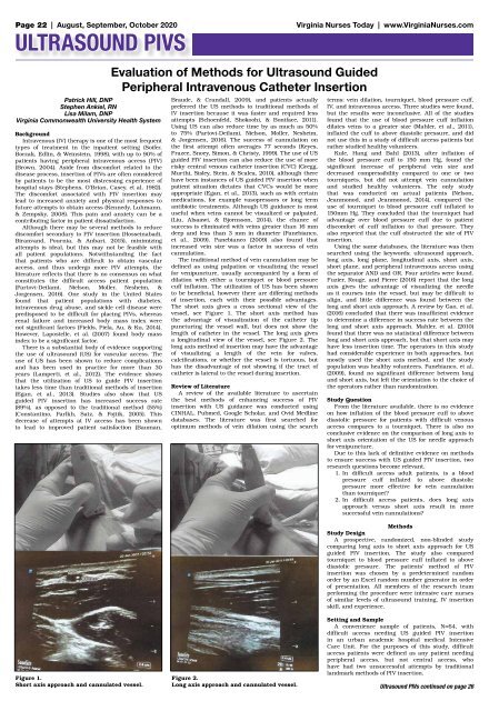
The short axis gives a cross sectional view of the<br />
vessel, see Figure 1. The short axis method has<br />
the advantage of visualization of the catheter tip<br />
puncturing the vessel wall, but does not show the<br />
length of catheter in the vessel. The long axis gives<br />
a longitudinal view of the vessel, see Figure 2. The<br />
long axis method of insertion may have the advantage<br />
of visualizing a length of the vein for valves,<br />
calcifications, or whether the vessel is tortuous, but<br />
has the disadvantage of not showing if the tract of<br />
catheter is lateral to the vessel during insertion.<br />
Review of Literature<br />
A review of the available literature to ascertain<br />
the best methods of enhancing success of PIV<br />
insertion with US guidance was conducted using<br />
CINHAL, Pubmed, Google Scholar, and Ovid Medline<br />
databases. The literature was first searched for<br />
optimum methods of vein dilation using the search<br />
terms: vein dilation, tourniquet, blood pressure cuff,<br />
IV, and intravenous access. Three studies were found,<br />
but the results were inconclusive. All of the studies<br />
found that the use of blood pressure cuff inflation<br />
dilates veins to a greater size (Mahler, et al., 2011),<br />
inflated the cuff to above diastolic pressure, and did<br />
not use this in a study of difficult access patients but<br />
rather studied healthy volunteers.<br />
Kule, Hang and Bahl (2013), after inflation of<br />
the blood pressure cuff to 150 mm Hg, found the<br />
significant increase of peripheral vein size and<br />
decreased compressibility compared to one or two<br />
tourniquets, but did not attempt vein cannulation<br />
and studied healthy volunteers. The only study<br />
that was conducted on actual patients (Nelson,<br />
Jeanmonod, and Jeanmonod, 2014), compared the<br />
use of tourniquet to blood pressure cuff inflated to<br />
150mm Hg. They concluded that the tourniquet had<br />
advantage over blood pressure cuff due to patient<br />
discomfort of cuff inflation to that pressure. They<br />
also reported that the cuff obstructed the site of PIV<br />
insertion.<br />
Using the same databases, the literature was then<br />
searched using the keywords: ultrasound approach,<br />
long axis, long plane, longitudinal axis, short axis,<br />
short plane, and peripheral intravenous access using<br />
the separator AND and OR. Four articles were found.<br />
Fuzier, Rouge, and Pierre (2016) report that the long<br />
axis gives the advantage of visualizing the needle<br />
as it courses into the vessel, but may be difficult to<br />
align, and little difference was found between the<br />
long and short axis approach. A review by Gao, et al.<br />
(2016) concluded that there was insufficient evidence<br />
to determine a difference in success rate between the<br />
long and short axis approach. Mahler, et al. (2010)<br />
found that there was no statistical difference between<br />
long and short axis approach, but that short axis may<br />
have less insertion time. The operators in this study<br />
had considerable experience in both approaches, but<br />
mostly used the short axis method, and the study<br />
population was healthy volunteers. Panebianco, et al.<br />
(2009), found no significant difference between long<br />
and short axis, but left the orientation to the choice of<br />
the operators rather than randomization.<br />
Study Question<br />
From the literature available, there is no evidence<br />
on how inflation of the blood pressure cuff to above<br />
diastolic pressure for patients with difficult venous<br />
access compares to a tourniquet. There is also no<br />
conclusive evidence on the comparison of long axis to<br />
short axis orientation of the US for needle approach<br />
for venipuncture.<br />
Due to this lack of definitive evidence on methods<br />
to ensure success with US guided PIV insertion, two<br />
research questions become relevant.<br />
1. In difficult access adult patients, is a blood<br />
pressure cuff inflated to above diastolic<br />
pressure more effective for vein cannulation<br />
than tourniquet?<br />
2. In difficult access patients, does long axis<br />
approach versus short axis result in more<br />
successful vein cannulations?<br />
Methods<br />
Study Design<br />
A prospective, randomized, non-blinded study<br />
comparing long axis to short axis approach for US<br />
guided PIV insertion. The study also compared<br />
tourniquet to blood pressure cuff inflated to above<br />
diastolic pressure. The patients’ method of PIV<br />
insertion was chosen by a predetermined random<br />
order by an Excel random number generator in order<br />
of presentation. All members of the research team<br />
performing the procedure were intensive care nurses<br />
of similar levels of ultrasound training, IV insertion<br />
skill, and experience.<br />
Figure 1.<br />
Short axis approach and cannulated vessel.<br />
Figure 2.<br />
Long axis approach and cannulated vessel.<br />
Setting and Sample<br />
A convenience sample of patients, N=64, with<br />
difficult access needing US guided PIV insertion<br />
in an urban academic hospital medical Intensive<br />
Care Unit. For the purposes of this study, difficult<br />
access patients were defined as any patient needing<br />
peripheral access, but not central access, who<br />
have had two unsuccessful attempts by traditional<br />
landmark methods of PIV insertion.<br />
Ultrasound PIVs continued on page 26