TECHNOLOGY DIGEST - Draper Laboratory
TECHNOLOGY DIGEST - Draper Laboratory
TECHNOLOGY DIGEST - Draper Laboratory

You also want an ePaper? Increase the reach of your titles
YUMPU automatically turns print PDFs into web optimized ePapers that Google loves.
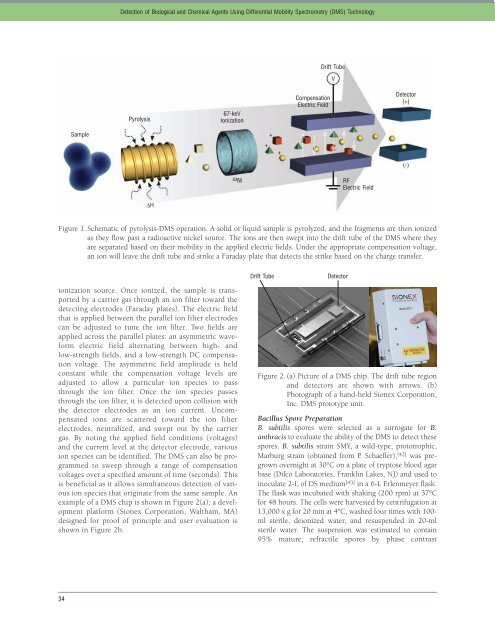
ionization source. Once ionized, the sample is transported<br />
by a carrier gas through an ion filter toward the<br />
detecting electrodes (Faraday plates). The electric field<br />
that is applied between the parallel ion filter electrodes<br />
can be adjusted to tune the ion filter. Two fields are<br />
applied across the parallel plates: an asymmetric waveform<br />
electric field alternating between high- and<br />
low-strength fields, and a low-strength DC compensation<br />
voltage. The asymmetric field amplitude is held<br />
constant while the compensation voltage levels are<br />
adjusted to allow a particular ion species to pass<br />
through the ion filter. Once the ion species passes<br />
through the ion filter, it is detected upon collision with<br />
the detector electrodes as an ion current. Uncompensated<br />
ions are scattered toward the ion filter<br />
electrodes, neutralized, and swept out by the carrier<br />
gas. By noting the applied field conditions (voltages)<br />
and the current level at the detector electrode, various<br />
ion species can be identified. The DMS can also be programmed<br />
to sweep through a range of compensation<br />
voltages over a specified amount of time (seconds). This<br />
is beneficial as it allows simultaneous detection of various<br />
ion species that originate from the same sample. An<br />
example of a DMS chip is shown in Figure 2(a); a development<br />
platform (Sionex Corporation, Waltham, MA)<br />
designed for proof of principle and user evaluation is<br />
shown in Figure 2b.<br />
34<br />
Sample<br />
Detection of Biological and Chemical Agents Using Differential Mobility Spectrometry (DMS) Technology<br />
Pyrolysis<br />
∆H<br />
67-keV<br />
Ionization<br />
63Ni<br />
Figure 1. Schematic of pyrolysis-DMS operation. A solid or liquid sample is pyrolyzed, and the fragments are then ionized<br />
as they flow past a radioactive nickel source. The ions are then swept into the drift tube of the DMS where they<br />
are separated based on their mobility in the applied electric fields. Under the appropriate compensation voltage,<br />
an ion will leave the drift tube and strike a Faraday plate that detects the strike based on the charge transfer.<br />
+<br />
+<br />
Drift Tube<br />
+<br />
Compensation<br />
Electric Field<br />
Drift Tube<br />
V<br />
Detector<br />
RF<br />
Electric Field<br />
Detector<br />
(+)<br />
Figure 2. (a) Picture of a DMS chip. The drift tube region<br />
and detectors are shown with arrows. (b)<br />
Photograph of a hand-held Sionex Corporation,<br />
Inc. DMS prototype unit.<br />
Bacillus Spore Preparation<br />
B. subtilis spores were selected as a surrogate for B.<br />
anthracis to evaluate the ability of the DMS to detect these<br />
spores. B. subtilis strain SMY, a wild-type, prototrophic,<br />
Marburg strain (obtained from P. Schaeffer), [42] was pregrown<br />
overnight at 30°C on a plate of tryptose blood agar<br />
base (Difco Laboratories, Franklin Lakes, NJ) and used to<br />
inoculate 2-L of DS medium [43] in a 6-L Erlenmeyer flask.<br />
The flask was incubated with shaking (200 rpm) at 37°C<br />
for 48 hours. The cells were harvested by centrifugation at<br />
13,000 x g for 20 min at 4°C, washed four times with 100ml<br />
sterile, deionized water, and resuspended in 20-ml<br />
sterile water. The suspension was estimated to contain<br />
95% mature, refractile spores by phase contrast<br />
(-)