Texas Journal of Microscopy - Texas Society for Microscopy
Texas Journal of Microscopy - Texas Society for Microscopy
Texas Journal of Microscopy - Texas Society for Microscopy
Create successful ePaper yourself
Turn your PDF publications into a flip-book with our unique Google optimized e-Paper software.
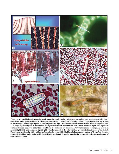
Plate 2. A series <strong>of</strong> light micrographs which show the graphic colors <strong>of</strong>ten seen when observing plant crystal cells either<br />
directly or under polarized light. 1. Micrographs showing a cleared leaf <strong>of</strong> Ginkgo biloba. Upper figure clearing as seen<br />
in normal light, lower right figure as seen in polarized light. Note the numerous druses which occur along each vein.<br />
2. A single styloid crystal in Eichhornia crassipies (the water hyacinth) as seen under polarized light. The styloid is<br />
contained within a cell but under these conditions the cell walls are not seen. 3. Crystal sclereids <strong>of</strong> Nymphaea as seen in<br />
normal light (left) and polarized light (right). The lower part <strong>of</strong> the sclereids has grown into the airspace <strong>of</strong> the leaf. 4.<br />
Paradermal section <strong>of</strong> a Vitis vinifera leaf showing large raphide idioblast. 5. Paradermal section <strong>of</strong> V. vinifera showing<br />
a raphide idioblast under polarized light. 6. Living section <strong>of</strong> V. vulpine showing large raphide cell with small group <strong>of</strong><br />
crystals in its center.<br />
Tex. J. Micros. 38: , 2007<br />
2


