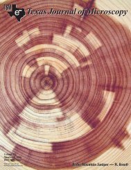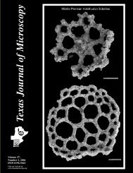
Texas Journal of Microscopy - Texas Society for Microscopy
Texas Journal of Microscopy - Texas Society for Microscopy
Texas Journal of Microscopy - Texas Society for Microscopy

You also want an ePaper? Increase the reach of your titles
YUMPU automatically turns print PDFs into web optimized ePapers that Google loves.
during the class break between lecture and lab. We generally<br />
talked about students because then, as today, they are always<br />
<strong>of</strong> interest to instructors. However, on the day in question, we<br />
discussed the use <strong>of</strong> electron microscopy in the study <strong>of</strong> plant<br />
cells. Specifically, we spoke about a recent paper by Dr. Flora<br />
Murray Scott, a distinguished plant anatomist at UCLA, and her<br />
colleagues. Her paper presented electron micrographs <strong>of</strong> onion<br />
epidermal cells (Scott, et al., 956). However, we could not relate<br />
her micrographs to plant cells as seen in the light microscope.<br />
In fact, “we” dubbed her micrographs “rug patterns.” We had<br />
a great laugh about these “useless pictures!” (I think that “Rug<br />
patterns” was probably Foster’s idiom, in fact sometimes I used<br />
“Wallpaper patterns” when relating this incident). Obviously,<br />
we did not have much regard <strong>for</strong> the use <strong>of</strong> electron microscopy<br />
in the study <strong>of</strong> plant cells. That “contempt” especially on my part,<br />
is incredible, when you realize that in just few years, I was fully<br />
engaged in studying plant cells with the electron microscope. By<br />
the early 960’s Dr. Scott published electron micrographs that<br />
contained obvious plant cells components. Her 956 paper was<br />
apparently near the beginning <strong>of</strong> her use <strong>of</strong> electron microscopy<br />
to study plant cells. Foster stuck with the tried and true and never<br />
moved on to electron microscopy.<br />
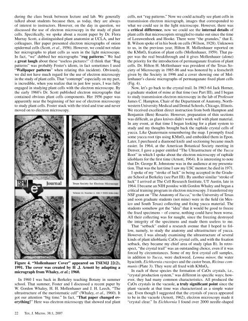
Figure 4. “Mollenhauer Cover” appeared on TSEMJ 22(2),<br />
1991. The cover was created by H .J. Arnott by adapting a<br />
micrograph from Whaley, et al.; 1960.<br />
In 960 I was back in Berkeley teaching Botany in summer<br />
school. That summer, Foster and I discussed a recent paper by<br />
W. Gordon Whaley, H. H. Mollenhauer and J. H. Leech, “The<br />
ultrastructure <strong>of</strong> the meristematic cell” (Whaley, et al., 960). It<br />
got our attention “big time.” In fact, “That paper changed everything!”<br />
Here was electron microscopy that showed real plant<br />
22 Tex. J. Micros. 38: , 2007<br />
cells, not “rug patterns.” Now we could actually see plant cells in<br />
transmission electron micrograph, images that corresponded to<br />
what we could see in the light microscope. However, there was<br />
a critical difference, now we could see the internal details <strong>of</strong><br />
plant cells that microscopists struggled to make out since the time<br />
<strong>of</strong> Leeuwenhoek and Hooke. There were “the granules,” that E.<br />
B. Wilson spoke <strong>of</strong> in his book on cells (Wilson, 928). Unknown<br />
to us, in the previous year, Hilton H. Mollenhauer reported on<br />
the KMnO4 fixation <strong>of</strong> plant cells (Mollenhauer, 959). That paper<br />
was the real breakthrough and it gives Mollenhauer (alone)<br />
the priority <strong>for</strong> the introduction <strong>of</strong> permanganate fixation <strong>of</strong> plant<br />
cells. Dr. Hilton H. Mollenhauer was president <strong>of</strong> the <strong>Texas</strong> <strong>Society</strong><br />
<strong>of</strong> <strong>Microscopy</strong> in 985-86 and was honored by a festschrift<br />
given by the <strong>Society</strong> in 996 and a cover showing one <strong>of</strong> Mollenhauer’s<br />
classic micrographs <strong>of</strong> permanganate fixed plant cells<br />
(Fig. 4).<br />
Now, let’s go back to the crystal trail. In 963-64 Jack Horner,<br />
a graduate student <strong>of</strong> mine at that time (see Part III), and I began<br />
to study transmission electron microscopy in the lab <strong>of</strong> Pr<strong>of</strong>essor<br />
James C. Hampton, Chair <strong>of</strong> the Department <strong>of</strong> Anatomy, Northwestern<br />
University Medical and Dental Schools, Chicago, Illinois.<br />
We received excellent direct instruction from both Hampton and<br />
Benjamin (Ben) Rosario. However, preparation <strong>of</strong> thin sections<br />
was difficult, as glass knives didn’t work well with plant material.<br />
In any event, at that time I began looking <strong>for</strong> a plant system to<br />
study and my thoughts brought back the raphide crystal cells <strong>of</strong><br />
yucca. Like Quatermain remembering the map. I promptly fixed<br />
some yucca root tips using KMnO4 and embedded them in Epon.<br />
Later, I purchased a diamond knife and sectioning became much<br />
easier. In 964, at the American Botanical <strong>Society</strong> meeting in<br />
Denver, I gave a paper entitled “The Ultrastructure <strong>of</strong> the Yucca<br />
Root” in which I spoke about the electron microscopy <strong>of</strong> raphide<br />
idioblasts <strong>for</strong> the first time (Arnott, 964). It is interesting to note<br />
that Dr. George R. Johnstone was in the audience at my presentation.<br />
That was the last time I saw my USC mentor; he died in 97 .<br />
I spoke <strong>of</strong> my “stroke <strong>of</strong> luck” in being accepted in the Graduate<br />
School at Berkeley (see Part III). By another similar “stroke <strong>of</strong><br />
luck” I arrived at The Cell Research Institute, UT Austin, late in<br />
964. I became an NIH postdoc with Gordon Whaley and began a<br />
critical training program in electron microscopy. I transferred my<br />
NSF grant on “The Anatomy <strong>of</strong> Yucca,” to the University <strong>of</strong> <strong>Texas</strong><br />
and soon graduate students (not mine) were in the field (in Mexico<br />
and South <strong>Texas</strong>) collecting and fixing yucca material. The<br />
students somehow got the “idea” that it would be good to freeze<br />
the fixed specimens – <strong>of</strong> course, nothing could have been worse.<br />
All their collecting was <strong>for</strong> naught, since the freezing destroyed<br />
the integrity <strong>of</strong> the specimens and made them useless to me.<br />
That “setback” ended a research avenue that I hoped to follow,<br />
namely, to study the anatomy and ultrastructure <strong>of</strong> yucca.<br />
However, I was already examining the ultrastructure <strong>of</strong> several<br />
kinds <strong>of</strong> plant idioblastic CaOx crystal cells, and with the fixation<br />
setback, they became my chief area <strong>of</strong> study (plan B). In retrospect,<br />
“the crystal trail” was an outstanding choice, even if it was<br />
<strong>for</strong>ced by circumstances. Some <strong>of</strong> my first crystal cell samples,<br />
in addition to Yucca, were duckweed, Lemna minor, the water<br />
hyacinth, Eichhornia crassipes and the castor bean, Ricinus communis<br />
(Plate 3). They were all fixed with KMnO4.<br />
In each <strong>of</strong> these species the <strong>for</strong>mation <strong>of</strong> CaOx crystals, i.e.,<br />
“crystal production system,” was different in specific ways; however,<br />
they had many common characteristics. All produced the<br />
CaOx crystals in the vacuole, a truly significant point since the<br />
plant vacuole at that time was characterized as a simple water<br />
sac. Even though I suggested that the crystals <strong>of</strong> yucca appeared<br />
to be in the vacuole (Arnott, 962), electron microscopy made it<br />
“crystal clear.” In Eichhornia I found over 2000 needle-shaped