Texas Journal of Microscopy - Texas Society for Microscopy
Texas Journal of Microscopy - Texas Society for Microscopy
Texas Journal of Microscopy - Texas Society for Microscopy

Create successful ePaper yourself
Turn your PDF publications into a flip-book with our unique Google optimized e-Paper software.
she worked on the seeds <strong>of</strong> zucchini (Curcubita pepo) finishing<br />
her thesis in 980. Her study <strong>of</strong> the dormant (completely dry) seeds<br />
<strong>of</strong> zucchini is unique. Most ultrastructural research on seeds was<br />
(is) done with seeds that had been hydrated. A set <strong>of</strong> surprising<br />
results came from this research in which the fixation was by osmium<br />
vapor. For example, the cell walls <strong>of</strong> the dormant cells are<br />
folded in regular arrays that help maintain wall structure during<br />
the dormant period and allow <strong>for</strong> the imbibition <strong>of</strong> water and expansion<br />
during germination. A uniquely interesting finding from<br />
this research was a material that filled the intracellular spaces in<br />
dry seeds. This material “falls out” <strong>of</strong> its position in specimens<br />
when they are fractured. The material is flexible and apparently<br />
gel-like in consistency. Through a series <strong>of</strong> excellent micrographs,<br />
Mary Alice also showed exactly how the protein bodies <strong>of</strong> the<br />
dry seed become protein vacuoles and then develop into vacuoles<br />
during the germination process. Like me, Mary Alice’s first research<br />
was on seeds. I have examined and re-examined her thesis<br />
recently, and without doubt, it is equivalent <strong>of</strong> most Ph.D. dissertations<br />
at UTA or in the other four institutions where I taught.<br />
Un<strong>for</strong>tunately, at the time, because <strong>of</strong> pointless state government<br />
interference, we were not authorized to <strong>of</strong>fer a Ph.D. degree (See<br />
Part II). Working out the methods with which to examine seeds in<br />
their dormant condition was a bona fide <strong>for</strong>ward step in the study<br />
<strong>of</strong> seeds that can<br />
be attributed to Dr.<br />
Webb.<br />
Following her<br />
graduation, Mary<br />
Alice moved to<br />
North Carolina<br />
where she worked<br />
<strong>for</strong> Dr. J. David<br />
Robertson at Duke<br />
Medical Center.<br />
Dr. Robertson, well<br />
known <strong>for</strong> his studies<br />
<strong>of</strong> membranes,<br />
is perhaps generally<br />
remembered<br />
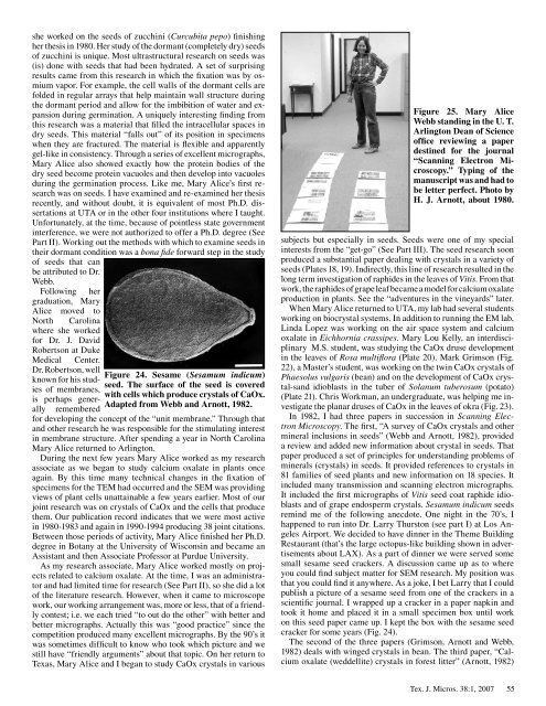
Figure 24. Sesame (Sesamum indicum)<br />
seed. The surface <strong>of</strong> the seed is covered<br />
with cells which produce crystals <strong>of</strong> CaOx.<br />
Adapted from Webb and Arnott, 1982.<br />
<strong>for</strong> developing the concept <strong>of</strong> the “unit membrane.” Through that<br />
and other research he was responsible <strong>for</strong> the stimulating interest<br />
in membrane structure. After spending a year in North Carolina<br />
Mary Alice returned to Arlington.<br />
During the next few years Mary Alice worked as my research<br />
associate as we began to study calcium oxalate in plants once<br />
again. By this time many technical changes in the fixation <strong>of</strong><br />
specimens <strong>for</strong> the TEM had occurred and the SEM was providing<br />
views <strong>of</strong> plant cells unattainable a few years earlier. Most <strong>of</strong> our<br />
joint research was on crystals <strong>of</strong> CaOx and the cells that produce<br />
them. Our publication record indicates that we were most active<br />
in 980- 983 and again in 990- 994 producing 38 joint citations.<br />
Between those periods <strong>of</strong> activity, Mary Alice finished her Ph.D.<br />
degree in Botany at the University <strong>of</strong> Wisconsin and became an<br />
Assistant and then Associate Pr<strong>of</strong>essor at Purdue University.<br />
As my research associate, Mary Alice worked mostly on projects<br />
related to calcium oxalate. At the time, I was an administrator<br />
and had limited time <strong>for</strong> research (See Part II), so she did a lot<br />
<strong>of</strong> the literature research. However, when it came to microscope<br />
work, our working arrangement was, more or less, that <strong>of</strong> a friendly<br />
contest; i.e. we each tried “to out do the other” with better and<br />
better micrographs. Actually this was “good practice” since the<br />
competition produced many excellent micrographs. By the 90’s it<br />
was sometimes difficult to know who took which picture and we<br />
still have “friendly arguments” about that topic. On her return to<br />
<strong>Texas</strong>, Mary Alice and I began to study CaOx crystals in various<br />
Figure 25. Mary Alice<br />
Webb standing in the U. T.<br />
Arlington Dean <strong>of</strong> Science<br />
<strong>of</strong>fice reviewing a paper<br />
destined <strong>for</strong> the journal<br />
“Scanning Electron <strong>Microscopy</strong>.”<br />
Typing <strong>of</strong> the<br />
manuscript was and had to<br />
be letter perfect. Photo by<br />
H. J. Arnott, about 1980.<br />
subjects but especially in seeds. Seeds were one <strong>of</strong> my special<br />
interests from the “get-go” (See Part III). The seed research soon<br />
produced a substantial paper dealing with crystals in a variety <strong>of</strong><br />
seeds (Plates 8, 9). Indirectly, this line <strong>of</strong> research resulted in the<br />
long term investigation <strong>of</strong> raphides in the leaves <strong>of</strong> Vitis. From that<br />
work, the raphides <strong>of</strong> grape leaf became a model <strong>for</strong> calcium oxalate<br />
production in plants. See the “adventures in the vineyards” later.<br />
When Mary Alice returned to UTA, my lab had several students<br />
working on biocrystal systems. In addition to running the EM lab,<br />
Linda Lopez was working on the air space system and calcium<br />
oxalate in Eichhornia crassipes. Mary Lou Kelly, an interdisciplinary<br />
M.S. student, was studying the CaOx druse development<br />
in the leaves <strong>of</strong> Rosa multiflora (Plate 20). Mark Grimson (Fig.<br />
22), a Master’s student, was working on the twin CaOx crystals <strong>of</strong><br />
Phaesolus vulgaris (bean) and on the development <strong>of</strong> CaOx crystal-sand<br />
idioblasts in the tuber <strong>of</strong> Solanum tuberosum (potato)<br />
(Plate 2 ). Chris Workman, an undergraduate, was helping me investigate<br />
the planar druses <strong>of</strong> CaOx in the leaves <strong>of</strong> okra (Fig. 23).<br />
In 982, I had three papers in succession in Scanning Electron<br />
<strong>Microscopy</strong>. The first, “A survey <strong>of</strong> CaOx crystals and other<br />
mineral inclusions in seeds” (Webb and Arnott, 982), provided<br />
a review and added new in<strong>for</strong>mation about crystal in seeds. That<br />
paper produced a set <strong>of</strong> principles <strong>for</strong> understanding problems <strong>of</strong><br />
minerals (crystals) in seeds. It provided references to crystals in<br />
8 families <strong>of</strong> seed plants and new in<strong>for</strong>mation on 8 species. It<br />
included many transmission and scanning electron micrographs.<br />
It included the first micrographs <strong>of</strong> Vitis seed coat raphide idioblasts<br />
and <strong>of</strong> grape endosperm crystals. Sesamum indicum seeds<br />
remind me <strong>of</strong> the following anecdote. One night in the 70’s, I<br />
happened to run into Dr. Larry Thurston (see part I) at Los Angeles<br />
Airport. We decided to have dinner in the Theme Building<br />
Restaurant (that’s the large octopus-like building shown in advertisements<br />
about LAX). As a part <strong>of</strong> dinner we were served some<br />
small sesame seed crackers. A discussion came up as to where<br />
you could find subject matter <strong>for</strong> SEM research. My position was<br />
that you could find it anywhere. As a joke, I bet Larry that I could<br />
publish a picture <strong>of</strong> a sesame seed from one <strong>of</strong> the crackers in a<br />
scientific journal. I wrapped up a cracker in a paper napkin and<br />
took it home and placed it in a small specimen box until work<br />
on this seed paper came up. I kept the box with the sesame seed<br />
cracker <strong>for</strong> some years (Fig. 24).<br />
The second <strong>of</strong> the three papers (Grimson, Arnott and Webb,<br />
982) deals with winged crystals in bean. The third paper, “Calcium<br />
oxalate (weddellite) crystals in <strong>for</strong>est litter” (Arnott, 982)<br />
Tex. J. Micros. 38: , 2007<br />
55