Texas Journal of Microscopy - Texas Society for Microscopy
Texas Journal of Microscopy - Texas Society for Microscopy
Texas Journal of Microscopy - Texas Society for Microscopy

You also want an ePaper? Increase the reach of your titles
YUMPU automatically turns print PDFs into web optimized ePapers that Google loves.
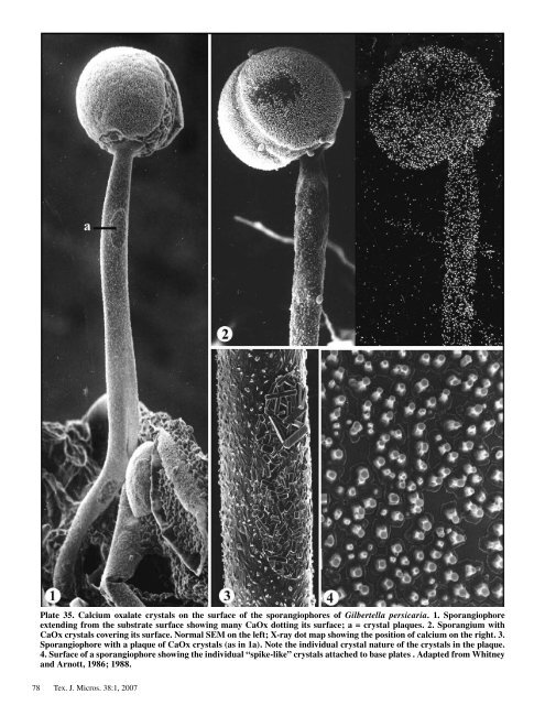
Plate 35. Calcium oxalate crystals on the surface <strong>of</strong> the sporangiophores <strong>of</strong> Gilbertella persicaria. 1. Sporangiophore<br />
extending from the substrate surface showing many CaOx dotting its surface; a = crystal plaques. 2. Sporangium with<br />
CaOx crystals covering its surface. Normal SEM on the left; X-ray dot map showing the position <strong>of</strong> calcium on the right. 3.<br />
Sporangiophore with a plaque <strong>of</strong> CaOx crystals (as in 1a). Note the individual crystal nature <strong>of</strong> the crystals in the plaque.<br />
4. Surface <strong>of</strong> a sporangiophore showing the individual “spike-like” crystals attached to base plates . Adapted from Whitney<br />
and Arnott, 1986; 1988.<br />
78 Tex. J. Micros. 38: , 2007