Colin Nicol was always keen to get on with his work; in all probability, his colleagues would describe him as focused, or single-minded, or driven, or even obsessed by his research. I would probably choose the latter description. If it meant working all night, working in the dark, or working in an inconvenient place (boat) that was just part <strong>of</strong> research, his drive certainly had a great deal to do with the fact that Colin Nicol was “a very good scientist.” On one occasion when I was visiting him at Port Aransas, he was in the process <strong>of</strong> fixing the eyes <strong>of</strong> some stingrays. He wanted the eyes to be dark adapted so the stingrays were in buckets in a “very dark room;” perhaps there was a tiny red light in the room. In any event, I was glad to learn that he cut the stingers <strong>of</strong>f the fish be<strong>for</strong>e putting them in the dark. Colin used a rather large machete to deal the fatal blow, all done in “that very dark room,” while I remained outside fighting <strong>of</strong>f the famous bird-like mosquitoes <strong>of</strong> Port Aransas. Colin had hyperopia (farsightedness) and used two pairs <strong>of</strong> glasses to deal with that situation. Depending on the circumstances he used one set <strong>for</strong> normal viewing and a second <strong>for</strong> close-up vision. I am very familiar with that situation since I too have hyperopia. Benjamin Franklin invented bifocals to solve this problem. However, <strong>for</strong> me (and I guess Colin) reading glasses are better <strong>for</strong> reading and an absolute requirement <strong>for</strong> using the SEM. The remarkable thing about Colin was that he could switch from one pair to the other incredibly fast and did so <strong>of</strong>ten. The only comparison I can make is with the “fast guns” <strong>of</strong> the old west. Colin Nicol involved me deeply in the study <strong>of</strong> animals’ eyes; like the viruses in the Kenneth Smith saga, this was another subject about which I knew almost nothing at the outset. You might infer, correctly I think, that I did a great deal <strong>of</strong> “on the job training.” However, I believe that in both cases I contributed more than might be imagined at the outset. The interaction with Nicol was important to my development in that it taught me to get on with things quickly and to sort out what was important from the rest; working with Colin there was no time <strong>for</strong> “diddling around.” Observers, noting his level <strong>of</strong> activity might have called it the “Nicol syndrome.” I have just written a paragraph telling you about a “whirling dervish” but now, to set things straight, I must tell you about Colin Nicol the host, family man and friend. On a couple <strong>of</strong> occasions Jean and I spent an evening with Colin and his wife in their home at Aransas Pass; they shared that home with a very large, black dog (I think it was a Mastiff). As a host, Colin was very gracious, considerate, completely relaxed. Like night and day, the drive associated with his research activities was absent and he was the most affable host you can imagine, in fact, he was always a big hit with Jean. Colin and I published 6 papers together, most dealing with eye structure (Fig. 3). In eyes we studied the following: the tapetum lucidum <strong>of</strong> the stingray (Arnott, Best and Nicol, 970); reflecting spheres in the eyes <strong>of</strong> weakfish (Arnott, Nicol and Querfeld, 97 ); the tapetum lucidum in the eyes <strong>of</strong> seatrout (Arnott, Nicol, and Querfeld, 972); rib<strong>of</strong>lavin containing spheres in the eyes <strong>of</strong> gars (Nicol and Arnott 972; Fig. 4); the tapetum lucidum in the eyes <strong>of</strong> gar (Nicol and Arnott, 973); the tapetum lucidum in the eyes <strong>of</strong> catfish (Arnott, et al., 974); tapetum lucidum in the eyes <strong>of</strong> goatsuckers [birds] (Nicol and Arnott, 974); and the diffuse reflectance <strong>of</strong> retinal tapetum lucidum in drum (Nicol, Best and Arnott, 973). We also studied the reflection <strong>of</strong> the ratfish (Hydrolagus colliei) skin 32 Tex. J. Micros. 38: , 2007 Figure 14. Ordered arrangement <strong>of</strong> spheres which <strong>for</strong>m the reflecting layer in the eyes <strong>of</strong> the alligator gar. Adapted from Nicol and Arnott, 1973. (Arnott and Nicol, 970). I also started a study on the guppy skin (Plate 8) but it has never been published. A few <strong>of</strong> these studies deal directly with crystals. Many <strong>of</strong> these studies were reminiscent <strong>of</strong> the swamps, sand dunes, and mountains <strong>of</strong> Africa that sidetracked the Quatermain party from their travels on the trail to King Solomon’s Mines. While traveling on my crystal trail I was <strong>of</strong>ten diverted, even the smallest dune (interesting subject or student complaint) could provide a distraction. After I moved to South Florida, Nicol and I tried to continue joint research but the distance and the complexities were too much. That pleasant and effective cooperative relationship was killed by my “urge to administrate.” Which <strong>of</strong> my studies with Nicol contributed to the crystal trail? First let’s reflect on the silver color seen in the skin <strong>of</strong> many fish, <strong>for</strong> example the skin <strong>of</strong> a guppy (Plate 8), sardine or king salmon. Where does that silver color come from? The short answer is that silver color is caused by the reflection <strong>of</strong> certain wavelengths <strong>of</strong> light (silver light) by special layers <strong>of</strong> cells in the skin. The cells in these special layers contain guanine crystals. Each cell, termed a guanophore, or sometimes an iridiophore, may contain ten or more large flat guanine crystals arranged in an oriented array. For example, consider the well known neon tetra. Guanophores are involved in the bright blue lateral stripe and the red posterior. In the neon tetra the red and blue colors come from cells exterior to a guanophore layer. Light from outside passes though these pigments, which act like filters, and are reflected back by the guanine layer, hence the colors seem to glow. When there is very low room light the fish are almost transparent. Likewise the angle <strong>of</strong> view can cause the blue line to change to green. We studied the origin <strong>of</strong> the silver/gold color(s) in the ratfish skin (Arnott and Nicol, 970). The general structure <strong>of</strong> the reflecting cells (guanophores) is presented in a line drawing (Fig. 2). In that case we see an elongate cell section containing: five guanine crystals, a nucleus, mitochondria, Golgi body, rough endoplasmic reticulum and a series <strong>of</strong> vesicles. In actuality the cells are flattened perpendicular to the plane <strong>of</strong> cut seen in the diagram. Each plate-like guanine crystal was produced and is contained in a chamber. The organization seen in the ratfish skin and guppy are very much like that seen in plants where the CaOx crystals, regardless <strong>of</strong> their shape, develop and are contained in crystal chambers. One interesting question regarding this system is what physical <strong>of</strong> biochemical <strong>for</strong>ces are involved in the precise arrangement and orientation <strong>of</strong> the crystals. In some fish there is clear evidence that the distance between the guanine crystals can be “manipulated” by the guanophores. A change in the distance between the members <strong>of</strong> a guanine stack can change the wavelength <strong>of</strong> the reflected light because the individual crystals work as a quarter wave reflecting system. Thus, when the distance between guanine crystals changes, the light reflected from skin can vary in color. Colin Nicol also introduced me to eyeshine in fish. We investigated the ultrastructure in several examples <strong>of</strong> fish with eye-shine. The cause <strong>of</strong> eye-shine in these fish is a layer called the tapetum lucidum (reflecting layer). The tapetum lucidum is situated behind (outside) the retina. The two are <strong>for</strong>med as concentric zones in the eye. Various substances are involved in fish eye-shine. For example spheres are used by
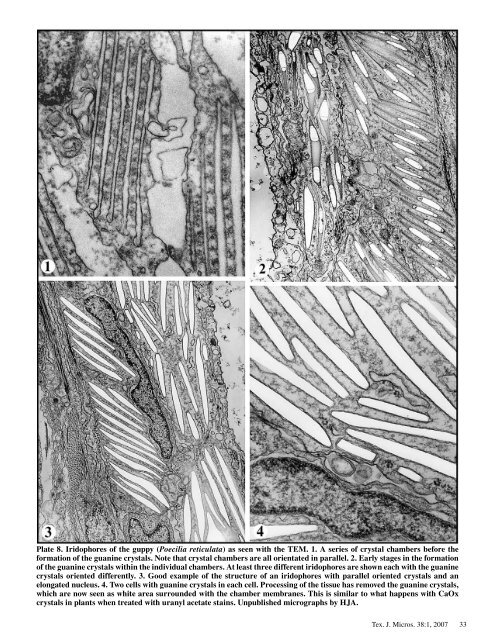
Plate 8. Iridophores <strong>of</strong> the guppy (Poecilia reticulata) as seen with the TEM. 1. A series <strong>of</strong> crystal chambers be<strong>for</strong>e the <strong>for</strong>mation <strong>of</strong> the guanine crystals. Note that crystal chambers are all orientated in parallel. 2. Early stages in the <strong>for</strong>mation <strong>of</strong> the guanine crystals within the individual chambers. At least three different iridophores are shown each with the guanine crystals oriented differently. 3. Good example <strong>of</strong> the structure <strong>of</strong> an iridophores with parallel oriented crystals and an elongated nucleus. 4. Two cells with guanine crystals in each cell. Processing <strong>of</strong> the tissue has removed the guanine crystals, which are now seen as white area surrounded with the chamber membranes. This is similar to what happens with CaOx crystals in plants when treated with uranyl acetate stains. Unpublished micrographs by HJA. Tex. J. Micros. 38: , 2007 33