Texas Journal of Microscopy - Texas Society for Microscopy
Texas Journal of Microscopy - Texas Society for Microscopy
Texas Journal of Microscopy - Texas Society for Microscopy

You also want an ePaper? Increase the reach of your titles
YUMPU automatically turns print PDFs into web optimized ePapers that Google loves.
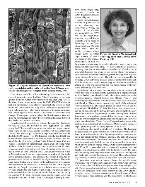
Figure 47. Crystal sclereids <strong>of</strong> Nymphaea mexicana. Note<br />
CaOx crystals imbedded in the cell wall <strong>of</strong> four different sclereids<br />
in the storage root. Adapted from Van De Veire, 1997.<br />
On a visit to the JOEL <strong>of</strong>fice in Peabody, Massachusetts, I discovered<br />
some interesting leaf-like “plants” growing on the large<br />
granite boulders which were frequent in the area (Fig. 45). At<br />
the time, I was taking a course on the EMS 200 TEM that we<br />
had just purchased. I took a few <strong>of</strong> these leaf-like structures back<br />
home and determined that they were Umbilicaria pennsylvanica,<br />
a lichen species. I also found that one <strong>of</strong> its common names<br />
was “Washington’s Rock Tripe.” This lichen is associated with<br />
George Washington because when the Revolutionary War soldiers<br />
he commanded at Valley Forge were hard pressed <strong>for</strong> food,<br />
they boiled and ate the rock tripe.<br />
I was fascinated by rock tripe, not only because they had many<br />
crystals <strong>of</strong> CaOx, but because <strong>of</strong> their “reputation” as “emergency<br />
food.” Large multi-interpenetrant twins and single crystals<br />
were found on the surface and in the interior <strong>of</strong> these interesting<br />
“plants.” On a later trip, I collected a large number <strong>of</strong> the leaf-like<br />
thalli <strong>for</strong> SEM studies (Fig. 46). I studied both the dried thalli and<br />
hydrated fixed specimens. I also boiled them <strong>for</strong> some time and<br />
found that the plentiful crystals <strong>of</strong> CaOx did not dissolve. Calcium<br />
oxalate is insoluble in water (Merck Index), even in boiling water.<br />
Soldiers <strong>of</strong> the Revolution and/or others that have eaten U. pennsylvanica<br />
would have got a good dose <strong>of</strong> oxalate when the crystals<br />
dissolve in their stomach acids. On another occasion, while<br />
in Massachusetts, I had the opportunity to work with (then) the<br />
newly developed ESEM, and among other things, I collected U.<br />
pennsylvanica to look at the thalli in a hydrated state. The walls<br />
were much thicker and wrinkles in the cell contours were erased<br />
when hydrated. In some cases, it appears that the CaOx crystals<br />
<strong>for</strong>m in the fungal component <strong>of</strong> the lichen since examples <strong>of</strong><br />
crystals attached to the hyphae were found. The crystals appear<br />
to be embedded on the surface <strong>of</strong> the thallus and thereby partly<br />
surrounded by hyphae. However, some crystals seem to be free<br />
within the “tissues” <strong>of</strong> this “plant.” Although twinning is com-<br />
mon, many single large<br />
prismatic crystals <strong>of</strong><br />
CaOx dihydrate were also<br />
present (Fig. 46).<br />
One <strong>of</strong> the last students<br />
to work on plant crystals<br />
in my laboratory was<br />
Jackie Van De Veire. The<br />
subject <strong>of</strong> Jackie’s thesis,<br />
completed in 997,<br />
was on the large multi<br />
branched crystalliferous<br />
sclereids which occur in<br />
the storage roots <strong>of</strong> Nymphaea<br />
mexicana (Van De<br />
Veire, 997). This water<br />
lily produces unique<br />
storage roots in which<br />
large masses <strong>of</strong> starch<br />
are stored in the cortical<br />
parenchyma. In addition,<br />
Figure 48. Sandra Westmoreland,<br />
“The egg shell lady”, about 1998.<br />
Photo by HJA.<br />
these roots produce very large sclereids which have crystals embedded<br />
in their cell walls (Fig. 47). The sclereids are unique as<br />
they may have five or more cellular branches, all <strong>of</strong> which run in<br />
a parallel direction and more or less in one plane. The arms <strong>of</strong><br />
these sclereids extend by intrusive growth <strong>for</strong>cing their way between<br />
other cells in the cortex. The sclereids are also notable <strong>for</strong><br />
the large CaOx dihydrate crystals that are embedded in the cell<br />
wall. Jackie worked out the morphology and development <strong>of</strong> these<br />
unique cells and she established the role these storage roots play<br />
in the life history <strong>of</strong> N. mexicana.<br />
Crystals are not just found in association with specialized cell<br />
types. They are <strong>of</strong>ten found in the cytoplasm or in organelles such<br />
as microbodies, mitochondria and chloroplasts and even in the<br />
cytoplasm proper. I studied them in corn phloem (Plate 37) and<br />
in Malpigia glabra flowers, which have cells with crystals in their<br />
mitochondria. These crystals may occupy much <strong>of</strong> the volume <strong>of</strong><br />
some mitochondria. The lattice planes <strong>of</strong> these crystals can be<br />
seen with the TEM (Plate 37: -6). The line to line measurements<br />
can be on the order <strong>of</strong> 50- 00 Å. Proteins are probably the only<br />
molecules which are large enough to <strong>for</strong>m lattice planes <strong>of</strong> these<br />
dimensions. I always have assumed that the above crystals were<br />
proteins which underwent crystallization being present in excess<br />
in the mitochondrion. These “protein” crystals <strong>of</strong>ten seem to be<br />
nucleated on mitochondrial membranes. Malpigia also produces<br />
large “protein crystals” in the cytoplasm (Plate 37: 4).<br />
Crystals are also found in single cell organisms. The bacterial<br />
magnetosomes are partly crystalline iron sulfide crystals and are<br />
found in a few bacterial species. Euglena granulata is a eukaryotic<br />
single cell organism that produces crystals. The cells <strong>of</strong> Euglena<br />
are somewhat complex; they have a nucleus, mitochondria, chloroplasts,<br />
an intricate folded external structure called the pellicle<br />
which allows them to change shape, a very large Golgi apparatus<br />
with 30 to 50 or more cisternae, a contractile vacuole, and two<br />
flagella, one which is associated with the large eyespot. The cells<br />
have the ability to change their shape from spherical to elongated<br />
and back, and to swim backward or <strong>for</strong>ward. These characteristics<br />
certainly establish Euglena as one <strong>of</strong> the “one-cell wonders”.<br />
E. granulata also makes crystals, hence the specific name<br />
granulata. The crystals can be seen as bright spots when viewing<br />
cells under polarized light (each spot representing a single crystal).<br />
The E. granulata crystals are believed to be calcium oxalate<br />
as they are extracted in the same manner as the CaOx crystals<br />
in higher plants; however, unambiguous identification <strong>of</strong> these<br />
crystals has not been achieved yet. The specimens I studied came<br />
Tex. J. Micros. 38: , 2007<br />
83