Role of the ubiquitin-like modifier FAT10 in protein degradation and ...
Role of the ubiquitin-like modifier FAT10 in protein degradation and ...
Role of the ubiquitin-like modifier FAT10 in protein degradation and ...

Create successful ePaper yourself
Turn your PDF publications into a flip-book with our unique Google optimized e-Paper software.
<strong>Role</strong> <strong>of</strong> <strong>the</strong> <strong>ubiquit<strong>in</strong></strong>-<strong>like</strong><br />
<strong>modifier</strong> <strong>FAT10</strong> <strong>in</strong><br />
prote<strong>in</strong> <strong>degradation</strong> <strong>and</strong> immunity<br />
Dissertation<br />
zur Erlangung des akademischen Grades<br />
des Doktors der Naturwissenschaften<br />
(Dr. rer. nat.)<br />
an der Universität Konstanz<br />
Fachbereich Biologie<br />
vorgelegt von<br />
Birte Kathar<strong>in</strong>a Henriette Kalveram<br />
Tag der mündlichen Prüfung: 06.04.09<br />
1. Referent: Pr<strong>of</strong>. Dr. Marcus Groettrup, Universität Konstanz<br />
2. Referent: Pr<strong>of</strong>. Dr. Mart<strong>in</strong> Scheffner, Universität Konstanz
Contents<br />
Acknowledgements 3<br />
Summary / Zusammenfassung 4<br />
English . . . . . . . . . . . . . . . . . . . . . . . . . . . . . . . . . . . . . . . 4<br />
Deutsch . . . . . . . . . . . . . . . . . . . . . . . . . . . . . . . . . . . . . . . 6<br />
Introduction 9<br />
Prote<strong>in</strong> Degradation . . . . . . . . . . . . . . . . . . . . . . . . . . . . . . . 9<br />
The Ubiquit<strong>in</strong>-Proteasome-System . . . . . . . . . . . . . . . . . . . . . . . 10<br />
The Proteasome . . . . . . . . . . . . . . . . . . . . . . . . . . . . . . . 10<br />
The 20S Proteasome . . . . . . . . . . . . . . . . . . . . . . . . 10<br />
The 26S Proteasome . . . . . . . . . . . . . . . . . . . . . . . . 11<br />
Ubiquit<strong>in</strong> <strong>and</strong> Ubiquit<strong>in</strong>-Like Prote<strong>in</strong>s . . . . . . . . . . . . . . . . . . 14<br />
Ubiquit<strong>in</strong> . . . . . . . . . . . . . . . . . . . . . . . . . . . . . . . 14<br />
Ubiquit<strong>in</strong>-Like Prote<strong>in</strong>s . . . . . . . . . . . . . . . . . . . . . . 18<br />
Ubiquit<strong>in</strong>-Like Modifiers . . . . . . . . . . . . . . . . . . . . . . 18<br />
<strong>FAT10</strong> . . . . . . . . . . . . . . . . . . . . . . . . . . . . . . . . . 21<br />
Ubiquit<strong>in</strong> Doma<strong>in</strong> Prote<strong>in</strong>s . . . . . . . . . . . . . . . . . . . . 24<br />
NUB1 . . . . . . . . . . . . . . . . . . . . . . . . . . . . . . . . . 26<br />
The Autophagy-Lysosome System . . . . . . . . . . . . . . . . . . . . . . . . 28<br />
Misfolded Prote<strong>in</strong> Stress, Aggresomes <strong>and</strong> <strong>the</strong> Connection between Au-<br />
tophagy <strong>and</strong> <strong>the</strong> UPS . . . . . . . . . . . . . . . . . . . . . . . . . . . . 29<br />
HDAC6 . . . . . . . . . . . . . . . . . . . . . . . . . . . . . . . . 33<br />
Chapter 1: The UBA doma<strong>in</strong>s <strong>of</strong> NUB1L are required for b<strong>in</strong>d<strong>in</strong>g but not for<br />
accelerated <strong>degradation</strong> <strong>of</strong> <strong>the</strong> <strong>ubiquit<strong>in</strong></strong>-<strong>like</strong> <strong>modifier</strong> <strong>FAT10</strong> 36<br />
Abstract . . . . . . . . . . . . . . . . . . . . . . . . . . . . . . . . . . . . 37<br />
Introduction . . . . . . . . . . . . . . . . . . . . . . . . . . . . . . . . . 38<br />
Results . . . . . . . . . . . . . . . . . . . . . . . . . . . . . . . . . . . . 40<br />
Discussion . . . . . . . . . . . . . . . . . . . . . . . . . . . . . . . . . . 51<br />
Experimental Procedures . . . . . . . . . . . . . . . . . . . . . . . . . 56<br />
1
Acknowledgements . . . . . . . . . . . . . . . . . . . . . . . . . . . . . 59<br />
Chapter 2: Degradation <strong>of</strong> <strong>FAT10</strong> by <strong>the</strong> 26S proteasome <strong>in</strong> vitro is <strong>in</strong>depen-<br />
dent <strong>of</strong> ubiquitylation but relies on NUB1L 60<br />
Abstract . . . . . . . . . . . . . . . . . . . . . . . . . . . . . . . . . . . . 61<br />
Introduction . . . . . . . . . . . . . . . . . . . . . . . . . . . . . . . . . 62<br />
Results . . . . . . . . . . . . . . . . . . . . . . . . . . . . . . . . . . . . 64<br />
Discussion . . . . . . . . . . . . . . . . . . . . . . . . . . . . . . . . . . 69<br />
Materials <strong>and</strong> methods . . . . . . . . . . . . . . . . . . . . . . . . . . . 72<br />
Acknowledgements . . . . . . . . . . . . . . . . . . . . . . . . . . . . . 76<br />
Chapter 3: The <strong>ubiquit<strong>in</strong></strong>-<strong>like</strong> <strong>modifier</strong> <strong>FAT10</strong> <strong>in</strong>teracts with HDAC6 <strong>and</strong> lo-<br />
calizes to aggresomes under proteasome <strong>in</strong>hibition 77<br />
Abstract . . . . . . . . . . . . . . . . . . . . . . . . . . . . . . . . . . . . 78<br />
Introduction . . . . . . . . . . . . . . . . . . . . . . . . . . . . . . . . . 79<br />
Results . . . . . . . . . . . . . . . . . . . . . . . . . . . . . . . . . . . . 81<br />
Discussion . . . . . . . . . . . . . . . . . . . . . . . . . . . . . . . . . . 95<br />
Materials <strong>and</strong> Methods . . . . . . . . . . . . . . . . . . . . . . . . . . . 100<br />
Acknowledgements . . . . . . . . . . . . . . . . . . . . . . . . . . . . . 103<br />
Chapter 4: <strong>FAT10</strong>-deficient mice display a prolonged CD8 + T-cell response<br />
after LCMV <strong>in</strong>fection 104<br />
Introduction . . . . . . . . . . . . . . . . . . . . . . . . . . . . . . . . . 105<br />
Results <strong>and</strong> Discussion . . . . . . . . . . . . . . . . . . . . . . . . . . . 106<br />
Materials <strong>and</strong> Methods . . . . . . . . . . . . . . . . . . . . . . . . . . . 109<br />
Chapter 5: Generation <strong>of</strong> mouse monoclonal antibodies directed aga<strong>in</strong>st<br />
human <strong>FAT10</strong> 111<br />
Introduction . . . . . . . . . . . . . . . . . . . . . . . . . . . . . . . . . 112<br />
Results <strong>and</strong> Discussion . . . . . . . . . . . . . . . . . . . . . . . . . . . 112<br />
Materials <strong>and</strong> Methods . . . . . . . . . . . . . . . . . . . . . . . . . . . 117<br />
Discussion 120<br />
References 126<br />
Abbreviations 142<br />
Record <strong>of</strong> Contributions 145<br />
2
Acknowledgements<br />
First <strong>of</strong> all, I would <strong>like</strong> to thank Pr<strong>of</strong>. Dr. Marcus Groettrup for giv<strong>in</strong>g me<br />
<strong>the</strong> opportunity to do this work <strong>and</strong> for teach<strong>in</strong>g me a lot dur<strong>in</strong>g <strong>the</strong>se past few<br />
years.<br />
Many thanks also go to...<br />
Dr. Gunter Schmidtke for his biochemistry toolbox <strong>and</strong> for teach<strong>in</strong>g me <strong>the</strong><br />
“appropriate” way to ask questions.<br />
My fellow (PhD) students on <strong>the</strong> <strong>FAT10</strong> project, expecially Sebastian<br />
Lukasiak, Anja Holtz <strong>and</strong> Christiane Peltzer, for many stimulat<strong>in</strong>g discussions<br />
<strong>and</strong> many bars <strong>of</strong> chocolate.<br />
Dr. Matthias Langhorst for tak<strong>in</strong>g <strong>the</strong> time to teach me confocal microscopy,<br />
Dr. (<strong>in</strong> spe) Jaquel<strong>in</strong>e Möbius for teach<strong>in</strong>g me how to do an ICS <strong>and</strong> Dr. Eva<br />
Schlosser for show<strong>in</strong>g me how to make BMDCs.<br />
All <strong>of</strong> my colleagues <strong>in</strong> <strong>the</strong> Groettrup lab (whose names could not be cited due<br />
to space constra<strong>in</strong>ts) for always lend<strong>in</strong>g a h<strong>and</strong> when I needed it <strong>and</strong> for generally<br />
mak<strong>in</strong>g this an enjoyable place to work <strong>in</strong>.<br />
Dr. Henner Br<strong>in</strong>kmann for help<strong>in</strong>g me take my first steps <strong>in</strong> <strong>the</strong> excit<strong>in</strong>g<br />
world <strong>of</strong> scientific research.<br />
Dr. Marian Schwartz for show<strong>in</strong>g me how much fun biology could be.<br />
1000 Dank auch den “Lustigen Biologen”, <strong>in</strong>sbesondere Drs. Isa Pochic und<br />
Katr<strong>in</strong> Styp von Rekowski sowie Drs. (<strong>in</strong> spe) Eva Billerbeck, Helgard Fischer,<br />
Sonja Fraas, N<strong>in</strong>a Jagmann, Silke Litz<strong>in</strong>ger und Sonja We<strong>in</strong>itschke für die unzähligen<br />
geme<strong>in</strong>samen Stunden und die vielen Blicke über den Tellerr<strong>and</strong> h<strong>in</strong>aus.<br />
Last but not least gilt me<strong>in</strong> Dank me<strong>in</strong>en Eltern für ihre Liebe, ihre Unterstützung<br />
– und die frühk<strong>in</strong>dliche Prägung auf die wissenschaftliche Denkweise.<br />
3
Summary / Zusammenfassung<br />
English<br />
<strong>FAT10</strong> is a <strong>ubiquit<strong>in</strong></strong>-<strong>like</strong> prote<strong>in</strong> which is encoded <strong>in</strong> <strong>the</strong> major histocompatibil-<br />
ity complex class I locus <strong>and</strong> is synergistically <strong>in</strong>ducible with <strong>the</strong> pro<strong>in</strong>flamma-<br />
tory cytok<strong>in</strong>es IFN-γ <strong>and</strong> TNF-α <strong>in</strong> cells <strong>of</strong> nearly every tissue orig<strong>in</strong>. It consists<br />
<strong>of</strong> two <strong>ubiquit<strong>in</strong></strong>-<strong>like</strong> doma<strong>in</strong>s, which are connected by a short l<strong>in</strong>ker, <strong>and</strong> bears<br />
a free diglyc<strong>in</strong>e-motif at its C-term<strong>in</strong>us through which it can become covalently<br />
conjugated to so far unidentified target prote<strong>in</strong>s. Overexpression <strong>of</strong> <strong>the</strong> wild-type<br />
prote<strong>in</strong> – but not a diglyc<strong>in</strong>e-deficient mutant – leads to <strong>the</strong> <strong>in</strong>duction <strong>of</strong> caspase-<br />
dependent apoptosis <strong>in</strong> mur<strong>in</strong>e as well as human cell l<strong>in</strong>es. Both <strong>FAT10</strong> <strong>and</strong> its<br />
conjugates are rapidly degraded by <strong>the</strong> proteasome at a similar rate, <strong>and</strong> <strong>the</strong> N-<br />
term<strong>in</strong>al fusion <strong>of</strong> <strong>FAT10</strong> to long-lived prote<strong>in</strong>s such as GFP or DHFR reduces<br />
<strong>the</strong>ir half-life as potently as <strong>the</strong>ir fusion with <strong>ubiquit<strong>in</strong></strong>. The proteasomal degra-<br />
dation <strong>of</strong> <strong>FAT10</strong>, which occurs <strong>in</strong>dependently <strong>of</strong> ubiquitylation, can fur<strong>the</strong>r be ac-<br />
celerated by non-covalent <strong>in</strong>teraction with <strong>the</strong> UBL-UBA doma<strong>in</strong> prote<strong>in</strong> NEDD8<br />
ultimate buster 1-long (NUB1L).<br />
The primary purpose <strong>of</strong> this <strong>the</strong>sis was to ga<strong>in</strong> a more detailed <strong>in</strong>sight <strong>in</strong>to <strong>the</strong><br />
mechanistics <strong>of</strong> <strong>FAT10</strong>-mediated prote<strong>in</strong> <strong>degradation</strong>. Fur<strong>the</strong>rmore, a monoclonal<br />
antibody directed aga<strong>in</strong>st human <strong>FAT10</strong> was raised <strong>and</strong> <strong>FAT10</strong> could be shown to<br />
be required for <strong>the</strong> proper down-regulation <strong>of</strong> <strong>the</strong> antiviral CD8 + T cell response.<br />
NUB1L, which was identified through a yeast two-hybrid screen as a non-covalent<br />
b<strong>in</strong>d<strong>in</strong>g partner <strong>of</strong> <strong>FAT10</strong>, could previously be shown to accelerate <strong>the</strong> proteaso-<br />
mal <strong>degradation</strong> <strong>of</strong> <strong>FAT10</strong> when <strong>the</strong> two prote<strong>in</strong>s were co-expressed. This <strong>the</strong>sis<br />
dissects <strong>the</strong> molecular determ<strong>in</strong>ants <strong>of</strong> <strong>the</strong> aforementioned acceleration <strong>and</strong> un-<br />
covers a dual role <strong>of</strong> NUB1L as a proteasomal receptor for <strong>FAT10</strong> as well as a<br />
facilitator <strong>of</strong> its <strong>degradation</strong>. Although <strong>FAT10</strong> is capable <strong>of</strong> direct <strong>in</strong>teraction<br />
with <strong>the</strong> 26S proteasome, NUB1L can function as a soluble <strong>FAT10</strong>-receptor by<br />
4
Summary<br />
b<strong>in</strong>d<strong>in</strong>g to <strong>the</strong> proteasome with its UBL doma<strong>in</strong> <strong>and</strong> to <strong>FAT10</strong> with its UBA do-<br />
ma<strong>in</strong>s. Interest<strong>in</strong>gly, <strong>the</strong> <strong>in</strong>teraction <strong>of</strong> <strong>FAT10</strong> with NUB1L is, at least <strong>in</strong> vivo,<br />
strictly dependent on <strong>the</strong> presence <strong>of</strong> all three <strong>of</strong> its UBA doma<strong>in</strong>s. The facilita-<br />
tion <strong>of</strong> <strong>FAT10</strong> <strong>degradation</strong>, on <strong>the</strong> o<strong>the</strong>r h<strong>and</strong>, is strictly dependent on <strong>the</strong> abil-<br />
ity <strong>of</strong> NUB1L to <strong>in</strong>teract with <strong>the</strong> 26S proteasome through its UBL doma<strong>in</strong> <strong>and</strong><br />
shows no requirement for any <strong>of</strong> its three UBA doma<strong>in</strong>s. Fur<strong>the</strong>rmore, degrada-<br />
tion <strong>of</strong> <strong>the</strong> N-term<strong>in</strong>al UBL doma<strong>in</strong> <strong>of</strong> <strong>FAT10</strong> – which can <strong>in</strong>teract with <strong>the</strong> 26S<br />
proteasome, but not with NUB1L – can still be accelerated by co-expression <strong>of</strong><br />
NUB1L.<br />
Through experiments us<strong>in</strong>g an <strong>in</strong> vitro system consist<strong>in</strong>g <strong>of</strong> purified 26S protea-<br />
some <strong>and</strong> recomb<strong>in</strong>antly expressed substrates, this <strong>the</strong>sis demonstrates <strong>the</strong> 26S<br />
proteasome to be able to degrade <strong>the</strong> model substrate <strong>FAT10</strong>-dihydr<strong>of</strong>olate reduc-<br />
tase (DHFR) – although only <strong>in</strong> <strong>the</strong> presence <strong>of</strong> NUB1L. Fur<strong>the</strong>rmore, <strong>the</strong> study<br />
<strong>of</strong> <strong>FAT10</strong>-DHFR <strong>degradation</strong> <strong>in</strong> NUB1L knock-down cells revealed this absolute<br />
requirement for NUB1L to also apply to <strong>in</strong> vivo situations, suggest<strong>in</strong>g that – at<br />
least <strong>in</strong> <strong>the</strong> case <strong>of</strong> this model substrate – mere b<strong>in</strong>d<strong>in</strong>g <strong>of</strong> <strong>FAT10</strong> to <strong>the</strong> protea-<br />
some is not sufficient to ensure <strong>the</strong> <strong>degradation</strong> <strong>of</strong> its target prote<strong>in</strong>s.<br />
Ano<strong>the</strong>r non-covalent <strong>in</strong>teraction partner <strong>of</strong> <strong>FAT10</strong>, histone deacetylase 6<br />
(HDAC6), was also identified through a yeast two-hybrid screen. HDAC6, which<br />
is a primarily cytoplasmatic prote<strong>in</strong>, is <strong>in</strong>volved <strong>in</strong> <strong>the</strong> deactylation <strong>of</strong> α-tubul<strong>in</strong>,<br />
HSP90 <strong>and</strong> cortact<strong>in</strong>. In addition to its two deacetylase doma<strong>in</strong>s, it also encom-<br />
passes a dyne<strong>in</strong>-b<strong>in</strong>d<strong>in</strong>g doma<strong>in</strong> as well as a ubiquitn-b<strong>in</strong>d<strong>in</strong>g z<strong>in</strong>c f<strong>in</strong>ger (BUZ<br />
doma<strong>in</strong>) <strong>and</strong> te<strong>the</strong>rs polyubiquitylated prote<strong>in</strong>s to <strong>the</strong> dyne<strong>in</strong>-motor dur<strong>in</strong>g <strong>the</strong>ir<br />
microtubule-dependent transport to <strong>the</strong> aggresome. The here<strong>in</strong> presented results<br />
uncover a role for HDAC6 not only <strong>in</strong> <strong>the</strong> transport <strong>of</strong> polyubiquitylated cargo,<br />
but also <strong>in</strong> <strong>the</strong> transport <strong>of</strong> <strong>the</strong> <strong>ubiquit<strong>in</strong></strong>-<strong>like</strong> <strong>modifier</strong> <strong>FAT10</strong> <strong>and</strong> its conjugates.<br />
Under conditions <strong>of</strong> misfolded prote<strong>in</strong> stress <strong>FAT10</strong> <strong>in</strong>teracts with HDAC6 <strong>and</strong><br />
localizes to <strong>the</strong> aggresome <strong>in</strong> a microtubule-dependent manner. The b<strong>in</strong>d<strong>in</strong>g <strong>of</strong><br />
<strong>FAT10</strong> is mediated by two different doma<strong>in</strong>s <strong>of</strong> HDAC6, <strong>the</strong> BUZ doma<strong>in</strong> <strong>and</strong><br />
surpris<strong>in</strong>gly also <strong>the</strong> first catalytic doma<strong>in</strong>, but it is not dependent on <strong>the</strong> cat-<br />
alytic activity <strong>of</strong> HDAC6. Fur<strong>the</strong>rmore, this <strong>the</strong>sis showed <strong>FAT10</strong>-conta<strong>in</strong><strong>in</strong>g as<br />
well as <strong>ubiquit<strong>in</strong></strong>-conta<strong>in</strong><strong>in</strong>g aggresomes to be reduced <strong>in</strong> both size <strong>and</strong> number<br />
<strong>in</strong> HDAC6 deficient fibroblasts, which suggests that, although HDAC6 plays an<br />
important role <strong>in</strong> aggresomal target<strong>in</strong>g, it is not essential for <strong>the</strong> transport <strong>of</strong><br />
proteasomal substrates to <strong>the</strong> aggresome.<br />
5
Summary<br />
In conclusion, <strong>the</strong>se results suggest <strong>FAT10</strong> to mediate <strong>the</strong> rapid <strong>and</strong> <strong>in</strong>ducible<br />
destruction <strong>of</strong> its target prote<strong>in</strong>s through association with two different adaptor<br />
prote<strong>in</strong>s. With help <strong>of</strong> <strong>the</strong> UBL-UBA doma<strong>in</strong> prote<strong>in</strong> NUB1L, <strong>FAT10</strong> is able to<br />
promote <strong>the</strong> <strong>degradation</strong> <strong>of</strong> its conjugates by <strong>the</strong> 26S proteasome. Under condi-<br />
tions <strong>of</strong> proteasome impairment, however, <strong>FAT10</strong> <strong>in</strong>teracts with <strong>the</strong> l<strong>in</strong>ker prote<strong>in</strong><br />
HDAC6 <strong>and</strong> <strong>in</strong>stead mediates <strong>the</strong> sequestration <strong>of</strong> its substrates <strong>in</strong> <strong>the</strong> aggre-<br />
some.<br />
Deutsch<br />
<strong>FAT10</strong> ist e<strong>in</strong> Ubiquit<strong>in</strong>-ähnliches Prote<strong>in</strong>, welches im Haupt-Gewebe-<br />
Kompatibilitäts (MHC) Lokus der Klasse I kodiert liegt. Mit den pro<strong>in</strong>flam-<br />
matorischen Zytok<strong>in</strong>en TNF-α und IFN-γ ist es <strong>in</strong> Zellen fast jeder Herkunft<br />
<strong>in</strong>duzierbar. <strong>FAT10</strong> besteht aus zwei durch e<strong>in</strong>en kurzen L<strong>in</strong>ker verbundenen<br />
Ubiquit<strong>in</strong>-ähnlichen Domänen und besitzt an se<strong>in</strong>em C-term<strong>in</strong>us e<strong>in</strong> Di-Glyc<strong>in</strong>-<br />
Motiv, über welches es an se<strong>in</strong>e Zielprote<strong>in</strong>e kovalent konjugiert werden kann.<br />
Überexpression des wild-typ Prote<strong>in</strong>s – aber nicht e<strong>in</strong>er Mutante ohne Di-Glyc<strong>in</strong>-<br />
Motiv – führt zur Induktion von Caspase-abhängiger Apoptose <strong>in</strong> mur<strong>in</strong>en und<br />
humanen Zelll<strong>in</strong>ien. Sowohl <strong>FAT10</strong> als auch se<strong>in</strong>e Konjugate werden vom Protea-<br />
som mit der gleichen Geschw<strong>in</strong>digkeit abgebaut; und die N-term<strong>in</strong>ale Fusion von<br />
<strong>FAT10</strong> an langlebige Prote<strong>in</strong>e wie GFP oder DHFR reduziert deren Halbwertszeit<br />
ebenso potent wie e<strong>in</strong>e Fusion mit Ubiquit<strong>in</strong>. Der proteasomale Abbau von <strong>FAT10</strong><br />
– welcher unabhängig von Ubiquit<strong>in</strong> erfolgt – kann zusätzlich beschleunigt<br />
werden durch nicht-kovalente Interaktion von <strong>FAT10</strong> mit dem UBL-UBA Prote<strong>in</strong><br />
NEDD8 Ultimate Buster 1-Long (NUB1L).<br />
Ziel dieser Doktorarbeit war es, e<strong>in</strong>en tieferen E<strong>in</strong>blick <strong>in</strong> den Prozess des <strong>FAT10</strong>-<br />
vermittelten Prote<strong>in</strong>abbaus zu erlangen. Des weiteren wurde e<strong>in</strong> monoklonaler<br />
Antikörper gegen humanes <strong>FAT10</strong> hergestellt und es konnte gezeigt werden, dass<br />
<strong>FAT10</strong> für die korrekte Herrunterregulation der CD8 + T Zell-vermittelten anti-<br />
viralen Immunantwort benötigt wird.<br />
Bereits im Vorfeld konnte gezeigt werden, dass NUB1L, welches über e<strong>in</strong>en<br />
Yeast Two-Hybrid Screen als nicht-kovalenter Interaktionspartner von <strong>FAT10</strong><br />
identifiziert wurde, durch se<strong>in</strong>e Co-Expression zusammen mit <strong>FAT10</strong> dessen Ab-<br />
bau beschleunigen konnte. Die vorliegende Arbeit untersucht die molekularen<br />
Faktoren dieser Beschleunigung und deckt e<strong>in</strong>e doppelte Rolle für NUB1L sowohl<br />
6
Summary<br />
als proteasomaler Rezeptor für <strong>FAT10</strong> als auch als sogenannter “Facilitator”<br />
se<strong>in</strong>es Abbaus auf. Obwohl <strong>FAT10</strong> <strong>in</strong> der Lage ist, direkt mit dem 26S Protea-<br />
som zu <strong>in</strong>teragieren, kann NUB1L als löslicher <strong>FAT10</strong>-Rezeptor fungieren <strong>in</strong>dem<br />
es über se<strong>in</strong>e UBL Domäne an das Proteasom und über se<strong>in</strong>e drei UBA Domä-<br />
nen an <strong>FAT10</strong> b<strong>in</strong>det. Interessanterweise ist die Interaktion zwischen NUB1L<br />
und <strong>FAT10</strong> – zum<strong>in</strong>dest <strong>in</strong> vivo – abhängig vom Vorh<strong>and</strong>ense<strong>in</strong> aller drei UBA<br />
Domänen. Im Gegensatz dazu beruht Beschleunigung des <strong>FAT10</strong>-Abbaus alle<strong>in</strong>e<br />
auf der Fähigkeit von NUB11L, über se<strong>in</strong>e UBL Domäne an das 26S Proteasom<br />
zu b<strong>in</strong>den. Des weiteren kann der Abbau der N-term<strong>in</strong>alen UBL Domäne von<br />
<strong>FAT10</strong> – welche mit dem 26S Proteasom, aber nicht mit NUB1L <strong>in</strong>teragieren<br />
kann – immer noch durch die Co-Expression von NUB1L beschleunigt werden.<br />
Mit Hilfe von Experimenten <strong>in</strong> e<strong>in</strong>em <strong>in</strong> vitro System, welches aus aufgere<strong>in</strong>igtem<br />
26S Proteasom sowie rekomb<strong>in</strong>ant exprimierten Substraten best<strong>and</strong>, zeigt diese<br />
Arbeit, dass das 26S Proteasom durchaus <strong>in</strong> der Lage ist das Modell-Substrat<br />
<strong>FAT10</strong>-DHFR <strong>in</strong> vitro abzubauen – allerd<strong>in</strong>gs nur <strong>in</strong> der Gegenwart von NUB1L.<br />
Versuche <strong>in</strong> NUB1L knock-down Zellen lassen den Schluss zu, dass diese ab-<br />
solute Abhängigkeit des <strong>FAT10</strong>-gesteuerten Abbaus von NUB1L auch auf die<br />
Situation <strong>in</strong> vivo zutrifft. Dies ist e<strong>in</strong> H<strong>in</strong>weis darauf, dass – zum<strong>in</strong>dest im Falle<br />
des hier untersuchten Modell-Substrats – die B<strong>in</strong>dung von <strong>FAT10</strong> an das Protea-<br />
som alle<strong>in</strong>e nicht ausreicht, um e<strong>in</strong>en effizienten Abbau se<strong>in</strong>er Zielprote<strong>in</strong>e zu<br />
ermöglichen.<br />
Wiederum durch e<strong>in</strong>en Yeast Two-Hybrid Screen wurde Histon Deacetylase 6<br />
(HDAC6) als weiterer, nicht-kovalenter Interaktionspartner von <strong>FAT10</strong> identi-<br />
fiziert. HDAC6 ist e<strong>in</strong> Prote<strong>in</strong> mit größtenteils cytoplasmatischer Lokalisierung,<br />
welches für die Deacetylierung von α-Tubul<strong>in</strong>, HSP90 und Cortact<strong>in</strong> verant-<br />
wortlich ist. Zusätzlich zu se<strong>in</strong>en beiden katalytischen Domänen besitzt es<br />
e<strong>in</strong>e Dyne<strong>in</strong>-B<strong>in</strong>dedomäne sowie e<strong>in</strong>en Ubiquit<strong>in</strong>-b<strong>in</strong>denden Z<strong>in</strong>k-F<strong>in</strong>ger (BUZ<br />
Domäne). Mit Hilfe dieser beiden Domänen fungiert es als Verb<strong>in</strong>dung zwi-<br />
schen dem Dyne<strong>in</strong>-Motor sowie polyubiquitylierten Prote<strong>in</strong>en und ermöglicht auf<br />
diese Weise deren Transport entlang von Mikrotubuli zum Aggresom. Diese<br />
Arbeit zeigt, dass HDAC6 nicht nur für den Transport von polyubiquitylierten<br />
Prote<strong>in</strong>en, sondern auch für den Transport von <strong>FAT10</strong> und se<strong>in</strong>en Konjugaten<br />
zuständig ist. Unter Bed<strong>in</strong>gungen <strong>in</strong> denen das Proteasom nur e<strong>in</strong>geschränkt<br />
funktioniert b<strong>in</strong>det HDAC6 an <strong>FAT10</strong> und vermittelt se<strong>in</strong>en Transport zum<br />
Aggresom. Die Interaktion mit <strong>FAT10</strong> wird durch zwei verschiedene Domänen<br />
von HDAC6 vermittelt, nämlich die BUZ Domäne und – überraschenderweise<br />
– die erste katalytische Domäne, obwohl die Interaktion der beiden Prote<strong>in</strong>e<br />
7
Summary<br />
nicht von der katalytischen Aktivität von HDAC6 abhängig ist. Des weiteren<br />
zeigt diese Arbeit, dass sowohl die Anzahl als auch die Größe von <strong>FAT10</strong>- und<br />
Poly<strong>ubiquit<strong>in</strong></strong>-haltigen Aggresomen <strong>in</strong> HDAC6-defizienten Zellen stark reduziert<br />
s<strong>in</strong>d. Dies deutet darauf h<strong>in</strong>, dass HDAC6 e<strong>in</strong>e wichtige Rolle im Transport von<br />
proteasomalen Substraten zum Aggresom spielt, obgleich es hierfür nicht essen-<br />
tiell ist.<br />
Zusammengenommen weisen diese Ergebnisse darauf h<strong>in</strong>, dass <strong>FAT10</strong> den<br />
schnellen und <strong>in</strong>duzierbaren Abbau se<strong>in</strong>er Zielprote<strong>in</strong> durch nicht-kovalente<br />
Interaktion mit zwei verschiedenen Adapterprote<strong>in</strong>en vermittelt. Mit Hilfe des<br />
UBL-UBA Prote<strong>in</strong>s NUB1L ist <strong>FAT10</strong> <strong>in</strong> der Lage, se<strong>in</strong>e Zielprote<strong>in</strong>e dem Abbau<br />
durch das 26S Proteasom zuzuführen. Ist das Proteasom jedoch bee<strong>in</strong>trächtigt,<br />
vermittelt <strong>FAT10</strong> stattdessen über se<strong>in</strong>e B<strong>in</strong>dung an HDAC6 die Isolierung<br />
se<strong>in</strong>er Zielprote<strong>in</strong>e im Aggresom.<br />
8
Introduction<br />
Prote<strong>in</strong> Degradation<br />
Nearly all prote<strong>in</strong>s <strong>in</strong> our body are not stable constituents but are ra<strong>the</strong>r sub-<br />
ject to dynamic turnover through <strong>degradation</strong> <strong>and</strong> neosyn<strong>the</strong>sis. Prote<strong>in</strong> degra-<br />
dation is a highly complex <strong>and</strong> tightly regulated process <strong>and</strong> is essential for a<br />
broad range <strong>of</strong> cellular functions. These <strong>in</strong>clude processes spann<strong>in</strong>g from <strong>the</strong><br />
response to nutrient starvation over quality control to activation <strong>and</strong> silenc<strong>in</strong>g<br />
<strong>of</strong> transcription, regulation <strong>of</strong> <strong>the</strong> cell cycle, signal transduction, apoptosis <strong>and</strong><br />
antigen presentation. Cellular proteolysis <strong>in</strong> eukaryotic cells is mediated by two<br />
dist<strong>in</strong>ct systems, <strong>the</strong> proteasome <strong>and</strong> <strong>the</strong> lysosome. The proteasome is a large,<br />
barrel-shaped protease which is responsible for <strong>the</strong> <strong>degradation</strong> <strong>of</strong> about 80% <strong>of</strong><br />
all <strong>in</strong>tracellular prote<strong>in</strong>s, <strong>the</strong> majority <strong>of</strong> which are short-lived. Access to <strong>the</strong><br />
proteolytic core <strong>of</strong> <strong>the</strong> proteasome is regulated by covalent modification <strong>of</strong> <strong>the</strong> tar-<br />
geted prote<strong>in</strong>s with a <strong>ubiquit<strong>in</strong></strong> “tag”. The lysosome, <strong>in</strong> contrast, is a membrane-<br />
enclosed organelle which conta<strong>in</strong>s various proteolytic enzymes with a functional<br />
optimum at an acidic pH. Exogenous prote<strong>in</strong>s can be targeted to <strong>the</strong> lysosome<br />
ei<strong>the</strong>r by receptor-mediated endocytosis or by p<strong>in</strong>o- or phagocytosis, which can<br />
be summarized as heterophagy. In addition, <strong>the</strong> lysosome is also <strong>in</strong>volved <strong>in</strong> <strong>the</strong><br />
bulk <strong>degradation</strong> <strong>of</strong> cellular prote<strong>in</strong>s as well as <strong>the</strong> <strong>degradation</strong> <strong>of</strong> organelles <strong>in</strong> a<br />
process termed autophagy.<br />
9
The Ubiquit<strong>in</strong>-Proteasome-System<br />
The Proteasome<br />
The 20S Proteasome<br />
Introduction<br />
The proteasome, a large, cyl<strong>in</strong>drical multi-enzyme complex, is <strong>the</strong> central ele-<br />
ment <strong>of</strong> <strong>the</strong> <strong>ubiquit<strong>in</strong></strong>-proteasome system (UPS). It can be subdivided <strong>in</strong>to two<br />
dissociable assemblies; <strong>the</strong> core complex, also known as <strong>the</strong> 20S proteasome, <strong>and</strong><br />
one or two regulatory particles which cap <strong>the</strong> ends <strong>of</strong> <strong>the</strong> core particle. Four<br />
hetero-heptameric r<strong>in</strong>gs are axially stacked to form <strong>the</strong> 20S proteasome, where<br />
subunits α1-α7 make up <strong>the</strong> outer <strong>and</strong> subunits β1-β7 <strong>the</strong> <strong>in</strong>ner two r<strong>in</strong>gs (Fig. 1).<br />
Enzymatic activity is restricted to <strong>the</strong> central lumen <strong>of</strong> <strong>the</strong> cyl<strong>in</strong>der, which effec-<br />
tively prevents unspecific access to <strong>the</strong> active sites <strong>and</strong> thus aberrant degrada-<br />
tion. Through N-term<strong>in</strong>al extensions, <strong>the</strong> α-subunits fur<strong>the</strong>r occlude <strong>the</strong> cen-<br />
tral pore, which can only be opened by association with a regulatory complex<br />
(Groll et al., 2000). Only three <strong>of</strong> <strong>the</strong> β-subunits <strong>in</strong> each r<strong>in</strong>g (β1, β2 <strong>and</strong> β5)<br />
display catalytic activity, <strong>and</strong> <strong>the</strong>ir active sites po<strong>in</strong>t toward <strong>the</strong> <strong>in</strong>side <strong>of</strong> <strong>the</strong><br />
proteolytic chamber. All <strong>of</strong> <strong>the</strong>se subunits are threon<strong>in</strong>e-proteases, but each <strong>of</strong><br />
<strong>the</strong>m displays different proteolytic preferences, which have been classified ac-<br />
cord<strong>in</strong>g to <strong>the</strong> residue N-term<strong>in</strong>al <strong>of</strong> <strong>the</strong> cleaved peptide-bond: acidic (caspase-<br />
<strong>like</strong>, β1), basic (tryps<strong>in</strong>-<strong>like</strong>, β2), <strong>and</strong> hydrophobic (chymotryps<strong>in</strong>-<strong>like</strong>, β5) (Groll<br />
<strong>and</strong> Clausen, 2003). The cyl<strong>in</strong>drical shape <strong>of</strong> <strong>the</strong> 20S proteasome promotes a pro-<br />
cessive <strong>degradation</strong> <strong>of</strong> its substrates by prevent<strong>in</strong>g <strong>the</strong> dissociation <strong>of</strong> partially<br />
processed polypeptides, <strong>and</strong> thus releases short peptides rang<strong>in</strong>g from three to 25<br />
residues. These peptides can now be fur<strong>the</strong>r processed <strong>in</strong>to s<strong>in</strong>gle am<strong>in</strong>o acids by<br />
cytoplasmatic am<strong>in</strong>opeptidases, or <strong>the</strong>y can be – if <strong>the</strong>y meet <strong>the</strong> requirements –<br />
loaded onto MHC class I molecules <strong>and</strong> subsequently be presented to circulat<strong>in</strong>g<br />
lymphocytes (Coux et al., 1996; Baumeister et al., 1998).<br />
In higher vertebrates, <strong>the</strong> release <strong>of</strong> <strong>in</strong>terferon (IFN)-γ leads to <strong>the</strong> <strong>in</strong>duction<br />
<strong>of</strong> three additional, non-essential subunits: β1i (LMP2), β2i (MECL-1) <strong>and</strong> β5i<br />
(LMP7), which are <strong>in</strong>corporated <strong>in</strong>to <strong>the</strong> proteasome dur<strong>in</strong>g neosyn<strong>the</strong>sis. Pro-<br />
teasomes which conta<strong>in</strong> <strong>the</strong>se <strong>in</strong>ducible subunits are termed “immunoprotea-<br />
somes” <strong>and</strong> display an altered cleavage pattern which could be shown to gen-<br />
erate more peptides suitable for presentation (Groettrup et al., 2001a,b; Goldberg<br />
et al., 2002). In addition to <strong>the</strong>se three <strong>in</strong>ducible subunits, one o<strong>the</strong>r differentially<br />
10
Introduction<br />
expressed subunit, called β5t, has been found. Its expression is restricted exclu-<br />
sively to cortical thymic epi<strong>the</strong>lial cells, which are responsible for <strong>the</strong> positive<br />
selection <strong>of</strong> develop<strong>in</strong>g thymocytes (Murata et al., 2007).<br />
The 26S Proteasome<br />
The 26S proteasome is a 2.4 MDa supercomplex which consists <strong>of</strong> <strong>the</strong> 20S core <strong>and</strong><br />
two 19S regulatory subunits which cap both ends <strong>of</strong> <strong>the</strong> core particle (Fig. 1). The<br />
latter are responsible for mediat<strong>in</strong>g functions associated with substrate recogni-<br />
tion <strong>and</strong> <strong>in</strong>sertion, conta<strong>in</strong> <strong>in</strong>tr<strong>in</strong>sic unfoldase activity, which is ATP-dependent<br />
(Liu et al., 2002), <strong>and</strong> serve as a dock<strong>in</strong>g platform for substoichiometric com-<br />
ponents <strong>of</strong> <strong>the</strong> proteasome. The 19S regulatory complex, which is also known<br />
as PA700, can be fur<strong>the</strong>r subdivided <strong>in</strong>to <strong>the</strong> proximal “base”, which directly<br />
abuts to <strong>the</strong> α-r<strong>in</strong>gs, <strong>and</strong> <strong>the</strong> distal “lid”. The “base” consists <strong>of</strong> six AAA-family<br />
ATPases (Rpt1-Rpt6) <strong>and</strong> four non-ATPase subunits (Rpn1, 2, 10 <strong>and</strong> 13). The<br />
ATPases form a hexameric r<strong>in</strong>g which mediates ATP-dependent open<strong>in</strong>g <strong>of</strong> <strong>the</strong><br />
pores formed by <strong>the</strong> α-subunit r<strong>in</strong>gs <strong>and</strong> unfold<strong>in</strong>g <strong>of</strong> substrate prote<strong>in</strong>s (Glickman<br />
et al., 1999). The rema<strong>in</strong>der <strong>of</strong> <strong>the</strong> approximately 20 PA700 subunits make up <strong>the</strong><br />
“lid”; <strong>and</strong> while some <strong>of</strong> <strong>the</strong>m could be shown to display deubiquitylat<strong>in</strong>g activity<br />
– among <strong>the</strong>m Rpn11/S13 – most <strong>of</strong> <strong>the</strong>ir functions rema<strong>in</strong> unknown (Smith et al.,<br />
2006).<br />
The majority <strong>of</strong> prote<strong>in</strong>s is targeted for proteasomal <strong>degradation</strong> through cova-<br />
lent attachment <strong>of</strong> a small prote<strong>in</strong> called <strong>ubiquit<strong>in</strong></strong>. This <strong>ubiquit<strong>in</strong></strong>-tag is <strong>the</strong>n<br />
recognized by <strong>the</strong> 26S proteasome, which leads to <strong>the</strong> subsequent unfold<strong>in</strong>g <strong>and</strong><br />
<strong>degradation</strong> <strong>of</strong> <strong>the</strong> target prote<strong>in</strong>. Five components <strong>of</strong> <strong>the</strong> 19S regulator have been<br />
implicated <strong>in</strong> <strong>ubiquit<strong>in</strong></strong>-recognition, although this list is <strong>in</strong> all <strong>like</strong>lihood far from<br />
complete. These <strong>ubiquit<strong>in</strong></strong>-receptors employ two different modes <strong>of</strong> operation:<br />
<strong>the</strong> subunits S5a/Rpn10 (Deveraux et al., 1994), S6’/Rpt5 (Lam et al., 2002) <strong>and</strong><br />
ADRM1/Rpn13 (Husnjak et al., 2008; Schre<strong>in</strong>er et al., 2008) are able to b<strong>in</strong>d to<br />
<strong>ubiquit<strong>in</strong></strong> directly, whereas S1/Rpn2 <strong>and</strong> S2/Rpn1 recognize substrates <strong>in</strong>directly<br />
by b<strong>in</strong>d<strong>in</strong>g to l<strong>in</strong>ker prote<strong>in</strong>s such as hHR23/Rad23 <strong>and</strong> hPLIC/Dsk2 (Elsasser<br />
et al., 2002; Saeki et al., 2002b; Seeger et al., 2003). Interest<strong>in</strong>gly, Rpn13 is also<br />
responsible for te<strong>the</strong>r<strong>in</strong>g <strong>the</strong> deubiquitylat<strong>in</strong>g enzyme UCH37 to <strong>the</strong> proteasome<br />
through a second doma<strong>in</strong> (Yao et al., 2006).<br />
Apart from <strong>the</strong> 19S regulator, <strong>the</strong> 20S proteasome can pair with several alterna-<br />
tive regulatory particles to form a number <strong>of</strong> structurally different proteasome-<br />
11
Introduction<br />
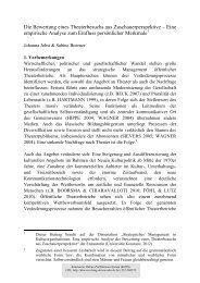
Figure 1: The 26S proteasome. The proteasome is a large, multicatalytic protease that<br />
degrades polyubiquitylated prote<strong>in</strong>s to produce small peptides. It is composed <strong>of</strong><br />
two subcomplexes – a 20S core particle (CP) that carries <strong>the</strong> catalytic activity, <strong>and</strong> a<br />
19S regulatory particle (RP). The 20S CP is a barrel-shaped structure that is composed<br />
<strong>of</strong> four stacked r<strong>in</strong>gs, two identical outer α-r<strong>in</strong>gs <strong>and</strong> two identical <strong>in</strong>ner β-r<strong>in</strong>gs. The<br />
eukaryotic α- <strong>and</strong> β-r<strong>in</strong>gs are each composed <strong>of</strong> seven dist<strong>in</strong>ct subunits, which gives<br />
<strong>the</strong> 20S complex <strong>the</strong> general structure <strong>of</strong> α1–7β1–7β1–7α1–7. The catalytic sites are localized<br />
to some <strong>of</strong> <strong>the</strong> β-subunits. One or both ends <strong>of</strong> <strong>the</strong> 20S barrel can be capped<br />
by a 19S RP. Follow<strong>in</strong>g substrate <strong>degradation</strong>, short peptides that have been derived<br />
from <strong>the</strong> substrate are released, as is reusable <strong>ubiquit<strong>in</strong></strong>. Part (A) <strong>of</strong> <strong>the</strong> figure shows<br />
an electron-microscopy image <strong>of</strong> a 26S proteasome from Saccharomyces cerevisiae,<br />
<strong>and</strong> part (B) shows a schematic representation <strong>of</strong> <strong>the</strong> structure <strong>and</strong> function <strong>of</strong> <strong>the</strong><br />
26S proteasome. Ub, <strong>ubiquit<strong>in</strong></strong>. (Modified from Ciechanover, 2005).<br />
regulatory complexes (Schmidt et al., 2005). These alternative regulators, which<br />
<strong>in</strong>clude PA28αβ (also known as <strong>the</strong> 11S regulator), PA28γ as well as PA200, do<br />
not conta<strong>in</strong> ATPases <strong>and</strong> stimulate proteasome activity solely by remov<strong>in</strong>g <strong>the</strong><br />
occlusions at <strong>the</strong> outer pores <strong>of</strong> <strong>the</strong> 20S core particle. As a consequence, <strong>the</strong>y are<br />
unable to unfold complex substrates <strong>and</strong> can only enhance <strong>the</strong> hydrolysis <strong>of</strong> short<br />
peptides. In addition, <strong>the</strong>y are devoid <strong>of</strong> any <strong>ubiquit<strong>in</strong></strong>-b<strong>in</strong>d<strong>in</strong>g activity (Rech-<br />
ste<strong>in</strong>er <strong>and</strong> Hill, 2005). Interest<strong>in</strong>gly, <strong>the</strong> PA28α <strong>and</strong> PA28β genes are encoded <strong>in</strong><br />
<strong>the</strong> major histocompatibility class I locus adjacent to <strong>the</strong> <strong>in</strong>ducible 20S subunits<br />
LMP2 <strong>and</strong> LMP7, <strong>and</strong> expression <strong>of</strong> PA28αβ is also upregulated <strong>in</strong> response to<br />
cytok<strong>in</strong>es such as IFN-γ. Fur<strong>the</strong>rmore, <strong>the</strong> 20S core particle can associate with<br />
two different regulators at ei<strong>the</strong>r end, creat<strong>in</strong>g a hybrid-proteasome with catalytic<br />
properties which differ from those displayed by proteasomes conta<strong>in</strong><strong>in</strong>g only one<br />
type <strong>of</strong> regulator. Indeed, proteasomes capped with both <strong>the</strong> 19S <strong>and</strong> 11S reg-<br />
ulator are able to generate a unique pool <strong>of</strong> peptides with an <strong>in</strong>creased aff<strong>in</strong>ity<br />
for MHC class I molecules (Rechste<strong>in</strong>er et al., 2000). Alternatively, proteasomal<br />
activity can not only be stimulated, but also attenuated by a different class <strong>of</strong><br />
<strong>in</strong>hibitory regulators such as PI31, which competes with PA28αβ for proteasome<br />
b<strong>in</strong>d<strong>in</strong>g (Rechste<strong>in</strong>er <strong>and</strong> Hill, 2005).<br />
12
Introduction<br />
The 26S proteasome plays a pivotal role <strong>in</strong> many cellular processes <strong>and</strong> its sub-<br />
strates come from a variety <strong>of</strong> sources <strong>and</strong> are targeted for a number <strong>of</strong> different<br />
reasons. These reasons can be grouped <strong>in</strong>to two categories: recycl<strong>in</strong>g <strong>and</strong> regula-<br />
tion. The former is perhaps <strong>the</strong> more obvious: prote<strong>in</strong>s which are defective or no<br />
longer needed are disassembled <strong>in</strong>to s<strong>in</strong>gle am<strong>in</strong>o acids which can <strong>the</strong>n be used for<br />
neosyn<strong>the</strong>sis. The latter utilizes <strong>degradation</strong> as a fast, efficient <strong>and</strong> irreversible<br />
means to <strong>the</strong> <strong>in</strong>activation <strong>of</strong> regulatory prote<strong>in</strong>s such as cell-cycle regulators or<br />
components <strong>of</strong> signal<strong>in</strong>g cascades. Of course, <strong>the</strong>se two functions are not entirely<br />
unrelated, as even those prote<strong>in</strong>s which are primarily targeted for <strong>degradation</strong><br />
to ensure <strong>the</strong>ir <strong>in</strong>activation are ultimately reduced to <strong>the</strong>ir build<strong>in</strong>g blocks <strong>and</strong><br />
subsequently recycled. Conversely, even peptides derived from defective prote<strong>in</strong>s<br />
which were targeted <strong>in</strong> <strong>the</strong> course <strong>of</strong> waste disposal might end up be<strong>in</strong>g presented<br />
on MHC class I molecules (Yewdell et al., 1996; Schubert et al., 2000).<br />
One prom<strong>in</strong>ent example for <strong>the</strong> regulatory function <strong>of</strong> <strong>the</strong> proteasome is <strong>the</strong> tar-<br />
geted distruction <strong>of</strong> cycl<strong>in</strong>s <strong>and</strong> Cdk <strong>in</strong>hibitors – which, through <strong>the</strong>ir periodic<br />
<strong>degradation</strong> <strong>and</strong> neosyn<strong>the</strong>sis, ultimately drive <strong>the</strong> cell-cycle (Nigg, 1995; Obaya<br />
<strong>and</strong> Sedivy, 2002). Ano<strong>the</strong>r is that <strong>of</strong> <strong>the</strong> tumor suppressor p53, which, when<br />
stabilized by stress stimuli such as DNA-damage, is responsible for <strong>the</strong> transcrip-<br />
tional activation <strong>of</strong> a broad array <strong>of</strong> prote<strong>in</strong>s <strong>in</strong>volved <strong>in</strong> cell-cycle control, apop-<br />
tosis <strong>and</strong> senescence (Lav<strong>in</strong> <strong>and</strong> Gueven, 2006). A third example is that <strong>of</strong> <strong>the</strong> en-<br />
zyme ornith<strong>in</strong>e-decarboxylase (ODC), which catalyzes <strong>the</strong> first <strong>and</strong> rate-limit<strong>in</strong>g<br />
step <strong>in</strong> <strong>the</strong> syn<strong>the</strong>sis <strong>of</strong> cellular polyam<strong>in</strong>es. Interest<strong>in</strong>gly, ODC also doubles as<br />
<strong>the</strong> most prom<strong>in</strong>ent example <strong>of</strong> <strong>ubiquit<strong>in</strong></strong>-<strong>in</strong>dependent proteasomal <strong>degradation</strong><br />
(Kahana et al., 2005).<br />
The non-functional prote<strong>in</strong>s which are targeted for proteasomal <strong>degradation</strong> as a<br />
means <strong>of</strong> waste-disposal orig<strong>in</strong>ate from several sources: Ag<strong>in</strong>g, previously fully<br />
functional prote<strong>in</strong>s can become become damaged, primarily through oxidation,<br />
over <strong>the</strong> course <strong>of</strong> <strong>the</strong>ir lifetime (Grune et al., 1997). Alternatively, prote<strong>in</strong>s can<br />
be targeted to <strong>the</strong> proteasome right after syn<strong>the</strong>sis due to failure to pass <strong>the</strong><br />
str<strong>in</strong>gent cellular quality control mechanisms. As many as 30% <strong>of</strong> all newly<br />
syn<strong>the</strong>sized prote<strong>in</strong>s are ubiquitylated <strong>and</strong> degraded by <strong>the</strong> proteasome. Most<br />
<strong>of</strong> <strong>the</strong>se prote<strong>in</strong>s, which are called defective ribosomal products (DRiPs), result<br />
from errors <strong>in</strong> <strong>the</strong> process <strong>of</strong> prote<strong>in</strong> syn<strong>the</strong>sis, such as mis<strong>in</strong>corporation <strong>of</strong> am<strong>in</strong>o<br />
acids, premature term<strong>in</strong>ation or deletion <strong>of</strong> residues. Post-translational mistakes<br />
which can occur dur<strong>in</strong>g fold<strong>in</strong>g, oligomer assembly or <strong>in</strong>tracellular sort<strong>in</strong>g can<br />
also lead to <strong>the</strong> generation <strong>of</strong> DRiPs. Importantly, even though <strong>the</strong>y are tar-<br />
geted for <strong>degradation</strong> through <strong>the</strong> action <strong>of</strong> quality control mechanisms, DRiPs<br />
13
Introduction<br />
are <strong>the</strong> major source <strong>of</strong> antigenic peptides for MHC class I restricted presentation<br />
(Yewdell et al., 1996, 2001).<br />
Ubiquit<strong>in</strong> <strong>and</strong> Ubiquit<strong>in</strong>-Like Prote<strong>in</strong>s<br />
Ubiquit<strong>in</strong><br />
Ubiquit<strong>in</strong> is a small, highly conserved prote<strong>in</strong> <strong>of</strong> 76 am<strong>in</strong>o acids <strong>and</strong> <strong>of</strong> compact,<br />
globular structure. It exerts its function ma<strong>in</strong>ly through covalent attachment to<br />
o<strong>the</strong>r prote<strong>in</strong>s <strong>and</strong> is coded for by a number <strong>of</strong> different genes, which are scattered<br />
throughout <strong>the</strong> genome. In all cases, <strong>ubiquit<strong>in</strong></strong> is expressed as a proprote<strong>in</strong> with<br />
a C-term<strong>in</strong>al extension <strong>and</strong> needs to be proteolytically processed before yield<strong>in</strong>g<br />
functional, monomeric <strong>ubiquit<strong>in</strong></strong> (Jentsch et al., 1991). The conjugation <strong>of</strong> ubiqui-<br />
t<strong>in</strong> to its target prote<strong>in</strong>s, which is termed <strong>ubiquit<strong>in</strong></strong>ation, <strong>ubiquit<strong>in</strong></strong>ylation or ubiq-<br />
uitylation, is dependent on <strong>the</strong> presence <strong>of</strong> two consecutive glyc<strong>in</strong>e residues at its<br />
very C-term<strong>in</strong>us <strong>and</strong> requires <strong>the</strong> sequential action <strong>of</strong> three enzymes. In a first<br />
step, <strong>the</strong> C-term<strong>in</strong>al glyc<strong>in</strong>e residue <strong>of</strong> <strong>ubiquit<strong>in</strong></strong> is activated <strong>in</strong> an ATP-dependent<br />
step by a specific <strong>ubiquit<strong>in</strong></strong> activat<strong>in</strong>g enzyme, or E1. This step consists <strong>of</strong> an <strong>in</strong>-<br />
termediate formation <strong>of</strong> <strong>ubiquit<strong>in</strong></strong> adenylate, with <strong>the</strong> release <strong>of</strong> pyrophosphate<br />
(PPi), followed by <strong>the</strong> b<strong>in</strong>d<strong>in</strong>g <strong>of</strong> <strong>ubiquit<strong>in</strong></strong> to a cyste<strong>in</strong>e residue <strong>of</strong> <strong>the</strong> E1 enzyme<br />
<strong>in</strong> a thiolester l<strong>in</strong>kage, with <strong>the</strong> release <strong>of</strong> AMP. In a second step, <strong>ubiquit<strong>in</strong></strong> is<br />
transferred to <strong>the</strong> active site cyste<strong>in</strong>e residue <strong>of</strong> a <strong>ubiquit<strong>in</strong></strong>-conjugation enzyme,<br />
or E2. The third enzyme <strong>in</strong> this cascade, <strong>the</strong> <strong>ubiquit<strong>in</strong></strong>-prote<strong>in</strong> ligase, or E3, cat-<br />
alyzes <strong>the</strong> isopeptide l<strong>in</strong>kage <strong>of</strong> <strong>the</strong> <strong>ubiquit<strong>in</strong></strong> C-term<strong>in</strong>us to an ε-am<strong>in</strong>o group <strong>in</strong><br />
<strong>of</strong> one <strong>of</strong> <strong>the</strong> substrate prote<strong>in</strong>’s lys<strong>in</strong>e residues (Hershko <strong>and</strong> Ciechanover, 1998<br />
<strong>and</strong> Fig. 2).<br />
In most cases, modification <strong>of</strong> <strong>the</strong> substrate prote<strong>in</strong> is not limited to <strong>the</strong> attach-<br />
ment <strong>of</strong> a s<strong>in</strong>gle <strong>ubiquit<strong>in</strong></strong> moiety. Instead, <strong>the</strong> newly attached <strong>ubiquit<strong>in</strong></strong> now<br />
serves as <strong>the</strong> acceptor for a new round <strong>of</strong> ubiquitylation, which eventually results<br />
<strong>in</strong> <strong>the</strong> formation <strong>of</strong> <strong>ubiquit<strong>in</strong></strong> cha<strong>in</strong>s. In some cases, <strong>the</strong> action <strong>of</strong> an additional<br />
enzyme – a multi<strong>ubiquit<strong>in</strong></strong>-cha<strong>in</strong> assembly factor, termed E4 – is required for<br />
extension <strong>of</strong> <strong>the</strong> poly<strong>ubiquit<strong>in</strong></strong> cha<strong>in</strong> (Hoppe, 2005). Ubiquit<strong>in</strong> possesses seven ly-<br />
s<strong>in</strong>e residues, all <strong>of</strong> which are potentially <strong>in</strong>volved <strong>in</strong> cha<strong>in</strong> formation. However,<br />
only l<strong>in</strong>kages through K48, K63 <strong>and</strong> sometimes also K29 are readily observed <strong>in</strong><br />
nature (Haglund <strong>and</strong> Dikic, 2005). Ubiquit<strong>in</strong> was first identified as <strong>the</strong> tag which<br />
targets prote<strong>in</strong>s for ATP-dependent <strong>degradation</strong> (Ciechanover et al., 1978), which<br />
14
Introduction<br />
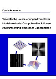
Figure 2: The ubiquitylation pathway. Free <strong>ubiquit<strong>in</strong></strong> (Ub) is activated <strong>in</strong> an ATPdependent<br />
manner with <strong>the</strong> formation <strong>of</strong> a thiol-ester-l<strong>in</strong>kage between E1 <strong>and</strong> <strong>the</strong><br />
carboxyl term<strong>in</strong>us <strong>of</strong> <strong>ubiquit<strong>in</strong></strong>. Ubiquit<strong>in</strong> is <strong>the</strong>n transferred to one <strong>of</strong> a number <strong>of</strong> different<br />
E2s. E2s associate with E3s, which may have substrate already bound. For HECT<br />
doma<strong>in</strong> E3s, <strong>ubiquit<strong>in</strong></strong> is next transferred to <strong>the</strong> active-site cyste<strong>in</strong>e <strong>of</strong> <strong>the</strong> HECT doma<strong>in</strong><br />
followed by transfer to substrate(s) (as shown) or to a substrate-bound poly<strong>ubiquit<strong>in</strong></strong>cha<strong>in</strong>.<br />
For RING E3s, current evidence <strong>in</strong>dicates that <strong>ubiquit<strong>in</strong></strong> is transferred directly<br />
from <strong>the</strong> E2 to <strong>the</strong> substrate. (Modified from Fang <strong>and</strong> Weissman, 2004).<br />
turned out to <strong>in</strong>deed be <strong>the</strong> most prevalent function <strong>of</strong> <strong>ubiquit<strong>in</strong></strong> conjugation. Pro-<br />
teasomal target<strong>in</strong>g <strong>of</strong> prote<strong>in</strong>s is mediated by <strong>the</strong> attachment <strong>of</strong> a cha<strong>in</strong> <strong>of</strong> four or<br />
more K48-l<strong>in</strong>ked <strong>ubiquit<strong>in</strong></strong> moieties (Thrower et al., 2000). In a limited number <strong>of</strong><br />
cases, <strong>the</strong> attachment <strong>of</strong> a K29-l<strong>in</strong>ked cha<strong>in</strong> also leads to proteasomal <strong>degradation</strong><br />
(Johnson et al., 1995), however, most o<strong>the</strong>r variations <strong>of</strong> <strong>ubiquit<strong>in</strong></strong> attachment are<br />
means to a different end. K63-l<strong>in</strong>ked <strong>ubiquit<strong>in</strong></strong> displays a dist<strong>in</strong>ct cha<strong>in</strong> topology<br />
<strong>and</strong> is <strong>in</strong>volved <strong>in</strong> signal transduction (Krappmann <strong>and</strong> Scheidereit, 2005) <strong>and</strong><br />
DNA repair (Friedberg et al., 2005). In some <strong>in</strong>stances, modification <strong>of</strong> <strong>the</strong> target<br />
prote<strong>in</strong> with a s<strong>in</strong>gle <strong>ubiquit<strong>in</strong></strong> moiety (monoubiquitylation), or with several sep-<br />
arate <strong>ubiquit<strong>in</strong></strong>s at different lys<strong>in</strong>es (multiubiquitylation) is sufficient to modify<br />
a prote<strong>in</strong>’s activity or to create a new b<strong>in</strong>d<strong>in</strong>g site. As such, monoubiquitylation<br />
has been implicated <strong>in</strong> endocytosis, vesicular sort<strong>in</strong>g, histone regulation <strong>and</strong> <strong>the</strong><br />
budd<strong>in</strong>g <strong>of</strong> retroviruses (Hicke, 2001; Di Fiore et al., 2003).<br />
Substrate specificity <strong>of</strong> <strong>the</strong> <strong>ubiquit<strong>in</strong></strong> system is conferred by a multitude <strong>of</strong> E3<br />
enzymes, which are responsible for recogniz<strong>in</strong>g <strong>the</strong>ir specific target prote<strong>in</strong>. In<br />
genomics studies, several hundered putative E3 c<strong>and</strong>idate genes have been iden-<br />
tified. E3 <strong>ubiquit<strong>in</strong></strong>-prote<strong>in</strong> ligases are def<strong>in</strong>ed as enzymes which b<strong>in</strong>d, through<br />
direct <strong>in</strong>teraction or with <strong>the</strong> help <strong>of</strong> additional adaptor prote<strong>in</strong>s, specific prote<strong>in</strong><br />
substrates <strong>and</strong> facilitate <strong>the</strong> transfer <strong>of</strong> <strong>ubiquit<strong>in</strong></strong> from a thiolester <strong>in</strong>termediate<br />
<strong>of</strong> <strong>the</strong>ir cognate E2 enzyme to an isopeptide l<strong>in</strong>kage with <strong>the</strong> target prote<strong>in</strong>. The<br />
15
Introduction<br />
majority <strong>of</strong> <strong>ubiquit<strong>in</strong></strong>-prote<strong>in</strong> ligases fall <strong>in</strong>to one <strong>of</strong> two subcategories, <strong>the</strong> HECT-<br />
doma<strong>in</strong> E3s or <strong>the</strong> RING-f<strong>in</strong>ger E3s (Fang <strong>and</strong> Weissman, 2004).<br />
The HECT (Homologous to E6-AP C-term<strong>in</strong>us)-doma<strong>in</strong> family was <strong>the</strong> first family<br />
<strong>of</strong> <strong>ubiquit<strong>in</strong></strong>-ligases to be identified. The first member <strong>of</strong> this family, E6-AP, was<br />
discovered as a cellular prote<strong>in</strong> which was required for <strong>the</strong> ubiquitylation <strong>and</strong> sub-<br />
sequent <strong>degradation</strong> <strong>of</strong> <strong>the</strong> p53 tumor suppressor by <strong>the</strong> human pappilomavirus<br />
E6 oncoprote<strong>in</strong> (Scheffner et al., 1994). HECT-doma<strong>in</strong> E3s first transfer <strong>ubiquit<strong>in</strong></strong><br />
from <strong>the</strong> bound E2 onto <strong>the</strong>ir active site cyste<strong>in</strong>e <strong>in</strong> <strong>the</strong> C-term<strong>in</strong>al HECT-doma<strong>in</strong><br />
before <strong>the</strong>y pass it on to <strong>the</strong>ir substrate prote<strong>in</strong>.<br />
The RING (Really Interest<strong>in</strong>g New Gene)-doma<strong>in</strong> family is <strong>the</strong> largest family <strong>of</strong><br />
<strong>ubiquit<strong>in</strong></strong>-ligases. The RING-f<strong>in</strong>ger is def<strong>in</strong>ed by eight conserved cyste<strong>in</strong>es <strong>and</strong><br />
histid<strong>in</strong>es which toge<strong>the</strong>r coord<strong>in</strong>ate two z<strong>in</strong>c ions (Borden <strong>and</strong> Freemont, 1996).<br />
The first member <strong>of</strong> this family which was identified is <strong>the</strong> <strong>ubiquit<strong>in</strong></strong>-ligase E3α,<br />
which is responsible for ubiquitylat<strong>in</strong>g substrates <strong>of</strong> <strong>the</strong> N-end rule pathway.<br />
RING-doma<strong>in</strong> E3s do not transfer <strong>the</strong> activated <strong>ubiquit<strong>in</strong></strong> onto <strong>the</strong>mselves, but<br />
ra<strong>the</strong>r facilitate its transfer from <strong>the</strong> tightly bound E2 directly onto <strong>the</strong> substrate<br />
prote<strong>in</strong>. This family <strong>of</strong> E3s consists <strong>of</strong> both s<strong>in</strong>gle subunit enzymes, which conta<strong>in</strong><br />
all <strong>the</strong> necessary doma<strong>in</strong>s <strong>in</strong> one prote<strong>in</strong>, for example Cbl, as well as large multi-<br />
meric complexes such as <strong>the</strong> SCF-complex, or <strong>the</strong> anaphase-promot<strong>in</strong>g-complex,<br />
<strong>in</strong> which <strong>the</strong> RING doma<strong>in</strong> exists as a separate subunit (Joazeiro <strong>and</strong> Weissman,<br />
2000).<br />
The o<strong>the</strong>r two subfamilies are relatively small <strong>and</strong> both conta<strong>in</strong> variants <strong>of</strong><br />
<strong>the</strong> RING-doma<strong>in</strong>. The PHD-f<strong>in</strong>ger was first identified as a component <strong>of</strong> viral<br />
<strong>ubiquit<strong>in</strong></strong>-ligases, but has subsequently also been found <strong>in</strong> mammalian prote<strong>in</strong>s<br />
(Coscoy <strong>and</strong> Ganem, 2003). The U-box, <strong>in</strong> turn, is a modified RING-doma<strong>in</strong> with<br />
no coord<strong>in</strong>ated z<strong>in</strong>c ions. The <strong>ubiquit<strong>in</strong></strong>-ligase CHIP for example, which functions<br />
as an E3 for HSP90 <strong>in</strong>teract<strong>in</strong>g prote<strong>in</strong>s, is a member <strong>of</strong> this last family (Jiang<br />
et al., 2001).<br />
In contrast to <strong>the</strong> large number or E3 enzymes, <strong>the</strong> mammalian genome encodes<br />
for only about 30 different E2 <strong>ubiquit<strong>in</strong></strong>-conjugat<strong>in</strong>g enzymes (von Arnim, 2001).<br />
As a consequence, each E2 enzyme is responsible for serv<strong>in</strong>g a number <strong>of</strong> differ-<br />
ent E3s. All known E2 enzymes belong to <strong>the</strong> same family, which is characterized<br />
by <strong>the</strong> presence <strong>of</strong> a catalytic doma<strong>in</strong> called <strong>the</strong> UBC-doma<strong>in</strong>. Some E2s possess<br />
additional C-term<strong>in</strong>al or N-term<strong>in</strong>al extensions, which are responsible for medi-<br />
at<strong>in</strong>g subcellular localization or recognition <strong>of</strong> E3 enzymes. With a few exceptions<br />
16
Introduction<br />
– among <strong>the</strong>m BRUCE, a 528 kDa <strong>in</strong>hibitor <strong>of</strong> apoptosis (Hauser et al., 1998) –<br />
most E2s are <strong>of</strong> ra<strong>the</strong>r small size (Jentsch et al., 1990).<br />
Two different E1 enzymes are able to activate <strong>ubiquit<strong>in</strong></strong> <strong>in</strong> <strong>the</strong> first step <strong>of</strong> <strong>the</strong><br />
<strong>ubiquit<strong>in</strong></strong> conjugation process. These <strong>ubiquit<strong>in</strong></strong>-activat<strong>in</strong>g enzymes, which are<br />
called UBE1 <strong>and</strong> UBA6, serve dist<strong>in</strong>ct, but overlapp<strong>in</strong>g pools <strong>of</strong> E2s. Although<br />
both E1s are broadly expressed <strong>in</strong> all tissue types, deletion <strong>of</strong> ei<strong>the</strong>r one <strong>of</strong> <strong>the</strong>m<br />
is lethal, <strong>in</strong>dicat<strong>in</strong>g that <strong>the</strong>re is little redundancy <strong>in</strong> <strong>the</strong> system. Interest<strong>in</strong>gly,<br />
UBE1 – which was long thought to be <strong>the</strong> only <strong>ubiquit<strong>in</strong></strong>-conjugat<strong>in</strong>g enzyme <strong>in</strong><br />
existence – is expressed at a much higher level <strong>in</strong> most tissues <strong>and</strong> accounts for<br />
up to 85% <strong>of</strong> all <strong>ubiquit<strong>in</strong></strong> conjugates observed (Groettrup et al., 2008).<br />
A fur<strong>the</strong>r layer <strong>of</strong> complexity is added to <strong>the</strong> <strong>ubiquit<strong>in</strong></strong>-system through <strong>the</strong> actions<br />
<strong>of</strong> a group <strong>of</strong> deubiquitylat<strong>in</strong>g enzymes, or DUBs, <strong>the</strong> majority <strong>of</strong> which fall <strong>in</strong>to<br />
one <strong>of</strong> two subgroups: The family <strong>of</strong> <strong>ubiquit<strong>in</strong></strong> C-term<strong>in</strong>al hydrolases (UCHs) or<br />
<strong>the</strong> family <strong>of</strong> <strong>ubiquit<strong>in</strong></strong>-specific process<strong>in</strong>g proteases (UBPs). DUBs carry out a<br />
variety <strong>of</strong> process<strong>in</strong>g or edit<strong>in</strong>g functions. Some <strong>of</strong> <strong>the</strong>m are responsible for <strong>the</strong><br />
process<strong>in</strong>g <strong>of</strong> l<strong>in</strong>ear <strong>ubiquit<strong>in</strong></strong>-fusions <strong>and</strong> are required for <strong>the</strong> generation <strong>of</strong> ac-<br />
tive <strong>ubiquit<strong>in</strong></strong> from newly syn<strong>the</strong>sized precursor prote<strong>in</strong>s. O<strong>the</strong>rs are responsible<br />
for <strong>the</strong> removal <strong>of</strong> poly<strong>ubiquit<strong>in</strong></strong>-cha<strong>in</strong>s from prote<strong>in</strong>s targeted for proteasomal<br />
<strong>degradation</strong>, ensur<strong>in</strong>g that <strong>the</strong>se are not degraded along with <strong>the</strong>ir substrates<br />
but <strong>in</strong>stead recycled. Once <strong>the</strong> <strong>ubiquit<strong>in</strong></strong> cha<strong>in</strong>s are liberated, a second group <strong>of</strong><br />
DUBs <strong>the</strong>n disassembles <strong>the</strong>m <strong>in</strong>to <strong>ubiquit<strong>in</strong></strong> monomers. Yet ano<strong>the</strong>r group is <strong>in</strong>-<br />
volved <strong>in</strong> <strong>the</strong> modulation <strong>of</strong> various <strong>ubiquit<strong>in</strong></strong> signals. They can, for example, res-<br />
cue prote<strong>in</strong>s from <strong>degradation</strong> by remov<strong>in</strong>g <strong>the</strong>ir <strong>ubiquit<strong>in</strong></strong>-tags before <strong>the</strong>y come<br />
<strong>in</strong>to contact with <strong>the</strong> proteasome, or attenuate signal transduction cascades by<br />
remov<strong>in</strong>g <strong>the</strong> activat<strong>in</strong>g K63-l<strong>in</strong>ked cha<strong>in</strong>s (Amerik <strong>and</strong> Hochstrasser, 2004).<br />
Both <strong>the</strong> enzymes <strong>in</strong>volved <strong>in</strong> <strong>ubiquit<strong>in</strong></strong>-process<strong>in</strong>g as well as <strong>the</strong> effector pro-<br />
te<strong>in</strong>s which act downstream <strong>of</strong> <strong>the</strong> <strong>ubiquit<strong>in</strong></strong> signal need to <strong>in</strong>teract with ubiqui-<br />
t<strong>in</strong>, <strong>and</strong> this recognition is mediated by a plethora <strong>of</strong> different <strong>ubiquit<strong>in</strong></strong>-b<strong>in</strong>d<strong>in</strong>g<br />
doma<strong>in</strong>s. These <strong>in</strong>clude – but may not be limited to – sixteen different doma<strong>in</strong>s:<br />
UBA, UIM, MIU, DUIM, CUE, GAT, NZF, A20 ZnF, BUZ, UBZ, Ubc, UEV, UBM,<br />
GLUE, Jab1/MPN <strong>and</strong> PFU. Interest<strong>in</strong>gly, although several <strong>of</strong> <strong>the</strong>se doma<strong>in</strong>s dis-<br />
play absolutely no sequence similarity <strong>and</strong> seem to have evolved <strong>in</strong>dependently<br />
<strong>of</strong> each o<strong>the</strong>r, most <strong>of</strong> <strong>the</strong>m b<strong>in</strong>d to <strong>ubiquit<strong>in</strong></strong> through a conserved surface encom-<br />
pass<strong>in</strong>g residues Leu8, Ile44 <strong>and</strong> Val70, which is called <strong>the</strong> “hydrophobic patch”.<br />
A notable exception to this rule is <strong>the</strong> BUZ doma<strong>in</strong>, a z<strong>in</strong>c-f<strong>in</strong>ger which <strong>in</strong>stead<br />
<strong>in</strong>teracts with <strong>ubiquit<strong>in</strong></strong> through its free C-term<strong>in</strong>al diglyc<strong>in</strong>e-motif, <strong>and</strong> is for<br />
17
Introduction<br />
example conta<strong>in</strong>ed <strong>in</strong> <strong>the</strong> deubiquitylat<strong>in</strong>g enzyme isopeptidase T or <strong>the</strong> tubul<strong>in</strong><br />
deacetylase HDAC6. Most <strong>ubiquit<strong>in</strong></strong>-b<strong>in</strong>d<strong>in</strong>g doma<strong>in</strong>s display a preference for ei-<br />
<strong>the</strong>r mono<strong>ubiquit<strong>in</strong></strong>, different poly<strong>ubiquit<strong>in</strong></strong> cha<strong>in</strong> topologies or <strong>ubiquit<strong>in</strong></strong>-<strong>like</strong> do-<br />
ma<strong>in</strong>s, although <strong>the</strong> exact molecular determ<strong>in</strong>ants <strong>of</strong> this ability to discrim<strong>in</strong>ate<br />
rema<strong>in</strong> unknown (Hurley et al., 2006).<br />
Ubiquit<strong>in</strong>-Like Prote<strong>in</strong>s<br />
All eukaryotic cells conta<strong>in</strong> several additional prote<strong>in</strong>s which are related to ubiq-<br />
uit<strong>in</strong> <strong>in</strong> ei<strong>the</strong>r sequence or structure. Like <strong>ubiquit<strong>in</strong></strong>, <strong>the</strong>se prote<strong>in</strong>s are <strong>in</strong>-<br />
volved <strong>in</strong> a vast number <strong>of</strong> fundamental cellular processes. Collectively known<br />
as <strong>ubiquit<strong>in</strong></strong>-<strong>like</strong> prote<strong>in</strong>s, <strong>the</strong>y can be subdivided <strong>in</strong>to <strong>the</strong> families <strong>of</strong> <strong>ubiquit<strong>in</strong></strong>-<br />
<strong>like</strong> <strong>modifier</strong>s <strong>and</strong> <strong>ubiquit<strong>in</strong></strong>-doma<strong>in</strong> prote<strong>in</strong>s (Fig. 3). Members <strong>of</strong> <strong>the</strong> first group<br />
function – as <strong>the</strong>ir name suggests – as <strong>modifier</strong>s, <strong>and</strong> are covalently attached to<br />
o<strong>the</strong>r prote<strong>in</strong>s <strong>in</strong> a manner analogous to <strong>ubiquit<strong>in</strong></strong> conjugation. Members <strong>of</strong> <strong>the</strong><br />
second group are larger prote<strong>in</strong>s <strong>and</strong> conta<strong>in</strong> a <strong>ubiquit<strong>in</strong></strong>-<strong>like</strong> doma<strong>in</strong> as an <strong>in</strong>-<br />
tegral part <strong>of</strong> <strong>the</strong>ir sequence, but are o<strong>the</strong>rwise funct<strong>in</strong>ally diverse (Jentsch <strong>and</strong><br />
Pyrowolakis, 2000).<br />
Ubiquit<strong>in</strong>-Like Modifiers<br />
With <strong>the</strong> exception <strong>of</strong> ATG8, ATG12 <strong>and</strong> URM1, all <strong>ubiquit<strong>in</strong></strong>-<strong>like</strong> <strong>modifier</strong>s<br />
(UbLs) display a high level <strong>of</strong> sequence homology to <strong>ubiquit<strong>in</strong></strong> <strong>and</strong>, regardless<br />
<strong>of</strong> <strong>the</strong>ir sequence, <strong>the</strong>y all share essentially <strong>the</strong> same three-dimensional struc-<br />
ture. In addition, those UbLs which are capable <strong>of</strong> covalent attachment to an-<br />
o<strong>the</strong>r prote<strong>in</strong> <strong>in</strong>variably share a conserved glyc<strong>in</strong>e residue at <strong>the</strong>ir C-term<strong>in</strong>us,<br />
<strong>and</strong> <strong>the</strong> carboxyl group <strong>of</strong> this glyc<strong>in</strong>e is <strong>the</strong> site <strong>of</strong> attachment to <strong>the</strong>ir substrates.<br />
Like <strong>ubiquit<strong>in</strong></strong>, most <strong>of</strong> <strong>the</strong>m are generated as precursors with C-term<strong>in</strong>al exten-<br />
sions <strong>of</strong> vari<strong>in</strong>g length <strong>and</strong> need to be activated through endoproteolytic process-<br />
<strong>in</strong>g. This step is mediated by UbL-specific proteases (ULPs), which function <strong>in</strong><br />
a manner analogous to DUBs <strong>and</strong> are also capable <strong>of</strong> hydroliz<strong>in</strong>g <strong>the</strong> isopeptide<br />
bond between <strong>the</strong>ir specific UbL <strong>and</strong> its target prote<strong>in</strong>s. Exceptions to this rule<br />
are ATG12, URM1 <strong>and</strong> <strong>FAT10</strong>, which already conta<strong>in</strong> a free glyc<strong>in</strong>e at <strong>the</strong>ir C-<br />
term<strong>in</strong>us <strong>and</strong> have no requirement for process<strong>in</strong>g.<br />
Ubiquit<strong>in</strong>-<strong>like</strong> <strong>modifier</strong>s are also conjugated to <strong>the</strong>ir target prote<strong>in</strong>s by way <strong>of</strong> a<br />
multi-enzyme cascade, <strong>in</strong> a manner analogous to that <strong>of</strong> <strong>ubiquit<strong>in</strong></strong> conjugation,<br />
18
Introduction<br />
Figure 3: Two families <strong>of</strong> <strong>ubiquit<strong>in</strong></strong>-<strong>like</strong> prote<strong>in</strong>s. Ubiquit<strong>in</strong>-<strong>like</strong> <strong>modifier</strong>s (UbLs, red)<br />
are conjugated to <strong>the</strong>ir target prote<strong>in</strong>s <strong>in</strong> a manner analogous to that <strong>of</strong> <strong>ubiquit<strong>in</strong></strong>.<br />
Ubiquit<strong>in</strong>-doma<strong>in</strong> prote<strong>in</strong>s (UBDs) conta<strong>in</strong> an <strong>in</strong>tegral <strong>ubiquit<strong>in</strong></strong>-<strong>like</strong> doma<strong>in</strong> (green),<br />
but do not form conjugates. (Modified from Jentsch <strong>and</strong> Pyrowolakis, 2000).<br />
which ultimately results <strong>in</strong> <strong>the</strong> formation <strong>of</strong> an isopeptide l<strong>in</strong>kage between <strong>the</strong><br />
C-term<strong>in</strong>al glyc<strong>in</strong>e <strong>of</strong> <strong>the</strong> UbL <strong>and</strong> a lys<strong>in</strong>e residue <strong>of</strong> <strong>the</strong> target prote<strong>in</strong>. Many <strong>of</strong><br />
<strong>the</strong> enzymes <strong>in</strong>volved <strong>in</strong> <strong>the</strong> conjugation <strong>of</strong> UbLs have recently been discovered,<br />
<strong>and</strong> although <strong>the</strong> pathways appear to be for <strong>the</strong> most part dist<strong>in</strong>ct, <strong>the</strong>re appears<br />
to be a certa<strong>in</strong> amount <strong>of</strong> enzyme shar<strong>in</strong>g between different <strong>modifier</strong>s. ATG8 <strong>and</strong><br />
ATG12, <strong>the</strong> two UbLs <strong>in</strong>volved <strong>in</strong> autophagosome formation, for example share a<br />
s<strong>in</strong>gle E1, ATG7, but each <strong>of</strong> <strong>the</strong>m has <strong>the</strong>ir own E2. Conversely, two different<br />
E1s specific for different UbLs can share <strong>the</strong> same E2; UBEL1, <strong>the</strong> E1 for ISG15,<br />
<strong>and</strong> UBE1, <strong>the</strong> E1 for <strong>ubiquit<strong>in</strong></strong>, both can both transfer <strong>the</strong>ir respective <strong>modifier</strong><br />
to <strong>the</strong> E2 UbcH8. Apart from <strong>FAT10</strong>, none <strong>of</strong> <strong>the</strong> o<strong>the</strong>r UbLs are directly <strong>in</strong>volved<br />
<strong>in</strong> <strong>the</strong> target<strong>in</strong>g <strong>of</strong> prote<strong>in</strong>s for proteasomal <strong>degradation</strong>. Instead, through <strong>the</strong>ir<br />
attachment, <strong>the</strong>y are for example able to alter <strong>the</strong> function or subcellular local-<br />
ization <strong>of</strong> <strong>the</strong>ir target prote<strong>in</strong> (Welchman et al., 2005; Kerscher et al., 2006).<br />
SUMO (Small <strong>ubiquit<strong>in</strong></strong>-<strong>like</strong> <strong>modifier</strong>), which is perhaps <strong>the</strong> most well-studied<br />
member <strong>of</strong> this group, exists <strong>in</strong> <strong>the</strong> form <strong>of</strong> three is<strong>of</strong>orms, SUMO-1, SUMO-<br />
2 <strong>and</strong> SUMO-3, <strong>of</strong> which <strong>the</strong> latter two are virtually identical <strong>in</strong> sequence <strong>and</strong><br />
are functionally dist<strong>in</strong>ct from SUMO-1. Interest<strong>in</strong>gly, SUMO-2/3 are <strong>the</strong> only<br />
19
Introduction<br />
two <strong>modifier</strong>s apart from <strong>ubiquit<strong>in</strong></strong> which could be shown to form polyUbL-cha<strong>in</strong>s<br />
(Tatham et al., 2001). In many cases, <strong>the</strong> attachment <strong>of</strong> SUMO leads to a change<br />
<strong>in</strong> <strong>the</strong> subcellular localization <strong>of</strong> its target prote<strong>in</strong>s, for example <strong>in</strong> <strong>the</strong> case <strong>of</strong><br />
RanGAP1, which is <strong>in</strong>volved <strong>in</strong> nuclear import/export <strong>and</strong> is targeted to <strong>the</strong> nu-<br />
clear pore complex by modification with SUMO-1 (Matunis et al., 1996). In <strong>the</strong><br />
case <strong>of</strong> <strong>the</strong> promyelotic leukemia (PML) prote<strong>in</strong>, SUMOylation is required for<br />
its function as a scaffold promot<strong>in</strong>g <strong>the</strong> formation <strong>of</strong> PML nuclear bodies, which<br />
are subnuclear structures implicated <strong>in</strong> transcriptional regulation <strong>and</strong> DNA re-<br />
pair (Zhong et al., 2000). Interest<strong>in</strong>gly, PML can also be polySUMOylated, <strong>and</strong><br />
this leads to <strong>the</strong> recruitment <strong>of</strong> <strong>the</strong> SUMO-<strong>in</strong>teraction (SIM) doma<strong>in</strong> conta<strong>in</strong><strong>in</strong>g<br />
<strong>ubiquit<strong>in</strong></strong>-ligase RNF4/SNURF. Recruitment <strong>of</strong> RNF4 <strong>the</strong>n facilitates <strong>the</strong> polyu-<br />
biquitylation <strong>of</strong> PML which ultimately results <strong>in</strong> <strong>the</strong> destruction <strong>of</strong> PML via pro-<br />
teasomal <strong>degradation</strong> (Tatham et al., 2008; Lallem<strong>and</strong>-Breitenbach et al., 2008).<br />
Fur<strong>the</strong>rmore, several prote<strong>in</strong>s can be modified by ei<strong>the</strong>r SUMO or <strong>ubiquit<strong>in</strong></strong> at<br />
<strong>the</strong> same lys<strong>in</strong>e residue. The attachment <strong>of</strong> ei<strong>the</strong>r SUMO or <strong>ubiquit<strong>in</strong></strong> is mutually<br />
exclusive, <strong>and</strong> for a number <strong>of</strong> prote<strong>in</strong>s – among <strong>the</strong>m <strong>the</strong> DNA-replication pro-<br />
cessivity factor PCNA (Hoege et al., 2002), or <strong>the</strong> tumor suppresor p53 (Rodriguez<br />
et al., 1999) – it could be shown that SUMOylation can prevent ubiquitylation at<br />
<strong>the</strong> same residue. Interest<strong>in</strong>gly, many <strong>of</strong> SUMO’s target prote<strong>in</strong>s conta<strong>in</strong> a con-<br />
sensus SUMOylation site, which can be directly recognized by <strong>the</strong> E2, <strong>the</strong>reby<br />
obliterat<strong>in</strong>g <strong>the</strong> need for an E3 (Johnson, 2004).<br />
NEDD8 (Neuronal precursor cell-expressed developmentally down-regulated 8)<br />
was orig<strong>in</strong>ally characterized as a prote<strong>in</strong> able to regulate <strong>the</strong> activity <strong>of</strong> <strong>ubiquit<strong>in</strong></strong>-<br />
ligases. NEDDylation <strong>of</strong> cull<strong>in</strong>s, which are scaffolid<strong>in</strong>g subunits <strong>of</strong> multi-<br />
component RING E3s, enables <strong>the</strong> <strong>in</strong>teraction <strong>of</strong> <strong>the</strong> E3 with its cognate E2 (Pan<br />
et al., 2004). Ano<strong>the</strong>r example <strong>of</strong> a target <strong>of</strong> NEDDylation is <strong>the</strong> tumor suppressor<br />
p53, which <strong>in</strong> addition to be<strong>in</strong>g subject to both ubiquitylation <strong>and</strong> SUMOylation,<br />
can also be modified by NEDD8. Like ubiquitylation, attachment <strong>of</strong> NEDD8 can<br />
<strong>in</strong>hibit p53 activity <strong>and</strong> is mediated by <strong>the</strong> same E3 ligase, Mdm2 (Xirodimas<br />
et al., 2004).<br />
ISG15 (Interferon-stimulated gene 15) consists <strong>of</strong> two UBL doma<strong>in</strong>s <strong>in</strong> a tan-<br />
dem array <strong>and</strong> is <strong>the</strong> only <strong>ubiquit<strong>in</strong></strong>-<strong>like</strong> <strong>modifier</strong> which can be secreted <strong>in</strong>to <strong>the</strong><br />
extracellular space (Knight <strong>and</strong> Cordova, 1991). Several components <strong>of</strong> <strong>the</strong> ISG-<br />
conjugation <strong>and</strong> deconjugation mach<strong>in</strong>ery, such as its E1 UBE1L (Yuan <strong>and</strong> Krug,<br />
2001), its E2 UbcH8 (Kim et al., 2004) <strong>and</strong> <strong>the</strong> deconjugat<strong>in</strong>g enzyme UBP43<br />
(Malakhov et al., 2002), as well as ISG15 itself are <strong>in</strong>ducible with IFN-α <strong>and</strong><br />
IFN-β. Free ISG15 displays immunemodulatory functions <strong>and</strong> has been shown<br />
20
Introduction<br />
to stimulate <strong>the</strong> IFN-γ production by CD3 + cells (Recht et al., 1991) <strong>and</strong> acti-<br />
vate <strong>the</strong> proliferation <strong>of</strong> CD56 + natural killer cells (D’Cunha et al., 1996). Fur-<br />
<strong>the</strong>rmore, <strong>in</strong> accordance with its function as a putative antiviral effector, several<br />
viruses actively block <strong>the</strong> conjugation <strong>of</strong> ISG15 to its target prote<strong>in</strong>s (Yuan <strong>and</strong><br />
Krug, 2001; Guerra et al., 2008). Several key factors <strong>in</strong>volved <strong>in</strong> signal trans-<br />
duction, among <strong>the</strong>m phospholipase Cγ1, <strong>the</strong> k<strong>in</strong>ases JAK1 <strong>and</strong> ERK1, as well<br />
as <strong>the</strong> transcription factor STAT1, have been reported to be substrates <strong>of</strong> ISG15<br />
(Malakhov et al., 2003), however, <strong>the</strong> biological significance <strong>of</strong> <strong>the</strong>ir modification<br />
rema<strong>in</strong>s unknown.<br />
<strong>FAT10</strong><br />
<strong>FAT10</strong> (HLA-F locus associated transcript 10) is a <strong>ubiquit<strong>in</strong></strong>-<strong>like</strong> <strong>modifier</strong> <strong>of</strong> 165<br />
am<strong>in</strong>o acids <strong>in</strong> length <strong>and</strong> encompasses two <strong>ubiquit<strong>in</strong></strong>-<strong>like</strong> doma<strong>in</strong>s <strong>in</strong> a head-<br />
to-tail formation, which are connected by a short l<strong>in</strong>ker. Ow<strong>in</strong>g to its resem-<br />
blance to a t<strong>and</strong>em fusion <strong>of</strong> two <strong>ubiquit<strong>in</strong></strong>s, it was orig<strong>in</strong>ally also called “diubiq-<br />
uit<strong>in</strong>” or “<strong>ubiquit<strong>in</strong></strong> D”. Its N-term<strong>in</strong>al <strong>and</strong> C-term<strong>in</strong>al <strong>ubiquit<strong>in</strong></strong>-<strong>like</strong> doma<strong>in</strong>s are<br />
more closely related to <strong>ubiquit<strong>in</strong></strong> than to each o<strong>the</strong>r <strong>and</strong> show 29% <strong>and</strong> 36% se-<br />
quence identity to <strong>ubiquit<strong>in</strong></strong>, respectively. Four <strong>of</strong> <strong>the</strong> lys<strong>in</strong>e residues <strong>in</strong>volved <strong>in</strong><br />
poly<strong>ubiquit<strong>in</strong></strong>-cha<strong>in</strong> formation – correspond<strong>in</strong>g to K27, K33 <strong>and</strong> most notably K48<br />
<strong>and</strong> K63 – are conserved <strong>in</strong> both <strong>ubiquit<strong>in</strong></strong>-<strong>like</strong> doma<strong>in</strong>s <strong>of</strong> <strong>FAT10</strong>. In addition, it<br />
does not carry a C-term<strong>in</strong>al extension, but ra<strong>the</strong>r ends with a free diglyc<strong>in</strong>e-motif<br />
(Fig. 4). The fat10 gene lies encoded <strong>in</strong> <strong>the</strong> MHC class I HLA-F locus on chro-<br />
mosome 6, adjacent to <strong>the</strong> genes for <strong>the</strong> <strong>in</strong>terferon-<strong>in</strong>ducible subunits LMP2 <strong>and</strong><br />
LMP7 <strong>of</strong> <strong>the</strong> proteasome <strong>and</strong> was thus orig<strong>in</strong>ally suspected to have a function <strong>in</strong><br />
antigen process<strong>in</strong>g <strong>and</strong> presentation (Fan et al., 1996; Bates et al., 1997).<br />
<strong>FAT10</strong> is constitutively expressed <strong>in</strong> dendritic cells <strong>and</strong> mature B cells (Bates<br />
et al., 1997), <strong>and</strong> <strong>FAT10</strong> mRNA was readily detectable <strong>in</strong> spleen <strong>and</strong> thymus<br />
both by Nor<strong>the</strong>rn blot, <strong>in</strong> situ hybridization (Liu et al., 1999) as well as qRT-PCR<br />
(Lukasiak et al., 2008). However, expression <strong>of</strong> <strong>FAT10</strong> is not restricted to cells <strong>of</strong><br />
<strong>the</strong> immune system, but can be synergistically <strong>in</strong>duced with <strong>the</strong> pro<strong>in</strong>flammatory<br />
cytok<strong>in</strong>es TNF-α <strong>and</strong> IFN-γ <strong>in</strong> cells <strong>of</strong> almost every tissue orig<strong>in</strong>. Induction <strong>of</strong> <strong>the</strong><br />
<strong>FAT10</strong> mRNA did not rely on de novo prote<strong>in</strong> syn<strong>the</strong>sis <strong>and</strong> was partially depen-<br />
dent on proteasomal activity, as treatment with proteasome <strong>in</strong>hibitors prevented<br />
<strong>in</strong>duction <strong>of</strong> <strong>FAT10</strong> with TNF-α, but not IFN-γ (Raasi et al., 1999). In addition,<br />
<strong>the</strong> fat10 promotor was shown to conta<strong>in</strong> a p53-b<strong>in</strong>d<strong>in</strong>g site <strong>and</strong> transcription <strong>of</strong><br />
<strong>the</strong> fat10 gene appeared to be negatively regulated by p53 (Zhang et al., 2006a).<br />
21
Introduction<br />
Figure 4: Comparison <strong>of</strong> sequence as well as predicted tertiary structure <strong>of</strong> <strong>FAT10</strong><br />
with <strong>ubiquit<strong>in</strong></strong>. (A) <strong>FAT10</strong> encompasses two <strong>ubiquit<strong>in</strong></strong>-<strong>like</strong> doma<strong>in</strong>s (UBLs) which are<br />
29% <strong>and</strong> 36% identical to <strong>ubiquit<strong>in</strong></strong>. The C-term<strong>in</strong>al diglyc<strong>in</strong>e-motif is conserved <strong>in</strong> <strong>the</strong><br />
second doma<strong>in</strong> <strong>of</strong> <strong>FAT10</strong>. In addition, four <strong>of</strong> <strong>the</strong> lys<strong>in</strong>es <strong>in</strong>volved <strong>in</strong> poly<strong>ubiquit<strong>in</strong></strong>-cha<strong>in</strong><br />
formation – correspond<strong>in</strong>g to K27, K33, K48 <strong>and</strong> K63 – are conserved. (B) Ribbon<br />
diagrams <strong>of</strong> <strong>the</strong> <strong>ubiquit<strong>in</strong></strong> structure <strong>and</strong> <strong>the</strong> predicted structure <strong>of</strong> <strong>FAT10</strong>. (Modified<br />
from Groettrup et al., 2008). (C) Sequence alignment <strong>of</strong> <strong>the</strong> N- <strong>and</strong> C-term<strong>in</strong>al parts<br />
<strong>of</strong> <strong>FAT10</strong> with <strong>ubiquit<strong>in</strong></strong>.<br />
Although <strong>the</strong> identity <strong>of</strong> its target prote<strong>in</strong>s rema<strong>in</strong>s elusive, <strong>FAT10</strong> could be shown<br />
to form covalent conjugates through its C-term<strong>in</strong>al glyc<strong>in</strong>e (Raasi et al., 2001),<br />
<strong>and</strong> <strong>the</strong> E1 enzyme responsible for its activation was recently discovered (Chiu<br />
et al., 2007). Interest<strong>in</strong>gly, <strong>FAT10</strong> shares its activat<strong>in</strong>g enzyme, UBA6, with ubiq-<br />
uit<strong>in</strong>, although both <strong>modifier</strong>s appear to be passed on to different E2 enzymes.<br />
<strong>FAT10</strong> shares one additional important property with <strong>ubiquit<strong>in</strong></strong>, as both <strong>modifier</strong>s<br />
can promote <strong>the</strong> proteasomal <strong>degradation</strong> <strong>of</strong> <strong>the</strong>ir substrates with equal potency,<br />
an ability <strong>in</strong>tr<strong>in</strong>sic to no o<strong>the</strong>r member <strong>of</strong> <strong>the</strong> <strong>ubiquit<strong>in</strong></strong> superfamily. Interest-<br />
<strong>in</strong>gly, although <strong>ubiquit<strong>in</strong></strong> is a remarkably stable prote<strong>in</strong>, both <strong>FAT10</strong> as well as<br />
its conjugates are rapidly degraded. This f<strong>in</strong>d<strong>in</strong>g, toge<strong>the</strong>r with <strong>the</strong> apparent lack<br />
<strong>of</strong> a <strong>FAT10</strong>-specific protease suggests that, ra<strong>the</strong>r than be<strong>in</strong>g recycled, <strong>FAT10</strong> is<br />
degraded along with its substrates (Hipp et al., 2005), <strong>and</strong> experiments with a<br />
22
Introduction<br />
diglyc<strong>in</strong>e-deficient mutant <strong>of</strong> <strong>FAT10</strong> revealed that conjugation to a substrate was<br />
not required for its <strong>degradation</strong> (Hipp, 2004). In addition, <strong>the</strong> <strong>degradation</strong> <strong>of</strong><br />
<strong>FAT10</strong> could be fur<strong>the</strong>r accelerated through coexpression <strong>of</strong> a non-covalent <strong>in</strong>ter-<br />
action partner, <strong>the</strong> UBL-UBA doma<strong>in</strong> prote<strong>in</strong> NUB1L (Hipp et al., 2004). Despite<br />
<strong>the</strong> strik<strong>in</strong>g similarities between <strong>ubiquit<strong>in</strong></strong>- <strong>and</strong> <strong>FAT10</strong>-mediated <strong>degradation</strong>,<br />
FATylation <strong>of</strong> a prote<strong>in</strong> appears to be sufficient to promote its <strong>degradation</strong>, as<br />
two experimental approaches demonstrated no fur<strong>the</strong>r requirement for ubiquity-<br />
lation. A mutant version <strong>of</strong> <strong>FAT10</strong>, which due to a lack <strong>of</strong> lys<strong>in</strong>es is unable to<br />
serve as a substrate <strong>of</strong> <strong>ubiquit<strong>in</strong></strong> conjugation, was still able to promote <strong>the</strong> degra-<br />
dation <strong>of</strong> a model substrate. Likewise, a cell l<strong>in</strong>e carry<strong>in</strong>g a <strong>the</strong>rmolabile mutant<br />
<strong>of</strong> <strong>the</strong> major <strong>ubiquit<strong>in</strong></strong>-activat<strong>in</strong>g enzyme showed no accumulation <strong>of</strong> an artificial<br />
<strong>FAT10</strong>-conjugate (Hipp et al., 2005).<br />
Evidence for a role <strong>of</strong> <strong>FAT10</strong> <strong>in</strong> immunity has rema<strong>in</strong>ed sparse. In fact, <strong>the</strong> only<br />
two connections to <strong>the</strong> immune response derive from <strong>the</strong> study <strong>of</strong> <strong>FAT10</strong> gene-<br />
targeted mice, which display a hypersensitivity to LPS as <strong>the</strong>ir major phenotype<br />
to date (Canaan et al., 2006), <strong>and</strong> <strong>the</strong> f<strong>in</strong>d<strong>in</strong>g that <strong>FAT10</strong> might be able to <strong>in</strong>hibit<br />
hepatitis B virus expression <strong>in</strong> a hepatoblastoma cell l<strong>in</strong>e (Xiong et al., 2003).<br />
A recent number <strong>of</strong> publications have, however, implicated <strong>FAT10</strong> <strong>in</strong> cancer, al-<br />
though it is still a matter <strong>of</strong> debate whe<strong>the</strong>r it functions as a tumor suppressor or<br />
ra<strong>the</strong>r an oncogene.<br />
One study found <strong>FAT10</strong> to be highly upregulated <strong>in</strong> hepatocellular carc<strong>in</strong>oma as<br />
well as o<strong>the</strong>r cancers <strong>of</strong> <strong>the</strong> gastro<strong>in</strong>test<strong>in</strong>al tract <strong>and</strong> female reproductive sys-<br />
tem (Lee et al., 2003). Ano<strong>the</strong>r identified <strong>FAT10</strong> as a potential marker for liver<br />
preneoplasia, as it was highly overexpressed <strong>in</strong> a model <strong>of</strong> Mallory-Denk body<br />
conta<strong>in</strong><strong>in</strong>g chronic liver diseases, which are thought to progress to hepatocellular<br />
carc<strong>in</strong>oma (Oliva et al., 2008). Both suggested an active role <strong>of</strong> <strong>FAT10</strong> <strong>in</strong> tumori-<br />
genesis based on its <strong>in</strong>teraction with <strong>the</strong> sp<strong>in</strong>dle assembly checkpo<strong>in</strong>t prote<strong>in</strong><br />
MAD2 (Liu et al., 1999). Through non-covalent <strong>in</strong>teraction, <strong>FAT10</strong> is thought to<br />
displace MAD2 from <strong>the</strong> k<strong>in</strong>etochore dur<strong>in</strong>g prometaphase, which leads to faulty<br />
chromosomal segregation <strong>and</strong> ultimately results <strong>in</strong> <strong>the</strong> formation <strong>of</strong> cells conta<strong>in</strong>-<br />
<strong>in</strong>g abnormal chromosome numbers (Ren et al., 2006). This hypo<strong>the</strong>sis is fur<strong>the</strong>r<br />
corroborated by <strong>the</strong> f<strong>in</strong>d<strong>in</strong>g that <strong>the</strong> expression <strong>of</strong> <strong>FAT10</strong> is cell-cyle regulated<br />
at transcript level <strong>and</strong> is kept low <strong>in</strong> G2/M phase, presumably to prevent <strong>FAT10</strong><br />
from <strong>in</strong>terfer<strong>in</strong>g with chromosomal segregation (Lim et al., 2006).<br />
Strong evidence for <strong>the</strong> function <strong>of</strong> <strong>FAT10</strong> as a tumor suppressor, on <strong>the</strong> o<strong>the</strong>r<br />
h<strong>and</strong>, comes from <strong>the</strong> f<strong>in</strong>d<strong>in</strong>g that expression <strong>of</strong> <strong>FAT10</strong> is able to <strong>in</strong>duce caspase-<br />
23
Introduction<br />
dependent apoptosis <strong>in</strong> a variety <strong>of</strong> models. Studies <strong>in</strong> a mouse fibroblast<br />
l<strong>in</strong>e demonstrated that overexpression <strong>of</strong> <strong>FAT10</strong> resulted <strong>in</strong> massive caspase-<br />
dependent cell death with<strong>in</strong> 24 to 48 hours, <strong>and</strong> that this <strong>in</strong>duction <strong>of</strong> apoptosis<br />
was dependent on <strong>the</strong> conjugation <strong>of</strong> <strong>FAT10</strong> to so far unidentified target prote<strong>in</strong>s<br />
(Raasi et al., 2001). A different study found <strong>FAT10</strong> to be highly upregulated <strong>in</strong><br />
<strong>the</strong> HIV-1 <strong>in</strong>duced dysregulated apoptosis <strong>of</strong> renal tubular epi<strong>the</strong>lial cells <strong>and</strong> was<br />
able to show that expression <strong>of</strong> <strong>FAT10</strong> promoted apoptosis <strong>in</strong> a culture <strong>of</strong> those<br />
cells (Ross et al., 2006). In addition, two groups described <strong>the</strong>ir <strong>in</strong>ability to gen-<br />
erate stable transfectants <strong>of</strong> <strong>FAT10</strong> as a result <strong>of</strong> pr<strong>of</strong>ound cell death caused by<br />
<strong>the</strong> overexpression <strong>of</strong> <strong>FAT10</strong> (Liu et al., 1999; Raasi et al., 1999). Fur<strong>the</strong>rmore,<br />
NIH3T3 transformation assays revealed <strong>FAT10</strong> to have no transform<strong>in</strong>g capabil-<br />
ity <strong>and</strong> <strong>the</strong> same study also demonstrated that <strong>the</strong> upregulation <strong>of</strong> <strong>FAT10</strong> <strong>in</strong> liver<br />
<strong>and</strong> colon cancer cells could be attributed to <strong>the</strong> synergistic action <strong>of</strong> IFN-γ <strong>and</strong><br />
TNF-α due to an ongo<strong>in</strong>g immune response (Lukasiak et al., 2008).<br />
Ubiquit<strong>in</strong> Doma<strong>in</strong> Prote<strong>in</strong>s<br />
Ubiquit<strong>in</strong> doma<strong>in</strong> prote<strong>in</strong>s (UBDs) are a structurally <strong>and</strong> functionally heteroge-<br />
neous group <strong>and</strong> are characterized by <strong>the</strong> presence <strong>of</strong> an <strong>in</strong>tegral <strong>ubiquit<strong>in</strong></strong>-<strong>like</strong><br />
(UBL) doma<strong>in</strong>. This doma<strong>in</strong> lacks <strong>the</strong> C-term<strong>in</strong>al diglyc<strong>in</strong>e-motif found <strong>in</strong> ubiq-<br />
uit<strong>in</strong> <strong>and</strong> most <strong>ubiquit<strong>in</strong></strong>-<strong>like</strong> <strong>modifier</strong>s <strong>and</strong> can nei<strong>the</strong>r be conjugated to o<strong>the</strong>r<br />
prote<strong>in</strong>s nor is it a signal for <strong>degradation</strong>. Instead, it functions as a mediator<br />
<strong>of</strong> prote<strong>in</strong>-prote<strong>in</strong> <strong>in</strong>teractions through recognition by <strong>ubiquit<strong>in</strong></strong>-b<strong>in</strong>d<strong>in</strong>g doma<strong>in</strong>s.<br />
In most cases, <strong>the</strong> UBL doma<strong>in</strong> is situated near or at <strong>the</strong> N-term<strong>in</strong>us <strong>of</strong> <strong>the</strong> UBD<br />
while <strong>the</strong> C-term<strong>in</strong>us conta<strong>in</strong>s one or more additional, unrelated doma<strong>in</strong>s per-<br />
ta<strong>in</strong><strong>in</strong>g to <strong>the</strong> function <strong>of</strong> <strong>the</strong> prote<strong>in</strong>. (Jentsch <strong>and</strong> Pyrowolakis, 2000). Interest-<br />
<strong>in</strong>gly, many <strong>ubiquit<strong>in</strong></strong>-doma<strong>in</strong> prote<strong>in</strong>s are able to <strong>in</strong>teract with <strong>the</strong> 19S protea-<br />
somal regulator, which appears to be a common mechanism <strong>of</strong> te<strong>the</strong>r<strong>in</strong>g prote<strong>in</strong>s<br />
with different functions to <strong>the</strong> 26S proteasome (Schauber et al., 1998). Among<br />
<strong>the</strong>m are deubiquitylat<strong>in</strong>g enzymes, such as Ubp4/Doa4 (Papa et al., 1999) <strong>and</strong><br />
Usp14/Ubp6, which b<strong>in</strong>ds to <strong>the</strong> S1 <strong>and</strong> S2 subunits <strong>of</strong> <strong>the</strong> 19S regulator (Leggett<br />
et al., 2002) <strong>and</strong> is also able to delay proteasomal <strong>degradation</strong> through a second,<br />
noncatalytic mechanism (Hanna et al., 2006). E3s can also associate directly with<br />
<strong>the</strong> proteasome; for example <strong>the</strong> <strong>ubiquit<strong>in</strong></strong>-ligase park<strong>in</strong> (Shimura et al., 2000),<br />
which b<strong>in</strong>ds to <strong>the</strong> S5a subunit via its UBL doma<strong>in</strong> (Sakata et al., 2003). O<strong>the</strong>r<br />
prote<strong>in</strong>s simply serve as adaptors, such as BAG-1 (Bcl2-associated athanogene-1),<br />
which is responsible for te<strong>the</strong>r<strong>in</strong>g both HSP70 as well as <strong>the</strong> HSP70-<strong>in</strong>teract<strong>in</strong>g<br />
E3 ligase CHIP to <strong>the</strong> proteasome (Lüders et al., 2000; Alberti et al., 2002).<br />
24
Introduction<br />
A dist<strong>in</strong>ct subgroup consists <strong>of</strong> <strong>ubiquit<strong>in</strong></strong> doma<strong>in</strong> prote<strong>in</strong>s which are character-<br />
ized by <strong>the</strong> possession <strong>of</strong> one or more <strong>ubiquit<strong>in</strong></strong>-associated (UBA) doma<strong>in</strong>s <strong>and</strong><br />
are thought to function as soluble <strong>ubiquit<strong>in</strong></strong> receptors <strong>and</strong> facilitators <strong>of</strong> protea-<br />
somal <strong>degradation</strong>. Members <strong>of</strong> this family <strong>of</strong> UBL-UBA doma<strong>in</strong> prote<strong>in</strong>s <strong>in</strong>-<br />
clude hHR23/Rad23 (Chen <strong>and</strong> Madura, 2002), hPLIC/ubiquil<strong>in</strong>/Dsk2 (Funakoshi<br />
et al., 2002), NUB1 (Kito et al., 2001), <strong>and</strong> Ddi1/Vsm1 (Bertolaet et al., 2001),<br />
which is <strong>the</strong> only member <strong>of</strong> this family conta<strong>in</strong><strong>in</strong>g a putative aspartyl protease<br />
doma<strong>in</strong> (Sirkis et al., 2006). Although <strong>the</strong>re are conflict<strong>in</strong>g data, a grow<strong>in</strong>g<br />
body <strong>of</strong> evidence now supports a model <strong>in</strong> which UBL-UBA prote<strong>in</strong>s function as<br />
proteasome-substrate carriers by b<strong>in</strong>d<strong>in</strong>g to <strong>the</strong> proteasome through <strong>the</strong>ir UBL<br />
doma<strong>in</strong> <strong>and</strong> to polyubiquitylated cargo through <strong>the</strong>ir UBA doma<strong>in</strong>(s).<br />
Most <strong>of</strong> <strong>the</strong> evidence comes from <strong>the</strong> study <strong>of</strong> Rad23, which was orig<strong>in</strong>ally dis-<br />
covered as a component <strong>of</strong> nuclear excision repair, <strong>and</strong> Dsk2. Both prote<strong>in</strong>s could<br />
be shown to <strong>in</strong>teract via <strong>the</strong>ir UBL doma<strong>in</strong> with <strong>the</strong> S5a (Hiyama et al., 1999;<br />
Walters et al., 2002) <strong>and</strong> S2 (Elsasser et al., 2002) subunits <strong>of</strong> <strong>the</strong> proteasome<br />
<strong>and</strong> with polyubiquitylated cargo through <strong>the</strong>ir UBA doma<strong>in</strong>s (Chen et al., 2001;<br />
Zhang et al., 2008a). Through genetic studies, which were mostly performed<br />
<strong>in</strong> yeast, it could be shown that deletion <strong>of</strong> ei<strong>the</strong>r <strong>of</strong> <strong>the</strong>se prote<strong>in</strong>s resulted <strong>in</strong><br />
<strong>the</strong> accumulation <strong>of</strong> high molecular weight <strong>ubiquit<strong>in</strong></strong> conjugates <strong>and</strong> impaired<br />
<strong>degradation</strong> <strong>of</strong> a limited number <strong>of</strong> proteasomal substrates (Chen <strong>and</strong> Madura,<br />
2002; Funakoshi et al., 2002; Rao <strong>and</strong> Sastry, 2002). It would be expected that<br />
<strong>the</strong> overexpression <strong>of</strong> UBL-UBA prote<strong>in</strong>s, <strong>in</strong> turn, stimulates <strong>the</strong> <strong>degradation</strong> <strong>of</strong><br />
<strong>the</strong>se substrates, which could <strong>in</strong>deed be observed <strong>in</strong> some cases (Funakoshi et al.,<br />
2002; Verma et al., 2004b). However, it appears that <strong>the</strong> system is highly sen-<br />
sitive to pertubations <strong>in</strong> <strong>the</strong> level <strong>of</strong> UBL-UBA prote<strong>in</strong>s, as <strong>the</strong>ir overexpression<br />
<strong>of</strong>ten leads to <strong>the</strong> stabilization, ra<strong>the</strong>r than accelerated <strong>degradation</strong>, <strong>of</strong> protea-<br />
somal substrates (Kleijnen et al., 2000; Ortolan et al., 2000). This effect can<br />
probably be attributed to <strong>the</strong> sequestration <strong>of</strong> polyubiqu<strong>in</strong> cha<strong>in</strong>s by an excess<br />
<strong>of</strong> soluble UBA doma<strong>in</strong> conta<strong>in</strong><strong>in</strong>g prote<strong>in</strong>s, which effectively prevents <strong>the</strong>ir as-<br />
sociation with all sorts <strong>of</strong> <strong>ubiquit<strong>in</strong></strong>-<strong>in</strong>teract<strong>in</strong>g prote<strong>in</strong>s <strong>in</strong>clud<strong>in</strong>g not only pro-<br />
teasomal subunits (Raasi <strong>and</strong> Pickart, 2003), but also for example enzymes <strong>in</strong>-<br />
volved <strong>in</strong> poly<strong>ubiquit<strong>in</strong></strong>-cha<strong>in</strong> assembly (Ortolan et al., 2000), or deubiquitylation<br />
(Hartmann-Petersen et al., 2003). As most manipulations concern<strong>in</strong>g a s<strong>in</strong>gle<br />
proteasome receptor have only mild effects on <strong>the</strong> bulk turnover <strong>of</strong> polyubiquity-<br />
lated prote<strong>in</strong>s, <strong>the</strong>re appears to be a high level <strong>of</strong> redundancy <strong>in</strong> <strong>the</strong> delivery <strong>of</strong><br />
polyubiquitylated substrates to <strong>the</strong> proteasome. Accord<strong>in</strong>gly, only <strong>the</strong> comb<strong>in</strong>ed<br />
deletion <strong>of</strong> <strong>the</strong> three proteasome receptors Rpn10, Rad23 <strong>and</strong> Dsk2 results <strong>in</strong> a<br />
severe (Saeki et al., 2002a) or lethal (Wilk<strong>in</strong>son et al., 2001) phenotype <strong>in</strong> yeast.<br />
25
Introduction<br />
Interest<strong>in</strong>gly, a recent study has uncovered a dual role <strong>of</strong> <strong>the</strong>se <strong>ubiquit<strong>in</strong></strong>-receptors<br />
<strong>in</strong> both substrate delivery <strong>and</strong> <strong>the</strong> stimulation <strong>of</strong> proteasomal activity (Verma<br />
et al., 2004a). Although Rad23 was sufficient to physically l<strong>in</strong>k a polyubiquity-<br />
lated substrate to <strong>the</strong> proteasome, <strong>the</strong> presence <strong>of</strong> <strong>the</strong> VWA doma<strong>in</strong> <strong>of</strong> Rpn10<br />
– but not its UIM doma<strong>in</strong> – was required to facilitate its <strong>degradation</strong>. As this<br />
requirement for facilitation was only observed for one <strong>of</strong> <strong>the</strong> substrates under<br />
<strong>in</strong>vestigation, it appears that despite <strong>of</strong> all redunancy, different substrates are<br />
preferrentially targeted to <strong>the</strong> proteasome via different mechanisms.<br />
NUB1<br />
NUB1 was orig<strong>in</strong>ally identified as a prote<strong>in</strong> able to <strong>in</strong>teract with <strong>and</strong> down-<br />
regulate cellular NEDD8 levels via proteasomal <strong>degradation</strong> (Kito et al., 2001;<br />
Kamitani et al., 2001) <strong>and</strong> was thus christened “NEDD8 ultimate buster 1”. It is<br />
a member <strong>of</strong> <strong>the</strong> family <strong>of</strong> UBL-UBA doma<strong>in</strong> prote<strong>in</strong>s <strong>and</strong> exists <strong>in</strong> <strong>the</strong> form <strong>of</strong><br />
two splic<strong>in</strong>g variants, a short version <strong>of</strong> 601 am<strong>in</strong>o acids <strong>in</strong> length (NUB1), which<br />
conta<strong>in</strong>s two UBA doma<strong>in</strong>s, <strong>and</strong> a long version (termed NUB1long or NUB1L)<br />
conta<strong>in</strong><strong>in</strong>g an <strong>in</strong>sertion <strong>of</strong> 14 am<strong>in</strong>o acids, which generates a third UBA doma<strong>in</strong><br />
s<strong>and</strong>wiched <strong>in</strong>between <strong>the</strong> two exist<strong>in</strong>g ones (Tanaka et al., 2003). NUB1 has a<br />
somewhat unique status among <strong>the</strong> UBL-UBA prote<strong>in</strong>s, as it is <strong>the</strong> only mem-<br />
ber <strong>of</strong> this family which conta<strong>in</strong>s UBA doma<strong>in</strong>s that are unable to associate<br />
with ei<strong>the</strong>r poly<strong>ubiquit<strong>in</strong></strong>-cha<strong>in</strong>s or mono<strong>ubiquit<strong>in</strong></strong> (Raasi et al., 2005). The b<strong>in</strong>d-<br />
<strong>in</strong>g <strong>of</strong> NEDD8 was mapped to <strong>the</strong> second UBA doma<strong>in</strong> (UBA2) as well as a<br />
short sequence near <strong>the</strong> C-term<strong>in</strong>us <strong>of</strong> NUB1, which <strong>in</strong>cludes <strong>the</strong> C-term<strong>in</strong>al half<br />
<strong>of</strong> a PEST doma<strong>in</strong>. In addition, NUB1 was also reported to <strong>in</strong>teract with <strong>the</strong><br />
<strong>ubiquit<strong>in</strong></strong>-precursor UbC1, however, <strong>the</strong> functional significance <strong>of</strong> this f<strong>in</strong>d<strong>in</strong>g re-<br />
ma<strong>in</strong>s unknown (Tanaka et al., 2004). In addition, it is able to associate with<br />
<strong>the</strong> proteasomal subunit S5a/Rpn10 (Kamitani et al., 2001), <strong>and</strong> this <strong>in</strong>teraction<br />
has surpris<strong>in</strong>gly been reported to be mediated not through its UBL doma<strong>in</strong> but<br />
ra<strong>the</strong>r through a short C-term<strong>in</strong>al region between am<strong>in</strong>o acids 536 <strong>and</strong> 584 (Tanji<br />
et al., 2005). NUB1 is <strong>in</strong>ducible with IFN-β as well as IFN-γ <strong>and</strong> its subcellular<br />
localization is ma<strong>in</strong>ly nuclear, ow<strong>in</strong>g to <strong>the</strong> presense <strong>of</strong> a nuclear localization sig-<br />
nal (NLS) between residues 414 <strong>and</strong> 431 (Kito et al., 2001). Even though it is<br />
cytok<strong>in</strong>e-<strong>in</strong>ducible, NUB1/NUB1L appears to be universally expressed <strong>in</strong> all tis-<br />
sue types, although <strong>the</strong> two is<strong>of</strong>orms differ <strong>in</strong> <strong>the</strong>ir level <strong>of</strong> mRNA expression<br />
(Tanaka et al., 2003).<br />
26
Introduction<br />
NUB1 has recently been implicated <strong>in</strong> two different types <strong>of</strong> neurodegenerative<br />
diseases, although <strong>the</strong> evidence is, admittedly, only circumstantial. NUB1 has<br />
been found to <strong>in</strong>teract with AIPL1 (Aryl hydrocarbon receptor-<strong>in</strong>teract<strong>in</strong>g prote<strong>in</strong>-<br />
<strong>like</strong> 1), a prote<strong>in</strong> which is expressed solely <strong>in</strong> <strong>the</strong> rod photoreceptors <strong>of</strong> <strong>the</strong> adult<br />
human ret<strong>in</strong>a <strong>and</strong> is mutated <strong>in</strong> patients with Leber congenital amaurosis (LCA),<br />
a severe, early-onset form <strong>of</strong> ret<strong>in</strong>al degeneration (Akey et al., 2002). AIPL1 func-<br />
tions as part <strong>of</strong> a chaperone heterocomplex conta<strong>in</strong><strong>in</strong>g both HSP70 <strong>and</strong> HSP90<br />
(de Qu<strong>in</strong>tana et al., 2008) <strong>and</strong> is able to promote <strong>the</strong> translocation <strong>of</strong> NUB1 from<br />
<strong>the</strong> nucleus to <strong>the</strong> cytoplasm as well suppress <strong>the</strong> formation <strong>of</strong> NUB1 conta<strong>in</strong>-<br />
<strong>in</strong>g cytoplasmatic <strong>in</strong>clusions (van der Spuy <strong>and</strong> Cheetham, 2004). Interest<strong>in</strong>gly,<br />
mutations <strong>in</strong> both <strong>the</strong> chaperone (de Qu<strong>in</strong>tana et al., 2008) as well as <strong>the</strong> NUB1<br />
(Kanaya et al., 2004) b<strong>in</strong>d<strong>in</strong>g sites have been found <strong>in</strong> patients with Leber con-<br />
genital amaurosis. The second l<strong>in</strong>k to neurodegeneration derives from <strong>the</strong> <strong>in</strong>ter-<br />
action <strong>of</strong> NUB1 with synphil<strong>in</strong>-1, which is a major component <strong>of</strong> <strong>in</strong>clusion bodies<br />
found <strong>in</strong> <strong>the</strong> bra<strong>in</strong>s <strong>of</strong> patients with neurodegenerative α-synucle<strong>in</strong>opathies, such<br />
as Park<strong>in</strong>son’s disease. It could be shown that NUB1 is able to suppress <strong>the</strong><br />
formation <strong>of</strong> synphil<strong>in</strong>-1 conta<strong>in</strong><strong>in</strong>g <strong>in</strong>clusion bodies, presumably by proteasomal<br />
<strong>degradation</strong> <strong>of</strong> synphil<strong>in</strong>-1 (Tanji et al., 2006). The same study also demonstrated<br />
<strong>the</strong> presence <strong>of</strong> NUB1 <strong>in</strong> synphil<strong>in</strong>-1 positive Lewy bodies <strong>of</strong> Park<strong>in</strong>son’s disease,<br />
while a follow-up study revealed NUB1 to only be present <strong>in</strong> <strong>in</strong>clusions found <strong>in</strong><br />
patients with α-synucle<strong>in</strong>opathies, but not <strong>in</strong> <strong>in</strong>clusions observed <strong>in</strong> patients with<br />
o<strong>the</strong>r neurodegenerative diseases (Tanji et al., 2007).<br />
Through a yeast two-hybrid screen with <strong>the</strong> <strong>ubiquit<strong>in</strong></strong>-<strong>like</strong> <strong>modifier</strong> <strong>FAT10</strong> as a<br />
bait, NUB1L was recently identified as a non-covalent b<strong>in</strong>d<strong>in</strong>g partner <strong>of</strong> <strong>FAT10</strong>,<br />
which could be verified both <strong>in</strong> vivo as well as <strong>in</strong> vitro. In addition, NUB1L<br />
was shown to specifically <strong>in</strong>teract with <strong>FAT10</strong> <strong>and</strong> failed to b<strong>in</strong>d ei<strong>the</strong>r ubiqui-<br />
t<strong>in</strong> or SUMO. Strik<strong>in</strong>gly, this study was unable to reproduce <strong>the</strong> <strong>in</strong>teraction with<br />
NEDD8, nei<strong>the</strong>r <strong>in</strong> vivo nor <strong>in</strong> vitro, although under <strong>the</strong> same conditions, <strong>FAT10</strong><br />
displayed robust <strong>in</strong>teraction with NUB1L. Fur<strong>the</strong>rmore, even a 10-fold excess <strong>of</strong><br />
NEDD8 was unable to compete with <strong>FAT10</strong> for NUB1L b<strong>in</strong>d<strong>in</strong>g. Overexpres-<br />
sion <strong>of</strong> NUB1L had no <strong>in</strong>fluence on <strong>the</strong> subcellular localization <strong>of</strong> <strong>FAT10</strong>. It did,<br />
however, lead to a marked <strong>in</strong>crease <strong>in</strong> <strong>the</strong> rate <strong>of</strong> <strong>FAT10</strong> <strong>degradation</strong>, which is<br />
consistent with its role as a putative proteasomal <strong>FAT10</strong>-receptor. (Hipp et al.,<br />
2004).<br />
27
The Autophagy-Lysosome System<br />
Introduction<br />
Autophagy is an <strong>in</strong>tracellular <strong>degradation</strong> system which delivers cytoplasmatic<br />
compontents to <strong>the</strong> lysosome. There are three dist<strong>in</strong>ct types <strong>of</strong> autophagy –<br />
microautophagy, chaperone-mediated autophagy <strong>and</strong> macroautophagy – <strong>and</strong> <strong>the</strong><br />
name “autophagy” usually refers to <strong>the</strong> process <strong>of</strong> macroautophagy unless o<strong>the</strong>r-<br />
wise specified (Mizushima <strong>and</strong> Klionsky, 2007). Microautophagy <strong>in</strong>volves <strong>the</strong> bud-<br />
d<strong>in</strong>g <strong>of</strong> small cytosol-conta<strong>in</strong><strong>in</strong>g vessels directly <strong>in</strong>to <strong>the</strong> lysosomal lumen. Dur<strong>in</strong>g<br />
chaperone-mediated autophagy, prote<strong>in</strong>s carry<strong>in</strong>g a signal-peptide for lysosomal<br />
sort<strong>in</strong>g are directly transported <strong>in</strong>to <strong>the</strong> lysosome by <strong>the</strong> transporter LAMP-2a,<br />
which is assisted by cytosolic <strong>and</strong> lysosomal chaperones <strong>of</strong> <strong>the</strong> HSC70 family<br />
(Massey et al., 2006). Macroautophagy is mediated by a unique organelle termed<br />
<strong>the</strong> autophagosome. Dur<strong>in</strong>g its process, a crescent-shaped isolation membrane<br />
forms de novo <strong>and</strong> encloses cytoplasmatic components – sometimes <strong>in</strong>clud<strong>in</strong>g<br />
whole organelles – <strong>in</strong> a double-membraned vesicle, which subsequently fuses with<br />
a lysosome for <strong>the</strong> <strong>degradation</strong> <strong>of</strong> its cargo <strong>and</strong> <strong>in</strong>ner membrane (Ohsumi, 2001<br />
<strong>and</strong> Fig. 5).<br />
Nutrient starvation – <strong>and</strong> <strong>the</strong> lack <strong>of</strong> am<strong>in</strong>o acids <strong>in</strong> particular – is <strong>the</strong> most typi-<br />
cal trigger <strong>of</strong> autophagy, although a number <strong>of</strong> o<strong>the</strong>r factors <strong>in</strong>cluc<strong>in</strong>g, but not lim-<br />
ited to, <strong>in</strong>sul<strong>in</strong>, growth factors or TRAIL are also able to negatively or positively<br />
affect autophagy (Mizushima, 2007). Many <strong>of</strong> <strong>the</strong> signals modulat<strong>in</strong>g autophagy<br />
converge at mTOR (mammalian target <strong>of</strong> rapamyc<strong>in</strong>), which is a master regula-<br />
tor <strong>of</strong> nutrient signal<strong>in</strong>g <strong>and</strong> potent <strong>in</strong>hibitor <strong>of</strong> autophagy. Inhibition <strong>of</strong> mTOR<br />
ultimately results <strong>in</strong> autophagosome formation, which is mediated through <strong>the</strong><br />
concerted action <strong>of</strong> approximately 20 autophagy-related (ATG) prote<strong>in</strong>s <strong>and</strong> <strong>in</strong>-<br />
volves <strong>the</strong> action <strong>of</strong> two <strong>ubiquit<strong>in</strong></strong>-<strong>like</strong> conjugation systems. In a first step, <strong>the</strong><br />
<strong>ubiquit<strong>in</strong></strong>-<strong>like</strong> <strong>modifier</strong>s ATG8 – which requires prior process<strong>in</strong>g by ATG4 to re-<br />
move its C-term<strong>in</strong>al extension – <strong>and</strong> ATG12 are activated through <strong>the</strong>ir common<br />
E1 enzyme ATG7. ATG8, which <strong>in</strong> mammals is known as LC3 (light cha<strong>in</strong> 3),<br />
<strong>the</strong>n becomes conjugated to <strong>the</strong> lipid phosphatidyl-ethanolam<strong>in</strong>e <strong>in</strong> <strong>the</strong> form<strong>in</strong>g<br />
isolation membrane through its E2 enzyme ATG3. Follow<strong>in</strong>g completion <strong>of</strong> <strong>the</strong><br />
autophagosome, ATG8 gets recycled from <strong>the</strong> outer autophagosomal membrane<br />
by deconjugation from <strong>the</strong> phospholipid, however, it rema<strong>in</strong>s attached to <strong>the</strong> <strong>in</strong>ner<br />
membrane <strong>and</strong> this portion is degraded upon fusion with <strong>the</strong> lysosome. Ow<strong>in</strong>g to<br />
its prom<strong>in</strong>ent localization to <strong>the</strong> <strong>in</strong>side <strong>of</strong> <strong>the</strong> autophagosomal membrane, ATG8<br />
has been suggested to function as an anchor for autophagy substrates. ATG12 <strong>in</strong><br />
turn becomes conjugated to ATG5 through <strong>the</strong> action <strong>of</strong> its E2 enzyme ATG10.<br />
28
Introduction<br />
The ATG12-ATG5 complex <strong>the</strong>n associates with ATG16 <strong>and</strong> is thought to create<br />
a scaffold for <strong>the</strong> formation <strong>of</strong> <strong>the</strong> isolation membrane (Xie <strong>and</strong> Klionsky, 2007).<br />
As autophagy functions by engulf<strong>in</strong>g a portion <strong>of</strong> <strong>the</strong> cytoplasm, it was generally<br />
thought to be a non-selective <strong>degradation</strong> system. However, this view has recently<br />
been challenged by a number <strong>of</strong> reports which describe <strong>the</strong> specific target<strong>in</strong>g <strong>of</strong><br />
certa<strong>in</strong> substrates to <strong>the</strong> autophagosome.<br />
Misfolded Prote<strong>in</strong> Stress, Aggresomes <strong>and</strong> <strong>the</strong><br />
Connection between Autophagy <strong>and</strong> <strong>the</strong> UPS<br />
The existence <strong>of</strong> misfolded prote<strong>in</strong>s <strong>in</strong> cells is an <strong>in</strong>evitable consequence <strong>of</strong> par-<br />
tial unfold<strong>in</strong>g dur<strong>in</strong>g <strong>the</strong>rmal or oxidative stress <strong>and</strong> <strong>of</strong> alterations <strong>in</strong> primary<br />
structure caused by mutation, translational mis<strong>in</strong>corporation or premature ter-<br />
m<strong>in</strong>ation. Due to <strong>the</strong> exposure <strong>of</strong> hydrophobic regions, misfolded prote<strong>in</strong>s are<br />
prone to aggregation, <strong>and</strong> – if not immediately cleared – ultimately accumulate<br />
<strong>in</strong> <strong>in</strong>soluble cytoplasmatic or nuclear <strong>in</strong>clusions (Kopito, 2000). The accumulation<br />
<strong>of</strong> prote<strong>in</strong> aggregates has been tightly l<strong>in</strong>ked to neuronal degeneration or organ<br />
failure <strong>in</strong> a number <strong>of</strong> diseases such as Alzheimer’s, Park<strong>in</strong>son’s or Hunt<strong>in</strong>gton’s<br />
disease <strong>and</strong> alcohol or drug <strong>in</strong>duced liver damage (Gregersen et al., 2006). Al-<br />
though <strong>the</strong> exact reasons beh<strong>in</strong>d <strong>the</strong> cytotoxicity <strong>of</strong> prote<strong>in</strong> aggregates rema<strong>in</strong><br />
unknown, a number <strong>of</strong> <strong>like</strong>ly mechanisms have been proposed: First <strong>of</strong> all, <strong>the</strong><br />
accumulation <strong>of</strong> aggregates refractory to unfold<strong>in</strong>g could lead to impairment <strong>of</strong><br />
<strong>the</strong> <strong>ubiquit<strong>in</strong></strong>-proteasome system simply by “clogg<strong>in</strong>g” <strong>the</strong> proteasome, as <strong>the</strong><br />
unfold<strong>in</strong>g <strong>of</strong> its substrates is a prerequisite for <strong>the</strong>ir <strong>degradation</strong> (Bence et al.,<br />
2001; Bennett et al., 2005). Alternatively, aggregates could lead to <strong>the</strong> disruption<br />
<strong>of</strong> microtubule-dependent axonal transport (Gunawardena et al., 2003), or <strong>the</strong>y<br />
could directly <strong>in</strong>teract with transcription factors or membrane receptors, which<br />
might result <strong>in</strong> an aberrant activation <strong>of</strong> signal transduction pathways (Yan et al.,<br />
2000).<br />
In any case, whatever <strong>the</strong> reason for <strong>the</strong> cytotoxicity <strong>of</strong> misfolded prote<strong>in</strong>s, cells<br />
have evolved a number <strong>of</strong> ways to counter <strong>the</strong> formation <strong>of</strong> prote<strong>in</strong> aggregates.<br />
First <strong>in</strong> l<strong>in</strong>e are <strong>the</strong> molecular chaperones, which transiently associate with un-<br />
folded or partially folded <strong>in</strong>termediates <strong>and</strong> shelter hydrophobic surfaces from<br />
form<strong>in</strong>g <strong>in</strong>appropriate <strong>in</strong>tra- or <strong>in</strong>termolecular contacts. Chaperones are con-<br />
stitutively active <strong>in</strong> <strong>the</strong> quality control <strong>of</strong> nascent polypepdides, but can also be<br />
29
Introduction<br />
Figure 5: Molecular mach<strong>in</strong>ery <strong>of</strong> autophagy. Two <strong>ubiquit<strong>in</strong></strong>-<strong>like</strong> systems are required<br />
for formation <strong>of</strong> <strong>the</strong> isolation membrane <strong>and</strong> couple ATG8 (LC3) <strong>and</strong> ATG12 to phosphatidylethanolam<strong>in</strong>e<br />
(PE) <strong>and</strong> ATG5, respectively. The five C-term<strong>in</strong>al am<strong>in</strong>o acids<br />
<strong>of</strong> ATG8 are cleaved <strong>of</strong> by ATG4 to reveal glyc<strong>in</strong>e 120 (G120), which is required to l<strong>in</strong>k<br />
<strong>the</strong> prote<strong>in</strong> after activation by ATG7 <strong>and</strong> ligation by ATG3 to PE <strong>in</strong> <strong>the</strong> autophagosomal<br />
membrane (green circles). Similarly, glyc<strong>in</strong>e 140 (G140) is used by ATG7 <strong>and</strong><br />
ATG10 to couple ATG12 to ATG5. This complex <strong>the</strong>n localizes to <strong>the</strong> outer membrane<br />
<strong>of</strong> <strong>the</strong> form<strong>in</strong>g autophagosome (blue squares). Upon autophagosome completion,<br />
<strong>the</strong> ATG12-ATG5 complex recycles from <strong>the</strong> outer membrane, <strong>and</strong> only ATG8 rema<strong>in</strong>s<br />
associated with <strong>the</strong> completed autophagosome. Autophagosomes <strong>the</strong>n fuse with<br />
late endosomes <strong>and</strong> lysosomes for <strong>degradation</strong> <strong>of</strong> <strong>the</strong>ir cargo <strong>and</strong> <strong>the</strong>ir <strong>in</strong>travesicular<br />
membranes. (Modified from Schmid <strong>and</strong> Münz, 2007).<br />
30
Introduction<br />
strongly <strong>in</strong>duced under conditions which lead to <strong>the</strong> massive generation <strong>of</strong> mis-<br />
folded prote<strong>in</strong>s, such as heat-shock or oxidative stress (Hendrick <strong>and</strong> Hartl, 1995;<br />
Bukau et al., 2006).<br />
Prote<strong>in</strong>s which do not pass quality control mechanisms or fail to successfully<br />
complete chaperone-mediated refold<strong>in</strong>g are eventually targeted for proteasomal<br />
<strong>degradation</strong> through <strong>the</strong> attachment <strong>of</strong> a poly<strong>ubiquit<strong>in</strong></strong>-cha<strong>in</strong>. This mechanism<br />
has been well established for prote<strong>in</strong>s degraded via <strong>the</strong> ER-associated degrada-<br />
tion pathway (ERAD), but is also thought to apply to misfolded prote<strong>in</strong>s orig<strong>in</strong>at-<br />
<strong>in</strong>g from <strong>the</strong> cytoplasm or nucleus (Meusser et al., 2005). The <strong>ubiquit<strong>in</strong></strong>-ligase<br />
CHIP, for example, represents an <strong>in</strong>terest<strong>in</strong>g connnection between <strong>the</strong> chaperone<br />
mach<strong>in</strong>ery <strong>and</strong> <strong>the</strong> <strong>ubiquit<strong>in</strong></strong>-proteasome system. It is able to simultaneously as-<br />
sociate with both <strong>the</strong> proteasome as well as chaperones <strong>of</strong> <strong>the</strong> HSC70 family <strong>and</strong> is<br />
responsible for <strong>the</strong> poly<strong>ubiquit<strong>in</strong></strong>ylation <strong>of</strong> those chaperone substrates which fail<br />
to aquire proper fold<strong>in</strong>g (McDonough <strong>and</strong> Patterson, 2003). Removal <strong>of</strong> misfolded<br />
prote<strong>in</strong>s by <strong>the</strong> proteasome, however, is complicated by <strong>the</strong> fact that substrate un-<br />
fold<strong>in</strong>g is a prerequisite for proteasomal <strong>degradation</strong>. Thus, for misfolded prote<strong>in</strong>s<br />
to be cleared by <strong>the</strong> proteasome, <strong>the</strong>y need to be present <strong>in</strong> soluble, monomeric<br />
form. As soon as <strong>the</strong>y aggregate <strong>in</strong>to <strong>in</strong>soluble polymers, <strong>the</strong> proteasome is no<br />
longer able to mediate <strong>the</strong>ir destruction, result<strong>in</strong>g <strong>in</strong> a vicious circle as <strong>in</strong>solu-<br />
ble prote<strong>in</strong> aggregates <strong>in</strong> turn are potent <strong>in</strong>hibitors <strong>of</strong> proteasomal <strong>degradation</strong><br />
(Bence et al., 2001).<br />
Fortunately, cells have evolved a way out <strong>of</strong> this predicament through <strong>the</strong> se-<br />
questration <strong>of</strong> misfolded prote<strong>in</strong>s <strong>in</strong> a subcellular structure called <strong>the</strong> aggresome.<br />
The aggresome is a pericentriolar body which forms ma<strong>in</strong>ly under conditions <strong>of</strong><br />
misfolded prote<strong>in</strong> stress <strong>and</strong> is encaged by a tight mesh <strong>of</strong> viment<strong>in</strong> filaments,<br />
presumably to prevent its contents from dissociat<strong>in</strong>g. In addition, it conta<strong>in</strong>s a<br />
large amount <strong>of</strong> prote<strong>in</strong>s associated with prote<strong>in</strong> refold<strong>in</strong>g or removal, such as<br />
heat-shock prote<strong>in</strong>s or proteasomes (Kopito, 2000; Garcia-Mata et al., 2002). Pri-<br />
marily <strong>in</strong> <strong>the</strong> form <strong>of</strong> microaggregates, misfolded prote<strong>in</strong>s are actively delivered<br />
to <strong>the</strong> aggresome via dyne<strong>in</strong>-dependent transport along <strong>the</strong> microtubule network<br />
(Johnston et al., 2002). Although <strong>the</strong> majority <strong>of</strong> misfolded prote<strong>in</strong>s is polyubiq-<br />
uitylated prior to <strong>the</strong>ir delivery to <strong>the</strong> aggresome, a small number <strong>of</strong> prote<strong>in</strong>s can<br />
apparently be targeted without <strong>the</strong> <strong>in</strong>volvement <strong>of</strong> <strong>ubiquit<strong>in</strong></strong>, as overexpression <strong>of</strong><br />
<strong>in</strong>herently unstable prote<strong>in</strong>s such as GFP-250 (Garcia-Mata et al., 1999) as well<br />
as mutant SOD (Johnston et al., 2000) or ATP7B (Johnston et al., 1998) leads to<br />
<strong>the</strong> formation <strong>of</strong> aggresomes which do not sta<strong>in</strong> positively for poly<strong>ubiquit<strong>in</strong></strong>.<br />
31
Introduction<br />
The transport <strong>of</strong> misfolded prote<strong>in</strong>s to <strong>the</strong> aggresome serves a dual purpose. As<br />
an immediate response to <strong>the</strong> danger posed by toxic prote<strong>in</strong> aggregates, it collects<br />
<strong>the</strong>m <strong>in</strong> a centralized “l<strong>and</strong>fill” where <strong>the</strong>y can no longer do <strong>the</strong> cell any harm.<br />
Later on, <strong>the</strong> centralized location <strong>of</strong> <strong>the</strong>se aggregates simplifies <strong>the</strong>ir complete<br />
removal, as <strong>the</strong>y can be more easily be picked up <strong>and</strong> disposed <strong>of</strong> via autophagy<br />
(Iwata et al., 2005; P<strong>and</strong>ey et al., 2007b). Interest<strong>in</strong>gly, <strong>the</strong> cytoplasmatic <strong>in</strong>-<br />
clusions which are a hallmark <strong>of</strong> many conformational diseases, such as Lewy<br />
bodies <strong>of</strong> Park<strong>in</strong>son’s disease (Olanow et al., 2004), or Mallory-Denk bodies <strong>of</strong><br />
chronic liver disease (French et al., 2001), display a strik<strong>in</strong>g resemblance to ag-<br />
gresomes.<br />
Autophagy <strong>and</strong> <strong>the</strong> aggresome are not only components <strong>of</strong> an emergency waste<br />
disposal mechanism, but are also also <strong>in</strong>volved <strong>in</strong> a number <strong>of</strong> o<strong>the</strong>r fundamen-<br />
tal processes such as cell death or immunity. Both aggresomes as well as au-<br />
tophagy have been implicated <strong>in</strong> pathogen defense through <strong>the</strong> <strong>in</strong>nate immune<br />
response, <strong>in</strong> addition, autophagy has emerged as a source <strong>of</strong> <strong>in</strong>tracellular pep-<br />
tides for MHC class II presentation (Schmid <strong>and</strong> Münz, 2007). DALIs, <strong>in</strong> turn,<br />
which are aggresome-<strong>like</strong> structures specific to activated dendritic cells, have<br />
been shown to function as storage compartments <strong>of</strong> prote<strong>in</strong>s targeted for pre-<br />
sentation via <strong>the</strong> class I pathway (Pierre, 2005). Fur<strong>the</strong>rmore, autophagy is able<br />
to both positively <strong>and</strong> negatively regulate apoptosis through <strong>in</strong>teraction with <strong>the</strong><br />
anti-apoptotic prote<strong>in</strong>s BCL-2 <strong>and</strong> BCL-XL, as well as through additional, so far<br />
unidentified mechanisms (Thorburn, 2008).<br />
Two prote<strong>in</strong>s are responsible for mak<strong>in</strong>g <strong>the</strong> connection between <strong>the</strong> <strong>ubiquit<strong>in</strong></strong>-<br />
proteasome system <strong>and</strong> autophagy. These are <strong>the</strong> cytoplasmatic deacetylase<br />
HDAC6 <strong>and</strong> p62, which is also known as sequestosome 1 (SQSTM1). Both func-<br />
tion as adaptor prote<strong>in</strong>s <strong>and</strong> are able to recognize polyubiquitylated substrates<br />
through a <strong>ubiquit<strong>in</strong></strong>-b<strong>in</strong>d<strong>in</strong>g doma<strong>in</strong> <strong>in</strong> <strong>the</strong>ir C-term<strong>in</strong>us. p62 conta<strong>in</strong>s a UBA<br />
doma<strong>in</strong> <strong>in</strong> its C-term<strong>in</strong>us, which is responsible for mediat<strong>in</strong>g <strong>the</strong> recognition <strong>of</strong><br />
poly<strong>ubiquit<strong>in</strong></strong>, as well as a PB1 doma<strong>in</strong> near its N-term<strong>in</strong>us. Interest<strong>in</strong>gly, PB1<br />
doma<strong>in</strong>s display significant homology to UBL doma<strong>in</strong>s <strong>and</strong> <strong>the</strong> PB1 doma<strong>in</strong> <strong>of</strong><br />
p62 is <strong>in</strong>deed able to te<strong>the</strong>r <strong>the</strong> prote<strong>in</strong> to <strong>the</strong> S5a subunit <strong>of</strong> <strong>the</strong> 26S proteasome<br />
(Seibenhener et al., 2004). This doma<strong>in</strong> also mediates dimerization <strong>of</strong> p62 as well<br />
as its association with a number <strong>of</strong> additional prote<strong>in</strong>s (Wilson et al., 2003). In<br />
addition to be<strong>in</strong>g able to attach to <strong>the</strong> proteasome, p62 is also able to localize to<br />
<strong>the</strong> aggresome through association with ATG8/LC3 (Pankiv et al., 2007; Shvets<br />
et al., 2008). Fur<strong>the</strong>rmore, p62 has been shown to be capable <strong>of</strong> sequester<strong>in</strong>g poly-<br />
<strong>ubiquit<strong>in</strong></strong> <strong>in</strong> aggresomes (Sh<strong>in</strong>, 1998), <strong>and</strong> was found to be a component <strong>of</strong> Lewy<br />
32
Introduction<br />
bodies, Mallory-Denk bodies, <strong>the</strong> neur<strong>of</strong>ibrillary tangles <strong>of</strong> Alzheimer’s disease<br />
<strong>and</strong> hunt<strong>in</strong>gt<strong>in</strong> aggregates (Zatloukal et al., 2002; Kuusisto et al., 2001; Nagaoka<br />
et al., 2004), although <strong>the</strong> doma<strong>in</strong> responsible for mediat<strong>in</strong>g its localization to ag-<br />
gresomes has yet to be identified. Thus, p62 appears to function as a shuttl<strong>in</strong>g<br />
factor which can alternate between <strong>the</strong> delivery <strong>of</strong> polyubiquitylated prote<strong>in</strong>s to<br />
ei<strong>the</strong>r <strong>the</strong> proteasome or <strong>the</strong> aggresome or even directly <strong>in</strong>to <strong>the</strong> autophagosomal<br />
vesicle. Indeed, p62 could be shown to target tau for proteasomal <strong>degradation</strong><br />
(Babu et al., 2005), while it was also able to direct mutated hunt<strong>in</strong>gt<strong>in</strong> to <strong>the</strong><br />
autophagosome (Bjørkøy et al., 2005).<br />
HDAC6<br />
HDAC6 (histone deacetylase 6) is a cytoplasmatic class II deacetylase (Verdel <strong>and</strong><br />
Khochb<strong>in</strong>, 1999; Yang <strong>and</strong> Grégoire, 2005) <strong>and</strong> is responsible for <strong>the</strong> deactylation<br />
<strong>of</strong> α-tubul<strong>in</strong> (Hubbert et al., 2002; Matsuyama et al., 2002; Zhang et al., 2003b),<br />
HSP90 (Bali et al., 2005) <strong>and</strong> cortact<strong>in</strong> (Zhang et al., 2007), although it could be<br />
shown to also deacetylate histones <strong>in</strong> vitro (Haggarty et al., 2003). HDAC6 is<br />
<strong>the</strong> only member <strong>of</strong> <strong>the</strong> histone deacetylase family which encompasses two cat-<br />
alytic doma<strong>in</strong>s <strong>and</strong> presumably does not rely on dimerization to exert its catalytic<br />
function (Zhang et al., 2006b). In addition to its two catalytic doma<strong>in</strong>s, HDAC6<br />
possesses a SE14 repeat doma<strong>in</strong>, which acts as a cytoplasmatic retention signal<br />
(Bertos et al., 2004), a dyne<strong>in</strong> b<strong>in</strong>d<strong>in</strong>g doma<strong>in</strong> (Kawaguchi et al., 2003) <strong>and</strong> a C-<br />
term<strong>in</strong>al BUZ doma<strong>in</strong>, which is able to <strong>in</strong>teract with poly<strong>ubiquit<strong>in</strong></strong> cha<strong>in</strong>s as well<br />
as monomeric <strong>ubiquit<strong>in</strong></strong> (Seigneur<strong>in</strong>-Berny et al., 2001; Hook et al., 2002).<br />
Under conditions <strong>of</strong> misfolded prote<strong>in</strong> stress HDAC6 is able to associate simul-<br />
taneously with <strong>the</strong> dyne<strong>in</strong> motor complex through its dyne<strong>in</strong> b<strong>in</strong>d<strong>in</strong>g doma<strong>in</strong> as<br />
well as with polyubiquitylated prote<strong>in</strong>s through its BUZ doma<strong>in</strong>. Through <strong>the</strong>se<br />
<strong>in</strong>teractions, it is able mediate <strong>the</strong> relocalization <strong>of</strong> polyubiquitylated cargo to <strong>the</strong><br />
aggresome via retrograde transport along <strong>the</strong> microtubule network (Kawaguchi<br />
et al., 2003). HDAC6 associates with <strong>the</strong> chaperone-<strong>like</strong> AAA ATPase p97/Cdc48<br />
(Seigneur<strong>in</strong>-Berny et al., 2001), which is also able to associate with <strong>the</strong> protea-<br />
some <strong>and</strong> assists <strong>in</strong> <strong>the</strong> <strong>degradation</strong> <strong>of</strong> misfolded prote<strong>in</strong>s (Römisch, 2006; Rumpf<br />
<strong>and</strong> Jentsch, 2006). p97/Cdc48 is able to control <strong>the</strong> fate <strong>of</strong> misfolded polyubiqui-<br />
tylated prote<strong>in</strong>s by dissociat<strong>in</strong>g <strong>the</strong>m from HDAC6 <strong>and</strong> redirect<strong>in</strong>g <strong>the</strong>m to pro-<br />
teasomal <strong>degradation</strong> (Boyault et al., 2006). Fur<strong>the</strong>rmore, HDAC6 is responsible<br />
for transport<strong>in</strong>g prote<strong>in</strong>s modfied with K63-l<strong>in</strong>ked poly<strong>ubiquit<strong>in</strong></strong> cha<strong>in</strong>s directly to<br />
<strong>the</strong> aggresome <strong>and</strong> for subsequent autophagic <strong>degradation</strong> even under conditions<br />
33
Introduction<br />
where <strong>the</strong> proteasome is function<strong>in</strong>g normally (Olzmann et al., 2007; Olzmann<br />
<strong>and</strong> Ch<strong>in</strong>, 2008). Moreover, HDAC6 has been found to be a component <strong>of</strong> Lewy<br />
bodies as well as <strong>of</strong> aggresomes <strong>in</strong>duced by proteasome <strong>in</strong>hibition or ER stress<br />
(Kawaguchi et al., 2003), is required for <strong>the</strong> clearance <strong>of</strong> mutated hunt<strong>in</strong>gt<strong>in</strong><br />
via autophagy (Iwata et al., 2005) <strong>and</strong> could be shown to mediate <strong>the</strong> rescue <strong>of</strong><br />
neurodegeneration by compensatory autophagy (P<strong>and</strong>ey et al., 2007b,a). In ad-<br />
dition to its role <strong>in</strong> <strong>the</strong> transport <strong>of</strong> polyubiquitylated prote<strong>in</strong>s to <strong>the</strong> aggresome,<br />
HDAC6 is also <strong>in</strong>volved <strong>in</strong> its autophagic clearance by transport<strong>in</strong>g autophago-<br />
somal membranes as well as lysosomes to <strong>the</strong> pericentriolar region, although <strong>the</strong><br />
doma<strong>in</strong> which is responsible for <strong>the</strong> b<strong>in</strong>d<strong>in</strong>g <strong>of</strong> <strong>the</strong>se structures rema<strong>in</strong>s uniden-<br />
tified (Iwata et al., 2005). Interest<strong>in</strong>gly, although catalytic activity <strong>of</strong> HDAC6 is<br />
nei<strong>the</strong>r required for <strong>the</strong> assiociation with dyne<strong>in</strong> nor for <strong>the</strong> b<strong>in</strong>d<strong>in</strong>g <strong>of</strong> polyubiq-<br />
uitylated cargo, most studies reveal <strong>the</strong> actual transport <strong>of</strong> cargo to be dependent<br />
on its enzymatic function. Although <strong>the</strong> reasons for this requirement are un-<br />
known, this effect could be attributed to <strong>the</strong> modulation <strong>of</strong> tubul<strong>in</strong> stability or<br />
k<strong>in</strong>es<strong>in</strong>-1 association through its acetylation status (Palazzo et al., 2003; Reed<br />
et al., 2006).<br />
In addition to its role <strong>in</strong> <strong>the</strong> transport <strong>of</strong> polyubiquitylated prote<strong>in</strong>s, HDAC6 is<br />
also <strong>in</strong>volved <strong>in</strong> controll<strong>in</strong>g <strong>the</strong> cellular response to prote<strong>in</strong> aggregates. A re-<br />
cent report has demonstrated HDAC6 to act as a sensor for <strong>the</strong> accumulation <strong>of</strong><br />
polyubiquitylated prote<strong>in</strong>s <strong>and</strong> to mediate <strong>the</strong> <strong>in</strong>duction <strong>of</strong> major cellular chap-<br />
erones by trigger<strong>in</strong>g <strong>the</strong> release <strong>of</strong> heat-shock factor 1 (HSF1) from a repressive<br />
HDAC6/HSF1/HSP90 complex (Boyault et al., 2007). Interest<strong>in</strong>gly, this function<br />
<strong>of</strong> HDAC6 is <strong>in</strong>dependent <strong>of</strong> its catalytic activity, although HSP90 has been iden-<br />
tified as a substrate <strong>of</strong> HDAC6-mediated deacetylation. Fur<strong>the</strong>rmore, HDAC6<br />
has been found to be a critical component <strong>of</strong> stress granules, which are dynamic<br />
cytoplasmatic structures <strong>in</strong>volved <strong>in</strong> reversible translational repression as a re-<br />
sponse to environmental stress (Kwon et al., 2007).<br />
Through deactylation <strong>of</strong> HSP90, HDAC6 is able to modulate its chaperone activ-<br />
ity (Bali et al., 2005) <strong>and</strong> ultimately control activation <strong>of</strong> <strong>the</strong> glucocorticoid re-<br />
ceptor (Kovacs et al., 2005; Murphy et al., 2005) <strong>and</strong> growth factor-<strong>in</strong>duced act<strong>in</strong><br />
remodel<strong>in</strong>g as well as endocytosis (Gao et al., 2007). Probably through a com-<br />
b<strong>in</strong>ation <strong>of</strong> HSP90 mediated act<strong>in</strong> remodel<strong>in</strong>g <strong>and</strong> deacetylation <strong>of</strong> cortact<strong>in</strong> as<br />
well as α-tubul<strong>in</strong>, HDAC6 is <strong>in</strong>volved <strong>in</strong> <strong>the</strong> control <strong>of</strong> cell motility (Tran et al.,<br />
2007; Zhang et al., 2007), lymphocyte chemotaxis (Cabrero et al., 2006) <strong>and</strong> for-<br />
mation <strong>of</strong> <strong>the</strong> immune synapse (Serrador et al., 2004), although it might fur<strong>the</strong>r<br />
contribute to <strong>the</strong>se processes via deacetylase-<strong>in</strong>dependent scaffold<strong>in</strong>g functions<br />
34
Introduction<br />
(Cabrero et al., 2006). Interest<strong>in</strong>gly, HDAC6 overexpression could be shown to<br />
prevent HIV-1 envelope-dependent cell fusion <strong>and</strong> <strong>in</strong>fection through <strong>the</strong> deacety-<br />
lation <strong>of</strong> α-tubul<strong>in</strong> (Valenzuela-Fern<strong>and</strong>ez et al., 2005). Fur<strong>the</strong>rmore, HDAC6<br />
seems to be <strong>in</strong>volved <strong>in</strong> osteoclast maturation through <strong>in</strong>teraction with <strong>the</strong> RhoA-<br />
effector mDia2 (Desta<strong>in</strong>g et al., 2005), however, it is <strong>in</strong>terest<strong>in</strong>g to note that<br />
HDAC6-deficient mice have only m<strong>in</strong>or differences <strong>in</strong> bone density (Zhang et al.,<br />
2008b). In addition, <strong>the</strong>y display <strong>in</strong>creased levels <strong>of</strong> α-tubul<strong>in</strong> <strong>and</strong> HSP90 acety-<br />
lation, although lymphoid development as well as microtubule organization <strong>and</strong><br />
stability rema<strong>in</strong> remarkably unaffected.<br />
Recently, HDAC6 has emerged as a target <strong>of</strong> <strong>the</strong> pharmaceutical <strong>in</strong>dustry, al-<br />
though whe<strong>the</strong>r <strong>the</strong> observed tumor-selective activity <strong>of</strong> HDAC6 <strong>in</strong>hibitors <strong>in</strong>-<br />
deed results from its <strong>in</strong>volvement <strong>in</strong> <strong>the</strong> clearance <strong>of</strong> misfolded prote<strong>in</strong>s – as sug-<br />
gested by <strong>the</strong>ir synergism with proteasome <strong>in</strong>hibitors (Hideshima et al., 2005) –<br />
rema<strong>in</strong>s unclear. Especially as HDAC6 has also been implicated <strong>in</strong> tumor cell<br />
<strong>in</strong>vasion <strong>and</strong> metastasis through <strong>in</strong>teraction with BRMS1 (breast cancer metas-<br />
tasis suppressor 1, Hurst et al., 2006) <strong>and</strong> HSP90 deacetylation (Bali et al., 2005;<br />
Fiskus et al., 2007), or its <strong>in</strong>volvement <strong>in</strong> Akt-PP1 (prote<strong>in</strong> phosphatase 1) medi-<br />
ated signal<strong>in</strong>g (Brush et al., 2004; Chen et al., 2005; Park et al., 2008).<br />
35
Chapter 1<br />
The UBA doma<strong>in</strong>s <strong>of</strong> NUB1L are required for<br />
b<strong>in</strong>d<strong>in</strong>g but not for accelerated <strong>degradation</strong><br />
<strong>of</strong> <strong>the</strong> <strong>ubiquit<strong>in</strong></strong>-<strong>like</strong> <strong>modifier</strong> <strong>FAT10</strong><br />
Gunter Schmidtke, Birte Kalveram, Elvira Weber * , Petra Bochtler ‡ ,<br />
Sebastian Lukasiak, Mark Steffen Hipp § <strong>and</strong> Marcus Groettrup<br />
Department <strong>of</strong> Biology, Division <strong>of</strong> Immunology, University <strong>of</strong> Constance,<br />
78457 Konstanz, Germany<br />
‡ Present address: IZKF, Department <strong>of</strong> Internal Medic<strong>in</strong>e I, University <strong>of</strong> Ulm, 89081 Ulm,<br />
Germany<br />
* Present address: Department <strong>of</strong> Biological Sciences, Stanford University, Stanford, CA 94305,<br />
USA<br />
Journal <strong>of</strong> Biological Chemistry. 281(29): 20045-20054. July 21, 2006.<br />
36
Abstract<br />
Chapter 1<br />
Prote<strong>in</strong>s selected for <strong>degradation</strong> are labeled with multiple molecules <strong>of</strong> ubiqui-<br />
t<strong>in</strong> <strong>and</strong> are subsequently cleaved by <strong>the</strong> 26S proteasome. A family <strong>of</strong> prote<strong>in</strong>s<br />
conta<strong>in</strong><strong>in</strong>g at least one <strong>ubiquit<strong>in</strong></strong>-associated (UBA) doma<strong>in</strong> <strong>and</strong> one <strong>ubiquit<strong>in</strong></strong>-<strong>like</strong><br />
(UBL) doma<strong>in</strong> have been shown to act as soluble <strong>ubiquit<strong>in</strong></strong> receptors <strong>of</strong> <strong>the</strong> 26S<br />
proteasome <strong>and</strong> <strong>in</strong>troduce a new level <strong>of</strong> specificity <strong>in</strong>to <strong>the</strong> <strong>degradation</strong> system.<br />
They b<strong>in</strong>d ubiquitylated prote<strong>in</strong>s via <strong>the</strong>ir UBA-doma<strong>in</strong>s <strong>and</strong> <strong>the</strong> 26S proteasome<br />
via <strong>the</strong>ir UBL-doma<strong>in</strong> <strong>and</strong> facilitate <strong>the</strong> contact between substrate <strong>and</strong> protease.<br />
NEDD8 ultimate buster-1 long (NUB1L) belongs to this class <strong>of</strong> prote<strong>in</strong>s <strong>and</strong> con-<br />
ta<strong>in</strong>s one UBL- <strong>and</strong> three UBA-doma<strong>in</strong>s. We recently reported that NUB1L <strong>in</strong>ter-<br />
acts with <strong>the</strong> <strong>ubiquit<strong>in</strong></strong>-<strong>like</strong> <strong>modifier</strong> <strong>FAT10</strong> <strong>and</strong> accelerates its <strong>degradation</strong> <strong>and</strong><br />
that <strong>of</strong> its conjugates.<br />
Here we show that a deletion mutant <strong>of</strong> NUB1L lack<strong>in</strong>g <strong>the</strong> UBL-doma<strong>in</strong> is still<br />
able to b<strong>in</strong>d <strong>FAT10</strong> but not <strong>the</strong> proteasome <strong>and</strong> no longer accelerates <strong>FAT10</strong><br />
<strong>degradation</strong>. A version <strong>of</strong> NUB1L lack<strong>in</strong>g all three UBA-doma<strong>in</strong>s, on <strong>the</strong> o<strong>the</strong>r<br />
h<strong>and</strong>, looses <strong>the</strong> ability to b<strong>in</strong>d <strong>FAT10</strong> but is still able to <strong>in</strong>teract with <strong>the</strong> pro-<br />
teasome <strong>and</strong> accelerates <strong>the</strong> <strong>degradation</strong> <strong>of</strong> <strong>FAT10</strong>. The <strong>degradation</strong> <strong>of</strong> a <strong>FAT10</strong><br />
mutant conta<strong>in</strong><strong>in</strong>g only <strong>the</strong> C-term<strong>in</strong>al UBL-doma<strong>in</strong> is also still accelerated by<br />
NUB1L, even though <strong>the</strong> two prote<strong>in</strong>s do not <strong>in</strong>teract. In addition, we show that<br />
<strong>FAT10</strong> <strong>and</strong> ei<strong>the</strong>r one <strong>of</strong> its UBL-doma<strong>in</strong>s alone can <strong>in</strong>teract directly with <strong>the</strong> 26S<br />
proteasome. We propose that NUB1L not only acts as a l<strong>in</strong>ker between <strong>the</strong> 26S<br />
proteasome <strong>and</strong> <strong>ubiquit<strong>in</strong></strong>-<strong>like</strong> prote<strong>in</strong>s, but also as a facilitator <strong>of</strong> proteasomal<br />
<strong>degradation</strong>.<br />
37
Introduction<br />
Chapter 1<br />
Ubiquit<strong>in</strong>, a small prote<strong>in</strong> <strong>of</strong> 76 am<strong>in</strong>o acids is one <strong>of</strong> <strong>the</strong> most conserved pro-<br />
te<strong>in</strong>s known <strong>and</strong> has been found <strong>in</strong> all eukaryotic cells studied. It is essen-<br />
tial for a variety <strong>of</strong> cellular processes, <strong>in</strong>clud<strong>in</strong>g <strong>degradation</strong>, cell-cycle regula-<br />
tion, DNA repair, stress response, embryogenesis, apoptosis, signal transduc-<br />
tion, <strong>and</strong> transmembrane- <strong>and</strong> vesicular transport. (Pickart, 2001; Hicke, 2001;<br />
Jesenberger <strong>and</strong> Jentsch, 2002; Lai, 2002; Ma <strong>and</strong> Hendershot, 2001; Koepp<br />
et al., 1999). Throughout <strong>the</strong> past years, a family <strong>of</strong> prote<strong>in</strong>s conta<strong>in</strong><strong>in</strong>g struc-<br />
tural motives related to <strong>ubiquit<strong>in</strong></strong> has been described which can be grouped<br />
<strong>in</strong>to <strong>the</strong> <strong>ubiquit<strong>in</strong></strong>-<strong>like</strong> <strong>modifier</strong>s <strong>and</strong> <strong>the</strong> <strong>ubiquit<strong>in</strong></strong>-doma<strong>in</strong> prote<strong>in</strong>s (Jentsch <strong>and</strong><br />
Pyrowolakis, 2000). The <strong>ubiquit<strong>in</strong></strong>-<strong>like</strong> <strong>modifier</strong>s have substantial sequence or<br />
structural homology to <strong>ubiquit<strong>in</strong></strong> <strong>and</strong> form covalent conjugates with <strong>the</strong>ir target<br />
prote<strong>in</strong>s. However, un<strong>like</strong> <strong>ubiquit<strong>in</strong></strong>, which can form large polymeric conjugates,<br />
usually only monomeric modifications are observed. As is <strong>the</strong> case for ubiqui-<br />
t<strong>in</strong>, a C-term<strong>in</strong>al diglyc<strong>in</strong>e motif is essential for conjugation <strong>of</strong> most <strong>modifier</strong>s to<br />
<strong>the</strong>ir target prote<strong>in</strong>s (Yeh et al., 2000). Prom<strong>in</strong>ent members <strong>of</strong> this group <strong>in</strong>clude<br />
SUMO-1, which serves several functions <strong>in</strong>clud<strong>in</strong>g nuclear transport <strong>and</strong> bud-<br />
d<strong>in</strong>g, (Matunis et al., 1996; Müller et al., 2001), NEDD8, which regulates SCF<br />
<strong>ubiquit<strong>in</strong></strong>-ligases via cull<strong>in</strong> modification (Osaka et al., 2000), ISG15, which plays<br />
a role <strong>in</strong> <strong>in</strong>nate immunity <strong>and</strong> <strong>in</strong> <strong>the</strong> response to alpha <strong>in</strong>terferon, (Ritchie et al.,<br />
2004; Kim et al., 2005) <strong>and</strong> <strong>FAT10</strong>, which is <strong>in</strong>ducible with gamma <strong>in</strong>terferon<br />
(IFN)-γ <strong>and</strong> tumour necrosis factor alpha (TNF)-α (Raasi et al., 1999; Liu et al.,<br />
1999), <strong>and</strong> has been shown to cause apoptosis upon ectopic expression (Raasi<br />
et al., 2001).<br />
The <strong>ubiquit<strong>in</strong></strong>-<strong>like</strong> prote<strong>in</strong> <strong>FAT10</strong> consists <strong>of</strong> two UBL doma<strong>in</strong>s <strong>in</strong> a head to tail<br />
formation, which are about 29% <strong>and</strong> 36% identical to <strong>ubiquit<strong>in</strong></strong>, respectively. Sev-<br />
eral key features <strong>of</strong> <strong>ubiquit<strong>in</strong></strong>, <strong>like</strong> <strong>the</strong> lys<strong>in</strong>e residues 48, 63, <strong>and</strong> 29, which are<br />
required for poly<strong>ubiquit<strong>in</strong></strong> cha<strong>in</strong> formation, are conserved <strong>in</strong> both UBL-doma<strong>in</strong>s<br />
(Fan et al., 1996). Its C-term<strong>in</strong>us bears a free diglyc<strong>in</strong>e motif, which is neces-<br />
sary for <strong>the</strong> conjugation to so far unidentified target prote<strong>in</strong>s (Raasi et al., 2001).<br />
Like <strong>ubiquit<strong>in</strong></strong> (Johnson et al., 1992), <strong>FAT10</strong> causes rapid <strong>degradation</strong> <strong>of</strong> long-<br />
lived prote<strong>in</strong>s when fused to <strong>the</strong> N-term<strong>in</strong>us (Hipp et al., 2005). However, un<strong>like</strong><br />
<strong>ubiquit<strong>in</strong></strong>, which is recycled from <strong>the</strong> degraded target prote<strong>in</strong>s, <strong>FAT10</strong> is digested<br />
along with its substrates. <strong>FAT10</strong> thus has a relatively short half life (Hipp et al.,<br />
2004), which decreases dramatically by coexpression <strong>of</strong> a member <strong>of</strong> <strong>the</strong> group<br />
38
Chapter 1<br />
<strong>of</strong> <strong>ubiquit<strong>in</strong></strong>-doma<strong>in</strong> prote<strong>in</strong>s named NEDD8 ultimate buster 1 long (NUB1L)<br />
(Kamitani et al., 2001; Kito et al., 2001).<br />
Several <strong>ubiquit<strong>in</strong></strong>-doma<strong>in</strong> prote<strong>in</strong>s consist <strong>of</strong> an N-term<strong>in</strong>al UBL-doma<strong>in</strong> which<br />
b<strong>in</strong>ds to <strong>the</strong> 19S regulator <strong>of</strong> <strong>the</strong> 26S proteasome (Schauber et al., 1998) <strong>and</strong> one<br />
or more UBA-doma<strong>in</strong>s that b<strong>in</strong>d mono- or poly<strong>ubiquit<strong>in</strong></strong> (H<strong>of</strong>mann <strong>and</strong> Bucher,<br />
1996; Raasi et al., 2005). Members <strong>of</strong> this group <strong>in</strong>clude Rad23, which is <strong>in</strong>-<br />
volved <strong>in</strong> nucleotide excision repair, <strong>and</strong> Dsk2, which plays a role <strong>in</strong> sp<strong>in</strong>dle pole<br />
duplication (Miller et al., 1982; Raasi et al., 2004; Bigg<strong>in</strong>s et al., 1996). It has<br />
been suggested that <strong>the</strong>se <strong>ubiquit<strong>in</strong></strong>-doma<strong>in</strong> prote<strong>in</strong>s serve as l<strong>in</strong>kers <strong>of</strong> ubiquity-<br />
lated prote<strong>in</strong>s <strong>and</strong> <strong>the</strong> 26S proteasome (Miller <strong>and</strong> Gordon, 2005), but <strong>the</strong> role <strong>of</strong><br />
<strong>the</strong>se prote<strong>in</strong>s <strong>in</strong> <strong>degradation</strong> is still controversial. Some artificial reporter sub-<br />
strates accumulate <strong>in</strong> rad23-deleted <strong>and</strong> dsk2-deleted cells, as do high molecu-<br />
lar weight <strong>ubiquit<strong>in</strong></strong> conjugates (Wilk<strong>in</strong>son et al., 2001; Chen <strong>and</strong> Madura, 2002;<br />
Funakoshi et al., 2002; Rao <strong>and</strong> Sastry, 2002; Saeki et al., 2002a), but even <strong>in</strong><br />
rad23/rpn10 doubly deleted cells, <strong>the</strong> bulk turnover <strong>of</strong> short-lived prote<strong>in</strong>s is not<br />
affected (Lambertson et al., 1999). Overexpression <strong>of</strong> ei<strong>the</strong>r hPlic or Rad23 leads<br />
to <strong>in</strong>hibition <strong>of</strong> substrate turnover by <strong>the</strong> 26S proteasome (Kleijnen et al., 2000;<br />
Ortolan et al., 2000), <strong>and</strong> an excess <strong>of</strong> Rad23 <strong>in</strong>hibits <strong>the</strong> <strong>in</strong> vitro <strong>degradation</strong> <strong>of</strong><br />
Ub5-DHFR by <strong>the</strong> 26S proteasome (Raasi <strong>and</strong> Pickart, 2003). A recent study sug-<br />
gested that UBL-UBA prote<strong>in</strong>s, act<strong>in</strong>g as multi<strong>ubiquit<strong>in</strong></strong> cha<strong>in</strong> b<strong>in</strong>d<strong>in</strong>g prote<strong>in</strong>s<br />
(MCBP), def<strong>in</strong>e a new layer <strong>of</strong> substrate specificity, where different MCBP are<br />
<strong>in</strong>volved <strong>in</strong> <strong>the</strong> <strong>degradation</strong> <strong>of</strong> different prote<strong>in</strong>s (Verma et al., 2004a). The UBA-<br />
doma<strong>in</strong>s are usually essential for this function. Rad23 lack<strong>in</strong>g its UBA-doma<strong>in</strong>s<br />
cannot <strong>in</strong>hibit <strong>degradation</strong> <strong>in</strong> vitro anymore (Raasi <strong>and</strong> Pickart, 2003), nor can<br />
it b<strong>in</strong>d poly<strong>ubiquit<strong>in</strong></strong>. The UBL-doma<strong>in</strong>s, on <strong>the</strong> o<strong>the</strong>r h<strong>and</strong>, are required for<br />
b<strong>in</strong>d<strong>in</strong>g to <strong>the</strong> 26S proteasome (Schauber et al., 1998). For example, only Rad23<br />
with both UBA- <strong>and</strong> UBL-doma<strong>in</strong>s can rescue <strong>degradation</strong> by ei<strong>the</strong>r Rad23- or<br />
Rpn10-depleted proteasomes <strong>in</strong> vitro (Verma et al., 2004a).<br />
In this study we <strong>in</strong>vestigated <strong>the</strong> impact <strong>of</strong> <strong>the</strong> UBL- <strong>and</strong> UBA-doma<strong>in</strong>s <strong>of</strong> NUB1L<br />
on <strong>the</strong> b<strong>in</strong>d<strong>in</strong>g <strong>and</strong> <strong>degradation</strong> <strong>of</strong> <strong>FAT10</strong> as well as <strong>the</strong> role <strong>of</strong> <strong>the</strong> two UBL-<br />
doma<strong>in</strong>s <strong>of</strong> <strong>FAT10</strong>. We were able to show that all three UBA-doma<strong>in</strong>s <strong>of</strong> NUB1L<br />
are required for <strong>FAT10</strong> b<strong>in</strong>d<strong>in</strong>g whereas <strong>the</strong> NUB1L UBL-doma<strong>in</strong> mediates <strong>in</strong>-<br />
teraction with <strong>the</strong> 26S proteasome. Surpris<strong>in</strong>gly, a NUB1L mutant lack<strong>in</strong>g <strong>the</strong><br />
UBA-doma<strong>in</strong>s was still able to accelerate <strong>the</strong> <strong>degradation</strong> <strong>of</strong> <strong>FAT10</strong>, even though<br />
<strong>the</strong> two prote<strong>in</strong>s no longer <strong>in</strong>teracted. This apparent contradiction could be rec-<br />
onciled by <strong>the</strong> f<strong>in</strong>d<strong>in</strong>g that <strong>FAT10</strong> <strong>and</strong> NUB1L as well as both UBL-doma<strong>in</strong>s <strong>of</strong><br />
<strong>FAT10</strong> separately <strong>in</strong>teracted with <strong>the</strong> 26S proteasome. Taken toge<strong>the</strong>r, we found<br />
39
Chapter 1<br />
no correlation between <strong>the</strong> b<strong>in</strong>d<strong>in</strong>g <strong>of</strong> target prote<strong>in</strong>s to NUB1L <strong>and</strong> <strong>the</strong> ability<br />
<strong>of</strong> NUB1L to accelerate <strong>the</strong>ir <strong>degradation</strong>, suggest<strong>in</strong>g that NUB1L, by b<strong>in</strong>d<strong>in</strong>g<br />
to <strong>the</strong> proteasome via its UBL doma<strong>in</strong>, functions as a facilitator <strong>of</strong> proteasomal<br />
<strong>degradation</strong> <strong>of</strong> <strong>FAT10</strong> without <strong>the</strong> necessity to serve as a l<strong>in</strong>ker.<br />
Results<br />
All three UBA-doma<strong>in</strong>s <strong>of</strong> NUB1L are required for <strong>the</strong> <strong>in</strong>teraction with <strong>FAT10</strong>, but<br />
<strong>the</strong> UBL-doma<strong>in</strong> is dispensable<br />
We recently showed that NUB1L <strong>in</strong>teracts with <strong>FAT10</strong> <strong>and</strong> accelerates its<br />
<strong>degradation</strong> <strong>and</strong> that <strong>of</strong> <strong>FAT10</strong>-conjugated prote<strong>in</strong>s (Hipp et al., 2004, 2005).<br />
To elucidate <strong>the</strong> role <strong>of</strong> <strong>the</strong> different UBL- <strong>and</strong> UBA-doma<strong>in</strong>s, we created<br />
several deletion mutants <strong>of</strong> NUB1L. It should be po<strong>in</strong>ted out that NUB1<br />
is a natural splic<strong>in</strong>g variant <strong>of</strong> NUB1L, which has a deletion <strong>of</strong> 14 am<strong>in</strong>o<br />
acids, encompass<strong>in</strong>g <strong>the</strong> C-term<strong>in</strong>al third <strong>of</strong> a UBA-doma<strong>in</strong> (UBA2 <strong>in</strong> Fig. 6)<br />
(Tanaka et al., 2003). This flexibility <strong>in</strong> <strong>the</strong> structure should allow us to<br />
remove all three UBA-doma<strong>in</strong>s toge<strong>the</strong>r (NUB1L∆UBA1-3), or two <strong>of</strong> <strong>the</strong>m<br />
(NUB1L∆UBA2/3, NUB1L∆UBA1/3, NUB1L∆UBA2/3) or only one <strong>of</strong> <strong>the</strong>m<br />
(NUB1L∆UBA3, NUB1L∆UBA2, NUB1L∆UBA1) without compromis<strong>in</strong>g <strong>the</strong><br />
fold<strong>in</strong>g. We also deleted <strong>the</strong> <strong>ubiquit<strong>in</strong></strong>-<strong>like</strong> doma<strong>in</strong> (NUB1L∆UBL) as <strong>in</strong>dicated<br />
<strong>in</strong> figure 6. First we analysed <strong>the</strong> <strong>in</strong>teraction <strong>of</strong> <strong>FAT10</strong> <strong>and</strong> <strong>the</strong> NUB1L mutants<br />
<strong>in</strong> vitro. After <strong>in</strong> vitro transcription <strong>and</strong> translation 10% <strong>of</strong> <strong>the</strong> reactions were<br />
analysed by SDS-PAGE <strong>and</strong> autoradiography. All analysed mutants appeared as<br />
a s<strong>in</strong>gle b<strong>and</strong> <strong>and</strong> were expressed as soluble prote<strong>in</strong>s <strong>in</strong> amounts comparable to<br />
wild-type NUB1L (Fig. 6B). Half <strong>of</strong> <strong>the</strong> rema<strong>in</strong>der <strong>of</strong> <strong>the</strong> reaction was <strong>in</strong>cubated<br />
with Glutathione-S-transferase (GST) coupled to glutathione (GSH)-sepharose,<br />
<strong>the</strong> o<strong>the</strong>r half was <strong>in</strong>cubated with GST-<strong>FAT10</strong> coupled to GSH-sepharose. After<br />
wash<strong>in</strong>g, <strong>the</strong> bound prote<strong>in</strong>s were analysed by SDS-PAGE <strong>and</strong> autoradiography.<br />
None <strong>of</strong> <strong>the</strong> analysed prote<strong>in</strong>s could be detected after pull-down with GST alone<br />
(Fig. 6C). Only wild-type NUB1L, NUB1<strong>and</strong> NUB1L∆UBL were pulled down<br />
<strong>in</strong> significant amounts by GST-<strong>FAT10</strong>. NUB1L∆UBA3 was also bound, but to<br />
a much lesser extent, whereas none <strong>of</strong> <strong>the</strong> o<strong>the</strong>r mutants <strong>in</strong>teracted with GST-<br />
<strong>FAT10</strong> <strong>in</strong> this assay (Fig. 6D).<br />
To study <strong>the</strong> <strong>in</strong>teraction <strong>of</strong> <strong>FAT10</strong> <strong>and</strong> NUB1L <strong>in</strong> vivo, we transfected wild-type<br />
<strong>and</strong> mutant HA-NUB1L ei<strong>the</strong>r alone or toge<strong>the</strong>r with His6-tagged <strong>FAT10</strong> <strong>in</strong>to<br />
HEK293T cells. After metabolic labell<strong>in</strong>g with [ 35 S]-methion<strong>in</strong>e, <strong>the</strong> cells were<br />
lysed <strong>and</strong> subjected to immunoprecipitation with anti-HA antibodies. The re-<br />
40
Chapter 1<br />
Figure 6: Doma<strong>in</strong> structure <strong>of</strong> NUB1 <strong>and</strong> NUB1L, some deletion mutants, <strong>and</strong> <strong>the</strong>ir <strong>in</strong>teraction<br />
with GST <strong>and</strong> GST-<strong>FAT10</strong>. (A) The positions <strong>of</strong> <strong>the</strong> doma<strong>in</strong>s <strong>of</strong> NUB1L are <strong>in</strong>dicated<br />
by <strong>the</strong> number <strong>of</strong> <strong>the</strong> am<strong>in</strong>o acids (aa). The <strong>ubiquit<strong>in</strong></strong>-<strong>like</strong> (UBL) doma<strong>in</strong>s are<br />
shown <strong>in</strong> grey, <strong>the</strong> <strong>ubiquit<strong>in</strong></strong>-associated (UBA) doma<strong>in</strong>s are shown <strong>in</strong> white. (B) Autoradiography<br />
<strong>of</strong> <strong>in</strong> vitro transcribed <strong>and</strong> translated NUB1L mutants after separation by<br />
SDS-PAGE. A 10% aliquot <strong>of</strong> each reaction was loaded to demonstrate <strong>the</strong> amount<br />
available for <strong>the</strong> GST pull-down. The positions <strong>of</strong> molecular weight markers (<strong>in</strong> kD) are<br />
<strong>in</strong>dicated on <strong>the</strong> right. (C) Autoradiography <strong>of</strong> <strong>the</strong> NUB1L mutants shown <strong>in</strong> A after<br />
<strong>in</strong>cubation with GST coupled GSH-sepharose, wash<strong>in</strong>g <strong>and</strong> separation by SDS-PAGE.<br />
The lane occupation is <strong>the</strong> same as <strong>in</strong> B. (D) Autoradiography <strong>of</strong> <strong>the</strong> NUB1L mutants<br />
shown <strong>in</strong> A after <strong>in</strong>cubation with GST-<strong>FAT10</strong> coupled GSH-sepharose, wash<strong>in</strong>g <strong>and</strong><br />
separation by SDS-PAGE. Lane occupation is <strong>the</strong> same as <strong>in</strong> B. Only wild-type NUB1L,<br />
NUB1, <strong>and</strong> NUB1L∆UBL <strong>in</strong>teract strongly, NUB1L∆UBA3 <strong>in</strong>teracts weakly with GST-<strong>FAT10</strong>.<br />
The experiments were repeated three times with similar outcomes.<br />
41
Chapter 1<br />
covered prote<strong>in</strong>s were separated by SDS-PAGE <strong>and</strong> analysed by autoradiogra-<br />
phy. As can be seen <strong>in</strong> figure 7A, C <strong>and</strong> 9C, only wild-type NUB1L, NUB1, <strong>and</strong><br />
NUB1L∆UBL were able to pull down His6-<strong>FAT10</strong> from <strong>the</strong> lysates. A subsequent<br />
immunoprecipitation <strong>of</strong> <strong>the</strong> supernatants with anti-His6 antibody demonstrated<br />
that sufficient His6-<strong>FAT10</strong> was available <strong>in</strong> all reactions, so lack <strong>of</strong> expression can<br />
be excluded as a reason for <strong>the</strong> negative results. From <strong>the</strong> experiments presented<br />
<strong>in</strong> figure 6, figure 7 <strong>and</strong> figure 9C, D we conclude that <strong>the</strong> <strong>in</strong>teraction <strong>of</strong> <strong>FAT10</strong><br />
<strong>and</strong> NUB1L depends on <strong>the</strong> presence <strong>of</strong> all three UBA-doma<strong>in</strong>s, but is <strong>in</strong>depen-<br />
dent <strong>of</strong> <strong>the</strong> UBL-doma<strong>in</strong> <strong>of</strong> NUB1L. The deletion <strong>of</strong> only 14 am<strong>in</strong>o acids <strong>in</strong> NUB1,<br />
which is about one third <strong>of</strong> <strong>the</strong> UBA2 doma<strong>in</strong> is not sufficient to abrogate <strong>the</strong><br />
<strong>in</strong>teraction <strong>of</strong> NUB1 <strong>and</strong> <strong>FAT10</strong>.<br />
Figure 7: Co-immunoprecipitation <strong>of</strong> NUB1L mutants <strong>and</strong> <strong>FAT10</strong>. (A, C)Empty vector<br />
(mock) or HA-tagged NUB1L mutants lack<strong>in</strong>g one or more UBA-doma<strong>in</strong>s were transfected<br />
ei<strong>the</strong>r alone (- His-<strong>FAT10</strong>) or toge<strong>the</strong>r with His 6-tagged <strong>FAT10</strong> (+ His-<strong>FAT10</strong>) <strong>in</strong>to<br />
HEK293T cells. The cells were lysed <strong>and</strong> an anti-HA immunoprecipitation was performed.<br />
The prote<strong>in</strong>s bound by <strong>the</strong> antibody were separated by SDS-PAGE <strong>and</strong> analyzed<br />
by autoradiography. The NUB1L mutants appear between <strong>the</strong> 60 <strong>and</strong> <strong>the</strong> 90<br />
kD molecular weight markers <strong>in</strong>dicated at <strong>the</strong> right. An arrow on <strong>the</strong> right side <strong>of</strong> <strong>the</strong><br />
gel denotes <strong>the</strong> position <strong>of</strong> co-immunoprecipitated <strong>FAT10</strong>. Only wild-type HA-NUB1L<br />
<strong>and</strong> HA-NUB1L∆UBL coprecipitated significant amounts <strong>of</strong> His 6-<strong>FAT10</strong>. (B, D) To analyze<br />
<strong>the</strong> amount <strong>of</strong> <strong>FAT10</strong> available, <strong>the</strong> supernatants after <strong>the</strong> immunoprecipitation<br />
shown <strong>in</strong> A <strong>and</strong> C were precipitated with anti-His 6 antibodies, <strong>and</strong> <strong>the</strong> bound prote<strong>in</strong>s<br />
separated by SDS-PAGE. Lane occupation is <strong>the</strong> same as <strong>in</strong> A <strong>and</strong> C, respectively. His 6-<br />
<strong>FAT10</strong> was present <strong>in</strong> all <strong>FAT10</strong> transfected cells. The experiments were repeated three<br />
times yield<strong>in</strong>g similar results.<br />
42
Chapter 1<br />
Only <strong>the</strong> UBL-doma<strong>in</strong> <strong>of</strong> NUB1L is essential for <strong>the</strong> accelerated <strong>degradation</strong> <strong>of</strong><br />
<strong>FAT10</strong><br />
To analyze <strong>the</strong> impact <strong>of</strong> <strong>the</strong> UBA- <strong>and</strong> UBL-doma<strong>in</strong>s <strong>of</strong> NUB1L on <strong>the</strong> acceler-<br />
ated <strong>degradation</strong> <strong>of</strong> <strong>FAT10</strong>, we cotransfected NUB1L∆UBA1-3 <strong>and</strong> NUB1L∆UBL<br />
toge<strong>the</strong>r with <strong>FAT10</strong> <strong>in</strong> HEK293T cells <strong>and</strong> determ<strong>in</strong>ed <strong>the</strong> half-life <strong>of</strong> <strong>FAT10</strong> by<br />
pulse-chase analysis. For comparison, we also transfected <strong>FAT10</strong> alone <strong>and</strong> <strong>in</strong><br />
comb<strong>in</strong>ation with wild-type NUB1L (Fig. 8). As can be seen <strong>in</strong> figure 8A <strong>and</strong><br />
B, wild-type NUB1L accelerates <strong>the</strong> <strong>degradation</strong> <strong>of</strong> <strong>FAT10</strong> by a factor <strong>of</strong> about 4,<br />
which is consistent with our results previously obta<strong>in</strong>ed <strong>in</strong> HeLa cells (Hipp et al.,<br />
2004). In contrast, <strong>the</strong> cotransfection <strong>of</strong> NUB1L∆UBL did not have an effect on<br />
<strong>the</strong> half-life <strong>of</strong> <strong>FAT10</strong> (compare Fig. 8A <strong>and</strong> C). Unexpectedly, <strong>the</strong> cotransfec-<br />
tion <strong>of</strong> NUB1L∆UBA1-3 – which does not <strong>in</strong>teract with <strong>FAT10</strong> – accelerated<br />
<strong>the</strong> <strong>degradation</strong> <strong>of</strong> <strong>FAT10</strong> almost as potently as wild-type NUB1L (compare Fig.<br />
8B <strong>and</strong> D, for quantifications on a radio-imager see Fig. 8E). We also analysed<br />
<strong>the</strong> impact <strong>of</strong> <strong>the</strong> small splic<strong>in</strong>g variant NUB1 on <strong>FAT10</strong> <strong>degradation</strong>. We found<br />
that NUB1 is able to accelerate <strong>the</strong> <strong>degradation</strong> <strong>of</strong> <strong>FAT10</strong> as potently as NUB1L<br />
(Fig. 9).<br />
NUB1L does not <strong>in</strong>fluence prote<strong>in</strong> <strong>degradation</strong> <strong>in</strong> general<br />
We already published that expression <strong>of</strong> NUB1L does not have any effect on <strong>the</strong><br />
<strong>degradation</strong> <strong>of</strong> <strong>the</strong> model substrates Ubiquit<strong>in</strong>-GFP <strong>and</strong> Ubiquit<strong>in</strong>-DHFR (Hipp<br />
et al., 2005). To extend this study <strong>and</strong> to <strong>in</strong>vestigate <strong>the</strong> role <strong>of</strong> NUB1L <strong>in</strong> protea-<br />
some dependent prote<strong>in</strong> <strong>degradation</strong> we analysed <strong>the</strong> <strong>degradation</strong> <strong>of</strong> short lived<br />
prote<strong>in</strong>s <strong>in</strong> mock transfected cells <strong>and</strong> NUB1L transfected cells. Western blot<br />
analysis <strong>of</strong> lysates from <strong>the</strong>se cells showed a clear overexpression <strong>of</strong> NUB1L <strong>in</strong><br />
<strong>the</strong> NUB1L transfectant (Fig. 10A). Aliquots <strong>of</strong> <strong>the</strong>se cells were labeled with<br />
[ 35 S]-methion<strong>in</strong>e, washed <strong>and</strong> chased for one hour. After this <strong>in</strong>cubation cells were<br />
treated with TCA, <strong>and</strong> <strong>the</strong> acid soluble radioactivity, represent<strong>in</strong>g <strong>the</strong> amount <strong>of</strong><br />
degraded prote<strong>in</strong>, was counted. Mock transfected cells converted 31% (+/-5%)<br />
<strong>and</strong> NUB1L transfected cells 27% (+/-4%) <strong>of</strong> <strong>the</strong> activity <strong>in</strong>to acid soluble counts.<br />
The <strong>ubiquit<strong>in</strong></strong>-proteasome system is responsible for <strong>degradation</strong> <strong>of</strong> about 75% <strong>of</strong><br />
bulk cellular prote<strong>in</strong>s occur<strong>in</strong>g with<strong>in</strong> one hour after syn<strong>the</strong>sis. Hence, a ma-<br />
jor effect <strong>of</strong> NUB1L overexpression on <strong>degradation</strong> by <strong>the</strong> <strong>ubiquit<strong>in</strong></strong>-proteasome<br />
system should have been detectable <strong>in</strong> this assay. S<strong>in</strong>ce this was not <strong>the</strong> case,<br />
NUB1L is un<strong>like</strong>ly to generally affect <strong>ubiquit<strong>in</strong></strong> mediated <strong>degradation</strong>. Moreover,<br />
<strong>the</strong> <strong>in</strong>tracellular proteasome activity as measured by <strong>degradation</strong> <strong>of</strong> <strong>the</strong> cell per-<br />
meable fluorogenic proteasome substrate MeO-Suc-GLF-AMC was not affected by<br />
NUB1L overexpression.<br />
43
Chapter 1<br />
Figure 8: Accelerated <strong>degradation</strong> <strong>of</strong> <strong>FAT10</strong> by NUB1L <strong>and</strong> NUB1L∆UBA1-3, but not by<br />
NUB1L∆UBL. (A) Pulse-chase analysis <strong>of</strong> <strong>FAT10</strong> <strong>degradation</strong>. HEK293T cells were transfected<br />
with HA-<strong>FAT10</strong> <strong>and</strong> labeled with [ 35 S]-methion<strong>in</strong>e. At <strong>in</strong>dicated time po<strong>in</strong>ts <strong>of</strong><br />
chase, aliquots <strong>of</strong> <strong>the</strong> cells were lysed <strong>and</strong> anti-HA immunoprecipitations were performed.<br />
Bound prote<strong>in</strong>s were analyzed by SDS-PAGE <strong>and</strong> autoradiography.(B) Same<br />
experimental setup as <strong>in</strong> A, but HA-NUB1L was cotransfected. An open arrow at <strong>the</strong><br />
right <strong>of</strong> <strong>the</strong> gel denotes <strong>the</strong> position <strong>of</strong> NUB1L, a closed arrow denotes <strong>FAT10</strong>. Positions<br />
<strong>of</strong> molecular weight markers from 21 to 90 kD are at <strong>the</strong> right <strong>of</strong> <strong>the</strong> gel. (C)<br />
Same experimental setup as <strong>in</strong> A, but HA-NUB1L∆UBL was cotransfected. (D) Same<br />
experimental setup as <strong>in</strong> A, but HA-NUB1L∆UBA1-3 was cotransfected. (E) Graphic<br />
representation <strong>of</strong> <strong>degradation</strong> rates as determ<strong>in</strong>ed for <strong>FAT10</strong> alone <strong>in</strong> A (diamonds),<br />
with NUB1L <strong>in</strong> B (squares), with NUB1L ∆UBL <strong>in</strong> C (triangles) <strong>and</strong> with NUB1L∆UBA1-3 <strong>in</strong><br />
D (filled circles). The amount <strong>of</strong> radioactivity recovered at 0 hours was set to 100%. The<br />
activity immunoprecipitated at 0.5, 1, 2 <strong>and</strong> 4 hours was calculated relative to <strong>the</strong> 0<br />
hour value. Values represent <strong>the</strong> mean <strong>of</strong> five <strong>in</strong>dependent experiments +/- SEM.<br />
44
Chapter 1<br />
Figure 9: NUB1 <strong>in</strong>teracts with <strong>FAT10</strong> <strong>and</strong> accelerates its <strong>degradation</strong>. (A) HEK293 T cells<br />
were transiently transfected with HA-<strong>FAT10</strong>. After pulse labell<strong>in</strong>g with [ 35 S]-methion<strong>in</strong>e,<br />
aliquots <strong>of</strong> <strong>the</strong> cells were harvested at <strong>the</strong> <strong>in</strong>dicated time po<strong>in</strong>ts <strong>of</strong> chase, lysed <strong>and</strong><br />
anti-HA immunoprecipitations were performed. Bound prote<strong>in</strong>s were analyzed by<br />
SDS-PAGE <strong>and</strong> autoradiography. (B) Same experiment as <strong>in</strong> A, but NUB1 was coexpressed.<br />
The position <strong>of</strong> NUB1 is <strong>in</strong>dicated by an arrowhead. Molecular weight<br />
markers (<strong>in</strong> kD) are shown at <strong>the</strong> right. (C) Co-immunoprecipitation <strong>of</strong> HA-NUB1 <strong>and</strong><br />
His-<strong>FAT10</strong>. Empty vector (mock), or His-Fat10 or HA-tagged NUB1 were transfected ei<strong>the</strong>r<br />
alone or toge<strong>the</strong>r with His-<strong>FAT10</strong> (His-<strong>FAT10</strong> + HA-Nub1) <strong>in</strong>to HEK293T cells. As a<br />
positive control we used His-<strong>FAT10</strong> <strong>and</strong> HA-NUB1L. The cells were lysed <strong>and</strong> an anti-HA<br />
immunoprecipitation was performed. The prote<strong>in</strong>s bound by <strong>the</strong> antibody were separated<br />
by SDS-PAGE <strong>and</strong> analyzed by autoradiography. An arrowhead on <strong>the</strong> right<br />
side <strong>of</strong> <strong>the</strong> gel denotes <strong>the</strong> position <strong>of</strong> co-immunoprecipitated <strong>FAT10</strong>. (D) The supernatants<br />
<strong>of</strong> C were immunoprecipitated with anti His 6-antibodies to show <strong>the</strong> amount<br />
<strong>of</strong> <strong>FAT10</strong> available, <strong>the</strong> position <strong>of</strong> which is <strong>in</strong>dicated by an arrowhead. (E) Graphic<br />
evaluation <strong>of</strong> <strong>the</strong> different <strong>degradation</strong> rates <strong>of</strong> <strong>FAT10</strong> as determ<strong>in</strong>ed <strong>in</strong> A (diamonds)<br />
<strong>and</strong> B (squares).<br />
45
Chapter 1<br />
Treatment with IFN-γ <strong>and</strong> TNF-α enhances <strong>the</strong> expression <strong>of</strong> NUB1L <strong>and</strong> <strong>the</strong><br />
<strong>degradation</strong> <strong>of</strong> endogenous <strong>FAT10</strong><br />
Next we <strong>in</strong>vestigated whe<strong>the</strong>r <strong>the</strong> endogenous upregulation <strong>of</strong> NUB1L expres-<br />
sion by TNF-α <strong>and</strong> IFN-γ suffices to accelerate <strong>the</strong> <strong>degradation</strong> <strong>of</strong> endogenous<br />
<strong>FAT10</strong>. The comb<strong>in</strong>ed treatment <strong>of</strong> HeLa cells with IFN-γ <strong>and</strong> TNF-α for one day<br />
resulted <strong>in</strong> an over n<strong>in</strong>e fold <strong>in</strong>crease <strong>in</strong> <strong>FAT10</strong> mRNA expression <strong>and</strong> over five<br />
fold <strong>in</strong>crease <strong>in</strong> NUB1L mRNA expression as determ<strong>in</strong>ed by qRT PCR (Fig. 10C).<br />
Also on prote<strong>in</strong> level we detected an upregulation <strong>of</strong> NUB1L by western analysis<br />
(Fig. 10B). The <strong>degradation</strong> rate <strong>of</strong> endogenous <strong>FAT10</strong> was <strong>the</strong>n analysed by<br />
pulse-chase analysis us<strong>in</strong>g an anti-<strong>FAT10</strong> antibody. As shown <strong>in</strong> figure 10E <strong>and</strong>F,<br />
<strong>FAT10</strong> <strong>degradation</strong> was accelerated threefold as compared to <strong>FAT10</strong> transfected<br />
HeLa cells without cytok<strong>in</strong>e mediated NUB1L <strong>in</strong>duction (Fig. 10D). Transient<br />
<strong>FAT10</strong> transfection <strong>of</strong> unstimulated HeLa cells was necessary because <strong>in</strong> <strong>the</strong> ab-<br />
sence <strong>of</strong> cytok<strong>in</strong>e stimulation no <strong>FAT10</strong> prote<strong>in</strong> was detectable. Taken toge<strong>the</strong>r,<br />
we f<strong>in</strong>d a threefold acceleration <strong>of</strong> <strong>FAT10</strong> <strong>degradation</strong> <strong>in</strong> cytok<strong>in</strong>e treated cells<br />
which correlates with an upregulation <strong>of</strong> NUB1L.<br />
The N-term<strong>in</strong>al UBL-doma<strong>in</strong> <strong>of</strong> <strong>FAT10</strong> is sufficient for <strong>in</strong>teraction with NUB1L<br />
We previously reported that <strong>the</strong> <strong>in</strong>teraction <strong>of</strong> NUB1L with <strong>FAT10</strong> is specific for<br />
this prote<strong>in</strong>, because b<strong>in</strong>d<strong>in</strong>g <strong>of</strong> NUB1L to NEDD8, SUMO-1, <strong>and</strong> <strong>ubiquit<strong>in</strong></strong> was<br />
not detected by co-immunoprecipitation (Hipp et al., 2004). <strong>FAT10</strong> consists <strong>of</strong><br />
two <strong>ubiquit<strong>in</strong></strong>-<strong>like</strong> doma<strong>in</strong>s <strong>in</strong> t<strong>and</strong>em array (Bates et al., 1997). To determ<strong>in</strong>e<br />
whe<strong>the</strong>r <strong>the</strong> N- <strong>and</strong> <strong>the</strong> C-term<strong>in</strong>al doma<strong>in</strong>s can <strong>in</strong>teract with NUB1L <strong>in</strong>depen-<br />
dently, we performed co-immunoprecipitation experiments. However, <strong>the</strong> persis-<br />
tence <strong>of</strong> <strong>the</strong>se s<strong>in</strong>gle UBL prote<strong>in</strong>s was so short that we could only detect <strong>the</strong>m <strong>in</strong><br />
<strong>the</strong> presence <strong>of</strong> proteasome <strong>in</strong>hibitors (data not shown). To overcome this obsta-<br />
cle, we fused each <strong>of</strong> <strong>the</strong>m to <strong>the</strong> N-term<strong>in</strong>us <strong>of</strong> green fluorescent prote<strong>in</strong> (GFP),<br />
as we did for <strong>FAT10</strong> as positive <strong>and</strong> SUMO-1 as negative control (Fig. 11). Mock<br />
transfected cells, or cells transfected with <strong>the</strong> constructs mentioned above ei<strong>the</strong>r<br />
alone or toge<strong>the</strong>r with HA-NUB1L were metabolically labeled, lysed <strong>and</strong> sub-<br />
jected to anti-GFP immunoprecipitation. After wash<strong>in</strong>g, <strong>the</strong> bound prote<strong>in</strong>s were<br />
analysed by SDS-PAGE <strong>and</strong> autoradiography. The supernatants <strong>of</strong> this first pre-<br />
cipitation were <strong>the</strong>n subjected to immunoprecipitation with anti-HA antibodies<br />
to determ<strong>in</strong>e <strong>the</strong> level <strong>of</strong> NUB1L expression. As shown <strong>in</strong> figure 11A, only <strong>the</strong> N-<br />
term<strong>in</strong>al UBL-doma<strong>in</strong> <strong>of</strong> <strong>FAT10</strong> (<strong>FAT10</strong>-N-GFP) <strong>and</strong> <strong>FAT10</strong>-GFP coprecipitated<br />
NUB1L. There was no NUB1L signal <strong>in</strong> mock transfected cells <strong>and</strong> cells express-<br />
46
Chapter 1<br />
Figure 10: Treatment with IFN-γ <strong>and</strong> TNF-α upregulates NUB1L expression <strong>and</strong> accelerates<br />
<strong>the</strong> <strong>degradation</strong> <strong>of</strong> <strong>FAT10</strong>. (A) Anti-NUB1L Western blot <strong>of</strong> lysates from mock<br />
transfected <strong>and</strong> HA-NUB1 transfected HEK293T cells (top panel), Western analysis <strong>of</strong><br />
GAPDH prote<strong>in</strong> is used as load<strong>in</strong>g control (bottom panel). (B) Anti-NUB1L Western<br />
blot <strong>of</strong> lysates from untreated HeLa cells <strong>and</strong> HeLa cells <strong>in</strong>duced with IFN-γ <strong>and</strong> TNF-α<br />
for one day. An arrow at <strong>the</strong> right <strong>in</strong>dicates <strong>the</strong> position <strong>of</strong> NUB1L (top panel); <strong>the</strong><br />
same lysates probed with anti-GAPDH antibodies serves as load<strong>in</strong>g control (bottom<br />
panel). (C) Real time RT-PCR analysis <strong>of</strong> <strong>FAT10</strong> <strong>and</strong> NUB1L mRNA expression <strong>in</strong> HeLa<br />
cells before (-) <strong>and</strong> after (+) treatment with TNF-α <strong>and</strong> IFN-γ for one day. (D) HeLa cells<br />
were transfected with an HA-<strong>FAT10</strong> expression construct <strong>and</strong> <strong>the</strong> <strong>degradation</strong> rate <strong>of</strong><br />
<strong>FAT10</strong> was determ<strong>in</strong>ed <strong>in</strong> a pulse-chase experiment. K- denotes an IP with irrelevant<br />
antibody. (E) Analysis <strong>of</strong> <strong>the</strong> <strong>degradation</strong> rate <strong>of</strong> endogenous <strong>FAT10</strong> after <strong>in</strong>duction<br />
<strong>of</strong> <strong>FAT10</strong> <strong>and</strong> NUB1L by IFN-γ <strong>and</strong> TNF-α. The pulse-chase experiment was performed<br />
as <strong>in</strong> E us<strong>in</strong>g an anti-<strong>FAT10</strong> antibody. K- is an IP with pre-immune serum. The position<br />
<strong>of</strong> <strong>FAT10</strong> is <strong>in</strong>dicated by an arrowhead. (F) Graphic evaluation <strong>of</strong> <strong>the</strong> different<br />
<strong>degradation</strong> pr<strong>of</strong>iles <strong>of</strong> <strong>FAT10</strong> as determ<strong>in</strong>ed <strong>in</strong> D (squares) <strong>and</strong> E (triangles).<br />
47
Chapter 1<br />
<strong>in</strong>g SUMO-1-GFP or <strong>the</strong> C-term<strong>in</strong>al UBL-doma<strong>in</strong> <strong>of</strong> <strong>FAT10</strong> (<strong>FAT10</strong>-C-GFP). Fig-<br />
ure 11B demonstrates that <strong>the</strong> amounts <strong>of</strong> NUB1L available were sufficient <strong>in</strong> all<br />
co-expression reactions to allow detection if bound to <strong>the</strong> respective GFP fusion<br />
prote<strong>in</strong>.<br />
Figure 11: The N-term<strong>in</strong>al-, but not <strong>the</strong> C-term<strong>in</strong>al UBL-doma<strong>in</strong> <strong>of</strong> <strong>FAT10</strong> <strong>in</strong>teracts with<br />
NUB1L. (A) Empty vector (mock), <strong>the</strong> C-term<strong>in</strong>al UBL-doma<strong>in</strong> <strong>of</strong> <strong>FAT10</strong> (<strong>FAT10</strong>-C), <strong>the</strong><br />
N-term<strong>in</strong>al UBL-doma<strong>in</strong> <strong>of</strong> <strong>FAT10</strong> (<strong>FAT10</strong>-N), wild-type <strong>FAT10</strong> as positive- <strong>and</strong> SUMO-1<br />
as negative control, all as GFP-fusions, were transfected ei<strong>the</strong>r alone (- HA-NUB1L)<br />
or toge<strong>the</strong>r with HA-tagged NUB1L (+ HA-NUB1L) <strong>in</strong>to HEK293T cells. The cells were<br />
lysed <strong>and</strong> an anti-GFP immunoprecipitation was performed. The prote<strong>in</strong>s bound by<br />
<strong>the</strong> antibody were separated by SDS-PAGE <strong>and</strong> analyzed by autoradiography. An arrow<br />
denotes <strong>the</strong> position <strong>of</strong> NUB1L. Molecular weight markers are shown at <strong>the</strong> right.<br />
(B) To analyze <strong>the</strong> amount <strong>of</strong> NUB1L available, <strong>the</strong> supernatants after <strong>the</strong> immunoprecipitation<br />
shown <strong>in</strong> A were precipitated with anti-HA antibodies, <strong>and</strong> <strong>the</strong> bound<br />
prote<strong>in</strong>s separated by SDS-PAGE. Lane occupation is <strong>the</strong> same as <strong>in</strong> A. An arrow denotes<br />
<strong>the</strong> position <strong>of</strong> NUB1L. HA-NUB1L was present <strong>in</strong> all HA-NUB1L transfected cells.<br />
The experiments were repeated two times with similar outcome.<br />
NUB1L accelerates both <strong>FAT10</strong>-N-GFP <strong>and</strong> <strong>FAT10</strong>-C-GFP <strong>degradation</strong><br />
To analyse <strong>the</strong> impact <strong>of</strong> NUB1L expression on <strong>the</strong> half-lives <strong>of</strong> <strong>the</strong> two isolated<br />
UBL-doma<strong>in</strong>s <strong>of</strong> <strong>FAT10</strong>, we first determ<strong>in</strong>ed <strong>the</strong> rate <strong>of</strong> <strong>degradation</strong> <strong>of</strong> <strong>FAT10</strong>-<br />
N-GFP <strong>and</strong> <strong>FAT10</strong>-C-GFP alone. As can be seen <strong>in</strong> figure 12A <strong>and</strong> B, <strong>the</strong> degra-<br />
dation rates <strong>of</strong> <strong>the</strong> two UBL-doma<strong>in</strong>-GFP fusion prote<strong>in</strong>s <strong>in</strong> HEK293T cells were<br />
significantly different from each o<strong>the</strong>r. <strong>FAT10</strong>-C-GFP was degraded with a rate<br />
comparable to that <strong>of</strong> wild-type <strong>FAT10</strong>-GFP (Hipp et al., 2005) but slower than<br />
<strong>FAT10</strong> itself (compare Fig. 8E <strong>and</strong> Fig. 12I). <strong>FAT10</strong>-N-GFP, <strong>in</strong> comparison, is a<br />
48
Chapter 1<br />
stable prote<strong>in</strong> show<strong>in</strong>g only m<strong>in</strong>imal <strong>degradation</strong> with<strong>in</strong> 5 hours. Co-expression<br />
<strong>of</strong> NUB1L lead to <strong>degradation</strong> <strong>of</strong> both UBL-doma<strong>in</strong>-GFP fusion prote<strong>in</strong>s at al-<br />
most <strong>the</strong> same rate (Fig. 12C, D <strong>and</strong> I). In <strong>the</strong> presence <strong>of</strong> NUB1L, <strong>FAT10</strong>-C-<br />
GFP <strong>degradation</strong> was accelerated by a factor <strong>of</strong> two to three. The enhancement<br />
<strong>in</strong> <strong>FAT10</strong>-N-GFP <strong>degradation</strong> caused by NUB1L was much higher, as it was de-<br />
graded very slowly alone. To determ<strong>in</strong>e whe<strong>the</strong>r <strong>the</strong> <strong>degradation</strong> <strong>of</strong> <strong>FAT10</strong>-N-GFP<br />
<strong>and</strong> <strong>FAT10</strong>-C-GFP is mediated by <strong>the</strong> proteasome <strong>and</strong> not by ano<strong>the</strong>r protease,<br />
we repeated <strong>the</strong> experiments <strong>in</strong> <strong>the</strong> presence <strong>of</strong> <strong>the</strong> proteasome <strong>in</strong>hibitor lacta-<br />
cyst<strong>in</strong>. As shown <strong>in</strong> figure 12E <strong>and</strong> F, both UBL-doma<strong>in</strong>-GFP fusion prote<strong>in</strong>s<br />
were not degraded at all. Even co-expression <strong>of</strong> NUB1L did not lead to detectable<br />
<strong>degradation</strong> <strong>of</strong> ei<strong>the</strong>r prote<strong>in</strong> <strong>in</strong> <strong>the</strong> presence <strong>of</strong> lactacyst<strong>in</strong>.<br />
<strong>FAT10</strong> <strong>and</strong> NUB1L <strong>in</strong>teract with <strong>the</strong> 26S proteasome<br />
To <strong>in</strong>vestigate why NUB1L∆UBA1-3, but not NUB1L∆UBL, is able to accelerate<br />
<strong>the</strong> <strong>degradation</strong> <strong>of</strong> <strong>FAT10</strong>, we tested <strong>the</strong> work<strong>in</strong>g hypo<strong>the</strong>sis that <strong>FAT10</strong> may <strong>in</strong>-<br />
teract directly with <strong>the</strong> proteasome <strong>and</strong> that NUB1L with its UBL doma<strong>in</strong> serves<br />
as a facilitator <strong>of</strong> <strong>degradation</strong> <strong>of</strong> <strong>FAT10</strong> by <strong>the</strong> 26S proteasome. B<strong>in</strong>d<strong>in</strong>g <strong>of</strong> NUB1<br />
to <strong>the</strong> S5a (Rpn10) subunit <strong>of</strong> <strong>the</strong> 26S proteasome has been shown <strong>in</strong> vitro be-<br />
fore, as well as detection <strong>of</strong> NUB1 <strong>in</strong> preparations <strong>of</strong> purified 26S proteasome<br />
(Kamitani et al., 2001). We decided to use co-immunoprecipitation from trans-<br />
fected HEK293T cells to determ<strong>in</strong>e whe<strong>the</strong>r <strong>FAT10</strong>, NUB1L <strong>and</strong> <strong>the</strong>ir mutants<br />
are able to <strong>in</strong>teract with <strong>the</strong> 26S proteasome. We used a bi-directional vector<br />
express<strong>in</strong>g GFP from one side, <strong>and</strong> <strong>FAT10</strong> or NUB1L from <strong>the</strong> o<strong>the</strong>r site <strong>of</strong> <strong>the</strong><br />
promoter. Aliquots <strong>of</strong> lysates from transfected cells were analysed for expres-<br />
sion <strong>of</strong> <strong>the</strong> respective prote<strong>in</strong>s (Fig. 13A lane 2 <strong>and</strong> 3 for NUB1L <strong>and</strong> <strong>FAT10</strong>,<br />
<strong>and</strong> Fig. 11B lane 5 <strong>and</strong> 6 for GFP). After immunoprecipitation with <strong>the</strong> mon-<br />
oclonal antibody MCP444 specific for <strong>the</strong> β-type proteasome core subunit HN3<br />
(β7), we looked for coprecipitation <strong>of</strong> <strong>the</strong> expressed prote<strong>in</strong>s with <strong>the</strong> proteasome.<br />
<strong>FAT10</strong> <strong>and</strong> NUB1L, but not GFP, were co-immunoprecipitated by MCP444, <strong>in</strong>di-<br />
cat<strong>in</strong>g <strong>the</strong> specificity <strong>of</strong> <strong>the</strong> <strong>in</strong>teraction (Fig. 13C lane 2 <strong>and</strong> 3 for NUB1L <strong>and</strong><br />
<strong>FAT10</strong>, <strong>and</strong> Fig. 13D lane 5 <strong>and</strong> 6 for GFP). We also performed this experi-<br />
ment with <strong>FAT10</strong> <strong>and</strong> NUB1L expressed toge<strong>the</strong>r by <strong>the</strong> bi-directional promoter,<br />
<strong>and</strong> found that both can be co-immunoprecipitated at <strong>the</strong> same time (Fig. 13C<br />
lane 4). To determ<strong>in</strong>e <strong>the</strong> b<strong>in</strong>d<strong>in</strong>g <strong>of</strong> <strong>the</strong> NUB1L mutants, we used NUB1L as<br />
control for NUB1L∆UBA1-3 <strong>and</strong> NUB1L∆UBL, which were all expressed from<br />
<strong>the</strong> vector pcDNA3.1. After <strong>the</strong> immunoprecipitation with MCP444, we found<br />
that NUB1L∆UBA1-3 <strong>in</strong>teracted with <strong>the</strong> proteasome, but not NUB1L∆UBL<br />
(Fig. 13C lanes 5-7). A western blot aga<strong>in</strong>st a subunit <strong>of</strong> <strong>the</strong> 19S regulatory com-<br />
49
Chapter 1<br />
Figure 12: Accelerated <strong>degradation</strong> <strong>of</strong> <strong>the</strong> <strong>FAT10</strong> UBL-doma<strong>in</strong>-GFP fusion prote<strong>in</strong>s<br />
by NUB1L. (A) Pulse-chase analysis <strong>of</strong> <strong>the</strong> <strong>FAT10</strong>-N-term<strong>in</strong>al UBL-doma<strong>in</strong> fused to GFP.<br />
HEK293T cells were transfected with HA-<strong>FAT10</strong>-N-term<strong>in</strong>us-GFP <strong>and</strong> labeled with [ 35 S]methion<strong>in</strong>e.<br />
After <strong>in</strong>dicated time periods <strong>of</strong> chase, aliquots <strong>of</strong> <strong>the</strong> cells were lysed <strong>and</strong><br />
anti-HA immunoprecipitations were performed. Bound prote<strong>in</strong>s were analyzed by autoradiography<br />
after SDS-PAGE. An arrowhead at <strong>the</strong> right denotes <strong>the</strong> position <strong>of</strong> <strong>the</strong><br />
GFP fusion prote<strong>in</strong>s. (B) Same experimental setup as <strong>in</strong> A, but HA-<strong>FAT10</strong>-C-term<strong>in</strong>us-<br />
GFP was transfected. The positions <strong>of</strong> <strong>the</strong> molecular weight markers are shown on<br />
<strong>the</strong> right. (C) Same experimental setup as <strong>in</strong> A, but HA-NUB1L was cotransfected.<br />
(D) Same experimental setup as <strong>in</strong> B, but HA-NUB1L was cotransfected. An open arrow<br />
at <strong>the</strong> right <strong>in</strong>dicates <strong>the</strong> position <strong>of</strong> NUB1L. (E, F) Experiments were performed<br />
as <strong>in</strong> A <strong>and</strong> B, but <strong>in</strong> <strong>the</strong> presence <strong>of</strong> 50 µM lactacyst<strong>in</strong>. (G, H) Experiments were<br />
performed as <strong>in</strong> C <strong>and</strong> D, but <strong>in</strong> <strong>the</strong> presence <strong>of</strong> 50 µM lactacyst<strong>in</strong>. (I) Graphic representation<br />
<strong>of</strong> <strong>degradation</strong> rates as determ<strong>in</strong>ed for <strong>FAT10</strong>-N-term<strong>in</strong>us-GFP alone <strong>in</strong> A<br />
(diamonds), for <strong>FAT10</strong>-C-term<strong>in</strong>us-GFP <strong>in</strong> B (triangles), for <strong>FAT10</strong>-N-term<strong>in</strong>us-GFP with<br />
NUB1L <strong>in</strong> C (squares) <strong>and</strong> for <strong>FAT10</strong>-C-term<strong>in</strong>us-GFP with NUB1L <strong>in</strong> D (closed circles).<br />
The amount <strong>of</strong> radioactivity recovered at 0 hours was set to 100%. The activity immunoprecipitated<br />
at 1, 3 <strong>and</strong> 5 hours was calculated relative to <strong>the</strong> 0 hour value.<br />
Values represent <strong>the</strong> means <strong>of</strong> three experiments +/- SEM.<br />
50
Chapter 1<br />
plex, Rpt6, demonstrates successful immunoprecipitation <strong>of</strong> <strong>the</strong> 26S proteasome<br />
<strong>in</strong> all cases (Fig. 13E <strong>and</strong> F). The ability to <strong>in</strong>teract with <strong>the</strong> 26S proteasome<br />
was shown for <strong>FAT10</strong>-N-GFP <strong>and</strong> <strong>FAT10</strong>-C-GFP <strong>in</strong> <strong>the</strong> same manner, us<strong>in</strong>g GFP<br />
alone as negative <strong>and</strong> <strong>FAT10</strong>-GFP as positive control (Fig. 13B <strong>and</strong> D). The speci-<br />
ficity <strong>of</strong> <strong>the</strong> b<strong>in</strong>d<strong>in</strong>g was aga<strong>in</strong> demonstrated by <strong>the</strong> lack <strong>of</strong> co-immunoprecipitation<br />
<strong>of</strong> GFP alone, despite successful immunoprecipitation <strong>of</strong> <strong>the</strong> 26S complex<br />
(Fig. 13F).<br />
To <strong>in</strong>vestigate whe<strong>the</strong>r <strong>the</strong> <strong>in</strong>teraction between <strong>FAT10</strong> <strong>and</strong> <strong>the</strong> proteasome is di-<br />
rect, an <strong>in</strong> vitro pull-down assay was performed where recomb<strong>in</strong>ant GST-<strong>FAT10</strong><br />
was <strong>in</strong>cubated toge<strong>the</strong>r with purified 26S proteasome. Only GST-<strong>FAT10</strong>, but not<br />
GST alone, was able to pull down <strong>the</strong> proteasome, as determ<strong>in</strong>ed by a western<br />
blot aga<strong>in</strong>st <strong>the</strong> 20S subunit iota (α1, Fig. 13G) although <strong>the</strong> same amount <strong>of</strong><br />
proteasomes was available for <strong>in</strong>teraction as demonstrated by western blott<strong>in</strong>g<br />
(Fig. 13H).<br />
Discussion<br />
The role <strong>of</strong> prote<strong>in</strong>s which conta<strong>in</strong> both a UBL doma<strong>in</strong> <strong>and</strong> one or more UBA do-<br />
ma<strong>in</strong>s for <strong>the</strong> recruitment <strong>of</strong> polyubiquitylated substrates for <strong>degradation</strong> by <strong>the</strong><br />
proteasome is currently a subject <strong>of</strong> <strong>in</strong>tensive <strong>in</strong>vestigations (Miller <strong>and</strong> Gordon,<br />
2005). While it was previously believed that polyubiquitylation is a sufficient la-<br />
bel for dock<strong>in</strong>g to <strong>the</strong> 26S proteasome via <strong>the</strong> subunits Rpn10 (van Nocker et al.,<br />
1996) or Rpt5 (Lam et al., 2002), it is now becom<strong>in</strong>g clear that UBL-UBA pro-<br />
te<strong>in</strong>s, which b<strong>in</strong>d to proteasomes via <strong>the</strong>ir UBL doma<strong>in</strong>, can be required to target<br />
a subset <strong>of</strong> ubiquitylated substrates to <strong>the</strong> proteasome thus <strong>in</strong>troduc<strong>in</strong>g a fur<strong>the</strong>r<br />
layer <strong>of</strong> regulation. Here we <strong>in</strong>vestigated <strong>the</strong> molecular <strong>in</strong>teractions required<br />
for a novel proteasomal target<strong>in</strong>g system consist<strong>in</strong>g <strong>of</strong> <strong>the</strong> <strong>ubiquit<strong>in</strong></strong>-<strong>like</strong> prote<strong>in</strong><br />
<strong>FAT10</strong> <strong>and</strong> <strong>the</strong> UBL-UBA prote<strong>in</strong> NUB1L. In table 1 we summarize <strong>the</strong> data we<br />
obta<strong>in</strong>ed while dissect<strong>in</strong>g <strong>the</strong> different doma<strong>in</strong>s <strong>of</strong> NUB1L <strong>and</strong> <strong>FAT10</strong> for <strong>in</strong>ter-<br />
action <strong>and</strong> accelerated <strong>degradation</strong>.<br />
When we identified NUB1L as a non-covalent <strong>in</strong>teraction partner <strong>of</strong> <strong>FAT10</strong> that<br />
conta<strong>in</strong>s three bona fide UBA doma<strong>in</strong>s, it was an obvious assumption that <strong>the</strong>se<br />
are required for <strong>the</strong> <strong>in</strong>teraction with <strong>FAT10</strong>. S<strong>in</strong>ce <strong>the</strong> alternative splice vari-<br />
ant NUB1 can be viewed as a ‘naturally occurr<strong>in</strong>g’ deletion mutant <strong>of</strong> NUB1L, it<br />
appeared reasonable to delete <strong>the</strong> UBA doma<strong>in</strong>s s<strong>in</strong>gly, <strong>in</strong> pairs, <strong>and</strong> altoge<strong>the</strong>r.<br />
We found <strong>in</strong> GST-pulldown experiments (Fig. 6B) <strong>and</strong> co-immunoprecipitation<br />
51
Chapter 1<br />
Figure 13: Co-immunoprecipitation <strong>of</strong> NUB1L, NUB1L∆UBA1-3, <strong>and</strong> <strong>FAT10</strong>, but not<br />
<strong>of</strong> NUB1L∆UBL with <strong>the</strong> 26S proteasome. (A) HEK293T cells were transfected with<br />
empty vector (mock), HA-NUB1L toge<strong>the</strong>r with GFP (vector: pBI), HA-<strong>FAT10</strong> toge<strong>the</strong>r<br />
with GFP (vector: pBI), HA-NUB1L toge<strong>the</strong>r with <strong>FAT10</strong> (vector: pBI), HA-NUB1L (vector:<br />
pcDNA3.1), HA-NUB1L∆UBA1-3 (vector: pcDNA3.1) <strong>and</strong> HA-NUB1L∆UBL (vector:<br />
pcDNA3.1). The cells were <strong>the</strong>n lysed <strong>and</strong> about 2% <strong>of</strong> <strong>the</strong> lysate was analyzed by<br />
SDS-PAGE <strong>and</strong> anti-HA Western blott<strong>in</strong>g (WB). (B) HEK293T cells were transfected with<br />
GFP, HA-<strong>FAT10</strong>-GFP fusion prote<strong>in</strong>, HA-<strong>FAT10</strong>-N-term<strong>in</strong>al UBL doma<strong>in</strong>-GFP fusion prote<strong>in</strong><br />
(HA-Fat10-N-GFP), HA-<strong>FAT10</strong>-C-term<strong>in</strong>al UBL doma<strong>in</strong>-GFP fusion prote<strong>in</strong> (HA-<strong>FAT10</strong>-C-<br />
GFP), HA-NUB1L toge<strong>the</strong>r with GFP (vector: pBI) <strong>and</strong> HA-<strong>FAT10</strong> toge<strong>the</strong>r with GFP (vector:<br />
pBI). The cells were lysed <strong>and</strong> about 2% <strong>of</strong> <strong>the</strong> lysate was analyzed by SDS-PAGE<br />
<strong>and</strong> anti-GFP Western blott<strong>in</strong>g (WB). The positions <strong>of</strong> molecular weight markers are<br />
shown on <strong>the</strong> right side <strong>of</strong> <strong>the</strong> gel. (C, D) The lysates shown <strong>in</strong> A <strong>and</strong> B were subjected<br />
to immunoprecipitation with an antibody aga<strong>in</strong>st <strong>the</strong> 20S proteasome subunit<br />
β7 (MCP444, anti-HN3) under conditions preserv<strong>in</strong>g <strong>the</strong> 26S proteasome. The bound<br />
prote<strong>in</strong>s were analyzed by WB with an anti-HA antibody <strong>in</strong> C, or with an anti-GFP antibody<br />
<strong>in</strong> D. The lane occupation <strong>in</strong> C is <strong>the</strong> same as <strong>in</strong> A, <strong>the</strong> lane occupation <strong>in</strong> D is<br />
<strong>the</strong> same as <strong>in</strong> B. (E, F) To demonstrate <strong>the</strong> successful immunoprecipitation <strong>of</strong> <strong>the</strong> 26S<br />
proteasome, <strong>the</strong> samples shown <strong>in</strong> C <strong>and</strong> D were also analyzed by WB with an antibody<br />
recognis<strong>in</strong>g <strong>the</strong> 19S regulator subunit Rpt6. The lane occupation <strong>in</strong> E is <strong>the</strong> same<br />
as <strong>in</strong> A; <strong>the</strong> lane occupation <strong>in</strong> F is <strong>the</strong> same as <strong>in</strong> B. (G) GST-pull down experiment.<br />
GST (lanes 1 <strong>and</strong> 2) or GST-<strong>FAT10</strong> (lanes 3 <strong>and</strong> 4) were <strong>in</strong>cubated with purified 26S proteasome<br />
<strong>in</strong> <strong>the</strong> presence or absence <strong>of</strong> recomb<strong>in</strong>ant His 6-NUB1L <strong>and</strong> analyzed by WB<br />
with an antibody recogniz<strong>in</strong>g <strong>the</strong> 20S proteasome subunit iota (α1). (H) Supernatants<br />
<strong>of</strong> <strong>the</strong> GST-pull down shown <strong>in</strong> G were analyzed on a Western blot for iota content.<br />
The data were confirmed <strong>in</strong> three <strong>in</strong>dependent experiments.<br />
52
<strong>in</strong>d<strong>in</strong>g b<strong>in</strong>d<strong>in</strong>g accelerated<br />
to to <strong>degradation</strong><br />
<strong>FAT10</strong> proteasome <strong>of</strong> <strong>FAT10</strong><br />
NUB1L + + +<br />
NUB1∆UBA1-3 - + +<br />
NUB1∆UBL + - -<br />
<strong>FAT10</strong>-GFP + + +<br />
<strong>FAT10</strong>-N-GFP - + +<br />
<strong>FAT10</strong>-C-GFP + + +<br />
Chapter 1<br />
Table 1: A summary <strong>of</strong> <strong>the</strong> results obta<strong>in</strong>ed. Results are classified with (+) for a positive<br />
result <strong>in</strong> a b<strong>in</strong>d<strong>in</strong>g assay or for accelerated <strong>degradation</strong> <strong>and</strong> with (-) for a negative<br />
result.<br />
studies (Fig. 7) that all three UBA-doma<strong>in</strong>s <strong>of</strong> NUB1L were required for <strong>the</strong><br />
b<strong>in</strong>d<strong>in</strong>g <strong>of</strong> <strong>FAT10</strong>, whereas <strong>the</strong> UBL-doma<strong>in</strong> was dispensable for this <strong>in</strong>teraction.<br />
We were able to detect a weak <strong>in</strong>teraction <strong>of</strong> <strong>FAT10</strong> <strong>and</strong> NUB1L∆UBL3 <strong>in</strong> pull-<br />
down studies, suggest<strong>in</strong>g that <strong>the</strong> comb<strong>in</strong>ation <strong>of</strong> <strong>the</strong> first two UBA doma<strong>in</strong>s can<br />
mediate a weak <strong>in</strong>teraction, but s<strong>in</strong>ce this <strong>in</strong>teraction was not confirmed <strong>in</strong> co-<br />
immunoprecipitation studies <strong>in</strong> vivo, <strong>the</strong> significance <strong>of</strong> this <strong>in</strong>teraction rema<strong>in</strong>s<br />
uncerta<strong>in</strong>. Interest<strong>in</strong>gly, <strong>the</strong> deletion <strong>of</strong> <strong>the</strong> 14 am<strong>in</strong>o acids occur<strong>in</strong>g <strong>in</strong> <strong>the</strong> natural<br />
splice variant NUB1 was <strong>in</strong>sufficient to abolish <strong>the</strong> b<strong>in</strong>d<strong>in</strong>g to <strong>FAT10</strong>.<br />
While study<strong>in</strong>g <strong>the</strong> <strong>in</strong>teraction <strong>of</strong> isolated UBA doma<strong>in</strong>s with poly<strong>ubiquit<strong>in</strong></strong><br />
cha<strong>in</strong>s, Raasi et al. divided <strong>the</strong> UBA-doma<strong>in</strong>s <strong>in</strong>to four different groups, de-<br />
pend<strong>in</strong>g on <strong>the</strong>ir ability to discrim<strong>in</strong>ate between differently l<strong>in</strong>ked poly<strong>ubiquit<strong>in</strong></strong><br />
cha<strong>in</strong>s <strong>and</strong> mono<strong>ubiquit<strong>in</strong></strong>. The members <strong>of</strong> <strong>the</strong> third group – which <strong>in</strong>cluded all<br />
three UBA doma<strong>in</strong>s <strong>of</strong> NUB1L - were not able to <strong>in</strong>teract with <strong>ubiquit<strong>in</strong></strong> at all<br />
<strong>and</strong> may hence be <strong>in</strong> charge <strong>of</strong> recogniz<strong>in</strong>g <strong>ubiquit<strong>in</strong></strong>-<strong>like</strong> prote<strong>in</strong>s <strong>in</strong>stead (Raasi<br />
et al., 2005). While most reports about UBL-UBA doma<strong>in</strong> prote<strong>in</strong>s focus on ubiq-<br />
uit<strong>in</strong> or polyubiquitylated substrates, very little is known about <strong>the</strong>ir <strong>in</strong>teraction<br />
with <strong>ubiquit<strong>in</strong></strong>-<strong>like</strong> <strong>modifier</strong>s. In <strong>the</strong> three first reports on this issue, NUB1 <strong>and</strong><br />
NUB1L were found to b<strong>in</strong>d to <strong>the</strong> <strong>ubiquit<strong>in</strong></strong>-<strong>like</strong> <strong>modifier</strong> NEDD8 <strong>and</strong> to acceler-<br />
ate its <strong>degradation</strong> (Kamitani et al., 2001; Kito et al., 2001; Tanaka et al., 2003).<br />
Unexpectedly, <strong>the</strong> <strong>in</strong>teraction <strong>of</strong> NUB1 <strong>and</strong> NEDD8 was not mediated by <strong>the</strong> three<br />
UBA doma<strong>in</strong>s but ra<strong>the</strong>r by a short C-term<strong>in</strong>al doma<strong>in</strong>. Only <strong>the</strong> second UBA do-<br />
ma<strong>in</strong> <strong>of</strong> NUB1L appeared to <strong>in</strong>teract weakly with NEDD8 but it was not required<br />
to promote NEDD8 <strong>degradation</strong> (Tanaka et al., 2003). Subsequently, we reported<br />
<strong>the</strong> robust non-covalent <strong>in</strong>teraction between <strong>FAT10</strong> <strong>and</strong> NUB1L, but <strong>in</strong> <strong>the</strong> same<br />
series <strong>of</strong> experiments we failed to detect an <strong>in</strong>teraction <strong>of</strong> NEDD8 <strong>and</strong> NUB1L<br />
(Hipp et al., 2004). In this study we found that <strong>the</strong> concerted action <strong>of</strong> three UBA<br />
53
Chapter 1<br />
doma<strong>in</strong>s <strong>of</strong> NUB1L is required for <strong>the</strong> b<strong>in</strong>d<strong>in</strong>g <strong>of</strong> <strong>FAT10</strong>, a <strong>ubiquit<strong>in</strong></strong>-<strong>like</strong> prote<strong>in</strong><br />
conta<strong>in</strong><strong>in</strong>g two UBL doma<strong>in</strong>s.<br />
In order to determ<strong>in</strong>e if only one or both <strong>of</strong> <strong>the</strong> UBL doma<strong>in</strong>s <strong>of</strong> <strong>FAT10</strong> are<br />
required for b<strong>in</strong>d<strong>in</strong>g NUB1L, co-immunoprecipitation studies were performed.<br />
These experiments revealed that only <strong>the</strong> N-term<strong>in</strong>al but not <strong>the</strong> C-term<strong>in</strong>al<br />
UBL-doma<strong>in</strong> <strong>of</strong> <strong>FAT10</strong> <strong>in</strong>teracted with NUB1L (Fig. 11). The residues L8, I44,<br />
<strong>and</strong> V70 <strong>of</strong> <strong>ubiquit<strong>in</strong></strong>, known to be important for <strong>the</strong> <strong>in</strong>teraction <strong>of</strong> <strong>ubiquit<strong>in</strong></strong> with<br />
o<strong>the</strong>r UBA doma<strong>in</strong>s, (Vijay-Kumar et al., 1987; Beal et al., 1996; Sloper-Mould<br />
et al., 2001; Raasi et al., 2004), are only partially conserved <strong>in</strong> <strong>FAT10</strong>. The<br />
N-term<strong>in</strong>al UBL-doma<strong>in</strong> <strong>of</strong> <strong>FAT10</strong> conta<strong>in</strong>s <strong>the</strong> L8 residue <strong>and</strong> bears a leuc<strong>in</strong>e<br />
<strong>in</strong> position 44, but a threon<strong>in</strong>e <strong>in</strong> position 70. The C-term<strong>in</strong>al doma<strong>in</strong>, <strong>in</strong> con-<br />
trast, shows <strong>in</strong> <strong>the</strong> correspond<strong>in</strong>g residues G8, T44, <strong>and</strong> A70. While this seems<br />
to be <strong>in</strong> good agreement with our results, it can not be excluded that different<br />
residues might be responsible for <strong>the</strong> <strong>in</strong>teraction between NUB1L <strong>and</strong> <strong>ubiquit<strong>in</strong></strong>-<br />
<strong>like</strong> <strong>modifier</strong>s, especially s<strong>in</strong>ce it could be shown that NUB1L does not b<strong>in</strong>d a<br />
<strong>ubiquit<strong>in</strong></strong>-GFP fusion prote<strong>in</strong> (Hipp et al., 2004) or poly<strong>ubiquit<strong>in</strong></strong>-cha<strong>in</strong>s (Raasi<br />
et al., 2005).<br />
While <strong>in</strong>teraction <strong>of</strong> mono<strong>ubiquit<strong>in</strong></strong> to UBA doma<strong>in</strong> prote<strong>in</strong>s has been reported<br />
(Wilk<strong>in</strong>son et al., 2001), usually polyubiquitylated prote<strong>in</strong>s are better b<strong>in</strong>ders <strong>of</strong><br />
UBL-UBA doma<strong>in</strong> prote<strong>in</strong>s. This can be rationalized by data on <strong>the</strong> b<strong>in</strong>d<strong>in</strong>g aff<strong>in</strong>-<br />
ity between <strong>ubiquit<strong>in</strong></strong> <strong>and</strong> <strong>the</strong> UBL-UBA prote<strong>in</strong> Rad23, where <strong>the</strong> strength <strong>of</strong><br />
b<strong>in</strong>d<strong>in</strong>g <strong>in</strong>creased exponentially when <strong>the</strong> cha<strong>in</strong> length <strong>of</strong> <strong>ubiquit<strong>in</strong></strong> was extended<br />
from 1 to 6 units (Raasi et al., 2004). We take our result that one <strong>of</strong> <strong>the</strong> two <strong>FAT10</strong><br />
UBL doma<strong>in</strong>s is sufficient for mediat<strong>in</strong>g <strong>the</strong> <strong>in</strong>teraction with NUB1L (Fig. 11)<br />
as fur<strong>the</strong>r support for <strong>the</strong> specificity <strong>of</strong> this <strong>in</strong>teraction. Our data suggest that<br />
this <strong>in</strong>teraction is not merely a result <strong>of</strong> an <strong>in</strong>crease <strong>in</strong> avidity by connect<strong>in</strong>g two<br />
<strong>ubiquit<strong>in</strong></strong>-<strong>like</strong> doma<strong>in</strong>s <strong>and</strong> thus mimick<strong>in</strong>g a short poly<strong>ubiquit<strong>in</strong></strong> cha<strong>in</strong>. Never-<br />
<strong>the</strong>less, s<strong>in</strong>ce <strong>the</strong> b<strong>in</strong>d<strong>in</strong>g assays performed <strong>in</strong> our study are <strong>of</strong> qualitative ra<strong>the</strong>r<br />
than quantitative nature, we can not rule out that <strong>FAT10</strong> or <strong>FAT10</strong>-GFP b<strong>in</strong>d<br />
with a higher aff<strong>in</strong>ity than <strong>FAT10</strong>-N-GFP.<br />
Several reports about <strong>the</strong> <strong>in</strong>fluence <strong>of</strong> UBL-UBA prote<strong>in</strong>s on <strong>degradation</strong> have<br />
already appeared. The deletion <strong>of</strong> <strong>the</strong>se prote<strong>in</strong>s lead to an <strong>in</strong>hibition <strong>of</strong> degrada-<br />
tion <strong>and</strong> accumulation <strong>of</strong> high molecular weight <strong>ubiquit<strong>in</strong></strong> conjugates (Wilk<strong>in</strong>son<br />
et al., 2001; Chen <strong>and</strong> Madura, 2002; Funakoshi et al., 2002; Rao <strong>and</strong> Sastry,<br />
2002; Saeki et al., 2002a). Conversely, overexpression <strong>of</strong> Rad23 or Dsk2 lead to<br />
<strong>in</strong>hibition <strong>of</strong> substrate turnover by <strong>the</strong> 26S proteasome (Kleijnen et al., 2000;<br />
54
Chapter 1<br />
Ortolan et al., 2000), <strong>and</strong> Rad23 <strong>in</strong>hibited <strong>the</strong> <strong>in</strong> vitro <strong>degradation</strong> <strong>of</strong> Ub5-DHFR<br />
(Raasi <strong>and</strong> Pickart, 2003). With respect to <strong>the</strong> function <strong>of</strong> UBA-UBL prote<strong>in</strong>s <strong>in</strong><br />
<strong>degradation</strong> it was proposed that <strong>the</strong>y could operate as <strong>in</strong>hibitors <strong>of</strong> multiubiqui-<br />
t<strong>in</strong> cha<strong>in</strong> assembly (Ortolan et al., 2000), <strong>in</strong>hibitors <strong>of</strong> deubiquitylation (Raasi <strong>and</strong><br />
Pickart, 2003), l<strong>in</strong>kers to <strong>the</strong> proteasome, <strong>and</strong> substrate carriers (Wilk<strong>in</strong>son et al.,<br />
2001). A careful <strong>in</strong>vestigation based on <strong>the</strong> reconstitution <strong>of</strong> Rad23- <strong>and</strong> Rpn10-<br />
deficient proteasomes with recomb<strong>in</strong>ant prote<strong>in</strong>s <strong>and</strong> analysis <strong>of</strong> <strong>the</strong> <strong>degradation</strong><br />
<strong>of</strong> different substrates clearly showed a substrate specificity <strong>of</strong> <strong>the</strong> UBL-UBA pro-<br />
te<strong>in</strong>s beyond <strong>the</strong> modification with poly<strong>ubiquit<strong>in</strong></strong> (Verma et al., 2004a). This study<br />
demonstrated that deletion <strong>of</strong> a specific UBL-UBA prote<strong>in</strong> lead to a slow down but<br />
not to a complete <strong>in</strong>hibition <strong>of</strong> <strong>the</strong> proteolysis <strong>of</strong> some substrates, but left o<strong>the</strong>rs<br />
unaffected. However, <strong>in</strong> all cases <strong>the</strong> effect <strong>of</strong> <strong>the</strong> UBL-UBA prote<strong>in</strong> was strictly<br />
dependent on <strong>the</strong> presence <strong>of</strong> <strong>the</strong> UBA-doma<strong>in</strong>.<br />
Here we report <strong>the</strong> unexpected f<strong>in</strong>d<strong>in</strong>g that <strong>the</strong> accelerated <strong>degradation</strong> <strong>of</strong><br />
<strong>FAT10</strong> by NUB1L is <strong>in</strong>dependent <strong>of</strong> all three <strong>of</strong> its UBA-doma<strong>in</strong>s. S<strong>in</strong>ce <strong>FAT10</strong><br />
<strong>and</strong> NUB1L∆UBA1-3 could not be co-immunoprecipitated, this enhancement<br />
<strong>in</strong> <strong>degradation</strong> does not depend on <strong>the</strong> <strong>in</strong>teraction <strong>of</strong> both prote<strong>in</strong>s. Moreover,<br />
NUB1L was able to accelerate <strong>the</strong> <strong>degradation</strong> <strong>of</strong> <strong>the</strong> two isolated UBL-doma<strong>in</strong>s<br />
<strong>of</strong> <strong>FAT10</strong> when fused to GFP. S<strong>in</strong>ce only <strong>the</strong> N-term<strong>in</strong>al UBL-doma<strong>in</strong> <strong>of</strong> <strong>FAT10</strong><br />
bound to NUB1L (Fig. 11), this is <strong>the</strong> second <strong>in</strong>dependent experiment <strong>in</strong> which we<br />
found no correlation between <strong>the</strong> ability <strong>of</strong> NUB1L to b<strong>in</strong>d a <strong>FAT10</strong> variant <strong>and</strong> to<br />
accelerate its <strong>degradation</strong>. Therefore, a role <strong>of</strong> NUB1L as a proteasomal receptor<br />
for <strong>FAT10</strong> can not be <strong>the</strong> only mode <strong>of</strong> action. Because <strong>FAT10</strong> <strong>degradation</strong> occurs<br />
also <strong>in</strong>dependently <strong>of</strong> ubiquitylation (Hipp et al., 2005), a role for NUB1L <strong>in</strong> <strong>the</strong><br />
protection from deubiquitylation is un<strong>like</strong>ly as well. Also a protection from pu-<br />
tative <strong>FAT10</strong>-specific proteases is un<strong>like</strong>ly, s<strong>in</strong>ce we failed to obta<strong>in</strong> evidence for<br />
<strong>the</strong>ir existence <strong>in</strong> two <strong>in</strong>dependent cell l<strong>in</strong>es <strong>in</strong> <strong>the</strong> presence or absence <strong>of</strong> IFN-γ<br />
(Hipp et al., 2005). In our experiments, <strong>the</strong> ability <strong>of</strong> NUB1L to accelerate <strong>FAT10</strong><br />
<strong>degradation</strong> relied solely on <strong>the</strong> UBL doma<strong>in</strong>-dependent <strong>in</strong>teraction with <strong>the</strong> 26S<br />
proteasome, s<strong>in</strong>ce both NUB1L <strong>and</strong> NUB1L∆UBA1-3 had a similar effect on <strong>the</strong><br />
<strong>degradation</strong> rate <strong>of</strong> <strong>FAT10</strong> while NUB1L∆UBL was <strong>in</strong>effective (Fig. 8). Given<br />
that NUB1L <strong>and</strong> <strong>FAT10</strong> can b<strong>in</strong>d <strong>in</strong>dependently to <strong>the</strong> 26S proteasome (Fig. 13),<br />
we favour <strong>the</strong> hypo<strong>the</strong>sis that b<strong>in</strong>d<strong>in</strong>g <strong>of</strong> NUB1L to <strong>the</strong> 26S proteasome with its<br />
N-term<strong>in</strong>al UBL doma<strong>in</strong> <strong>in</strong>duces a conformational change <strong>in</strong> <strong>the</strong> 19S regulator<br />
that favours <strong>the</strong> b<strong>in</strong>d<strong>in</strong>g <strong>and</strong>/or <strong>degradation</strong> <strong>of</strong> <strong>FAT10</strong> <strong>and</strong> <strong>FAT10</strong>-conjugated pro-<br />
te<strong>in</strong>s at an <strong>in</strong>dependent dock<strong>in</strong>g site. This scenario would be analogous to <strong>the</strong> one<br />
suggested by Verma et al. who showed that b<strong>in</strong>d<strong>in</strong>g <strong>of</strong> <strong>the</strong> VWA doma<strong>in</strong> <strong>of</strong> Rpn10<br />
serves to facilitate <strong>the</strong> <strong>degradation</strong> <strong>of</strong> polyubiquitylated substrates delivered by<br />
55
Chapter 1<br />
Rad23 (Verma et al., 2004a). In order to address this hypo<strong>the</strong>sis for NUB1L <strong>and</strong><br />
<strong>FAT10</strong>, it will be important to map <strong>the</strong> b<strong>in</strong>d<strong>in</strong>g site <strong>of</strong> <strong>FAT10</strong> at <strong>the</strong> 26S protea-<br />
some.<br />
Curiously, <strong>the</strong> mutant <strong>FAT10</strong>-N-GFP is quite a stable prote<strong>in</strong> (Fig. 12A), despite<br />
its ability to <strong>in</strong>teract with <strong>the</strong> proteasome (Fig. 13D). Degradation <strong>of</strong> <strong>FAT10</strong>-N-<br />
GFP, however, can be readily <strong>in</strong>duced by coexpression <strong>of</strong> NUB1L. These results<br />
comb<strong>in</strong>ed argue aga<strong>in</strong>st <strong>the</strong> recently proposed model that mere b<strong>in</strong>d<strong>in</strong>g to <strong>the</strong><br />
proteasome is sufficient to target for proteasomal <strong>degradation</strong> (Janse et al., 2004).<br />
Verma et al. found that <strong>the</strong> polyubiquitylated proteasome substrate Sic1 bound<br />
to Rpn10-deficient 26S proteasomes <strong>in</strong> dependency <strong>of</strong> Rad23, but no <strong>degradation</strong><br />
was observed (Verma et al., 2004a). Thus b<strong>in</strong>d<strong>in</strong>g seems to be a necessary but<br />
not a sufficient prerequisite for proteasomal <strong>degradation</strong> <strong>of</strong> at least some sub-<br />
strates. For <strong>the</strong> proteolysis <strong>of</strong> such substrates <strong>like</strong> Sic1 or <strong>FAT10</strong>, <strong>the</strong> b<strong>in</strong>d<strong>in</strong>g<br />
<strong>of</strong> <strong>the</strong>ir respective facilitators Rpn10 or NUB1L to <strong>the</strong> proteasome appears to be<br />
important.<br />
Experimental Procedures<br />
Antibodies. The anti-HA mAb clone HA7 (Sigma) was used for immunoprecip-<br />
itation <strong>in</strong> figures 7, 8, 9, 10, 11, 12 <strong>and</strong> for <strong>the</strong> Western blot <strong>in</strong> figure 13A. The<br />
anti-His6 mAb clone BMG His-1 (Roche) was used for immunoprecipitation shown<br />
<strong>in</strong> figures 7B, D <strong>and</strong> 9D. A mixture <strong>of</strong> monoclonal anti-GFP clones 7.1 <strong>and</strong> 13.1<br />
(Roche) was used for <strong>the</strong> immunoprecipitation <strong>in</strong> figure 11A <strong>and</strong> for <strong>the</strong> western<br />
blot <strong>in</strong> figure 13B. An anti-HA mAb clone HA7 antibody peroxidase-conjugate<br />
(Sigma) was used for <strong>the</strong> western blot shown <strong>in</strong> figure 13C, <strong>and</strong> a polyclonal<br />
rabbit anti-GFP antibody (Sigma) for <strong>the</strong> western blot <strong>in</strong> figure 13D. The west-<br />
ern blots presented <strong>in</strong> figure 13E <strong>and</strong> F were performed with a rabbit polyclonal<br />
anti-Rpt6 (S8) from Biomol. The immunoprecipitation <strong>of</strong> <strong>the</strong> proteasome was per-<br />
formed with 5 µl <strong>of</strong> ascitis fluid <strong>of</strong> <strong>the</strong> anti-HN3 antibody MCP444 (Hendil et al.,<br />
1995). The mouse monoclonal anti-iota antibody 27K was k<strong>in</strong>dly provided by Dr.<br />
Klaus Scherrer (Paris). Anti-<strong>FAT10</strong> antibody has been described (Hipp et al.,<br />
2005), anti-NUB1 antibody was a gift from Dr. Michael E. Cheetham (London).<br />
The anti-GAPDH antibody was purchased from Ambion. All antibodies were cou-<br />
pled to EZview Red Prote<strong>in</strong> A or G Aff<strong>in</strong>ity Gel (Sigma) for immunoprecipitation.<br />
All horseradish peroxidase-conjugated secondary antibodies were purchased from<br />
DAKO.<br />
56
Chapter 1<br />
Tissue culture. HEK293T <strong>and</strong> HeLa cells were kept <strong>in</strong> IMDM. Induction <strong>of</strong><br />
<strong>FAT10</strong> <strong>and</strong> NUB1L was performed by over night <strong>in</strong>cubation with <strong>in</strong>terferon-γ<br />
(IFN-γ, Endogen) at a f<strong>in</strong>al concentration <strong>of</strong> 200 U/ml <strong>and</strong> tumor necrosis factor-α<br />
(TNF-α, Endogen) at 400 U/ml.<br />
qRT-PCR. The RNA was isolated with a kit from Machery-Nagel <strong>and</strong> <strong>the</strong> cDNA<br />
was generated with a kit from Promega accord<strong>in</strong>g to <strong>the</strong> <strong>in</strong>struction <strong>of</strong> <strong>the</strong> manu-<br />
facturer. We use <strong>the</strong> follow<strong>in</strong>g primers <strong>and</strong> conditions: for <strong>FAT10</strong> forward primer:<br />
5’-TTGTTCTTGTGGAGTCAGGTG-3’; reverse primer: 5’-AGTAAGTTGCCCTTT<br />
CTGATGC-3’ with cycl<strong>in</strong>g parameters 95°C 10 m<strong>in</strong>, 95°C 15 s, 60°C 5 s, 72°C<br />
9 s 40 times; for NUB1L forward primer: 5’-AAAGGGATGGGCTACTCCAC-<br />
3’, reverse primer: 5’-CGTCTGTTGAGGCACTAGAGG-3’ with cycl<strong>in</strong>g param-<br />
eters 95°C 10 m<strong>in</strong>, 95°C 15 s, 60°C 5 s, 72°C 13 s 40 times; for GAPDH<br />
forward primer: 5’-GAAGGTGAAGGTCGGAGTC-3’, reverse primer: 5’-<br />
GAAGATGGTGATGGGATTTC-3’, with cycl<strong>in</strong>g parameters 95°C 10 m<strong>in</strong>, 95°C<br />
15 s, 60°C 5 s, 72°C 11 s 40 times.<br />
Generation <strong>of</strong> cDNA constructs. The generation <strong>of</strong> HA-NUB1L (Figs. 6-11)<br />
<strong>and</strong> His6-<strong>FAT10</strong> (Fig. 7) both <strong>in</strong> pcDNA3.1, HA-<strong>FAT10</strong> <strong>in</strong> pBI, HA-<strong>FAT10</strong> <strong>in</strong><br />
pBI+GFP, HA-NUB1L <strong>in</strong> pBI+GFP, HA-<strong>FAT10</strong>+HA-NUB1L <strong>in</strong> pBI, <strong>FAT10</strong>-GFP,<br />
SUMO-1-GFP <strong>and</strong> GST-<strong>FAT10</strong> has been described (Hipp et al., 2004). The<br />
GST-express<strong>in</strong>g vector pGEX-4T-3 <strong>and</strong> <strong>the</strong> GFP-express<strong>in</strong>g vector pN1EGFP are<br />
commercially available from Amersham Biosciences <strong>and</strong> Clontech. Generation<br />
<strong>of</strong> expression constructs for NUB1L∆UBA1, NUB1L∆UBA2, NUB1L∆UBA3,<br />
NUB1L∆UBA1/2, NUB1L∆UBA1/3, NUB1L∆UBA2/3, NUB1L∆UBA1-3,<br />
NUB1L∆UBL <strong>and</strong> NUB1 was performed by PCR. A list <strong>of</strong> <strong>the</strong> primers used will<br />
be available on request. First we made <strong>the</strong> deletion mutant NUB1L∆UBA1-3.<br />
The parts <strong>of</strong> <strong>the</strong> DNA from <strong>the</strong> 5’-end <strong>of</strong> <strong>the</strong> full length cDNA to <strong>the</strong> 5’-end <strong>of</strong><br />
UBA1, <strong>and</strong> from <strong>the</strong> 3’-end <strong>of</strong> UBA3 to <strong>the</strong> 3’-end <strong>of</strong> <strong>the</strong> wild-type cDNA were<br />
amplified separately by PCR. A short overlap <strong>of</strong> <strong>the</strong> primers flank<strong>in</strong>g <strong>the</strong> region<br />
to be deleted allowed for assembly <strong>of</strong> <strong>the</strong>se two fragments that were completed<br />
by amplification us<strong>in</strong>g <strong>the</strong> 5’ <strong>and</strong> 3’ primers or <strong>the</strong> full length cDNA. The short<br />
overlap <strong>of</strong> <strong>the</strong> two primers conta<strong>in</strong>ed three unique restriction sites, which were<br />
<strong>in</strong>troduced by conservative mutations. The UBA doma<strong>in</strong> or doma<strong>in</strong>s <strong>of</strong> <strong>the</strong><br />
o<strong>the</strong>r mutants were amplified by PCR <strong>and</strong> <strong>in</strong>troduced <strong>in</strong>to NUB1L∆UBA1-3<br />
via <strong>the</strong> unique restriction sites. Nub1L∆UBL was generated <strong>the</strong> same way as<br />
NUB1L∆UBA1-3, but different primers were used. <strong>FAT10</strong>-N-GFP <strong>and</strong> <strong>FAT10</strong>-<br />
C-GFP were generated by PCR amplification <strong>of</strong> <strong>the</strong> N-term<strong>in</strong>al (FAT-N-GFP) or<br />
C-term<strong>in</strong>al (<strong>FAT10</strong>-C-GFP) UBL doma<strong>in</strong> <strong>of</strong> <strong>FAT10</strong> <strong>and</strong> replac<strong>in</strong>g <strong>FAT10</strong> <strong>in</strong> a<br />
57
Chapter 1<br />
<strong>FAT10</strong>-GFP express<strong>in</strong>g vector by restriction digestion. The two doma<strong>in</strong>s were<br />
cloned <strong>in</strong>to pcDNA3.1 as well. All sequences were verified by sequenc<strong>in</strong>g.<br />
Immunoprecipitation. Co-immunoprecipitation <strong>of</strong> NUB1L mutants <strong>and</strong> His6-<br />
<strong>FAT10</strong>, <strong>and</strong> NUB1L <strong>and</strong> <strong>FAT10</strong> mutants was performed as previously described<br />
(Hipp et al., 2004). For <strong>the</strong> immunoprecipitation (IP) <strong>of</strong> <strong>the</strong> 26S proteasome, one<br />
well <strong>of</strong> a 6-well plate <strong>of</strong> transfected cells was used per IP. Cells were lysed <strong>in</strong><br />
25 mM Tris pH 7.8, 2 mM MgCl2, 20% glycerol, 1 mM DTT, <strong>and</strong> 2 mM ATP by<br />
sonification. After centrifugation for 15m<strong>in</strong> at 20000 × g, <strong>the</strong> supernatant was<br />
supplemented with BSA to a f<strong>in</strong>al concentration <strong>of</strong> 1 mg/ml, 15 mM creat<strong>in</strong>e phos-<br />
phate <strong>and</strong> 15 U/ml creat<strong>in</strong>e k<strong>in</strong>ase. After pre<strong>in</strong>cubation <strong>of</strong> <strong>the</strong> lysates for one hour<br />
with Sepharose CL-4B (Amersham), <strong>the</strong> supernatant was immunoprecipitated for<br />
four hours with 5 µl <strong>of</strong> MCP444 ascitis fluid. After wash<strong>in</strong>g three times with lysis<br />
buffer, <strong>the</strong> matrix was washed once with 25 mM Tris pH 7.8, 2 mM MgCl2, 20%<br />
glycerol, 1 mM DTT, 2 mM ATP, 150 mM KCl, <strong>and</strong> 0.05% Triton X-100. Bound<br />
prote<strong>in</strong>s were eluted by boil<strong>in</strong>g <strong>in</strong> SDS sample buffer <strong>and</strong> analysed by western<br />
blott<strong>in</strong>g.<br />
Pulse-chase experiments. Tissue culture <strong>and</strong> transfection <strong>of</strong> HEK293T cells,<br />
metabolic labell<strong>in</strong>g, <strong>and</strong> pulse-chase analysis were performed as described (Hipp<br />
et al., 2004).<br />
GST-pulldown assays. Glutathione Sepharose 4B (Amersham) <strong>and</strong> recombi-<br />
nant GST or GST-<strong>FAT10</strong> Hipp et al., 2004, purified as previously described <strong>in</strong><br />
were <strong>in</strong>cubated over night at 4°C with 20 µg 26S proteasome <strong>in</strong> <strong>the</strong> presence or<br />
absence <strong>of</strong> recomb<strong>in</strong>ant His6-NUB1L Hipp et al., 2004, purified as previously de-<br />
scribed <strong>in</strong>. The <strong>in</strong>cubation buffer conta<strong>in</strong>ed 20% glycerol, 2 mM ATP, 1 mM DTT,<br />
<strong>and</strong> 100 µM LLnL (Roche). The matrix was washed four times with <strong>in</strong>cubation<br />
buffer <strong>and</strong> analysed by western analysis for <strong>the</strong> 20S subunit iota. All o<strong>the</strong>r GST-<br />
pulldown assays <strong>and</strong> coupled <strong>in</strong> vitro transcription/translation reactions were per-<br />
formed as described (Hipp et al., 2004).<br />
Purification <strong>of</strong> 26S proteasomes. Purification <strong>of</strong> 26S proteasomes was per-<br />
formed as detailed elsewhere (Khan et al., 2004).<br />
Analysis <strong>of</strong> <strong>in</strong>tracellular prote<strong>in</strong> <strong>degradation</strong> <strong>and</strong> proteasome activity.<br />
The experiments were performed exactly as previously described (Moradpour<br />
et al., 2001).<br />
58
Acknowledgements<br />
Chapter 1<br />
We thank Elisabeth Naidoo for excellent technical support <strong>and</strong> Sel<strong>in</strong>a Khan for<br />
contribut<strong>in</strong>g a preparation <strong>of</strong> 26S proteasome. Klaus Hendil, Klaus Scherrer, <strong>and</strong><br />
Michael E. Cheetham are acknowledged for <strong>the</strong> contribution <strong>of</strong> antibodies. This<br />
work was supported by Deutsche Forschungsgeme<strong>in</strong>schaft (grants GR1517/2-1<br />
<strong>and</strong> GR1517/3-1) <strong>and</strong> <strong>the</strong> Fritz Thyssen Foundation.<br />
59
Chapter 2<br />
Degradation <strong>of</strong> <strong>FAT10</strong> by <strong>the</strong> 26S proteasome<br />
<strong>in</strong> vitro is <strong>in</strong>dependent <strong>of</strong> ubiquitylation but<br />
relies on NUB1L<br />
Gunter Schmidtke * , Birte Kalveram * <strong>and</strong> Marcus Groettrup<br />
Department <strong>of</strong> Biology, Division <strong>of</strong> Immunology, University <strong>of</strong> Constance,<br />
78457 Konstanz, Germany<br />
*The authors contributed equally to this work<br />
Manuscript submitted.<br />
60
Abstract<br />
Chapter 2<br />
<strong>FAT10</strong> is a <strong>ubiquit<strong>in</strong></strong>-<strong>like</strong> prote<strong>in</strong> which is encoded <strong>in</strong> <strong>the</strong> major histocompatibil-<br />
ity complex class I locus <strong>and</strong> is synergistically <strong>in</strong>ducible with <strong>in</strong>terferon-gamma<br />
<strong>and</strong> tumor necrosis factor-alpha. It encompasses two <strong>ubiquit<strong>in</strong></strong>-<strong>like</strong> doma<strong>in</strong>s<br />
connected by a short l<strong>in</strong>ker, <strong>and</strong> bears a free diglyc<strong>in</strong>e-motif at its C-term<strong>in</strong>us<br />
through which it forms covalent conjugates with its target prote<strong>in</strong>s. Previously,<br />
we have shown that both monomeric <strong>FAT10</strong> as well as its conjugates are rapidly<br />
degraded by <strong>the</strong> proteasome <strong>in</strong> <strong>in</strong>tact cells <strong>and</strong> <strong>the</strong>ir <strong>degradation</strong> was fur<strong>the</strong>r<br />
accelerated by expression <strong>of</strong> <strong>the</strong> UBL-UBA doma<strong>in</strong> prote<strong>in</strong> NEDD8 ultimate<br />
buster 1-long (NUB1L). This <strong>degradation</strong> was <strong>in</strong>dependent <strong>of</strong> a <strong>ubiquit<strong>in</strong></strong> acti-<br />
vat<strong>in</strong>g enzyme <strong>and</strong> did not rely on <strong>the</strong> lys<strong>in</strong>e residues <strong>of</strong> <strong>FAT10</strong>, suggest<strong>in</strong>g that<br />
poly-ubiquitylation was not <strong>in</strong>volved. Here we show that <strong>the</strong> 26S proteasome<br />
readily degrades <strong>the</strong> model substrate <strong>FAT10</strong>-dihydr<strong>of</strong>olate reductase (DHFR) but<br />
not <strong>ubiquit<strong>in</strong></strong>-DHFR <strong>in</strong> vitro, demonstrat<strong>in</strong>g conclusively that <strong>FAT10</strong>-mediated<br />
<strong>degradation</strong> does not rely on poly-ubiquitylation. Interest<strong>in</strong>gly, <strong>the</strong> purified 26S<br />
proteasome could only degrade <strong>FAT10</strong>-DHFR when NUB1L was present. Knock-<br />
down <strong>of</strong> NUB1L attenuated <strong>the</strong> <strong>degradation</strong> <strong>of</strong> <strong>FAT10</strong>-DHFR, suggest<strong>in</strong>g that<br />
NUB1L modulates <strong>the</strong> <strong>degradation</strong> rate <strong>of</strong> <strong>FAT10</strong>-l<strong>in</strong>ked prote<strong>in</strong>s <strong>in</strong> cells. In<br />
conclusion, our data establish <strong>FAT10</strong> as a <strong>ubiquit<strong>in</strong></strong>-<strong>in</strong>dependent but NUB1L-<br />
dependent target<strong>in</strong>g signal for proteasomal <strong>degradation</strong>.<br />
61
Introduction<br />
Chapter 2<br />
The proteasome is responsible for <strong>the</strong> turnover <strong>of</strong> <strong>the</strong> majority <strong>of</strong> <strong>in</strong>tracellular pro-<br />
te<strong>in</strong>s <strong>in</strong> eukaryotes <strong>and</strong> is essential for <strong>the</strong> <strong>degradation</strong> <strong>of</strong> misfolded prote<strong>in</strong>s <strong>and</strong><br />
<strong>the</strong> destruction <strong>of</strong> short-lived regulatory prote<strong>in</strong>s. Most prote<strong>in</strong>s are degraded by<br />
<strong>the</strong> 26S proteasome, a 2.4M Da multi-subunit complex consist<strong>in</strong>g <strong>of</strong> <strong>the</strong> 20S pro-<br />
teasome core particle capped by one or two 19S regulatory particles (Hanna <strong>and</strong><br />
F<strong>in</strong>ley, 2007). The 20S proteasome is a cyl<strong>in</strong>drical stack <strong>of</strong> four 7-membered r<strong>in</strong>gs<br />
around a central pore, where subunits α1-α7 make up <strong>the</strong> outer <strong>and</strong> subunits β1-<br />
β7 <strong>the</strong> <strong>in</strong>ner two r<strong>in</strong>gs. Six <strong>of</strong> those subunits – two copies <strong>of</strong> β1, β2 <strong>and</strong> β5 – are<br />
catalytically active, <strong>and</strong> <strong>the</strong>ir active sites po<strong>in</strong>t toward <strong>the</strong> central lumen <strong>of</strong> <strong>the</strong><br />
core particle. The 19S regulator, also known as PA700, can be fur<strong>the</strong>r subdivided<br />
<strong>in</strong>to <strong>the</strong> proximal “base”, which abuts to <strong>the</strong> outer α-r<strong>in</strong>gs <strong>of</strong> <strong>the</strong> 20S proteasome,<br />
<strong>and</strong> <strong>the</strong> distal “lid”. The “base” consists <strong>of</strong> six AAA-family ATPases (Rpt1-Rpt6)<br />
<strong>and</strong> four non-ATPase subunits (Rpn1, 2, 10 <strong>and</strong> 13). The ATPases form a hex-<br />
americ r<strong>in</strong>g, which mediates ATP-dependent open<strong>in</strong>g <strong>of</strong> <strong>the</strong> pores formed by <strong>the</strong><br />
α-subunit r<strong>in</strong>gs <strong>and</strong> unfold<strong>in</strong>g <strong>of</strong> substrate prote<strong>in</strong>s. The rema<strong>in</strong>der <strong>of</strong> <strong>the</strong> approx-<br />
imately 20 PA700 subunits make up <strong>the</strong> “lid”; <strong>and</strong> while some <strong>of</strong> <strong>the</strong>m could be<br />
shown to display deubiquitylat<strong>in</strong>g activity, most <strong>of</strong> <strong>the</strong>ir functions are unknown.<br />
Prote<strong>in</strong>s are targeted for proteasomal <strong>degradation</strong> by <strong>the</strong> covalent attachment<br />
<strong>of</strong> a cha<strong>in</strong> <strong>of</strong> <strong>ubiquit<strong>in</strong></strong> molecules by way <strong>of</strong> a multi-enzyme cascade. Ubiquit<strong>in</strong><br />
first gets activated by an E1 enzyme, <strong>the</strong>n it is transferred to an E2 enzyme <strong>and</strong><br />
f<strong>in</strong>ally, through <strong>the</strong> help <strong>of</strong> an E3 enzyme, becomes conjugated to an acceptor<br />
lys<strong>in</strong>e <strong>of</strong> a target prote<strong>in</strong>. After attachment <strong>of</strong> a s<strong>in</strong>gle <strong>ubiquit<strong>in</strong></strong> molecule, ad-<br />
ditional <strong>ubiquit<strong>in</strong></strong>s can become conjugated to each <strong>of</strong> its seven lys<strong>in</strong>e residues<br />
to form a poly<strong>ubiquit<strong>in</strong></strong> cha<strong>in</strong>. Poly<strong>ubiquit<strong>in</strong></strong> cha<strong>in</strong>s l<strong>in</strong>ked through K48 are <strong>the</strong><br />
pr<strong>in</strong>cipal target<strong>in</strong>g signal, with <strong>the</strong> attachment <strong>of</strong> a tetra<strong>ubiquit<strong>in</strong></strong> cha<strong>in</strong> serv-<br />
<strong>in</strong>g as <strong>the</strong> m<strong>in</strong>imal signal required for proteasomal <strong>degradation</strong> (Thrower et al.,<br />
2000). Substrate recognition <strong>of</strong> <strong>the</strong> proteasome is mediated by four subunits <strong>of</strong><br />
<strong>the</strong> 19S regulator; S5a/Rpn10 <strong>and</strong> S6’/Rpt5, which directly recruit K48-l<strong>in</strong>ked<br />
poly<strong>ubiquit<strong>in</strong></strong> cha<strong>in</strong>s (Deveraux et al., 1994; Lam et al., 2002) as well as S1/Rpn2<br />
<strong>and</strong> S2/Rpn1, which <strong>in</strong>directly recognize substrates by b<strong>in</strong>d<strong>in</strong>g to l<strong>in</strong>ker prote<strong>in</strong>s<br />
such as Rad23/hHR23 <strong>and</strong> Dsk2/hPLIC (Elsasser et al., 2002; Saeki et al., 2002b;<br />
Walters et al., 2002).<br />
Aside from this canonical mode <strong>of</strong> proteasomal <strong>degradation</strong>, a few alternative<br />
routes <strong>in</strong>to <strong>the</strong> proteasome exist. The most prom<strong>in</strong>ent <strong>and</strong> well-studied case is<br />
that <strong>of</strong> ornith<strong>in</strong>e decarboxylase (ODC), <strong>the</strong> first <strong>and</strong> rate-limit<strong>in</strong>g enzyme <strong>in</strong> <strong>the</strong><br />
62
Chapter 2<br />
syn<strong>the</strong>sis <strong>of</strong> polyam<strong>in</strong>es. Cellular ODC levels are controlled through a negative<br />
feedback mechanism <strong>in</strong>volv<strong>in</strong>g <strong>the</strong> polyam<strong>in</strong>e-<strong>in</strong>duced <strong>in</strong>hibitor antizyme (AZ)<br />
(Murakami et al., 1992). AZ serves to <strong>in</strong>activate ODC by two different mecha-<br />
nisms. Firstly, it disrupts <strong>the</strong> enzymatically active ODC homodimer, form<strong>in</strong>g an<br />
<strong>in</strong>active AZ/ODC heterodimer <strong>in</strong>stead. Secondly, it alters ODC conformation, ex-<br />
pos<strong>in</strong>g a C-term<strong>in</strong>al segment which, <strong>in</strong> comb<strong>in</strong>ation with <strong>the</strong> bound AZ, serves as<br />
a recognition signal for <strong>the</strong> 26S proteasome. Analogous to <strong>ubiquit<strong>in</strong></strong>, AZ is not de-<br />
graded along with ODC but can promote its destruction even <strong>in</strong> substoichiometric<br />
amounts (Zhang et al., 2003a).<br />
A small group <strong>of</strong> prote<strong>in</strong>s can be degraded by <strong>the</strong> proteasome without <strong>the</strong> need<br />
for prior attachment <strong>of</strong> a poly<strong>ubiquit<strong>in</strong></strong> cha<strong>in</strong> or <strong>the</strong> <strong>in</strong>tervention <strong>of</strong> a negative<br />
regulator such as AZ. Among <strong>the</strong>m are p21 Waf1/Cip1 , thymidylate synthase, IκBα,<br />
c-Jun, <strong>and</strong> α-synucle<strong>in</strong>, with <strong>the</strong>ir common denom<strong>in</strong>ator be<strong>in</strong>g <strong>the</strong> presence <strong>of</strong> a<br />
loosely folded, disordered doma<strong>in</strong>, which <strong>in</strong> some cases suffices as a signal for<br />
proteasomal process<strong>in</strong>g (Forsthoefel et al., 2004; Liu et al., 2003; Asher et al.,<br />
2005). Fur<strong>the</strong>rmore, <strong>the</strong> proteasome regulator PA28γ has been shown to facilitate<br />
<strong>the</strong> <strong>ubiquit<strong>in</strong></strong>- <strong>and</strong> ATP-<strong>in</strong>dependent <strong>degradation</strong> <strong>of</strong> p21 (Li et al., 2007; Chen<br />
et al., 2007) <strong>and</strong> <strong>the</strong> steroid receptor coactivator-3 (Li et al., 2006).<br />
A different case entirely is that <strong>of</strong> <strong>the</strong> <strong>ubiquit<strong>in</strong></strong>-<strong>like</strong> <strong>modifier</strong> <strong>FAT10</strong>, which en-<br />
compasses two <strong>ubiquit<strong>in</strong></strong>-<strong>like</strong> doma<strong>in</strong>s connected by a short l<strong>in</strong>ker. <strong>FAT10</strong> lies<br />
encoded <strong>in</strong> <strong>the</strong> major histocompatibility complex class I locus (Fan et al., 1996)<br />
<strong>and</strong> is <strong>in</strong>ducible with <strong>the</strong> pro<strong>in</strong>flammatory cytok<strong>in</strong>es IFN-γ <strong>and</strong> TNF-α (Raasi<br />
et al., 1999; Liu et al., 1999). <strong>FAT10</strong> can become activated by <strong>the</strong> E1 type enzyme<br />
UBA6 (E1-L2, UBE1L2) <strong>and</strong> becomes covalently conjugated to a number <strong>of</strong> so<br />
far unidentified target prote<strong>in</strong>s through a free diglyc<strong>in</strong>e-motif at its C-term<strong>in</strong>us<br />
(Raasi et al., 2001; Chiu et al., 2007). Both monomeric <strong>FAT10</strong> as well as <strong>FAT10</strong>-<br />
conjugates are rapidly degraded by <strong>the</strong> proteasome <strong>in</strong> a <strong>ubiquit<strong>in</strong></strong>-<strong>in</strong>dependent<br />
manner; <strong>and</strong> N-term<strong>in</strong>al fusions <strong>of</strong> <strong>FAT10</strong> to long-lived prote<strong>in</strong>s such as GFP<br />
<strong>and</strong> DHFR are degraded with a k<strong>in</strong>etic similar to that <strong>of</strong> <strong>ubiquit<strong>in</strong></strong>-fusions (Hipp<br />
et al., 2005). In addition, <strong>degradation</strong> <strong>of</strong> <strong>FAT10</strong> can be fur<strong>the</strong>r accelerated by <strong>the</strong><br />
UBL-UBA doma<strong>in</strong> prote<strong>in</strong> NEDD8 ultimate buster 1-long (NUB1L) (Hipp et al.,<br />
2004), which <strong>in</strong>teracts with both <strong>FAT10</strong> <strong>and</strong> <strong>the</strong> 26S proteasome (Tanji et al.,<br />
2005). S<strong>in</strong>ce <strong>FAT10</strong> can also directly <strong>in</strong>teract with <strong>the</strong> 26S proteasome <strong>and</strong> s<strong>in</strong>ce<br />
NUB1L can promote <strong>FAT10</strong> <strong>degradation</strong> even without b<strong>in</strong>d<strong>in</strong>g to <strong>FAT10</strong>, we pos-<br />
tulated that NUB1L may serve as a facilitator <strong>of</strong> <strong>FAT10</strong> <strong>degradation</strong> (Schmidtke<br />
et al., 2006).<br />
63
Chapter 2<br />
From our previous data it appeared that <strong>FAT10</strong> functions as an <strong>in</strong>ducible alterna-<br />
tive to <strong>ubiquit<strong>in</strong></strong>-mediated proteasomal <strong>degradation</strong>, but it needs to be rigorously<br />
<strong>in</strong>vestigated whe<strong>the</strong>r <strong>the</strong> <strong>FAT10</strong> system can operate <strong>in</strong>dependently <strong>of</strong> <strong>ubiquit<strong>in</strong></strong><br />
conjugation. Two approaches have been used so far to address this issue <strong>in</strong> <strong>in</strong>tact<br />
cells: <strong>the</strong> first was <strong>the</strong> mutation <strong>of</strong> all 17 lys<strong>in</strong>e residues to arg<strong>in</strong><strong>in</strong>e which did not<br />
affect proteasomal <strong>degradation</strong> <strong>of</strong> <strong>FAT10</strong> (Hipp et al., 2005). Although we could<br />
not detect any ubiquitylation <strong>of</strong> lys<strong>in</strong>eless <strong>FAT10</strong>, one can not absolutely exclude<br />
that residual ubiquitylation <strong>of</strong> <strong>the</strong> N-term<strong>in</strong>us <strong>of</strong> <strong>FAT10</strong> below detection level may<br />
contribute to proteasome target<strong>in</strong>g. The second approach was <strong>the</strong> <strong>in</strong>vestigation<br />
<strong>of</strong> <strong>FAT10</strong> <strong>degradation</strong> <strong>in</strong> E36ts20 cells which bear a temperature sensitive muta-<br />
tion <strong>of</strong> <strong>the</strong> <strong>ubiquit<strong>in</strong></strong> activat<strong>in</strong>g enzyme UBE1. In <strong>the</strong>se cells <strong>FAT10</strong> <strong>degradation</strong><br />
occured normally at <strong>the</strong> restrictive temperature (Hipp et al., 2005). However, <strong>the</strong><br />
recent discovery <strong>of</strong> UBA6 (UBE1L2, E1-L2) as a second <strong>ubiquit<strong>in</strong></strong> activat<strong>in</strong>g en-<br />
zyme (Pelzer et al., 2007; Chiu et al., 2007; J<strong>in</strong> et al., 2007) opens <strong>the</strong> possibility<br />
that ubiquitylation <strong>of</strong> <strong>FAT10</strong> may rely on UBA6 activity thus render<strong>in</strong>g experi-<br />
ments <strong>in</strong> ts20 cells <strong>in</strong>conclusive.<br />
In this study we have addressed whe<strong>the</strong>r <strong>FAT10</strong> can target a l<strong>in</strong>ked prote<strong>in</strong> to<br />
<strong>the</strong> 26S proteasome <strong>in</strong>dependently <strong>of</strong> ubiquitylation <strong>in</strong> vitro. We found that an<br />
N-term<strong>in</strong>al <strong>FAT10</strong>-DHFR fusion prote<strong>in</strong> can be degraded <strong>in</strong> vitro by purified 26S<br />
proteasomes, but only <strong>in</strong> <strong>the</strong> presence <strong>of</strong> <strong>the</strong> UBL-UBA doma<strong>in</strong> prote<strong>in</strong> NUB1L.<br />
Experiments <strong>in</strong> NUB1L knock-down cells show that NUB1L is <strong>in</strong>dispensable for<br />
proteasome-mediated <strong>FAT10</strong> turnover <strong>in</strong> cells. The here<strong>in</strong> presented results con-<br />
clusively demonstrate <strong>the</strong> <strong>ubiquit<strong>in</strong></strong>-<strong>in</strong>dependence <strong>of</strong> <strong>FAT10</strong>-mediated degrada-<br />
tion <strong>and</strong>, given that <strong>FAT10</strong> can also directly <strong>in</strong>teract with <strong>the</strong> proteasome, allow<br />
for <strong>the</strong> conclusion that mere association <strong>of</strong> <strong>FAT10</strong> with <strong>the</strong> 26S proteasome is not<br />
enough to promote its <strong>degradation</strong>.<br />
Results<br />
In order to <strong>in</strong>vestigate whe<strong>the</strong>r purified 26S proteasome can degrade a <strong>FAT10</strong>-<br />
l<strong>in</strong>ked prote<strong>in</strong> <strong>in</strong> <strong>the</strong> absence <strong>of</strong> ubiquitylation we decided to use an N-term<strong>in</strong>al<br />
fusion <strong>of</strong> <strong>FAT10</strong> to DHFR as a model substrate because Ub5-DHFR has been<br />
shown to be a well behaved substrate for <strong>degradation</strong> by 26S proteasomes <strong>in</strong> vitro<br />
(Thrower et al., 2000) <strong>and</strong> because HA-<strong>FAT10</strong>-DHFR fusions could be degraded<br />
<strong>in</strong> a proteasome-dependent manner <strong>in</strong> <strong>in</strong>tact cells (Hipp et al., 2005). To exclude<br />
any residual ubiquitylat<strong>in</strong>g activity <strong>in</strong> our reactions, we decided on Ub-DHFR<br />
as a negative control. In cells, both yeast <strong>and</strong> mammalian, Ub-DHFR is readily<br />
64
Chapter 2<br />
polyubiquitylated <strong>and</strong> rapidly degraded by <strong>the</strong> proteasome (Johnson et al., 1995;<br />
Hipp et al., 2005), however, a s<strong>in</strong>gle <strong>ubiquit<strong>in</strong></strong> moiety is <strong>in</strong>sufficient to promote<br />
proteasomal <strong>degradation</strong> by 26S proteasomes <strong>in</strong> vitro (Thrower et al., 2000). A C-<br />
term<strong>in</strong>al fusion <strong>of</strong> <strong>FAT10</strong> to GST (GST-<strong>FAT10</strong>) was selected as a second negative<br />
control on <strong>the</strong> premise that, if <strong>the</strong> C-term<strong>in</strong>al attachment <strong>of</strong> <strong>ubiquit<strong>in</strong></strong> to a prote<strong>in</strong><br />
has no effect on its <strong>degradation</strong> (Lévy et al., 1996), <strong>the</strong> same should also hold true<br />
for <strong>FAT10</strong>.<br />
<strong>FAT10</strong>-DHFR, Ub-DHFR as well as GST-<strong>FAT10</strong> were recomb<strong>in</strong>antly produced <strong>in</strong><br />
E. coli <strong>and</strong> metabolically labeled with [ 35 S]-methion<strong>in</strong>e. Degradation was mea-<br />
sured by <strong>the</strong> release <strong>of</strong> acid soluble counts <strong>and</strong> was l<strong>in</strong>ear over time (data not<br />
shown). [ 35 S]-labeled, <strong>in</strong> vitro transcribed/translated <strong>and</strong> aff<strong>in</strong>ity purified or-<br />
nith<strong>in</strong>e decarboxylase (ODC) served as <strong>the</strong> positive control for <strong>degradation</strong>. The<br />
prote<strong>in</strong>aceous components <strong>of</strong> our <strong>in</strong> vitro <strong>degradation</strong> assays are depicted <strong>in</strong> fig-<br />
ure 14. In panel A a coomassie sta<strong>in</strong><strong>in</strong>g <strong>of</strong> <strong>the</strong> purified prote<strong>in</strong>s is shown while <strong>in</strong><br />
panel B <strong>the</strong> autoradiogram <strong>of</strong> <strong>the</strong> same prote<strong>in</strong>s is presented.<br />
In accordance with <strong>the</strong> literature (Zhang et al., 2003a), <strong>degradation</strong> <strong>of</strong> ODC al-<br />
ready occurred to a m<strong>in</strong>or extent when <strong>in</strong>cubated with 26S proteasome alone<br />
(Fig. 15A, compare bars 1 <strong>and</strong> 3) <strong>and</strong> could be greatly <strong>in</strong>creased by <strong>the</strong> addition<br />
<strong>of</strong> recomb<strong>in</strong>ant, unlabeled AZ (Fig. 15A, bar 4). Incubation <strong>of</strong> <strong>the</strong> reaction mix <strong>in</strong><br />
<strong>the</strong> presence <strong>of</strong> <strong>the</strong> proteasome <strong>in</strong>hibitor epoxomyc<strong>in</strong> reduced <strong>the</strong> release <strong>of</strong> acid<br />
soluble counts to background levels (Fig. 15A, bar 5). The observed <strong>degradation</strong> <strong>of</strong><br />
ODC <strong>in</strong> <strong>the</strong> absence <strong>of</strong> AZ is most <strong>like</strong>ly due to <strong>the</strong> <strong>degradation</strong> <strong>of</strong> monomeric ODC<br />
by dissociated 20S proteasome (Asher et al., 2005), <strong>and</strong> native SDS-PAGE did <strong>in</strong>-<br />
deed reveal low levels <strong>of</strong> 20S proteasome <strong>in</strong> our preparations <strong>of</strong> 26S proteasome<br />
(data not shown). Fig. 15B reveals our assay to be devoid <strong>of</strong> any ubiquitylat-<br />
<strong>in</strong>g activity, as Ub-DHFR was not degraded by <strong>the</strong> proteasome under any <strong>of</strong> <strong>the</strong><br />
conditions <strong>in</strong>vestigated.<br />
In a previous study, we were able to show <strong>FAT10</strong> to be capable <strong>of</strong> direct <strong>in</strong>ter-<br />
action with <strong>the</strong> 26S proteasome (Schmidtke et al., 2006), however, <strong>in</strong>cubation <strong>of</strong><br />
<strong>FAT10</strong>-DHFR with purified 26S proteasome alone was not sufficient to promote<br />
its <strong>degradation</strong> over background level <strong>in</strong> vitro (Fig. 15C, bar 2) which also oc-<br />
cured <strong>in</strong> <strong>the</strong> presence <strong>of</strong> epoxomyc<strong>in</strong> (Fig. 15C, bar 5). Remarkably, <strong>the</strong> addition<br />
<strong>of</strong> purified, recomb<strong>in</strong>ant NUB1L was able to <strong>in</strong>duce <strong>degradation</strong> <strong>of</strong> <strong>FAT10</strong>-DHFR<br />
by <strong>the</strong> 26S proteasome <strong>in</strong> a dose dependent manner (Fig. 15C, bar 3 <strong>and</strong> 4). In<br />
contrast, <strong>FAT10</strong>-DHFR was completely stable when <strong>in</strong>cubated with purified 20S<br />
proteasomes, even <strong>in</strong> <strong>the</strong> presence <strong>of</strong> recomb<strong>in</strong>ant NUB1L (Fig. 15C, bars 8 <strong>and</strong><br />
65
Chapter 2<br />
Figure 14: Purified components for <strong>in</strong> vitro <strong>degradation</strong>. (A) Silver sta<strong>in</strong>ed SDS-gel <strong>of</strong><br />
purified 26S proteasome (about 500ng), purified 20S proteasome (about 250ng) <strong>and</strong><br />
<strong>the</strong> recomb<strong>in</strong>ant purified prote<strong>in</strong>s NUB1L, antizyme, ornith<strong>in</strong>e decarboxylase (ODC),<br />
GST-<strong>FAT10</strong>, <strong>ubiquit<strong>in</strong></strong>-dihydr<strong>of</strong>olate reductase (Ub-DHFR) <strong>and</strong> <strong>FAT10</strong>-DHFR (between 10<br />
<strong>and</strong> 50ng each). (B) Autoradiogram <strong>of</strong> <strong>the</strong> radioactively labelled ODC, GST-<strong>FAT10</strong>,<br />
Ub-DHFR, <strong>and</strong> <strong>FAT10</strong>-DHFR.<br />
9). Experiments with GST-<strong>FAT10</strong> as a substrate showed that modification with a<br />
C-term<strong>in</strong>al <strong>FAT10</strong>-tag was <strong>in</strong>sufficient to promote <strong>degradation</strong> even under condi-<br />
tions where N-term<strong>in</strong>al fusions were efficiently degraded, <strong>and</strong> despite <strong>the</strong> ability<br />
<strong>of</strong> GST-<strong>FAT10</strong> to <strong>in</strong>teract with both NUB1L <strong>and</strong> <strong>the</strong> 26S proteasome <strong>in</strong> vitro (Hipp<br />
et al., 2004; Schmidtke et al., 2006, <strong>and</strong> data not shown).<br />
To determ<strong>in</strong>e whe<strong>the</strong>r NUB1L is essential for <strong>the</strong> <strong>degradation</strong> <strong>of</strong> <strong>FAT10</strong>-DHFR<br />
<strong>in</strong> vivo, we created HeLa cell transfectants stably express<strong>in</strong>g two shRNAs di-<br />
rected aga<strong>in</strong>st both NUB1 as well as NUB1L. We selected three different clones<br />
with knock-down efficiencies <strong>of</strong> 91.2% (clone G6), 90.5% (clone F9) <strong>and</strong> 89.1%<br />
(clone H10), as determ<strong>in</strong>ed by quantitative RT-PCR, to use <strong>in</strong> <strong>the</strong> subsequent<br />
pulse-chase experiments. Western blots <strong>of</strong> cell lysates probed with two differ-<br />
ent antibodies aga<strong>in</strong>st NUB1 as well as an anti-HSP90 antibody as a load<strong>in</strong>g<br />
control revealed fa<strong>in</strong>tly detectable levels <strong>of</strong> NUB1L prote<strong>in</strong> <strong>in</strong> only one <strong>of</strong> <strong>the</strong><br />
clones (F9) <strong>and</strong> with only one <strong>of</strong> <strong>the</strong> antibodies (Fig. 16A <strong>and</strong> C). Densitometric<br />
66
Chapter 2<br />
Figure 15: The <strong>degradation</strong> <strong>of</strong> <strong>FAT10</strong>-DHFR by <strong>the</strong> 26S proteasome <strong>in</strong> vitro is <strong>in</strong>dependent<br />
<strong>of</strong> ubiquitylation but relies on NUB1L. The Y-axis always represents percent <strong>of</strong><br />
acid soluble counts per hour. The <strong>in</strong> vitro reactions were performed with <strong>the</strong> components<br />
<strong>in</strong>dicated above each panel. Inhib. refers to <strong>degradation</strong> obta<strong>in</strong>ed <strong>in</strong> <strong>the</strong><br />
presence <strong>of</strong> <strong>the</strong> specific proteasome <strong>in</strong>hibitor epoxomyc<strong>in</strong>e. (A) Antizyme (AZ) dependent<br />
<strong>degradation</strong> <strong>of</strong> ornith<strong>in</strong> decarboxylase (ODC) as positive control for 26S proteasome<br />
activity. (B) HA-Ubiquit<strong>in</strong>-DHFR (Ub-DHFR) is not significanly degraded by <strong>the</strong><br />
purified 26S proteasome. (C) NUB1L-dependent <strong>degradation</strong> <strong>of</strong> HA-<strong>FAT10</strong>-DHFR by<br />
purified 26S proteasome. A small <strong>and</strong> a normal sized (+) sign designate <strong>degradation</strong><br />
<strong>in</strong> <strong>the</strong> presence <strong>of</strong> low (2ng) <strong>and</strong> high (20ng) amounts <strong>of</strong> NUB1L, respectively.<br />
67
Chapter 2<br />
evaluation <strong>of</strong> <strong>the</strong> NUB1L b<strong>and</strong>s obta<strong>in</strong>ed by three dilutions <strong>of</strong> lysates on graded<br />
semi-quantitative Western blots (Fig. 16A) revealed that <strong>the</strong> residual expression<br />
<strong>of</strong> NUB1L prote<strong>in</strong> <strong>in</strong> <strong>the</strong> shRNA transfectants was 2% (G6), 8.5% (F9), <strong>and</strong> 4%<br />
(H10), respectively. As NUB1L is <strong>the</strong> dom<strong>in</strong>ant is<strong>of</strong>orm <strong>in</strong> HeLa cells, no NUB1<br />
was detectable even <strong>in</strong> wild-type cells.<br />
Figure 16: Efficient knock down <strong>of</strong> NUB1L <strong>in</strong> stable HeLa transfectants express<strong>in</strong>g two<br />
NUB1L shRNAs. Shown are Western blots <strong>of</strong> wild-type HeLa-cells (WT) <strong>and</strong> 3 different<br />
stable transfectants express<strong>in</strong>g two NUB1L specific shRNAs (clones F9, G6 <strong>and</strong> H10). 10,<br />
5 <strong>and</strong> 1µl <strong>of</strong> cell lysate (<strong>in</strong>dicated above <strong>the</strong> panels) were loaded for each cell type.<br />
(A,C Western blots probed with two different NUB1/NUB1L peptide specific antibodies<br />
(A, van der Spuy et al., 2003); (B, Biomol). The arrowheads on <strong>the</strong> left show <strong>the</strong> position<br />
<strong>of</strong> NUB1L.(B, D Load<strong>in</strong>g control for panels (A) <strong>and</strong> (C) with anti-HSP90 antibody.<br />
Next, we transiently transfected <strong>the</strong> three NUB1L knock-down clones as well<br />
as wild-type HeLa cells with an expression contruct for HA-<strong>FAT10</strong>-DHFR (Hipp<br />
et al., 2005). Subsequent to transfection, <strong>the</strong> <strong>degradation</strong> <strong>of</strong> HA-<strong>FAT10</strong>-DHFR<br />
was monitored <strong>in</strong> pulse-chase experiments. As shown <strong>in</strong> figure 17, <strong>the</strong> degrada-<br />
tion <strong>of</strong> HA-<strong>FAT10</strong>-DHFR was markedly slowed down <strong>in</strong> <strong>the</strong> three NUB1L knock-<br />
down clones compared to wild-type HeLa cells, <strong>and</strong> <strong>the</strong> reduction <strong>in</strong> HA-<strong>FAT10</strong>-<br />
DHFR <strong>degradation</strong> correlated with <strong>the</strong> extent <strong>of</strong> NUB1L deficiency. This result<br />
strongly suggests that <strong>in</strong> <strong>in</strong>tact cells, NUB1L is required for <strong>the</strong> <strong>degradation</strong> <strong>of</strong><br />
<strong>FAT10</strong>-l<strong>in</strong>ked prote<strong>in</strong>s by <strong>the</strong> proteasome.<br />
68
Chapter 2<br />
Figure 17: Knock-down <strong>of</strong> NUB1L attenuates <strong>the</strong> <strong>degradation</strong> <strong>of</strong> HA-<strong>FAT10</strong>-DHFR. (A)<br />
Shown are autoradiograms <strong>of</strong> HA-<strong>FAT10</strong>-DHFR immunoprecipitations from wild-type<br />
HeLa cells (WT) <strong>and</strong> <strong>the</strong> NUB1L shRNA transfected clones G6, F9, und H10 described<br />
<strong>in</strong> figure 3. Cells were pulse labeled with [ 35 S]-methion<strong>in</strong>e/cyste<strong>in</strong>e for one hour <strong>and</strong><br />
subsequently chased for <strong>the</strong> <strong>in</strong>dicated time periods (<strong>in</strong> h). (B) Graphic representation<br />
<strong>of</strong> <strong>the</strong> phosphorimager-assisted quantitative evaluation <strong>of</strong> three different experiments<br />
as exemplified <strong>in</strong> panel (A); <strong>the</strong> extent <strong>of</strong> <strong>FAT10</strong> <strong>degradation</strong> <strong>in</strong> percent is plotted versus<br />
<strong>the</strong> chase time. Shown are <strong>the</strong> means +/-SD for HeLa wild-type cells (diamonds),<br />
<strong>and</strong> <strong>the</strong> transfectants F9 (dots), H10 (triangles), <strong>and</strong> G6 (cubes).<br />
Discussion<br />
Ubiquit<strong>in</strong> has long lost its unique status as <strong>the</strong> sole mediator <strong>of</strong> proteasomal<br />
<strong>degradation</strong>. While o<strong>the</strong>r, non-proteolytic functions <strong>of</strong> <strong>ubiquit<strong>in</strong></strong> have been dis-<br />
covered, several alternative routes <strong>in</strong>to <strong>the</strong> proteasome have also been scouted.<br />
The <strong>ubiquit<strong>in</strong></strong>-<strong>like</strong> <strong>modifier</strong> <strong>FAT10</strong> was shown to be as potent as <strong>ubiquit<strong>in</strong></strong> <strong>in</strong> tar-<br />
get<strong>in</strong>g artificial fusion prote<strong>in</strong>s for proteasomal <strong>degradation</strong>. The present study<br />
shows that <strong>FAT10</strong>-l<strong>in</strong>ked prote<strong>in</strong>s are efficiently degraded by <strong>the</strong> 26S proteasome<br />
<strong>in</strong> vitro, but only <strong>in</strong> <strong>the</strong> presence <strong>of</strong> <strong>the</strong> UBL-UBA doma<strong>in</strong> prote<strong>in</strong> NUB1L.<br />
Degradation by <strong>the</strong> proteasome can be divided <strong>in</strong>to two ma<strong>in</strong> categories: <strong>in</strong>-<br />
ducible destruction, which is chiefly mediated by attachment <strong>of</strong> a K48-l<strong>in</strong>ked<br />
poly<strong>ubiquit<strong>in</strong></strong> cha<strong>in</strong> to <strong>the</strong> target prote<strong>in</strong>, <strong>and</strong> <strong>degradation</strong> “by default” <strong>of</strong> pro-<br />
69
Chapter 2<br />
te<strong>in</strong>s which conta<strong>in</strong> <strong>in</strong>tr<strong>in</strong>sic target<strong>in</strong>g signals (Asher et al., 2006). A diverse<br />
group <strong>of</strong> prote<strong>in</strong>s has been described which can be characterized by <strong>the</strong> presence<br />
<strong>of</strong> one or more loosely folded doma<strong>in</strong>s <strong>and</strong> which can be degraded by <strong>the</strong> 20S pro-<br />
teasome without <strong>the</strong> need for prior ubiquitylation or ATP-dependent unfold<strong>in</strong>g.<br />
Members <strong>of</strong> this group <strong>in</strong>clude IκBα (Alvarez-Castelao <strong>and</strong> Castaño, 2005), p53<br />
(Asher et al., 2005), p21 Waf1/Cip1 (Touitou et al., 2001), α-synucle<strong>in</strong> (T<strong>of</strong>aris et al.,<br />
2001), thymidylate synthase (Forsthoefel et al., 2004), <strong>and</strong> ODC (Asher et al.,<br />
2005). As many <strong>of</strong> <strong>the</strong>se prote<strong>in</strong>s function as part <strong>of</strong> a multimeric complex, <strong>and</strong><br />
“default” <strong>degradation</strong> can only be observed when <strong>the</strong>y are present <strong>in</strong> monomeric<br />
form, it has been suggested that assembly <strong>in</strong>to a larger complex masks <strong>the</strong> un-<br />
structured regions – which are considered to function as <strong>the</strong> target<strong>in</strong>g signal –<br />
<strong>and</strong> <strong>the</strong>reby protects <strong>the</strong>m from <strong>degradation</strong>. It is <strong>in</strong>terest<strong>in</strong>g to note that most,<br />
if not all <strong>of</strong> <strong>the</strong>se prote<strong>in</strong>s can also be actively targeted to <strong>the</strong> 26S proteasome by<br />
attachment <strong>of</strong> a poly<strong>ubiquit<strong>in</strong></strong> cha<strong>in</strong> or – <strong>in</strong> <strong>the</strong> case <strong>of</strong> ODC – by <strong>in</strong>teraction with<br />
AZ.<br />
The structure <strong>of</strong> <strong>FAT10</strong> rema<strong>in</strong>s yet to be solved, however, <strong>the</strong> high sequence sim-<br />
ilarity to <strong>ubiquit<strong>in</strong></strong> <strong>and</strong> ISG15 suggests that it is <strong>in</strong> all <strong>like</strong>lihood composed <strong>of</strong> two<br />
<strong>ubiquit<strong>in</strong></strong>-fold doma<strong>in</strong>s (Narasimhan et al., 2005), which are anyth<strong>in</strong>g but loosely<br />
folded. One can thus safely assume that <strong>FAT10</strong> has evolved as a component <strong>of</strong><br />
a specialized regulatory system responsible for target<strong>in</strong>g a subset <strong>of</strong> prote<strong>in</strong>s for<br />
proteasomal <strong>degradation</strong>, <strong>and</strong> is not merely degraded due to an <strong>in</strong>tr<strong>in</strong>sic <strong>in</strong>stabil-<br />
ity. This notion is fur<strong>the</strong>r supported by <strong>the</strong> f<strong>in</strong>d<strong>in</strong>g that purified 20S proteasome<br />
– which was fully competent <strong>in</strong> <strong>the</strong> cleavage <strong>of</strong> short peptide substrates (data not<br />
shown) – was unable to degrade HA-<strong>FAT10</strong>-DHFR <strong>in</strong> vitro (Fig. 15C), <strong>and</strong> that<br />
C-term<strong>in</strong>al attachment <strong>of</strong> a <strong>FAT10</strong>-tag does not promote <strong>degradation</strong> (data not<br />
shown).<br />
The precise mechanism by which <strong>FAT10</strong> is targeted to <strong>the</strong> proteasome has some-<br />
what rema<strong>in</strong>ed a mystery. Discovery <strong>of</strong> <strong>the</strong> UBL-UBA doma<strong>in</strong> prote<strong>in</strong> NUB1L as<br />
a non-covalent <strong>in</strong>teraction partner <strong>and</strong> an accelerator <strong>of</strong> <strong>FAT10</strong> <strong>degradation</strong> (Hipp<br />
et al., 2004) suggested that it might <strong>in</strong>teract with <strong>FAT10</strong> through its C-term<strong>in</strong>al<br />
UBA doma<strong>in</strong>s <strong>and</strong> with <strong>the</strong> 19S regulator through its N-term<strong>in</strong>al UBL doma<strong>in</strong><br />
<strong>and</strong> thus physically l<strong>in</strong>k <strong>FAT10</strong> to <strong>the</strong> proteasome. It was soon revealed, however,<br />
that while NUB1L did <strong>in</strong>teract with <strong>FAT10</strong> through all three <strong>of</strong> its UBA doma<strong>in</strong>s,<br />
only <strong>the</strong> UBL doma<strong>in</strong> was required to accelerate its <strong>degradation</strong>, <strong>and</strong> that <strong>FAT10</strong><br />
was perfectly able to <strong>in</strong>teract with <strong>the</strong> 26S proteasome on its own (Schmidtke<br />
et al., 2006). Recent studies <strong>in</strong>vestigat<strong>in</strong>g o<strong>the</strong>r members <strong>of</strong> this family, most<br />
notably Rad23/hHR23 <strong>and</strong> Dsk2/hPLIC, have revealed functional redundancy <strong>in</strong><br />
70
Chapter 2<br />
<strong>the</strong> target<strong>in</strong>g <strong>of</strong> polyubiquitylated substrates to <strong>the</strong> proteasome (Elsasser et al.,<br />
2004) <strong>and</strong> have uncovered, <strong>in</strong> at least some cases, an uncoupl<strong>in</strong>g <strong>of</strong> proteasomal<br />
anchor<strong>in</strong>g <strong>and</strong> <strong>degradation</strong> (Verma et al., 2004a). The current results provide <strong>the</strong><br />
f<strong>in</strong>al piece <strong>of</strong> evidence that <strong>the</strong> same also applies to <strong>the</strong> <strong>degradation</strong> <strong>of</strong> <strong>FAT10</strong> <strong>and</strong><br />
its conjugates. While <strong>the</strong> 26S proteasome is able to directly <strong>in</strong>teract with <strong>FAT10</strong>,<br />
it fails to degrade <strong>FAT10</strong>-DHFR <strong>in</strong> vitro. Never<strong>the</strong>less, <strong>the</strong> addition <strong>of</strong> NUB1L<br />
is sufficient to facilitate <strong>FAT10</strong> <strong>degradation</strong> (Fig. 15C). It rema<strong>in</strong>s subject to <strong>in</strong>-<br />
vestigation whe<strong>the</strong>r <strong>the</strong> requirement for a facilitator applies to all <strong>of</strong> <strong>FAT10</strong>’s<br />
conjugates or whe<strong>the</strong>r it can perhaps be circumvented by <strong>the</strong> presence <strong>of</strong> <strong>in</strong>tr<strong>in</strong>sic<br />
<strong>degradation</strong> signals <strong>in</strong> some <strong>of</strong> <strong>the</strong> target prote<strong>in</strong>s.<br />
As <strong>of</strong> yet, <strong>the</strong> b<strong>in</strong>d<strong>in</strong>g sites <strong>of</strong> <strong>FAT10</strong> <strong>and</strong> NUB1L to <strong>the</strong> proteasome are still un-<br />
known. Although NUB1 was shown to <strong>in</strong>teract with S5a/Rpn10 through a short<br />
C-term<strong>in</strong>al region between am<strong>in</strong>o acids 536 <strong>and</strong> 584 (Tanji et al., 2005), o<strong>the</strong>r<br />
studies demonstrated an absolute requirement <strong>of</strong> <strong>the</strong> N-term<strong>in</strong>al UBL doma<strong>in</strong><br />
for its <strong>in</strong>teraction with <strong>the</strong> 26S proteasome (Schmidtke et al., 2006). Interest-<br />
<strong>in</strong>gly, b<strong>in</strong>d<strong>in</strong>g <strong>of</strong> <strong>the</strong> UBL doma<strong>in</strong> does not seem to be mediated by S5a/Rpn10.<br />
Likewise, S5a/Rpn10, S6’/Rpt5 <strong>and</strong> S2/Rpn1 are no <strong>like</strong>ly c<strong>and</strong>idates as receptors<br />
for <strong>FAT10</strong>, as <strong>the</strong>y all failed to <strong>in</strong>teract <strong>in</strong> GST-pulldown assays (data now shown).<br />
Because DHFR needs to be unfolded <strong>in</strong> an ATP-dependent manner before it can<br />
be processed by <strong>the</strong> proteasome (Liu et al., 2002), PA700 is <strong>the</strong> most <strong>like</strong>ly can-<br />
didate for regulat<strong>in</strong>g <strong>FAT10</strong> access to <strong>the</strong> proteasome, as it is <strong>the</strong> only regulator<br />
which conta<strong>in</strong>s ATPases to confer unfoldase activity.<br />
The important role for NUB1L <strong>in</strong> mediat<strong>in</strong>g <strong>the</strong> <strong>degradation</strong> <strong>of</strong> <strong>FAT10</strong> <strong>and</strong> <strong>FAT10</strong>-<br />
l<strong>in</strong>ked prote<strong>in</strong> has been confirmed <strong>in</strong> three <strong>in</strong>dependent HeLa clones stably ex-<br />
press<strong>in</strong>g two different NUB1L shRNAs each. Compared to <strong>the</strong> parental cells<br />
<strong>the</strong> <strong>degradation</strong> rate <strong>of</strong> HA-<strong>FAT10</strong>-DHFR was reduced by approximately 50%<br />
(Fig. 17), <strong>in</strong>dicat<strong>in</strong>g that <strong>the</strong> expression <strong>of</strong> NUB1L mRNA <strong>and</strong> prote<strong>in</strong>, which<br />
was reduced by over 90%, became a rate limit<strong>in</strong>g factor for HA-<strong>FAT10</strong>-DHFR<br />
<strong>degradation</strong> (Fig. 16). It hence appears, that both <strong>in</strong> vitro <strong>and</strong> <strong>in</strong> vivo, NUB1L<br />
is <strong>in</strong>dispensable for <strong>the</strong> proteasomal <strong>degradation</strong> <strong>of</strong> HA-<strong>FAT10</strong>-DHFR. Whe<strong>the</strong>r<br />
<strong>the</strong> residual <strong>degradation</strong> <strong>of</strong> HA-<strong>FAT10</strong>-DHFR relies on a second factor that can<br />
mediate <strong>degradation</strong> <strong>of</strong> this substrate by <strong>the</strong> proteasome or whe<strong>the</strong>r it is due to<br />
<strong>the</strong> residual expression <strong>of</strong> m<strong>in</strong>ute amounts <strong>of</strong> NUB1L <strong>in</strong> <strong>the</strong> knock-down l<strong>in</strong>es<br />
rema<strong>in</strong>s to be determ<strong>in</strong>ed <strong>in</strong> NUB1L gene deficient mice which are not yet avail-<br />
able.<br />
71
Chapter 2<br />
Comb<strong>in</strong>ed with our studies on <strong>FAT10</strong> <strong>degradation</strong> <strong>in</strong> UBE1 mutant ts20 cells<br />
<strong>and</strong> <strong>the</strong> fact that deletion <strong>of</strong> all lys<strong>in</strong>es <strong>of</strong> <strong>FAT10</strong> abolished <strong>the</strong> prom<strong>in</strong>ent mono-<br />
ubiquitylation <strong>and</strong> fa<strong>in</strong>t poly-ubiquitylation <strong>of</strong> wild-type <strong>FAT10</strong> <strong>in</strong> HEK293 cells<br />
but never<strong>the</strong>less had not effect on <strong>FAT10</strong> <strong>degradation</strong> (Hipp et al., 2005), <strong>the</strong><br />
present <strong>in</strong> vitro studies establish <strong>FAT10</strong> as <strong>the</strong> second member <strong>of</strong> <strong>the</strong> <strong>ubiquit<strong>in</strong></strong><br />
family that serves as an autonomous signal for target<strong>in</strong>g prote<strong>in</strong>s for <strong>degradation</strong><br />
by <strong>the</strong> 26S proteasome.<br />
Materials <strong>and</strong> methods<br />
Proteasome purification. Purification <strong>of</strong> <strong>the</strong> 20S proteasome was performed<br />
exactly as described elsewhere (Schmidtke et al., 1996). 26S proteasome was pu-<br />
rified as described (Ben-Shahar et al., 1999) with <strong>the</strong> follow<strong>in</strong>g modifications: all<br />
<strong>in</strong>itial purification procedures were performed at 4°C. 26S proteasome was pre-<br />
pared from 10 livers <strong>of</strong> 8-13 week old C57BL/6 mice. The livers were cut <strong>and</strong> ho-<br />
mogenized <strong>in</strong> 10 ml/liver <strong>of</strong> buffer A (20 mM Tris-HCl (pH 7.5), 1 mM DTT, 1 mM<br />
EDTA, 1.5 mM ATP, <strong>and</strong> 0.25 M Sucrose) us<strong>in</strong>g a blender <strong>and</strong> a Potter-Elvehjem<br />
gr<strong>in</strong>der. After ammonium sulfate precipitation as described (Ben-Shahar et al.,<br />
1999), gel filtration chromatography was performed at 15°C on a Sepharose 6B<br />
fast-flow column (1.6 × 100 cm, GE Healthcare) equilibrated <strong>in</strong> buffer B (20 mM<br />
Tris-HCl (pH 7.5), 1 mM DTT, 1 mM ATP, <strong>and</strong> 20% (v/v) glycerol). The pellet <strong>of</strong> <strong>the</strong><br />
38% ammonium sulfate precipitation was dissolved <strong>in</strong> 10 ml <strong>of</strong> buffer B <strong>and</strong> was<br />
loaded onto <strong>the</strong> column. 10 ml fractions were collected, <strong>and</strong> 26S proteasome ac-<br />
tivity was assayed with 50 µl samples <strong>of</strong> column fractions as described previously<br />
(Schmidtke et al., 2000). The fractions with proteasome peak activity were com-<br />
b<strong>in</strong>ed <strong>and</strong> loaded onto a 6 ml Resource-Q column (GE Healthcare) equilibrated <strong>in</strong><br />
buffer B. The column was <strong>the</strong>n washed with 5 column volumes <strong>of</strong> buffer B <strong>and</strong><br />
developed with a l<strong>in</strong>ear gradient from 0 to 0.8 M NaCl <strong>in</strong> buffer B over 60 ml.<br />
The 26S proteasome eluted from <strong>the</strong> column between 0.35 <strong>and</strong> 0.4 M salt. The<br />
proteolytically active fractions from <strong>the</strong> ion exchange column were concentrated<br />
to 500 µl by ultrafiltration <strong>in</strong> a Centricon 100 device (Amicon). The sample was<br />
loaded onto a 20-40% (v/v) glycerol gradient <strong>in</strong> buffe B (12.5 ml <strong>in</strong> a 14 × 89 mM<br />
tube). After centrifugation at 28,000rpm for 18 h at 4°C, fractions <strong>of</strong> 0.6 ml were<br />
collected, <strong>and</strong> 26S proteasome activity was assayed <strong>in</strong> 20 µl samples. The qual-<br />
ity <strong>of</strong> <strong>the</strong> preparation was tested with <strong>in</strong> vitro transcribed/translated ODC <strong>and</strong><br />
recomb<strong>in</strong>ant antizyme. The proteasome preparation was stored <strong>in</strong> aliquots at<br />
80°C.<br />
72
Chapter 2<br />
Expression constructs. Plasmids for ODC <strong>and</strong> antizyme were k<strong>in</strong>dly provided<br />
by Dr. Philip C<strong>of</strong>f<strong>in</strong>o (San Francisco, USA). Expression constructs for GST-<strong>FAT10</strong><br />
<strong>and</strong> His-NUB1L have been described (Hipp et al., 2004). HA-<strong>FAT10</strong>-DHFR <strong>and</strong><br />
HA-<strong>ubiquit<strong>in</strong></strong>-AV-DHFR expression constructs (Hipp et al., 2005) were ampli-<br />
fied by PCR <strong>and</strong> cloned <strong>in</strong>to an expression vector with a Tev-protease cleavage<br />
site after <strong>the</strong> GST read<strong>in</strong>g frame, which was k<strong>in</strong>dly provided by Dr. Michael<br />
Groll (Munich, Germany). Because <strong>the</strong> HA-<strong>ubiquit<strong>in</strong></strong>-AV-DHFR construct could<br />
not be cleaved with <strong>the</strong> Tev-protease, it was also cloned <strong>in</strong>to <strong>the</strong> pQE30 vector<br />
(Qiagen).<br />
Generation <strong>of</strong> shRNA constructs. Three siRNA oligonucleotides aga<strong>in</strong>st<br />
NUB1L were designed <strong>and</strong> provided by Dharmacon Inc. <strong>and</strong> tested <strong>in</strong> transient<br />
transfection us<strong>in</strong>g HeLa cells <strong>and</strong> X-tremeGENE siRNA (Roche) for transfection.<br />
The degree <strong>of</strong> RNA repression was determ<strong>in</strong>ed by quantitative reverse transcrip-<br />
tase PCR (qRT PCR) as described (Schmidtke et al., 2006). The most effec-<br />
tive <strong>of</strong> our oligonucleotides (5’-CCTCAGATGTGGTGGTTAA-3’) <strong>and</strong> one reported<br />
earlier (5’-CGATGGTGCTTGAACTAAA-3’) (Tanji et al., 2006) were selected for<br />
shRNAi expression. Overlapp<strong>in</strong>g double-str<strong>and</strong> DNA oligonucleotides with <strong>the</strong>se<br />
sequences <strong>and</strong> <strong>the</strong> antisense sequences separated by <strong>the</strong> hairp<strong>in</strong> loop were gener-<br />
ated, <strong>the</strong> miss<strong>in</strong>g parts from <strong>the</strong> overlap filled with Pfu-polymerase <strong>and</strong> <strong>the</strong> con-<br />
structs were cloned <strong>in</strong>to pSUPER vector (Oligoeng<strong>in</strong>e) via BamHI <strong>and</strong> H<strong>in</strong>dIII.<br />
From <strong>the</strong>se constructs we cleaved out <strong>the</strong> H1 promoter <strong>and</strong> <strong>the</strong> shRNA oligo via<br />
EcoRI <strong>and</strong> XhoI <strong>and</strong> cloned <strong>the</strong>m <strong>in</strong>to <strong>the</strong> vectors pRETRO-SUPER (Screen<strong>in</strong>c)<br />
<strong>and</strong> pMSVneo (Clontech). This allowed us to transfer <strong>and</strong> express both shRNA<br />
constructs with two different retroviruses <strong>in</strong> parallel <strong>in</strong>to <strong>the</strong> same cell <strong>and</strong> se-<br />
lect with two different resistance markers. Pack<strong>in</strong>g <strong>of</strong> <strong>the</strong> shRNA constructs <strong>in</strong>to<br />
retroviruses, propagation, titration, <strong>and</strong> transfection was performed exactly as<br />
described by <strong>the</strong> supplier (Clontech). Tissue culture <strong>and</strong> selection <strong>of</strong> stable clones<br />
has been described elsewhere (Schmidtke et al., 2006).<br />
Expression <strong>and</strong> purification <strong>of</strong> recomb<strong>in</strong>ant prote<strong>in</strong>s. The plasmids cod<strong>in</strong>g<br />
for <strong>the</strong> respective prote<strong>in</strong>s were transformed <strong>in</strong>to BL21 (DE3) [pLysE] cells. Each<br />
transformation was allowed to grow over night <strong>in</strong> 200 ml LB-medium. The next<br />
morn<strong>in</strong>g, <strong>the</strong> culture was diluted 1:200 <strong>in</strong> 500 ml fresh LB-medium <strong>and</strong> allowed to<br />
grow until an OD600 <strong>of</strong> 0.6 to 0.8 was reached. The expression <strong>of</strong> <strong>the</strong> prote<strong>in</strong>s was<br />
<strong>in</strong>duced with IPTG( 1 mM f<strong>in</strong>al concentration) for 4-6 hours. Next, <strong>the</strong> culture<br />
was centrifuged at 5000 × g for 10 m<strong>in</strong>utes, <strong>the</strong> supernatant removed <strong>and</strong> <strong>the</strong><br />
pellet washed once <strong>in</strong> PBS. The pellets were frozen at -80°C if not immediately<br />
processed.<br />
73
Chapter 2<br />
Recomb<strong>in</strong>ant expression <strong>of</strong> radioactively labelled prote<strong>in</strong>s was a modification <strong>of</strong><br />
methods described previously (Ben-Shahar et al., 1999; Zhang et al., 2003a). Plas-<br />
mids were transformed <strong>in</strong>to <strong>the</strong> methion<strong>in</strong>e-auxotroph E. coli stra<strong>in</strong> B834 (DE3)<br />
(Novagen Inc.) <strong>and</strong> allowed to grow over night <strong>in</strong> 200 ml <strong>of</strong> LB-medium. The<br />
next morn<strong>in</strong>g, <strong>the</strong> culture was diluted 1:200 <strong>in</strong> 100 ml fresh LB-medium <strong>and</strong> al-<br />
lowed to grow to an OD600 <strong>of</strong> 0.5. The culture was centrifuged at 5000 × g for<br />
10 m<strong>in</strong>utes <strong>and</strong> <strong>the</strong> pellet washed twice <strong>in</strong> PBS. After <strong>the</strong> f<strong>in</strong>al wash <strong>the</strong> cells<br />
were resuspended <strong>in</strong> 50 ml M9 m<strong>in</strong>imal medium supplemented with thiam<strong>in</strong>e<br />
(20 µg/ml) <strong>and</strong> all am<strong>in</strong>o acids (except methion<strong>in</strong>e <strong>and</strong> cyste<strong>in</strong>e) at 50 or 100 mg/l<br />
<strong>and</strong> <strong>in</strong>cubated for 1 hour at 37°C with agitation. The expression <strong>of</strong> <strong>the</strong> prote<strong>in</strong>s<br />
was <strong>in</strong>duced with IPTG (1 mM f<strong>in</strong>al concentration) for 4-6 hours <strong>in</strong> <strong>the</strong> presence<br />
<strong>of</strong> 1 mCi TranS 35 Label (ICN). After <strong>the</strong> expression, <strong>the</strong> culture was processed as<br />
described above.<br />
Purification <strong>of</strong> His-NUB1L, His-antizyme1 (AZ), His-ornith<strong>in</strong>e decarboxy-<br />
lase (ODC), <strong>and</strong> His-<strong>ubiquit<strong>in</strong></strong>-dihydr<strong>of</strong>olatereductase (DHFR). After bac-<br />
terial expression <strong>in</strong> E. coli, <strong>the</strong> cell pellets were resuspended <strong>in</strong> PBS <strong>and</strong> lysozyme<br />
(100ng/ml) was added. The suspension was kept on ice for 30 m<strong>in</strong> followed by<br />
sonication <strong>and</strong> centrifugation at 20000 × g for 20 m<strong>in</strong>. The supernatants were<br />
adjusted to 500 mM NaCl <strong>and</strong> 0.1% Triton X-100 before load<strong>in</strong>g on Prot<strong>in</strong>o Ni-<br />
IDA res<strong>in</strong> (Macherey Nagel). The res<strong>in</strong> was washed with PBS conta<strong>in</strong><strong>in</strong>g 500 mM<br />
NaCl <strong>and</strong> 1% Triton X-100, followed by 50 mM Tris-HCl (pH 7.5), 2 mM DTT,<br />
5 mM MgCl2, 10% glycerol. The prote<strong>in</strong>s were eluted with 50 mM Tris-HCl (pH<br />
7.5), 2 mM DTT, 5 mM MgCl2, 20% glycerol, 200 mM Imidazole. The samples were<br />
concentrated <strong>and</strong> <strong>the</strong> imidazole was removed by ultrafiltration <strong>in</strong> a Centricon 10<br />
or Centricon 30 concentrator (Millipore).<br />
Purification <strong>of</strong> GST-<strong>FAT10</strong>-DHFR <strong>and</strong> GST-<strong>FAT10</strong>. The cells were lysed as<br />
described above. Triton X-100 was added to <strong>the</strong> supernatant after centrifuga-<br />
tion to a f<strong>in</strong>al concentration <strong>of</strong> 1%, <strong>and</strong> <strong>the</strong> prote<strong>in</strong>s were <strong>in</strong>cubated with GSH-<br />
Sepharose 4B (GE Healthcare) over night. The GSH-res<strong>in</strong> was washed with PBS/<br />
Triton X-100. The GST-<strong>FAT10</strong> preparation was washed with 50 mM Tris-HCl<br />
(pH 7.5), 2 mM DTT, 5 mM MgCl2, 20% glycerol, <strong>and</strong> <strong>the</strong> bound prote<strong>in</strong> eluted<br />
with <strong>the</strong> same buffer conta<strong>in</strong><strong>in</strong>g 20 mM reduced glutathione. The samples were<br />
concentrated <strong>and</strong> <strong>the</strong> glutathione was removed by ultrafiltration <strong>in</strong> a Centricon<br />
10 concentrator (Millipore). The GST-<strong>FAT10</strong>-DHFR preparation was washed two<br />
times with Tev-protease buffer. A 50% slurry <strong>of</strong> beads <strong>in</strong> Tev-buffer was prepared<br />
<strong>and</strong> 2 units <strong>of</strong> Tev-protease (Invitrogen) were added. The reaction was allowed to<br />
proceed for six hours at room temperature. After <strong>the</strong> <strong>in</strong>cubation <strong>the</strong> beads were<br />
74
Chapter 2<br />
centrifuged, <strong>the</strong> supernatant was removed, <strong>and</strong> <strong>the</strong> beads were washed with Tev-<br />
buffer. The supernatant <strong>and</strong> <strong>the</strong> wash were comb<strong>in</strong>ed <strong>and</strong> diluted <strong>in</strong> 50 mM Tris-<br />
HCl (pH 7.5), 2 mM DTT, 5 mM MgCl2, 20% glycerol. The DTT was removed <strong>and</strong><br />
<strong>the</strong> prote<strong>in</strong> was concentrated by ultrafiltration. The Tev-protease was removed<br />
by Ni ++ -aff<strong>in</strong>ity chromatography as suggested by <strong>the</strong> supplier (Invitrogen).<br />
Preparation <strong>of</strong> radiolabeled <strong>in</strong> vitro transcribed/translated ODC was performed<br />
by a T7 polymerase-dependent transcription-translation (TNT)-coupled system<br />
from reticulocyte lysate (Promega) <strong>in</strong> <strong>the</strong> presence <strong>of</strong> TranS 35 label as recom-<br />
mended by <strong>the</strong> supplier. Follow<strong>in</strong>g <strong>the</strong> translation reaction, un<strong>in</strong>corporated [ 35 S]-<br />
labeled methion<strong>in</strong>e/cyste<strong>in</strong>e was removed <strong>and</strong> <strong>the</strong> buffer was exchanged to 50 mM<br />
Tris-HCl (pH 7.5), 2 mM DTT, 5 mM MgCl2, 20% glycerol by gel filtration chro-<br />
matography on a NICK column (GE healthcare). The prote<strong>in</strong> was concentrated<br />
by ultrafiltration <strong>in</strong> a Centricon 30 unit. Specific activity for [ 35 S]-labeled pro-<br />
te<strong>in</strong>s was estimated to be about 5 × 10 4 c.p.m./pmol. After buffer exchange to<br />
50 mM Tris-HCl pH 7.5, 10 mM KCl, 5 mM MgCl2, 1 mM DTT, 1 mM ATP <strong>and</strong><br />
20% glycerol, <strong>the</strong> concentration <strong>of</strong> labeled prote<strong>in</strong>s was estimated by SDS-PAGE<br />
<strong>and</strong> coomassie blue sta<strong>in</strong><strong>in</strong>g, us<strong>in</strong>g unlabeled prote<strong>in</strong>s as a st<strong>and</strong>ard.<br />
In vitro <strong>degradation</strong> assays. Prote<strong>in</strong> <strong>degradation</strong> assays were performed<br />
as previously outl<strong>in</strong>ed (Ben-Shahar et al., 1999; Hoyt et al., 2006) with some<br />
modifications. The assays were performed <strong>in</strong> 50 µl reaction volumes at 37°C<br />
for one hour <strong>and</strong> conta<strong>in</strong>ed 50 mM Tris-HCl, pH 7.5, 5 mM MgCl2, 1 mM ATP,<br />
20% glycerol, an ATP regenerat<strong>in</strong>g system (2 mM dithiothreitol, 10 mM creat<strong>in</strong>e<br />
phosphate, 1.6 mg/ml creat<strong>in</strong>e k<strong>in</strong>ase), 1 mg/ml acetylated bov<strong>in</strong>e serum album<strong>in</strong><br />
(Promega) <strong>and</strong> proteasomes. Reactions were <strong>in</strong>itiated by addition <strong>of</strong> substrate<br />
(usually 20000cpm) <strong>and</strong> quenched by addition <strong>of</strong> 50 µl <strong>of</strong> 20% (w/v) trichloroacetic<br />
acid. The trichloroacetic acid-<strong>in</strong>soluble material was removed by centrifugation<br />
(14,000 × g, 15 m<strong>in</strong>) at 4°C, <strong>and</strong> 3 × 30 µl <strong>of</strong> <strong>the</strong> supernatant was removed <strong>and</strong> re-<br />
leased counts were measured <strong>in</strong> a sc<strong>in</strong>tillation counter. Percentage <strong>degradation</strong> <strong>of</strong><br />
radiolabeled prote<strong>in</strong>s was determ<strong>in</strong>ed as <strong>the</strong> released counts/m<strong>in</strong> divided by total<br />
<strong>in</strong>put <strong>in</strong> counts/m<strong>in</strong>. Total <strong>in</strong>put counts were determ<strong>in</strong>ed by substitut<strong>in</strong>g water<br />
for trichloroacetic acid <strong>in</strong> <strong>the</strong> reactions. Background counts/m<strong>in</strong> were determ<strong>in</strong>ed<br />
<strong>in</strong> reactions devoid <strong>of</strong> proteasomes, <strong>and</strong> were typically 1% to 3% <strong>of</strong> total <strong>in</strong>put<br />
counts.<br />
Western blot <strong>and</strong> immunoprecipitation. Determ<strong>in</strong>ation <strong>of</strong> NUB1L prote<strong>in</strong><br />
expression was performed by Western blott<strong>in</strong>g us<strong>in</strong>g anti-NUB1L peptide anti-<br />
bodies provided by Biomol <strong>and</strong> Dr. Michal E. Cheetham (London) (van der Spuy<br />
75
Chapter 2<br />
et al., 2003), both at a 1:10000 dilution. Cells were harvested, washed, <strong>and</strong> lysed<br />
<strong>in</strong> 200 µl 50 mM Tris-HCl, pH 7.8 <strong>and</strong> 0.01% SDS. After sonication <strong>and</strong> centrifuga-<br />
tion, <strong>the</strong> supernatant was removed <strong>and</strong> <strong>the</strong> OD at 260nm <strong>and</strong> 280nm determ<strong>in</strong>ed.<br />
All lysates were diluted to an OD280 <strong>of</strong> 0.15 <strong>and</strong> 10, 5 <strong>and</strong> 1 µl was loaded onto<br />
SDS-PAGE. As a load<strong>in</strong>g control <strong>the</strong> HSP90-specific antibody H-114 (Santa Cruz<br />
Biotechnology) was used. Cultivation <strong>and</strong> transfection <strong>of</strong> HeLa cells, pulse chase<br />
analysis, immunoprecipitation, SDS-PAGE <strong>and</strong> audioradiography have been de-<br />
scribed <strong>in</strong> detail before (Hipp et al., 2004).<br />
Acknowledgements<br />
We thank E. Naidoo for excellent technical support. We acknowledge Dr. P.<br />
C<strong>of</strong>f<strong>in</strong>o <strong>and</strong> Dr. M. Groll for <strong>the</strong> donation <strong>of</strong> plasmids <strong>and</strong> Dr. M. E. Cheetham for<br />
contribut<strong>in</strong>g <strong>the</strong> NUB1 antibody. This work was funded by <strong>the</strong> German Research<br />
Foundation (grants GR1517/2-3 <strong>and</strong> GR1517/3-1).<br />
76
Chapter 3<br />
The <strong>ubiquit<strong>in</strong></strong>-<strong>like</strong> <strong>modifier</strong> <strong>FAT10</strong> <strong>in</strong>teracts<br />
with HDAC6 <strong>and</strong> localizes to aggresomes<br />
under proteasome <strong>in</strong>hibition<br />
Birte Kalveram, Gunter Schmidtke <strong>and</strong> Marcus Groettrup<br />
Department <strong>of</strong> Biology, Division <strong>of</strong> Immunology, University <strong>of</strong> Constance,<br />
Universitätsstrasse 10, 78457 Konstanz, Germany<br />
Journal <strong>of</strong> Cell Science, 121(24): 4079-4088. December 15, 2008.<br />
77
Abstract<br />
Chapter 3<br />
Dur<strong>in</strong>g misfolded prote<strong>in</strong> stress, <strong>the</strong> cytoplasmatic prote<strong>in</strong> Histone Deacetylase<br />
(HDAC) 6 functions as a l<strong>in</strong>ker between <strong>the</strong> dyne<strong>in</strong> motor <strong>and</strong> poly<strong>ubiquit<strong>in</strong></strong> to<br />
mediate <strong>the</strong> transport <strong>of</strong> polyubiquitylated cargo to <strong>the</strong> aggresome. Here we iden-<br />
tify a new b<strong>in</strong>d<strong>in</strong>g partner <strong>of</strong> HDAC6, <strong>the</strong> <strong>ubiquit<strong>in</strong></strong>-<strong>like</strong> <strong>modifier</strong> <strong>FAT10</strong>, which<br />
is cytok<strong>in</strong>e-<strong>in</strong>ducible <strong>and</strong> – <strong>like</strong> <strong>ubiquit<strong>in</strong></strong> – serves as a signal for proteasomal<br />
<strong>degradation</strong>. In vivo, <strong>the</strong> two prote<strong>in</strong>s only <strong>in</strong>teracted under conditions <strong>of</strong> pro-<br />
teasome impairment. The b<strong>in</strong>d<strong>in</strong>g <strong>of</strong> HDAC6 to <strong>FAT10</strong> was mediated by two<br />
separate doma<strong>in</strong>s, its C-term<strong>in</strong>al <strong>ubiquit<strong>in</strong></strong>-b<strong>in</strong>d<strong>in</strong>g z<strong>in</strong>c-f<strong>in</strong>ger (BUZ doma<strong>in</strong>) as<br />
well as its first catalytic doma<strong>in</strong>, even though catalytic activity <strong>of</strong> HDAC6 was<br />
not required for this <strong>in</strong>teraction. Both endogenous <strong>and</strong> ectopically expressed<br />
<strong>FAT10</strong> as well as <strong>the</strong> model conjugate <strong>FAT10</strong>-GFP localized to <strong>the</strong> aggresome<br />
<strong>in</strong> a microtubule-dependent manner. Fur<strong>the</strong>rmore, <strong>FAT10</strong>-conta<strong>in</strong><strong>in</strong>g as well<br />
as <strong>ubiquit<strong>in</strong></strong>-conta<strong>in</strong><strong>in</strong>g aggresomes were reduced <strong>in</strong> both size <strong>and</strong> number <strong>in</strong><br />
HDAC6 deficient fibroblasts. We conclude that if <strong>FAT10</strong> fails to subject is tar-<br />
get prote<strong>in</strong>s for proteasomal <strong>degradation</strong>, an alternative route is taken to ensure<br />
<strong>the</strong>ir sequestration <strong>and</strong> possibly also <strong>the</strong>ir subsequent removal by transport<strong>in</strong>g<br />
<strong>the</strong>m to <strong>the</strong> aggresome via <strong>the</strong> association with HDAC6.<br />
78
Introduction<br />
Chapter 3<br />
The <strong>ubiquit<strong>in</strong></strong>-proteasome system (UPS) is responsible for <strong>the</strong> targeted destruc-<br />
tion <strong>of</strong> <strong>the</strong> majority <strong>of</strong> <strong>in</strong>tracellular prote<strong>in</strong>s. Proteasomal <strong>degradation</strong> is a tightly<br />
controlled process <strong>and</strong> can serve ei<strong>the</strong>r <strong>of</strong> <strong>the</strong> follow<strong>in</strong>g two functions <strong>in</strong> <strong>the</strong> cell –<br />
it can have a regulatory role by destroy<strong>in</strong>g <strong>and</strong> thus <strong>in</strong>activat<strong>in</strong>g specific prote<strong>in</strong>s<br />
such as cell cycle regulators, key enzymes <strong>and</strong> transcription factors (Hershko <strong>and</strong><br />
Ciechanover, 1998), or it can ensure <strong>the</strong> removal <strong>of</strong> non-functional, damaged or<br />
misfolded prote<strong>in</strong>s from <strong>the</strong> cell (Goldberg, 2003). Substrates are targeted to <strong>the</strong><br />
26S proteasome by covalent modification with Lys48-l<strong>in</strong>ked poly<strong>ubiquit<strong>in</strong></strong> cha<strong>in</strong>s.<br />
Enzymatic cascades <strong>in</strong>volv<strong>in</strong>g a <strong>ubiquit<strong>in</strong></strong>-activat<strong>in</strong>g enzyme (E1), a <strong>ubiquit<strong>in</strong></strong>-<br />
conjugat<strong>in</strong>g enzyme (E2), <strong>and</strong> a <strong>ubiquit<strong>in</strong></strong>-ligase (E3), catalyze <strong>the</strong> formation <strong>of</strong><br />
an isopeptide bond between <strong>the</strong> ε-am<strong>in</strong>o group <strong>of</strong> a lys<strong>in</strong>e residue <strong>in</strong> <strong>the</strong> target<br />
prote<strong>in</strong> <strong>and</strong> <strong>the</strong> C-term<strong>in</strong>al diglyc<strong>in</strong>e motif <strong>of</strong> <strong>ubiquit<strong>in</strong></strong>. Specificity <strong>in</strong> <strong>ubiquit<strong>in</strong></strong><br />
conjugation is conferred by a multitude <strong>of</strong> different E3 enzymes, which recruit<br />
one <strong>of</strong> a limited number <strong>of</strong> E2 enzymes as well as <strong>the</strong> target prote<strong>in</strong>, to facilitate<br />
attachment <strong>of</strong> <strong>the</strong> poly<strong>ubiquit<strong>in</strong></strong> cha<strong>in</strong> (Hershko <strong>and</strong> Ciechanover, 1998; Pickart<br />
<strong>and</strong> Fushman, 2004). While Lys48-l<strong>in</strong>ked polyubiquitylation is <strong>the</strong> ma<strong>in</strong> route<br />
<strong>of</strong> target<strong>in</strong>g for proteasomal <strong>degradation</strong>, <strong>the</strong>re are a few exceptions to this rule.<br />
These <strong>in</strong>clude <strong>the</strong> <strong>ubiquit<strong>in</strong></strong>-<strong>in</strong>dependent <strong>degradation</strong> <strong>of</strong> select prote<strong>in</strong>s such as<br />
ornith<strong>in</strong>e decarboxylase (Murakami et al., 1992) or p21 (Chen et al., 2004) as<br />
well as <strong>the</strong> attachment <strong>of</strong> poly<strong>ubiquit<strong>in</strong></strong> cha<strong>in</strong>s <strong>of</strong> unconventional l<strong>in</strong>kage, such as<br />
Lys29-l<strong>in</strong>ked cha<strong>in</strong>s (Pickart <strong>and</strong> Fushman, 2004), or <strong>the</strong> modification with <strong>the</strong><br />
<strong>ubiquit<strong>in</strong></strong>-<strong>like</strong> <strong>modifier</strong> <strong>FAT10</strong> (Hipp et al., 2005).<br />
Under normal condidions, <strong>the</strong> proteasome is responsible for <strong>the</strong> destruction <strong>of</strong> <strong>the</strong><br />
majority <strong>of</strong> misfolded prote<strong>in</strong>s <strong>in</strong> <strong>the</strong> cell. However, when <strong>the</strong> capacity <strong>of</strong> <strong>the</strong> pro-<br />
teasome is exceeded, <strong>the</strong> result<strong>in</strong>g accumulation <strong>of</strong> aggregation-prone misfolded<br />
prote<strong>in</strong>s tends to have deleterious effects on <strong>the</strong> cell, which might contribute to<br />
a variety <strong>of</strong> conformational diseases such as Park<strong>in</strong>son’s disease, amyotrophic<br />
lateral sclerosis or dementia with Lewy bodies (Gregersen et al., 2006). Over<br />
<strong>the</strong> past few years, evidence has been uncovered for a compensatory mechanism<br />
by which, under conditions where proteasomal <strong>degradation</strong> is no longer possi-<br />
ble, misfolded prote<strong>in</strong>s are transported to pericentriolar <strong>in</strong>clusions termed ag-<br />
gresomes via dyne<strong>in</strong>-dependent retrograde transport along <strong>the</strong> microtubule net-<br />
work (Johnston et al., 1998, 2002; Garcia-Mata et al., 2002). Once sequestered<br />
<strong>in</strong> <strong>the</strong> aggresome, <strong>the</strong>se prote<strong>in</strong>s can now be removed by <strong>the</strong> lysosomal pathway<br />
via autophagy (Iwata et al., 2005; P<strong>and</strong>ey et al., 2007b). Mak<strong>in</strong>g <strong>the</strong> connec-<br />
79
Chapter 3<br />
tion between <strong>the</strong> UPS <strong>and</strong> aggresome formation is one <strong>of</strong> <strong>the</strong> proposed functions<br />
<strong>of</strong> histone deacetylase 6 (HDAC6) which, <strong>in</strong> addition to two catalytic doma<strong>in</strong>s,<br />
conta<strong>in</strong>s a <strong>ubiquit<strong>in</strong></strong> b<strong>in</strong>d<strong>in</strong>g z<strong>in</strong>c f<strong>in</strong>ger (BUZ, also known as PAZ, ZnF-UBP or<br />
DAUP doma<strong>in</strong>) <strong>and</strong> a dyne<strong>in</strong> b<strong>in</strong>d<strong>in</strong>g doma<strong>in</strong>. The latter two doma<strong>in</strong>s are uti-<br />
lized by HDAC6 to function as a l<strong>in</strong>ker between <strong>the</strong> dyne<strong>in</strong> motor complex <strong>and</strong><br />
polyubiquitylated cargo dur<strong>in</strong>g its transport to <strong>the</strong> aggresome (Hook et al., 2002;<br />
Seigneur<strong>in</strong>-Berny et al., 2001; Kawaguchi et al., 2003). In addition to its function<br />
as a l<strong>in</strong>ker, HDAC6 is also <strong>in</strong>volved <strong>in</strong> aggresome formation <strong>and</strong> prote<strong>in</strong> qual-<br />
ity control <strong>in</strong> o<strong>the</strong>r ways – it mediates microtubule stability by deacetylation <strong>of</strong><br />
α-tubul<strong>in</strong> (Hubbert et al., 2002), it is <strong>in</strong>volved <strong>in</strong> <strong>the</strong> transport <strong>of</strong> components<br />
<strong>of</strong> <strong>the</strong> autophagy apparatus to <strong>the</strong> aggresome (Iwata et al., 2005) <strong>and</strong> it is re-<br />
quired for <strong>the</strong> <strong>in</strong>duction <strong>of</strong> major cellular chaperones via HSF1 after misfolded<br />
prote<strong>in</strong> stress (Boyault et al., 2007). In addition, it can also deliver prote<strong>in</strong>s to <strong>the</strong><br />
aggresome directly, even under conditions where <strong>the</strong> proteasome is function<strong>in</strong>g<br />
normally, if <strong>the</strong>y are marked with a Lys63-l<strong>in</strong>ked poly<strong>ubiquit<strong>in</strong></strong> cha<strong>in</strong> (Olzmann<br />
et al., 2007).<br />
In addition to <strong>ubiquit<strong>in</strong></strong>, <strong>the</strong> <strong>ubiquit<strong>in</strong></strong>-<strong>like</strong> <strong>modifier</strong> <strong>FAT10</strong> can serve as a sig-<br />
nal for proteasomal <strong>degradation</strong> (Kerscher et al., 2006). Like <strong>ubiquit<strong>in</strong></strong>, <strong>FAT10</strong><br />
can become covalently conjugated to its target prote<strong>in</strong>s via a free diglyc<strong>in</strong>e mo-<br />
tif at its C-term<strong>in</strong>us (Raasi et al., 2001; Chiu et al., 2007). It is encoded <strong>in</strong> <strong>the</strong><br />
major histocompatibility class I locus, encompasses two <strong>ubiquit<strong>in</strong></strong>-<strong>like</strong> doma<strong>in</strong>s<br />
(UBLs) connected by a short l<strong>in</strong>ker (Fan et al., 1996) <strong>and</strong> can be <strong>in</strong>duced with<br />
<strong>the</strong> pro<strong>in</strong>flammatory cytok<strong>in</strong>es IFN-γ <strong>and</strong> TNF-α (Raasi et al., 1999; Liu et al.,<br />
1999). Overexpression <strong>of</strong> <strong>FAT10</strong> leads to apoptosis <strong>in</strong> a conjugation-dependent<br />
manner (Raasi et al., 2001). Attachment <strong>of</strong> <strong>FAT10</strong> causes <strong>the</strong> rapid <strong>degradation</strong><br />
<strong>of</strong> long-lived prote<strong>in</strong>s, which is dependent on <strong>the</strong> 26S proteasome but occurs <strong>in</strong>-<br />
dependently <strong>of</strong> ubiquitylation (Hipp et al., 2005). In contrast to <strong>ubiquit<strong>in</strong></strong>, <strong>FAT10</strong><br />
has a relatively short half-life s<strong>in</strong>ce it is also subject to proteasomal <strong>degradation</strong><br />
<strong>in</strong> its monomeric form <strong>and</strong>, additionally, it is probably not recycled but <strong>in</strong>stead<br />
degraded along with its substrates (Hipp et al., 2004).<br />
The here<strong>in</strong> presented results uncover a role for HDAC6 not only <strong>in</strong> <strong>the</strong> trans-<br />
port <strong>of</strong> polyubiquitylated cargo, but also <strong>in</strong> <strong>the</strong> transport <strong>of</strong> <strong>the</strong> <strong>ubiquit<strong>in</strong></strong>-<strong>like</strong><br />
<strong>modifier</strong> <strong>FAT10</strong> <strong>and</strong> a <strong>FAT10</strong>-l<strong>in</strong>ked prote<strong>in</strong>. We show that, after <strong>in</strong>hibition <strong>of</strong><br />
proteasome activity, <strong>FAT10</strong> <strong>in</strong>teracts with HDAC6 <strong>and</strong> localizes to <strong>the</strong> aggre-<br />
some <strong>in</strong> a microtubule-dependent manner. This <strong>in</strong>teraction is mediated by <strong>the</strong><br />
<strong>ubiquit<strong>in</strong></strong>-b<strong>in</strong>d<strong>in</strong>g z<strong>in</strong>c f<strong>in</strong>ger <strong>and</strong> <strong>the</strong> first catalytic doma<strong>in</strong> <strong>of</strong> HDAC6 as well as<br />
both <strong>ubiquit<strong>in</strong></strong>-<strong>like</strong> doma<strong>in</strong>s <strong>of</strong> <strong>FAT10</strong> <strong>and</strong> is not dependent on catalytic activity<br />
80
Chapter 3<br />
<strong>of</strong> HDAC6. Comb<strong>in</strong>ed with previous f<strong>in</strong>d<strong>in</strong>gs, <strong>the</strong>se results suggest that <strong>the</strong> role<br />
<strong>of</strong> <strong>FAT10</strong> is <strong>the</strong> rapid <strong>and</strong> <strong>in</strong>ducible destruction <strong>of</strong> its target prote<strong>in</strong>s via conju-<br />
gation <strong>and</strong> subsequent <strong>degradation</strong>. If <strong>the</strong> primary way <strong>of</strong> <strong>degradation</strong> by <strong>the</strong><br />
proteasome fails, an alternative route is taken to ensure <strong>the</strong>ir removal by seques-<br />
ter<strong>in</strong>g <strong>the</strong>m <strong>in</strong> <strong>the</strong> aggresome.<br />
Results<br />
<strong>FAT10</strong> <strong>in</strong>teracts with HDAC6 after proteasome <strong>in</strong>hibition<br />
A yeast two-hybrid screen us<strong>in</strong>g <strong>FAT10</strong> as a bait aga<strong>in</strong>st a human lymph node<br />
cDNA library (Hipp et al., 2004) identified HDAC6 as a novel <strong>in</strong>teraction part-<br />
ner <strong>of</strong> <strong>FAT10</strong>. Sequenc<strong>in</strong>g <strong>of</strong> <strong>the</strong> plasmid recovered from <strong>the</strong> transformant re-<br />
vealed that <strong>the</strong> <strong>in</strong>sert did not encode a full length copy <strong>of</strong> HDAC6 but ra<strong>the</strong>r an<br />
N-term<strong>in</strong>al truncation conta<strong>in</strong><strong>in</strong>g only <strong>the</strong> last 106 am<strong>in</strong>o acids <strong>of</strong> <strong>the</strong> prote<strong>in</strong>.<br />
This C-term<strong>in</strong>al part <strong>of</strong> HDAC6 encompasses little more than <strong>the</strong> z<strong>in</strong>c-f<strong>in</strong>ger do-<br />
ma<strong>in</strong> (BUZ) which is responsible for <strong>the</strong> <strong>in</strong>teraction with free <strong>ubiquit<strong>in</strong></strong> <strong>and</strong> ubiq-<br />
uit<strong>in</strong> conjugates (Seigneur<strong>in</strong>-Berny et al., 2001; Hook et al., 2002). To confirm <strong>the</strong><br />
<strong>in</strong>teraction with <strong>FAT10</strong> <strong>in</strong> vivo, we transfected HEK293T cells with FLAG-tagged<br />
HDAC6 as well as hemagglut<strong>in</strong><strong>in</strong> (HA)-tagged <strong>FAT10</strong> <strong>and</strong> treated <strong>the</strong>m with pro-<br />
teasome <strong>in</strong>hibitor before perform<strong>in</strong>g co-immunoprecipitation experiments. In ac-<br />
cordance with <strong>the</strong> f<strong>in</strong>d<strong>in</strong>g that HDAC6 only associated non-covalently with polyu-<br />
biquitylated prote<strong>in</strong>s if <strong>the</strong> proteasome is impaired (Kawaguchi et al., 2003), we<br />
observed robust <strong>in</strong>teraction <strong>of</strong> <strong>FAT10</strong> with wild-type HDAC6 only after protea-<br />
some <strong>in</strong>hibition (Fig. 18A, lanes 1 <strong>and</strong> 2). As shown <strong>in</strong> figure 18B, transiently<br />
transfected HDAC6 was also capable <strong>of</strong> <strong>in</strong>teract<strong>in</strong>g with endogenous <strong>FAT10</strong> af-<br />
ter proteasome <strong>in</strong>hibition. To our surprise, deletion <strong>of</strong> <strong>the</strong> BUZ doma<strong>in</strong> did not<br />
abolish <strong>the</strong> <strong>in</strong>teraction between <strong>FAT10</strong> <strong>and</strong> HDAC6 (Fig. 18A, lanes 3 <strong>and</strong> 4), sug-<br />
gest<strong>in</strong>g that – while be<strong>in</strong>g able to b<strong>in</strong>d <strong>FAT10</strong> on its own as shown by <strong>the</strong> yeast<br />
two-hybrid screen – <strong>the</strong> BUZ doma<strong>in</strong> is not <strong>the</strong> only doma<strong>in</strong> <strong>of</strong> HDAC6 which is<br />
capable <strong>of</strong> b<strong>in</strong>d<strong>in</strong>g to <strong>FAT10</strong>.<br />
HDAC6 has no <strong>in</strong>fluence on <strong>the</strong> rate <strong>of</strong> <strong>FAT10</strong> <strong>degradation</strong><br />
S<strong>in</strong>ce overexpression <strong>of</strong> <strong>the</strong> o<strong>the</strong>r <strong>in</strong>teraction partner discovered <strong>in</strong> <strong>the</strong> yeast-<br />
two hybrid screen, <strong>the</strong> <strong>ubiquit<strong>in</strong></strong>-doma<strong>in</strong> prote<strong>in</strong> NEDD8 ultimate buster-1 long<br />
(NUB1L), leads to a marked acceleration <strong>of</strong> <strong>FAT10</strong> <strong>degradation</strong> (Hipp et al.,<br />
2004), <strong>and</strong> s<strong>in</strong>ce HDAC6 is able to attenuate <strong>the</strong> proteasomal <strong>degradation</strong> <strong>of</strong> poly-<br />
81
Chapter 3<br />
Figure 18: HDAC6 <strong>in</strong>teracts with <strong>FAT10</strong> only under proteasome <strong>in</strong>hibition <strong>and</strong> has no<br />
<strong>in</strong>fluence on <strong>the</strong> <strong>degradation</strong> rate <strong>of</strong> <strong>FAT10</strong>. (A) HEK293T cells, transiently transfected<br />
with HA-<strong>FAT10</strong> <strong>and</strong> ei<strong>the</strong>r wild-type (wt) FLAG-HDAC6, a ∆BUZ mutant, or empty vector,<br />
were treated with 5µM <strong>of</strong> <strong>the</strong> proteasome <strong>in</strong>hibitor MG132 or DMSO as a negative<br />
control for 6 hours before lysis, anti-FLAG immunoprecipitation (IP) <strong>and</strong> analysis by<br />
Western blot (WB). (B) HEK293T cells were transiently transfected with FLAG-HDAC6<br />
followed by treatment with 500 U/ml TNF-α <strong>and</strong> 200 U/ml IFN-γ for 16 hours <strong>and</strong> 5µM<br />
MG132 for ano<strong>the</strong>r 6 hours before lysis, anti-FLAG immunoprecipitation (IP) <strong>and</strong> analysis<br />
by Western blot (WB). (C) HEK293T cells transfected with ei<strong>the</strong>r HA-<strong>FAT10</strong> alone (left<br />
panel), HA-<strong>FAT10</strong> <strong>and</strong> FLAG-HDAC6 toge<strong>the</strong>r (center panel) or FLAG-HDAC6 alone<br />
(right panel) were pulse labeled with [ 35 S]-methion<strong>in</strong>e <strong>and</strong> chased for <strong>the</strong> <strong>in</strong>dicated<br />
times. Cell lysates were subjected to anti-HA <strong>and</strong> anti-FLAG immunoprecipitation followed<br />
by SDS-PAGE <strong>and</strong> autoradiography.<br />
ubiquitylated misfolded prote<strong>in</strong>s (Boyault et al., 2006), we decided to <strong>in</strong>vestigate<br />
whe<strong>the</strong>r overexpression <strong>of</strong> HDAC6 had an effect on <strong>the</strong> rate <strong>of</strong> <strong>FAT10</strong> degrada-<br />
tion. We performed pulse-chase assays with <strong>FAT10</strong> <strong>in</strong> <strong>the</strong> presence or absence <strong>of</strong><br />
HDAC6, however, as can be seen <strong>in</strong> 18C, coexpression <strong>of</strong> HDAC6 did not affect<br />
<strong>the</strong> <strong>degradation</strong> <strong>of</strong> <strong>FAT10</strong>.<br />
HDAC6 b<strong>in</strong>ds <strong>FAT10</strong> via <strong>the</strong> z<strong>in</strong>c f<strong>in</strong>ger (BUZ) <strong>and</strong> <strong>the</strong> first catalytic doma<strong>in</strong><br />
(CAT1)<br />
To identify additional <strong>FAT10</strong>-b<strong>in</strong>d<strong>in</strong>g doma<strong>in</strong>s <strong>of</strong> HDAC6, we performed co-<br />
immunoprecipitation experiments with truncation mutants <strong>of</strong> HDAC6. To this<br />
aim, cells were transfected with <strong>FAT10</strong> <strong>and</strong> each <strong>of</strong> <strong>the</strong> HDAC6 mutants depicted<br />
82
Chapter 3<br />
<strong>in</strong> figure 19B <strong>and</strong> treated with proteasome <strong>in</strong>hibitor. In <strong>the</strong>se experiments, we<br />
were able to identify <strong>the</strong> first deacetylase (CAT1) doma<strong>in</strong> <strong>of</strong> HDAC6 as a sec-<br />
ond doma<strong>in</strong> responsible for <strong>in</strong>teract<strong>in</strong>g with <strong>FAT10</strong>. Interest<strong>in</strong>gly, this is not<br />
<strong>the</strong> doma<strong>in</strong> which exhibits α-tubul<strong>in</strong> deacetylase activity (Haggarty et al., 2003).<br />
Both <strong>the</strong> isolated BUZ as well as <strong>the</strong> CAT1 doma<strong>in</strong> were able to associate with<br />
<strong>FAT10</strong> after proteasome <strong>in</strong>hibition, but not as strongly as <strong>the</strong> full length prote<strong>in</strong>.<br />
However, <strong>the</strong> longer prote<strong>in</strong>s which lack only <strong>the</strong> N-term<strong>in</strong>al (∆N) or C-term<strong>in</strong>al<br />
(∆BUZ) doma<strong>in</strong> were able to precipitate amounts <strong>of</strong> <strong>FAT10</strong> which were compara-<br />
ble to those pulled down by wild-type HDAC6. The 1-503 mutant showed weak<br />
<strong>in</strong>teraction with <strong>FAT10</strong> only <strong>in</strong> some <strong>of</strong> <strong>the</strong> experiments performed, while <strong>the</strong><br />
1-840 mutant was never able to b<strong>in</strong>d <strong>FAT10</strong>, which might be expla<strong>in</strong>ed by <strong>in</strong>cor-<br />
rect fold<strong>in</strong>g or <strong>the</strong> formation <strong>of</strong> multimers which could result <strong>in</strong> blockage <strong>of</strong> <strong>the</strong><br />
<strong>FAT10</strong> b<strong>in</strong>d<strong>in</strong>g site.<br />
To confirm <strong>the</strong> results obta<strong>in</strong>ed <strong>in</strong> cell l<strong>in</strong>es <strong>in</strong> an <strong>in</strong> vitro sett<strong>in</strong>g, we performed<br />
a GST-pulldown assay us<strong>in</strong>g recomb<strong>in</strong>ant GST-<strong>FAT10</strong> <strong>and</strong> <strong>in</strong> vitro transcribed<br />
<strong>and</strong> translated [ 35 S]-methion<strong>in</strong>e labeled HDAC6. As shown <strong>in</strong> figure 20A, only<br />
GST-<strong>FAT10</strong>, but not GST alone, was able to pull down <strong>the</strong> CAT1 <strong>and</strong> <strong>the</strong> BUZ<br />
doma<strong>in</strong> as well as full length HDAC6. Both <strong>of</strong> <strong>the</strong> isolated doma<strong>in</strong>s showed robust<br />
<strong>in</strong>teraction with <strong>FAT10</strong>, while <strong>the</strong> isolated BUZ doma<strong>in</strong> only <strong>in</strong>teracted weakly<br />
<strong>in</strong> <strong>the</strong> co-immunoprecipitation experiments. In vitro as well as <strong>in</strong> <strong>in</strong>tact cells, <strong>the</strong><br />
full length prote<strong>in</strong> displayed <strong>the</strong> highest aff<strong>in</strong>ity for <strong>FAT10</strong>. Taken toge<strong>the</strong>r, <strong>the</strong>se<br />
results demonstrate that both <strong>the</strong> BUZ as well as <strong>the</strong> CAT1 doma<strong>in</strong> are sufficient<br />
to b<strong>in</strong>d <strong>FAT10</strong> on <strong>the</strong>ir own, but do so much more efficiently <strong>in</strong> <strong>the</strong> context <strong>of</strong> <strong>the</strong><br />
full length prote<strong>in</strong>.<br />
B<strong>in</strong>d<strong>in</strong>g <strong>of</strong> <strong>FAT10</strong> to HDAC6 is not mediated through its C-term<strong>in</strong>al diglyc<strong>in</strong>e<br />
motif<br />
Previous studies have suggested <strong>the</strong> <strong>in</strong>teraction between HDAC6 <strong>and</strong> <strong>ubiquit<strong>in</strong></strong> to<br />
be largely mediated by its free C-term<strong>in</strong>al diglyc<strong>in</strong>e (Pai et al., 2007; Reyes-Turcu<br />
et al., 2006), <strong>the</strong>refore we decided to <strong>in</strong>vestigate whe<strong>the</strong>r <strong>the</strong> C-term<strong>in</strong>al diglyc<strong>in</strong>e<br />
motif <strong>of</strong> <strong>FAT10</strong> was required for its <strong>in</strong>teraction with HDAC6. As demonstrated<br />
<strong>in</strong> figure 20C, both wild-type <strong>FAT10</strong> as well as a mutant lack<strong>in</strong>g <strong>the</strong> C-term<strong>in</strong>al<br />
diglyc<strong>in</strong>e were able to pull down comparable amounts <strong>of</strong> radiolabeled HDAC6.<br />
Fur<strong>the</strong>r evidence that its C-term<strong>in</strong>al diglyc<strong>in</strong>e is not required comes from ex-<br />
periments <strong>in</strong> which a l<strong>in</strong>ear fusion <strong>of</strong> <strong>FAT10</strong> to GFP was transiently transfected<br />
<strong>in</strong>to HEK293T cells. In this fusion prote<strong>in</strong>, <strong>the</strong> two C-term<strong>in</strong>al glyc<strong>in</strong>es <strong>of</strong> <strong>FAT10</strong><br />
were exchanged for alan<strong>in</strong>e <strong>and</strong> val<strong>in</strong>e (AV), yet it was still able to localize to<br />
aggresomes (Fig. 23A) <strong>and</strong> could still be coprecipitated with HDAC6 (Fig. 26A).<br />
83
Chapter 3<br />
Figure 19: Both <strong>the</strong> BUZ doma<strong>in</strong> <strong>and</strong> <strong>the</strong> first catalytic doma<strong>in</strong> (CAT1) <strong>of</strong> HDAC6 are<br />
able to b<strong>in</strong>d <strong>FAT10</strong>. (A) HEK293T cells, transfected with HA-<strong>FAT10</strong> <strong>and</strong> <strong>the</strong> <strong>in</strong>dicated<br />
FLAG-HDAC6 mutants, were treated with 5µM MG132 or DMSO as a negative control<br />
for 6 hours. Cell lysates were subjected to anti-FLAG immunoprecipitation (IP) <strong>and</strong><br />
analysis by Western blot (WB). Asterisks denote <strong>the</strong> position <strong>of</strong> antibody light cha<strong>in</strong>s.<br />
(B) Schematic representation <strong>of</strong> <strong>the</strong> mutants used <strong>in</strong> A. CAT1 = first catalytic doma<strong>in</strong>,<br />
CAT2 = second catalytic doman, BUZ = <strong>ubiquit<strong>in</strong></strong> b<strong>in</strong>d<strong>in</strong>g z<strong>in</strong>c f<strong>in</strong>ger.<br />
84
Chapter 3<br />
Figure 20: Both full length HDAC6 as well as <strong>the</strong> isolated CAT1 <strong>and</strong> BUZ doma<strong>in</strong>s <strong>in</strong>teract<br />
with <strong>FAT10</strong> <strong>in</strong> vitro. The diglyc<strong>in</strong>e motif <strong>of</strong> <strong>FAT10</strong> is not required for its <strong>in</strong>teraction<br />
with HDAC6. (A) GST-pulldown <strong>of</strong> [ 35 S]-methion<strong>in</strong>e labeled <strong>in</strong> vitro transcibed <strong>and</strong><br />
translated HDAC6 mutants with recomb<strong>in</strong>ant <strong>FAT10</strong>. Bound prote<strong>in</strong>s were analyzed by<br />
SDS-PAGE <strong>and</strong> autoradiography. CAT1 = first catalytic doma<strong>in</strong>, BUZ = <strong>ubiquit<strong>in</strong></strong> b<strong>in</strong>d<strong>in</strong>g<br />
z<strong>in</strong>c f<strong>in</strong>ger. (B) Coomassie-sta<strong>in</strong><strong>in</strong>g <strong>of</strong> <strong>the</strong> recomb<strong>in</strong>ant prote<strong>in</strong>s used <strong>in</strong> A. (C)<br />
GST-pulldown <strong>of</strong> [ 35 S]-methion<strong>in</strong>e labeled HDAC6 with recomb<strong>in</strong>ant wild-type <strong>FAT10</strong><br />
or <strong>FAT10</strong> lack<strong>in</strong>g <strong>the</strong> C-term<strong>in</strong>al diglyc<strong>in</strong>e motif (∆GG). (D) Coomassie-sta<strong>in</strong><strong>in</strong>g <strong>of</strong> recomb<strong>in</strong>ant<br />
<strong>FAT10</strong> used <strong>in</strong> C.<br />
The catalytic activity <strong>of</strong> HDAC6 is not required for <strong>in</strong>teraction with <strong>FAT10</strong><br />
The role <strong>of</strong> <strong>the</strong> deacetylase activity <strong>of</strong> HDAC6 <strong>in</strong> aggresome formation rema<strong>in</strong>s<br />
somewhat mysterious. On one h<strong>and</strong>, re<strong>in</strong>troduction <strong>of</strong> ei<strong>the</strong>r <strong>the</strong> ∆BUZ or a cat-<br />
alytically <strong>in</strong>active mutant <strong>in</strong>to HDAC6 knock-down cells resulted <strong>in</strong> little to no<br />
rescue <strong>of</strong> aggresome formation. On <strong>the</strong> o<strong>the</strong>r h<strong>and</strong>, formation <strong>of</strong> <strong>ubiquit<strong>in</strong></strong>-free ag-<br />
gresomes, which appear after expression <strong>of</strong> <strong>the</strong> aggregation-prone GFP chimera<br />
GFP-250, which is not ubituitylated (Garcia-Mata et al., 1999), does not seem<br />
to require HDAC6 at all (Kawaguchi et al., 2003). Then aga<strong>in</strong>, <strong>the</strong> <strong>in</strong>hibition<br />
<strong>of</strong> tubul<strong>in</strong> deacetylase activity has a pr<strong>of</strong>ound effect on <strong>the</strong> transport <strong>of</strong> LC3, a<br />
component <strong>of</strong> <strong>the</strong> autophagy apparatus, to <strong>the</strong> aggresome, but affects <strong>the</strong> trans-<br />
port <strong>of</strong> polyubiquitylated misfolded hunt<strong>in</strong>gt<strong>in</strong> to a much lesser extent (Iwata<br />
et al., 2005). We <strong>the</strong>refore decided to <strong>in</strong>vestigate whe<strong>the</strong>r <strong>the</strong> catalytic activity<br />
85
Chapter 3<br />
<strong>of</strong> HDAC6 was required for its <strong>in</strong>teraction with <strong>FAT10</strong>. In fact, this turned out<br />
not to be <strong>the</strong> case. A catalytically dead mutant, <strong>in</strong> which <strong>the</strong> deacetylase activity<br />
was <strong>in</strong>activated by two po<strong>in</strong>t mutations <strong>in</strong> <strong>the</strong> active sites, <strong>in</strong>teracted with <strong>FAT10</strong><br />
just as well as <strong>the</strong> catalytically active wild-type HDAC6 (Fig. 21A). Treatment <strong>of</strong><br />
cells with <strong>the</strong> broad-spectrum deacetylase <strong>in</strong>hibitor trichostat<strong>in</strong> A even lead to an<br />
<strong>in</strong>creased association <strong>of</strong> HDAC6 with <strong>FAT10</strong>, which could already be observed <strong>in</strong><br />
<strong>the</strong> absence <strong>of</strong> proteasome <strong>in</strong>hibition (compare Fig. 21B lanes 1 <strong>and</strong> 2 vs. 5 <strong>and</strong><br />
6). To elucidate whe<strong>the</strong>r this <strong>in</strong>crease was a direct effect <strong>of</strong> HDAC6 <strong>in</strong>hibition or<br />
a secondary consequence <strong>of</strong> <strong>in</strong>hibit<strong>in</strong>g one or more <strong>of</strong> <strong>the</strong> many deacetylases also<br />
affected by TSA, we repeated <strong>the</strong> experiment us<strong>in</strong>g <strong>the</strong> HDAC6 specific tubul<strong>in</strong><br />
deacetylase <strong>in</strong>hibitor tubac<strong>in</strong> (Haggarty et al., 2003). Treatment <strong>of</strong> cells with<br />
tubac<strong>in</strong> had hardly any effect on <strong>the</strong> association <strong>of</strong> <strong>FAT10</strong> with HDAC6 although<br />
it markedly <strong>in</strong>creased <strong>the</strong> level <strong>of</strong> acetylated α-tubul<strong>in</strong> (Fig. 21C). The addition <strong>of</strong><br />
nei<strong>the</strong>r <strong>in</strong>hibitor affected HDAC6-<strong>FAT10</strong> <strong>in</strong>teraction <strong>in</strong> <strong>in</strong> vitro pull-down exper-<br />
iments (Fig. 28), suggest<strong>in</strong>g that <strong>the</strong> <strong>in</strong>hibition <strong>of</strong> catalytic activity has no <strong>in</strong>flu-<br />
ence on <strong>the</strong> b<strong>in</strong>d<strong>in</strong>g capacity <strong>of</strong> HDAC6 for <strong>FAT10</strong>. We <strong>the</strong>refore conclude that <strong>the</strong><br />
deacetylase activity <strong>of</strong> HDAC6 is dispensable for its <strong>in</strong>teraction with <strong>FAT10</strong>.<br />
<strong>FAT10</strong> is acetylated, but acetylation is not required for <strong>in</strong>teraction with HDAC6<br />
The f<strong>in</strong>d<strong>in</strong>gs that (1) HDAC6 <strong>in</strong>teracts with <strong>FAT10</strong> through one <strong>of</strong> its catalytic<br />
doma<strong>in</strong>s, that (2) treatment with trichostat<strong>in</strong> A slightly <strong>in</strong>creases <strong>the</strong> <strong>in</strong>terac-<br />
tion between <strong>the</strong> two prote<strong>in</strong>s <strong>and</strong> that (3) two-dimensional gel electrophoresis <strong>of</strong><br />
<strong>FAT10</strong> reveals several closely migrat<strong>in</strong>g b<strong>and</strong>s with a similar molecular weight<br />
but dist<strong>in</strong>ct isoelectric po<strong>in</strong>ts (Raasi et al., 2001) collectively raise <strong>the</strong> question if<br />
<strong>FAT10</strong> is acetylated <strong>and</strong> whe<strong>the</strong>r this might be a prerequisite for its <strong>in</strong>teraction<br />
with HDAC6. To address this question, we <strong>in</strong>vestigated whe<strong>the</strong>r immunoprecip-<br />
itated <strong>FAT10</strong> showed reactivity with an antibody aga<strong>in</strong>st acetylated lys<strong>in</strong>e. As<br />
can be seen <strong>in</strong> Fig. 29A, <strong>FAT10</strong> can <strong>in</strong>deed be acetylated, although this is <strong>in</strong> all<br />
<strong>like</strong>lihood not a very dynamic process, as treatment <strong>of</strong> cells with trichostat<strong>in</strong> A<br />
for 6 hours did not change <strong>the</strong> level <strong>of</strong> <strong>FAT10</strong> acetylation. In addition, <strong>FAT10</strong><br />
does not appear to be a substrate <strong>of</strong> HDAC6 ei<strong>the</strong>r, s<strong>in</strong>ce <strong>FAT10</strong> exhibited sim-<br />
ilar levels <strong>of</strong> acetylation when expressed <strong>in</strong> <strong>the</strong> presence or absence <strong>of</strong> HDAC6<br />
(Fig. 29B). The degree <strong>of</strong> <strong>FAT10</strong> acetylation is estimated to be low as judged<br />
from 2D gels (Raasi et al., 2001) <strong>and</strong> s<strong>in</strong>ce exposure times for <strong>the</strong> anti-acetylated<br />
lys<strong>in</strong>e Western blots (Figs. 21E <strong>and</strong> 29) were at least 10-fold longer than for<br />
<strong>the</strong> anti-HA blots. In fact, <strong>the</strong> pool <strong>of</strong> <strong>FAT10</strong> which <strong>in</strong>teracted with HDAC6<br />
after proteasome <strong>in</strong>hibition did not appear to be acetylated at all, as <strong>in</strong>dicated<br />
by <strong>the</strong> complete absence <strong>of</strong> anti-acetyl-lys<strong>in</strong>e reactivity <strong>of</strong> <strong>the</strong> <strong>FAT10</strong> which co-<br />
86
Chapter 3<br />
Figure 21: Nei<strong>the</strong>r acetylation <strong>of</strong> <strong>FAT10</strong>, nor catalytic activity <strong>of</strong> HDAC6 are required for<br />
<strong>in</strong>teraction between <strong>the</strong> two prote<strong>in</strong>s. (A) HEK293T cells transfected with HA-<strong>FAT10</strong> <strong>and</strong><br />
ei<strong>the</strong>r wild-type (wt) FLAG-HDAC6 or a catalytically dead mutant (mut) were treated<br />
with 5µM MG132 or DMSO as a negative control for 6 hours before lysis, anti-FLAG<br />
immunoprecipitation (IP) <strong>and</strong> analysis by Western blot (WB). (B) HEK293T cells transfected<br />
with HA-<strong>FAT10</strong> <strong>and</strong> FLAG-HDAC6 were treated with 5µM MG132, 1µM nocodazole,<br />
5µM <strong>of</strong> <strong>the</strong> HDAC <strong>in</strong>hibitor trichostat<strong>in</strong> A (TSA) or mock treated for 6 hours<br />
before lysis, anti-FLAG immunoprecipitation (IP) <strong>and</strong> analysis by Western blot (WB).<br />
(C) HEK293T cells transfected with HA-<strong>FAT10</strong> <strong>and</strong> FLAG-HDAC6 were treated with 5µM<br />
MG132, 20µM tubac<strong>in</strong> or mock treated for 6 hours before lysis, anti-FLAG immunoprecipitation<br />
(IP) <strong>and</strong> analysis by Western blot (WB). (D) HEK293T cells, transfected<br />
with FLAG-HDAC6 <strong>and</strong> ei<strong>the</strong>r wild-type (wt) or a lys<strong>in</strong>e-less HA-<strong>FAT10</strong> mutant (K0), were<br />
treated with 5µM MG132 or DMSO for 6 hours before lysis, anti-FLAG immunoprecipitation<br />
(IP) <strong>and</strong> analysis by Western blot (WB). (E) HEK293T cells transfected with HA-<strong>FAT10</strong><br />
<strong>and</strong> FLAG-HDAC6 were treated with 5µM MG132, 5µM TSA or mock treated for 6 hours<br />
before lysis, anti-FLAG immunoprecipitation (IP) <strong>and</strong> analysis by Western blot (WB) with<br />
an acetyl-Lys<strong>in</strong>e antibody (AcK). An asterisk denotes <strong>the</strong> position <strong>of</strong> <strong>the</strong> antibody light<br />
cha<strong>in</strong>, an arrow <strong>the</strong> position <strong>of</strong> HA-<strong>FAT10</strong> on <strong>the</strong> anti-AcK Western blot.<br />
87
Chapter 3<br />
precipitates with HDAC6 (Fig. 21E). Moreover, a mutant <strong>of</strong> <strong>FAT10</strong> <strong>in</strong> which all<br />
lys<strong>in</strong>es were mutated to arg<strong>in</strong><strong>in</strong>e (Hipp et al., 2005) – thus prevent<strong>in</strong>g it from<br />
be<strong>in</strong>g ubiquitylated or acetylated – was still able to <strong>in</strong>teract with HDAC6 as well<br />
as, if not better than, wild-type <strong>FAT10</strong> (Fig. 21D). Still, <strong>the</strong> ability <strong>of</strong> catalyti-<br />
cally dead or <strong>in</strong>hibited HDAC6 to <strong>in</strong>teract with <strong>FAT10</strong> might solely be mediated<br />
through <strong>the</strong> C-term<strong>in</strong>al BUZ doma<strong>in</strong>. To exclude this possibility, we performed<br />
co-immunoprecipitation experiments with an isolated, catalytically dead CAT1<br />
doma<strong>in</strong>. As can be seen <strong>in</strong> figure 27A, <strong>the</strong> mutated CAT1 doma<strong>in</strong> was still able<br />
to pull down <strong>FAT10</strong>. In addition, <strong>the</strong> isolated CAT1 doma<strong>in</strong> was still capable <strong>of</strong><br />
<strong>in</strong>teract<strong>in</strong>g with <strong>the</strong> lys<strong>in</strong>e-less mutant <strong>of</strong> <strong>FAT10</strong>. Figure 27B demonstrates that<br />
recomb<strong>in</strong>ant GST-<strong>FAT10</strong> was able to pull down similar amounts <strong>of</strong> <strong>the</strong> wild-type<br />
<strong>and</strong> mutated <strong>in</strong> vitro transcribed <strong>and</strong> translated [ 35 S]-methion<strong>in</strong>e labeled CAT1<br />
doma<strong>in</strong>s. Toge<strong>the</strong>r, <strong>the</strong>se f<strong>in</strong>d<strong>in</strong>gs suggest that, while a small portion <strong>of</strong> <strong>the</strong> <strong>in</strong>tra-<br />
cellular <strong>FAT10</strong> is acetylated, acetylation is not required for its b<strong>in</strong>d<strong>in</strong>g to HDAC6<br />
nor is <strong>FAT10</strong> a substrate <strong>of</strong> HDAC6 deacetylase activity.<br />
<strong>FAT10</strong> <strong>and</strong> <strong>FAT10</strong>-GFP localize to <strong>the</strong> aggresome after proteasome <strong>in</strong>hibition<br />
As <strong>FAT10</strong> <strong>in</strong>teracts with HDAC6 after proteasome <strong>in</strong>hibition, <strong>and</strong> as HDAC6 is<br />
<strong>in</strong>volved <strong>in</strong> <strong>the</strong> transport <strong>of</strong> polyubiquitylated prote<strong>in</strong>s to <strong>the</strong> aggresome upon pro-<br />
teasome impairment (Kawaguchi et al., 2003), we <strong>in</strong>vestigated whe<strong>the</strong>r HDAC6<br />
may <strong>like</strong>wise transport <strong>FAT10</strong> to <strong>the</strong> aggresome. To test this hypo<strong>the</strong>sis, we<br />
<strong>in</strong>vestigated whe<strong>the</strong>r treatment <strong>of</strong> transiently HA-<strong>FAT10</strong>-tranfected HEK293T<br />
cells with proteasome <strong>in</strong>hibitor had an <strong>in</strong>fluence on <strong>the</strong> subcellular localization<br />
<strong>of</strong> <strong>FAT10</strong> as analyzed by confocal immun<strong>of</strong>luorescence microscopy. In untreated<br />
cells, <strong>FAT10</strong> was evenly distributed throughout <strong>the</strong> cytoplasm <strong>and</strong> showed vary-<br />
<strong>in</strong>g degrees <strong>of</strong> localization to <strong>the</strong> nucleus (Fig. 22A, a <strong>and</strong> i). HDAC6, on <strong>the</strong><br />
o<strong>the</strong>r h<strong>and</strong>, was completely excluded from <strong>the</strong> nucleus <strong>and</strong> showed only cytoplas-<br />
matic localization (Fig. 22A, b). As reported by Kawaguchi et al., proteasome<br />
<strong>in</strong>hibition <strong>in</strong>duced relocalization <strong>of</strong> HDAC6 to a s<strong>in</strong>gle prom<strong>in</strong>ent juxtanuclear<br />
structure – <strong>and</strong> <strong>in</strong>deed also caused <strong>FAT10</strong> to localize to <strong>the</strong> very same structure<br />
(Fig. 22A, e-h). To determ<strong>in</strong>e <strong>the</strong> identity <strong>of</strong> <strong>the</strong> observed <strong>in</strong>clusion bodies, we<br />
analyzed <strong>the</strong>m for <strong>the</strong> presence <strong>of</strong> known aggresome markers o<strong>the</strong>r than HDAC6.<br />
We never observed formation <strong>of</strong> more than one <strong>of</strong> <strong>the</strong>se <strong>in</strong>clusion bodies per cell,<br />
<strong>and</strong> comb<strong>in</strong>ed with <strong>the</strong> observation that <strong>the</strong>y colocalizd with γ-tubul<strong>in</strong> (Fig. 22A,<br />
m-p), a component <strong>of</strong> centromeres, this leads us to <strong>in</strong>fer that <strong>the</strong>y form at <strong>the</strong><br />
microtubule organiz<strong>in</strong>g center. In addition, we observed a characteristic distor-<br />
tion <strong>and</strong> enlargement <strong>of</strong> <strong>the</strong> centromeres upon proteasome <strong>in</strong>hibition (compare<br />
Fig. 22A, j <strong>and</strong> n), which is ano<strong>the</strong>r hallmark <strong>of</strong> aggresome formation (Johnston<br />
88
Chapter 3<br />
et al., 1998). The colocalization with <strong>ubiquit<strong>in</strong></strong> conjugates (Fig. 22A, q-t) <strong>and</strong> 20S<br />
proteasome (Fig. 22A, u-x) fur<strong>the</strong>r substantiates <strong>the</strong> evidence that <strong>the</strong> observed<br />
<strong>in</strong>clusion bodies are <strong>in</strong>deed bona fide aggresomes.<br />
To exclude <strong>the</strong> possibility that <strong>the</strong> localization <strong>of</strong> <strong>FAT10</strong> to aggresomes is only<br />
an artefact <strong>of</strong> overexpression, we treated cells with TNF-α <strong>and</strong> IFN-γ to <strong>in</strong>duce<br />
expression <strong>of</strong> <strong>FAT10</strong> <strong>and</strong> exam<strong>in</strong>ed whe<strong>the</strong>r <strong>the</strong> endogenous prote<strong>in</strong> behaved<br />
similarly to <strong>the</strong> overexpressed version. The localization <strong>of</strong> both exogenous <strong>and</strong><br />
cytok<strong>in</strong>e-<strong>in</strong>duced <strong>FAT10</strong> <strong>in</strong> cells not treated with proteasome <strong>in</strong>hibitor was abso-<br />
lutely identical (compare Fig. 22A, a <strong>and</strong> Fig. 22B, a), <strong>and</strong> while <strong>the</strong> aggresomes<br />
formed <strong>in</strong> <strong>the</strong> absence <strong>of</strong> overexpression tended to be smaller, <strong>the</strong>re is no ques-<br />
tion that endogenously expressed <strong>FAT10</strong> also localized to aggresomes (Fig. 22B,<br />
e-h).<br />
S<strong>in</strong>ce HEK293T cells produce extremely low levels <strong>of</strong> <strong>FAT10</strong> conjugates (Hipp<br />
et al., 2005; Chiu et al., 2007), we assume <strong>the</strong> <strong>FAT10</strong> observed <strong>in</strong> figure 22 to be<br />
mostly monomeric. To <strong>in</strong>vestigate whe<strong>the</strong>r <strong>FAT10</strong> l<strong>in</strong>ked prote<strong>in</strong>s are also trans-<br />
ported to <strong>the</strong> aggresome, we transfected cells with our model conjugate – a l<strong>in</strong>ear,<br />
N-term<strong>in</strong>al fusion <strong>of</strong> <strong>FAT10</strong> to GFP. This model conjugate is rapidly degraded by<br />
<strong>the</strong> proteasome (Hipp et al., 2005) <strong>and</strong>, as shown <strong>in</strong> figure 23A, m-p, also local-<br />
ized to <strong>the</strong> aggresome after proteasome <strong>in</strong>hibition. In fact, cells transfected with<br />
wild-type GFP did form aggresomes under proteasome <strong>in</strong>hibition, but <strong>the</strong>se did<br />
not conta<strong>in</strong> GFP (Fig. 23A, e-h), suggest<strong>in</strong>g that <strong>FAT10</strong>mediated target<strong>in</strong>g to <strong>the</strong><br />
aggresome is specific <strong>and</strong> not an artefact <strong>of</strong> prote<strong>in</strong> overexpression <strong>in</strong> <strong>the</strong> face <strong>of</strong><br />
proteasome <strong>in</strong>hibition. Quantification <strong>of</strong> three <strong>in</strong>dependent experiments revealed<br />
a statistically significant <strong>in</strong>crease <strong>in</strong> <strong>the</strong> formation <strong>of</strong> GFP-conta<strong>in</strong><strong>in</strong>g aggresomes<br />
only after proteasome <strong>in</strong>hibition <strong>and</strong> transfection with <strong>FAT10</strong>-GFP but not GFP<br />
alone (Fig. 23B). Taken toge<strong>the</strong>r, <strong>the</strong>se data provide compell<strong>in</strong>g evidence for a<br />
specific localization <strong>of</strong> <strong>FAT10</strong> to <strong>the</strong> aggresome after proteasome <strong>in</strong>hibition.<br />
Transport <strong>of</strong> <strong>FAT10</strong> to aggresomes depends on an <strong>in</strong>tact microtubule network<br />
To test whe<strong>the</strong>r <strong>FAT10</strong> also relies on <strong>the</strong> tubul<strong>in</strong> network for transport to aggre-<br />
somes, we treated cells with <strong>the</strong> microtubule depolymeriz<strong>in</strong>g agent nocodazole <strong>in</strong><br />
addition to proteasome <strong>in</strong>hibitors <strong>and</strong> observed <strong>the</strong> effect this had on <strong>the</strong> local-<br />
ization <strong>of</strong> <strong>FAT10</strong>. In accordance with <strong>the</strong> central role <strong>of</strong> retrograde microtubule-<br />
dependent transport <strong>in</strong> <strong>the</strong> formation <strong>of</strong> aggresomes (Kopito, 2000), depolymeriza-<br />
tion <strong>of</strong> microtubules also lead to <strong>the</strong> disruption <strong>of</strong> <strong>FAT10</strong> conta<strong>in</strong><strong>in</strong>g aggresomes.<br />
89
90<br />
Chapter 3
Chapter 3<br />
Figure 22: <strong>FAT10</strong> localizes to aggresomes under proteasome <strong>in</strong>hibition. (A) HEK293T<br />
cells transiently transfected with HA-<strong>FAT10</strong> <strong>and</strong> FLAG-HDAC6 were treated with 5µM<br />
MG132 or DMSO as a negative control for 6 hours <strong>and</strong> immunosta<strong>in</strong>ed with antibodies<br />
for HA (green), FLAG (red) <strong>and</strong> DAPI (blue; a-h). HEK293T cells transfected with<br />
HA-<strong>FAT10</strong> were treated with 5µM MG132 or DMSO for 6 hours <strong>and</strong> immunosta<strong>in</strong>ed with<br />
anti-HA (green), DAPI (blue) <strong>and</strong> anti-γ-tubul<strong>in</strong> (red; i-p), anti-Ubiquit<strong>in</strong> (red; q-t) or<br />
anti-20S proteasome (red; u-x). (B) HEK293T cells treated with TNF-α <strong>and</strong> IFN-γ for 16<br />
hours were <strong>in</strong>cubated <strong>in</strong> <strong>the</strong> presence <strong>of</strong> 5µM MG132 or DMSO for an additional 6<br />
hours <strong>and</strong> immunosta<strong>in</strong>ed with antibodies aga<strong>in</strong>st endogenous <strong>FAT10</strong> (green), Ubiquit<strong>in</strong><br />
(red) <strong>and</strong> DAPI (blue). Scale bars = 5µm.<br />
Instead <strong>of</strong> localiz<strong>in</strong>g to one prom<strong>in</strong>ent juxtanuclear aggresome upon proteasome<br />
<strong>in</strong>hibition (Fig. 24A, d-f), <strong>FAT10</strong> accumulated under nocodazole treatment <strong>in</strong><br />
several microaggregates which were evenly dispersed throughout <strong>the</strong> cytoplasm<br />
(Fig. 24A, g-i).<br />
S<strong>in</strong>ce catalytic activity <strong>of</strong> HDAC6 is required for <strong>the</strong> transport <strong>of</strong> poly-<br />
ubiquitylated prote<strong>in</strong>s to <strong>the</strong> aggresome (Kawaguchi et al., 2003), but is dispens-<br />
able for <strong>the</strong> <strong>in</strong>teraction with <strong>FAT10</strong> (Fig. 24 <strong>and</strong> 28), we set out to <strong>in</strong>vestigate <strong>the</strong><br />
effect <strong>of</strong> <strong>the</strong> HDAC6 specific <strong>in</strong>hibitor tubac<strong>in</strong> (Haggarty et al., 2003) on <strong>the</strong> for-<br />
mation <strong>of</strong> <strong>FAT10</strong> conta<strong>in</strong><strong>in</strong>g aggresomes. Unfortunately though, we were not able<br />
to draw many conclusions regard<strong>in</strong>g <strong>the</strong> formation <strong>of</strong> aggresomes from this exper-<br />
iment, s<strong>in</strong>ce <strong>the</strong> comb<strong>in</strong>ation <strong>of</strong> tubac<strong>in</strong> <strong>and</strong> MG132 lead to pr<strong>of</strong>ound cell death<br />
<strong>in</strong> <strong>FAT10</strong> transfected cells already a few hours after treatment. Up until that<br />
po<strong>in</strong>t, however, <strong>the</strong> formation <strong>of</strong> aggresomes appeared to be comparable between<br />
cells treated with tubac<strong>in</strong> or <strong>the</strong> <strong>in</strong>active control compound niltubac<strong>in</strong> (data not<br />
shown). Thus it is clear that <strong>the</strong> transport <strong>of</strong> <strong>FAT10</strong> to <strong>the</strong> aggresome requires<br />
a functional microtubule network, but <strong>the</strong> question whe<strong>the</strong>r it also requires <strong>the</strong><br />
deacetylase function <strong>of</strong> HDAC6 rema<strong>in</strong>s unsolved.<br />
91
Chapter 3<br />
Figure 23: The model conjugate <strong>FAT10</strong>-GFP localizes to aggresomes under proteasome<br />
<strong>in</strong>hibition. (A) HEK293T cells transfected with FLAG-HDAC6, GFP or <strong>FAT10</strong>-GFP<br />
were treated with 5µM MG132 or DMSO for 6 hours <strong>and</strong> immunosta<strong>in</strong>ed with anti-GFP<br />
antibody (green), anti-FLAG antibody (red) <strong>and</strong> DAPI (blue). Scale bars = 5µm. (B)<br />
Quantification <strong>of</strong> percentage <strong>of</strong> cells with GFP conta<strong>in</strong><strong>in</strong>g aggresomes, n ≥ 100. Error<br />
bars represent <strong>the</strong> st<strong>and</strong>ard error <strong>of</strong> <strong>the</strong> mean (s.e.m.), statistical analysis was carried<br />
out us<strong>in</strong>g Student's t test.<br />
92
Chapter 3<br />
Figure 24: Localization <strong>of</strong> <strong>FAT10</strong> to aggresomes is dependent on <strong>the</strong> tubul<strong>in</strong> network<br />
<strong>and</strong> on HDAC6. (A) HEK293T cells transfected with HA-<strong>FAT10</strong> were treated with 5µM<br />
MG132, 1µM nocodazole or mock treated for 6 hours <strong>and</strong> immunosta<strong>in</strong>ed with anti-<br />
HA antibody (green) <strong>and</strong> DAPI (blue). Scale bars = 5µm. (B) HDAC6 wild-type (a-h)<br />
or knock-out (i-p) cells were transiently transfected with HA-<strong>FAT10</strong> followed by treatment<br />
with 5µM MG132 or DMSO for 4 hours <strong>and</strong> immunosta<strong>in</strong>ed with anti-HA antibody<br />
(green), anti-Ubiquit<strong>in</strong> antibody (red) <strong>and</strong> DAPI (blue). Scale bars = 5µm.<br />
93
Chapter 3<br />
HDAC6 is required for <strong>the</strong> proper formation <strong>of</strong> <strong>FAT10</strong>-conta<strong>in</strong><strong>in</strong>g aggresomes<br />
To assay whe<strong>the</strong>r HDAC6 is essential for <strong>the</strong> localization <strong>of</strong> <strong>FAT10</strong> to <strong>the</strong> ag-<br />
gresome, we set out to <strong>in</strong>vestigate <strong>the</strong> formation <strong>of</strong> aggresomes <strong>and</strong> subcellular<br />
localization <strong>of</strong> <strong>FAT10</strong> <strong>in</strong> cells derived from mice lack<strong>in</strong>g <strong>the</strong> hdac6 gene (Zhang<br />
et al., 2008b). Cells stably express<strong>in</strong>g an siRNA directed aga<strong>in</strong>st HDAC6 have<br />
previously been shown to be defective <strong>in</strong> <strong>the</strong> formation <strong>of</strong> poly<strong>ubiquit<strong>in</strong></strong>-conta<strong>in</strong><strong>in</strong>g<br />
aggresomes. After <strong>in</strong>hibition <strong>of</strong> <strong>the</strong> proteasome, <strong>in</strong> comparison to wild-type cells,<br />
fewer <strong>of</strong> <strong>the</strong>se cells conta<strong>in</strong>ed aggresomes <strong>and</strong> those aggresomes which did form<br />
were smaller <strong>in</strong> size (Kawaguchi et al., 2003). Our experiments revealed HDAC6-<br />
deficient cells to have a similar phenotype. When treated with 5 µM <strong>of</strong> <strong>the</strong> protea-<br />
some <strong>in</strong>hibitor MG132 for 4 hours, HDAC6 knock-out cells were still able to form<br />
poly<strong>ubiquit<strong>in</strong></strong>-conta<strong>in</strong><strong>in</strong>g aggresomes (Fig. 24B). However, fewer cells (approx-<br />
imately 40% less) conta<strong>in</strong>ed an aggresome <strong>in</strong> comparison to HDAC6 wild-type<br />
cells (Fig. 25B) <strong>and</strong> <strong>the</strong> aggresomes which still formed were significantly smaller<br />
<strong>in</strong> size (Fig. 25C). In those cells which were transiently transfected with HA-<br />
<strong>FAT10</strong>, <strong>the</strong> subcellular localization – <strong>in</strong>clud<strong>in</strong>g <strong>the</strong> localization to aggresomes –<br />
<strong>of</strong> <strong>FAT10</strong> mirrored that <strong>of</strong> poly<strong>ubiquit<strong>in</strong></strong> (compare Figs. 24B h <strong>and</strong> p). The quan-<br />
tification <strong>of</strong> aggresome size as determ<strong>in</strong>ed by anti-HA immun<strong>of</strong>luorescence also<br />
revealed HDAC6 knock-out cells to have significantly smaller <strong>FAT10</strong>-conta<strong>in</strong><strong>in</strong>g<br />
aggresomes (Fig. 25D). As cells lack<strong>in</strong>g HDAC6 are partially deficient <strong>in</strong> <strong>the</strong> for-<br />
mation <strong>of</strong> <strong>FAT10</strong>- as well as poly<strong>ubiquit<strong>in</strong></strong>-conta<strong>in</strong><strong>in</strong>g aggresomes, HDAC6 cer-<br />
ta<strong>in</strong>ly plays an important role <strong>in</strong> <strong>the</strong>ir formation, even though it does not appear<br />
to be essential for <strong>the</strong> transport <strong>of</strong> nei<strong>the</strong>r poly<strong>ubiquit<strong>in</strong></strong> nor <strong>FAT10</strong> to <strong>the</strong> aggre-<br />
some or <strong>the</strong> formation <strong>of</strong> aggresomes <strong>in</strong> general.<br />
Both <strong>ubiquit<strong>in</strong></strong>-<strong>like</strong> doma<strong>in</strong>s <strong>of</strong> <strong>FAT10</strong> <strong>in</strong>teract with HDAC6 <strong>and</strong> localize to aggresomes<br />
under proteasome <strong>in</strong>hibition<br />
S<strong>in</strong>ce <strong>the</strong> isolated <strong>ubiquit<strong>in</strong></strong>-<strong>like</strong> doma<strong>in</strong>s <strong>of</strong> <strong>FAT10</strong> exhibit differences <strong>in</strong> <strong>the</strong>ir<br />
ability to be degraded by <strong>the</strong> 26S proteasome <strong>and</strong> to <strong>in</strong>teract with NEDD8 ul-<br />
timate buster 1 long (Schmidtke et al., 2006), we decided to exam<strong>in</strong>e whe<strong>the</strong>r<br />
<strong>the</strong>re were also differences <strong>in</strong> <strong>the</strong>ir ability to b<strong>in</strong>d to HDAC6 or to localize<br />
to aggresomes. To <strong>in</strong>vestigate <strong>the</strong> <strong>in</strong>teraction with HDAC6, we performed co-<br />
immunoprecipitation experiments similar to <strong>the</strong> ones described above, only this<br />
time we used wild-type HDAC6 <strong>and</strong> N-term<strong>in</strong>al fusions <strong>of</strong> ei<strong>the</strong>r full length<br />
<strong>FAT10</strong> or <strong>the</strong> isolated N- <strong>and</strong> C-term<strong>in</strong>al <strong>ubiquit<strong>in</strong></strong>-<strong>like</strong> doma<strong>in</strong>s <strong>of</strong> <strong>FAT10</strong> with<br />
GFP. We found that GFP-fusions with both <strong>the</strong> N- or C-term<strong>in</strong>al doma<strong>in</strong> <strong>of</strong> <strong>FAT10</strong><br />
as well as full length <strong>FAT10</strong> were able to <strong>in</strong>teract with HDAC6 under proteasome<br />
<strong>in</strong>hibition (Fig. 26A). In addition, immun<strong>of</strong>luorescence studies revealed that both<br />
94
Chapter 3<br />
Figure 25: HDAC6-deficient cells have defects <strong>in</strong> aggresome formation. (A) Verification<br />
<strong>of</strong> HDAC6 knock-out by Western blot. (B) Quantification <strong>of</strong> <strong>the</strong> percentage <strong>of</strong><br />
cells with <strong>ubiquit<strong>in</strong></strong> conta<strong>in</strong><strong>in</strong>g aggresomes, n ≥ 100. (C) Quantitative evaluation <strong>of</strong><br />
<strong>the</strong> size <strong>of</strong> <strong>ubiquit<strong>in</strong></strong> conta<strong>in</strong><strong>in</strong>g aggresomes, n ≥ 50. (D) Quantitative evaluation <strong>of</strong><br />
<strong>the</strong> size <strong>of</strong> <strong>FAT10</strong> conta<strong>in</strong><strong>in</strong>g aggresomes, n ≥ 10. Error bars represent <strong>the</strong> st<strong>and</strong>ard<br />
error <strong>of</strong> <strong>the</strong> mean (s.e.m.), statistical analysis was carried out us<strong>in</strong>g Student's t test.<br />
doma<strong>in</strong>s localized to aggresomes under proteasome <strong>in</strong>hibition (Fig. 26B). We can<br />
<strong>the</strong>refore conclude that each <strong>ubiquit<strong>in</strong></strong>-<strong>like</strong> doma<strong>in</strong> <strong>of</strong> <strong>FAT10</strong> is sufficient to <strong>in</strong>ter-<br />
act with HDAC6 <strong>and</strong> mediate localization to <strong>the</strong> aggresome.<br />
Discussion<br />
<strong>FAT10</strong> is unique among <strong>the</strong> family <strong>of</strong> <strong>ubiquit<strong>in</strong></strong>-<strong>like</strong> prote<strong>in</strong>s as it is <strong>the</strong> only<br />
member – aside from <strong>ubiquit<strong>in</strong></strong> – which can lead conjugated prote<strong>in</strong>s directly to<br />
proteasomal <strong>degradation</strong>. Here we report <strong>the</strong> discovery <strong>of</strong> a third non-covalent<br />
<strong>in</strong>teraction partner <strong>of</strong> <strong>FAT10</strong>, <strong>the</strong> cytosolic deacetylase HDAC6. HDAC6 has a<br />
b<strong>in</strong>d<strong>in</strong>g doma<strong>in</strong> for <strong>ubiquit<strong>in</strong></strong>-<strong>like</strong> prote<strong>in</strong>s <strong>in</strong> its C-term<strong>in</strong>us which belongs to <strong>the</strong><br />
family <strong>of</strong> ‘<strong>ubiquit<strong>in</strong></strong> carboxyl-term<strong>in</strong>al hydrolase-<strong>like</strong> z<strong>in</strong>c f<strong>in</strong>ger’ (Znf-UBP) do-<br />
95
Chapter 3<br />
Figure 26: Both <strong>ubiquit<strong>in</strong></strong>-<strong>like</strong> doma<strong>in</strong>s <strong>of</strong> <strong>FAT10</strong> <strong>in</strong>teract with HDAC6 <strong>and</strong> localize to<br />
aggresomes under proteasome <strong>in</strong>hibition. (A) HEK293T cells transfected with FLAG-<br />
HDAC6 <strong>and</strong> <strong>the</strong> <strong>in</strong>dicated GFP-fusions were treated with 5µM MG132 for 6 hours before<br />
lysis, anti-FLAG immunoprecipitation (IP) <strong>and</strong> analysis by Western blot (WB). (B)<br />
HEK293T cells transfected with ei<strong>the</strong>r <strong>the</strong> N-term<strong>in</strong>al (a-c) or C-term<strong>in</strong>al (d-f) doma<strong>in</strong><br />
<strong>of</strong> <strong>FAT10</strong> fused to GFP were treated with 5µM MG132 for 6 hours <strong>and</strong> immunosta<strong>in</strong>ed<br />
with anti-GFP antibody (green) <strong>and</strong> DAPI (blue). Scale bars = 5µm.<br />
ma<strong>in</strong>s, also known as ei<strong>the</strong>r ‘deacetylase/<strong>ubiquit<strong>in</strong></strong>-specific protease’ (DAUP) do-<br />
ma<strong>in</strong>, ‘poly<strong>ubiquit<strong>in</strong></strong>-associated z<strong>in</strong>c f<strong>in</strong>ger’ (PAZ) doma<strong>in</strong>, or ‘b<strong>in</strong>der <strong>of</strong> <strong>ubiquit<strong>in</strong></strong><br />
z<strong>in</strong>c f<strong>in</strong>ger’ (BUZ) doma<strong>in</strong>.<br />
S<strong>in</strong>ce NUB1L leads to an accelerated <strong>degradation</strong> <strong>of</strong> <strong>FAT10</strong> (Hipp et al., 2004)<br />
<strong>and</strong> HDAC6 has been shown to be <strong>in</strong>volved <strong>in</strong> <strong>the</strong> turnover <strong>of</strong> polyubiquitylated<br />
prote<strong>in</strong>s (Kawaguchi et al., 2003; Boyault et al., 2006), we <strong>in</strong>vestigated whe<strong>the</strong>r<br />
<strong>the</strong> overexpression <strong>of</strong> HDAC6 would have an effect on <strong>the</strong> rate <strong>of</strong> <strong>FAT10</strong> degra-<br />
dation – but this turned out not to be <strong>the</strong> case (Fig. 18C). We were able to obta<strong>in</strong><br />
evidence that <strong>FAT10</strong> is acetylated (Fig. 29), which may <strong>in</strong> part account for <strong>the</strong> iso-<br />
electric variants observed for <strong>FAT10</strong> on two-dimensional gels (Raasi et al., 2001).<br />
However, <strong>the</strong> degree <strong>of</strong> acetylation was nei<strong>the</strong>r affected by <strong>the</strong> overexpression <strong>of</strong><br />
HDAC6 nor by treatment <strong>of</strong> cells with <strong>the</strong> broad range deacetylase <strong>in</strong>hibitor tri-<br />
chostat<strong>in</strong> A (TSA), suggest<strong>in</strong>g that <strong>the</strong> acetylation <strong>of</strong> <strong>FAT10</strong> is not under dynamic<br />
regulation <strong>and</strong> that <strong>FAT10</strong> is not a deacetylation substrate <strong>of</strong> HDAC6. Acetyla-<br />
tion <strong>of</strong> <strong>FAT10</strong> was also not required for <strong>the</strong> <strong>in</strong>teraction with HDAC6 as (1) nei<strong>the</strong>r<br />
mutation <strong>of</strong> <strong>the</strong> active sites <strong>of</strong> HDAC6 nor treatment with TSA or tubac<strong>in</strong> abol-<br />
96
Chapter 3<br />
Figure 27: Nei<strong>the</strong>r acetylation <strong>of</strong> <strong>FAT10</strong> nor catalytic activity <strong>of</strong> <strong>the</strong> CAT1 doma<strong>in</strong> are<br />
required for its <strong>in</strong>teraction with <strong>FAT10</strong>. (A) HEK293T cells were transfected with ei<strong>the</strong>r<br />
catalytically active (wt) or <strong>in</strong>active (mut) FLAG-CAT1 toge<strong>the</strong>r with ei<strong>the</strong>r wild-type or<br />
lys<strong>in</strong>e-less (K0) HA-<strong>FAT10</strong>, followed by treatment with 5µM MG132 or DMSO for 6 h. Cell<br />
lysated were subjected to anti-FLAG immunoprecipitation <strong>and</strong> subsequent analysis by<br />
Western blot (WB). (B) GST-pulldown <strong>of</strong> an [ 35 S]-methion<strong>in</strong>e labeled <strong>in</strong> vitro transcibed<br />
<strong>and</strong> translated active (wt) or <strong>in</strong>active (mut) CAT1 doma<strong>in</strong> by recomb<strong>in</strong>ant GST-<strong>FAT10</strong>.<br />
ished <strong>FAT10</strong> b<strong>in</strong>d<strong>in</strong>g (Fig. 21A-C), as (2) <strong>the</strong> mutation <strong>of</strong> all lys<strong>in</strong>e residues <strong>of</strong><br />
<strong>FAT10</strong> did not alter its association with HDAC6 (Fig. 21D), <strong>and</strong> as (3) acetylated<br />
<strong>FAT10</strong> did not preferentially associate with HDAC6 (Fig. 21E).<br />
Much more reveal<strong>in</strong>g were our experiments <strong>in</strong>vestigat<strong>in</strong>g <strong>the</strong> hypo<strong>the</strong>sis that<br />
HDAC6 might also shuttle <strong>FAT10</strong> to <strong>the</strong> aggresome <strong>in</strong> situations <strong>of</strong> misfolded<br />
prote<strong>in</strong> stress or proteasome <strong>in</strong>hibition (Johnston et al., 1998; Kawaguchi et al.,<br />
2003). Indeed, proteasome <strong>in</strong>hibition lead to <strong>the</strong> accumulation <strong>of</strong> <strong>FAT10</strong> or <strong>FAT10</strong>-<br />
l<strong>in</strong>ked GFP <strong>in</strong> a s<strong>in</strong>gle, large juxtanuclear <strong>in</strong>clusion body. Compell<strong>in</strong>g evidence<br />
has been collected that this <strong>in</strong>clusion body is an aggresome: firstly, <strong>FAT10</strong> colo-<br />
calized <strong>in</strong> this structure with typical constituent prote<strong>in</strong>s <strong>of</strong> aggresomes such<br />
as HDAC6, γ-tubul<strong>in</strong>, poly<strong>ubiquit<strong>in</strong></strong>, <strong>and</strong> <strong>the</strong> proteasome (Fig. 21). Secondly,<br />
<strong>FAT10</strong> localized to this <strong>in</strong>clusion body only when <strong>the</strong> proteasome was <strong>in</strong>hibited,<br />
<strong>and</strong> did so with a k<strong>in</strong>etic similar to <strong>the</strong> one reported for aggresome formation<br />
(Garcia-Mata et al., 1999, Fig. 22 <strong>and</strong> data not shown). Thirdly, <strong>the</strong> presence <strong>of</strong><br />
an <strong>in</strong>tact microtubule network, which is essential for <strong>the</strong> formation <strong>of</strong> aggresomes,<br />
was also required as treatment <strong>of</strong> cells with <strong>the</strong> microtubule depolymeriz<strong>in</strong>g agent<br />
nocodazole <strong>in</strong> addition to proteasome <strong>in</strong>hibition resulted <strong>in</strong> <strong>the</strong> formation <strong>of</strong> mul-<br />
tiple <strong>FAT10</strong> conta<strong>in</strong><strong>in</strong>g microaggregates <strong>in</strong> <strong>the</strong> cytosol (Fig. 24A). Evidence that<br />
<strong>the</strong> transport <strong>of</strong> <strong>FAT10</strong> to aggresomes is not merely an artifact <strong>of</strong> overexpression<br />
comes from experiments <strong>in</strong> which endogenous <strong>FAT10</strong>, which was physiologically<br />
<strong>in</strong>duced with IFN-γ <strong>and</strong> TNF-α, displayed a localization similar to that <strong>of</strong> <strong>the</strong><br />
transiently transfected prote<strong>in</strong> (Fig. 22B). In addition, co-immunoprecipitation<br />
97
Chapter 3<br />
also revealed endogenous, cytok<strong>in</strong>e-<strong>in</strong>duced <strong>FAT10</strong> to be associated with HDAC6<br />
(Fig. 18B).<br />
The question whe<strong>the</strong>r transport <strong>of</strong> <strong>FAT10</strong> to <strong>the</strong> aggresome is solely depen-<br />
dent on HDAC6 was adressed <strong>in</strong> cells derived from HDAC6-deficient mice. A<br />
previous study with cells stably express<strong>in</strong>g an shRNA directed aga<strong>in</strong>st HDAC6<br />
(Kawaguchi et al., 2003) revealed HDAC6 to be required for <strong>the</strong> proper formation<br />
<strong>of</strong> poly<strong>ubiquit<strong>in</strong></strong>-conta<strong>in</strong><strong>in</strong>g aggresomes, as HDAC6 knock-down cells conta<strong>in</strong>ed<br />
fewer <strong>and</strong> smaller aggresomes as compared to wild-type cells. However, it was<br />
impossible to determ<strong>in</strong>e whe<strong>the</strong>r <strong>the</strong> residual formation <strong>of</strong> aggresomes was due<br />
to an <strong>in</strong>complete knock-down <strong>of</strong> HDAC6 or a functional redundancy. Our experi-<br />
ments <strong>in</strong> cells lack<strong>in</strong>g <strong>the</strong> hdac6 gene now po<strong>in</strong>t toward <strong>the</strong> latter, as <strong>the</strong>se cells<br />
display a similar phenotype (Fig. 24B, 25B <strong>and</strong> C). We were able to demonstrate<br />
<strong>the</strong> transport <strong>of</strong> <strong>FAT10</strong> <strong>and</strong> poly<strong>ubiquit<strong>in</strong></strong> to <strong>the</strong> aggresome to be equally depen-<br />
dent on HDAC6; both <strong>FAT10</strong>- as well as poly<strong>ubiquit<strong>in</strong></strong>-conta<strong>in</strong><strong>in</strong>g aggresomes<br />
were significantly reduced <strong>in</strong> size <strong>in</strong> HDAC6-deficient compared to wild-type cells<br />
(Fig. 25C <strong>and</strong> D).<br />
Figure 28: Inhibition <strong>of</strong> catalytic activity has no effect on <strong>the</strong> <strong>in</strong>teraction between<br />
HDAC6 <strong>and</strong> <strong>FAT10</strong> <strong>in</strong> vitro . GST-pulldown <strong>of</strong> [ 35 S]-methion<strong>in</strong>e labeled <strong>in</strong> vitro transrcibed<br />
<strong>and</strong> translated HDAC6 with recomb<strong>in</strong>ant GST-<strong>FAT10</strong> <strong>in</strong> <strong>the</strong> presence <strong>of</strong> 20µM<br />
TSA or 160µM tubac<strong>in</strong>.<br />
The role <strong>of</strong> tubul<strong>in</strong> deacetylation <strong>in</strong> <strong>the</strong> transport <strong>of</strong> <strong>FAT10</strong> to <strong>the</strong> aggresome how-<br />
ever rema<strong>in</strong>s unclear. We attempted to <strong>in</strong>vestigate whe<strong>the</strong>r <strong>the</strong> deacetylase activ-<br />
ity <strong>of</strong> HDAC6 was required for <strong>the</strong> transfer <strong>of</strong> <strong>FAT10</strong> <strong>in</strong>to aggresomes by us<strong>in</strong>g <strong>the</strong><br />
HDAC6 specific <strong>in</strong>hibitor tubac<strong>in</strong>. Unfortunately, cells treated with proteasome<br />
<strong>in</strong>hibitor <strong>and</strong> tubac<strong>in</strong> at <strong>the</strong> same time did not survive long enough to <strong>in</strong>vestigate<br />
aggresome formation. We only can say for certa<strong>in</strong> that <strong>the</strong> catalytic activity <strong>of</strong><br />
HDAC6 is dispensable for its <strong>in</strong>teraction with <strong>FAT10</strong>, as both <strong>in</strong>hibition as well as<br />
mutation <strong>of</strong> <strong>the</strong> active sites failed to abolish this <strong>in</strong>teraction (Figs. 21 <strong>and</strong> 28). In<br />
fact, <strong>the</strong> association <strong>of</strong> HDAC6 with <strong>FAT10</strong> was even <strong>in</strong>creased after treatment <strong>of</strong><br />
cells with <strong>the</strong> broad-spectrum deacetylase <strong>in</strong>hibitor TSA (Fig. 21B), which might<br />
be attributed to an <strong>in</strong>crease <strong>in</strong> α-tubul<strong>in</strong> acetylation as well as non-specific effects<br />
98
Chapter 3<br />
<strong>of</strong> TSA on o<strong>the</strong>r targets, especially as treatment <strong>of</strong> cells with tubac<strong>in</strong> has a much<br />
less pronounced effect (Fig. 21C).<br />
The question arises why <strong>the</strong> <strong>in</strong>teraction between HDAC6 <strong>and</strong> <strong>FAT10</strong> can only<br />
be observed under proteasome <strong>in</strong>hibition. Boyault et al. have proposed a model<br />
<strong>in</strong> which HDAC6 senses an overload <strong>of</strong> polyubiquitylated prote<strong>in</strong>s through <strong>the</strong><br />
b<strong>in</strong>d<strong>in</strong>g <strong>of</strong> poly<strong>ubiquit<strong>in</strong></strong> to its BUZ doma<strong>in</strong>. This <strong>the</strong>n results <strong>in</strong> <strong>the</strong> release <strong>of</strong><br />
heat-shock factor 1 <strong>and</strong> HDAC6 from a complex also conta<strong>in</strong><strong>in</strong>g p97/VCP <strong>and</strong><br />
HSP90 (Boyault et al., 2007). An alternative model (Pai et al., 2007) suggests<br />
that it is not <strong>the</strong> accumulation <strong>of</strong> polyubiquitylated prote<strong>in</strong>s but ra<strong>the</strong>r <strong>the</strong> lack<br />
<strong>of</strong> unconjugated <strong>ubiquit<strong>in</strong></strong> result<strong>in</strong>g from proteasome <strong>in</strong>hibition that facilitates<br />
<strong>the</strong> <strong>in</strong>teraction <strong>of</strong> HDAC6 <strong>and</strong> poly<strong>ubiquit<strong>in</strong></strong>. Ow<strong>in</strong>g to a much higher aff<strong>in</strong>ity <strong>of</strong><br />
<strong>the</strong> BUZ doma<strong>in</strong> for <strong>the</strong> C-term<strong>in</strong>us <strong>of</strong> <strong>ubiquit<strong>in</strong></strong> ra<strong>the</strong>r than for poly<strong>ubiquit<strong>in</strong></strong>-<br />
conjugates (Reyes-Turcu et al., 2006), <strong>the</strong> majority <strong>of</strong> HDAC6 would normally be<br />
bound to free <strong>ubiquit<strong>in</strong></strong>. Under conditions <strong>of</strong> proteasome impairment, <strong>the</strong> decl<strong>in</strong>e<br />
<strong>in</strong> <strong>the</strong> level <strong>of</strong> monomeric <strong>ubiquit<strong>in</strong></strong> releases HDAC6. In both models, HDACD6<br />
would <strong>the</strong>n be free to <strong>in</strong>teract with poly<strong>ubiquit<strong>in</strong></strong> - <strong>and</strong> also <strong>FAT10</strong>-conjugates.<br />
Figure 29: <strong>FAT10</strong> is acetylated but does not appear to be a substrate <strong>of</strong> HDAC6mediated<br />
deacetylation. (A) HEK293T cells transfected with HA-<strong>FAT10</strong> were treated<br />
with 5µM MG132, 5µM TSA or mock treated for 6 h before lysis, immunoprecipitation<br />
(IP) with anti-<strong>FAT10</strong> or control serum <strong>and</strong> subsequent analysis by Western blot (WB)<br />
with an acetyl-Lys<strong>in</strong>e antibody (AcK). (B) HEK293T cells transfected with ei<strong>the</strong>r HA-<br />
<strong>FAT10</strong>, FLAG-HDAC6 or empty vector were treated for 6 h with 5µM MG132 or DMSO<br />
before lysis, anti-<strong>FAT10</strong> immunoprecipitation (IP) <strong>and</strong> analysis by Western blot (WB).<br />
Asterisks denote <strong>the</strong> position <strong>of</strong> antibody light cha<strong>in</strong>s, arrows <strong>the</strong> position <strong>of</strong> HA-<strong>FAT10</strong><br />
on <strong>the</strong> anti-AcK Western blots.<br />
It is <strong>in</strong>terest<strong>in</strong>g that <strong>FAT10</strong>, which can target prote<strong>in</strong>s for proteasomal degrada-<br />
tion <strong>in</strong>dependently <strong>of</strong> <strong>the</strong> <strong>ubiquit<strong>in</strong></strong> system, uses <strong>the</strong> same rescue strategy when<br />
<strong>the</strong> proteasome is overwhelmed. This implies that <strong>the</strong> rapid removal <strong>of</strong> <strong>FAT10</strong><br />
99
Chapter 3<br />
<strong>and</strong>/or its conjugates is essential for <strong>the</strong> cell, for example to protect it from <strong>the</strong><br />
deleterious effects caused by an excess <strong>of</strong> free <strong>FAT10</strong>, which is suggested by <strong>the</strong><br />
f<strong>in</strong>d<strong>in</strong>g that <strong>the</strong> ectopic expression <strong>of</strong> <strong>FAT10</strong> <strong>in</strong>duces apoptosis <strong>in</strong> several cell<br />
types (Raasi et al., 2001; Ross et al., 2006).<br />
The formation <strong>of</strong> <strong>ubiquit<strong>in</strong></strong>-conta<strong>in</strong><strong>in</strong>g aggregates is a hallmark <strong>of</strong> many neurode-<br />
generative diseases <strong>like</strong> Hunt<strong>in</strong>gton’s or Park<strong>in</strong>son’s disease (PD) <strong>and</strong> dementia<br />
with Lewy bodies (DLB). It is notable that NUB1L, ano<strong>the</strong>r non-covalent <strong>in</strong>terac-<br />
tion partner <strong>of</strong> <strong>FAT10</strong> (Hipp et al., 2004), accumulates <strong>in</strong> Lewy bodies <strong>of</strong> PD <strong>and</strong><br />
DLB (Tanji et al., 2007) <strong>and</strong> localizes to aggresomes under proteasome <strong>in</strong>hibition<br />
(data not shown). It will hence be <strong>in</strong>terest<strong>in</strong>g to <strong>in</strong>vestigate whe<strong>the</strong>r <strong>FAT10</strong> is also<br />
a component <strong>of</strong> Lewy bodies <strong>and</strong> may even be responsible for target<strong>in</strong>g NUB1L<br />
to <strong>the</strong>se sites. S<strong>in</strong>ce <strong>the</strong> basal expression <strong>of</strong> <strong>FAT10</strong> <strong>in</strong> <strong>the</strong> bra<strong>in</strong> is extremely low<br />
(Canaan et al., 2006), an <strong>in</strong>volvement <strong>of</strong> <strong>FAT10</strong> would rely on a strong <strong>in</strong>duc-<br />
tion <strong>of</strong> fat10 expression with TNF-α <strong>and</strong> IFN-γ which is probably not prevalent<br />
<strong>in</strong> <strong>the</strong>se chronic neurological disorders. Recently, <strong>FAT10</strong> was shown to be highly<br />
upregulated <strong>in</strong> hepatocellular carc<strong>in</strong>oma (Lee et al., 2003), <strong>and</strong> <strong>in</strong> a drug <strong>in</strong>duced<br />
mouse model <strong>of</strong> hepatocellular carc<strong>in</strong>oma <strong>FAT10</strong> localized to Mallory-Denk bod-<br />
ies, which are a special form <strong>of</strong> aggresomes observed <strong>in</strong> a variety <strong>of</strong> liver diseases<br />
(Oliva et al., 2008; Zatloukal et al., 2007). On <strong>the</strong> o<strong>the</strong>r h<strong>and</strong>, <strong>the</strong> localization <strong>of</strong><br />
<strong>the</strong> fat10 gene <strong>in</strong> <strong>the</strong> major histocompatibility class I locus <strong>and</strong> <strong>the</strong> fact that it is<br />
<strong>in</strong>ducible by pro<strong>in</strong>flammatory cytok<strong>in</strong>es po<strong>in</strong>t toward a function <strong>in</strong> <strong>the</strong> immune<br />
response. It rema<strong>in</strong>s subject to <strong>in</strong>vestigation whe<strong>the</strong>r <strong>FAT10</strong> is perhaps <strong>in</strong>volved<br />
<strong>in</strong> <strong>the</strong> formation <strong>of</strong> DALIS – which are transient aggresome <strong>like</strong> structures that<br />
function as storage compartments <strong>of</strong> antigenic prote<strong>in</strong>s dur<strong>in</strong>g <strong>the</strong> maturation <strong>of</strong><br />
dendritic cells (Lelouard et al., 2002). In addition, as autophagy has been im-<br />
plicated <strong>in</strong> <strong>the</strong> <strong>in</strong>nate as well as <strong>the</strong> adaptive immune response to <strong>in</strong>tracellular<br />
pathogens, it is tempt<strong>in</strong>g to speculate whe<strong>the</strong>r <strong>FAT10</strong> might be <strong>in</strong>volved <strong>in</strong> those<br />
processes as well.<br />
Materials <strong>and</strong> Methods<br />
Tissue culture <strong>and</strong> transfection. Spontaneously immortalized HDAC6 +/+ <strong>and</strong><br />
HDAC6 -/- MEFs were generously provided by P. Matthias (Zhang et al., 2008b).<br />
MEFs <strong>and</strong> HEK293T cells were cultivated <strong>in</strong> DMEM supplemented with 10%<br />
FCS <strong>and</strong> 100 µg/ml penicill<strong>in</strong>/streptomyc<strong>in</strong> (all from Invitrogen). HEK293T cells<br />
were transiently transfected us<strong>in</strong>g FuGENE 6 (Roche) accord<strong>in</strong>g to <strong>the</strong> manufac-<br />
turer’s <strong>in</strong>structions. MEFs were transiently transfected us<strong>in</strong>g <strong>the</strong> Amaxa MEF2<br />
100
Chapter 3<br />
Kit accord<strong>in</strong>g to <strong>the</strong> manufacturer’s <strong>in</strong>structions. For <strong>the</strong> <strong>in</strong>duction <strong>of</strong> endogenous<br />
<strong>FAT10</strong>, cells were treated for 16-20 hours with 500 U/ml hTNF-α (Endogen) <strong>and</strong><br />
200 U/ml hIFN-γ (Endogen).<br />
Yeast two-hybrid screen. The yeast two-hybrid screen was performed as previ-<br />
ously described (Hipp et al., 2004).<br />
Antibodies <strong>and</strong> reagents. The antibodies used <strong>in</strong> this study were anti-FLAG<br />
M2 (Sigma), anti-FLAG polyclonal (Sigma), anti-HA HA-7 (Sigma), anti-HA poly-<br />
clonal (Sigma), anti-AcK 103 (Cell Signal<strong>in</strong>g), anti-γ-tubul<strong>in</strong> GTU-88 (Sigma),<br />
anti-Ubiquit<strong>in</strong> FK2 (Biomol), anti-HC9/20S (Aff<strong>in</strong>ity), anti-HDAC6 polyclonal<br />
(Cell Signal<strong>in</strong>g) <strong>and</strong> anti-GFP polyclonal (Sigma). The anti-<strong>FAT10</strong> polyclonal an-<br />
tibody was generated as previously described (Hipp et al., 2005). Horse radish<br />
peroxidase coupled anti-HA HA-7 antibody was purchased from Sigma, anti-<br />
rabbit <strong>and</strong> anti-mouse antibodies from DAKO. The antibodies used <strong>in</strong> immuno-<br />
fluorescence, anti-HA 16B12, Alexa Fluor TM 488-coupled, goat anti-rabbit IgG<br />
(H+L), Alexa Fluor TM 488-coupled <strong>and</strong> goat anti-mouse IgG (H+L), Alexa Fluor TM<br />
546-coupled were purchased from Molecular Probes. MG132, nocodazole <strong>and</strong> tri-<br />
chostat<strong>in</strong> A were purchased from Sigma.<br />
Plasmids <strong>and</strong> generation <strong>of</strong> expression constructs. pcDNA3-FLAG-tagged<br />
wt, ∆BUZ, 1-840, 1-503 <strong>and</strong> ∆N HDAC6 as well as <strong>the</strong> catalytically dead mutant<br />
were a k<strong>in</strong>d gift from T. P. Yao (Kawaguchi et al., 2003). The additional mutants<br />
were constructed by first <strong>in</strong>sert<strong>in</strong>g a FLAG-tag (5’-ATGGACTACAAGGACGACG<br />
ATGACAAG-3’) between <strong>the</strong> KpnI <strong>and</strong> BamHI restriction sites <strong>of</strong> pcDNA3.1. The<br />
desired fragments <strong>of</strong> HDAC6 were <strong>the</strong>n amplified by PCR us<strong>in</strong>g pcDNA3/FLAG-<br />
HDAC6 wt as a template <strong>and</strong> cloned <strong>in</strong>to <strong>the</strong> vector via BamHI <strong>and</strong> XbaI restric-<br />
tion sites. The follow<strong>in</strong>g primers were used for PCR: forward 5’-TAACGGATCCAC<br />
CTCAACCGGCCAGGATTC-3’, reverse 5’-TAAGTCTAGATTAGAAGCTGTCAT<br />
CCCAGAGGCA-3’ (1-105); forward 5’-TAACGGATCCTCTCCCAGTACACTGAT<br />
TGGG-3’, reverse 5’-TAAGTCTAGATTAGTGTGGGTGGGGCATATCCTC-3’<br />
(BUZ); forward 5’-GTATGGATCCGACAGAGAAGGACCCTCCAG-3’ (841-1215);<br />
forward 5’-TAACGGATCCCCGGAAGGCCCTGAGCGG-3’, reverse 5’-TAACTCT<br />
AGATTAAAGAACCTCCCAGAAGGGCTCA-3’ (CAT1); forward 5’-TCTAAGATC<br />
TGTACCCCAGCGCATCTTGC-3’, reverse 5’-ACAGTCTAGATTATTCTACCTTC<br />
ATGACCCGTAAG-3’ (CAT2). The catalytically <strong>in</strong>active CAT1 doma<strong>in</strong> was gen-<br />
erated us<strong>in</strong>g <strong>the</strong> catalytically dead full-length prote<strong>in</strong> as a template. All se-<br />
quences were verified by dideoxy sequenc<strong>in</strong>g. pcDNA3.1/HA-<strong>FAT10</strong> was gener-<br />
ated as previously described (Raasi et al., 2001), as were pcDNA3.1/HA-<strong>FAT10</strong> K0<br />
101
Chapter 3<br />
(Hipp et al., 2005), pEGFP-N1/<strong>FAT10</strong>-AV-GFP <strong>and</strong> pGEX-4T-3/GST-<strong>FAT10</strong> (Hipp<br />
et al., 2004), as well as EGFP-N1/<strong>FAT10</strong>-N-GFP <strong>and</strong> pEGFP-N1/<strong>FAT10</strong>-C-GFP<br />
(Schmidtke et al., 2006). pGEX-4T-3/GST-His6-<strong>FAT10</strong>∆GG was generated by mu-<br />
tat<strong>in</strong>g <strong>the</strong> C-term<strong>in</strong>al diglyc<strong>in</strong>e to AV. pEGFP-N1 was purchased from Clontech.<br />
Pulse-chase experiments <strong>and</strong> immunoprecipitation. Pulse-chase experi-<br />
ments were performed as previously described (Hipp et al., 2004). For <strong>the</strong> im-<br />
munoprecipitation experiments, transiently transfected cells were <strong>in</strong>cubated for<br />
6 hours <strong>in</strong> <strong>the</strong> presence <strong>of</strong> 5 µM MG132, 1 µM nocodazole, 5 µM trichostat<strong>in</strong> A or<br />
<strong>the</strong> respective solvent as a negative control. After harvest<strong>in</strong>g, cells were lysed<br />
<strong>in</strong> 20 mM Tris/HCl pH 7.8 conta<strong>in</strong><strong>in</strong>g 0.1% Triton X-100, 1 µM pepstat<strong>in</strong>, 10 µM<br />
leupept<strong>in</strong>, 5 µg/ml aprot<strong>in</strong><strong>in</strong>, 100 µM PMSF, 20 µM MG132, 2 mM ATP <strong>and</strong> 2 mM<br />
MgCl2 for 30 m<strong>in</strong> on ice, followed by sonication, addition <strong>of</strong> 150 mM NaCl <strong>and</strong><br />
centrifugation for 15 m<strong>in</strong> at 20000 × g. Cleared lysates were subjected to pre-<br />
clearance with ei<strong>the</strong>r prote<strong>in</strong> A or prote<strong>in</strong> G aff<strong>in</strong>ity gel (Sigma) for 30 m<strong>in</strong> at 4°C<br />
followed by <strong>in</strong>cubation over night at 4°C with 10 µg anti-FLAG M2 antibody <strong>and</strong><br />
prote<strong>in</strong> G or 15 µl anti-<strong>FAT10</strong> or control serum <strong>and</strong> prote<strong>in</strong> A, respectively. Im-<br />
munoprecipitates were washed 5 times <strong>in</strong> lysis buffer <strong>and</strong> analysed by SDS-PAGE<br />
<strong>and</strong> Western blott<strong>in</strong>g. For <strong>the</strong> experiments exam<strong>in</strong><strong>in</strong>g <strong>the</strong> acetylation <strong>of</strong> <strong>FAT10</strong>,<br />
5 µM trichostat<strong>in</strong> A was <strong>in</strong>cluded <strong>in</strong> <strong>the</strong> lysis buffer.<br />
GST-pulldown assay. GST-<strong>FAT10</strong> <strong>and</strong> GST were purified as previously de-<br />
scribed (Hipp et al., 2005). FLAG-tagged wild-type as well as mutant HDAC6<br />
was <strong>in</strong> vitro transcribed <strong>and</strong> translated us<strong>in</strong>g <strong>the</strong> TNT ® T7 Coupled Reticulo-<br />
cyte Lysate System (Promega) accord<strong>in</strong>g to <strong>the</strong> manufacturer’s <strong>in</strong>structions. Af-<br />
ter preclearance with GSH-Sepharose (GE Healthcare), half <strong>of</strong> each reaction was<br />
<strong>in</strong>cubated with 30 µl GSH-Sepharose (blocked with 1 mg/ml BSA) <strong>and</strong> ei<strong>the</strong>r 50 µg<br />
GST-<strong>FAT10</strong>, GST-His6-<strong>FAT10</strong> (∆GG) or GST as a negative control <strong>in</strong> 200 µl <strong>of</strong> <strong>the</strong><br />
lysis buffer used <strong>in</strong> <strong>the</strong> immunoprecipitation experiments. Samples were <strong>in</strong>cu-<br />
bated over night at 4°C, washed 5 times <strong>and</strong> analysed by SDS-PAGE followed by<br />
autoradiography.<br />
Immun<strong>of</strong>luorescence <strong>and</strong> confocal microscopy. For <strong>the</strong> anti-γ-tubul<strong>in</strong> (dilu-<br />
tion 1:500) <strong>and</strong> anti-20S proteasome (dilution 1:100) sta<strong>in</strong><strong>in</strong>gs, HEK293T cells<br />
were grown on poly-L-lys<strong>in</strong>e (Fluka) coated cover slips, <strong>in</strong>cubated with 5 µM<br />
MG132 or solvent only for 6 hours <strong>and</strong> fixed with ice-cold MeOH:acetic acid (3:1)<br />
for 10 m<strong>in</strong>. For <strong>the</strong> anti-FLAG M2 (dilution 1:300), anti-Ubiquit<strong>in</strong> FK2 (dilution<br />
1:100), anti-GFP (dilution 1:200) <strong>and</strong> anti-<strong>FAT10</strong> (dilution 1:200) sta<strong>in</strong><strong>in</strong>gs, cells<br />
were grown on poly-L-lys<strong>in</strong>e coated cover slips, <strong>in</strong>cubated with 5 µM MG132, 1 µM<br />
102
Chapter 3<br />
nocodazole or solvent only for 6 hours (HEK293T cells) or 4 hours (MEFs), fixed<br />
with 4% paraformaldehyde for 30 m<strong>in</strong> <strong>and</strong> permeabilized with 0.1% Triton X-100<br />
for 10 m<strong>in</strong> at room temperature. Cells were first labeled with <strong>the</strong> primary an-<br />
tibodies, followed by wash<strong>in</strong>g <strong>and</strong> <strong>in</strong>cubation with <strong>the</strong> respective Alexa Fluor TM<br />
labeled secondary antibodies (dilution 1:300), followed by wash<strong>in</strong>g <strong>and</strong> <strong>in</strong>cubation<br />
with <strong>the</strong> Alexa Fluor TM labeled anti-HA antibody (dilution 1:300) where applica-<br />
ble. All antibodies were diluted <strong>in</strong> 0.2% gelat<strong>in</strong>e, except for <strong>the</strong> anti-γ-tubul<strong>in</strong><br />
<strong>and</strong> anti-<strong>FAT10</strong> sta<strong>in</strong><strong>in</strong>gs, were 1.5% normal goat serum was added. All <strong>in</strong>cu-<br />
bations were carried out for 1 h at room temperature. Images were aquired <strong>and</strong><br />
analyzed with a LSM 510 confocal laser-scann<strong>in</strong>g microscope (Carl Zeiss) us<strong>in</strong>g<br />
a 63× plan-apochromat, oil-immersion objective (NA=1.4). Statistical analysis <strong>of</strong><br />
<strong>the</strong> number <strong>of</strong> aggresome-conta<strong>in</strong><strong>in</strong>g cells was performed by r<strong>and</strong>omly select<strong>in</strong>g<br />
approximately 100 cells <strong>and</strong> scor<strong>in</strong>g <strong>the</strong>m for <strong>the</strong> presence <strong>of</strong> a large, juxtanuclear<br />
aggresome. Statistical analysis <strong>of</strong> aggresome size was carried out by r<strong>and</strong>omly se-<br />
lect<strong>in</strong>g approximately 50 (for <strong>the</strong> anti-<strong>ubiquit<strong>in</strong></strong> sta<strong>in</strong><strong>in</strong>g) or 10 (for <strong>the</strong> anti-HA<br />
sta<strong>in</strong><strong>in</strong>g after HA-<strong>FAT10</strong> transfection) aggresome-conta<strong>in</strong><strong>in</strong>g cells. Aggresome<br />
sizes were <strong>the</strong>n quantified us<strong>in</strong>g ImageJ (Rasb<strong>and</strong>, 1997).<br />
Acknowledgements<br />
We thank Dr. P. Matthias for <strong>the</strong> donation <strong>of</strong> HDAC6 -/- cells, Dr. T. P. Yao for <strong>the</strong><br />
donation <strong>of</strong> plasmids, Dr. M. Langhorst for <strong>the</strong> k<strong>in</strong>d <strong>in</strong>struction <strong>in</strong> confocal mi-<br />
croscopy <strong>and</strong> Dr. S. L. Schreiber for <strong>the</strong> gift <strong>of</strong> tubac<strong>in</strong> <strong>and</strong> niltubac<strong>in</strong>. This work<br />
was supported by <strong>the</strong> German Research Foundation (DFG) grant GR1517/2-3.<br />
103
Chapter 4<br />
<strong>FAT10</strong>-deficient mice display a prolonged<br />
CD8 + T-cell response after LCMV <strong>in</strong>fection<br />
Birte Kalveram, Michael Basler <strong>and</strong> Marcus Groettrup<br />
Unpublished results.<br />
104
Introduction<br />
Chapter 4<br />
Nearly all types <strong>of</strong> cells present on <strong>the</strong>ir surface an array <strong>of</strong> peptides repre-<br />
sentative <strong>of</strong> <strong>the</strong>ir physiological state, which will be scanned by cytotoxic (CD8 + )<br />
T cells or natural killer (NK) cells for potential abnormalities, such as <strong>in</strong>fec-<br />
tion by viruses or <strong>in</strong>tracellular bacteria. These peptides are derived from en-<br />
dogenous prote<strong>in</strong>s, which <strong>in</strong>clude all normal cellular constituents as well as for-<br />
eign antigens <strong>in</strong>troduced <strong>in</strong>to <strong>the</strong> cell as components <strong>of</strong> <strong>the</strong> <strong>in</strong>vad<strong>in</strong>g pathogen<br />
(Germa<strong>in</strong>, 1994). The target<strong>in</strong>g <strong>and</strong> <strong>degradation</strong> <strong>of</strong> <strong>the</strong>se antigens is mostly<br />
achieved through polyubiquitylation <strong>and</strong> subsequent proteasomal <strong>degradation</strong><br />
(Kloetzel, 2004). The peptides produced by <strong>the</strong> proteasome are <strong>the</strong>n translocated<br />
<strong>in</strong>to <strong>the</strong> endoplasmatic reticulum (ER) by <strong>the</strong> MHC locus-encoded transporters<br />
associated with antigen process<strong>in</strong>g (TAPs) (Townsend <strong>and</strong> Trowsdale, 1993) <strong>and</strong><br />
are fur<strong>the</strong>r trimmed to <strong>the</strong> correct size by a set <strong>of</strong> ER resident am<strong>in</strong>opeptidases<br />
(Kloetzel <strong>and</strong> Ossendorp, 2004). With <strong>the</strong> help <strong>of</strong> several chaperones, such as<br />
tapas<strong>in</strong> as well <strong>the</strong> more ubiquitous chaperones calnex<strong>in</strong> <strong>and</strong> calreticul<strong>in</strong>, <strong>the</strong> gen-<br />
erated peptides are f<strong>in</strong>ally loaded onto MHC class I molecules <strong>and</strong> subsequently<br />
transported to <strong>the</strong> surface (Antoniou et al., 2003).<br />
For a cytotoxic T cell to take action upon recogniz<strong>in</strong>g an <strong>in</strong>fected cell, however,<br />
it first needs to become activated through <strong>the</strong> action <strong>of</strong> a pr<strong>of</strong>essional antigen-<br />
present<strong>in</strong>g cell. In most cases, this activation is mediated through <strong>the</strong> action<br />
<strong>of</strong> dendritic cells (DCs), which <strong>in</strong> <strong>the</strong>ir immature form patrol through periph-<br />
eral organs <strong>and</strong> cont<strong>in</strong>uously sample <strong>the</strong>ir surround<strong>in</strong>gs via phagocytosis or<br />
macrop<strong>in</strong>ocytosis. Upon contact with specific pathogen-associated molecular pat-<br />
terns such as LPS – which are recognized through Toll-<strong>like</strong> receptors (TLRs) –<br />
<strong>the</strong>y become activated, mature <strong>and</strong> migrate to <strong>the</strong> lymph knode, where <strong>the</strong>y<br />
present <strong>the</strong>ir collected antigens to circulat<strong>in</strong>g T cells. As pr<strong>of</strong>essional antigen-<br />
present<strong>in</strong>g cells, DCs not only present endogenous antigens on <strong>the</strong>ir MHC class<br />
I molecules but are also able to load <strong>the</strong>m with exogenous antigens <strong>in</strong> a process<br />
termed “cross presentation”. In addition, mature DCs express a number <strong>of</strong> co-<br />
stimulatory molecules (such as CD80, CD86 or CD40) on <strong>the</strong>ir surface, which, <strong>in</strong><br />
comb<strong>in</strong>ation with <strong>the</strong> recognition <strong>of</strong> <strong>the</strong>ir cognate peptide-MHC complex, mediate<br />
activation <strong>of</strong> circulat<strong>in</strong>g T cells. Follow<strong>in</strong>g its activation, <strong>the</strong> T cell proliferates<br />
vigorously <strong>and</strong> its decendants proceed to patrol <strong>the</strong> periphery <strong>and</strong> kill <strong>in</strong>fected<br />
cells upon recognition <strong>of</strong> <strong>the</strong>ir cognate peptide-MHC complex (Guermonprez et al.,<br />
2002).<br />
105
Chapter 4<br />
Once <strong>the</strong> <strong>in</strong>fection is cleared, massive cell death <strong>of</strong> <strong>of</strong> antigen-activated T cells<br />
occurs <strong>in</strong> order to ma<strong>in</strong>ta<strong>in</strong> homeostasis <strong>and</strong> only a few cells which have been ex-<br />
posed to <strong>the</strong> antigen rema<strong>in</strong> <strong>and</strong> develop <strong>in</strong>to memory T cells. Removal <strong>of</strong> super-<br />
fluous T cells is ei<strong>the</strong>r mediated by activation-<strong>in</strong>duced cell death (AICD), <strong>in</strong> which<br />
restimulation <strong>of</strong> exp<strong>and</strong>ed T cells <strong>in</strong> <strong>the</strong> absence <strong>of</strong> appropriate co-stimulation<br />
<strong>and</strong> <strong>in</strong> comb<strong>in</strong>ation with <strong>the</strong> engagement <strong>of</strong> death receptors such as Fas or TNFR<br />
triggers apoptosis, or by activated cell-autonomous death (ACAD), <strong>in</strong> which <strong>the</strong><br />
absence <strong>of</strong> survival signals such as IL-2 leads to <strong>the</strong> <strong>in</strong>duction <strong>of</strong> apoptosis via <strong>the</strong><br />
<strong>in</strong>tr<strong>in</strong>sic pathway (Krammer et al., 2007).<br />
Due to <strong>the</strong> localization <strong>of</strong> its gene <strong>in</strong> <strong>the</strong> MHC class I locus (Fan et al., 1996),<br />
as well as its expression pr<strong>of</strong>ile (ma<strong>in</strong>ly restricted to DCs <strong>and</strong> mature B cells,<br />
Bates et al., 1997) <strong>and</strong> cytok<strong>in</strong>e <strong>in</strong>ducibility (Raasi et al., 1999), <strong>the</strong> <strong>ubiquit<strong>in</strong></strong>-<strong>like</strong><br />
<strong>modifier</strong> <strong>FAT10</strong> has been suggested to play a role <strong>in</strong> MHC class I mediated anti-<br />
gen presentation. Until now, however, evidence support<strong>in</strong>g this hypo<strong>the</strong>sis has<br />
rema<strong>in</strong>ed sparse. Prompted by <strong>the</strong> f<strong>in</strong>d<strong>in</strong>gs that <strong>FAT10</strong> is capable <strong>of</strong> target<strong>in</strong>g<br />
prote<strong>in</strong>s for proteasomal <strong>degradation</strong> (Hipp et al., 2005) as well as <strong>in</strong>duc<strong>in</strong>g apop-<br />
tosis (Raasi et al., 2001; Ross et al., 2006), we set out to determ<strong>in</strong>e whe<strong>the</strong>r it was<br />
possibly <strong>in</strong>volved <strong>in</strong> <strong>the</strong> target<strong>in</strong>g <strong>of</strong> viral or bacterial antigens for <strong>degradation</strong><br />
<strong>and</strong> subsequent presentation, or perhaps <strong>in</strong> <strong>the</strong> downregulation <strong>of</strong> <strong>the</strong> immune<br />
response after elim<strong>in</strong>ation <strong>of</strong> <strong>the</strong> pathogen.<br />
To this aim, we used <strong>in</strong>fection with lymphocytic choriomen<strong>in</strong>gitis virus (LCMV) to<br />
study antigen presentation <strong>and</strong> antiviral response <strong>in</strong> <strong>FAT10</strong>-deficient mice, which<br />
have previously been shown to exhibit m<strong>in</strong>or differences <strong>in</strong> <strong>the</strong> number <strong>of</strong> DCs as<br />
well as <strong>the</strong> apoptosis <strong>of</strong> lymphocytes (Canaan et al., 2006). The cytotoxic T cell re-<br />
sponse to LCMV is essential for <strong>the</strong> elim<strong>in</strong>ation <strong>of</strong> virus from <strong>in</strong>fected mice <strong>and</strong>, <strong>in</strong><br />
C57BL/6 mice, is shaped by CD8 + T cells specific for <strong>the</strong> dom<strong>in</strong>ant GP-derived epi-<br />
topes GP33–41/D b , GP34–41/K b , GP276–286/D b , <strong>and</strong> NP-derived NP396–404/D b , as well<br />
as <strong>the</strong> subdom<strong>in</strong>ant epitopes GP92–101/D b , GP118–125/K b , <strong>and</strong> NP205–212/K b (van der<br />
Most et al., 1996; Gallimore et al., 1998; van der Most et al., 2003).<br />
Results <strong>and</strong> Discussion<br />
In order to uncover a potential role <strong>of</strong> <strong>FAT10</strong> <strong>in</strong> <strong>the</strong> antiviral response, <strong>FAT10</strong>-<br />
deficient <strong>and</strong> wild-type C57BL/6 mice were <strong>in</strong>fected with LCMV <strong>and</strong> <strong>the</strong> number<br />
<strong>of</strong> LCMV-specific cytotoxic T cells at seven, 14 <strong>and</strong> 20 days after <strong>in</strong>fection was<br />
analyzed by <strong>in</strong>tracellular cytok<strong>in</strong>e sta<strong>in</strong><strong>in</strong>g. Spleen cells were separately stimu-<br />
106
Chapter 4<br />
lated ex vivo with six different H-2 b -restricted epitopes <strong>and</strong> subsequently sta<strong>in</strong>ed<br />
for expression <strong>of</strong> CD8 as well as for production <strong>of</strong> IFN-γ. The total number <strong>of</strong><br />
LCMV-specific cytotoxic T cells was determ<strong>in</strong>ed by comb<strong>in</strong><strong>in</strong>g <strong>the</strong> number <strong>of</strong> cells<br />
specific for each <strong>of</strong> <strong>the</strong> epitopes. As figure 30 demonstrates clearly, no difference<br />
<strong>in</strong> <strong>the</strong> number <strong>of</strong> LCMV-specific CD8 + T cells could be observed on day seven post<br />
<strong>in</strong>fection. However, at both 14 <strong>and</strong> 20 days after <strong>in</strong>fection <strong>FAT10</strong>-deficient mice<br />
conta<strong>in</strong>ed significantly more LCMV-specific CD8 + T cells than C57BL/6 mice.<br />
Figure 30: Figure 30. <strong>FAT10</strong>-deficient mice reta<strong>in</strong> more LCMV-specific CD8 + T cells after<br />
<strong>in</strong>fection than wild-type mice. Mice were <strong>in</strong>fected with 200 pfu <strong>of</strong> LCMV-WE <strong>and</strong><br />
<strong>the</strong> frequency <strong>of</strong> specific IFN-γ produc<strong>in</strong>g CD8 + T cells was analyzed by <strong>in</strong>tracellular<br />
cytok<strong>in</strong>e sta<strong>in</strong><strong>in</strong>g (ICS) 7, 14 <strong>and</strong> 20 days post <strong>in</strong>fection. Statistical analysis was performed<br />
us<strong>in</strong>g Student's t test, error bars represent <strong>the</strong> st<strong>and</strong>ard deviation (SD). * = p <<br />
0.1, n.s. = not significant, n = 5. One representative experiment out <strong>of</strong> two is shown.<br />
It will now be important to determ<strong>in</strong>e whe<strong>the</strong>r this difference is due to <strong>the</strong> failure<br />
<strong>of</strong> <strong>FAT10</strong>-deficient mice to elim<strong>in</strong>ate <strong>the</strong> pathogen or ra<strong>the</strong>r <strong>the</strong> result <strong>of</strong> defects<br />
<strong>in</strong> T cell homeostasis. In wild-type mice, LCMV <strong>in</strong>fection is usually cleared after<br />
seven to n<strong>in</strong>e days, <strong>the</strong>refore <strong>the</strong> determ<strong>in</strong>ation <strong>of</strong> virus titers a week or longer<br />
after <strong>in</strong>fection should clarify whe<strong>the</strong>r or not <strong>the</strong> observed <strong>in</strong>crease <strong>in</strong> T cell num-<br />
bers is a result <strong>of</strong> cont<strong>in</strong>ued antigen supply due to persistent <strong>in</strong>fection. Should<br />
this be <strong>the</strong> case, it would be highly <strong>in</strong>terest<strong>in</strong>g to see whe<strong>the</strong>r one <strong>of</strong> LCMV’s pro-<br />
te<strong>in</strong>s was perhaps a target <strong>of</strong> <strong>FAT10</strong>-conjugation – suggest<strong>in</strong>g that <strong>FAT10</strong> could<br />
be required for <strong>the</strong> efficient presentation <strong>of</strong> LCMV-derived epitopes <strong>and</strong> thus for<br />
<strong>the</strong> recognition <strong>and</strong> elim<strong>in</strong>ation <strong>of</strong> <strong>in</strong>fected cells.<br />
107
Chapter 4<br />
As both wild-type as well as <strong>FAT10</strong>-deficient mice display similar numbers <strong>of</strong><br />
LCMV-specific T cells on day seven post <strong>in</strong>fection, however, it seems more <strong>like</strong>ly<br />
that <strong>the</strong> observed <strong>in</strong>crease <strong>in</strong> specific T cell numbers at later time po<strong>in</strong>ts is a<br />
result <strong>of</strong> defects <strong>in</strong> T cell homeostasis. In this case, it would be imporant to clarify<br />
whe<strong>the</strong>r this effect was due to a lack <strong>of</strong> T cell apoptosis or ra<strong>the</strong>r an <strong>in</strong>crease <strong>in</strong><br />
proliferation, which could for example be determ<strong>in</strong>ed though BrdU <strong>in</strong>corporation<br />
or CFSE-label<strong>in</strong>g <strong>of</strong> T cells. Aga<strong>in</strong>, it seems more <strong>like</strong>ly that <strong>the</strong> <strong>in</strong>creased cell<br />
numbers are a result <strong>of</strong> impaired T cell death, s<strong>in</strong>ce <strong>FAT10</strong> could be shown to<br />
<strong>in</strong>duce apoptosis <strong>in</strong> a number <strong>of</strong> different sett<strong>in</strong>gs (Raasi et al., 2001; Ross et al.,<br />
2006).<br />
The contraction phase <strong>of</strong> an immune response is characterized by massive cell<br />
death <strong>of</strong> antigen-specific T cells, which <strong>in</strong> most cases co<strong>in</strong>cides with <strong>the</strong> clear-<br />
ance <strong>of</strong> <strong>in</strong>fection (Sprent <strong>and</strong> Tough, 2001; Badov<strong>in</strong>ac <strong>and</strong> Harty, 2002). As DCs<br />
are <strong>in</strong>herently short-lived cells (Kamath et al., 2000), removal <strong>of</strong> <strong>the</strong> pathogen<br />
from <strong>the</strong> host results <strong>in</strong> <strong>the</strong> immediate loss <strong>of</strong> T cell activation as pathogen-<br />
derived epitopes are no longer presented to circulat<strong>in</strong>g T cells <strong>in</strong> comb<strong>in</strong>ation<br />
with co-stimulatory molecules. Interest<strong>in</strong>gly, apoptosis <strong>of</strong> DCs after maturation is<br />
mostly regulated through <strong>the</strong> <strong>in</strong>tr<strong>in</strong>sic pathway via <strong>the</strong> downregulation <strong>of</strong> BCL-2<br />
(Nopora <strong>and</strong> Brocker, 2002) <strong>and</strong> BCL-XL (Hon et al., 2004). Apoptosis <strong>of</strong> acti-<br />
vated T cells, <strong>in</strong> turn, is regulated through a comb<strong>in</strong>ation <strong>of</strong> both extr<strong>in</strong>sic as<br />
well as <strong>in</strong>tr<strong>in</strong>sic pathways. T cell receptor restimulation <strong>of</strong> already activated <strong>and</strong><br />
exp<strong>and</strong>ed T cells <strong>in</strong> <strong>the</strong> absence <strong>of</strong> appropriate co-stimulation can lead to <strong>the</strong><br />
<strong>in</strong>duction <strong>of</strong> activation-<strong>in</strong>duced cell death (AICD) (Green et al., 2003). ACID is<br />
mediated mostly through <strong>the</strong> engagement <strong>of</strong> death receptors such as Fas (Dhe<strong>in</strong><br />
et al., 1995) or TNFR1 (Sytwu et al., 1996) <strong>and</strong> can be modulated both through<br />
<strong>the</strong> upregulation <strong>of</strong> <strong>the</strong>se receptors (Li-Weber <strong>and</strong> Krammer, 2003) as well <strong>the</strong><br />
<strong>in</strong>hibitory prote<strong>in</strong> cFLIP (Refaeli et al., 1998; Schmitz et al., 2004). Activated<br />
cell-autonomous death (ACAD), on <strong>the</strong> o<strong>the</strong>r h<strong>and</strong>, is mostly mediated via <strong>the</strong> <strong>in</strong>-<br />
tr<strong>in</strong>sic pathway through <strong>the</strong> upregulation <strong>of</strong> <strong>the</strong> pro-apoptotic prote<strong>in</strong>s BIM <strong>and</strong><br />
PUMA (Hildeman et al., 2002; Erlacher et al., 2006) due to <strong>the</strong> absence <strong>of</strong> ap-<br />
propriate survival signals, such as cytok<strong>in</strong>e deprivation. In addition, ACAD can<br />
be fur<strong>the</strong>r promoted by <strong>the</strong> down-regulation <strong>of</strong> BCL-XL <strong>and</strong> BCL-2 dur<strong>in</strong>g T cell<br />
expansion (S<strong>and</strong>alova et al., 2004; Schmitz et al., 2003).<br />
Whe<strong>the</strong>r <strong>FAT10</strong> primarily plays a role <strong>in</strong> <strong>the</strong> apoptosis <strong>of</strong> T cells <strong>the</strong>mselves, or<br />
ra<strong>the</strong>r <strong>in</strong> <strong>the</strong> death <strong>of</strong> DCs <strong>and</strong> <strong>the</strong>refore <strong>the</strong> downregulation <strong>of</strong> T cell activation,<br />
could be determ<strong>in</strong>ed by isolat<strong>in</strong>g ei<strong>the</strong>r T cells or DCs <strong>and</strong> subject<strong>in</strong>g <strong>the</strong>m to<br />
different apoptotic stimuli. Prelim<strong>in</strong>ary results from <strong>the</strong> study <strong>of</strong> bone marrow-<br />
108
Chapter 4<br />
derived dendritic cells (BMDCs) argue aga<strong>in</strong>st a role <strong>of</strong> <strong>FAT10</strong> <strong>in</strong> <strong>the</strong> death <strong>of</strong> DCs,<br />
as BMDCs derived from <strong>FAT10</strong>-deficient as well as wild-type mice displayed no<br />
differences <strong>in</strong> <strong>the</strong> rate <strong>of</strong> maturation-<strong>in</strong>duced apoptosis (data not shown). How-<br />
ever, as BMDCs constitute a highly artificial system, <strong>the</strong>se experiments will need<br />
to be repeated with spleenic DCs for a def<strong>in</strong>ite answer. In addition, spleenic T cells<br />
could be assayed for differences <strong>in</strong> apoptosis after bulk stimulation through recep-<br />
tor cross-l<strong>in</strong>k<strong>in</strong>g with anti-CD3 <strong>and</strong> anti-CD28 antibodies <strong>and</strong> subsequent restim-<br />
ulation with ei<strong>the</strong>r anti-CD3 antibodies alone, treatment with PMA/ionomyc<strong>in</strong> or<br />
FasL to <strong>in</strong>duce ACID; or <strong>the</strong>y could be cultivated <strong>in</strong> cytok<strong>in</strong>e-free medium to <strong>in</strong>-<br />
duce ACAD.<br />
Materials <strong>and</strong> Methods<br />
Mice. <strong>FAT10</strong> gene-targeted mice were generated as previously described (Canaan<br />
et al., 2006) <strong>and</strong> were backcrossed on <strong>the</strong> C57BL/6 background until <strong>the</strong> 10 th gen-<br />
eration was obta<strong>in</strong>ed. Genotyp<strong>in</strong>g <strong>of</strong> mice was performed as described previously,<br />
except for an alternative set <strong>of</strong> primers which was used to amplify <strong>the</strong> neomyc<strong>in</strong><br />
gene: NeoF2 (5’-TATCGCCTTCTTGACGAGTTC-3’) <strong>and</strong> NeoR2 (5’-CATCAGGTG<br />
GAGTCTCTCTC-3’). C57BL/6 mice (H-2 b ) mice were obta<strong>in</strong>ed orig<strong>in</strong>ally from<br />
Charles River Laboratories. Mice were kept <strong>in</strong> a specific pathogen-free facility<br />
<strong>and</strong> used at 6 - 10 weeks <strong>of</strong> age. Animal experiments were approved by <strong>the</strong><br />
Regierungspräsidium Freiburg.<br />
Virus. LCMV-WE was obta<strong>in</strong>ed orig<strong>in</strong>ally from F. Lehmann-Grube (Hamburg,<br />
Germany) <strong>and</strong> propagated <strong>in</strong> <strong>the</strong> fibroblast l<strong>in</strong>e L929. Mice were <strong>in</strong>fected with a<br />
low dose <strong>of</strong> 200 pfu <strong>of</strong> LCMV-WE i.v., <strong>and</strong> <strong>the</strong> specific CTL response was analyzed<br />
at days 7, 14 <strong>and</strong> 20 post <strong>in</strong>fection.<br />
Intracellular sta<strong>in</strong><strong>in</strong>g (ICS) for IFN-γ. Splenocytes (2 × 10 6 ) were <strong>in</strong>cubated <strong>in</strong><br />
round-bottom 96-well plates with 10 -7 M <strong>of</strong> <strong>the</strong> specific peptide <strong>in</strong> 100 µl medium<br />
conta<strong>in</strong><strong>in</strong>g 10 µg/ml brefeld<strong>in</strong> A for 5 h at 37°C. The sta<strong>in</strong><strong>in</strong>g, fixation, <strong>and</strong> per-<br />
meabilization <strong>of</strong> <strong>the</strong> cells were performed exactly as previously described (Basler<br />
et al., 2004). Briefly, cells were first sta<strong>in</strong>ed with an anti-CD8-Tri-color conjugate<br />
(Caltag) at 4°C followed by fixation <strong>in</strong> 4% paraformaldehyde. Cells were <strong>the</strong>n<br />
permeabilized <strong>in</strong> 0.1% sapon<strong>in</strong>, sta<strong>in</strong>ed with an anti-IFN-γ-FITC conjugate (BD<br />
Biosciences) <strong>and</strong> analyzed <strong>in</strong> a FACScan flow cytometer.<br />
109
Chapter 4<br />
Peptides. The syn<strong>the</strong>tic peptides GP33–41 (KAVYNFATC), GP92–101 (CSANNS-<br />
HHYI), GP118–125 (ISHNFCNL), GP276–286 (SGVENPGGYCL), NP205–212 (YTVKY-<br />
PNL), <strong>and</strong> NP396–404 (FQPQNGQFI) were obta<strong>in</strong>ed from Echaz Microcollections.<br />
BMDCs. Bone marrow-derived dendritic cells were generated as previously de-<br />
scribed (Schlosser et al., 2008). Briefly, bone marrow cells from <strong>FAT10</strong> -/- as well<br />
as C57BL/6 mice were cultured <strong>in</strong> <strong>the</strong> presence <strong>of</strong> GM-CSF for seven days. Af-<br />
ter differentiation, non- <strong>and</strong> semiadherent cells were harvested <strong>and</strong> treated wih<br />
1µg/ml LPS to <strong>in</strong>duce maturation. 24 <strong>and</strong> 48 hours later, cells were sta<strong>in</strong>ed with<br />
FITC-coupled annex<strong>in</strong>-V <strong>and</strong> propidium iodide (both BD Biosciences) accord<strong>in</strong>g<br />
to <strong>the</strong> manufacturer’s <strong>in</strong>structions <strong>and</strong> analyzed for cell death <strong>in</strong> a FACScan flow<br />
cytometer.<br />
110
Chapter 5<br />
Generation <strong>of</strong> mouse monoclonal<br />
antibodies directed aga<strong>in</strong>st human <strong>FAT10</strong><br />
Birte Kalveram, Gunter Schmidtke <strong>and</strong> Marcus Groettrup<br />
Unpublished results.<br />
111
Introduction<br />
Chapter 5<br />
Antibodies are <strong>in</strong>valuable tools for <strong>the</strong> study <strong>of</strong> endogenous prote<strong>in</strong>s <strong>and</strong> can be<br />
utilized for example for <strong>the</strong> determ<strong>in</strong>ation <strong>of</strong> tissue specific expression patterns<br />
through western blot or histology or <strong>the</strong> analysis <strong>of</strong> <strong>in</strong>tracellular localization by<br />
immun<strong>of</strong>luorescence. We have previously generated two polyclonal antibodies by<br />
immuniz<strong>in</strong>g rabbits with ei<strong>the</strong>r recomb<strong>in</strong>ant GST-<strong>FAT10</strong> or KLH-coupled syn-<br />
<strong>the</strong>tic peptides (Hipp et al., 2005; Raasi, 2001), which have been successfully used<br />
<strong>in</strong> a number <strong>of</strong> applications such as western blott<strong>in</strong>g, immunoprecipitation <strong>and</strong><br />
immun<strong>of</strong>luorescence. Unfortunately, use <strong>of</strong> <strong>the</strong> GST-<strong>FAT10</strong> antiserum has been<br />
somewhat limited by its high level <strong>of</strong> cross-reactivity <strong>and</strong> we <strong>the</strong>refore decided to<br />
generate monoclonal antibodies directed aga<strong>in</strong>st human <strong>FAT10</strong> through <strong>the</strong> im-<br />
munization <strong>of</strong> mice with recomb<strong>in</strong>ant GST-<strong>FAT10</strong>. Fur<strong>the</strong>rmore, <strong>the</strong> availability<br />
<strong>of</strong> two antibodies derived from different species will enable us to perform a num-<br />
ber <strong>of</strong> additional applications, such as for example <strong>the</strong> detection <strong>of</strong> low levels <strong>of</strong><br />
<strong>FAT10</strong> prote<strong>in</strong> <strong>in</strong> different tissues through <strong>the</strong> comb<strong>in</strong>ation <strong>of</strong> immunoprecipita-<br />
tion <strong>and</strong> western blot.<br />
Results <strong>and</strong> Discussion<br />
S<strong>in</strong>ce a previous attempt to generate <strong>FAT10</strong>-specific monoclonal antibodies <strong>in</strong><br />
BALB/c mice failed to yield any viable hybridomas, we decided to use <strong>FAT10</strong>-<br />
deficient mice for <strong>the</strong> immunization <strong>in</strong> <strong>the</strong> hope that <strong>the</strong>se would carry a higher<br />
number <strong>of</strong> <strong>FAT10</strong>-specific precursor cells. Two mice were immunized with re-<br />
comb<strong>in</strong>ant human GST-<strong>FAT10</strong> <strong>and</strong> after three rounds <strong>of</strong> booster immunizations<br />
displayed a titer high enough to warrant perform<strong>in</strong>g <strong>the</strong> fusion. Unfortunately,<br />
immunizations had to be carried out with a GST-fusion prote<strong>in</strong> as proteolytic re-<br />
moval <strong>of</strong> GST resulted <strong>in</strong> <strong>the</strong> immediate precipitation <strong>of</strong> untagged <strong>FAT10</strong>. Sim-<br />
ilarly, expression <strong>of</strong> <strong>FAT10</strong> with a smaller tag (e.g. His6) did not yield any sol-<br />
uble prote<strong>in</strong> ei<strong>the</strong>r. To exclude those hybridomas which produced antibodies di-<br />
rected aga<strong>in</strong>st GST, hybridoma supernatants were screened by ELISA for reactiv-<br />
ity aga<strong>in</strong>st both GST-<strong>FAT10</strong> as well as GST alone. The <strong>in</strong>itial screen<strong>in</strong>g yielded<br />
five positive clones, two <strong>of</strong> which could be stabilized after two (<strong>in</strong> <strong>the</strong> case <strong>of</strong> 4F1)<br />
or three (<strong>in</strong> <strong>the</strong> case <strong>of</strong> 3A11) rounds <strong>of</strong> subclon<strong>in</strong>g. Isotyp<strong>in</strong>g by ELISA revealed<br />
both antibodies to be <strong>of</strong> <strong>the</strong> IgG2a isotype.<br />
112
Chapter 5<br />
The antibodies produced by both clones recognize transiently transfected <strong>and</strong><br />
endogenous <strong>FAT10</strong><br />
To determ<strong>in</strong>e whe<strong>the</strong>r <strong>the</strong> antibodies were able to recognize <strong>FAT10</strong> <strong>in</strong> a west-<br />
ern blot, HEK293T cells were transiently transfected with FLAG-<strong>FAT10</strong>, treated<br />
with 500 U/ml hTNF-α <strong>and</strong> 200 U/ml hIFN-γ for 16 hours or left untreated. The<br />
cell lysates were subsequently analyzed by western blot with a 1:1000 dilution<br />
<strong>of</strong> <strong>the</strong> purified antibodies <strong>in</strong> 3% BSA, which resulted <strong>in</strong> a 6.8 µg/ml (3A11) or<br />
4.4 µg/ml (4F1) work<strong>in</strong>g dilution, respectively. Both antibodies were able to rec-<br />
ognize <strong>FAT10</strong> equally well <strong>and</strong> <strong>the</strong> signal strength was comparable to that <strong>of</strong> an<br />
anti-FLAG western blot <strong>of</strong> <strong>the</strong> same lysates (Fig. 31). Interest<strong>in</strong>gly, <strong>the</strong> antibod-<br />
ies from both hybridomas produce <strong>in</strong> <strong>the</strong> same background b<strong>and</strong>s, are <strong>of</strong> <strong>the</strong> same<br />
isotype <strong>and</strong> recognize <strong>the</strong> N-term<strong>in</strong>al UBL doma<strong>in</strong> <strong>of</strong> <strong>FAT10</strong> (Fig. 36 <strong>and</strong> data<br />
now shown). This suggests that, even though both hybridomas are <strong>in</strong>dependent<br />
clones, <strong>the</strong>y probably produce <strong>the</strong> same antibody.<br />
Figure 31: The antibodies produced by both hybridomas recognize ectopically expressed<br />
as well as endogenous <strong>FAT10</strong> <strong>in</strong> a western blot. HEK293T cells were transiently<br />
transfected with human FLAG-<strong>FAT10</strong>, treated with IFN-γ <strong>and</strong> TNF-α or left untreated<br />
<strong>and</strong> cell lysates were analyzed by western blot with ei<strong>the</strong>r anti-FLAG antibody (right<br />
panel) or clone 3A11 (left panel) or clone 4F1 (middle panel) at work<strong>in</strong>g dilutions <strong>of</strong><br />
4.4µg/ml <strong>and</strong> 6.8µg/ml, respectively.<br />
Nei<strong>the</strong>r antibody is able to recognize mur<strong>in</strong>e <strong>FAT10</strong><br />
In order to determ<strong>in</strong>e whe<strong>the</strong>r <strong>the</strong> antibodies were perhaps also capable <strong>of</strong> recog-<br />
niz<strong>in</strong>g mur<strong>in</strong>e <strong>FAT10</strong> <strong>in</strong> addition to <strong>the</strong> human prote<strong>in</strong>, equal amounts <strong>of</strong> recom-<br />
113
Chapter 5<br />
b<strong>in</strong>ant human <strong>and</strong> mouse GST-<strong>FAT10</strong> were loaded onto an SDS-PAGE <strong>and</strong> sub-<br />
sequently probed with a 1:1000 dilution <strong>of</strong> <strong>the</strong> purified antibodies <strong>in</strong> 3% BSA. Un-<br />
fortunately, nei<strong>the</strong>r clone 3A11 nor clone 4F1 was capable <strong>of</strong> recogniz<strong>in</strong>g mur<strong>in</strong>e<br />
<strong>FAT10</strong> <strong>in</strong> a western blot (Fig. 32).<br />
Figure 32: The antibodies produced by both hybridomas recognize only human <strong>and</strong><br />
not mur<strong>in</strong>e <strong>FAT10</strong>. Clones 3A11 (left panel) <strong>and</strong> 4F1 (middle panel) at work<strong>in</strong>g dilutions<br />
<strong>of</strong> 4.4µg/ml <strong>and</strong> 6.8µg/ml, respectively, were tested for reactivity with recomb<strong>in</strong>ant<br />
human (GST-h<strong>FAT10</strong>) as well as mouse (GST-m<strong>FAT10</strong>) <strong>FAT10</strong> by western blot. A blot<br />
probed with anti-GST antibody (Sigma) serves as <strong>the</strong> load<strong>in</strong>g control (right panel).<br />
The antibodies produced by both hybridomas recognize <strong>FAT10</strong> <strong>in</strong> immun<strong>of</strong>luorescence<br />
To determ<strong>in</strong>e whe<strong>the</strong>r <strong>the</strong> antibodies could be used to detect <strong>FAT10</strong> <strong>in</strong> immuno-<br />
fluorescence sta<strong>in</strong><strong>in</strong>gs, HEK293T cells were grown on poly-L-lys<strong>in</strong>e-coated cover<br />
slips, transiently transfected with HA-<strong>FAT10</strong>, fixed with 4% paraformaldehyde<br />
<strong>and</strong> sta<strong>in</strong>ed with ei<strong>the</strong>r anti-HA antibody or ei<strong>the</strong>r 3A11 or 4F1 hybridoma su-<br />
pernatant which was first concentrated approximately 50-fold <strong>and</strong> subsequently<br />
diluted 1:5 <strong>in</strong> 1.5% normal goat serum. As can be seen <strong>in</strong> figure 33, <strong>the</strong> antibodies<br />
produced by both clones recognize <strong>FAT10</strong> equally well <strong>and</strong> with little background<br />
<strong>in</strong> immun<strong>of</strong>luorescence.<br />
The antibodies produced by both clones are able to immunoprecipitate <strong>FAT10</strong><br />
HEK293T cells were transiently transfected with HA-<strong>FAT10</strong> or left untreated <strong>and</strong><br />
<strong>the</strong>ir lysates were subjected to an immunoprecipitation with 5 µg <strong>of</strong> ei<strong>the</strong>r anti-<br />
HA antibody or anti-<strong>FAT10</strong> clone 3A11 or clone 4F1. The precipitates as well as<br />
<strong>the</strong> lysates before <strong>and</strong> after <strong>the</strong> IP were subsequently analyzed by western blot<br />
with an anti-HA antibody. As can be seen <strong>in</strong> figure 34, <strong>the</strong> antibodies produced<br />
by both clones are able to precipitate HA-<strong>FAT10</strong> with <strong>the</strong> same efficiency as an<br />
anti-HA antibody.<br />
114
Chapter 5<br />
Figure 33: Immun<strong>of</strong>luorescence <strong>of</strong> hybridoma supernatants. HEK293T cells were<br />
grown on poly-L-lys<strong>in</strong>e-coated coverslips, transiently transfected with human HA-<strong>FAT10</strong><br />
<strong>and</strong> treated with 5µM MG132 or solvent only for 6 hours. Cells were <strong>the</strong>n fixed <strong>and</strong><br />
sta<strong>in</strong>ed with ei<strong>the</strong>r an alexa-488-coupled anti-HA antibody (green) or 3A11 or 4F1 hybridoma<br />
supernatants followed by an alexa-546-coupled secondary anti-mouse antibody<br />
(red) as well as DAPI (blue). Untransfected cells serve as <strong>the</strong> negative control.<br />
Scale bar = 5µm.<br />
The monoclonal anti-<strong>FAT10</strong> antibody recognizes both monomeric <strong>FAT10</strong> as<br />
well as <strong>FAT10</strong>-conjugates<br />
As both hybridomas most <strong>like</strong>ly produce <strong>the</strong> same antibody, <strong>the</strong> optimal work<strong>in</strong>g<br />
dilution was only titrated for one <strong>of</strong> <strong>the</strong> clones (4F1) <strong>and</strong> was determ<strong>in</strong>ed to lie be-<br />
tween 50 <strong>and</strong> 100 ng/ml. HEK293T cells were ei<strong>the</strong>r left untreated or transiently<br />
transfected with HA-<strong>FAT10</strong> <strong>and</strong> cell lysates as well as 1:2 <strong>and</strong> 1:10 dilutions <strong>of</strong><br />
<strong>the</strong> HA-<strong>FAT10</strong> lysates were analyzed by western blot with a 100 ng/ml dilution <strong>of</strong><br />
anti-<strong>FAT10</strong> (clone 4F1) antibody <strong>in</strong> 3% BSA. Both short <strong>and</strong> long exposures <strong>of</strong> <strong>the</strong><br />
same blot are shown <strong>in</strong> figure 35 <strong>and</strong> <strong>the</strong> longer exposure reveals <strong>the</strong> presence <strong>of</strong><br />
high molecular weight <strong>FAT10</strong>-conjugates <strong>in</strong> <strong>the</strong> cells transfected with HA-<strong>FAT10</strong>.<br />
A Ponceau sta<strong>in</strong><strong>in</strong>g <strong>of</strong> <strong>the</strong> membrane serves as a load<strong>in</strong>g control.<br />
115
Chapter 5<br />
Figure 34: The antibodies produced by both hybridomas are able to immunoprecipitate<br />
<strong>FAT10</strong>. HEK293T cells were transiently transfected with HA-<strong>FAT10</strong> or left untreated<br />
<strong>and</strong> <strong>the</strong> cell lysates were subjected to immunoprecipitation with 5 µg <strong>of</strong> ei<strong>the</strong>r anti-<br />
HA antibody, clone 3A11 or clone 4F1. The precipitated prote<strong>in</strong>s as well as <strong>the</strong> lysates<br />
before (<strong>in</strong>put) as well as after (supernatants) <strong>the</strong> IP were analyzed by anti-HA western<br />
blot.<br />
The monoclonal anti-<strong>FAT10</strong> antibody specifically recognizes <strong>the</strong> N-term<strong>in</strong>al<br />
UBL doma<strong>in</strong> <strong>of</strong> <strong>FAT10</strong><br />
To acquire <strong>in</strong>formation about <strong>the</strong> specificity <strong>of</strong> <strong>the</strong> antibody <strong>and</strong> <strong>the</strong> localization<br />
<strong>of</strong> <strong>the</strong> epitope recognized by it, HEK293T cells were transiently transfected with<br />
constructs conta<strong>in</strong><strong>in</strong>g N-term<strong>in</strong>al fusions <strong>of</strong> <strong>the</strong> isolated UBL doma<strong>in</strong>s or full-<br />
length <strong>FAT10</strong> to GFP. Several o<strong>the</strong>r <strong>ubiquit<strong>in</strong></strong>-<strong>like</strong> <strong>modifier</strong>s, namely <strong>ubiquit<strong>in</strong></strong>-<br />
GFP, SUMO-GFP, LC3B-GFP <strong>and</strong> FLAG-NEDD8 were also transfected. In ad-<br />
dition, expression <strong>of</strong> endogenous <strong>FAT10</strong> was <strong>in</strong>duced by treatment <strong>of</strong> cells with<br />
IFN-γ <strong>and</strong> TNF-α. Lysates <strong>of</strong> <strong>the</strong>se cells were <strong>the</strong>n analysed by western blot<br />
with ei<strong>the</strong>r anti-GFP or anti-FLAG antibodies or a 50 ng/ml dilution <strong>of</strong> clone 4F1.<br />
Figure 36 reveals that <strong>the</strong> monoclonal anti-<strong>FAT10</strong> antibody recognized both full-<br />
length <strong>FAT10</strong> as well as its N-term<strong>in</strong>al UBL doma<strong>in</strong>. In addition, it demonstrates<br />
that this antibody did not cross-react with any <strong>of</strong> <strong>the</strong> o<strong>the</strong>r <strong>ubiquit<strong>in</strong></strong>-<strong>like</strong> modi-<br />
fiers <strong>in</strong>vestigated except for LC3 (ATG8). Interest<strong>in</strong>gly, LC3 is <strong>the</strong> only one among<br />
<strong>the</strong> prote<strong>in</strong>s assayed for cross-reactivity which shares no sequence homology with<br />
<strong>FAT10</strong>.<br />
116
Chapter 5<br />
Figure 35: The antibody recognizes both monomeric <strong>FAT10</strong> as well as <strong>FAT10</strong> conjugates.<br />
HEK293T cells were transiently transfected with HA-<strong>FAT10</strong> or left untreated <strong>and</strong><br />
cell lysates were analyzed by western blot with a 100 ng/ml work<strong>in</strong>g dilution <strong>of</strong> clone<br />
4F1. Both short as well as long exposures <strong>of</strong> <strong>the</strong> same blot are shown. A Ponceau<br />
sta<strong>in</strong><strong>in</strong>g <strong>of</strong> <strong>the</strong> membrane serves as a load<strong>in</strong>g control.<br />
Materials <strong>and</strong> Methods<br />
Purification <strong>of</strong> recomb<strong>in</strong>ant prote<strong>in</strong>s. The GST-<strong>FAT10</strong> expression construct<br />
was generated as previously described by clon<strong>in</strong>g <strong>the</strong> human <strong>FAT10</strong> cDNA <strong>in</strong>to<br />
pGEX-4T-3 (GE Healthcare) (Hipp et al., 2004). Recomb<strong>in</strong>ant prote<strong>in</strong>s were pu-<br />
rified <strong>in</strong> batch essentially accord<strong>in</strong>g to <strong>the</strong> manufacturer’s <strong>in</strong>structions. The pro-<br />
te<strong>in</strong>s used for coat<strong>in</strong>g ELISA plates were purified <strong>in</strong> <strong>the</strong> absence <strong>of</strong> Triton X-100.<br />
After purification, <strong>the</strong> buffer was exchanged to ei<strong>the</strong>r PBS (for use <strong>in</strong> ELISA)<br />
or 0.9% (w/v) NaCl (for use <strong>in</strong> immunization) us<strong>in</strong>g a HiTrap Desalt<strong>in</strong>g 5ml or<br />
NAP-5 column (both GE Healthcare).<br />
Western blot, immunoprecipitation <strong>and</strong> immun<strong>of</strong>luorescence. Western<br />
blots, immunoprecipitations <strong>and</strong> immun<strong>of</strong>luorescences were performed as previ-<br />
ously described (Kalveram et al., 2008).<br />
Expression constructs. HA-<strong>FAT10</strong>, HA-<strong>FAT10</strong>-GFP, HA-Ubiquit<strong>in</strong>-GFP, HA-<br />
SUMO-GFP (Hipp et al., 2005), HA-<strong>FAT10</strong>-N-GFP, HA-<strong>FAT10</strong>-C-GFP (Schmidtke<br />
et al., 2006) as well as GST-<strong>FAT10</strong>, FLAG-NEDD8 (Hipp et al., 2004) <strong>and</strong> FLAG-<br />
117
Chapter 5<br />
Figure 36: The antibody specifically recognizes <strong>the</strong> N-term<strong>in</strong>al UBL doma<strong>in</strong> <strong>of</strong> <strong>FAT10</strong>.<br />
Lysates <strong>of</strong> HEK293T cells transiently transfected with <strong>the</strong> <strong>in</strong>dicated constructs or treated<br />
with IFN-γ <strong>and</strong> TNF-α were analyzed by western blot with (A) ei<strong>the</strong>r a 50 ng/ml work<strong>in</strong>g<br />
dilution <strong>of</strong> clone 4F1 or (B) anti-GFP or anti-FLAG antibodies as a load<strong>in</strong>g control.<br />
118
Chapter 5<br />
<strong>FAT10</strong> (Chiu et al., 2007) have all been described previously. GFP-LC3B has<br />
been contributed by Trond Lamark (unpublished data). GST-m<strong>FAT10</strong> was gener-<br />
ated by amplify<strong>in</strong>g <strong>the</strong> mur<strong>in</strong>e fat10 cDNA by PCR <strong>and</strong> clon<strong>in</strong>g it <strong>in</strong>to <strong>the</strong> vector<br />
pGEX2-TK through its EcoRI <strong>and</strong> XbaI restriction sites (Neha Rani, unpublished<br />
results).<br />
Generation <strong>of</strong> monoclonal antibodies. Monoclonal antibodies were gener-<br />
ated <strong>in</strong> essence as previously described (Yokoyama et al., 2006). Briefly, two mice<br />
were each immunized with 100 µg <strong>of</strong> recomb<strong>in</strong>ant GST-<strong>FAT10</strong> <strong>in</strong> lipopeptide. Af-<br />
ter three rounds <strong>of</strong> booster immunizations, <strong>the</strong> animals were sacrificed <strong>and</strong> <strong>the</strong>ir<br />
spleens were harvested <strong>and</strong> a s<strong>in</strong>gle cell suspension was prepared. Spleen cells<br />
were <strong>the</strong>n fused with SP2/0-Ag14 myeloma cells at a 1:1 ratio <strong>and</strong> plated <strong>in</strong>to flat-<br />
bottom 96-well plates. After selection with HAT medium, <strong>the</strong> surviv<strong>in</strong>g cells were<br />
screened by ELISA for <strong>the</strong> production <strong>of</strong> GST-<strong>FAT10</strong> specific antibodies. A GST<br />
ELISA was performed to exclude those hybridomas which produced antibodies<br />
with anti-GST reactivity. The result<strong>in</strong>g hybridomas were <strong>the</strong>n subjected to three<br />
rounds <strong>of</strong> subclon<strong>in</strong>g. After expansion, antibodies were purified from hybridoma<br />
supernatants us<strong>in</strong>g a prote<strong>in</strong> G column (GE Healthcare).<br />
ELISA. Antisera <strong>and</strong> hybridoma supernatants were screened by ELISA essen-<br />
tially as previously described (Hornbeck, 1992). Briefly, Microlon ELISA-plates<br />
(Gre<strong>in</strong>er Bio-One) were coated with 20 µg/ml <strong>of</strong> recomb<strong>in</strong>ant prote<strong>in</strong>. Antisera<br />
were diluted 1:25 or 1:50 <strong>in</strong> block<strong>in</strong>g buffer, hybridoma supernatants were diluted<br />
1:2 <strong>in</strong> block<strong>in</strong>g buffer. Bound antibodies were detected with alkal<strong>in</strong>e phosphatase-<br />
coupled prote<strong>in</strong> G (Calbiochem) diluted 1:2500 <strong>in</strong> block<strong>in</strong>g buffer <strong>and</strong> developed<br />
with 3 mM p-nitrophenyl phosphate (Fluka). Substrate hydrolysis was measured<br />
at 405 nm wavelength <strong>in</strong> a Tecan microplate reader. Antibody isotypes were deter-<br />
m<strong>in</strong>ed us<strong>in</strong>g <strong>the</strong> Mouse Immunoglobul<strong>in</strong> Isotyp<strong>in</strong>g ELISA kit from BD Biosciences<br />
accord<strong>in</strong>g to <strong>the</strong> manufacturer’s <strong>in</strong>structions.<br />
119
Discussion<br />
30 years ago, <strong>ubiquit<strong>in</strong></strong> was first identified as a small prote<strong>in</strong> capable <strong>of</strong> target<strong>in</strong>g<br />
cellular prote<strong>in</strong>s for <strong>degradation</strong> through its covalent attachment (Ciechanover<br />
et al., 1978, 1980; Hershko et al., 1980). Later studies revealed it to be <strong>in</strong>volved<br />
<strong>in</strong> <strong>the</strong> <strong>degradation</strong> <strong>of</strong> <strong>the</strong> majority <strong>of</strong> <strong>in</strong>tracellular prote<strong>in</strong>s <strong>in</strong> eukaryotes – <strong>and</strong><br />
identified <strong>the</strong> 26S proteasome as <strong>the</strong> protease responsible for <strong>the</strong> proteolytic de-<br />
struction <strong>of</strong> its target prote<strong>in</strong>s (Hershko <strong>and</strong> Ciechanover, 1998). Polyubiquity-<br />
lated prote<strong>in</strong>s can ei<strong>the</strong>r b<strong>in</strong>d directly to <strong>the</strong> proteasome (Deveraux et al., 1994;<br />
Lam et al., 2002), or <strong>the</strong>y can be <strong>in</strong>directly delivered via l<strong>in</strong>ker prote<strong>in</strong>s such as<br />
hHR23/Rad23 or hPLIC/Dsk2 (Chen <strong>and</strong> Madura, 2002; Funakoshi et al., 2002).<br />
In addition, evidence has accumulated dur<strong>in</strong>g <strong>the</strong> past few years that, under<br />
conditions where <strong>the</strong> proteasome ceases to function, alternative adaptor prote<strong>in</strong>s<br />
such as HDAC6 or p62 can <strong>in</strong>stead target <strong>the</strong>se substrates for lysosomal degrada-<br />
tion via <strong>the</strong> autophagic pathway (Kawaguchi et al., 2003; Iwata et al., 2005; Sh<strong>in</strong>,<br />
1998; Bjørkøy et al., 2005).<br />
Although <strong>ubiquit<strong>in</strong></strong> was long thought to be <strong>the</strong> only prote<strong>in</strong> capable <strong>of</strong> directly<br />
target<strong>in</strong>g o<strong>the</strong>rs for destruction through its covalent attachment, <strong>the</strong> <strong>ubiquit<strong>in</strong></strong>-<br />
<strong>like</strong> <strong>modifier</strong> <strong>FAT10</strong> has recently emerged as cytok<strong>in</strong>e-<strong>in</strong>ducible alternative to<br />
<strong>ubiquit<strong>in</strong></strong>-mediated proteasomal <strong>degradation</strong>. Interest<strong>in</strong>gly, despite <strong>the</strong> fact that<br />
<strong>FAT10</strong> can mediate <strong>the</strong> <strong>degradation</strong> <strong>of</strong> its substrates <strong>in</strong>dependently <strong>of</strong> ubiqui-<br />
tylation (Hipp et al., 2005 <strong>and</strong> chapter 2), its modus oper<strong>and</strong>i is <strong>in</strong> many ways<br />
analogous to that <strong>of</strong> <strong>ubiquit<strong>in</strong></strong>. Like <strong>ubiquit<strong>in</strong></strong>, <strong>FAT10</strong> can become covalently<br />
conjugated to its target prote<strong>in</strong>s through its C-term<strong>in</strong>al glyc<strong>in</strong>e residue (Raasi<br />
et al., 2001) <strong>and</strong> can directly <strong>in</strong>teract with <strong>the</strong> 26S proteasome. Alternatively,<br />
<strong>FAT10</strong> can also be te<strong>the</strong>red to <strong>the</strong> proteasome through <strong>in</strong>teraction with <strong>the</strong> UBL-<br />
UBA doma<strong>in</strong> prote<strong>in</strong> NUB1L, which – <strong>in</strong> addition to its role as an alternative<br />
proteasomal receptor for <strong>FAT10</strong> – also functions as a facilitator <strong>of</strong> <strong>FAT10</strong> degra-<br />
dation (chapter 1). As could be demonstrated for some polyubiquitylated sub-<br />
strates, mere physical attachment <strong>of</strong> a model <strong>FAT10</strong>-conjugate to <strong>the</strong> proteasome<br />
was <strong>in</strong>sufficient to promote its <strong>degradation</strong>, which was <strong>in</strong>stead dependent on <strong>the</strong><br />
120
Discussion<br />
presence <strong>of</strong> NUB1L (chapter 2). Interest<strong>in</strong>gly, <strong>the</strong> facilitator-function <strong>of</strong> NUB1L<br />
relied solely on its ability to <strong>in</strong>teract with <strong>the</strong> proteasome <strong>and</strong> not with <strong>FAT10</strong><br />
(chapter 1). Whe<strong>the</strong>r this requirement for facilitation only holds true for some<br />
<strong>FAT10</strong>-conjugates, however, or whe<strong>the</strong>r <strong>the</strong> <strong>degradation</strong> <strong>of</strong> all <strong>of</strong> its target pro-<br />
te<strong>in</strong>s is dependent on NUB1L rema<strong>in</strong>s subject to <strong>in</strong>vestigation.<br />
Strik<strong>in</strong>gly, <strong>FAT10</strong> not only promotes <strong>the</strong> proteasomal <strong>degradation</strong> <strong>of</strong> its target<br />
prote<strong>in</strong>s <strong>in</strong> a manner analogous to that <strong>of</strong> <strong>ubiquit<strong>in</strong></strong>, but also relies on <strong>the</strong> same<br />
fail-safe mechanism under conditions <strong>of</strong> proteasome impairment. When a cell<br />
suffers from misfolded prote<strong>in</strong> stress, ei<strong>the</strong>r due to direct <strong>in</strong>hibition <strong>of</strong> <strong>the</strong> protea-<br />
some or because it becomes overwhelmed by <strong>the</strong> accumulation <strong>of</strong> a large number<br />
<strong>of</strong> <strong>in</strong>correctly folded prote<strong>in</strong>s, <strong>the</strong> cytoplasmatic deacetylase HDAC6 associates<br />
with both poly<strong>ubiquit<strong>in</strong></strong>- as well as <strong>FAT10</strong>-conjugates <strong>and</strong> mediates <strong>the</strong>ir dyne<strong>in</strong>-<br />
dependent transport to <strong>the</strong> aggresome – where <strong>the</strong>y are first sequestered <strong>and</strong><br />
subsequently cleared by autophagy (Kawaguchi et al., 2003; Iwata et al., 2005<br />
<strong>and</strong> chapter 3). In both cases, <strong>the</strong> <strong>in</strong>teraction with HDAC6 is strictly dependent<br />
on <strong>in</strong>hibition <strong>of</strong> <strong>the</strong> proteasome (Kawaguchi et al., 2003 <strong>and</strong> chapter 3), which<br />
raises <strong>the</strong> question <strong>of</strong> how <strong>the</strong> cell is able to recognize <strong>the</strong> accumulation <strong>of</strong> protea-<br />
somal substrates <strong>and</strong> how HDAC6 is prevented from <strong>in</strong>teract<strong>in</strong>g with <strong>the</strong>m under<br />
conditions where <strong>the</strong> proteasome is function<strong>in</strong>g normally.<br />
Two models have recently been proposed <strong>in</strong> which HDAC6 directly acts as a sen-<br />
sor <strong>of</strong> misfolded prote<strong>in</strong> stress. The first model is based on <strong>the</strong> structures <strong>of</strong><br />
<strong>the</strong> BUZ doma<strong>in</strong>s conta<strong>in</strong>ed <strong>in</strong> <strong>the</strong> <strong>ubiquit<strong>in</strong></strong>-specific proteases IsoT (Reyes-Turcu<br />
et al., 2006) <strong>and</strong> Ubp-M (Pai et al., 2007), which both b<strong>in</strong>d <strong>ubiquit<strong>in</strong></strong> through its<br />
free C-term<strong>in</strong>al diglyc<strong>in</strong>e motif. Pai et al. propose that HDAC6 acts as a sensor <strong>of</strong><br />
<strong>the</strong> level <strong>of</strong> free <strong>ubiquit<strong>in</strong></strong> <strong>in</strong> <strong>the</strong> cell by b<strong>in</strong>d<strong>in</strong>g to monomeric <strong>ubiquit<strong>in</strong></strong> through<br />
its BUZ doma<strong>in</strong> as well as through a second, so far unidentified, doma<strong>in</strong>. Ow-<br />
<strong>in</strong>g to a much higher aff<strong>in</strong>ity <strong>of</strong> <strong>the</strong> BUZ doma<strong>in</strong> for <strong>the</strong> C-term<strong>in</strong>us <strong>of</strong> <strong>ubiquit<strong>in</strong></strong><br />
ra<strong>the</strong>r than for poly<strong>ubiquit<strong>in</strong></strong>-conjugates, <strong>the</strong> majority <strong>of</strong> HDAC6 would normally<br />
be bound to free <strong>ubiquit<strong>in</strong></strong>. Under conditions <strong>of</strong> proteasome impairment, however,<br />
<strong>the</strong> decl<strong>in</strong>e <strong>in</strong> <strong>the</strong> level <strong>of</strong> monomeric <strong>ubiquit<strong>in</strong></strong> would release HDAC6, <strong>and</strong> pos-<br />
sibly also <strong>in</strong>duce a conformational change <strong>of</strong> HDAC6, thus enabl<strong>in</strong>g it to b<strong>in</strong>d to<br />
poly<strong>ubiquit<strong>in</strong></strong> cha<strong>in</strong>s – <strong>and</strong> to <strong>FAT10</strong>.<br />
Although this is certa<strong>in</strong>ly an elegant model which has <strong>the</strong> benefit <strong>of</strong> expla<strong>in</strong><strong>in</strong>g<br />
<strong>the</strong> necessity <strong>of</strong> an additional b<strong>in</strong>d<strong>in</strong>g doma<strong>in</strong> for <strong>ubiquit<strong>in</strong></strong>-<strong>like</strong> <strong>modifier</strong>s, <strong>the</strong>re<br />
are several pieces <strong>of</strong> evidence which argue aga<strong>in</strong>st it. First <strong>of</strong> all, several stud-<br />
ies have shown <strong>the</strong> BUZ doma<strong>in</strong> <strong>of</strong> HDAC6 to be capable <strong>of</strong> <strong>in</strong>teract<strong>in</strong>g with both<br />
121
Discussion<br />
monomeric <strong>ubiquit<strong>in</strong></strong> as well as poly<strong>ubiquit<strong>in</strong></strong> cha<strong>in</strong>s, <strong>and</strong> have also demonstrated<br />
<strong>the</strong> BUZ doma<strong>in</strong> to be essential for <strong>the</strong> latter (Seigneur<strong>in</strong>-Berny et al., 2001; Hook<br />
et al., 2002; Kawaguchi et al., 2003). As <strong>the</strong> structure <strong>of</strong> this particular BUZ do-<br />
ma<strong>in</strong> has yet to be solved, it is not entirely un<strong>like</strong>ly that it perhaps does not<br />
<strong>in</strong>teract with <strong>ubiquit<strong>in</strong></strong> through its C-term<strong>in</strong>al diglyc<strong>in</strong>e motif after all. Fur<strong>the</strong>r-<br />
more, even though <strong>the</strong> CAT1 doma<strong>in</strong> <strong>of</strong> HDAC6 could be identified as a second<br />
<strong>FAT10</strong>-b<strong>in</strong>d<strong>in</strong>g doma<strong>in</strong> <strong>in</strong> addition to its BUZ doma<strong>in</strong> (chapter 3), <strong>the</strong>re is no ev-<br />
idence which suggests <strong>the</strong> CAT1 doma<strong>in</strong> to be able to <strong>in</strong>teract with <strong>ubiquit<strong>in</strong></strong> as<br />
well.<br />
Ano<strong>the</strong>r group has subsequently suggested an alternative model <strong>in</strong> which HDAC6<br />
senses an overload <strong>of</strong> polyubiquitylated prote<strong>in</strong>s through <strong>the</strong> b<strong>in</strong>d<strong>in</strong>g <strong>of</strong> poly-<br />
<strong>ubiquit<strong>in</strong></strong> to its BUZ doma<strong>in</strong> (Boyault et al., 2007). They were able to show that,<br />
under normal physiological conditions, HDAC6 forms a repressive complex to-<br />
ge<strong>the</strong>r with heat-shock factor 1 (HSF1) as well as p97/VCP <strong>and</strong> HSP90. Based<br />
on this observation, <strong>the</strong>y proposed that upon proteasome impairment <strong>the</strong> accu-<br />
mulat<strong>in</strong>g polyubiquitylated prote<strong>in</strong>s would disrupt this complex, result<strong>in</strong>g <strong>in</strong> <strong>the</strong><br />
release <strong>of</strong> both active HSF1 as well as HDAC6 – <strong>and</strong> this effect could <strong>in</strong>deed be<br />
mimicked by <strong>the</strong> addition <strong>of</strong> penta<strong>ubiquit<strong>in</strong></strong>-cha<strong>in</strong>s, which did not conta<strong>in</strong> a free<br />
C-term<strong>in</strong>al diglyc<strong>in</strong>e, to <strong>the</strong> purified complex.<br />
Regardless <strong>of</strong> whichever mechanism regulates <strong>the</strong> association <strong>of</strong> <strong>FAT10</strong> with<br />
HDAC6, <strong>the</strong> f<strong>in</strong>d<strong>in</strong>g that it utilizes not only one, but both <strong>of</strong> <strong>the</strong> cell’s major path-<br />
ways for <strong>in</strong>tracellular prote<strong>in</strong> <strong>degradation</strong> suggests that <strong>the</strong> rapid removal <strong>of</strong> both<br />
monomeric <strong>FAT10</strong> as well as its conjugates is essential for <strong>the</strong> cell. Several o<strong>the</strong>r<br />
f<strong>in</strong>d<strong>in</strong>gs fur<strong>the</strong>r corroborate <strong>the</strong> notion that <strong>FAT10</strong> functions as a quick <strong>and</strong> tem-<br />
porally restricted means to promote <strong>the</strong> specific <strong>degradation</strong> <strong>of</strong> a select group <strong>of</strong><br />
prote<strong>in</strong>s. First <strong>of</strong> all, it is not constitutively expressed <strong>in</strong> most cell types (Raasi<br />
et al., 1999; Liu et al., 1999; Canaan et al., 2006) <strong>and</strong> <strong>FAT10</strong> gene-targeted mice<br />
kept under st<strong>and</strong>ard laboratory conditions display only m<strong>in</strong>imal phenotypic ab-<br />
normalities (Canaan et al., 2006). Both <strong>of</strong> <strong>the</strong>se f<strong>in</strong>d<strong>in</strong>gs strongly suggest that<br />
<strong>FAT10</strong> is not <strong>in</strong>volved <strong>in</strong> <strong>the</strong> day-to-day turnover <strong>of</strong> cellular prote<strong>in</strong>s. Secondly,<br />
<strong>FAT10</strong>-mediated <strong>degradation</strong> can be rapidly turned on by exposure <strong>of</strong> cells to<br />
pro<strong>in</strong>flammatory cytok<strong>in</strong>es (Raasi et al., 1999), but it can be just as quickly turned<br />
<strong>of</strong>f aga<strong>in</strong> as, <strong>in</strong> addition to be<strong>in</strong>g degraded along with its conjugates, unused<br />
<strong>FAT10</strong> is also rapidly removed from <strong>the</strong> cell (Hipp et al., 2004, 2005). Thirdly,<br />
even though both ectopic overexpression as well as physiological <strong>in</strong>duction by cy-<br />
tok<strong>in</strong>es – <strong>the</strong> latter <strong>of</strong> which would also serve to <strong>in</strong>duce any potentially miss<strong>in</strong>g<br />
factors required for <strong>the</strong> conjugation <strong>of</strong> <strong>FAT10</strong> to its target prote<strong>in</strong>s – result <strong>in</strong> a<br />
122
Discussion<br />
massive <strong>in</strong>crease <strong>of</strong> cellular <strong>FAT10</strong> levels, only very few <strong>FAT10</strong>-conjugates can be<br />
observed.<br />
Now, why would an organism evolve such a specialized “rapid response” degrada-<br />
tion system when it already has <strong>the</strong> fully functional, constitutive <strong>ubiquit<strong>in</strong></strong> system<br />
at its disposal? S<strong>in</strong>ce <strong>FAT10</strong> is only found <strong>in</strong> higher eukaryotes, it is <strong>in</strong> all <strong>like</strong>-<br />
lihood not a remnant <strong>of</strong> <strong>the</strong> times prior to <strong>the</strong> <strong>in</strong>vention <strong>of</strong> <strong>ubiquit<strong>in</strong></strong> – which is<br />
probably <strong>the</strong> reason for <strong>the</strong> existence <strong>of</strong> ano<strong>the</strong>r highly specialized system, namely<br />
<strong>the</strong> antizyme-mediated <strong>degradation</strong> <strong>of</strong> ODC.<br />
Both <strong>the</strong> localization <strong>of</strong> <strong>the</strong> fat10 gene <strong>in</strong> <strong>the</strong> MHC class I locus as well as its<br />
expression pr<strong>of</strong>ile <strong>and</strong> cytok<strong>in</strong>e <strong>in</strong>ducibility strongly po<strong>in</strong>t toward a role <strong>of</strong> <strong>FAT10</strong><br />
<strong>in</strong> <strong>the</strong> immune response. A large number <strong>of</strong> studies has implicated both <strong>ubiquit<strong>in</strong></strong><br />
as well as autophagy <strong>in</strong> <strong>the</strong> recognition <strong>and</strong> removal <strong>of</strong> <strong>in</strong>tracellular pathogens.<br />
The presence <strong>of</strong> a dedicated clearance system for <strong>the</strong>se pathogens would present<br />
several advantages for <strong>the</strong> cell. Firstly, competition for its components by normal<br />
cellular constituents would be avoided, so that, once <strong>in</strong>duced, it could concen-<br />
trate all its efforts on <strong>the</strong> removal <strong>of</strong> <strong>the</strong> dangerous <strong>in</strong>truders. Secondly, many<br />
pathogens actively evade or even manipulate <strong>the</strong> host’s mach<strong>in</strong>ery for ubiquityla-<br />
tion <strong>and</strong> autophagic <strong>degradation</strong> (Loureiro <strong>and</strong> Ploegh, 2006; Munro et al., 2007;<br />
Schmid <strong>and</strong> Münz, 2007), so this relatively new system could have evolved as a<br />
response to <strong>the</strong> pathogens’ evasive strategies.<br />
Up until now, no evidence for an <strong>in</strong>volvement <strong>of</strong> <strong>FAT10</strong> <strong>in</strong> MHC class I dependent<br />
antigen process<strong>in</strong>g has been found. Experiments performed <strong>in</strong> a mouse fibroblast<br />
cell-l<strong>in</strong>e revealed <strong>the</strong> overexpression <strong>of</strong> <strong>FAT10</strong> to have no <strong>in</strong>fluence on <strong>the</strong> level <strong>of</strong><br />
overall MHC class I surface expression as well as on <strong>the</strong> presentation <strong>of</strong> two viral<br />
epitopes, <strong>the</strong> MCMV-derived epitope pp89168-176/L d as well as <strong>the</strong> LCMV-derived<br />
epitope NP118-126/L d (Raasi et al., 2001). However, a function <strong>of</strong> <strong>FAT10</strong> <strong>in</strong> anti-<br />
gen process<strong>in</strong>g might rely on <strong>the</strong> action <strong>of</strong> additional cytok<strong>in</strong>e-<strong>in</strong>ducible prote<strong>in</strong>s,<br />
which would be miss<strong>in</strong>g <strong>in</strong> this model <strong>of</strong> ectopic overexpression. Fur<strong>the</strong>rmore,<br />
<strong>the</strong> two viral prote<strong>in</strong>s <strong>in</strong>vestigated may not be among <strong>the</strong> few which are targeted<br />
for proteasomal <strong>degradation</strong> by <strong>FAT10</strong>. Then aga<strong>in</strong>, <strong>FAT10</strong> might not only be <strong>in</strong>-<br />
volved <strong>in</strong> <strong>the</strong> target<strong>in</strong>g <strong>of</strong> s<strong>in</strong>gle prote<strong>in</strong>s to <strong>the</strong> proteasome but also <strong>in</strong> <strong>the</strong> trans-<br />
port <strong>of</strong> entire viral particles to <strong>the</strong> aggresome <strong>in</strong> an attempt to facilitate <strong>the</strong>ir re-<br />
moval via autophagy. Indeed, many viral core particles share characteristics with<br />
prote<strong>in</strong> aggregates <strong>and</strong> are actively transported to <strong>the</strong> microtubule-organiz<strong>in</strong>g<br />
center, although <strong>the</strong> precise details <strong>of</strong> this target<strong>in</strong>g mechanism rema<strong>in</strong> unknown<br />
(Wileman, 2006).<br />
123
Discussion<br />
Alternatively, <strong>FAT10</strong> could be responsible for <strong>the</strong> <strong>degradation</strong> <strong>of</strong> host factors <strong>in</strong>-<br />
volved <strong>in</strong> viral or bacterial replication. Depend<strong>in</strong>g on <strong>the</strong> nature <strong>of</strong> those prote<strong>in</strong>s,<br />
<strong>the</strong>ir prolonged removal after clearance <strong>of</strong> <strong>the</strong> pathogen could very well have dele-<br />
terious effects on <strong>the</strong> cell, which would be an elegant explanation for both <strong>the</strong><br />
ability <strong>of</strong> <strong>FAT10</strong> to <strong>in</strong>duce apoptosis upon 24 to 48 hours after expression (Raasi<br />
et al., 2001) as well as <strong>the</strong> requirement for <strong>the</strong> rapid removal <strong>of</strong> unconjugated<br />
<strong>FAT10</strong> (Hipp et al., 2004).<br />
Of course, ra<strong>the</strong>r than be<strong>in</strong>g a side-effect, <strong>the</strong> <strong>in</strong>duction <strong>of</strong> apoptosis – for example<br />
through proteasomal <strong>degradation</strong> <strong>of</strong> IAPs or o<strong>the</strong>r anti-apoptotic prote<strong>in</strong>s – could<br />
<strong>in</strong>stead be <strong>the</strong> pr<strong>in</strong>cipal function <strong>of</strong> <strong>FAT10</strong>. Indeed, several f<strong>in</strong>d<strong>in</strong>gs obta<strong>in</strong>ed<br />
through <strong>the</strong> study <strong>of</strong> <strong>FAT10</strong>-deficient mice argue <strong>in</strong> favor <strong>of</strong> this hypo<strong>the</strong>sis. In<br />
comparison to wild-type animals, <strong>the</strong>se mice display m<strong>in</strong>or differences <strong>in</strong> <strong>the</strong><br />
apoptosis <strong>of</strong> lymphocytes as well as a pronounced hypersensitivity to endotox<strong>in</strong>-<br />
mediated septic shock (Canaan et al., 2006), which could easily be expla<strong>in</strong>ed by<br />
a failure to remove cytok<strong>in</strong>e-produc<strong>in</strong>g cells by apoptosis. Fur<strong>the</strong>rmore, <strong>in</strong>ves-<br />
tigation <strong>of</strong> <strong>the</strong>ir antiviral response revealed <strong>the</strong>se mice to exhibit a prolonged<br />
CD8 + T-cell response after LCMV <strong>in</strong>fection, which is probably due to defects <strong>in</strong><br />
<strong>the</strong> removal <strong>of</strong> superfluous antigen-activated T-cells after clearance <strong>of</strong> <strong>the</strong> virus<br />
(chapter 4).<br />
The f<strong>in</strong>d<strong>in</strong>gs that <strong>FAT10</strong> is capable <strong>of</strong> target<strong>in</strong>g its substrates to <strong>the</strong> aggresome<br />
(chapter 3) <strong>and</strong> that it has been found to be a component <strong>of</strong> Mallory-Denk bodies<br />
(Oliva et al., 2008), which are a specialized type <strong>of</strong> aggresome found <strong>in</strong> chronic<br />
liver disease (Zatloukal et al., 2007), raise <strong>the</strong> question whe<strong>the</strong>r <strong>FAT10</strong> is per-<br />
haps <strong>in</strong>volved <strong>in</strong> <strong>the</strong> pathogenesis <strong>of</strong> degenerative diseases. Alternatively, as <strong>the</strong><br />
aggresome-<strong>like</strong> structures found <strong>in</strong> many neurodegenerative diseases have been<br />
found to exhibit cytoprotective functions by facilitat<strong>in</strong>g <strong>the</strong> removal <strong>of</strong> toxic pro-<br />
te<strong>in</strong> aggregates from <strong>the</strong> cell (Taylor et al., 2003; Olanow et al., 2004), <strong>FAT10</strong><br />
could also be actively <strong>in</strong>volved <strong>in</strong> <strong>the</strong> sequestration <strong>of</strong> <strong>the</strong>se aggregates. However,<br />
a widespread role <strong>of</strong> <strong>FAT10</strong> <strong>in</strong> <strong>the</strong> removal <strong>of</strong> misfolded prote<strong>in</strong>s seems un<strong>like</strong>ly,<br />
as it is only <strong>in</strong>duced <strong>in</strong> <strong>the</strong> course <strong>of</strong> an immune response <strong>and</strong> both <strong>the</strong> liver as well<br />
as <strong>the</strong> bra<strong>in</strong> <strong>of</strong> healthy <strong>in</strong>dividuals display no signs <strong>of</strong> <strong>FAT10</strong> expression (Canaan<br />
et al., 2006).<br />
The results described <strong>in</strong> this <strong>the</strong>sis represent a great advancement <strong>in</strong> underst<strong>and</strong>-<br />
<strong>in</strong>g <strong>the</strong> mechanistics <strong>of</strong> <strong>FAT10</strong>-mediated prote<strong>in</strong> <strong>degradation</strong>. In <strong>the</strong> future, it<br />
will be imperative to shed some more light onto <strong>the</strong> physiological functions <strong>of</strong><br />
<strong>FAT10</strong>-conjugation. To this aim, efforts are already well on <strong>the</strong> way to identify<br />
124
Discussion<br />
<strong>the</strong> nature <strong>of</strong> <strong>FAT10</strong>’s target prote<strong>in</strong>s. In addition, post-mortem bra<strong>in</strong>s <strong>of</strong> pa-<br />
tients with Hunt<strong>in</strong>gton’s or Alzheimer’s disease are be<strong>in</strong>g <strong>in</strong>vestigated for signs <strong>of</strong><br />
<strong>FAT10</strong> enrichment <strong>and</strong> <strong>FAT10</strong> gene-targeted mice will be utilized to exam<strong>in</strong>e <strong>the</strong><br />
requirement for <strong>FAT10</strong> <strong>in</strong> a drug-<strong>in</strong>duced model <strong>of</strong> Mallory-Denk body formation.<br />
Fur<strong>the</strong>rmore, cont<strong>in</strong>u<strong>in</strong>g studies <strong>in</strong>vestigat<strong>in</strong>g <strong>the</strong> role <strong>of</strong> <strong>FAT10</strong> <strong>in</strong> lymphocyte<br />
homeostasis as well as <strong>in</strong> <strong>the</strong> response to a number <strong>of</strong> additional <strong>in</strong>tracellular<br />
pathogens are also be<strong>in</strong>g conducted.<br />
125
References<br />
D. T. Akey, X. Zhu, M. Dyer, A. Li, A. Sorensen, S. Blackshaw, T. Fukuda-Kamitani, S. P. Daiger,<br />
C. M. Craft, T. Kamitani, <strong>and</strong> M. M. Sohocki. The <strong>in</strong>herited bl<strong>in</strong>dness associated prote<strong>in</strong> AIPL1<br />
<strong>in</strong>teracts with <strong>the</strong> cell cycle regulator prote<strong>in</strong> NUB1. Hum Mol Genet, 11(22):2723–2733, Oct<br />
2002.<br />
S. Alberti, J. Dem<strong>and</strong>, C. Esser, N. Emmerich, H. Schild, <strong>and</strong> J. Hohfeld. Ubiquitylation <strong>of</strong> BAG-<br />
1 suggests a novel regulatory mechanism dur<strong>in</strong>g <strong>the</strong> sort<strong>in</strong>g <strong>of</strong> chaperone substrates to <strong>the</strong><br />
proteasome. J Biol Chem, 277(48):45920–45927, Nov 2002.<br />
B. Alvarez-Castelao <strong>and</strong> J. G. Castaño. Mechanism <strong>of</strong> direct <strong>degradation</strong> <strong>of</strong> IkappaBalpha by 20S<br />
proteasome. FEBS Lett, 579(21):4797–4802, Aug 2005.<br />
A. Y. Amerik <strong>and</strong> M. Hochstrasser. Mechanism <strong>and</strong> function <strong>of</strong> de<strong>ubiquit<strong>in</strong></strong>at<strong>in</strong>g enzymes. Biochim<br />
Biophys Acta, 1695(1-3):189–207, Nov 2004.<br />
A. N. Antoniou, S. J. Powis, <strong>and</strong> T. Elliott. Assembly <strong>and</strong> export <strong>of</strong> MHC class I peptide lig<strong>and</strong>s.<br />
Curr Op<strong>in</strong> Immunol, 15(1):75–81, Feb 2003.<br />
G. Asher, Z. Bercovich, P. Tsvetkov, Y. Shaul, <strong>and</strong> C. Kahana. 20S proteasomal <strong>degradation</strong> <strong>of</strong><br />
ornith<strong>in</strong>e decarboxylase is regulated by NQO1. Mol Cell, 17(5):645–655, Mar 2005.<br />
G. Asher, N. Reuven, <strong>and</strong> Y. Shaul. 20S proteasomes <strong>and</strong> prote<strong>in</strong> <strong>degradation</strong> "by default". Bioessays,<br />
28(8):844–849, Aug 2006.<br />
J. R. Babu, T. Geetha, <strong>and</strong> M. W. Wooten. Sequestosome 1/p62 shuttles poly<strong>ubiquit<strong>in</strong></strong>ated tau for<br />
proteasomal <strong>degradation</strong>. J Neurochem, 94(1):192–203, Jul 2005.<br />
V. P. Badov<strong>in</strong>ac <strong>and</strong> J. T. Harty. CD8(+) T-cell homeostasis after <strong>in</strong>fection: sett<strong>in</strong>g <strong>the</strong> ’curve’.<br />
Microbes Infect, 4(4):441–447, Apr 2002.<br />
P. Bali, M. Pranpat, J. Bradner, M. Balasis, W. Fiskus, F. Guo, K. Rocha, S. Kumaraswamy, S. Boyapalle,<br />
P. Atadja, E. Seto, <strong>and</strong> K. Bhalla. Inhibition <strong>of</strong> histone deacetylase 6 acetylates <strong>and</strong> disrupts<br />
<strong>the</strong> chaperone function <strong>of</strong> heat shock prote<strong>in</strong> 90: a novel basis for antileukemia activity<br />
<strong>of</strong> histone deacetylase <strong>in</strong>hibitors. J Biol Chem., 280(29):26729–34. Epub 2005 Jun 2., 2005.<br />
M. Basler, N. Youhnovski, M. V. D. Broek, M. Przybylski, <strong>and</strong> M. Groettrup. Immunoproteasomes<br />
down-regulate presentation <strong>of</strong> a subdom<strong>in</strong>ant T cell epitope from lymphocytic choriomen<strong>in</strong>gitis<br />
virus. J Immunol, 173(6):3925–3934, Sep 2004.<br />
E. E. Bates, O. Ravel, M. C. Dieu, S. Ho, C. Guret, J. M. Bridon, S. Ait-Yahia, F. Brière, C. Caux,<br />
J. Banchereau, <strong>and</strong> S. Lebecque. Identification <strong>and</strong> analysis <strong>of</strong> a novel member <strong>of</strong> <strong>the</strong> <strong>ubiquit<strong>in</strong></strong><br />
family expressed <strong>in</strong> dendritic cells <strong>and</strong> mature B cells. Eur J Immunol, 27(10):2471–2477, Oct<br />
1997.<br />
W. Baumeister, J. Walz, F. Zühl, <strong>and</strong> E. Seemüller. The proteasome: paradigm <strong>of</strong> a selfcompartmentaliz<strong>in</strong>g<br />
protease. Cell, 92(3):367–380, Feb 1998.<br />
R. Beal, Q. Deveraux, G. Xia, M. Rechste<strong>in</strong>er, <strong>and</strong> C. Pickart. Surface hydrophobic residues <strong>of</strong><br />
multi<strong>ubiquit<strong>in</strong></strong> cha<strong>in</strong>s essential for proteolytic target<strong>in</strong>g. Proc Natl Acad Sci U S A, 93(2):861–<br />
866, Jan 1996.<br />
S. Ben-Shahar, A. Komlosh, E. Nadav, I. Shaked, T. Ziv, A. Admon, G. N. DeMart<strong>in</strong>o, <strong>and</strong><br />
Y. Reiss. 26 S proteasome-mediated production <strong>of</strong> an au<strong>the</strong>ntic major histocompatibility class<br />
I-restricted epitope from an <strong>in</strong>tact prote<strong>in</strong> substrate. J Biol Chem, 274(31):21963–21972, Jul<br />
1999.<br />
126
References<br />
N. F. Bence, R. M. Sampat, <strong>and</strong> R. R. Kopito. Impairment <strong>of</strong> <strong>the</strong> <strong>ubiquit<strong>in</strong></strong>-proteasome system by<br />
prote<strong>in</strong> aggregation. Science, 292(5521):1552–1555, May 2001.<br />
E. J. Bennett, N. F. Bence, R. Jayakumar, <strong>and</strong> R. R. Kopito. Global impairment <strong>of</strong> <strong>the</strong> <strong>ubiquit<strong>in</strong></strong>proteasome<br />
system by nuclear or cytoplasmic prote<strong>in</strong> aggregates precedes <strong>in</strong>clusion body formation.<br />
Mol Cell, 17(3):351–365, Feb 2005.<br />
B. L. Bertolaet, D. J. Clarke, M. Wolff, M. H. Watson, M. Henze, G. Divita, <strong>and</strong> S. I. Reed. UBA<br />
doma<strong>in</strong>s <strong>of</strong> DNA damage-<strong>in</strong>ducible prote<strong>in</strong>s <strong>in</strong>teract with <strong>ubiquit<strong>in</strong></strong>. Nat Struct Biol, 8(5):417–<br />
422, May 2001.<br />
N. R. Bertos, B. Gilqu<strong>in</strong>, G. K. Chan, T. J. Yen, S. Khochb<strong>in</strong>, <strong>and</strong> X. J. Yang. <strong>Role</strong> <strong>of</strong> <strong>the</strong> tetradecapeptide<br />
repeat doma<strong>in</strong> <strong>of</strong> human histone deacetylase 6 <strong>in</strong> cytoplasmic retention. J Biol Chem.,<br />
279(46):48246–54. Epub 2004 Sep 3., 2004.<br />
S. Bigg<strong>in</strong>s, I. Ivanovska, <strong>and</strong> M. D. Rose. Yeast <strong>ubiquit<strong>in</strong></strong>-<strong>like</strong> genes are <strong>in</strong>volved <strong>in</strong> duplication <strong>of</strong><br />
<strong>the</strong> microtubule organiz<strong>in</strong>g center. J Cell Biol, 133(6):1331–1346, Jun 1996.<br />
G. Bjørkøy, T. Lamark, A. Brech, H. Outzen, M. Per<strong>and</strong>er, A. Overvatn, H. Stenmark, <strong>and</strong> T. Johansen.<br />
p62/SQSTM1 forms prote<strong>in</strong> aggregates degraded by autophagy <strong>and</strong> has a protective<br />
effect on hunt<strong>in</strong>gt<strong>in</strong>-<strong>in</strong>duced cell death. J Cell Biol, 171(4):603–614, Nov 2005.<br />
K. L. Borden <strong>and</strong> P. S. Freemont. The RING f<strong>in</strong>ger doma<strong>in</strong>: a recent example <strong>of</strong> a sequencestructure<br />
family. Curr Op<strong>in</strong> Struct Biol, 6(3):395–401, Jun 1996.<br />
C. Boyault, B. Gilqu<strong>in</strong>, Y. Zhang, V. Ryb<strong>in</strong>, E. Garman, W. Meyer-Klaucke, P. Matthias, C. W.<br />
Müller, <strong>and</strong> S. Khochb<strong>in</strong>. HDAC6-p97/VCP controlled poly<strong>ubiquit<strong>in</strong></strong> cha<strong>in</strong> turnover. EMBO J,<br />
25(14):3357–3366, Jul 2006.<br />
C. Boyault, Y. Zhang, S. Fritah, C. Caron, B. Gilqu<strong>in</strong>, S. H. Kwon, C. Garrido, T.-P. Yao, C. Vourc’h,<br />
P. Matthias, <strong>and</strong> S. Khochb<strong>in</strong>. HDAC6 controls major cell response pathways to cytotoxic accumulation<br />
<strong>of</strong> prote<strong>in</strong> aggregates. Genes Dev, 21(17):2172–2181, Sep 2007.<br />
M. H. Brush, A. Guardiola, J. H. Connor, T. P. Yao, <strong>and</strong> S. Shenolikar. Deactylase <strong>in</strong>hibitors<br />
disrupt cellular complexes conta<strong>in</strong><strong>in</strong>g prote<strong>in</strong> phosphatases <strong>and</strong> deacetylases. J Biol Chem.,<br />
279(9):7685–91. Epub 2003 Dec 10., 2004.<br />
B. Bukau, J. Weissman, <strong>and</strong> A. Horwich. Molecular chaperones <strong>and</strong> prote<strong>in</strong> quality control. Cell,<br />
125(3):443–451, May 2006.<br />
J. R. Cabrero, J. M. Serrador, O. Barreiro, M. Mittelbrunn, S. Naranjo-Suárez, N. Martín-Cófreces,<br />
M. Vicente-Manzanares, R. Mazitschek, J. E. Bradner, J. Avila, A. Valenzuela-Fernández, <strong>and</strong><br />
F. Sánchez-Madrid. Lymphocyte chemotaxis is regulated by histone deacetylase 6, <strong>in</strong>dependently<br />
<strong>of</strong> its deacetylase activity. Mol Biol Cell, 17(8):3435–3445, Aug 2006.<br />
A. Canaan, X. Yu, C. J. Booth, J. Lian, I. Lazar, S. L. Gamfi, K. Castille, N. Kohya, Y. Nakayama,<br />
Y. C. Liu, E. Eynon, R. Flavell, <strong>and</strong> S. M. Weissman. <strong>FAT10</strong>/Di<strong>ubiquit<strong>in</strong></strong>-Like Prote<strong>in</strong>-Deficient<br />
Mice Exhibit M<strong>in</strong>imal Phenotypic Differences. Mol Cell Biol., 26(13):5180–9., 2006.<br />
C.-S. Chen, S.-C. Weng, P.-H. Tseng, H.-P. L<strong>in</strong>, <strong>and</strong> C.-S. Chen. Histone acetylation-<strong>in</strong>dependent<br />
effect <strong>of</strong> histone deacetylase <strong>in</strong>hibitors on Akt through <strong>the</strong> reshuffl<strong>in</strong>g <strong>of</strong> prote<strong>in</strong> phosphatase 1<br />
complexes. J Biol Chem, 280(46):38879–38887, Nov 2005.<br />
L. Chen <strong>and</strong> K. Madura. Rad23 promotes <strong>the</strong> target<strong>in</strong>g <strong>of</strong> proteolytic substrates to <strong>the</strong> proteasome.<br />
Mol Cell Biol, 22(13):4902–4913, Jul 2002.<br />
L. Chen, U. Sh<strong>in</strong>de, T. G. Ortolan, <strong>and</strong> K. Madura. Ubiquit<strong>in</strong>-associated (UBA) doma<strong>in</strong>s <strong>in</strong> Rad23<br />
b<strong>in</strong>d <strong>ubiquit<strong>in</strong></strong> <strong>and</strong> promote <strong>in</strong>hibition <strong>of</strong> multi-<strong>ubiquit<strong>in</strong></strong> cha<strong>in</strong> assembly. EMBO Rep, 2(10):<br />
933–938, Oct 2001.<br />
X. Chen, Y. Chi, A. Bloecher, R. Aebersold, B. E. Clurman, <strong>and</strong> J. M. Roberts. N-acetylation <strong>and</strong><br />
<strong>ubiquit<strong>in</strong></strong>-<strong>in</strong>dependent proteasomal <strong>degradation</strong> <strong>of</strong> p21(Cip1). Mol Cell, 16(5):839–847, Dec<br />
2004.<br />
X. Chen, L. F. Barton, Y. Chi, B. E. Clurman, <strong>and</strong> J. M. Roberts. Ubiquit<strong>in</strong>-<strong>in</strong>dependent <strong>degradation</strong><br />
<strong>of</strong> cell-cycle <strong>in</strong>hibitors by <strong>the</strong> REGgamma proteasome. Mol Cell, 26(6):843–852, Jun<br />
2007.<br />
127
References<br />
Y.-H. Chiu, Q. Sun, <strong>and</strong> Z. J. Chen. E1-L2 Activates Both Ubiquit<strong>in</strong> <strong>and</strong> <strong>FAT10</strong>. Mol Cell, 27(6):<br />
1014–1023, Sep 2007.<br />
A. Ciechanover. Proteolysis: from <strong>the</strong> lysosome to <strong>ubiquit<strong>in</strong></strong> <strong>and</strong> <strong>the</strong> proteasome. Nat Rev Mol Cell<br />
Biol, 6(1):79–87, Jan 2005.<br />
A. Ciechanover, Y. Hod, <strong>and</strong> A. Hershko. A heat-stable polypeptide component <strong>of</strong> an ATPdependent<br />
proteolytic system from reticulocytes. Biochem Biophys Res Commun, 81:1100–1105,<br />
1978.<br />
A. Ciechanover, H. Heller, S. Elias, A. L. Haas, <strong>and</strong> A. Hershko. ATP-dependent conjugation <strong>of</strong><br />
reticulocyte prote<strong>in</strong>s with <strong>the</strong> polypeptide required for prote<strong>in</strong> <strong>degradation</strong>. Proc Natl Acad Sci<br />
U S A, 77(3):1365–1368, Mar 1980.<br />
L. Coscoy <strong>and</strong> D. Ganem. PHD doma<strong>in</strong>s <strong>and</strong> E3 <strong>ubiquit<strong>in</strong></strong> ligases: viruses make <strong>the</strong> connection.<br />
Trends Cell Biol, 13(1):7–12, Jan 2003.<br />
O. Coux, K. Tanaka, <strong>and</strong> A. L. Goldberg. Structure <strong>and</strong> functions <strong>of</strong> <strong>the</strong> 20S <strong>and</strong> 26S proteasomes.<br />
Annu Rev Biochem, 65:801–847, 1996.<br />
J. D’Cunha, S. Ramanujam, R. J. Wagner, P. L. Witt, E. Knight, <strong>and</strong> E. C. Borden. In vitro <strong>and</strong> <strong>in</strong><br />
vivo secretion <strong>of</strong> human ISG15, an IFN-<strong>in</strong>duced immunomodulatory cytok<strong>in</strong>e. J Immunol, 157<br />
(9):4100–4108, Nov 1996.<br />
J. H. de Qu<strong>in</strong>tana, R. J. Evans, M. E. Cheetham, <strong>and</strong> J. van der Spuy. The Leber congenital<br />
amaurosis prote<strong>in</strong> AIPL1 functions as part <strong>of</strong> a chaperone heterocomplex. Invest Ophthalmol<br />
Vis Sci, 49(7):2878–2887, Jul 2008.<br />
O. Desta<strong>in</strong>g, F. Saltel, B. Gilqu<strong>in</strong>, A. Chabadel, S. Khochb<strong>in</strong>, S. Ory, <strong>and</strong> P. Jurdic. A novel RhomDia2-HDAC6<br />
pathway controls podosome pattern<strong>in</strong>g through microtubule acetylation <strong>in</strong> osteoclasts.<br />
J Cell Sci., 118(Pt 13):2901–11., 2005.<br />
Q. Deveraux, V. Ustrell, C. Pickart, <strong>and</strong> M. Rechste<strong>in</strong>er. A 26 S protease subunit that b<strong>in</strong>ds<br />
<strong>ubiquit<strong>in</strong></strong> conjugates. J Biol Chem, 269(10):7059–7061, Mar 1994.<br />
J. Dhe<strong>in</strong>, H. Walczak, C. Bäumler, K. M. Debat<strong>in</strong>, <strong>and</strong> P. H. Krammer. Autocr<strong>in</strong>e T-cell suicide<br />
mediated by APO-1/(Fas/CD95). Nature, 373(6513):438–441, Feb 1995.<br />
P. P. Di Fiore, S. Polo, <strong>and</strong> K. H<strong>of</strong>mann. When <strong>ubiquit<strong>in</strong></strong> meets <strong>ubiquit<strong>in</strong></strong> receptors: a signall<strong>in</strong>g<br />
connection. Nat Rev Mol Cell Biol, 4(6):491–497, Jun 2003.<br />
S. Elsasser, R. R. Gali, M. Schwickart, C. N. Larsen, D. S. Leggett, B. Müller, M. T. Feng, F. Tüb<strong>in</strong>g,<br />
G. A. G. Dittmar, <strong>and</strong> D. F<strong>in</strong>ley. Proteasome subunit Rpn1 b<strong>in</strong>ds <strong>ubiquit<strong>in</strong></strong>-<strong>like</strong> prote<strong>in</strong> doma<strong>in</strong>s.<br />
Nat Cell Biol, 4(9):725–730, Sep 2002.<br />
S. Elsasser, D. Ch<strong>and</strong>ler-Militello, B. Müller, J. Hanna, <strong>and</strong> D. F<strong>in</strong>ley. Rad23 <strong>and</strong> Rpn10 serve<br />
as alternative <strong>ubiquit<strong>in</strong></strong> receptors for <strong>the</strong> proteasome. J Biol Chem, 279(26):26817–26822, Jun<br />
2004.<br />
M. Erlacher, V. Labi, C. Manzl, G. Böck, A. Tzankov, G. Häcker, E. Michalak, A. Strasser, <strong>and</strong><br />
A. Villunger. Puma cooperates with Bim, <strong>the</strong> rate-limit<strong>in</strong>g BH3-only prote<strong>in</strong> <strong>in</strong> cell death dur<strong>in</strong>g<br />
lymphocyte development, <strong>in</strong> apoptosis <strong>in</strong>duction. J Exp Med, 203(13):2939–2951, Dec 2006.<br />
W. Fan, W. Cai, S. Parimoo, D. C. Schwarz, G. G. Lennon, <strong>and</strong> S. M. Weissman. Identification <strong>of</strong><br />
seven new human MHC class I region genes around <strong>the</strong> HLA-F locus. Immunogenetics, 44(2):<br />
97–103, 1996.<br />
S. Fang <strong>and</strong> A. M. Weissman. A field guide to ubiquitylation. Cell Mol Life Sci., 61(13):1546–61.,<br />
2004.<br />
W. Fiskus, Y. Ren, A. Mohapatra, P. Bali, A. M<strong>and</strong>awat, R. Rao, B. Herger, Y. Yang, P. Atadja,<br />
J. Wu, <strong>and</strong> K. Bhalla. Hydroxamic acid analogue histone deacetylase <strong>in</strong>hibitors attenuate estrogen<br />
receptor-alpha levels <strong>and</strong> transcriptional activity: a result <strong>of</strong> hyperacetylation <strong>and</strong> <strong>in</strong>hibition<br />
<strong>of</strong> chaperone function <strong>of</strong> heat shock prote<strong>in</strong> 90. Cl<strong>in</strong> Cancer Res, 13(16):4882–4890, Aug<br />
2007.<br />
A. M. Forsthoefel, M. M. O. Peña, Y. Y. X<strong>in</strong>g, Z. Rafique, <strong>and</strong> F. G. Berger. Structural determ<strong>in</strong>ants<br />
for <strong>the</strong> <strong>in</strong>tracellular <strong>degradation</strong> <strong>of</strong> human thymidylate synthase. Biochemistry, 43(7):1972–<br />
1979, Feb 2004.<br />
128
References<br />
B. A. French, F. van Leeuwen, N. E. Riley, Q. X. Yuan, F. Bardag-Gorce, K. Gaal, Y. H. Lue,<br />
N. Marceau, <strong>and</strong> S. W. French. Aggresome formation <strong>in</strong> liver cells <strong>in</strong> response to different toxic<br />
mechanisms: role <strong>of</strong> <strong>the</strong> <strong>ubiquit<strong>in</strong></strong>-proteasome pathway <strong>and</strong> <strong>the</strong> frameshift mutant <strong>of</strong> <strong>ubiquit<strong>in</strong></strong>.<br />
Exp Mol Pathol, 71(3):241–246, Dec 2001.<br />
E. C. Friedberg, A. R. Lehmann, <strong>and</strong> R. P. P. Fuchs. Trad<strong>in</strong>g places: how do DNA polymerases<br />
switch dur<strong>in</strong>g translesion DNA syn<strong>the</strong>sis? Mol Cell, 18(5):499–505, May 2005.<br />
M. Funakoshi, T. Sasaki, T. Nishimoto, <strong>and</strong> H. Kobayashi. Budd<strong>in</strong>g yeast Dsk2p is a poly<strong>ubiquit<strong>in</strong></strong>b<strong>in</strong>d<strong>in</strong>g<br />
prote<strong>in</strong> that can <strong>in</strong>teract with <strong>the</strong> proteasome. Proc Natl Acad Sci U S A, 99(2):745–750,<br />
Jan 2002.<br />
A. Gallimore, T. Dumrese, H. Hengartner, R. M. Z<strong>in</strong>kernagel, <strong>and</strong> H. G. Rammensee. Protective<br />
immunity does not correlate with <strong>the</strong> hierarchy <strong>of</strong> virus-specific cytotoxic T cell responses to<br />
naturally processed peptides. J Exp Med, 187(10):1647–1657, May 1998.<br />
Y.-S. Gao, C. C. Hubbert, J. Lu, Y.-S. Lee, J.-Y. Lee, <strong>and</strong> T.-P. Yao. Histone deacetylase 6 regulates<br />
growth factor-<strong>in</strong>duced act<strong>in</strong> remodel<strong>in</strong>g <strong>and</strong> endocytosis. Mol Cell Biol, 27(24):8637–8647, Dec<br />
2007.<br />
R. Garcia-Mata, Z. Bebok, E. J. Sorscher, <strong>and</strong> E. S. Sztul. Characterization <strong>and</strong> dynamics <strong>of</strong><br />
aggresome formation by a cytosolic GFP-chimera. J Cell Biol., 146(6):1239–54., 1999.<br />
R. Garcia-Mata, Y.-S. Gao, <strong>and</strong> E. Sztul. Hassles with tak<strong>in</strong>g out <strong>the</strong> garbage: aggravat<strong>in</strong>g aggresomes.<br />
Traffic, 3(6):388–396, Jun 2002.<br />
R. N. Germa<strong>in</strong>. MHC-dependent antigen process<strong>in</strong>g <strong>and</strong> peptide presentation: provid<strong>in</strong>g lig<strong>and</strong>s<br />
for T lymphocyte activation. Cell, 76(2):287–299, Jan 1994.<br />
M. H. Glickman, D. M. Rub<strong>in</strong>, H. Fu, C. N. Larsen, O. Coux, I. Wefes, G. Pfeifer, Z. Cjeka, R. Vierstra,<br />
W. Baumeister, V. Fried, <strong>and</strong> D. F<strong>in</strong>ley. Functional analysis <strong>of</strong> <strong>the</strong> proteasome regulatory<br />
particle. Mol Biol Rep, 26(1-2):21–28, Apr 1999.<br />
A. L. Goldberg. Prote<strong>in</strong> <strong>degradation</strong> <strong>and</strong> protection aga<strong>in</strong>st misfolded or damaged prote<strong>in</strong>s. Nature,<br />
426(6968):895–899, Dec 2003.<br />
A. L. Goldberg, P. Cascio, T. Saric, <strong>and</strong> K. L. Rock. The importance <strong>of</strong> <strong>the</strong> proteasome <strong>and</strong> subsequent<br />
proteolytic steps <strong>in</strong> <strong>the</strong> generation <strong>of</strong> antigenic peptides. Mol Immunol, 39(3-4):147–164,<br />
Oct 2002.<br />
D. R. Green, N. Dro<strong>in</strong>, <strong>and</strong> M. P<strong>in</strong>koski. Activation-<strong>in</strong>duced cell death <strong>in</strong> T cells. Immunol Rev,<br />
193:70–81, Jun 2003.<br />
N. Gregersen, P. Bross, S. Vang, <strong>and</strong> J. H. Christensen. Prote<strong>in</strong> Misfold<strong>in</strong>g <strong>and</strong> Human Disease.<br />
Annu Rev Genomics Hum Genet, 7:103–124, Sep 2006.<br />
M. Groettrup, S. Khan, K. Schwarz, <strong>and</strong> G. Schmidtke. Interferon-gamma <strong>in</strong>ducible exchanges <strong>of</strong><br />
20S proteasome active site subunits: why? Biochimie, 83(3-4):367–372, 2001a.<br />
M. Groettrup, M. van den Broek, K. Schwarz, A. Macagno, S. Khan, R. de Giuli, <strong>and</strong> G. Schmidtke.<br />
Structural plasticity <strong>of</strong> <strong>the</strong> proteasome <strong>and</strong> its function <strong>in</strong> antigen process<strong>in</strong>g. Crit Rev Immunol,<br />
21(4):339–358, 2001b.<br />
M. Groettrup, C. Pelzer, G. Schmidtke, <strong>and</strong> K. H<strong>of</strong>mann. Activat<strong>in</strong>g <strong>the</strong> <strong>ubiquit<strong>in</strong></strong> family: UBA6<br />
challenges <strong>the</strong> field. Trends Biochem Sci, 33(5):230–237, May 2008.<br />
M. Groll <strong>and</strong> T. Clausen. Molecular shredders: how proteasomes fulfill <strong>the</strong>ir role. Curr Op<strong>in</strong><br />
Struct Biol, 13(6):665–673, Dec 2003.<br />
M. Groll, M. Bajorek, A. Köhler, L. Moroder, D. M. Rub<strong>in</strong>, R. Huber, M. H. Glickman, <strong>and</strong> D. F<strong>in</strong>ley.<br />
A gated channel <strong>in</strong>to <strong>the</strong> proteasome core particle. Nat Struct Biol, 7(11):1062–1067, Nov 2000.<br />
T. Grune, T. Re<strong>in</strong>heckel, <strong>and</strong> K. J. Davies. Degradation <strong>of</strong> oxidized prote<strong>in</strong>s <strong>in</strong> mammalian cells.<br />
FASEB J, 11(7):526–534, Jun 1997.<br />
P. Guermonprez, J. Valladeau, L. Zitvogel, C. Théry, <strong>and</strong> S. Amigorena. Antigen presentation <strong>and</strong><br />
T cell stimulation by dendritic cells. Annu Rev Immunol, 20:621–667, 2002.<br />
S. Guerra, A. Cáceres, K.-P. Knobeloch, I. Horak, <strong>and</strong> M. Esteban. Vacc<strong>in</strong>ia virus E3 prote<strong>in</strong><br />
prevents <strong>the</strong> antiviral action <strong>of</strong> ISG15. PLoS Pathog, 4(7):e1000096, Jul 2008.<br />
129
References<br />
S. Gunawardena, L.-S. Her, R. G. Brusch, R. A. Laymon, I. R. Niesman, B. Gordesky-Gold, L. S<strong>in</strong>tasath,<br />
N. M. Bon<strong>in</strong>i, <strong>and</strong> L. S. B. Goldste<strong>in</strong>. Disruption <strong>of</strong> axonal transport by loss <strong>of</strong> hunt<strong>in</strong>gt<strong>in</strong><br />
or expression <strong>of</strong> pathogenic polyQ prote<strong>in</strong>s <strong>in</strong> Drosophila. Neuron, 40(1):25–40, Sep 2003.<br />
S. J. Haggarty, K. M. Koeller, J. C. Wong, C. M. Groz<strong>in</strong>ger, <strong>and</strong> S. L. Schreiber. Doma<strong>in</strong>-selective<br />
small-molecule <strong>in</strong>hibitor <strong>of</strong> histone deacetylase 6 (HDAC6)-mediated tubul<strong>in</strong> deacetylation.<br />
Proc Natl Acad Sci U S A., 100(8):4389–94. Epub 2003 Apr 3., 2003.<br />
K. Haglund <strong>and</strong> I. Dikic. Ubiquitylation <strong>and</strong> cell signal<strong>in</strong>g. EMBO J, 24(19):3353–3359, Oct 2005.<br />
J. Hanna <strong>and</strong> D. F<strong>in</strong>ley. A proteasome for all occasions. FEBS Lett, 581(15):2854–2861, Jun 2007.<br />
J. Hanna, N. A. Hathaway, Y. Tone, B. Crosas, S. Elsasser, D. S. Kirkpatrick, D. S. Leggett, S. P.<br />
Gygi, R. W. K<strong>in</strong>g, <strong>and</strong> D. F<strong>in</strong>ley. De<strong>ubiquit<strong>in</strong></strong>at<strong>in</strong>g enzyme Ubp6 functions noncatalytically to<br />
delay proteasomal <strong>degradation</strong>. Cell, 127(1):99–111, Oct 2006.<br />
R. Hartmann-Petersen, K. B. Hendil, <strong>and</strong> C. Gordon. Ubiquit<strong>in</strong> b<strong>in</strong>d<strong>in</strong>g prote<strong>in</strong>s protect <strong>ubiquit<strong>in</strong></strong><br />
conjugates from disassembly. FEBS Lett, 535(1-3):77–81, Jan 2003.<br />
H. P. Hauser, M. Bardr<strong>of</strong>f, G. Pyrowolakis, <strong>and</strong> S. Jentsch. A giant <strong>ubiquit<strong>in</strong></strong>-conjugat<strong>in</strong>g enzyme<br />
related to IAP apoptosis <strong>in</strong>hibitors. J Cell Biol, 141(6):1415–1422, Jun 1998.<br />
K. B. Hendil, P. Kristensen, <strong>and</strong> W. Uerkvitz. Human proteasomes analysed with monoclonal<br />
antibodies. Biochem J, 305 ( Pt 1):245–252, Jan 1995.<br />
J. P. Hendrick <strong>and</strong> F. U. Hartl. The role <strong>of</strong> molecular chaperones <strong>in</strong> prote<strong>in</strong> fold<strong>in</strong>g. FASEB J, 9<br />
(15):1559–1569, Dec 1995.<br />
A. Hershko <strong>and</strong> A. Ciechanover. The <strong>ubiquit<strong>in</strong></strong> system. Annu Rev Biochem, 67:425–479, 1998.<br />
A. Hershko, A. Ciechanover, H. Heller, A. L. Haas, <strong>and</strong> I. A. Rose. Proposed role <strong>of</strong> ATP <strong>in</strong> prote<strong>in</strong><br />
breakdown: conjugation <strong>of</strong> prote<strong>in</strong> with multiple cha<strong>in</strong>s <strong>of</strong> <strong>the</strong> polypeptide <strong>of</strong> ATP-dependent<br />
proteolysis. Proc Natl Acad Sci U S A, 77(4):1783–1786, Apr 1980.<br />
L. Hicke. A new ticket for entry <strong>in</strong>to budd<strong>in</strong>g vesicles-<strong>ubiquit<strong>in</strong></strong>. Cell, 106(5):527–530, Sep 2001.<br />
T. Hideshima, J. E. Bradner, J. Wong, D. Chauhan, P. Richardson, S. L. Schreiber, <strong>and</strong> K. C.<br />
Anderson. Small-molecule <strong>in</strong>hibition <strong>of</strong> proteasome <strong>and</strong> aggresome function <strong>in</strong>duces synergistic<br />
antitumor activity <strong>in</strong> multiple myeloma. Proc Natl Acad Sci U S A., 102(24):8567–72. Epub<br />
2005 Jun 3., 2005.<br />
D. A. Hildeman, Y. Zhu, T. C. Mitchell, P. Bouillet, A. Strasser, J. Kappler, <strong>and</strong> P. Marrack. Activated<br />
T cell death <strong>in</strong> vivo mediated by proapoptotic bcl-2 family member bim. Immunity, 16(6):<br />
759–767, Jun 2002.<br />
M. S. Hipp. NUB1L <strong>and</strong> <strong>FAT10</strong>, two <strong>ubiquit<strong>in</strong></strong>-<strong>like</strong> prote<strong>in</strong>s <strong>in</strong>volved <strong>in</strong> prote<strong>in</strong> <strong>degradation</strong>. PhD<br />
<strong>the</strong>sis, Universität Konstanz, 2004.<br />
M. S. Hipp, S. Raasi, M. Groettrup, <strong>and</strong> G. Schmidtke. NEDD8 ultimate buster-1L <strong>in</strong>teracts<br />
with <strong>the</strong> <strong>ubiquit<strong>in</strong></strong>-<strong>like</strong> prote<strong>in</strong> <strong>FAT10</strong> <strong>and</strong> accelerates its <strong>degradation</strong>. J Biol Chem., 279(16):<br />
16503–10. Epub 2004 Feb 2., 2004.<br />
M. S. Hipp, B. Kalveram, S. Raasi, M. Groettrup, <strong>and</strong> G. Schmidtke. <strong>FAT10</strong>, a <strong>ubiquit<strong>in</strong></strong><strong>in</strong>dependent<br />
signal for proteasomal <strong>degradation</strong>. Mol Cell Biol., 25(9):3483–91., 2005.<br />
H. Hiyama, M. Yokoi, C. Masutani, K. Sugasawa, T. Maekawa, K. Tanaka, J. H. Hoeijmakers,<br />
<strong>and</strong> F. Hanaoka. Interaction <strong>of</strong> hHR23 with S5a. The <strong>ubiquit<strong>in</strong></strong>-<strong>like</strong> doma<strong>in</strong> <strong>of</strong> hHR23 mediates<br />
<strong>in</strong>teraction with S5a subunit <strong>of</strong> 26 S proteasome. J Biol Chem, 274(39):28019–28025, Sep 1999.<br />
C. Hoege, B. Pf<strong>and</strong>er, G.-L. Moldovan, G. Pyrowolakis, <strong>and</strong> S. Jentsch. RAD6-dependent DNA<br />
repair is l<strong>in</strong>ked to modification <strong>of</strong> PCNA by <strong>ubiquit<strong>in</strong></strong> <strong>and</strong> SUMO. Nature, 419(6903):135–141,<br />
Sep 2002.<br />
K. H<strong>of</strong>mann <strong>and</strong> P. Bucher. The UBA doma<strong>in</strong>: a sequence motif present <strong>in</strong> multiple enzyme classes<br />
<strong>of</strong> <strong>the</strong> <strong>ubiquit<strong>in</strong></strong>ation pathway. Trends Biochem Sci, 21(5):172–173, May 1996.<br />
H. Hon, E. B. Rucker, L. Hennighausen, <strong>and</strong> J. Jacob. bcl-xL is critical for dendritic cell survival<br />
<strong>in</strong> vivo. J Immunol, 173(7):4425–4432, Oct 2004.<br />
130
References<br />
S. S. Hook, A. Orian, S. M. Cowley, <strong>and</strong> R. N. Eisenman. Histone deacetylase 6 b<strong>in</strong>ds poly<strong>ubiquit<strong>in</strong></strong><br />
through its z<strong>in</strong>c f<strong>in</strong>ger (PAZ doma<strong>in</strong>) <strong>and</strong> copurifies with de<strong>ubiquit<strong>in</strong></strong>at<strong>in</strong>g enzymes. Proc Natl<br />
Acad Sci U S A, 99(21):13425–13430, Oct 2002.<br />
T. Hoppe. Multiubiquitylation by E4 enzymes: ’one size’ doesn’t fit all. Trends Biochem Sci, 30(4):<br />
183–187, Apr 2005.<br />
P. Hornbeck. Current Protocols <strong>in</strong> Immunology: Unit 2.1 Enzyme-L<strong>in</strong>ked Immunosorbent Assays.<br />
John Wiley & Sons, 1992.<br />
M. A. Hoyt, J. Zich, J. Takeuchi, M. Zhang, C. Govaerts, <strong>and</strong> P. C<strong>of</strong>f<strong>in</strong>o. Glyc<strong>in</strong>e-alan<strong>in</strong>e repeats<br />
impair proper substrate unfold<strong>in</strong>g by <strong>the</strong> proteasome. EMBO J, 25(8):1720–1729, Apr 2006.<br />
C. Hubbert, A. Guardiola, R. Shao, Y. Kawaguchi, A. Ito, A. Nixon, M. Yoshida, X. F. Wang, <strong>and</strong><br />
T. P. Yao. HDAC6 is a microtubule-associated deacetylase. Nature., 417(6887):455–8., 2002.<br />
J. H. Hurley, S. Lee, <strong>and</strong> G. Prag. Ubiquit<strong>in</strong>-b<strong>in</strong>d<strong>in</strong>g doma<strong>in</strong>s. Biochem J, 399(3):361–372, Nov<br />
2006.<br />
D. R. Hurst, A. Mehta, B. P. Moore, P. A. Phadke, W. J. Meehan, M. A. Accavitti, L. A. Shevde, J. E.<br />
Hopper, Y. Xie, D. R. Welch, <strong>and</strong> R. S. Samant. Breast cancer metastasis suppressor 1 (BRMS1)<br />
is stabilized by <strong>the</strong> Hsp90 chaperone. Biochem Biophys Res Commun, 348(4):1429–1435, Oct<br />
2006.<br />
K. Husnjak, S. Elsasser, N. Zhang, X. Chen, L. R<strong>and</strong>les, Y. Shi, K. H<strong>of</strong>mann, K. J. Walters, D. F<strong>in</strong>ley,<br />
<strong>and</strong> I. Dikic. Proteasome subunit Rpn13 is a novel <strong>ubiquit<strong>in</strong></strong> receptor. Nature, 453(7194):<br />
481–488, May 2008.<br />
A. Iwata, B. E. Riley, J. A. Johnston, <strong>and</strong> R. R. Kopito. HDAC6 <strong>and</strong> microtubules are required<br />
for autophagic <strong>degradation</strong> <strong>of</strong> aggregated hunt<strong>in</strong>gt<strong>in</strong>. J Biol Chem, 280(48):40282–40292, Dec<br />
2005.<br />
D. M. Janse, B. Crosas, D. F<strong>in</strong>ley, <strong>and</strong> G. M. Church. Localization to <strong>the</strong> proteasome is sufficient<br />
for <strong>degradation</strong>. J Biol Chem, 279(20):21415–21420, May 2004.<br />
S. Jentsch <strong>and</strong> G. Pyrowolakis. Ubiquit<strong>in</strong> <strong>and</strong> its k<strong>in</strong>: how close are <strong>the</strong> family ties? Trends Cell<br />
Biol, 10(8):335–342, Aug 2000.<br />
S. Jentsch, W. Seufert, T. Sommer, <strong>and</strong> H. A. Re<strong>in</strong>s. Ubiquit<strong>in</strong>-conjugat<strong>in</strong>g enzymes: novel regulators<br />
<strong>of</strong> eukaryotic cells. Trends Biochem Sci, 15(5):195–198, May 1990.<br />
S. Jentsch, W. Seufert, <strong>and</strong> H. P. Hauser. Genetic analysis <strong>of</strong> <strong>the</strong> <strong>ubiquit<strong>in</strong></strong> system. Biochim<br />
Biophys Acta, 1089(2):127–139, Jun 1991.<br />
V. Jesenberger <strong>and</strong> S. Jentsch. Deadly encounter: <strong>ubiquit<strong>in</strong></strong> meets apoptosis. Nat Rev Mol Cell<br />
Biol, 3(2):112–121, Feb 2002.<br />
J. Jiang, C. A. Ball<strong>in</strong>ger, Y. Wu, Q. Dai, D. M. Cyr, J. Höhfeld, <strong>and</strong> C. Patterson. CHIP is a Ubox-dependent<br />
E3 <strong>ubiquit<strong>in</strong></strong> ligase: identification <strong>of</strong> Hsc70 as a target for ubiquitylation. J Biol<br />
Chem, 276(46):42938–42944, Nov 2001.<br />
J. J<strong>in</strong>, X. Li, S. P. Gygi, <strong>and</strong> J. W. Harper. Dual E1 activation systems for <strong>ubiquit<strong>in</strong></strong> differentially<br />
regulate E2 enzyme charg<strong>in</strong>g. Nature, 447(7148):1135–1138, Jun 2007.<br />
C. A. Joazeiro <strong>and</strong> A. M. Weissman. RING f<strong>in</strong>ger prote<strong>in</strong>s: mediators <strong>of</strong> <strong>ubiquit<strong>in</strong></strong> ligase activity.<br />
Cell, 102(5):549–552, Sep 2000.<br />
E. S. Johnson. Prote<strong>in</strong> modification by SUMO. Annu Rev Biochem, 73:355–382, 2004.<br />
E. S. Johnson, B. Bartel, W. Seufert, <strong>and</strong> A. Varshavsky. Ubiquit<strong>in</strong> as a <strong>degradation</strong> signal. EMBO<br />
J, 11(2):497–505, Feb 1992.<br />
E. S. Johnson, P. C. Ma, I. M. Ota, <strong>and</strong> A. Varshavsky. A proteolytic pathway that recognizes<br />
<strong>ubiquit<strong>in</strong></strong> as a <strong>degradation</strong> signal. J Biol Chem, 270(29):17442–17456, Jul 1995.<br />
J. A. Johnston, C. L. Ward, <strong>and</strong> R. R. Kopito. Aggresomes: a cellular response to misfolded prote<strong>in</strong>s.<br />
J Cell Biol, 143(7):1883–1898, Dec 1998.<br />
J. A. Johnston, M. J. Dalton, M. E. Gurney, <strong>and</strong> R. R. Kopito. Formation <strong>of</strong> high molecular weight<br />
complexes <strong>of</strong> mutant Cu, Zn-superoxide dismutase <strong>in</strong> a mouse model for familial amyotrophic<br />
lateral sclerosis. Proc Natl Acad Sci U S A, 97(23):12571–12576, Nov 2000.<br />
131
References<br />
J. A. Johnston, M. E. Ill<strong>in</strong>g, <strong>and</strong> R. R. Kopito. Cytoplasmic dyne<strong>in</strong>/dynact<strong>in</strong> mediates <strong>the</strong> assembly<br />
<strong>of</strong> aggresomes. Cell Motil Cytoskeleton, 53(1):26–38, Sep 2002.<br />
C. Kahana, G. Asher, <strong>and</strong> Y. Shaul. Mechanisms <strong>of</strong> prote<strong>in</strong> <strong>degradation</strong>: an odyssey with ODC.<br />
Cell Cycle, 4(11):1461–1464, Nov 2005.<br />
B. Kalveram, G. Schmidtke, <strong>and</strong> M. Groettrup. The <strong>ubiquit<strong>in</strong></strong>-<strong>like</strong> <strong>modifier</strong> <strong>FAT10</strong> <strong>in</strong>teracts with<br />
HDAC6 <strong>and</strong> localizes to aggresomes under proteasome <strong>in</strong>hibition. J. Cell Sci., <strong>in</strong> press, 2008.<br />
A. T. Kamath, J. Pooley, M. A. O’Keeffe, D. Vremec, Y. Zhan, A. M. Lew, A. D’Amico, L. Wu, D. F.<br />
Tough, <strong>and</strong> K. Shortman. The development, maturation, <strong>and</strong> turnover rate <strong>of</strong> mouse spleen<br />
dendritic cell populations. J Immunol, 165(12):6762–6770, Dec 2000.<br />
T. Kamitani, K. Kito, T. Fukuda-Kamitani, <strong>and</strong> E. T. Yeh. Target<strong>in</strong>g <strong>of</strong> NEDD8 <strong>and</strong> its conjugates<br />
for proteasomal <strong>degradation</strong> by NUB1. J Biol Chem, 276(49):46655–46660, Dec 2001.<br />
K. Kanaya, M. M. Sohocki, <strong>and</strong> T. Kamitani. Abolished <strong>in</strong>teraction <strong>of</strong> NUB1 with mutant AIPL1<br />
<strong>in</strong>volved <strong>in</strong> Leber congenital amaurosis. Biochem Biophys Res Commun, 317(3):768–773, May<br />
2004.<br />
Y. Kawaguchi, J. J. Kovacs, A. McLaur<strong>in</strong>, J. M. Vance, A. Ito, <strong>and</strong> T. P. Yao. The deacetylase HDAC6<br />
regulates aggresome formation <strong>and</strong> cell viability <strong>in</strong> response to misfolded prote<strong>in</strong> stress. Cell.,<br />
115(6):727–38., 2003.<br />
O. Kerscher, R. Felberbaum, <strong>and</strong> M. Hochstrasser. Modification <strong>of</strong> prote<strong>in</strong>s by <strong>ubiquit<strong>in</strong></strong> <strong>and</strong><br />
<strong>ubiquit<strong>in</strong></strong>-<strong>like</strong> prote<strong>in</strong>s. Annu Rev Cell Dev Biol, 22:159–180, 2006.<br />
S. Khan, A. Zimmermann, M. Basler, M. Groettrup, <strong>and</strong> H. Hengel. A cytomegalovirus <strong>in</strong>hibitor <strong>of</strong><br />
gamma <strong>in</strong>terferon signal<strong>in</strong>g controls immunoproteasome <strong>in</strong>duction. J Virol, 78(4):1831–1842,<br />
Feb 2004.<br />
K. I. Kim, N. V. Giannakopoulos, H. W. Virg<strong>in</strong>, <strong>and</strong> D. E. Zhang. Interferon-<strong>in</strong>ducible <strong>ubiquit<strong>in</strong></strong> E2,<br />
Ubc8, is a conjugat<strong>in</strong>g enzyme for prote<strong>in</strong> ISGylation. Mol Cell Biol., 24(21):9592–600., 2004.<br />
K. I. Kim, O. A. Malakhova, K. Hoebe, M. Yan, B. Beutler, <strong>and</strong> D.-E. Zhang. Enhanced antibacterial<br />
potential <strong>in</strong> UBP43-deficient mice aga<strong>in</strong>st Salmonella typhimurium <strong>in</strong>fection by upregulat<strong>in</strong>g<br />
type I IFN signal<strong>in</strong>g. J Immunol, 175(2):847–854, Jul 2005.<br />
K. Kito, E. T. Yeh, <strong>and</strong> T. Kamitani. NUB1, a NEDD8-<strong>in</strong>teract<strong>in</strong>g prote<strong>in</strong>, is <strong>in</strong>duced by <strong>in</strong>terferon<br />
<strong>and</strong> down-regulates <strong>the</strong> NEDD8 expression. J Biol Chem, 276(23):20603–20609, Jun 2001.<br />
M. F. Kleijnen, A. H. Shih, P. Zhou, S. Kumar, R. E. Soccio, N. L. Kedersha, G. Gill, <strong>and</strong> P. M.<br />
Howley. The hPLIC prote<strong>in</strong>s may provide a l<strong>in</strong>k between <strong>the</strong> <strong>ubiquit<strong>in</strong></strong>ation mach<strong>in</strong>ery <strong>and</strong> <strong>the</strong><br />
proteasome. Mol Cell, 6(2):409–419, Aug 2000.<br />
P.-M. Kloetzel. The proteasome <strong>and</strong> MHC class I antigen process<strong>in</strong>g. Biochim Biophys Acta, 1695<br />
(1-3):225–233, Nov 2004.<br />
P. M. Kloetzel <strong>and</strong> F. Ossendorp. Proteasome <strong>and</strong> peptidase function <strong>in</strong> MHC-class-I-mediated<br />
antigen presentation. Curr Op<strong>in</strong> Immunol, 16(1):76–81, Feb 2004.<br />
E. Knight <strong>and</strong> B. Cordova. IFN-<strong>in</strong>duced 15-kDa prote<strong>in</strong> is released from human lymphocytes <strong>and</strong><br />
monocytes. J Immunol, 146(7):2280–2284, Apr 1991.<br />
D. M. Koepp, J. W. Harper, <strong>and</strong> S. J. Elledge. How <strong>the</strong> cycl<strong>in</strong> became a cycl<strong>in</strong>: regulated proteolysis<br />
<strong>in</strong> <strong>the</strong> cell cycle. Cell, 97(4):431–434, May 1999.<br />
R. R. Kopito. Aggresomes, <strong>in</strong>clusion bodies <strong>and</strong> prote<strong>in</strong> aggregation. Trends Cell Biol., 10(12):<br />
524–30., 2000.<br />
J. J. Kovacs, P. J. M. Murphy, S. Gaillard, X. Zhao, J.-T. Wu, C. V. Nicchitta, M. Yoshida, D. O.<br />
T<strong>of</strong>t, W. B. Pratt, <strong>and</strong> T.-P. Yao. HDAC6 regulates Hsp90 acetylation <strong>and</strong> chaperone-dependent<br />
activation <strong>of</strong> glucocorticoid receptor. Mol Cell, 18(5):601–607, May 2005.<br />
P. H. Krammer, R. Arnold, <strong>and</strong> I. N. Lavrik. Life <strong>and</strong> death <strong>in</strong> peripheral T cells. Nat Rev Immunol,<br />
7(7):532–542, Jul 2007.<br />
D. Krappmann <strong>and</strong> C. Scheidereit. A pervasive role <strong>of</strong> <strong>ubiquit<strong>in</strong></strong> conjugation <strong>in</strong> activation <strong>and</strong><br />
term<strong>in</strong>ation <strong>of</strong> IkappaB k<strong>in</strong>ase pathways. EMBO Rep, 6(4):321–326, Apr 2005.<br />
132
References<br />
E. Kuusisto, A. Salm<strong>in</strong>en, <strong>and</strong> I. Alafuz<strong>of</strong>f. Ubiquit<strong>in</strong>-b<strong>in</strong>d<strong>in</strong>g prote<strong>in</strong> p62 is present <strong>in</strong> neuronal<br />
<strong>and</strong> glial <strong>in</strong>clusions <strong>in</strong> human tauopathies <strong>and</strong> synucle<strong>in</strong>opathies. Neuroreport, 12(10):2085–<br />
2090, Jul 2001.<br />
S. Kwon, Y. Zhang, <strong>and</strong> P. Matthias. The deacetylase HDAC6 is a novel critical component <strong>of</strong><br />
stress granules <strong>in</strong>volved <strong>in</strong> <strong>the</strong> stress response. Genes Dev, 21(24):3381–3394, Dec 2007.<br />
E. C. Lai. Prote<strong>in</strong> <strong>degradation</strong>: four E3s for <strong>the</strong> notch pathway. Curr Biol, 12(2):R74–R78, Jan<br />
2002.<br />
V. Lallem<strong>and</strong>-Breitenbach, M. Jeanne, S. Benhenda, R. Nasr, M. Lei, L. Peres, J. Zhou, J. Zhu,<br />
B. Raught, <strong>and</strong> H. de Thé. Arsenic degrades PML or PML-RARalpha through a SUMOtriggered<br />
RNF4/<strong>ubiquit<strong>in</strong></strong>-mediated pathway. Nat Cell Biol, 10(5):547–555, May 2008.<br />
Y. A. Lam, T. G. Lawson, M. Velayutham, J. L. Zweier, <strong>and</strong> C. M. Pickart. A proteasomal ATPase<br />
subunit recognizes <strong>the</strong> poly<strong>ubiquit<strong>in</strong></strong> <strong>degradation</strong> signal. Nature, 416(6882):763–767, Apr 2002.<br />
D. Lambertson, L. Chen, <strong>and</strong> K. Madura. Pleiotropic defects caused by loss <strong>of</strong> <strong>the</strong> proteasome<strong>in</strong>teract<strong>in</strong>g<br />
factors Rad23 <strong>and</strong> Rpn10 <strong>of</strong> Saccharomyces cerevisiae. Genetics, 153(1):69–79, Sep<br />
1999.<br />
M. F. Lav<strong>in</strong> <strong>and</strong> N. Gueven. The complexity <strong>of</strong> p53 stabilization <strong>and</strong> activation. Cell Death Differ,<br />
13(6):941–950, Jun 2006.<br />
C. G. L. Lee, J. Ren, I. S. Y. Cheong, K. H. K. Ban, L. L. P. J. Ooi, S. Y. Tan, A. Kan, I. Nuchprayoon,<br />
R. J<strong>in</strong>, K.-H. Lee, M. Choti, <strong>and</strong> L. A. Lee. Expression <strong>of</strong> <strong>the</strong> <strong>FAT10</strong> gene is highly upregulated<br />
<strong>in</strong> hepatocellular carc<strong>in</strong>oma <strong>and</strong> o<strong>the</strong>r gastro<strong>in</strong>test<strong>in</strong>al <strong>and</strong> gynecological cancers. Oncogene,<br />
22(17):2592–2603, May 2003.<br />
D. S. Leggett, J. Hanna, A. Borodovsky, B. Crosas, M. Schmidt, R. T. Baker, T. Walz, H. Ploegh,<br />
<strong>and</strong> D. F<strong>in</strong>ley. Multiple associated prote<strong>in</strong>s regulate proteasome structure <strong>and</strong> function. Mol<br />
Cell, 10(3):495–507, Sep 2002.<br />
H. Lelouard, E. Gatti, F. Cappello, O. Gresser, V. Camosseto, <strong>and</strong> P. Pierre. Transient aggregation<br />
<strong>of</strong> <strong>ubiquit<strong>in</strong></strong>ated prote<strong>in</strong>s dur<strong>in</strong>g dendritic cell maturation. Nature, 417(6885):177–182, May<br />
2002.<br />
X. Li, D. M. Lonard, S. Y. Jung, A. Malovannaya, Q. Feng, J. Q<strong>in</strong>, S. Y. Tsai, M.-J. Tsai, <strong>and</strong><br />
B. W. O’Malley. The SRC-3/AIB1 coactivator is degraded <strong>in</strong> a <strong>ubiquit<strong>in</strong></strong>- <strong>and</strong> ATP-<strong>in</strong>dependent<br />
manner by <strong>the</strong> REGgamma proteasome. Cell, 124(2):381–392, Jan 2006.<br />
X. Li, L. Amazit, W. Long, D. M. Lonard, J. J. Monaco, <strong>and</strong> B. W. O’Malley. Ubiquit<strong>in</strong>- <strong>and</strong> ATP<strong>in</strong>dependent<br />
proteolytic turnover <strong>of</strong> p21 by <strong>the</strong> REGgamma-proteasome pathway. Mol Cell, 26<br />
(6):831–842, Jun 2007.<br />
M. Li-Weber <strong>and</strong> P. H. Krammer. Function <strong>and</strong> regulation <strong>of</strong> <strong>the</strong> CD95 (APO-1/Fas) lig<strong>and</strong> <strong>in</strong> <strong>the</strong><br />
immune system. Sem<strong>in</strong> Immunol, 15(3):145–157, Jun 2003.<br />
C.-B. Lim, D. Zhang, <strong>and</strong> C. G. Lee. <strong>FAT10</strong>, a gene up-regulated <strong>in</strong> various cancers, is cell-cycle<br />
regulated. Cell Div, 1:20, 2006.<br />
C.-W. Liu, L. Millen, T. B. Roman, H. Xiong, H. F. Gilbert, R. Noiva, G. N. DeMart<strong>in</strong>o, <strong>and</strong> P. J.<br />
Thomas. Conformational remodel<strong>in</strong>g <strong>of</strong> proteasomal substrates by PA700, <strong>the</strong> 19 S regulatory<br />
complex <strong>of</strong> <strong>the</strong> 26 S proteasome. J Biol Chem, 277(30):26815–26820, Jul 2002.<br />
C.-W. Liu, M. J. Corboy, G. N. DeMart<strong>in</strong>o, <strong>and</strong> P. J. Thomas. Endoproteolytic activity <strong>of</strong> <strong>the</strong> proteasome.<br />
Science, 299(5605):408–411, Jan 2003.<br />
Y. C. Liu, J. Pan, C. Zhang, W. Fan, M. Coll<strong>in</strong>ge, J. R. Bender, <strong>and</strong> S. M. Weissman. A MHCencoded<br />
<strong>ubiquit<strong>in</strong></strong>-<strong>like</strong> prote<strong>in</strong> (<strong>FAT10</strong>) b<strong>in</strong>ds noncovalently to <strong>the</strong> sp<strong>in</strong>dle assembly checkpo<strong>in</strong>t<br />
prote<strong>in</strong> MAD2. Proc Natl Acad Sci U S A, 96(8):4313–4318, Apr 1999.<br />
J. Loureiro <strong>and</strong> H. L. Ploegh. Antigen presentation <strong>and</strong> <strong>the</strong> <strong>ubiquit<strong>in</strong></strong>-proteasome system <strong>in</strong> hostpathogen<br />
<strong>in</strong>teractions. Adv Immunol, 92:225–305, 2006.<br />
S. Lukasiak, C. Schiller, P. Oehlschlaeger, G. Schmidtke, P. Krause, D. F. Legler, F. Autschbach,<br />
P. Schirmacher, K. Breuhahn, <strong>and</strong> M. Groettrup. Pro<strong>in</strong>flammatory cytok<strong>in</strong>es cause <strong>FAT10</strong> upregulation<br />
<strong>in</strong> cancers <strong>of</strong> liver <strong>and</strong> colon. Oncogene, Jun 2008.<br />
133
References<br />
F. Lévy, N. Johnsson, T. Rümenapf, <strong>and</strong> A. Varshavsky. Us<strong>in</strong>g <strong>ubiquit<strong>in</strong></strong> to follow <strong>the</strong> metabolic<br />
fate <strong>of</strong> a prote<strong>in</strong>. Proc Natl Acad Sci U S A, 93(10):4907–4912, May 1996.<br />
J. Lüders, J. Dem<strong>and</strong>, <strong>and</strong> J. Höhfeld. The <strong>ubiquit<strong>in</strong></strong>-related BAG-1 provides a l<strong>in</strong>k between <strong>the</strong><br />
molecular chaperones Hsc70/Hsp70 <strong>and</strong> <strong>the</strong> proteasome. J Biol Chem, 275(7):4613–4617, Feb<br />
2000.<br />
Y. Ma <strong>and</strong> L. M. Hendershot. The unfold<strong>in</strong>g tale <strong>of</strong> <strong>the</strong> unfolded prote<strong>in</strong> response. Cell, 107(7):<br />
827–830, Dec 2001.<br />
M. P. Malakhov, O. A. Malakhova, K. I. Kim, K. J. Ritchie, <strong>and</strong> D.-E. Zhang. UBP43 (USP18)<br />
specifically removes ISG15 from conjugated prote<strong>in</strong>s. J Biol Chem, 277(12):9976–9981, Mar<br />
2002.<br />
M. P. Malakhov, K. I. Kim, O. A. Malakhova, B. S. Jacobs, E. C. Borden, <strong>and</strong> D.-E. Zhang. Highthroughput<br />
immunoblott<strong>in</strong>g. Ubiquiti<strong>in</strong>-<strong>like</strong> prote<strong>in</strong> ISG15 modifies key regulators <strong>of</strong> signal<br />
transduction. J Biol Chem, 278(19):16608–16613, May 2003.<br />
A. C. Massey, C. Zhang, <strong>and</strong> A. M. Cuervo. Chaperone-mediated autophagy <strong>in</strong> ag<strong>in</strong>g <strong>and</strong> disease.<br />
Curr Top Dev Biol, 73:205–235, 2006.<br />
A. Matsuyama, T. Shimazu, Y. Sumida, A. Saito, Y. Yoshimatsu, D. Seigneur<strong>in</strong>-Berny, H. Osada,<br />
Y. Komatsu, N. Nish<strong>in</strong>o, S. Khochb<strong>in</strong>, S. Hor<strong>in</strong>ouchi, <strong>and</strong> M. Yoshida. In vivo destabilization <strong>of</strong><br />
dynamic microtubules by HDAC6-mediated deacetylation. Embo J., 21(24):6820–31., 2002.<br />
M. J. Matunis, E. Coutavas, <strong>and</strong> G. Blobel. A novel <strong>ubiquit<strong>in</strong></strong>-<strong>like</strong> modification modulates <strong>the</strong> partition<strong>in</strong>g<br />
<strong>of</strong> <strong>the</strong> Ran-GTPase-activat<strong>in</strong>g prote<strong>in</strong> RanGAP1 between <strong>the</strong> cytosol <strong>and</strong> <strong>the</strong> nuclear<br />
pore complex. J Cell Biol, 135(6 Pt 1):1457–1470, Dec 1996.<br />
H. McDonough <strong>and</strong> C. Patterson. CHIP: a l<strong>in</strong>k between <strong>the</strong> chaperone <strong>and</strong> proteasome systems.<br />
Cell Stress Chaperones, 8(4):303–308, 2003.<br />
B. Meusser, C. Hirsch, E. Jarosch, <strong>and</strong> T. Sommer. ERAD: <strong>the</strong> long road to destruction. Nat Cell<br />
Biol, 7(8):766–772, Aug 2005.<br />
J. Miller <strong>and</strong> C. Gordon. The regulation <strong>of</strong> proteasome <strong>degradation</strong> by multi-<strong>ubiquit<strong>in</strong></strong> cha<strong>in</strong> b<strong>in</strong>d<strong>in</strong>g<br />
prote<strong>in</strong>s. FEBS Lett, 579(15):3224–3230, Jun 2005.<br />
R. D. Miller, L. Prakash, <strong>and</strong> S. Prakash. Defective excision <strong>of</strong> pyrimid<strong>in</strong>e dimers <strong>and</strong> <strong>in</strong>terstr<strong>and</strong><br />
DNA crossl<strong>in</strong>ks <strong>in</strong> rad7 <strong>and</strong> rad23 mutants <strong>of</strong> Saccharomyces cerevisiae. Mol Gen Genet, 188<br />
(2):235–239, 1982.<br />
N. Mizushima. Autophagy: process <strong>and</strong> function. Genes Dev, 21(22):2861–2873, Nov 2007.<br />
N. Mizushima <strong>and</strong> D. J. Klionsky. Prote<strong>in</strong> turnover via autophagy: implications for metabolism.<br />
Annu Rev Nutr, 27:19–40, 2007.<br />
D. Moradpour, B. Grabscheid, A. R. Kammer, G. Schmidtke, M. Groettrup, H. E. Blum, <strong>and</strong><br />
A. Cerny. Expression <strong>of</strong> hepatitis C virus prote<strong>in</strong>s does not <strong>in</strong>terfere with major histocompatibility<br />
complex class I process<strong>in</strong>g <strong>and</strong> presentation <strong>in</strong> vitro. Hepatology, 33(5):1282–1287,<br />
May 2001.<br />
P. Munro, G. Flatau, <strong>and</strong> E. Lemichez. Bacteria <strong>and</strong> <strong>the</strong> <strong>ubiquit<strong>in</strong></strong> pathway. Curr Op<strong>in</strong> Microbiol,<br />
10(1):39–46, Feb 2007.<br />
Y. Murakami, S. Matsufuji, T. Kameji, S. Hayashi, K. Igarashi, T. Tamura, K. Tanaka, <strong>and</strong> A. Ichihara.<br />
Ornith<strong>in</strong>e decarboxylase is degraded by <strong>the</strong> 26S proteasome without <strong>ubiquit<strong>in</strong></strong>ation. Nature,<br />
360(6404):597–599, Dec 1992.<br />
S. Murata, K. Sasaki, T. Kishimoto, S.-I. Niwa, H. Hayashi, Y. Takahama, <strong>and</strong> K. Tanaka. Regulation<br />
<strong>of</strong> CD8+ T cell development by thymus-specific proteasomes. Science, 316(5829):1349–<br />
1353, Jun 2007.<br />
P. J. Murphy, Y. Morishima, J. J. Kovacs, T. P. Yao, <strong>and</strong> W. B. Pratt. Regulation <strong>of</strong> <strong>the</strong> dynamics<br />
<strong>of</strong> hsp90 action on <strong>the</strong> glucocorticoid receptor by acetylation/deacetylation <strong>of</strong> <strong>the</strong> chaperone. J<br />
Biol Chem., 280(40):33792–9. Epub 2005 Aug 8., 2005.<br />
S. Müller, C. Hoege, G. Pyrowolakis, <strong>and</strong> S. Jentsch. SUMO, <strong>ubiquit<strong>in</strong></strong>’s mysterious cous<strong>in</strong>. Nat<br />
Rev Mol Cell Biol, 2(3):202–210, Mar 2001.<br />
134
References<br />
U. Nagaoka, K. Kim, N. R. Jana, H. Doi, M. Maruyama, K. Mitsui, F. Oyama, <strong>and</strong> N. Nuk<strong>in</strong>a.<br />
Increased expression <strong>of</strong> p62 <strong>in</strong> exp<strong>and</strong>ed polyglutam<strong>in</strong>e-express<strong>in</strong>g cells <strong>and</strong> its association<br />
with polyglutam<strong>in</strong>e <strong>in</strong>clusions. J Neurochem, 91(1):57–68, Oct 2004.<br />
J. Narasimhan, M. Wang, Z. Fu, J. M. Kle<strong>in</strong>, A. L. Haas, <strong>and</strong> J.-J. P. Kim. Crystal structure <strong>of</strong> <strong>the</strong><br />
<strong>in</strong>terferon-<strong>in</strong>duced <strong>ubiquit<strong>in</strong></strong>-<strong>like</strong> prote<strong>in</strong> ISG15. J Biol Chem, 280(29):27356–27365, Jul 2005.<br />
E. A. Nigg. Cycl<strong>in</strong>-dependent prote<strong>in</strong> k<strong>in</strong>ases: key regulators <strong>of</strong> <strong>the</strong> eukaryotic cell cycle. Bioessays,<br />
17(6):471–480, Jun 1995.<br />
A. Nopora <strong>and</strong> T. Brocker. Bcl-2 controls dendritic cell longevity <strong>in</strong> vivo. J Immunol, 169(6):<br />
3006–3014, Sep 2002.<br />
A. J. Obaya <strong>and</strong> J. M. Sedivy. Regulation <strong>of</strong> cycl<strong>in</strong>-Cdk activity <strong>in</strong> mammalian cells. Cell Mol Life<br />
Sci, 59(1):126–142, Jan 2002.<br />
Y. Ohsumi. Molecular dissection <strong>of</strong> autophagy: two <strong>ubiquit<strong>in</strong></strong>-<strong>like</strong> systems. Nat Rev Mol Cell Biol,<br />
2(3):211–216, Mar 2001.<br />
C. W. Olanow, D. P. Perl, G. N. DeMart<strong>in</strong>o, <strong>and</strong> K. S. P. McNaught. Lewy-body formation is an<br />
aggresome-related process: a hypo<strong>the</strong>sis. Lancet Neurol, 3(8):496–503, Aug 2004.<br />
J. Oliva, F. Bardag-Gorce, B. A. French, J. Li, L. McPhaul, F. Amidi, J. Dedes, A. Habibi, S. Nguyen,<br />
<strong>and</strong> S. W. French. Fat10 is an epigenetic marker for liver preneoplasia <strong>in</strong> a drug-primed mouse<br />
model <strong>of</strong> tumorigenesis. Exp Mol Pathol, 84(2):102–112, Apr 2008.<br />
J. A. Olzmann <strong>and</strong> L.-S. Ch<strong>in</strong>. Park<strong>in</strong>-mediated K63-l<strong>in</strong>ked poly<strong>ubiquit<strong>in</strong></strong>ation: a signal for target<strong>in</strong>g<br />
misfolded prote<strong>in</strong>s to <strong>the</strong> aggresome-autophagy pathway. Autophagy, 4(1):85–87, Jan<br />
2008.<br />
J. A. Olzmann, L. Li, M. V. Chudaev, J. Chen, F. A. Perez, R. D. Palmiter, <strong>and</strong> L.-S. Ch<strong>in</strong>. Park<strong>in</strong>mediated<br />
K63-l<strong>in</strong>ked poly<strong>ubiquit<strong>in</strong></strong>ation targets misfolded DJ-1 to aggresomes via b<strong>in</strong>d<strong>in</strong>g to<br />
HDAC6. J Cell Biol, 178(6):1025–1038, Sep 2007.<br />
T. G. Ortolan, P. Tongaonkar, D. Lambertson, L. Chen, C. Schauber, <strong>and</strong> K. Madura. The DNA<br />
repair prote<strong>in</strong> rad23 is a negative regulator <strong>of</strong> multi-<strong>ubiquit<strong>in</strong></strong> cha<strong>in</strong> assembly. Nat Cell Biol, 2<br />
(9):601–608, Sep 2000.<br />
F. Osaka, M. Saeki, S. Katayama, N. Aida, A. Toh-E, K. Kom<strong>in</strong>ami, T. Toda, T. Suzuki, T. Chiba,<br />
K. Tanaka, <strong>and</strong> S. Kato. Covalent <strong>modifier</strong> NEDD8 is essential for SCF <strong>ubiquit<strong>in</strong></strong>-ligase <strong>in</strong><br />
fission yeast. EMBO J, 19(13):3475–3484, Jul 2000.<br />
M.-T. Pai, S.-R. Tzeng, J. J. Kovacs, M. A. Keaton, S. S.-C. Li, T.-P. Yao, <strong>and</strong> P. Zhou. Solution<br />
structure <strong>of</strong> <strong>the</strong> Ubp-M BUZ doma<strong>in</strong>, a highly specific prote<strong>in</strong> module that recognizes <strong>the</strong> Cterm<strong>in</strong>al<br />
tail <strong>of</strong> free <strong>ubiquit<strong>in</strong></strong>. J Mol Biol, 370(2):290–302, Jul 2007.<br />
A. Palazzo, B. Ackerman, <strong>and</strong> G. G. Gundersen. Cell biology: Tubul<strong>in</strong> acetylation <strong>and</strong> cell motility.<br />
Nature, 421(6920):230, Jan 2003.<br />
Z.-Q. Pan, A. Kentsis, D. C. Dias, K. Yamoah, <strong>and</strong> K. Wu. Nedd8 on cull<strong>in</strong>: build<strong>in</strong>g an expressway<br />
to prote<strong>in</strong> destruction. Oncogene, 23(11):1985–1997, Mar 2004.<br />
U. B. P<strong>and</strong>ey, Y. Batlevi, E. H. Baehrecke, <strong>and</strong> J. P. Taylor. HDAC6 at <strong>the</strong> <strong>in</strong>tersection <strong>of</strong> autophagy,<br />
<strong>the</strong> <strong>ubiquit<strong>in</strong></strong>-proteasome system <strong>and</strong> neurodegeneration. Autophagy, 3(6):643–645, 2007a.<br />
U. B. P<strong>and</strong>ey, Z. Nie, Y. Batlevi, B. A. McCray, G. P. Ritson, N. B. Nedelsky, S. L. Schwartz, N. A.<br />
DiProspero, M. A. Knight, O. Schuld<strong>in</strong>er, R. Padmanabhan, M. Hild, D. L. Berry, D. Garza, C. C.<br />
Hubbert, T.-P. Yao, E. H. Baehrecke, <strong>and</strong> J. P. Taylor. HDAC6 rescues neurodegeneration <strong>and</strong><br />
provides an essential l<strong>in</strong>k between autophagy <strong>and</strong> <strong>the</strong> UPS. Nature, 447(7146):859–863, Jun<br />
2007b.<br />
S. Pankiv, T. H. Clausen, T. Lamark, A. Brech, J.-A. Bruun, H. Outzen, A. Øvervatn, G. Bjørkøy,<br />
<strong>and</strong> T. Johansen. p62/SQSTM1 b<strong>in</strong>ds directly to Atg8/LC3 to facilitate <strong>degradation</strong> <strong>of</strong> <strong>ubiquit<strong>in</strong></strong>ated<br />
prote<strong>in</strong> aggregates by autophagy. J Biol Chem, 282(33):24131–24145, Aug 2007.<br />
F. R. Papa, A. Y. Amerik, <strong>and</strong> M. Hochstrasser. Interaction <strong>of</strong> <strong>the</strong> Doa4 de<strong>ubiquit<strong>in</strong></strong>at<strong>in</strong>g enzyme<br />
with <strong>the</strong> yeast 26S proteasome. Mol Biol Cell, 10(3):741–756, Mar 1999.<br />
135
References<br />
J.-H. Park, H.-S. Jong, S. G. Kim, Y. Jung, K.-W. Lee, J.-H. Lee, D.-K. Kim, Y.-J. Bang, <strong>and</strong> T.-Y.<br />
Kim. Inhibitors <strong>of</strong> histone deacetylases <strong>in</strong>duce tumor-selective cytotoxicity through modulat<strong>in</strong>g<br />
Aurora-A k<strong>in</strong>ase. J Mol Med, 86(1):117–128, Jan 2008.<br />
C. Pelzer, I. Kassner, K. Matentzoglu, R. K. S<strong>in</strong>gh, H.-P. Wollscheid, M. Scheffner, G. Schmidtke,<br />
<strong>and</strong> M. Groettrup. UBE1L2, a novel E1 enzyme specific for <strong>ubiquit<strong>in</strong></strong>. J Biol Chem, 282(32):<br />
23010–23014, Aug 2007.<br />
C. M. Pickart. Mechanisms underly<strong>in</strong>g <strong>ubiquit<strong>in</strong></strong>ation. Annu Rev Biochem, 70:503–533, 2001.<br />
C. M. Pickart <strong>and</strong> D. Fushman. Poly<strong>ubiquit<strong>in</strong></strong> cha<strong>in</strong>s: polymeric prote<strong>in</strong> signals. Curr Op<strong>in</strong> Chem<br />
Biol, 8(6):610–616, Dec 2004.<br />
P. Pierre. Dendritic cells, DRiPs, <strong>and</strong> DALIS <strong>in</strong> <strong>the</strong> control <strong>of</strong> antigen process<strong>in</strong>g. Immunol Rev,<br />
207:184–190, Oct 2005.<br />
S. Raasi. Functional analysis <strong>of</strong> <strong>FAT10</strong>, a cytok<strong>in</strong>e-<strong>in</strong>ducible <strong>ubiquit<strong>in</strong></strong>-<strong>like</strong> prote<strong>in</strong>. PhD <strong>the</strong>sis,<br />
ETH Zürich, 2001.<br />
S. Raasi <strong>and</strong> C. M. Pickart. Rad23 <strong>ubiquit<strong>in</strong></strong>-associated doma<strong>in</strong>s (UBA) <strong>in</strong>hibit 26 S proteasomecatalyzed<br />
proteolysis by sequester<strong>in</strong>g lys<strong>in</strong>e 48-l<strong>in</strong>ked poly<strong>ubiquit<strong>in</strong></strong> cha<strong>in</strong>s. J Biol Chem, 278<br />
(11):8951–8959, Mar 2003.<br />
S. Raasi, G. Schmidtke, R. de Giuli, <strong>and</strong> M. Groettrup. A <strong>ubiquit<strong>in</strong></strong>-<strong>like</strong> prote<strong>in</strong> which is synergistically<br />
<strong>in</strong>ducible by <strong>in</strong>terferon-gamma <strong>and</strong> tumor necrosis factor-alpha. Eur J Immunol., 29<br />
(12):4030–6., 1999.<br />
S. Raasi, G. Schmidtke, <strong>and</strong> M. Groettrup. The <strong>ubiquit<strong>in</strong></strong>-<strong>like</strong> prote<strong>in</strong> <strong>FAT10</strong> forms covalent conjugates<br />
<strong>and</strong> <strong>in</strong>duces apoptosis. J Biol Chem., 276(38):35334–43. Epub 2001 Jul 9., 2001.<br />
S. Raasi, I. Orlov, K. G. Flem<strong>in</strong>g, <strong>and</strong> C. M. Pickart. B<strong>in</strong>d<strong>in</strong>g <strong>of</strong> poly<strong>ubiquit<strong>in</strong></strong> cha<strong>in</strong>s to <strong>ubiquit<strong>in</strong></strong>associated<br />
(UBA) doma<strong>in</strong>s <strong>of</strong> HHR23A. J Mol Biol., 341(5):1367–79., 2004.<br />
S. Raasi, R. Varadan, D. Fushman, <strong>and</strong> C. M. Pickart. Diverse poly<strong>ubiquit<strong>in</strong></strong> <strong>in</strong>teraction properties<br />
<strong>of</strong> <strong>ubiquit<strong>in</strong></strong>-associated doma<strong>in</strong>s. Nat Struct Mol Biol, 12(8):708–714, Aug 2005.<br />
H. Rao <strong>and</strong> A. Sastry. Recognition <strong>of</strong> specific <strong>ubiquit<strong>in</strong></strong> conjugates is important for <strong>the</strong> proteolytic<br />
functions <strong>of</strong> <strong>the</strong> <strong>ubiquit<strong>in</strong></strong>-associated doma<strong>in</strong> prote<strong>in</strong>s Dsk2 <strong>and</strong> Rad23. J Biol Chem, 277(14):<br />
11691–11695, Apr 2002.<br />
W. S. Rasb<strong>and</strong>. ImageJ. U. S. National Institutes <strong>of</strong> Health, Be<strong>the</strong>sda, Maryl<strong>and</strong>, USA,<br />
http://rsb.<strong>in</strong>fo.nih.gov/ij/, 1997-2008.<br />
M. Rechste<strong>in</strong>er <strong>and</strong> C. P. Hill. Mobiliz<strong>in</strong>g <strong>the</strong> proteolytic mach<strong>in</strong>e: cell biological roles <strong>of</strong> proteasome<br />
activators <strong>and</strong> <strong>in</strong>hibitors. Trends Cell Biol, 15(1):27–33, Jan 2005.<br />
M. Rechste<strong>in</strong>er, C. Real<strong>in</strong>i, <strong>and</strong> V. Ustrell. The proteasome activator 11 S REG (PA28) <strong>and</strong> class I<br />
antigen presentation. Biochem J, 345 Pt 1:1–15, Jan 2000.<br />
M. Recht, E. C. Borden, <strong>and</strong> E. Knight. A human 15-kDa IFN-<strong>in</strong>duced prote<strong>in</strong> <strong>in</strong>duces <strong>the</strong> secretion<br />
<strong>of</strong> IFN-gamma. J Immunol, 147(8):2617–2623, Oct 1991.<br />
N. A. Reed, D. Cai, T. L. Blasius, G. T. Jih, E. Meyh<strong>of</strong>er, J. Gaertig, <strong>and</strong> K. J. Verhey. Microtubule<br />
acetylation promotes k<strong>in</strong>es<strong>in</strong>-1 b<strong>in</strong>d<strong>in</strong>g <strong>and</strong> transport. Curr Biol, 16(21):2166–2172, Nov 2006.<br />
Y. Refaeli, L. V. Parijs, C. A. London, J. Tschopp, <strong>and</strong> A. K. Abbas. Biochemical mechanisms <strong>of</strong><br />
IL-2-regulated Fas-mediated T cell apoptosis. Immunity, 8(5):615–623, May 1998.<br />
J. Ren, A. Kan, S. H. Leong, L. L. Ooi, K. T. Jeang, S. S. Chong, O. L. Kon, <strong>and</strong> C. G. Lee. Fat10<br />
plays a role <strong>in</strong> <strong>the</strong> regulation <strong>of</strong> chromosomal stability. J Biol Chem, 22:22, 2006.<br />
F. E. Reyes-Turcu, J. R. Horton, J. E. Mullally, A. Heroux, X. Cheng, <strong>and</strong> K. D. Wilk<strong>in</strong>son. The<br />
<strong>ubiquit<strong>in</strong></strong> b<strong>in</strong>d<strong>in</strong>g doma<strong>in</strong> ZnF UBP recognizes <strong>the</strong> C-term<strong>in</strong>al diglyc<strong>in</strong>e motif <strong>of</strong> unanchored<br />
<strong>ubiquit<strong>in</strong></strong>. Cell, 124(6):1197–1208, Mar 2006.<br />
K. J. Ritchie, C. S. Hahn, K. I. Kim, M. Yan, D. Rosario, L. Li, J. C. de la Torre, <strong>and</strong> D. E. Zhang.<br />
<strong>Role</strong> <strong>of</strong> ISG15 protease UBP43 (USP18) <strong>in</strong> <strong>in</strong>nate immunity to viral <strong>in</strong>fection. Nat Med., 10<br />
(12):1374–8. Epub 2004 Nov 7., 2004.<br />
136
References<br />
M. S. Rodriguez, J. M. Desterro, S. La<strong>in</strong>, C. A. Midgley, D. P. Lane, <strong>and</strong> R. T. Hay. SUMO-1<br />
modification activates <strong>the</strong> transcriptional response <strong>of</strong> p53. EMBO J, 18(22):6455–6461, Nov<br />
1999.<br />
M. J. Ross, M. S. Wosnitzer, M. D. Ross, B. Granelli, G. L. Gusella, M. Husa<strong>in</strong>, L. Kaufman,<br />
M. Vasievich, V. D. D’Agati, P. D. Wilson, M. E. Klotman, <strong>and</strong> P. E. Klotman. <strong>Role</strong> <strong>of</strong> Ubiquit<strong>in</strong>-<br />
Like Prote<strong>in</strong> <strong>FAT10</strong> <strong>in</strong> Epi<strong>the</strong>lial Apoptosis <strong>in</strong> Renal Disease. J Am Soc Nephrol, 22:22, 2006.<br />
S. Rumpf <strong>and</strong> S. Jentsch. Functional division <strong>of</strong> substrate process<strong>in</strong>g c<strong>of</strong>actors <strong>of</strong> <strong>the</strong> <strong>ubiquit<strong>in</strong></strong>selective<br />
Cdc48 chaperone. Mol Cell, 21(2):261–269, Jan 2006.<br />
K. Römisch. Cdc48p is UBX-l<strong>in</strong>ked to ER <strong>ubiquit<strong>in</strong></strong> ligases. Trends Biochem Sci, 31(1):24–25, Jan<br />
2006.<br />
Y. Saeki, A. Saitoh, A. Toh-e, <strong>and</strong> H. Yokosawa. Ubiquit<strong>in</strong>-<strong>like</strong> prote<strong>in</strong>s <strong>and</strong> Rpn10 play cooperative<br />
roles <strong>in</strong> <strong>ubiquit<strong>in</strong></strong>-dependent proteolysis. Biochem Biophys Res Commun, 293(3):986–992,<br />
May 2002a.<br />
Y. Saeki, T. Sone, A. Toh-e, <strong>and</strong> H. Yokosawa. Identification <strong>of</strong> <strong>ubiquit<strong>in</strong></strong>-<strong>like</strong> prote<strong>in</strong>-b<strong>in</strong>d<strong>in</strong>g<br />
subunits <strong>of</strong> <strong>the</strong> 26S proteasome. Biochem Biophys Res Commun, 296(4):813–819, Aug 2002b.<br />
E. Sakata, Y. Yamaguchi, E. Kurimoto, J. Kikuchi, S. Yokoyama, S. Yamada, H. Kawahara,<br />
H. Yokosawa, N. Hattori, Y. Mizuno, K. Tanaka, <strong>and</strong> K. Kato. Park<strong>in</strong> b<strong>in</strong>ds <strong>the</strong> Rpn10 subunit<br />
<strong>of</strong> 26S proteasomes through its <strong>ubiquit<strong>in</strong></strong>-<strong>like</strong> doma<strong>in</strong>. EMBO Rep, 4(3):301–306, Mar<br />
2003.<br />
E. S<strong>and</strong>alova, C.-H. Wei, M. G. Masucci, <strong>and</strong> V. Levitsky. Regulation <strong>of</strong> expression <strong>of</strong> Bcl-2 prote<strong>in</strong><br />
family member Bim by T cell receptor trigger<strong>in</strong>g. Proc Natl Acad Sci U S A, 101(9):3011–3016,<br />
Mar 2004.<br />
C. Schauber, L. Chen, P. Tongaonkar, I. Vega, D. Lambertson, W. Potts, <strong>and</strong> K. Madura. Rad23<br />
l<strong>in</strong>ks DNA repair to <strong>the</strong> <strong>ubiquit<strong>in</strong></strong>/proteasome pathway. Nature, 391(6668):715–718, Feb 1998.<br />
M. Scheffner, J. M. Huibregtse, <strong>and</strong> P. M. Howley. Identification <strong>of</strong> a human <strong>ubiquit<strong>in</strong></strong>-conjugat<strong>in</strong>g<br />
enzyme that mediates <strong>the</strong> E6-AP-dependent <strong>ubiquit<strong>in</strong></strong>ation <strong>of</strong> p53. Proc Natl Acad Sci U S A,<br />
91(19):8797–8801, Sep 1994.<br />
E. Schlosser, M. Mueller, S. Fischer, S. Basta, D. H. Busch, B. G<strong>and</strong>er, <strong>and</strong> M. Groettrup. TLR<br />
lig<strong>and</strong>s <strong>and</strong> antigen need to be coencapsulated <strong>in</strong>to <strong>the</strong> same biodegradable microsphere for <strong>the</strong><br />
generation <strong>of</strong> potent cytotoxic T lymphocyte responses. Vacc<strong>in</strong>e, 26(13):1626–1637, Mar 2008.<br />
D. Schmid <strong>and</strong> C. Münz. Innate <strong>and</strong> adaptive immunity through autophagy. Immunity, 27(1):<br />
11–21, Jul 2007.<br />
M. Schmidt, J. Hanna, S. Elsasser, <strong>and</strong> D. F<strong>in</strong>ley. Proteasome-associated prote<strong>in</strong>s: regulation <strong>of</strong> a<br />
proteolytic mach<strong>in</strong>e. Biol Chem, 386(8):725–737, Aug 2005.<br />
G. Schmidtke, R. Kraft, S. Kostka, P. Henkle<strong>in</strong>, C. Frömmel, J. Löwe, R. Huber, P. M. Kloetzel,<br />
<strong>and</strong> M. Schmidt. Analysis <strong>of</strong> mammalian 20S proteasome biogenesis: <strong>the</strong> maturation <strong>of</strong> betasubunits<br />
is an ordered two-step mechanism <strong>in</strong>volv<strong>in</strong>g autocatalysis. EMBO J, 15(24):6887–<br />
6898, Dec 1996.<br />
G. Schmidtke, S. Emch, M. Groettrup, <strong>and</strong> H. G. Holzhutter. Evidence for <strong>the</strong> existence <strong>of</strong> a noncatalytic<br />
<strong>modifier</strong> site <strong>of</strong> peptide hydrolysis by <strong>the</strong> 20 S proteasome. J Biol Chem, 275(29):<br />
22056–22063, Jul 2000.<br />
G. Schmidtke, B. Kalveram, E. Weber, P. Bochtler, S. Lukasiak, M. S. Hipp, <strong>and</strong> M. Groettrup.<br />
The UBA doma<strong>in</strong>s <strong>of</strong> NUB1L are required for b<strong>in</strong>d<strong>in</strong>g but not for accelerated <strong>degradation</strong> <strong>of</strong><br />
<strong>the</strong> <strong>ubiquit<strong>in</strong></strong>-<strong>like</strong> <strong>modifier</strong> <strong>FAT10</strong>. J. Biol. Chem., 281(29):20045–20054, Jul 2006.<br />
I. Schmitz, A. Krueger, S. Baumann, H. Schulze-Bergkamen, P. H. Krammer, <strong>and</strong> S. Kirchh<strong>of</strong>f.<br />
An IL-2-dependent switch between CD95 signal<strong>in</strong>g pathways sensitizes primary human T cells<br />
toward CD95-mediated activation-<strong>in</strong>duced cell death. J Immunol, 171(6):2930–2936, Sep 2003.<br />
I. Schmitz, H. Weyd, A. Krueger, S. Baumann, S. C. Fas, P. H. Krammer, <strong>and</strong> S. Kirchh<strong>of</strong>f. Resistance<br />
<strong>of</strong> short term activated T cells to CD95-mediated apoptosis correlates with de novo<br />
prote<strong>in</strong> syn<strong>the</strong>sis <strong>of</strong> c-FLIPshort. J Immunol, 172(4):2194–2200, Feb 2004.<br />
137
References<br />
P. Schre<strong>in</strong>er, X. Chen, K. Husnjak, L. R<strong>and</strong>les, N. Zhang, S. Elsasser, D. F<strong>in</strong>ley, I. Dikic, K. J. Walters,<br />
<strong>and</strong> M. Groll. Ubiquit<strong>in</strong> dock<strong>in</strong>g at <strong>the</strong> proteasome through a novel pleckstr<strong>in</strong>-homology<br />
doma<strong>in</strong> <strong>in</strong>teraction. Nature, 453(7194):548–552, May 2008.<br />
U. Schubert, L. C. Antón, J. Gibbs, C. C. Norbury, J. W. Yewdell, <strong>and</strong> J. R. Benn<strong>in</strong>k. Rapid <strong>degradation</strong><br />
<strong>of</strong> a large fraction <strong>of</strong> newly syn<strong>the</strong>sized prote<strong>in</strong>s by proteasomes. Nature, 404(6779):<br />
770–774, Apr 2000.<br />
M. Seeger, R. Hartmann-Petersen, C. R. M. Wilk<strong>in</strong>son, M. Wallace, I. Samejima, M. S. Taylor, <strong>and</strong><br />
C. Gordon. Interaction <strong>of</strong> <strong>the</strong> anaphase-promot<strong>in</strong>g complex/cyclosome <strong>and</strong> proteasome prote<strong>in</strong><br />
complexes with multi<strong>ubiquit<strong>in</strong></strong> cha<strong>in</strong>-b<strong>in</strong>d<strong>in</strong>g prote<strong>in</strong>s. J Biol Chem, 278(19):16791–16796, May<br />
2003.<br />
M. L. Seibenhener, J. R. Babu, T. Geetha, H. C. Wong, N. R. Krishna, <strong>and</strong> M. W. Wooten. Sequestosome<br />
1/p62 is a poly<strong>ubiquit<strong>in</strong></strong> cha<strong>in</strong> b<strong>in</strong>d<strong>in</strong>g prote<strong>in</strong> <strong>in</strong>volved <strong>in</strong> <strong>ubiquit<strong>in</strong></strong> proteasome<br />
<strong>degradation</strong>. Mol Cell Biol, 24(18):8055–8068, Sep 2004.<br />
D. Seigneur<strong>in</strong>-Berny, A. Verdel, S. Curtet, C. Lemercier, J. Gar<strong>in</strong>, S. Rousseaux, <strong>and</strong> S. Khochb<strong>in</strong>.<br />
Identification <strong>of</strong> components <strong>of</strong> <strong>the</strong> mur<strong>in</strong>e histone deacetylase 6 complex: l<strong>in</strong>k between acetylation<br />
<strong>and</strong> <strong>ubiquit<strong>in</strong></strong>ation signal<strong>in</strong>g pathways. Mol Cell Biol., 21(23):8035–44., 2001.<br />
J. M. Serrador, J. R. Cabrero, D. Sancho, M. Mittelbrunn, A. Urza<strong>in</strong>qui, <strong>and</strong> F. Sanchez-Madrid.<br />
HDAC6 deacetylase activity l<strong>in</strong>ks <strong>the</strong> tubul<strong>in</strong> cytoskeleton with immune synapse organization.<br />
Immunity., 20(4):417–28., 2004.<br />
H. Shimura, N. Hattori, S. Kubo, Y. Mizuno, S. Asakawa, S. M<strong>in</strong>oshima, N. Shimizu, K. Iwai,<br />
T. Chiba, K. Tanaka, <strong>and</strong> T. Suzuki. Familial Park<strong>in</strong>son disease gene product, park<strong>in</strong>, is a<br />
<strong>ubiquit<strong>in</strong></strong>-prote<strong>in</strong> ligase. Nat Genet, 25(3):302–305, Jul 2000.<br />
J. Sh<strong>in</strong>. P62 <strong>and</strong> <strong>the</strong> sequestosome, a novel mechanism for prote<strong>in</strong> metabolism. Arch Pharm Res,<br />
21(6):629–633, Dec 1998.<br />
E. Shvets, E. Fass, R. Scherz-Shouval, <strong>and</strong> Z. Elazar. The N-term<strong>in</strong>us <strong>and</strong> Phe52 residue <strong>of</strong> LC3<br />
recruit p62/SQSTM1 <strong>in</strong>to autophagosomes. J Cell Sci, 121(Pt 16):2685–2695, Aug 2008.<br />
R. Sirkis, J. E. Gerst, <strong>and</strong> D. Fass. Ddi1, a eukaryotic prote<strong>in</strong> with <strong>the</strong> retroviral protease fold. J<br />
Mol Biol, 364(3):376–387, Dec 2006.<br />
K. E. Sloper-Mould, J. C. Jemc, C. M. Pickart, <strong>and</strong> L. Hicke. Dist<strong>in</strong>ct functional surface regions on<br />
<strong>ubiquit<strong>in</strong></strong>. J Biol Chem, 276(32):30483–30489, Aug 2001.<br />
D. M. Smith, N. Benaroudj, <strong>and</strong> A. Goldberg. Proteasomes <strong>and</strong> <strong>the</strong>ir associated ATPases: a destructive<br />
comb<strong>in</strong>ation. J Struct Biol, 156(1):72–83, Oct 2006.<br />
J. Sprent <strong>and</strong> D. F. Tough. T cell death <strong>and</strong> memory. Science, 293(5528):245–248, Jul 2001.<br />
H. K. Sytwu, R. S. Liblau, <strong>and</strong> H. O. McDevitt. The roles <strong>of</strong> Fas/APO-1 (CD95) <strong>and</strong> TNF <strong>in</strong> antigen<strong>in</strong>duced<br />
programmed cell death <strong>in</strong> T cell receptor transgenic mice. Immunity, 5(1):17–30, Jul<br />
1996.<br />
T. Tanaka, H. Kawashima, E. T. H. Yeh, <strong>and</strong> T. Kamitani. Regulation <strong>of</strong> <strong>the</strong> NEDD8 conjugation<br />
system by a splic<strong>in</strong>g variant, NUB1L. J Biol Chem, 278(35):32905–32913, Aug 2003.<br />
T. Tanaka, E. T. H. Yeh, <strong>and</strong> T. Kamitani. NUB1-mediated target<strong>in</strong>g <strong>of</strong> <strong>the</strong> <strong>ubiquit<strong>in</strong></strong> precursor<br />
UbC1 for its C-term<strong>in</strong>al hydrolysis. Eur J Biochem, 271(5):972–982, Mar 2004.<br />
K. Tanji, T. Tanaka, <strong>and</strong> T. Kamitani. Interaction <strong>of</strong> NUB1 with <strong>the</strong> proteasome subunit S5a.<br />
Biochem Biophys Res Commun, 337(1):116–120, Nov 2005.<br />
K. Tanji, T. Tanaka, F. Mori, K. Kito, H. Takahashi, K. Wakabayashi, <strong>and</strong> T. Kamitani. NUB1<br />
suppresses <strong>the</strong> formation <strong>of</strong> Lewy body-<strong>like</strong> <strong>in</strong>clusions by proteasomal <strong>degradation</strong> <strong>of</strong> synphil<strong>in</strong>-<br />
1. Am J Pathol, 169(2):553–565, Aug 2006.<br />
K. Tanji, F. Mori, A. Kakita, H. Zhang, K. Kito, T. Kamitani, H. Takahashi, <strong>and</strong> K. Wakabayashi.<br />
Immunohistochemical localization <strong>of</strong> NUB1, a synphil<strong>in</strong>-1-b<strong>in</strong>d<strong>in</strong>g prote<strong>in</strong>, <strong>in</strong> neurodegenerative<br />
disorders. Acta Neuropathol (Berl), 4:365–371, Jun 2007.<br />
M. H. Tatham, E. Jaffray, O. A. Vaughan, J. M. Desterro, C. H. Bott<strong>in</strong>g, J. H. Naismith, <strong>and</strong><br />
R. T. Hay. Polymeric cha<strong>in</strong>s <strong>of</strong> SUMO-2 <strong>and</strong> SUMO-3 are conjugated to prote<strong>in</strong> substrates by<br />
SAE1/SAE2 <strong>and</strong> Ubc9. J Biol Chem, 276(38):35368–35374, Sep 2001.<br />
138
References<br />
M. H. Tatham, M.-C. Ge<strong>of</strong>froy, L. Shen, A. Plechanovova, N. Hattersley, E. G. Jaffray, J. J. Palvimo,<br />
<strong>and</strong> R. T. Hay. RNF4 is a poly-SUMO-specific E3 <strong>ubiquit<strong>in</strong></strong> ligase required for arsenic-<strong>in</strong>duced<br />
PML <strong>degradation</strong>. Nat Cell Biol, 10(5):538–546, May 2008.<br />
J. P. Taylor, F. Tanaka, J. Robitschek, C. M. S<strong>and</strong>oval, A. Taye, S. Markovic-Plese, <strong>and</strong> K. H.<br />
Fischbeck. Aggresomes protect cells by enhanc<strong>in</strong>g <strong>the</strong> <strong>degradation</strong> <strong>of</strong> toxic polyglutam<strong>in</strong>econta<strong>in</strong><strong>in</strong>g<br />
prote<strong>in</strong>. Hum Mol Genet, 12(7):749–757, Apr 2003.<br />
A. Thorburn. Apoptosis <strong>and</strong> autophagy: regulatory connections between two supposedly different<br />
processes. Apoptosis, 13(1):1–9, Jan 2008.<br />
J. S. Thrower, L. H<strong>of</strong>fman, M. Rechste<strong>in</strong>er, <strong>and</strong> C. M. Pickart. Recognition <strong>of</strong> <strong>the</strong> poly<strong>ubiquit<strong>in</strong></strong><br />
proteolytic signal. EMBO J, 19(1):94–102, Jan 2000.<br />
G. K. T<strong>of</strong>aris, R. Layfield, <strong>and</strong> M. G. Spillant<strong>in</strong>i. alpha-synucle<strong>in</strong> metabolism <strong>and</strong> aggregation is<br />
l<strong>in</strong>ked to <strong>ubiquit<strong>in</strong></strong>-<strong>in</strong>dependent <strong>degradation</strong> by <strong>the</strong> proteasome. FEBS Lett, 509(1):22–26, Nov<br />
2001.<br />
R. Touitou, J. Richardson, S. Bose, M. Nakanishi, J. Rivett, <strong>and</strong> M. J. Allday. A <strong>degradation</strong> signal<br />
located <strong>in</strong> <strong>the</strong> C-term<strong>in</strong>us <strong>of</strong> p21WAF1/CIP1 is a b<strong>in</strong>d<strong>in</strong>g site for <strong>the</strong> C8 alpha-subunit <strong>of</strong> <strong>the</strong><br />
20S proteasome. EMBO J, 20(10):2367–2375, May 2001.<br />
A. Townsend <strong>and</strong> J. Trowsdale. The transporters associated with antigen presentation. Sem<strong>in</strong><br />
Cell Biol, 4(1):53–61, Feb 1993.<br />
A. D.-A. Tran, T. P. Marmo, A. A. Salam, S. Che, E. F<strong>in</strong>kelste<strong>in</strong>, R. Kabarriti, H. S. Xenias, R. Mazitschek,<br />
C. Hubbert, Y. Kawaguchi, M. P. Sheetz, T.-P. Yao, <strong>and</strong> J. C. Bul<strong>in</strong>ski. HDAC6 deacetylation<br />
<strong>of</strong> tubul<strong>in</strong> modulates dynamics <strong>of</strong> cellular adhesions. J Cell Sci, ?(3):?, Mar 2007.<br />
A. Valenzuela-Fern<strong>and</strong>ez, S. Alvarez, M. Gordon-Alonso, M. Barrero, A. Ursa, J. R. Cabrero,<br />
G. Fern<strong>and</strong>ez, S. Naranjo-Suarez, M. Yanez-Mo, J. M. Serrador, M. A. Munoz-Fern<strong>and</strong>ez, <strong>and</strong><br />
F. Sanchez-Madrid. Histone Deacetylase 6 Regulates Human Immunodeficiency Virus Type 1<br />
Infection. Mol Biol Cell, 7:7, 2005.<br />
R. G. van der Most, A. Sette, C. Oser<strong>of</strong>f, J. Alex<strong>and</strong>er, K. Murali-Krishna, L. L. Lau, S. Southwood,<br />
J. Sidney, R. W. Chesnut, M. Matloubian, <strong>and</strong> R. Ahmed. Analysis <strong>of</strong> cytotoxic T cell responses<br />
to dom<strong>in</strong>ant <strong>and</strong> subdom<strong>in</strong>ant epitopes dur<strong>in</strong>g acute <strong>and</strong> chronic lymphocytic choriomen<strong>in</strong>gitis<br />
virus <strong>in</strong>fection. J Immunol, 157(12):5543–5554, Dec 1996.<br />
R. G. van der Most, K. Murali-Krishna, J. G. Lanier, E. J. Wherry, M. T. Puglielli, J. N. Blattman,<br />
A. Sette, <strong>and</strong> R. Ahmed. Chang<strong>in</strong>g immunodom<strong>in</strong>ance patterns <strong>in</strong> antiviral CD8 T-cell responses<br />
after loss <strong>of</strong> epitope presentation or chronic antigenic stimulation. Virology, 315(1):<br />
93–102, Oct 2003.<br />
J. van der Spuy <strong>and</strong> M. E. Cheetham. The Leber congenital amaurosis prote<strong>in</strong> AIPL1 modulates<br />
<strong>the</strong> nuclear translocation <strong>of</strong> NUB1 <strong>and</strong> suppresses <strong>in</strong>clusion formation by NUB1 fragments. J<br />
Biol Chem, 279(46):48038–48047, Nov 2004.<br />
J. van der Spuy, J. H. Kim, Y. S. Yu, A. Szel, P. J. Lu<strong>the</strong>rt, B. J. Clark, <strong>and</strong> M. E. Cheetham.<br />
The expression <strong>of</strong> <strong>the</strong> Leber congenital amaurosis prote<strong>in</strong> AIPL1 co<strong>in</strong>cides with rod <strong>and</strong> cone<br />
photoreceptor development. Invest Ophthalmol Vis Sci, 44(12):5396–5403, Dec 2003.<br />
S. van Nocker, S. Sadis, D. M. Rub<strong>in</strong>, M. Glickman, H. Fu, O. Coux, I. Wefes, D. F<strong>in</strong>ley, <strong>and</strong> R. D.<br />
Vierstra. The multi<strong>ubiquit<strong>in</strong></strong>-cha<strong>in</strong>-b<strong>in</strong>d<strong>in</strong>g prote<strong>in</strong> Mcb1 is a component <strong>of</strong> <strong>the</strong> 26S proteasome<br />
<strong>in</strong> Saccharomyces cerevisiae <strong>and</strong> plays a nonessential, substrate-specific role <strong>in</strong> prote<strong>in</strong><br />
turnover. Mol Cell Biol, 16(11):6020–6028, Nov 1996.<br />
A. Verdel <strong>and</strong> S. Khochb<strong>in</strong>. Identification <strong>of</strong> a new family <strong>of</strong> higher eukaryotic histone deacetylases.<br />
Coord<strong>in</strong>ate expression <strong>of</strong> differentiation-dependent chromat<strong>in</strong> <strong>modifier</strong>s. J Biol Chem,<br />
274(4):2440–2445, Jan 1999.<br />
R. Verma, R. Oania, J. Graumann, <strong>and</strong> R. J. Deshaies. Multi<strong>ubiquit<strong>in</strong></strong> cha<strong>in</strong> receptors def<strong>in</strong>e<br />
a layer <strong>of</strong> substrate selectivity <strong>in</strong> <strong>the</strong> <strong>ubiquit<strong>in</strong></strong>-proteasome system. Cell, 118(1):99–110, Jul<br />
2004a.<br />
R. Verma, N. R. Peters, M. D’On<strong>of</strong>rio, G. P. Tochtrop, K. M. Sakamoto, R. Varadan, M. Zhang,<br />
P. C<strong>of</strong>f<strong>in</strong>o, D. Fushman, R. J. Deshaies, <strong>and</strong> R. W. K<strong>in</strong>g. Ubistat<strong>in</strong>s <strong>in</strong>hibit proteasomedependent<br />
<strong>degradation</strong> by b<strong>in</strong>d<strong>in</strong>g <strong>the</strong> <strong>ubiquit<strong>in</strong></strong> cha<strong>in</strong>. Science., 306(5693):117–20., 2004b.<br />
139
References<br />
S. Vijay-Kumar, C. E. Bugg, <strong>and</strong> W. J. Cook. Structure <strong>of</strong> <strong>ubiquit<strong>in</strong></strong> ref<strong>in</strong>ed at 1.8 A resolution. J<br />
Mol Biol, 194(3):531–544, Apr 1987.<br />
A. G. von Arnim. A hitchhiker’s guide to <strong>the</strong> proteasome. Sci STKE, 2001(97):PE2, Aug 2001.<br />
K. J. Walters, M. F. Kleijnen, A. M. Goh, G. Wagner, <strong>and</strong> P. M. Howley. Structural studies <strong>of</strong> <strong>the</strong><br />
<strong>in</strong>teraction between <strong>ubiquit<strong>in</strong></strong> family prote<strong>in</strong>s <strong>and</strong> proteasome subunit S5a. Biochemistry., 41<br />
(6):1767–77., 2002.<br />
R. L. Welchman, C. Gordon, <strong>and</strong> R. J. Mayer. Ubiquit<strong>in</strong> <strong>and</strong> <strong>ubiquit<strong>in</strong></strong>-<strong>like</strong> prote<strong>in</strong>s as multifunctional<br />
signals. Nat Rev Mol Cell Biol, 6(8):599–609, Aug 2005.<br />
T. Wileman. Aggresomes <strong>and</strong> autophagy generate sites for virus replication. Science, 312(5775):<br />
875–878, May 2006.<br />
C. R. Wilk<strong>in</strong>son, M. Seeger, R. Hartmann-Petersen, M. Stone, M. Wallace, C. Semple, <strong>and</strong> C. Gordon.<br />
Prote<strong>in</strong>s conta<strong>in</strong><strong>in</strong>g <strong>the</strong> UBA doma<strong>in</strong> are able to b<strong>in</strong>d to multi-<strong>ubiquit<strong>in</strong></strong> cha<strong>in</strong>s. Nat Cell<br />
Biol, 3(10):939–943, Oct 2001.<br />
M. I. Wilson, D. J. Gill, O. Perisic, M. T. Qu<strong>in</strong>n, <strong>and</strong> R. L. Williams. PB1 doma<strong>in</strong>-mediated heterodimerization<br />
<strong>in</strong> NADPH oxidase <strong>and</strong> signal<strong>in</strong>g complexes <strong>of</strong> atypical prote<strong>in</strong> k<strong>in</strong>ase C with<br />
Par6 <strong>and</strong> p62. Mol Cell, 12(1):39–50, Jul 2003.<br />
Z. Xie <strong>and</strong> D. J. Klionsky. Autophagosome formation: core mach<strong>in</strong>ery <strong>and</strong> adaptations. Nat Cell<br />
Biol, 9(10):1102–1109, Oct 2007.<br />
W. Xiong, X. Wang, X.-Y. Liu, L. Xiang, L.-J. Zheng, J.-X. Liu, <strong>and</strong> Z.-H. Yuan. Analysis <strong>of</strong> gene<br />
expression <strong>in</strong> hepatitis B virus transfected cell l<strong>in</strong>e <strong>in</strong>duced by <strong>in</strong>terferon. Sheng Wu Hua Xue<br />
Yu Sheng Wu Wu Li Xue Bao (Shanghai), 35(12):1053–1060, Dec 2003.<br />
D. P. Xirodimas, M. K. Saville, J. C. Bourdon, R. T. Hay, <strong>and</strong> D. P. Lane. Mdm2-mediated NEDD8<br />
conjugation <strong>of</strong> p53 <strong>in</strong>hibits its transcriptional activity. Cell., 118(1):83–97., 2004.<br />
S. D. Yan, H. Zhu, A. Zhu, A. Golabek, H. Du, A. Roher, J. Yu, C. Soto, A. M. Schmidt, D. Stern, <strong>and</strong><br />
M. K<strong>in</strong>dy. Receptor-dependent cell stress <strong>and</strong> amyloid accumulation <strong>in</strong> systemic amyloidosis.<br />
Nat Med, 6(6):643–651, Jun 2000.<br />
X.-J. Yang <strong>and</strong> S. Grégoire. Class II histone deacetylases: from sequence to function, regulation,<br />
<strong>and</strong> cl<strong>in</strong>ical implication. Mol Cell Biol, 25(8):2873–2884, Apr 2005.<br />
T. Yao, L. Song, W. Xu, G. N. DeMart<strong>in</strong>o, L. Florens, S. K. Swanson, M. P. Washburn, R. C.<br />
Conaway, J. W. Conaway, <strong>and</strong> R. E. Cohen. Proteasome recruitment <strong>and</strong> activation <strong>of</strong> <strong>the</strong> Uch37<br />
de<strong>ubiquit<strong>in</strong></strong>at<strong>in</strong>g enzyme by Adrm1. Nat Cell Biol, 8(9):994–1002, Sep 2006.<br />
E. T. Yeh, L. Gong, <strong>and</strong> T. Kamitani. Ubiquit<strong>in</strong>-<strong>like</strong> prote<strong>in</strong>s: new w<strong>in</strong>es <strong>in</strong> new bottles. Gene, 248<br />
(1-2):1–14, May 2000.<br />
J. W. Yewdell, L. C. Antón, <strong>and</strong> J. R. Benn<strong>in</strong>k. Defective ribosomal products (DRiPs): a major<br />
source <strong>of</strong> antigenic peptides for MHC class I molecules? J Immunol, 157(5):1823–1826, Sep<br />
1996.<br />
J. W. Yewdell, U. Schubert, <strong>and</strong> J. R. Benn<strong>in</strong>k. At <strong>the</strong> crossroads <strong>of</strong> cell biology <strong>and</strong> immunology:<br />
DRiPs <strong>and</strong> o<strong>the</strong>r sources <strong>of</strong> peptide lig<strong>and</strong>s for MHC class I molecules. J Cell Sci, 114(Pt 5):<br />
845–851, Mar 2001.<br />
W. M. Yokoyama, M. Christensen, G. Dos Santos, <strong>and</strong> D. Miller. Current Protocols <strong>in</strong> Immunology:<br />
Unit 2.5 Production <strong>of</strong> Monoclonal Antibodies. John Wiley & Sons, 2006.<br />
W. Yuan <strong>and</strong> R. M. Krug. Influenza B virus NS1 prote<strong>in</strong> <strong>in</strong>hibits conjugation <strong>of</strong> <strong>the</strong> <strong>in</strong>terferon<br />
(IFN)-<strong>in</strong>duced <strong>ubiquit<strong>in</strong></strong>-<strong>like</strong> ISG15 prote<strong>in</strong>. EMBO J, 20(3):362–371, Feb 2001.<br />
K. Zatloukal, C. Stumptner, A. Fuchsbichler, H. Heid, M. Schnoelzer, L. Kenner, R. Kle<strong>in</strong>ert,<br />
M. Pr<strong>in</strong>z, A. Aguzzi, <strong>and</strong> H. Denk. p62 Is a common component <strong>of</strong> cytoplasmic <strong>in</strong>clusions <strong>in</strong><br />
prote<strong>in</strong> aggregation diseases. Am J Pathol, 160(1):255–263, Jan 2002.<br />
K. Zatloukal, S. W. French, C. Stumptner, P. Strnad, M. Harada, D. M. Toivola, M. Cadr<strong>in</strong>, <strong>and</strong><br />
M. B. Omary. From Mallory to Mallory-Denk bodies: what, how <strong>and</strong> why? Exp Cell Res, 313<br />
(10):2033–2049, Jun 2007.<br />
140
References<br />
D. Zhang, S. Raasi, <strong>and</strong> D. Fushman. Aff<strong>in</strong>ity makes <strong>the</strong> difference: nonselective <strong>in</strong>teraction <strong>of</strong><br />
<strong>the</strong> UBA doma<strong>in</strong> <strong>of</strong> Ubiquil<strong>in</strong>-1 with monomeric <strong>ubiquit<strong>in</strong></strong> <strong>and</strong> poly<strong>ubiquit<strong>in</strong></strong> cha<strong>in</strong>s. J Mol Biol,<br />
377(1):162–180, Mar 2008a.<br />
D. W. Zhang, K. T. Jeang, <strong>and</strong> C. G. Lee. p53 negatively regulates <strong>the</strong> expression <strong>of</strong> <strong>FAT10</strong>, a gene<br />
upregulated <strong>in</strong> various cancers. Oncogene, 27:27, 2006a.<br />
M. Zhang, C. M. Pickart, <strong>and</strong> P. C<strong>of</strong>f<strong>in</strong>o. Determ<strong>in</strong>ants <strong>of</strong> proteasome recognition <strong>of</strong> ornith<strong>in</strong>e<br />
decarboxylase, a <strong>ubiquit<strong>in</strong></strong>-<strong>in</strong>dependent substrate. EMBO J, 22(7):1488–1496, Apr 2003a.<br />
X. Zhang, Z. Yuan, Y. Zhang, S. Yong, A. Salas-Burgos, J. Koomen, N. Olashaw, J. T. Parsons, X.-J.<br />
Yang, S. R. Dent, T.-P. Yao, W. S. Lane, <strong>and</strong> E. Seto. HDAC6 modulates cell motility by alter<strong>in</strong>g<br />
<strong>the</strong> acetylation level <strong>of</strong> cortact<strong>in</strong>. Mol Cell, 27(2):197–213, Jul 2007.<br />
Y. Zhang, N. Li, C. Caron, G. Matthias, D. Hess, S. Khochb<strong>in</strong>, <strong>and</strong> P. Matthias. HDAC-6 <strong>in</strong>teracts<br />
with <strong>and</strong> deacetylates tubul<strong>in</strong> <strong>and</strong> microtubules <strong>in</strong> vivo. Embo J., 22(5):1168–79., 2003b.<br />
Y. Zhang, B. Gilqu<strong>in</strong>, S. Khochb<strong>in</strong>, <strong>and</strong> P. Matthias. Two catalytic doma<strong>in</strong>s are required for prote<strong>in</strong><br />
deacetylation. J Biol Chem., 281(5):2401–4. Epub 2005 Nov 6., 2006b.<br />
Y. Zhang, S. Kwon, T. Yamaguchi, F. Cubizolles, S. Rousseaux, M. Kneissel, C. Cao, N. Li, H.-<br />
L. Cheng, K. Chua, D. Lombard, A. Mizeracki, G. Matthias, F. W. Alt, S. Khochb<strong>in</strong>, <strong>and</strong><br />
P. Matthias. Mice lack<strong>in</strong>g histone deacetylase 6 have hyperacetylated tubul<strong>in</strong> but are viable<br />
<strong>and</strong> develop normally. Mol Cell Biol, 28(5):1688–1701, Mar 2008b.<br />
S. Zhong, S. Müller, S. Ronchetti, P. S. Freemont, A. Dejean, <strong>and</strong> P. P. P<strong>and</strong>olfi. <strong>Role</strong> <strong>of</strong> SUMO-1modified<br />
PML <strong>in</strong> nuclear body formation. Blood, 95(9):2748–2752, May 2000.<br />
141
Abbreviations<br />
aa am<strong>in</strong>o acid<br />
AAA ATPase associated with various cellular activities<br />
Ab antibody<br />
ACAD activated cell-autonomous death<br />
AICD activation-<strong>in</strong>duced cell death<br />
AIPL1 aryl hydrocarbon receptor-<strong>in</strong>teract<strong>in</strong>g prote<strong>in</strong>-<strong>like</strong> 1<br />
APC anaphase promot<strong>in</strong>g complex<br />
ATG autophagy related<br />
ATP adenos<strong>in</strong>e-5’-triphosphate<br />
AZ antizyme<br />
BAG Bcl-2-associated athanogene<br />
BMDCs bone marrow-derived dendritic cells<br />
BrdU bromodeoxyurid<strong>in</strong>e<br />
BUZ b<strong>in</strong>der <strong>of</strong> <strong>ubiquit<strong>in</strong></strong> z<strong>in</strong>c-f<strong>in</strong>ger<br />
C-term<strong>in</strong>al carboxy-term<strong>in</strong>al<br />
CAT catalytic (doma<strong>in</strong>)<br />
cDNA complementary DNA<br />
CHIP carboxyl-term<strong>in</strong>us <strong>of</strong> HSC70 <strong>in</strong>teract<strong>in</strong>g prote<strong>in</strong><br />
cpm counts per m<strong>in</strong>ute<br />
DALIS dendritic cell aggresome-<strong>like</strong> <strong>in</strong>duced structures<br />
DC dendritic cell<br />
DHFR dihydr<strong>of</strong>olate reductase<br />
DLB dementia with Lewy bodies<br />
DNA deoxyribonucleic acid<br />
DUB de-<strong>ubiquit<strong>in</strong></strong>at<strong>in</strong>g enzyme<br />
E6-AP E6-associated prote<strong>in</strong><br />
ER endoplasmatic reticulum<br />
ERAD endoplasmatic reticulum-associated <strong>degradation</strong><br />
<strong>FAT10</strong> HLA-F locus associated transcript<br />
GAPDH glyceraldehyde 3-phosphate dehydrogenase<br />
GFP green fluorescent prote<strong>in</strong><br />
GSH glutathione<br />
GST glutathione-S-transferase<br />
HA hemagglut<strong>in</strong><strong>in</strong><br />
HDAC histone deacetylase<br />
HECT homologous to E6-AP carboxyl-term<strong>in</strong>us<br />
HIV human immunodeficiency virus<br />
HSC heat-shock cognate<br />
HSF1 heat-shock factor 1<br />
HSP heat-shock prote<strong>in</strong><br />
142
Abbreviations<br />
IAP <strong>in</strong>hibitor <strong>of</strong> apoptosis<br />
IF immun<strong>of</strong>luorescence<br />
IFN <strong>in</strong>terferon<br />
IκBα NF-κB <strong>in</strong>hibitory prote<strong>in</strong> α<br />
IL <strong>in</strong>terleuk<strong>in</strong><br />
IP immunoprecipitation<br />
IPTG isopropylβ-D-1-thiogalactopyranoside<br />
ISG15 <strong>in</strong>terferon-stimulated gene 15<br />
LB Lewy body<br />
LCA Leber congenital amaurosis<br />
LCMV lymphocytic choriomen<strong>in</strong>gitis virus<br />
LMP low molecular-weight prote<strong>in</strong><br />
LPS lipopolysaccharide<br />
MCMV mouse cytomegalovirus<br />
MHC major histocompatibility locus<br />
mRNA messenger ribonucleic acid<br />
MTOC microtubule organiz<strong>in</strong>g center<br />
N-term<strong>in</strong>al am<strong>in</strong>o-term<strong>in</strong>al<br />
NEDD8 neuronal precursor cell-expressed developmentally down-regulated 8<br />
NF-κB nuclear factor-κB<br />
NLS nuclear localization signal<br />
NUB1 NEDD8 ultimate buster 1<br />
NUB1L NEDD8 ultimate buster 1-Long<br />
ODC ornith<strong>in</strong>e decarboxylase<br />
PCR polymerase cha<strong>in</strong> reaction<br />
PD Park<strong>in</strong>son's disease<br />
Pi orthophosphate ion<br />
PML promyelotic leukemia<br />
qRT quantitative real-time<br />
RING really <strong>in</strong>terest<strong>in</strong>g new gene<br />
RT room temperature<br />
SCF Skp1-Cull<strong>in</strong>-F-box prote<strong>in</strong><br />
shRNA short hairp<strong>in</strong> RNA<br />
siRNA small <strong>in</strong>terfer<strong>in</strong>g RNA<br />
SUMO small <strong>ubiquit<strong>in</strong></strong>-<strong>like</strong> <strong>modifier</strong><br />
TLR Toll-<strong>like</strong> receptor<br />
TNF tumor necrosis factor<br />
TNFR tumor necrosis factor receptor<br />
TSA trichostat<strong>in</strong> A<br />
Ub <strong>ubiquit<strong>in</strong></strong><br />
UBA <strong>ubiquit<strong>in</strong></strong> associated<br />
UBD <strong>ubiquit<strong>in</strong></strong> doma<strong>in</strong> prote<strong>in</strong><br />
UbL <strong>ubiquit<strong>in</strong></strong>-<strong>like</strong> <strong>modifier</strong><br />
UBL <strong>ubiquit<strong>in</strong></strong>-<strong>like</strong><br />
ULP UbL-specific protease<br />
UPS <strong>ubiquit<strong>in</strong></strong> proteasome system<br />
VCP valois<strong>in</strong> conta<strong>in</strong><strong>in</strong>g prote<strong>in</strong><br />
WB Western blot<br />
wt wild-type<br />
143
Am<strong>in</strong>o Acids<br />
Alan<strong>in</strong>e Ala A<br />
Arg<strong>in</strong><strong>in</strong>e Arg R<br />
Asparag<strong>in</strong>e Asn N<br />
Aspartic acid Asp D<br />
Cyste<strong>in</strong>e Cys C<br />
Glutam<strong>in</strong>e Gln Q<br />
Glutamic acid Glu E<br />
Glyc<strong>in</strong>e Gly G<br />
Histid<strong>in</strong>e His H<br />
Isoleuc<strong>in</strong>e Ile I<br />
Leuc<strong>in</strong>e Leu L<br />
Lys<strong>in</strong>e Lys K<br />
Methion<strong>in</strong>e Met K<br />
Phenylalan<strong>in</strong>e Phe F<br />
Prol<strong>in</strong>e Pro P<br />
Ser<strong>in</strong>e Ser S<br />
Threon<strong>in</strong>e Thr T<br />
Tryptophane Trp W<br />
Tyros<strong>in</strong>e Tyr Y<br />
Val<strong>in</strong>e Val V<br />
144<br />
Abbreviations
Record <strong>of</strong> Contributions<br />
Chapter 1: The UBA doma<strong>in</strong>s <strong>of</strong> NUB1L are required for b<strong>in</strong>d<strong>in</strong>g but not<br />
for accelerated <strong>degradation</strong> <strong>of</strong> <strong>the</strong> <strong>ubiquit<strong>in</strong></strong>-<strong>like</strong> <strong>modifier</strong> <strong>FAT10</strong><br />
I performed <strong>the</strong> experiments depicted <strong>in</strong> figures 6, 13G <strong>and</strong> 13H <strong>and</strong> helped write<br />
<strong>the</strong> paper. This chapter was published <strong>in</strong> J. Biol. Chem. 281(29): 20045-54.<br />
2006.<br />
Chapter 2: Degradation <strong>of</strong> <strong>FAT10</strong> by <strong>the</strong> 26S proteasome <strong>in</strong> vitro is <strong>in</strong>de-<br />
pendent <strong>of</strong> ubiquitylation but relies on NUB1L<br />
I established <strong>the</strong> <strong>in</strong> vitro <strong>degradation</strong> assay <strong>and</strong> wrote <strong>the</strong> paper. This chapter<br />
was submitted for publication.<br />
Chapter 3: The <strong>ubiquit<strong>in</strong></strong>-<strong>like</strong> <strong>modifier</strong> <strong>FAT10</strong> <strong>in</strong>teracts with HDAC6 <strong>and</strong><br />
localizes to aggresomes under proteasome <strong>in</strong>hibition<br />
I designed <strong>and</strong> performed all <strong>of</strong> <strong>the</strong> experiments <strong>in</strong> this chapter <strong>and</strong> wrote <strong>the</strong><br />
paper. This chapter was published <strong>in</strong> J. Cell Sci. 121(24): 4079-4088. 2008.<br />
Chapter 4: <strong>FAT10</strong>-deficient mice display a prolonged CD8 + T-cell re-<br />
sponse after LCMV <strong>in</strong>fection<br />
I performed <strong>the</strong> genotyp<strong>in</strong>g <strong>and</strong> supervised <strong>the</strong> breed<strong>in</strong>g <strong>of</strong> <strong>FAT10</strong> -/- mice. In ad-<br />
dition, I performed <strong>the</strong> experiments <strong>in</strong>vestigat<strong>in</strong>g <strong>the</strong> maturation-<strong>in</strong>duced apop-<br />
tosis <strong>of</strong> BMDCs as well as prelim<strong>in</strong>ary experiments <strong>in</strong>vestigat<strong>in</strong>g <strong>the</strong> CD8 + T cell<br />
response to LCMV.<br />
Chapter 5: Generation <strong>of</strong> mouse monoclonal antibodies directed aga<strong>in</strong>st<br />
human <strong>FAT10</strong><br />
I generated <strong>the</strong> antibodies <strong>and</strong> performed <strong>the</strong> experiments depicted <strong>in</strong> all figures<br />
except for figure 32.<br />
145