NSLS Activity Report 2006 - Brookhaven National Laboratory
NSLS Activity Report 2006 - Brookhaven National Laboratory
NSLS Activity Report 2006 - Brookhaven National Laboratory

You also want an ePaper? Increase the reach of your titles
YUMPU automatically turns print PDFs into web optimized ePapers that Google loves.
β-phase takes place, giving rise to the formation of<br />
the β-cylindritic crystalline superstructure.<br />
Our main goal was to obtain direct structural<br />
information from the fiber-matrix interphase. We<br />
have studied samples where fiber-pulling at 140<br />
ºC generated shear in the polymer melt, and after<br />
isothermal crystallization and subsequent cooling<br />
to room temperature, selected regions of<br />
the samples were examined using both infrared<br />
microspectroscopy and wide-angle x-ray microdiffraction.<br />
Mapping relative IR band intensities at specific<br />
sampling geometries in the highly polarized synchrotron<br />
beam allows us to differentiate between<br />
the two crystalline polymorphs (Figure 1), and<br />
a false-color image of the marked area clearly<br />
shows evidence of a layer of α-phase iPP around<br />
the fiber. Unequivocal identification depends on<br />
the relative orientation of the polymer chains with<br />
wavenumber, cm -1<br />
x axis axis ( (µm) m)<br />
(A) (B) (C)<br />
Figure 1. Synchrotron infrared microspectroscopy. (A)<br />
Polarized light microscopy of polymorphic iPP interphase,<br />
(B) IR spectra recorded through an 8 µm aperture at<br />
positions marked, and (C) false-color IR imaging of the<br />
interphase region using relative band intensities indicated.<br />
The LCP fi ber position is also shown.<br />
2-103<br />
respect to the polarization axis of the synchrotron<br />
beam.<br />
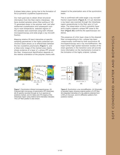
This is confirmed with wide-angle x-ray microdiffraction<br />
experiments (Figure 2). A 3 μm diameter<br />
x-ray beam was scanned through the interphase<br />
region perpendicular to the fiber axis in 5 μm<br />
steps. The observation of the β-cylindritic layer<br />
and a highly ordered α-phase iPP close to the<br />
fiber (Figure 2C) confirms the spectroscopic evidence.<br />
The presence of a thin layer close to the sheared<br />
fiber corresponding to the α-phase has been<br />
unequivocally confirmed by both synchrotron IR<br />
microspectroscopy and x-ray microdiffraction. We<br />
hope further high spatial resolution studies of the<br />
chain geometry in the transition zone will provide<br />
more information on the mechanisms involved in<br />
the formation of the highly ordered β-phase.<br />
spherulitic<br />
cylindritic<br />
cylindritic<br />
spherulitic<br />
distance, µm<br />
β−phase<br />
reflections<br />
diffraction angle, 2θ<br />
diffraction angle, 2θ<br />
diffraction angle, 2θ<br />
(A) (B) (C)<br />
Figure 2. Synchrotron x-ray microdiffraction. (A) Schematic<br />
of sampled region showing original position of LCP fi ber,<br />
(B) integrated intensities of x-ray patterns obtained from<br />
a 400 µm line-scan, and (C) diffraction patterns from<br />
positions marked.<br />
SOFT CONDENSED MATTER AND BIOPHYSICS