NSLS Activity Report 2006 - Brookhaven National Laboratory
NSLS Activity Report 2006 - Brookhaven National Laboratory
NSLS Activity Report 2006 - Brookhaven National Laboratory

Create successful ePaper yourself
Turn your PDF publications into a flip-book with our unique Google optimized e-Paper software.
SCIENTISTS TAKE 'SNAPSHOTS' OF ENZYME ACTION<br />
Scientists at <strong>Brookhaven</strong> <strong>National</strong> <strong>Laboratory</strong>,<br />
the New York Structural Biology Center, and SGX<br />
Pharmaceuticals, Inc., have determined the atomic<br />
crystal structure and functional mechanism of an<br />
enzyme essential for eliminating unwanted, nonnutritional<br />
compounds such as drugs, industrial<br />
chemicals, and toxic compounds from the body.<br />
The detailed mechanism of action will help scientists<br />
understand how these compounds are eliminated<br />
and what goes wrong in cases where normal<br />
metabolism fails. The research was published in<br />
the June 27, <strong>2006</strong> edition of the Proceedings of the<br />
<strong>National</strong> Academy of Sciences.<br />
According to <strong>Brookhaven</strong><br />
biologists Eswaramoorthy<br />
Subramaniam, the<br />
lead author, and Subramanyam<br />
Swaminathan,<br />
who led the research,<br />
most non-nutritional,<br />
foreign substances such<br />
as drugs and industrial<br />
chemicals are insoluble<br />
in water. The body uses<br />
two main groups of<br />
S. Swaminathan<br />
enzymes — flavin-containing<br />
monooxygenases<br />
(FMOs) and cytochrome P450s — to convert these<br />
compounds to soluble forms that can be easily<br />
excreted.<br />
“For FMOs, the end result — that an oxygen atom<br />
gets added to make these compounds soluble — is<br />
simple,” Swaminathan says, “but the reactions<br />
2-28<br />
require additional participants, or cofactors.” In<br />
order to understand the molecular mechanism, the<br />
scientists used high-intensity x-ray beams at the<br />
<strong>NSLS</strong> to identify the positions of individual atoms<br />
and produce crystal structures of the enzyme, the<br />
enzyme plus its cofactor, and the enzyme plus the<br />
cofactor plus the compound to be oxidized (the<br />
substrate).<br />
“These crystal structures give step-by-step snapshots<br />
of different stages of the catalytic action,”<br />
Swaminathan says, “and reveal a mechanism that<br />
is different from what had been known about this<br />
process.”<br />
Previously, it had been believed that all the<br />
“players” — the enzyme, cofactor and substrate<br />
— came together at a particular time to perform the<br />
function of transferring an oxygen atom from the<br />
enzyme to the substrate. “Our finding shows that<br />
the substrate and cofactor are binding to the enzyme<br />
alternately, not together,” Swaminathan says.<br />
First, the cofactor (known as NADPH) binds to a<br />
molecule known as FAD, which is a coenzyme attached<br />
to the FMO, and transfers a hydride ion to<br />
it. That makes the FAD group capable of accepting<br />
molecular oxygen. Then, when the substrate<br />
arrives, the cofactor leaves so that the substrate<br />
can bind to the same site on the FAD group. At this<br />
moment an oxygen atom from molecular oxygen<br />
is attached to the substrate, and the hydride ion<br />
obtained from the cofactor combines with the other<br />
oxygen atom to form a water molecule, which is released.<br />
Once the substrate is oxygenated, it leaves<br />
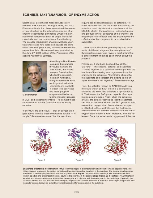
Figure 1 Figure 2 Figure 3<br />
Snapshots of catalytic mechanism of FMO: The three stages in the mechanism of action of FMO are depicted here. The<br />
ribbon diagram represents the protein consisting of two domains with a long loop in the interface. The big and small domains<br />
are shown in red and purple with the interface in golden color. Figure 1 represents the fi rst stage with the coenzyme FAD<br />
(shown as a ball and stick model in green) bound to the enzyme. In the second stage (Figure 2), the cofactor NADPH (shown<br />
as a ball and stick model in cyan) approaches the enzyme and interacts with the coenzyme. In the third stage (Figure 3), the<br />
substrate (shown as a ball and stick model in cyan) displaces the cofactor and binds in the same place to get oxygenated. The<br />
molecular oxygen (shown as a dumbbell in red) is required for oxygenation of the substrate.