NSLS Activity Report 2006 - Brookhaven National Laboratory
NSLS Activity Report 2006 - Brookhaven National Laboratory
NSLS Activity Report 2006 - Brookhaven National Laboratory

Create successful ePaper yourself
Turn your PDF publications into a flip-book with our unique Google optimized e-Paper software.
BEAMLINE<br />
X26C<br />
PUBLICATION<br />
L.J. Higgins, F. Yan, P. Liu, H.-W.<br />
Liu, and C.L. Drennan, "Structural<br />
Insight into Antibiotic Fosfomycin<br />
Biosynthesis by a Mononuclear<br />
Iron Enzyme," Nature, 437, 838-<br />
844 (2005).<br />
FUNDING<br />
<strong>National</strong> Institutes of Health;<br />
<strong>National</strong> Institute of Environmental<br />
Health Sciences; Searle<br />
Scholars Program; Alfred P. Sloan<br />
Foundation; A Lester Wolfe<br />
Predoctoral Fellowship<br />
FOR MORE INFORMATION<br />
Catherine L. Drennan<br />
Department of Chemistry<br />
Massachusetts Institute of Technology<br />
cdrennan@mit.edu<br />
Mononuclear non-heme iron enzymes use their<br />
metal cofactor to activate dioxygen (O 2 ) for difficult<br />
redox processes. One of these enzymes,<br />
S-(2)-Hydroxypropylphosphonic acid epoxidase<br />
(HppE), from Streptomyces wedmorensis (Figure<br />
1, overall structure), employs its mononuclear iron<br />
center and molecular oxygen for the two-electron<br />
oxidation of S-(2)-hydroxypropylphosphonic acid<br />
(S-HPP) to catalyze the formation of the antibiotic<br />
(1R,2S)-(1,2-epoxypropyl)phosphonic acid (fosfomycin).<br />
This reaction is essentially a dehydrogenation<br />
reaction (loss of hydrogen). In order to<br />
balance the four-electron reduction of oxygen to<br />
water, we have proposed a putative two-electron<br />
reductant (or reductase). Fosfomycin is an unusual<br />
C-P-bond-containing epoxide that covalently<br />
modifies UDP-GlcNAc enolpyruvyl transferase,<br />
consequently inhibiting bacterial cell-wall peptidoglycan<br />
biosynthesis and bacterial growth. Since it<br />
accumulates in the kidneys and bladder, fosfomycin<br />
has been used clinically for the treatment<br />
of lower-urinary-tract infections. The structures<br />
of the apo-HppE (metal-free), native Fe(II)-HppE,<br />
tris(hydroxymethyl)aminomethane (Tris)-Co(II)-<br />
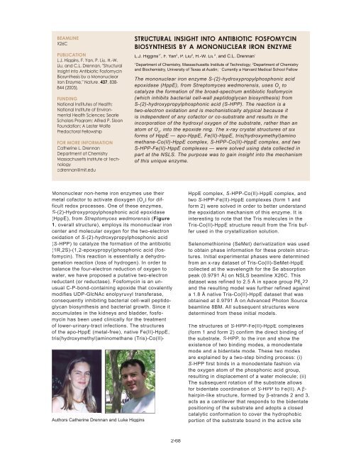
Authors Catherine Drennan and Luke Higgins<br />
STRUCTURAL INSIGHT INTO ANTIBIOTIC FOSFOMYCIN<br />
BIOSYNTHESIS BY A MONONUCLEAR IRON ENZYME<br />
L.J. Higgins 1* , F. Yan 2 , P. Liu 2 , H.-W. Liu 2 , and C.L. Drennan 1<br />
1 Department of Chemistry, Massachusetts Institute of Technology; 2 Department of Chemistry<br />
and Biochemistry, University of Texas at Austin; * Currently a Harvard Medical School Fellow<br />
The mononuclear iron enzyme S-(2)-hydroxypropylphosphonic acid<br />
epoxidase (HppE), from Streptomyces wedmorensis, uses O 2 to<br />
catalyze the formation of the broad-spectrum antibiotic fosfomycin<br />
(which inhibits bacterial cell-wall peptidoglycan biosynthesis) from<br />
S-(2)-hydroxypropylphosphonic acid (S-HPP). The reaction is a<br />
two-electron oxidation and is mechanistically atypical because it<br />
is independent of any cofactor or co-substrate and results in the<br />
incorporation of the hydroxyl oxygen of the substrate, rather than an<br />
atom of O 2 , into the epoxide ring. The x-ray crystal structures of six<br />
forms of HppE — apo-HppE, Fe(II)-HppE, tris(hydroxymethyl)amino<br />
methane-Co(II)-HppE complex, S-HPP-Co(II)-HppE complex, and two<br />
S-HPP-Fe(II)-HppE complexes — were solved using data collected in<br />
part at the <strong>NSLS</strong>. The purpose was to gain insight into the mechanism<br />
of this unique enzyme.<br />
2-68<br />
HppE complex, S-HPP-Co(II)-HppE complex, and<br />
two S-HPP-Fe(II)-HppE complexes (form 1 and<br />
form 2) were solved in order to better understand<br />
the epoxidation mechanism of this enzyme. It is<br />
interesting to note that the Tris molecules in the<br />
Tris-Co(II)-HppE structure result from the Tris buffer<br />
used in the crystallization solution.<br />
Selenomethionine (SeMet) derivatization was used<br />
to obtain phase information for these protein structures.<br />
Initial experimental phases were determined<br />
from an x-ray dataset of Tris-Co(II)-SeMet-HppE<br />
collected at the wavelength for the Se absorption<br />
peak (0.9791 Å) on <strong>NSLS</strong> beamline X26C. This<br />
dataset was refined to 2.5 Å in space group P6 5 22<br />
and the resulting model was further refined against<br />
a 1.8 Å native Tris-Co(II)-HppE dataset that was<br />
obtained at 0.9791 Å on Advanced Photon Source<br />
beamline 8BM. All subsequent structures were<br />
determined from these initial models.<br />
The structures of S-HPP-Fe(II)-HppE complexes<br />
(form 1 and form 2) confirm the direct binding of<br />
the substrate, S-HPP, to the iron and show the<br />
existence of two binding modes, a monodentate<br />
mode and a bidentate mode. These two modes<br />
are explained by a two-step binding process: (i)<br />
S-HPP first binds in a monodentate fashion via<br />
the oxygen atom of the phosphonic acid group,<br />
resulting in displacement of a water molecule; (ii)<br />
The subsequent rotation of the substrate allows<br />
for bidentate coordination of S-HPP to Fe(II). A βhairpin-like<br />
structure, formed by β-strands 2 and 3,<br />
acts as a cantilever that responds to the bidentate<br />
positioning of the substrate and adopts a closed<br />
catalytic conformation to cover the hydrophobic<br />
portion of the substrate bound in the active site