Evaluación de nuevos compuestos como potenciales - Acceda ...
Evaluación de nuevos compuestos como potenciales - Acceda ...
Evaluación de nuevos compuestos como potenciales - Acceda ...

Create successful ePaper yourself
Turn your PDF publications into a flip-book with our unique Google optimized e-Paper software.
Departamento <strong>de</strong> Química<br />
y<br />
Departamento <strong>de</strong> Bioquímica y Biología Molecular,<br />
Fisiología, Genética e Inmunología<br />
TESIS DOCTORAL<br />
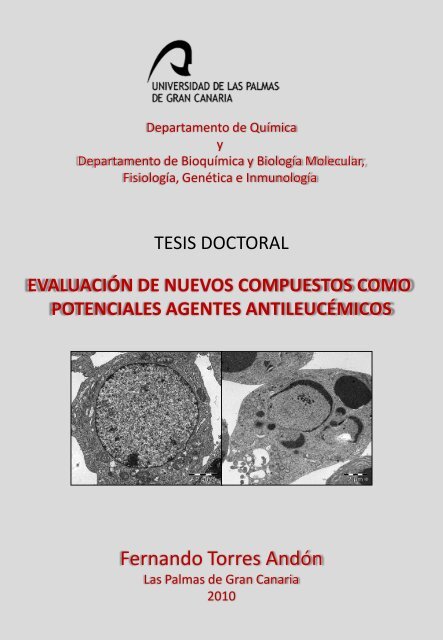
EVALUACIÓN DE NUEVOS COMPUESTOS COMO<br />
POTENCIALES AGENTES ANTILEUCÉMICOS<br />
Fernando Torres Andón<br />
Las Palmas <strong>de</strong> Gran Canaria<br />
2010
El tratamiento <strong>de</strong>l cáncer ha evolucionado mucho en los últimos años y<br />
aunque se ha conseguido aumentar la supervivencia frente a algunos tipos <strong>de</strong><br />
cáncer, no se ha logrado la efectividad <strong>de</strong>seada <strong>de</strong>bido fundamentalmente a<br />
una limitada actividad terapéutica o a una excesiva toxicidad. La lucha contra<br />
esta enfermedad sólo podrá solucionarse seleccionando las mejores dianas<br />
terapéuticas. Las neoplasias hematológicas forman uno <strong>de</strong> los grupos <strong>de</strong><br />
cáncer más estudiados y el análisis <strong>de</strong> los mecanismos implicados en la<br />
supervivencia o muerte <strong>de</strong> estas células tumorales ha proporcionado en<br />
numerosas ocasiones nuevas dianas terapéuticas eficaces en el tratamiento<br />
<strong>de</strong> éste y otros tipos <strong>de</strong> cáncer.<br />
Los principios activos obtenidos <strong>de</strong> fuentes naturales o sus <strong>de</strong>rivados<br />
semisintéticos han dado lugar a los principales fármacos utilizados contra el<br />
cáncer. Los flavonoi<strong>de</strong>s son <strong>compuestos</strong> con una estructura fenilbenzo-γpirona<br />
<strong>de</strong> origen natural, que abundan en frutas, verduras y bebidas <strong>de</strong>rivadas<br />
<strong>de</strong> plantas, y a<strong>de</strong>más poseen propieda<strong>de</strong>s antitumorales. Los datos <strong>de</strong>l<br />
presente estudio proporcionan evi<strong>de</strong>ncias acerca <strong>de</strong>l mecanismo por el que el<br />
trifolín acetato y la 3´,5,7-trihidroxi-3,4´-dimetoxiflavona inducen apoptosis<br />
en diferentes líneas celulares tumorales humanas y sientan las bases<br />
científicas para la utilización <strong>de</strong> estos <strong>compuestos</strong> en el <strong>de</strong>sarrollo <strong>de</strong> <strong>nuevos</strong><br />
fármacos antitumorales.
“¿Que cómo veo el futuro?...<br />
El futuro no se ve, el futuro se hace.”<br />
ÍNDICE 9<br />
J. M. Keynes
10 ÍNDICE<br />
Este trabajo ha sido financiado por:<br />
- Ministerio <strong>de</strong> Educación y Ciencia <strong>de</strong> España y FEDER. Plan Nacional <strong>de</strong><br />
Biomedicina. SAF 2004-07928 y SAF 2007-62536.<br />
- Subvención para equipamiento e infraestructura científico-tecnológica.Dirección<br />
General <strong>de</strong> Universida<strong>de</strong>s e Investigación. Resoluciones <strong>de</strong> 21 <strong>de</strong> diciembre <strong>de</strong> 2006 y<br />
21 <strong>de</strong> diciembre <strong>de</strong> 2007.<br />
- Beca <strong>de</strong>l Gobierno Autónomo <strong>de</strong> Canarias. Resolución <strong>de</strong> 30 <strong>de</strong> diciembre <strong>de</strong> 2005.<br />
BOC 7 feb 2006.<br />
- Consejería <strong>de</strong> Educación, Cultura y Deportes. Gobierno <strong>de</strong> Canarias y FEDER. GRUP<br />
2004-44.<br />
- Agencia Canaria <strong>de</strong> Investigación, Innovación y Sociedad <strong>de</strong> la Información <strong>de</strong>l<br />
Gobierno <strong>de</strong> Canarias. PI 2007/045.<br />
- Instituto Canario <strong>de</strong> Investigación <strong>de</strong>l Cáncer. RED PRODNATCANCER- 2008 y<br />
2009.<br />
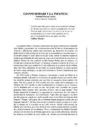
Ilustración <strong>de</strong> portada: Microfotografías <strong>de</strong> una célula HL-60 normal (izquierda) y una célula<br />
en apoptosis (<strong>de</strong>recha) obtenida mediante microscropía electrónica <strong>de</strong> transmisión.<br />
Ilustración <strong>de</strong> contraportada: Estructura química <strong>de</strong> la 3´,5,7-trihidroxi-3,4´-dimetoxiflavona<br />
(izquierda) y <strong>de</strong>l trifolín acetato (<strong>de</strong>recha).
AGRADECIMIENTOS 1<br />
ÍNDICE
1.<br />
ÍNDICE<br />
ÍNDICE 1<br />
AGRADECIMIENTOS 9<br />
ABREVIATURAS 13<br />
FIGURAS Y TABLAS 19<br />
INTRODUCCIÓN 23<br />
Tratamiento <strong>de</strong>l cáncer 26<br />
1.1. Selección <strong>de</strong> nuevas dianas terapéuticas 26<br />
1.2. Utilización <strong>de</strong> fármacos <strong>de</strong> origen natural 27<br />
2. Flavonoi<strong>de</strong>s 28<br />
2.1. Fuentes <strong>de</strong> flavonoi<strong>de</strong>s y su presencia en la dieta 29<br />
2.2. Absorción, metabolismo y actividad 30<br />
2.3. Utilización <strong>de</strong> flavonoi<strong>de</strong>s contra el cáncer 31<br />
3. Inhibición <strong>de</strong> la proliferación celular 32<br />
3.1. Inducción <strong>de</strong> la parada <strong>de</strong>l ciclo celular 32<br />
3.2. Interacción con los microtúbulos 35<br />
4. Apoptosis 36<br />
4.1. Vía extrínseca: receptores <strong>de</strong> muerte celular 38<br />
4.1.1. Receptor Fas (CD95) 39<br />
4.1.2. Receptor <strong>de</strong> TNF-α: TNFR1 40<br />
4.2. Vía intrínseca: inicio <strong>de</strong> la señal <strong>de</strong> apoptosis en la mitocondria 41<br />
4.3. Proteínas reguladoras <strong>de</strong> la apoptosis 42<br />
4.3.1. Proteínas <strong>de</strong> la familia Bcl-2 42<br />
4.3.2. Proteínas <strong>de</strong> la familia <strong>de</strong> las caspasas 45<br />
4.3.2.1 Sustratos <strong>de</strong> caspasas 47<br />
4.3.2.2. Mecanismos inhibidores <strong>de</strong> caspasas 48<br />
4.4. Vía <strong>de</strong> las MAPKs 49<br />
4.4.1. ERK1/2 (p42/44 MAPK) 51<br />
4.4.2. Familia JNK 52<br />
4.4.3. Familia p38 54<br />
4.5. Esfingolípidos 55<br />
3
4 ÍNDICE<br />
4.5.1. Ceramidas 56<br />
OBJETIVOS 59<br />
MATERIAL Y MÉTODOS 63<br />
1. Agentes farmacológicos 65<br />
2. Productos y material 65<br />
3. Mo<strong>de</strong>lo experimental 68<br />
3.1. Cultivo <strong>de</strong> células leucémicas humanas HL-60, JURKAT y MOLT-3, y<br />
células <strong>de</strong> linfoma histiocítico humano U937 68<br />
3.2. Cultivo <strong>de</strong> células <strong>de</strong> melanoma humanas SK-MEL-1 69<br />
3.3. Cultivo <strong>de</strong> células A549 (cáncer <strong>de</strong> pulmón humano) 69<br />
3.4. Cultivo <strong>de</strong> células HL-60 transfectadas con el gen humano Bcl-xL y con<br />
el vector control, y <strong>de</strong> células U937 que expresan niveles elevados <strong>de</strong>l<br />
gen humano Bcl-2 69<br />
3.5. Células mononucleares <strong>de</strong> sangre periférica <strong>de</strong> origen humano 70<br />
3.6. Tratamiento con los <strong>compuestos</strong> 70<br />
4. Métodos 70<br />
4.1. <strong>Evaluación</strong> <strong>de</strong> la citotoxicidad in vitro y estudios <strong>de</strong> la proliferación<br />
celular: MTT 70<br />
4.2. Estudio <strong>de</strong> la apoptosis celular 71<br />
4.2.1. Tinción con el fluorocromo bisbenzimida 71<br />
4.2.2. Fragmentación <strong>de</strong>l ADN 72<br />
4.2.3. Cuantificación <strong>de</strong> las células hipodiploi<strong>de</strong>s por citometría <strong>de</strong> flujo.<br />
Cuantificación <strong>de</strong> la fracción SubG1, G1, S y G2/M mediante el análisis<br />
<strong>de</strong>l contenido en ADN<br />
4.2.4. Determinación <strong>de</strong> las células apoptóticas mediante el análisis <strong>de</strong> la<br />
externalización <strong>de</strong> fosfatidilserina 73<br />
4.2.4. Microscopía electrónica <strong>de</strong> transmisión (MET) 74<br />
4.3. Determinación <strong>de</strong> la actividad caspasa 74<br />
4.4 Inmuno<strong>de</strong>tección <strong>de</strong> proteínas (western blot) 75<br />
4.5. Análisis <strong>de</strong> la tubulina por western blot 76<br />
4.6. Determinación <strong>de</strong> la generación <strong>de</strong> ROS intracelular 77<br />
4.7. Análisis <strong>de</strong> la <strong>de</strong>spolarización <strong>de</strong> la membrana mitocondrial (∆Ψm) 78<br />
72
ÍNDICE<br />
4.8. Análisis <strong>de</strong> la red <strong>de</strong> microtúbulos por microscopía <strong>de</strong> fluorescencia 78<br />
4.9. Análisis <strong>de</strong> la polimerización <strong>de</strong> tubulina in vitro 79<br />
4.10. Ensayo <strong>de</strong> competición por la unión a tubulina 79<br />
4.11. Determinación <strong>de</strong> la actividad esfingomielinasa ácida y neutra 79<br />
4.12. Determinación <strong>de</strong> la cantidad <strong>de</strong> ceramidas 80<br />
5. Métodos estadísticos 82<br />
RESULTADOS<br />
1. Los <strong>de</strong>rivados <strong>de</strong> la fenilbenzopirona inhiben el crecimiento y la<br />
viabilidad <strong>de</strong> las líneas celulares estudiadas 85<br />
2. Trifolín acetato induce apoptosis en células <strong>de</strong> leucemia mieloi<strong>de</strong> HL-60<br />
3. Trifolín acetato induce muerte celular mediada por la activación <strong>de</strong><br />
caspasas 91<br />
4. Trifolín acetato induce liberación <strong>de</strong> citocromo c al citosol, sin producir<br />
cambios en el potencial <strong>de</strong> membrana mitocondrial 93<br />
5. Trifolín acetato no induce apoptosis en líneas celulares <strong>de</strong> leucemia<br />
humana que sobreexpresan Bcl-2 ó Bcl-xL<br />
6. Trifolín acetato activa la vía MAPKs 95<br />
7. Las especies reactivas <strong>de</strong> oxígeno no están involucradas en la muerte<br />
celular inducida por el trifolín acetato<br />
8. THDF inhibe la viabilidad <strong>de</strong> células tumorales humanas y no presenta<br />
citotoxicidad frente a linfocitos normales 99<br />
9. THDF induce apoptosis en células <strong>de</strong> leucemia mieloi<strong>de</strong> humana 101<br />
10. THDF induce muerte celular a través <strong>de</strong> la activación <strong>de</strong> caspasas 104<br />
11. THDF induce liberación <strong>de</strong> citocromo c mitocondrial y disminución <strong>de</strong><br />
Bax citosólico 106<br />
12. La sobreexpresión <strong>de</strong> las proteínas mitocondriales Bcl-2 y Bcl-xL<br />
confiere resistencia parcial a la apoptosis inducida por THDF 108<br />
13. THDF induce cambios en el potencial <strong>de</strong> menbrana mitocondrial 108<br />
14. THDF activa las proteínas quinasas activadas por mitógenos (MAPKs) 108<br />
15. THDF aumenta los niveles <strong>de</strong> las especies reactivas <strong>de</strong> oxígeno<br />
intracelulares 110<br />
16. THDF induce parada <strong>de</strong>l ciclo celular en fase M 112<br />
17. Efectos <strong>de</strong>l THDF sobre la expresión y fosforilación <strong>de</strong> las 114<br />
83<br />
88<br />
95<br />
97<br />
5
6 ÍNDICE<br />
proteínasreguladoras <strong>de</strong> la fase M<br />
18. THDF <strong>de</strong>sestabiliza la polimerización <strong>de</strong> los microtúbulos 116<br />
19. THDF inhibe la polimerización <strong>de</strong> tubulina “in vitro” 119<br />
20. THDF se une específicamente al sitio <strong>de</strong> unión <strong>de</strong> la colchicina 119<br />
21. THDF induce la generación <strong>de</strong> ceramidas y la activación <strong>de</strong> SMasa 119<br />
DISCUSIÓN 135<br />
CONCLUSIONES 151<br />
SUMMARY 155<br />
CONCLUSIONS 171<br />
BIBLIOGRAFÍA 175<br />
ANEXOS: 191
AGRADECIMIENTOS 7<br />
AGRADECIMIENTOS
AGRADECIMIENTOS<br />
En primer lugar, me gustaría dar las gracias a mis directores <strong>de</strong> tesis, Dr. Francisco Estévez<br />
Rosas y Dr. José Quintana Aguiar, por introducirme en el mundo <strong>de</strong> la investigación. Gracias<br />
por haber compartido conmigo sus conocimientos y su pasión por la investigación, por su<br />
ayuda y <strong>de</strong>dicación en el trabajo diario.<br />
Quiero agra<strong>de</strong>cer a todos los que son o han sido compañeros <strong>de</strong> laboratorio, personas que han<br />
seguido más <strong>de</strong> cerca y saben perfectamente lo que conlleva la lectura <strong>de</strong> esta tesis. Hago<br />
especial mención a Sara y a Gledy por su amistad y ayuda a lo largo <strong>de</strong> estos años. Gracias a<br />
Olga, Carol, Ana, Cristina, Fabio, Mayte, Dionisio y Juan, por haberme acompañado durante<br />
estos años, por su ayuda, amistad y por traer el buen humor a estas cuatro pare<strong>de</strong>s.<br />
Merci à mes collègues <strong>de</strong> la Faculté <strong>de</strong> Pharmacie <strong>de</strong> l’Université Paris-Sud 11, Aida, Jöel e<br />
spécialement à la Dr. Jacqueline Bréard par vivre chaque découverte comme un grand<br />
événement et <strong>de</strong> m’avoir contaminés sa illusion.<br />
Gracias al Dr. Javier Cabrera, y al Dr. Juan Francisco Loro por su inestimable ayuda y<br />
consejos en los inicios <strong>de</strong> mi trabajo, a la Dra. Mª <strong>de</strong>l Pino Santana, a la Dra. Inmaculada<br />
Servanda Hernán<strong>de</strong>z, al Dr. Ignacio Javier González, al Dr. Enrique Castro, al Dr. Germán<br />
Gallardo, y al Dr. Carlos Tabraue por haber sido un gran apoyo en estos años, por sus<br />
consejos y por haberme ayudado a resolver todo tipo <strong>de</strong> problemas. También quiero agra<strong>de</strong>cer<br />
su ayuda a Jose Manuel Pérez y Olivia Rodriguez, <strong>de</strong>l Servicio <strong>de</strong> Microscopía Electrónica.<br />
Grazas aos meus amigos, Fernando, Fran, Luis, David y Joana, Pipo, Juanma, Iván, Jacobo,<br />
Mariña, Jose Carlos, Pedro y Marcos… porque hai cousas mellores que o traballo. Y sobre<br />
todo gracias a lo mejor que me pasó durante esta tesis, gracias Ana por cada segundo que<br />
estás conmigo, por tu cariño, alegría y por todo lo que nos queda por vivir. Gracias por<br />
ayudarme tanto y por hacerme conocer a tu familia, que me hacen sentir uno más.<br />
Finalmente, quero dar as grazas á miña familia, que sempre me axudou, aínda <strong>de</strong>n<strong>de</strong> a<br />
distancia. Grazas Suso e Nati, porque grazas ao voso esforzo pui<strong>de</strong>n chegar ata aquí. Grazas<br />
David pola túa paciencia e por axudarme sempre que o necesitei. Grazas aos meus avós, Xosé<br />
e Concha, Suso e Benjamina; e aos meus tíos, Conchi, Pili e Manel, por ser parte da miña<br />
educación e da miña vida. Grazas a Luís Miguel, Bruno, Pedro e Sara por renovar a ledicia da<br />
familia.<br />
9
ABREVIATURAS
ABREVIATURAS<br />
∆Ψm<br />
Potencial <strong>de</strong> membrana mitocondrial<br />
µM Micromolar<br />
ADN Ácido <strong>de</strong>soxirribonucleico<br />
AIF Factor inductor <strong>de</strong> apoptosis<br />
Akt Quinasa involucrada en la supervivencia celular<br />
Apaf-1 Factor activador <strong>de</strong> proteasas apoptóticas<br />
ARC Inhibidor general <strong>de</strong> la apoptosis<br />
ARN Ácido ribonucleico<br />
ATP A<strong>de</strong>nosín trifosfato<br />
Bad Proteína pro-apoptótica perteneciente a la familia Bcl-2<br />
Bak Proteína pro-apoptótica perteneciente a la familia Bcl-2<br />
Bax Proteína pro-apoptótica perteneciente a la familia Bcl-2<br />
Bcl-2 Proteína anti-apoptótica<br />
Bcl-xL<br />
Proteína anti-apoptótica perteneciente a la familia Bcl-2<br />
Bid Proteína pro-apoptótica<br />
BIR<br />
CAK<br />
Dominio <strong>de</strong> interacción proteína-proteína presente en las IAPS<br />
Proteína quinasa constitutiva activadora <strong>de</strong> CDK<br />
CARD Dominio <strong>de</strong> reclutamiento <strong>de</strong> caspasa<br />
CAT Catalasa<br />
CCCP Carbonil Cianuro m-Clorofenil- Hidrazona<br />
CDK Proteína quinasa <strong>de</strong>pendiente <strong>de</strong> ciclina<br />
CIP Proteínas inhibidoras <strong>de</strong> CDKs<br />
CKI Inhibidores <strong>de</strong> las quinasas <strong>de</strong>pendientes <strong>de</strong> ciclinas<br />
CoA Coenzima A<br />
DAG Diacilglicerol<br />
DED Dominio efector <strong>de</strong> muerte<br />
DETAPAC Ácido dietilen-triamina-penta-acético<br />
DEVD-pNA Sustrato específico <strong>de</strong> caspasa -3/-7<br />
DGK Diacilglicerol quinasa<br />
DISC Complejo inductor <strong>de</strong> muerte<br />
DMSO Dimetilsulfóxido<br />
DNA-PK Proteína quinasa <strong>de</strong>pendiente <strong>de</strong> ADN<br />
DTT Ditiotreitol<br />
EDTA Ácido etilen-diamin-tetra-acético<br />
EGFR Receptores <strong>de</strong>l factor <strong>de</strong> cremiento epidérmico<br />
EGTA Ácido etilenglicol-bis-N,N'-tetraacético<br />
ERK1/2 Proteína quinasa regulada por señales extracelulares 1 y 2<br />
ETO Etopósido<br />
FADD Dominio <strong>de</strong> muerte asociado a Fas<br />
Fas Receptor <strong>de</strong> muerte, también llamado Apo1 o CD95<br />
FasL Ligando <strong>de</strong>l receptor <strong>de</strong> muerte Fas<br />
FBS Suero bovino fetal<br />
GlcCer Glucosilceramida<br />
GPCR Receptor asociado a proteína G<br />
GTP Guanosín trifosfato<br />
GW4869 Inhibidor <strong>de</strong> la esfingomielinasa neutra<br />
H2-DCF-DA Diacetato 2´,7´-dicloro-dihidro-fluoresceína<br />
HEPES N-[2-hidroxietil] piperazino N'-[2-etanosulfanílico]<br />
IAP Proteína inhibidora <strong>de</strong> apoptosis<br />
IC50<br />
Concentración <strong>de</strong> producto que inhibe el crecimiento celular a la<br />
mitad<br />
IP Yoduro <strong>de</strong> propidio<br />
JC-1 Yoduro<strong>de</strong>5,5',6,6'–tetracloro-1,1',3,3'tetraetilbenzimidazolocarbocianina<br />
13
14 ABREVIATURAS<br />
JNK Proteína quinasa N-terminal <strong>de</strong> jun<br />
Jun Factor <strong>de</strong> transcripcion <strong>de</strong> la familia <strong>de</strong> AP-1<br />
MAPK Proteina quinasa activada por mitógenos<br />
MEK Proteína quinasa MAPK<br />
MIF Factor inhibitorio <strong>de</strong> la migración<br />
MK Proteínas quinasas activadas por MAPKs<br />
MKP MAP quinasas fosfatasas<br />
MMP Metaloproteinasa <strong>de</strong> la matriz extracelular<br />
MSK Proteína quinasa activada por mitógenos y estrés<br />
MTT Bromuro <strong>de</strong> 3-[4,5- Dimetiltiazol-2-il]-2,5-difeniltetrazolio<br />
MYT1 Factor <strong>de</strong> transcripción <strong>de</strong> mielina<br />
Na3VO4<br />
Ortovanadato sódico<br />
NAC N-acetil-L-cisteína<br />
NaCl Cloruro <strong>de</strong> sodio<br />
NADPH Nicotinamida a<strong>de</strong>nina dinucleótido fosfato<br />
NaOH Hidróxido <strong>de</strong> sodio<br />
NF-κB<br />
NO<br />
Factor nuclear - κB<br />
p21<br />
p38<br />
Inhibidor <strong>de</strong> quinasas <strong>de</strong>pendientes <strong>de</strong> ciclinas<br />
MAPK<br />
p53 Proteína supresora <strong>de</strong> tumores<br />
PA Plasminógeno tisular<br />
PARP-1 Poli (ADPribosa) polimerasa-1<br />
PBMC Células mononucleares humanas <strong>de</strong> sangre periférica<br />
PBS Tampón fosfato salino<br />
PD98059 Inhibidor específico <strong>de</strong> la quinasa reguladora MEK1<br />
PHA Fitohemaglutinina<br />
PI3K Fosfatidil inositol 3quinasa<br />
PK Proteína quinasa<br />
PL Fosfolipasa<br />
PMSF Fluoruro <strong>de</strong> metilsulfonilfenilo<br />
pRB Proteína retinoblastoma<br />
PVDF Fluoruro <strong>de</strong> Poli-vinili<strong>de</strong>no<br />
Raf MAPKKK<br />
Ras Proteína G pequeña<br />
RING Dominio interacción proteína-proteína<br />
RIP Proteína <strong>de</strong> interacción con el receptor<br />
ROS Especies reactivas <strong>de</strong> oxígeno<br />
RTK Receptores <strong>de</strong> tirosina quinasas<br />
S1P<br />
SB203580<br />
Esfingosina 1-fosfato<br />
Inhibidor específico <strong>de</strong> p38 MAPK<br />
SDS Do<strong>de</strong>cil sulfato sódico<br />
Ser Serina<br />
SIDA Síndrome <strong>de</strong> inmuno<strong>de</strong>ficiencia adquirida<br />
SK Esfingosina quinasa<br />
Smac Segundo activador <strong>de</strong> caspasas <strong>de</strong>rivado <strong>de</strong> la mitocondria<br />
SMasa Esfingomielinasa<br />
SOD Superóxido dismutasa<br />
SOS Factor <strong>de</strong> intercambio <strong>de</strong> nucleótidos <strong>de</strong> guanina<br />
SP600125 Inhibidor específico <strong>de</strong> JNK<br />
TA Trifolín heptaacetato<br />
TBST 20 mM Tris-HCl (pH 7,5), 137 mM NaCl, 0,1 % Tween-20<br />
TEMED N,N,N,N,-tetrametil-etilendiamina<br />
THDF 3´,5,7-Trihidroxi-3,4´-dimetoxiflavona
ABREVIATURAS<br />
Thr Treonina<br />
TLC Cromatografia en capa fina.<br />
TNF Factor <strong>de</strong> necrosis tumoral<br />
TNFR1 Receptor 1 <strong>de</strong>l factor necrótico tumoral<br />
TRADD Dominio <strong>de</strong> muerte asociado al TNFR<br />
TRAF2 Factor asociado a al receptor <strong>de</strong> TNF<br />
TRAIL Ligando inductor <strong>de</strong> apoptosis relacionado con TNF<br />
U0126 Inhibidor selectivo <strong>de</strong> las quinasa reguladoras MEK1 y MEK2<br />
UV Ultravioleta<br />
VEGF Factor <strong>de</strong> crecimiento endotelial vascular<br />
Wee1 Tirosina quinasa Wee1<br />
XIAP Proteína inhibidora <strong>de</strong> la apoptosis<br />
z-DEVD-fmk Inhibidor específico <strong>de</strong> caspasa-3 y -7<br />
z-IETD-fmk Inhibidor específico <strong>de</strong> caspasa-8<br />
z-LEHD-fmk Inhibidor específico <strong>de</strong> caspasa-9<br />
z-VAD-fmk Inhibidor general <strong>de</strong> caspasas<br />
z-VDVAD-fmk Inhibidor específico <strong>de</strong> caspasa-2<br />
z-VEID-fmk Inhibidor específico <strong>de</strong> caspasa-6<br />
z-YVAD-fmk Inhibidor específico <strong>de</strong> caspasa-1<br />
15
FIGURAS Y TABLAS<br />
FIGURAS<br />
Y<br />
TABLAS<br />
17
INTRODUCCIÓN<br />
FIGURAS Y TABLAS<br />
Figura 1 Clasificación <strong>de</strong> los flavonoi<strong>de</strong>s. 26<br />
Figura 2 Esquema <strong>de</strong> las quinasas <strong>de</strong>pendientes <strong>de</strong> ciclina implicadas en<br />
el paso <strong>de</strong> la fase G2 a la mitosis. 30<br />
Figura 3 Vías <strong>de</strong> activación <strong>de</strong> la apoptosis. 35<br />
Figura 4 Intercciones entre los miembros más importantes <strong>de</strong> la familia<br />
Bcl-2. 41<br />
Figura 5 (A) Estructura <strong>de</strong> las caspasas implicadas en la apoptosis. (B)<br />
Activacíón <strong>de</strong> las caspasas por procesamiento proteolítico. 43<br />
Figura 6 Vías <strong>de</strong> activación <strong>de</strong> las MAPKs. 46<br />
RESULTADOS<br />
Tabla 1 Estructura química <strong>de</strong> los flavonoi<strong>de</strong>s evaluados. 78<br />
Tabla 2 Efectos <strong>de</strong> los flavonoi<strong>de</strong>s sobre el crecimiento <strong>de</strong> células<br />
tumorales humanas. 79<br />
Figura 7 Estructura química <strong>de</strong>l trifolín acetato. 80<br />
Figura 8 Efectos <strong>de</strong>l TA sobre la inducción <strong>de</strong> apoptosis. 81<br />
Figura 9 Efecto <strong>de</strong>l TA en la proliferación <strong>de</strong> células mononucleares<br />
humanas <strong>de</strong> sangre periférica (PBMC) y <strong>de</strong> células HL-60. 82<br />
Figura 10 Influencia <strong>de</strong> la activación <strong>de</strong> caspasas en la apoptosis inducida<br />
por TA en células tumorales. 83<br />
Figura 11 El tratamiento con TA induce liberación <strong>de</strong> citocromo cen<br />
células tumorales. 85<br />
Figura 12 A) El TA no disminuye el potencial <strong>de</strong> membrana mitocondrial<br />
(∆Ψm) (B) El TA no es capaz <strong>de</strong> inducir apoptosis en líneas<br />
celulares que sobreexpresan Bcl-2 y Bcl-xL<br />
Figura 13 Papel <strong>de</strong> la vía <strong>de</strong> las MAPKs en al apoptosis inducida por TA<br />
en células HL-60. 87<br />
Figura 14 TA aumenta la generación <strong>de</strong> ROS en células HL-60 89<br />
Figura 15 Estructura química <strong>de</strong>l THDF. 90<br />
Tabla 3 Efecto <strong>de</strong>l THDF sobre la proliferación <strong>de</strong> células tumorales<br />
humanas. 90<br />
Figura 16 (A) Efecto <strong>de</strong>l THDF sobre la viabilidad <strong>de</strong> las células HL-60 y<br />
SK-MEL-1 (B) Efecto <strong>de</strong>l THDF sobre la proliferación <strong>de</strong><br />
células mononucleare humanas <strong>de</strong> sangre periférica. 91<br />
Figura 17 <strong>Evaluación</strong> mediante citometría <strong>de</strong> flujo <strong>de</strong>l efecto <strong>de</strong>l THDF<br />
sobre la inducción <strong>de</strong> apoptosis en células HL-60 y U937. 92<br />
Figura 18 Efecto <strong>de</strong>l THDF sobre la inducción <strong>de</strong> apoptosis en células<br />
HL-60 y U937. 93<br />
Figura 19 Estudio <strong>de</strong> la apoptosis inducida por THDF en células HL-60 y<br />
U937. 94<br />
Figura 20 <strong>Evaluación</strong> <strong>de</strong> la activación <strong>de</strong> caspasas en células <strong>de</strong> leucemia<br />
HL-60 y U937. 95<br />
Figura 21 <strong>Evaluación</strong> <strong>de</strong>l papel <strong>de</strong> las mitocondrias en la apoptosis<br />
inducida por THDF en células HL-60 y U937. 97<br />
Figura 22 (A) THDF induce la fosforilación <strong>de</strong> MAPKs. (B) Impacto <strong>de</strong><br />
86<br />
19
20 FIGURAS Y TABLAS<br />
los inhibidores <strong>de</strong> MAPKs en la apoptosis inducida por THDF 99<br />
Figura 23 ROS no está involucrado en la muerte celular inducida por<br />
THDF. 101<br />
Tabla 4 Efecto <strong>de</strong>l THDF sobre las diferentes fases <strong>de</strong>l ciclo celular en<br />
HL-60 y U937. 102<br />
Figura 24 Distribución <strong>de</strong>l ciclo celular durante la incubación en<br />
presencia o ausencia <strong>de</strong> THDF en células HL-60 previamente<br />
sincronizadas en fase M. 103<br />
Figura 25 THDF induce parada en mitosis. 103<br />
Figura 26 Efecto <strong>de</strong> THDF en los niveles <strong>de</strong> expresión <strong>de</strong> las proteínas<br />
que regulan el ciclo celular. 104<br />
Figura 27 THDF inhibe la polimerización <strong>de</strong> la tubulina en células HL-60<br />
y U937. 106<br />
Figura 28 (A) THDF induce la <strong>de</strong>spolimerización <strong>de</strong> tubulina in vitro (B)<br />
Ensayo <strong>de</strong> competición entre [ 3 H]-colchicina y THDF por la<br />
unión a la tubulina. 107<br />
Figura 29 Efecto <strong>de</strong> THDF sobre la vía <strong>de</strong> la esfingomielina en células<br />
HL-60. 109
INTRODUCCIÓN
INTRODUCCIÓN<br />
El cáncer pue<strong>de</strong> ser consi<strong>de</strong>rado una epi<strong>de</strong>mia. Su inci<strong>de</strong>ncia y prevalencia están aumentando<br />
rápidamente, <strong>de</strong>bido entre otras causas, al crecimiento y envejecimiento <strong>de</strong> la población.<br />
Alre<strong>de</strong>dor <strong>de</strong> 2,3 millones <strong>de</strong> <strong>nuevos</strong> casos <strong>de</strong> cáncer se contabilizaron en la Unión Europea<br />
en el año 2006, y el número <strong>de</strong> casos aumenta cada año. Una <strong>de</strong> cada cuatro muertes en la<br />
Unión Europea se atribuye al cáncer, y en el rango <strong>de</strong> edad entre 45 y 64 años, la proporción<br />
es cercana al 50% [1]. Proporciones similares se han observado en los Estados Unidos, dón<strong>de</strong><br />
se <strong>de</strong>tectaron en el año 2009 un total <strong>de</strong> 1,5 millones <strong>de</strong> casos y alre<strong>de</strong>dor <strong>de</strong> 500.000 muertes<br />
[2]. La Organización Mundial <strong>de</strong> la Salud ha estimado que alre<strong>de</strong>dor <strong>de</strong> 12 millones <strong>de</strong><br />
personas morirán, en todo el mundo, a causa <strong>de</strong> esta enfermedad en el año 2030 [3].<br />
El cáncer se produce <strong>como</strong> consecuencia <strong>de</strong> una acumulación <strong>de</strong> errores en el material<br />
genético <strong>de</strong> las células, que adquieren la capacidad <strong>de</strong> propagarse <strong>de</strong> forma in<strong>de</strong>finida<br />
(generando sus propias señales mitógenas), en su localización normal o en otras zonas<br />
(invadiendo otros tejidos y/o produciendo metástasis), <strong>como</strong> consecuencia <strong>de</strong> una pérdida <strong>de</strong><br />
control <strong>de</strong> su proliferación, diferenciación o <strong>de</strong> la respuesta ante señales <strong>de</strong> apoptosis (muerte<br />
celular programada).<br />
Existen más <strong>de</strong> 200 tipos diferentes <strong>de</strong> cáncer, la mayoría <strong>de</strong> los cuales <strong>de</strong>ben su nombre al<br />
órgano o al tejido en el que se forman, y pue<strong>de</strong>n ser agrupados en las siguientes categorías:<br />
- Carcinoma – tejido epitelial.<br />
- Sarcoma – tejido conjuntivo (hueso, cartílago, grasa, músculo, vasos sanguíneos u<br />
otros).<br />
- Glioma – tejido nervioso.<br />
- Neoplasias hematológicas (Leucemia, Linfoma y Mieloma) – tejido sanguíneo,<br />
médula ósea y ganglios linfáticos.<br />
- Otros – melanoma, hepatoma, etc.<br />
En la mayoría <strong>de</strong> estos tipos <strong>de</strong> cáncer las células no se mueren y se forman nuevas células<br />
innecesarias para el organismo, formando una masa llamada tumor (“tumor maligno”). En<br />
cambio algunos tipos <strong>de</strong> cáncer, <strong>como</strong> la leucemia, pue<strong>de</strong>n no formar un tumor.<br />
La leucemia es un grupo <strong>de</strong> enfermeda<strong>de</strong>s <strong>de</strong> la médula ósea y <strong>de</strong> la sangre, que implica un<br />
aumento incontrolado <strong>de</strong> glóbulos blancos (leucocitos). Es el cáncer más frecuente en los<br />
23
24 INTRODUCCIÓN<br />
niños menores <strong>de</strong> 15 años a nivel mundial [4], y en Estados Unidos en hombres menores <strong>de</strong><br />
40 años y mujeres menores <strong>de</strong> 20 años [2]. De acuerdo con la población leucocitaria que<br />
afecten, se clasifican en: leucemia mieloi<strong>de</strong> crónica, leucemia linfoi<strong>de</strong> crónica, leucemia<br />
linfoi<strong>de</strong> aguda, leucemia mieloi<strong>de</strong> aguda ó leucemia <strong>de</strong> células pilosas.<br />
1. Tratamiento <strong>de</strong>l cáncer.<br />
1.1. Selección <strong>de</strong> nuevas dianas terapéuticas.<br />
El tratamiento <strong>de</strong>l cáncer ha evolucionado mucho en los últimos años, y se ha conseguido<br />
aumentar la supervivencia frente a algunos tipos <strong>de</strong> cáncer (por ejemplo, leucemias o<br />
linfomas), pero no se ha conseguido la efectividad <strong>de</strong>seada <strong>de</strong>bido fundamentalmente a una<br />
insuficiente actividad terapéutica (30%) ó a una excesiva toxicidad (30%). Estos problemas<br />
sólo podrán solucionarse seleccionando las mejores dianas terapeúticas y mediante el uso <strong>de</strong><br />
biomarcadores para i<strong>de</strong>ntificar a los pacientes apropiados para recibir cada tratamiento [5,6].<br />
El conocimiento <strong>de</strong> los mecanismos moleculares implicados en el cáncer ha mejorado <strong>de</strong><br />
forma significativa, y actualmente, el <strong>de</strong>sarrollo <strong>de</strong> <strong>nuevos</strong> fármacos antitumorales está<br />
dirigido al estudio <strong>de</strong> los mecanismos moleculares y genéticos implicados, con el objetivo <strong>de</strong><br />
<strong>de</strong>sarrollar una medicina “personalizada”. La activación <strong>de</strong> oncogenes y la inactivación <strong>de</strong><br />
genes supresores <strong>de</strong> tumores en la célula tumoral implican una modificación <strong>de</strong> las vías <strong>de</strong><br />
señalización induciendo: la falta <strong>de</strong> respuesta frente a factores homeostáticos, la pérdida <strong>de</strong>l<br />
control <strong>de</strong>l ciclo celular y la proliferación <strong>de</strong>scontrolada, el aumento <strong>de</strong> la supervivencia<br />
celular, la disminución <strong>de</strong> la apoptosis, la inmortalización y la estimulación <strong>de</strong> procesos <strong>como</strong><br />
invasión, angiogénesis y metástasis (propagación a otras partes <strong>de</strong>l cuerpo por vía sanguínea o<br />
linfática). Todos estos procesos presentan <strong>potenciales</strong> dianas <strong>de</strong>s<strong>de</strong> el punto <strong>de</strong> vista <strong>de</strong> una<br />
intervención terapéutica [7].<br />
El gran avance que se ha producido en el conocimiento <strong>de</strong> la muerte celular por apoptosis,<br />
mecanismo mediante el cual las células son eliminadas con el propósito <strong>de</strong> controlar la<br />
proliferación celular ó en respuesta a un daño en el ADN, nos proporciona las líneas a seguir<br />
en el <strong>de</strong>sarrollo <strong>de</strong> nuevas terapias dirigidas, induciendo la muerte <strong>de</strong> las células tumorales <strong>de</strong><br />
forma específica o sensibilizándolas frente a los tratamientos antitumorales actuales.<br />
Las neoplasias hematológicas forman uno <strong>de</strong> los grupos <strong>de</strong> cáncer más estudiados. En 1980 se<br />
<strong>de</strong>scubrió que los glucocorticoi<strong>de</strong>s inducen fragmentación <strong>de</strong>l ADN y apoptosis en los<br />
timocitos, y <strong>de</strong>s<strong>de</strong> entonces el sistema hematopoyético ha <strong>de</strong>svelado numerosas situaciones en
INTRODUCCIÓN<br />
las que los mecanismos <strong>de</strong> muerte celular resultan clave en el control <strong>de</strong> la homeostasis. El<br />
análisis <strong>de</strong> este mecanismo ha proporcionado nuevas dianas terapéuticas eficaces en el<br />
tratamiento <strong>de</strong> este y otros tipos <strong>de</strong> cáncer [8]. Entre las neoplasias hematológicas, la leucemia<br />
mieloi<strong>de</strong> crónica es una <strong>de</strong> las más estudiadas, y constituye uno <strong>de</strong> los mejores ejemplos <strong>de</strong><br />
cómo una enfermedad pue<strong>de</strong> ser tratada mediante una terapia dirigida a una diana molecular<br />
específica. El tratamiento con Imatinib ha supuesto una importante mejora en la lucha contra<br />
la enfermedad y a<strong>de</strong>más ha sido utilizado en el tratamiento <strong>de</strong> otros tipos <strong>de</strong> cáncer [9,10]<br />
(tumores <strong>de</strong>l estroma gastrointestinal).<br />
El esfuerzo realizado en la i<strong>de</strong>ntificación <strong>de</strong> nuevas dianas terapéuticas ha tenido en los<br />
últimos años múltiples frutos, ejemplos <strong>de</strong> ello son: el Bortezomib, tratamiento <strong>de</strong> elección en<br />
mieloma múltiple, el Trastuzumab <strong>de</strong> aplicación en cáncer <strong>de</strong> mama, ó el Cetuximab que se<br />
utiliza en el tratamiento <strong>de</strong>l cáncer <strong>de</strong> colon metastásico con sobreexpresión <strong>de</strong> receptores <strong>de</strong>l<br />
factor <strong>de</strong> crecimiento epidérmico (EGFR) [7].<br />
1.2. Utilización <strong>de</strong> fármacos <strong>de</strong> origen natural.<br />
En la búsqueda <strong>de</strong> <strong>nuevos</strong> fármacos antitumorales no <strong>de</strong>bemos olvidarnos <strong>de</strong> los productos<br />
naturales. Las plantas han sido utilizadas con fines médicos durante miles <strong>de</strong> años y la<br />
búsqueda <strong>de</strong> <strong>nuevos</strong> <strong>compuestos</strong> activos <strong>de</strong> origen natural se ha intensificado en la última<br />
década [11]. De todos los fármacos antitumorales <strong>de</strong>scubiertos entre 1940 y 2002, el 40%<br />
fueron productos naturales o <strong>de</strong>rivados.<br />
El área <strong>de</strong> la fitoterapia tiene una influencia cada vez mayor en la nutrición y en el control <strong>de</strong><br />
enfermeda<strong>de</strong>s. Los <strong>compuestos</strong> fitoquímicos, también llamados <strong>compuestos</strong> bioactivos, tienen<br />
efectos beneficiosos para la salud cuando se usan <strong>de</strong> forma a<strong>de</strong>cuada con el objetivo <strong>de</strong><br />
modificar ciertas activida<strong>de</strong>s fisiológicas ó celulares, y presentan la gran ventaja, <strong>de</strong> que se<br />
consi<strong>de</strong>ran seguros farmacológicamente. No son esenciales para la vida, aparecen en<br />
pequeñas cantida<strong>de</strong>s en los alimentos y sus efectos sobre la salud son sutiles. Debido a su<br />
potencial aplicación en la prevención y curación <strong>de</strong> algunas enfermeda<strong>de</strong>s crónicas, existe un<br />
gran interés en el estudio <strong>de</strong> sus efectos sobre la salud y la elucidación <strong>de</strong> los mecanismos que<br />
median sus efectos [12].<br />
Des<strong>de</strong> la secuenciación <strong>de</strong>l genoma humano, se han i<strong>de</strong>ntificado miles <strong>de</strong> nuevas dianas<br />
moleculares en diferentes enfermeda<strong>de</strong>s [13], que junto con los ensayos <strong>de</strong> alto rendimiento,<br />
25
26 INTRODUCCIÓN<br />
hacen que el análisis <strong>de</strong> las dianas moleculares sobre las que pue<strong>de</strong>n actuar estos <strong>compuestos</strong><br />
resulte esencial para la i<strong>de</strong>ntificación <strong>de</strong> <strong>nuevos</strong> fármacos.<br />
A lo largo <strong>de</strong> la historia se ha i<strong>de</strong>ntificado una gran variedad <strong>de</strong> <strong>compuestos</strong> químicos en<br />
frutas, verduras y semillas [14], y <strong>de</strong> entre ellos, los flavonoi<strong>de</strong>s son los <strong>compuestos</strong><br />
fitoquímicos más abundantes. Su amplia bioactividad y su elevada presencia en nuestra dieta<br />
los hace merecedores <strong>de</strong> la atención <strong>de</strong> la investigación farmacológica.<br />
2. Flavonoi<strong>de</strong>s.<br />
Los flavonoi<strong>de</strong>s son un grupo <strong>de</strong> <strong>compuestos</strong> con una estructura común fenil benzo–γ-pirona<br />
(C6-C3-C6), que po<strong>de</strong>mos encontrar en muchas plantas, <strong>como</strong> producto <strong>de</strong> su metabolismo<br />
secundario, siendo abundantes en las Poligonaceae, Rutaceae, Leguminosae, Umbelliferae y<br />
Compositae. Po<strong>de</strong>mos clasificarlos fijándonos en los carbonos que forman la estructura<br />
común: antocianidinas (rojo, púrpura o azul), flavanonas, flavanoles (o catequinas), flavonas,<br />
flavonoles y sus glucósidos (amarillo pálido ó color marfil); e isoflavonas (sin color) [15].<br />
O<br />
O<br />
flavona<br />
O<br />
O<br />
O<br />
isoflavona flavanol<br />
O<br />
Figura 1. Clasificación <strong>de</strong> los flavonoi<strong>de</strong>s.<br />
En las plantas los flavonoi<strong>de</strong>s se encuentran en estado libre o unidos a azúcares formando<br />
heterósidos, que es lo más frecuente <strong>de</strong>bido a que les confiere una mayor estabilidad química.<br />
O<br />
flavanona flavonol<br />
O<br />
OH<br />
O<br />
+<br />
O<br />
antocianidina<br />
OH<br />
OH
INTRODUCCIÓN<br />
Estos heterósidos son polares por lo tanto son generalmente solubles en agua; se extraen y se<br />
solubilizan en disolventes polares: agua o etanol.<br />
2.1. Fuentes <strong>de</strong> flavonoi<strong>de</strong>s y su presencia en la dieta.<br />
Se han i<strong>de</strong>ntificado alre<strong>de</strong>dor <strong>de</strong> 9.000 flavonoi<strong>de</strong>s [16], que po<strong>de</strong>mos encontrar en frutas,<br />
verduras, semillas y flores, así <strong>como</strong> en cerveza, vino, té ver<strong>de</strong>, té negro y soja, los cuales se<br />
consumen en la dieta humana <strong>de</strong> forma habitual y también se utilizan en forma <strong>de</strong><br />
suplementos nutricionales, junto con ciertas vitaminas y minerales. Los flavonoi<strong>de</strong>s se<br />
encuentran también en extractos <strong>de</strong> plantas <strong>como</strong> arándano, Gingko biloba, cardo-mariano o<br />
crataegus. Se ubican principalmente en las partes aéreas <strong>de</strong> las plantas, a excepción <strong>de</strong> los<br />
tubérculos <strong>de</strong> cebolla, que contienen una gran cantidad <strong>de</strong> quercetina.<br />
Los hábitos alimenticios son muy diversos en el mundo, pero el valor medio <strong>de</strong> ingesta <strong>de</strong><br />
flavonoi<strong>de</strong>s se estima en torno a 23 mg/día [17], predominando los flavonoles, especialmente<br />
la quercetina. Las fuentes alimenticias principales <strong>de</strong> flavonoles son: el té negro, las cebollas,<br />
las manzanas, la pimienta negra y bebidas alcohólicas <strong>como</strong> el vino y la cerveza. En la dieta,<br />
el té es una <strong>de</strong> las fuentes principales <strong>de</strong> quercetina, principalmente en Japón y los Países<br />
Bajos, el vino tinto lo es en Italia, Francia, España y las cebollas en los Estados Unidos y<br />
Grecia.<br />
La ingesta promedio <strong>de</strong> flavonoles y flavonas se sitúa entre los 20 y 26 mg/día [18,19,20],<br />
excediendo a la <strong>de</strong> otros antioxidantes en la dieta, <strong>como</strong> el beta-caroteno (2-3 mg/día) y la<br />
vitamina E (7-10 mg/día) mientras que es aproximadamente un tercio <strong>de</strong> la vitamina C (70-<br />
100 mg/día). Así, pues los flavonoi<strong>de</strong>s representan una contribución importante al potencial<br />
antioxidante <strong>de</strong> la dieta humana.<br />
2.2. Absorción, metabolismo y actividad.<br />
Los flavonoi<strong>de</strong>s que ingerimos en la dieta son absorbidos en el tracto gastrointestinal,<br />
mientras que los utilizados con fines médicos son administrados directamente en el tejido<br />
enfermo si es accesible, y luego son excretados en heces y sobre todo en orina. La<br />
transformación <strong>de</strong> los flavonoi<strong>de</strong>s tiene lugar en dos localizaciones: en primer lugar en el<br />
hígado, por medio <strong>de</strong> reacciones <strong>de</strong> biotransformación <strong>de</strong> fase I en las que se introducen o<br />
exponen grupos polares; en segundo lugar en el colon mediante reacciones <strong>de</strong><br />
biotransformación <strong>de</strong> fase II, en las que los microorganismos <strong>de</strong>gradan los flavonoi<strong>de</strong>s no<br />
absorbidos [18]. La conjugación con el ácido glucurónico, sulfatos, o glicina, parecen tener<br />
27
28 INTRODUCCIÓN<br />
lugar tanto para los flavonoi<strong>de</strong>s <strong>como</strong> para sus metabolitos proce<strong>de</strong>ntes <strong>de</strong>l colon, y los<br />
conjugados, solubles en agua, pue<strong>de</strong>n excretarse por la orina [21]. La biodisponibilidad es uno<br />
<strong>de</strong> los factores más importantes a tener en cuenta al evaluar los efectos biológicos <strong>de</strong> los<br />
flavonoi<strong>de</strong>s, así <strong>como</strong> <strong>de</strong> cualquier fármaco o componente alimenticio.<br />
En cuanto a su actividad, los flavonoi<strong>de</strong>s cumplen funciones metabólicas importantes en las<br />
plantas: protección frente a la luz UV, <strong>de</strong>fensa ante los herbívoros, regulación <strong>de</strong>l transporte<br />
<strong>de</strong> la hormona auxina, atracción <strong>de</strong> animales polinizadores, atracción <strong>de</strong> presas, inducción <strong>de</strong><br />
la nodulación por parte <strong>de</strong> las bacterias fijadoras <strong>de</strong> nitrógeno, protección contra hongos [22].<br />
En los seres humanos, muestran un gran espectro <strong>de</strong> activida<strong>de</strong>s biológicas, haciéndose<br />
posible su aplicación en [22,23]: hipertensión y microe<strong>de</strong>mas, inflamación, diabetes, <strong>como</strong><br />
antioxidantes, <strong>como</strong> anestésicos locales, pérdida <strong>de</strong> tejido conectivo, alergia, asma, úlceras<br />
gastrointestinales, enfermeda<strong>de</strong>s reumáticas, infecciones bacterianas, propieda<strong>de</strong>s antivirales,<br />
alzheimer, regeneración tisular, hipercolesterolemia, estimulación <strong>de</strong>l sistema inmune,<br />
profilaxis o terapia <strong>de</strong>l síndrome <strong>de</strong> inmuno<strong>de</strong>ficiencia adquirida (SIDA), o fertilidad. Sus<br />
aplicaciones más importantes, y más estudiadas son las relacionadas con las enfermeda<strong>de</strong>s<br />
cardiovasculares [24,25] y el cáncer, enfermeda<strong>de</strong>s que según la última clasificación <strong>de</strong> la<br />
Organización Mundial <strong>de</strong> la Salud son la primera y la tercera causas <strong>de</strong> muerte en el mundo<br />
[26].<br />
2.3. Utilización <strong>de</strong> flavonoi<strong>de</strong>s contra el cáncer.<br />
Una amplia variedad <strong>de</strong> alimentos, especialmente frutas, contienen flavonoi<strong>de</strong>s que poseen<br />
propieda<strong>de</strong>s contra el cáncer [23,27]. Numerosos estudios epi<strong>de</strong>miológicos han <strong>de</strong>mostrado<br />
que existe una relación inversa entre el consumo <strong>de</strong> flavonoi<strong>de</strong>s y el riesgo <strong>de</strong> <strong>de</strong>sarrollar un<br />
cáncer [28,29,30,31]. Estudios experimentales y ensayos clínicos han indicado que poseen<br />
efectos importantes en la prevención y tratamiento <strong>de</strong>l cáncer, siendo capaces <strong>de</strong> interferir en<br />
las cuatro etapas <strong>de</strong> la carcinogénesis: iniciación, promoción, progresión e invasión [32].<br />
Los flavonoi<strong>de</strong>s modulan la acción y biosíntesis <strong>de</strong> varios citocromos P450 (CIPs) que están<br />
implicados en el cáncer y en el metabolismo <strong>de</strong> fármacos, aumentando su interés <strong>como</strong><br />
<strong>potenciales</strong> agentes terapéuticos [12]. Existen estudios que afirman que son capaces <strong>de</strong> inhibir<br />
la activación <strong>de</strong> la carcinogénesis, inhibir la proliferación <strong>de</strong> las células cancerígenas, inducir<br />
parada <strong>de</strong>l ciclo celular, inducir apoptosis <strong>de</strong> forma selectiva en las células tumorales, inducir<br />
diferenciación celular, inhibir la metástasis y la angiogénesis, activar la respuesta inmune
INTRODUCCIÓN<br />
frente a las células tumorales, y a<strong>de</strong>más, pue<strong>de</strong>n presentar efectos antioxidantes y/o modular<br />
la resistencia a fármacos [32].<br />
Algunos <strong>de</strong> los mecanismos mediante los cuales inducen los anteriores efectos implican:<br />
inhibición <strong>de</strong> la actividad ADN topoisomerasa I/II, modificación <strong>de</strong> la generación <strong>de</strong> especies<br />
reactivas <strong>de</strong> oxígeno (ROS), indución <strong>de</strong> la oxidación y fragmentación <strong>de</strong>l ADN, modulación<br />
<strong>de</strong> las vías celulares <strong>de</strong> supervivencia y/o proliferación, liberación <strong>de</strong> citocromo c al<br />
citoplasma celular con la consiguiente activación <strong>de</strong> caspasa-9 y caspasa-3, aumento <strong>de</strong> los<br />
niveles <strong>de</strong> caspasa-8 y t-Bid, disminución <strong>de</strong> la expresión <strong>de</strong> las proteínas Bcl-2 y Bcl-xL y<br />
aumento <strong>de</strong> la expresión <strong>de</strong> Bax y Bak, aumento <strong>de</strong>l plasminógeno tisular (PA) y u-PA,<br />
disminución <strong>de</strong>l factor <strong>de</strong> crecimiento endotelial vascular (VEGF) y/o los niveles <strong>de</strong><br />
metaloproteinasas <strong>de</strong> la matriz extracelular (MMPs), aumento <strong>de</strong>l factor inhibitorio <strong>de</strong> la<br />
migración (MIF), y modulación <strong>de</strong>l factor nuclear κB (NF-κB) [33].<br />
En este contexto, la apoptosis constituye un mecanismo fisiológico para la eliminación <strong>de</strong><br />
células anormales, y la indución <strong>de</strong> este tipo <strong>de</strong> muerte celular en un tumor, junto con la<br />
inhibición <strong>de</strong> su proliferación, son las principales metas en la lucha contra el cáncer<br />
[33,34,35].<br />
3. Inhibición <strong>de</strong> la proliferación celular.<br />
3.1. Inducción <strong>de</strong> la parada <strong>de</strong>l ciclo celular.<br />
En los últimos años, el avance en el conocimiento <strong>de</strong> las relaciones moleculares entre muerte,<br />
supervivencia y ciclo celular ha sido espectacular. El ciclo celular <strong>de</strong> una célula eucariota está<br />
dividido en cuatro fases [36]:<br />
- En la fase G1 la célula se está preparando para la síntesis <strong>de</strong> ADN, la célula está<br />
creciendo y las células diploi<strong>de</strong>s tienen 2n cromosomas.<br />
- En la siguiente fase, S, se produce la duplicación <strong>de</strong>l ADN (4n).<br />
- En la fase G2 se produce un crecimiento celular y las células se preparan para su<br />
división.<br />
- En la fase M (mitosis), las células se separan en dos células hijas.<br />
Las células en fase G0 (quiescentes), permanecen fuera <strong>de</strong>l ciclo celular. Existen mecanismos<br />
<strong>de</strong> control para evitar una proliferación ina<strong>de</strong>cuada, y las proteínas clave en la regulación <strong>de</strong>l<br />
29
30 INTRODUCCIÓN<br />
paso <strong>de</strong> una fase <strong>de</strong>l ciclo celular a la siguiente son las quinasas <strong>de</strong>pendientes <strong>de</strong> ciclina<br />
(CDKs), una familia <strong>de</strong> serina/treonina proteínquinasas que se activan en puntos específicos<br />
<strong>de</strong>l ciclo celular mediante fosforilación y asociación a las ciclinas [37].<br />
Los niveles <strong>de</strong> ciclinas activas van cambiando en las diferentes fases <strong>de</strong>l ciclo celular,<br />
mientras los niveles <strong>de</strong> CDKs permanecen estables [38]. La expresión <strong>de</strong> las ciclinas D1, D2,<br />
D3 y C aumenta en la transición <strong>de</strong>s<strong>de</strong> la fase G0 a la S. Las ciclinas D1, D2 y D3 se unen a<br />
CDK4 y CDK6, mientras la ciclina C se une a CDK8. La progresión a través <strong>de</strong> la fase G1<br />
está mediada por las diferentes isoformas <strong>de</strong> la ciclina D y CDK2, CDK4 y CDK6 [38]. La<br />
ciclina E se activa durante la transición <strong>de</strong> la fase G1 a S y activa a la CDK2. La ciclina A<br />
aumenta durante la fase S y se une a CDK1 y CDK2. Las ciclinas tipo B están presentes al<br />
final <strong>de</strong> la fase G2 y promueven la entrada en mitosis, estando asociadas a CDK1. Las ciclinas<br />
tipo G y T están asociadas con CDK5 y CDK9, respectivamente [36].<br />
CDK1<br />
B1<br />
Figura 2: Esquema <strong>de</strong> las quinasas <strong>de</strong>pendientes <strong>de</strong> ciclina implicadas en la transición <strong>de</strong> la fase G2 a<br />
la mitosis.<br />
B1<br />
CDK1<br />
S<br />
Wee1<br />
G 2<br />
G 1<br />
B1<br />
CDK1<br />
P P<br />
M<br />
CAK<br />
ACTIVO<br />
Las moléculas inhibidoras <strong>de</strong> CDKs impi<strong>de</strong>n su actividad uniéndose a ellas y frenan la<br />
progresión <strong>de</strong>l ciclo celular. Se pue<strong>de</strong>n dividir en dos familias, la familia <strong>de</strong>l inhibidor <strong>de</strong> la<br />
CDK4 (INK4), <strong>como</strong> p15, p16, p18 y p19 que inhiben específicamente CDK4 y CDK6 [39] y<br />
P<br />
B1<br />
CDK1<br />
P<br />
B1<br />
CDK1<br />
P P<br />
INACTIVO<br />
P<br />
P<br />
Cdc25<br />
P<br />
Chk1<br />
P<br />
Cdc25
INTRODUCCIÓN<br />
la familia inhibidora <strong>de</strong> CDK (CIP/KIP) <strong>como</strong> p21, p27 y p57 que son capaces <strong>de</strong> inhibir a<br />
todas las CDKs [40] en todas las fases <strong>de</strong>l ciclo. A<strong>de</strong>más, la activación <strong>de</strong> las CDKs pue<strong>de</strong><br />
estar inhibida mediante fosforilación por las quinasas Wee1 y MYT1. Las moléculas<br />
inhibidoras se unen a las CDKs e inhiben su actividad, regulando negativamente la progresión<br />
<strong>de</strong>l ciclo [41,42].<br />
La CDK1, que es la única proteína no-redundante <strong>de</strong> su familia (Figura 2), para ser activada<br />
<strong>de</strong>berá formar un complejo con la ciclina B1 (CDK1-ciclina B1), y ser fosforilada por una<br />
quinasa (CAK). Aún así, su actividad será inhibida por la fosforilación <strong>de</strong> un par <strong>de</strong><br />
aminoácidos en el sitio activo llevado a cabo por la proteína Wee1. Esta inhibición sólo podrá<br />
suprimida por las fosfatasas Cdc25C que pue<strong>de</strong>n <strong>de</strong>sfosforilar estos residuos aumentando la<br />
actividad CDK. CHK1 inactiva Cdc25C mediante su fosforilación. Este efecto <strong>de</strong> CHK1<br />
previene la <strong>de</strong>sfosforilación <strong>de</strong> CDK1, manteniendo el complejo CDK1-ciclina B1 en un<br />
estado inactivo. Un daño en el ADN activa CHK1, que luego inactivará Cdc25C <strong>de</strong>jando al<br />
complejo CDK1-ciclina B1 en un estado inactivo fosforilado. Por lo tanto, la activación <strong>de</strong> la<br />
proteína Cdc25C, es un evento muy importante en la progresión <strong>de</strong>l ciclo celular <strong>de</strong>s<strong>de</strong> la fase<br />
G2 a la fase M [43]. A<strong>de</strong>más, los inhibidores <strong>de</strong> CDK1 son capaces <strong>de</strong> parar el ciclo celular en<br />
este punto induciendo una acumulación <strong>de</strong> células, y posterior muerte por apoptosis.<br />
Cuando se produce un error o un daño en el ADN, <strong>como</strong> hemos visto, la célula dispone <strong>de</strong><br />
puntos <strong>de</strong> control que permiten <strong>de</strong>tectarlo y <strong>de</strong>tener la progresión <strong>de</strong>l ciclo celular, poniéndo<br />
en marcha el sistema <strong>de</strong> reparación. En el caso <strong>de</strong> no producirse la reparación la célula induce<br />
su propia muerte por apoptosis. La apoptosis se convierte así, una vez más, en un proceso<br />
esencial para la correcta progresión <strong>de</strong>l ciclo celular, protegiendo a los organismos<br />
multicelulares y permitiéndoles una eliminación selectiva <strong>de</strong> las células dañadas [36].<br />
Un amplio porcentaje <strong>de</strong> cánceres humanos (25-30%) está asociado con hiperactividad <strong>de</strong> las<br />
CDKs, cuya expresión suele estar aumentada <strong>como</strong> resultado <strong>de</strong> una mutación [44]. La<br />
alteración y pérdida <strong>de</strong> regulación en la actividad <strong>de</strong> las CDKs son marcas inequívocas <strong>de</strong><br />
neoplasia [45].<br />
Los inhibidores o moduladores <strong>de</strong> las CDKs representan un nuevo arsenal <strong>de</strong> agentes<br />
terapéuticos contra el cáncer [45], entre ellos, algunos flavonoi<strong>de</strong>s <strong>como</strong> la quercetina, la<br />
silimarina, la daidzeina, la luteolina, kaempherol, la apigenina y la genisteína, que presentan<br />
actividad inhibitoria sobre varias quinasas [46,47].<br />
31
32 INTRODUCCIÓN<br />
3.2. Interacción con los microtúbulos.<br />
Los microtúbulos son polímeros dinámicos en continua polimerización y <strong>de</strong>spolimerización<br />
que juegan un papel crucial en funciones celulares <strong>como</strong>: el crecimiento, la división celular, la<br />
movilidad, la fagocitosis, el transporte o la localización intracelular <strong>de</strong> orgánulos. Los<br />
filamentos <strong>de</strong>l citoesqueleto interaccionan con un gran número <strong>de</strong> proteínas que en<br />
condiciones normales podrían estar secuestrando e inhibiendo su función. Al producirse<br />
cualquier cambio en la dinámica <strong>de</strong> los microtúbulos, estas proteínas pue<strong>de</strong>n liberarse y<br />
participar <strong>de</strong> forma <strong>de</strong>cisiva en la modulación <strong>de</strong> múltiples procesos <strong>de</strong> señalización celular<br />
[48].<br />
El papel vital <strong>de</strong> los microtúbulos en la mitosis unido a que son la única diana celular con la<br />
que interaccionan directamente los agentes que afectan a los microtúbulos, los convierte en<br />
una diana muy atractiva para el <strong>de</strong>sarrollo <strong>de</strong> fármacos antitumorales. Estos fármacos poseen<br />
un gran interés y se utilizan habitualmente en quimioterapia. Los microtúbulos juegan un<br />
papel crucial en la regulación <strong>de</strong> la mitosis, y su <strong>de</strong>sorganización pue<strong>de</strong> inducir la parada <strong>de</strong>l<br />
ciclo celular en la fase M, formación <strong>de</strong> un huso mitótico anormal y muerte celular por<br />
apoptosis [49].<br />
Teniendo en cuenta su función, estos <strong>compuestos</strong> pue<strong>de</strong>n clasificarse en:<br />
- Inhibidores <strong>de</strong> la polimerización: colchicina, dolastatina, nocodazol y alcaloi<strong>de</strong>s <strong>de</strong> la<br />
Vinca (vincristina y vinblastina).<br />
- Potenciadores o estabilizantes <strong>de</strong> la polimerización: taxanos (paclitaxel) y epotilonas.<br />
A<strong>de</strong>más, podríamos aguparlos basándonos en sus sitios <strong>de</strong> unión a la tubulina. Los taxanos y<br />
el nocodazol se unen a la β-tubulina; los alcaloi<strong>de</strong>s <strong>de</strong> la Vinca, se unen en un lugar cercano al<br />
sitio <strong>de</strong> unión <strong>de</strong>l GTP en la β -tubulina, y la colchicina en la interfase entre los dímeros <strong>de</strong> α-<br />
y β-tubulina.<br />
Aunque el mecanismo <strong>de</strong> acción <strong>de</strong> estos fármacos no se conoce con <strong>de</strong>talle, todos ellos<br />
inducen una acumulación <strong>de</strong> células mitóticas asociada y muerte celular.<br />
El daño <strong>de</strong> los microtúbulos inhibe la <strong>de</strong>gradación <strong>de</strong> la ciclina B1 por el proteasoma e induce<br />
la expresión <strong>de</strong> CDK1, originando una activación continuada <strong>de</strong>l complejo CDK1-ciclina B1<br />
y parada <strong>de</strong>l ciclo celular en G2/M. La inhibición <strong>de</strong> CDK1 por olomoucina o la inhibición <strong>de</strong><br />
CDKs por medio <strong>de</strong>l inhibidor endógeno p21, previenen la apoptosis inducida por taxol. La
INTRODUCCIÓN<br />
activación <strong>de</strong> CDK1-ciclina B1 es necesaria para la fosforilación <strong>de</strong> Bcl-2, poniendo <strong>de</strong><br />
manifiesto la vinculación entre la alteración <strong>de</strong> la dinámica <strong>de</strong> los microtúbulos, la activación<br />
<strong>de</strong> CDKs y proteínas reguladoras <strong>de</strong> la apoptosis. Bcl-2 se inactiva por fosforilación, y Bax o<br />
Bad se pue<strong>de</strong>n activar disparando la apoptosis por via mitocondrial [50].<br />
El tratamiento con agentes que afectan a los microtúbulos también provoca un aumento <strong>de</strong> la<br />
expresión <strong>de</strong> p53 y <strong>de</strong>l inhibidor <strong>de</strong> quinasas <strong>de</strong>pendientes <strong>de</strong> ciclinas p21, activación <strong>de</strong><br />
proteínquinasas <strong>como</strong> Ras/Raf y MAPKs, aumento en la expresión <strong>de</strong> c-Mos, Mcl-1, DR4 y<br />
DR5, COX-2 y TNF-α [51,52,53]; y una disminución <strong>de</strong> los niveles <strong>de</strong> c-FLIPL o XIAP [54].<br />
Estos <strong>compuestos</strong> pue<strong>de</strong>n inducir apoptosis, o sensibilizar las células tumorales frente a otras<br />
drogas ancancerígenas.<br />
Algunos flavonoi<strong>de</strong>s ejercen su actividad antiproliferativa mediante interacción con la<br />
tubulina [55], siendo uno <strong>de</strong> los requerimientos estructurales la presencia <strong>de</strong> un grupo<br />
metoxilo en C-3. Por ejemplo, la casticina que se comporta <strong>como</strong> inhibidor <strong>de</strong> la<br />
polimerización produciendo una parada en la fase G2/M y activando la apoptosis. No obstante,<br />
esta no parece ser una condición indispensable, ya que existen ejemplos <strong>como</strong> la quercetina<br />
que no cumple esta condición, y sin embargo, es capaz <strong>de</strong> unirse a la tubulina impidiendo así<br />
la polimerización <strong>de</strong> los microtúbulos. Se ha <strong>de</strong>scrito que la quercetina se comporta <strong>como</strong> el<br />
antimitótico colchicina, tiene afinidad por el mismo sitio <strong>de</strong> unión, induce la actividad<br />
GTPasa <strong>de</strong> la tubulina soluble y perturbando la polimerización <strong>de</strong> los microtúbulos por<br />
inducción <strong>de</strong> cambios conformacionales en la tubulina [56].<br />
4. Apoptosis.<br />
Los organismos multicelulares <strong>de</strong>pen<strong>de</strong>n <strong>de</strong> la apoptosis para un apropiado <strong>de</strong>sarrollo y<br />
mantenimiento <strong>de</strong> sus tejidos. Este proceso permite la eliminación <strong>de</strong> las células que no están<br />
en su sitio, que no se necesitan o que están dañadas <strong>de</strong> manera irreparable [57].<br />
La apoptosis se pue<strong>de</strong> <strong>de</strong>finir <strong>como</strong> el conjunto <strong>de</strong> reacciones bioquímicas que tienen lugar en<br />
la célula, que concluyen con la muerte <strong>de</strong> la célula <strong>de</strong> una forma or<strong>de</strong>nada y silenciosa, sin<br />
producir ningún tipo <strong>de</strong> alteración en el resto <strong>de</strong>l tejido. La apoptosis es un proceso <strong>de</strong><br />
auto<strong>de</strong>strucción celular controlada, que permite al organismo su correcta morfogénesis y la<br />
eliminación <strong>de</strong> las células que amenacen su supervivencia, a<strong>de</strong>más <strong>de</strong> ser necesaria para evitar<br />
la sobreproducción celular. Este proceso es <strong>de</strong> vital importancia, tanto durante el <strong>de</strong>sarrollo<br />
embrionario <strong>como</strong> en la vida adulta [58,59], y po<strong>de</strong>mos observar su aparición en algunas<br />
33
34 INTRODUCCIÓN<br />
reacciones inmunes, o cuando las células son dañadas por alguna enfermedad o por la acción<br />
<strong>de</strong> un agente nocivo [60].<br />
Existe una gran variedad <strong>de</strong> estímulos y condiciones, fisiológicas o patológicas, que pue<strong>de</strong>n<br />
<strong>de</strong>senca<strong>de</strong>nar la apoptosis: ligandos que actúan sobre receptores específicos (TRAIL ó TNF),<br />
carencia <strong>de</strong> factores <strong>de</strong> crecimiento, la radiación, golpe <strong>de</strong> calor, hipoxia, toxinas, fármacos, la<br />
luz ultravioleta, la presencia <strong>de</strong> radicales libres, una infección viral o bacteriana, o un<br />
<strong>de</strong>sprendimiento <strong>de</strong> la célula <strong>de</strong> su tejido habitual [61]. Pero no todas las células mueren<br />
irremediablemente en respuesta a estos estímulos. Los corticoesteroi<strong>de</strong>s inducen apoptosis en<br />
timocitos, mientras otras células no resultan afectadas. La sensibilidad <strong>de</strong> la célula a un<br />
estímulo <strong>de</strong>pen<strong>de</strong>rá <strong>de</strong>l equilibrio entre la expresión <strong>de</strong> proteínas pro- y anti-apoptóticas, <strong>de</strong><br />
la potencia <strong>de</strong>l estímulo y <strong>de</strong>l estado <strong>de</strong>l ciclo celular en el que se encuentre la célula.<br />
La función crítica <strong>de</strong> la apoptosis en la regulación <strong>de</strong> la homeostasis tisular tiene importantes<br />
implicaciones en la salud. Niveles excesivos <strong>de</strong> apoptosis contribuyen al <strong>de</strong>sarrollo <strong>de</strong><br />
múltiples patologías, <strong>como</strong> el SIDA [62], trastornos neuro<strong>de</strong>generativos <strong>como</strong> el Alzheimer<br />
[63] y la enfermedad <strong>de</strong> Huntington [64], isquemia cardíaca [65] y daño renal [66], mientras<br />
que la <strong>de</strong>ficiencia <strong>de</strong> apoptosis es clave en el <strong>de</strong>sarrollo <strong>de</strong> enfermeda<strong>de</strong>s autoinmunes [67] y<br />
<strong>de</strong>l cáncer [68]. Esto hace que todos los estudios que contribuyan al conocimiento y<br />
manipulación <strong>de</strong> la apoptosis tengan un inmenso potencial, siendo la búsqueda <strong>de</strong> <strong>nuevos</strong><br />
fármacos antitumorales una <strong>de</strong> las áreas más prolíficas.<br />
Durante la apoptosis, la célula sufre una serie <strong>de</strong> cambios bioquímicos que afectan en último<br />
término a la morfología celular:<br />
- Cambios bioquímicos: Se produce hidrólisis <strong>de</strong> proteínas, jugando un papel esencial las<br />
caspasas, y rotura <strong>de</strong>l ADN en fragmentos <strong>de</strong> 180 a 200 pares <strong>de</strong> bases. También se produce<br />
la externalización <strong>de</strong> fosfatidilserina, anexina I y calreticulina que actúan <strong>como</strong> marcadores <strong>de</strong><br />
fagocitosis. Aumentan los niveles <strong>de</strong> calcio iónico libre y hay una <strong>de</strong>shidratación celular [69].<br />
- Cambios morfológicos: En el inicio <strong>de</strong> la apoptosis temprana po<strong>de</strong>mos observar una<br />
contracción celular y la con<strong>de</strong>nsación y fragmentación <strong>de</strong> la cromatina. Las células se hacen<br />
más pequeñas, el citoplasma más <strong>de</strong>nso y los orgánulos están más apretados. En la membrana<br />
plasmática se van formando evaginaciones, el núcleo se fragmenta y se forman cuerpos<br />
apoptóticos, fragmentos <strong>de</strong> la membrana celular que incluyen citoplasma con orgánulos<br />
<strong>de</strong>nsamente empaquetados con o sin fragmentos nucleares. Estos cuerpos apoptóticos son
INTRODUCCIÓN<br />
rápidamente fagocitados por macrófagos o por células vecinas <strong>de</strong>bido a la exposición <strong>de</strong><br />
marcadores <strong>de</strong> fagocitosis en la superficie celular <strong>como</strong> la fosfatidilserina, evitando la<br />
exposición <strong>de</strong>l material intracelular al sistema inmune que podría <strong>de</strong>senca<strong>de</strong>nar una respuesta<br />
inflamatoria [57].<br />
La activación <strong>de</strong> la apoptosis es controlada por dos vías <strong>de</strong> señalización diferentes pero<br />
interrelacionadas, las vías extrínseca e intrínseca.<br />
Receptor<br />
Pro-caspasa-8<br />
Caspasa-7<br />
Caspasa-8<br />
APOPTOSIS<br />
Pro-caspasa-7<br />
PARP<br />
Receptor<br />
+ Ligando<br />
TIPO II<br />
Caspasa-3<br />
TIPO I<br />
Caspasa-6<br />
Lamina A, B e ICAD<br />
Figura 3: Vías <strong>de</strong> activación <strong>de</strong> la apoptosis.<br />
4.1. Vía extrínseca: receptores <strong>de</strong> muerte celular.<br />
Bid<br />
IAPs<br />
Pro-caspasa-3<br />
Pro-caspasa-6<br />
La activación <strong>de</strong> la vía extrínseca se inicia mediante la unión <strong>de</strong> ligandos específicos a sus<br />
receptores <strong>de</strong> muerte, provocando la agregación <strong>de</strong>l receptor y el reclutamiento <strong>de</strong> proteínas<br />
adaptadoras, que mediante la interacción proteína-proteína transmiten la señal <strong>de</strong> muerte hasta<br />
las moléculas efectoras.<br />
Bax<br />
Caspasa-9<br />
BH3<br />
tBid<br />
Apoptosoma<br />
BH3<br />
Bak<br />
∆Ym<br />
Bax<br />
Bax<br />
Mitocondria<br />
Bcl2<br />
Bcl2<br />
Citocromo c<br />
Pro-caspase-9<br />
ATP<br />
Apaf-1<br />
35
36 INTRODUCCIÓN<br />
Los receptores <strong>de</strong> muerte pertenecen a la superfamilia <strong>de</strong>l receptor <strong>de</strong>l factor <strong>de</strong> necrosis<br />
tumoral (TNF), cuyos miembros tienen en común un dominio extracelular rico en cisteína y<br />
un dominio intra-celular que permite al receptor acoplarse con el resto <strong>de</strong> la maquinaría<br />
apoptótica. Los receptores <strong>de</strong> muerte mejor caracterizados y sus correspondientes ligandos<br />
son: TNFR1/TNF-α, FasR/FasL, DR3/Apo3L, DR4/TRAIL y DR5/TRAIL [70,71,72,73,74].<br />
4.1.1. Receptor Fas (CD95).<br />
Cuando se une FasR y Fas-L se produce la trimerización <strong>de</strong>l complejo ligando/receptor,<br />
reclutando otras moléculas adaptadoras asociadas al dominio <strong>de</strong> muerte <strong>como</strong> FADD<br />
(proteína con dominio <strong>de</strong> muerte asociado a Fas) o RIP (proteína <strong>de</strong> interacción con el<br />
receptor). Estas últimas, a su vez promueven el reclutamiento <strong>de</strong> las procaspasas iniciadoras -<br />
8 y -10, formándose así el complejo inductor <strong>de</strong> muerte (DISC).<br />
En la vía extrínseca existen dos mecanismos <strong>de</strong> señalización intracelular, que <strong>de</strong>pen<strong>de</strong>n <strong>de</strong>l<br />
tipo celular [75,76]:<br />
- Tipo I: la expresión <strong>de</strong> caspasa-8 es suficiente para activar las caspasas efectoras (-3, -6 y<br />
-7) y llevar a la célula a su muerte por apoptosis. Este tipo <strong>de</strong> inducción no precisa la<br />
mitocondria y por tanto no está inhibida por factores <strong>como</strong> Bcl-2 o Bcl-xL, ya que no requiere<br />
la amplificación <strong>de</strong> la señal mediante factores pro-apoptóticos mitocondriales.<br />
- Tipo II: la expresión <strong>de</strong> caspasa-8 no es suficiente para activar eficientemente otras<br />
caspasas y se hace necesaria la amplificación <strong>de</strong> la señal. En estas células la caspasa-8<br />
provoca la activación proteolítica <strong>de</strong> Bid, que se trasloca a la mitocondria e interacciona con<br />
moléculas anti-apoptóticas <strong>de</strong> la familia Bcl-2 <strong>como</strong> son Bax y Bak, induciendo la liberación<br />
<strong>de</strong> citocromo c y otros factores pro-apotóticos al citosol. En este compartimento, el citocromo<br />
c se une al dominio C-terminal <strong>de</strong> la proteína Apaf-1 (factor <strong>de</strong> activación <strong>de</strong> la apoptosis) y<br />
esta unión produce un cambio conformacional en Apaf-1 que es ahora más flexible,<br />
facilitando la unión <strong>de</strong>l ATP a sus dominios <strong>de</strong> unión a nucleótidos. Se forma así el<br />
apoptosoma, un complejo formado por siete moléculas <strong>de</strong> Apaf-1 interaccionando por los<br />
extremos N-terminal, a las que se une dATP, citocromo c y, finalmente caspasa-9 en su zona<br />
central por interacción <strong>de</strong> dominios CARD (dominios <strong>de</strong> reclutamiento <strong>de</strong> caspasas),<br />
provocando un cambio conformacional a una forma activa, capaz <strong>de</strong> activar a caspasas<br />
ejecutoras (-3, -6 y -7) [77].
4.1.2. Receptor <strong>de</strong> TNF-α: TNFR1.<br />
INTRODUCCIÓN<br />
El receptor <strong>de</strong> TNF-α (TNFR1) es una proteína <strong>de</strong> aproximadamente 55 kDa que se expresa<br />
en la mayoría <strong>de</strong> las líneas celulares. Comparte con CD95 los tres subdominios ricos en<br />
cisteina situados en la región extracelular, pero a diferencia <strong>de</strong> la pareja formada por<br />
CD95/CD95L, el par TNFR1/TNF-α es capaz <strong>de</strong> trasmitir a la célula dos tipos <strong>de</strong> señales muy<br />
distintas entre sí. Tras la unión <strong>de</strong> TNF-α a su receptor, el dominio intracelular es reconocido<br />
por una molécula que lleva un dominio <strong>de</strong> muerte asociado, TRADD, la cual pue<strong>de</strong> unirse a<br />
FADD, RIP o a TRAF-2 (TNF-R-asociado al factor 2). FADD se une a caspasa -8/-10<br />
activándola mediante autoproteolísis e induciendo apoptosis. TRAF2 recluta proteínas<br />
inhibidoras <strong>de</strong> la apoptosis <strong>como</strong> cIAP-1 y cIAP-2, aunque tambien pue<strong>de</strong> activar<br />
MAPKKKs, activando la cascada <strong>de</strong> quinasas y JNK. Si TRADD se une a la proteina RIP se<br />
induce la transcripción <strong>de</strong> los factores NF-kB y AP-1 promoviendo la inducción <strong>de</strong> genes <strong>de</strong><br />
carácter proinflamatorio e inmunomodulador, oponiéndose a la acción pro-apoptótica <strong>de</strong>l<br />
TNF. La unión <strong>de</strong>l ligando al receptor pue<strong>de</strong> dar lugar a dos tipos <strong>de</strong> complejos: Complejo I<br />
formado por TRADD unido a RIP y a TRAF2 que dará lugar a la señalizacion <strong>de</strong> NF-kB, y<br />
Complejo II: formado por TRADD unido a FADD que induce la autoproteolísis <strong>de</strong> caspasa -<br />
8/10 promoviendo la apotosis.<br />
Los niveles <strong>de</strong> c-FLIP van a <strong>de</strong>terminar la activación <strong>de</strong> una vía u otra. En el caso <strong>de</strong> TNFR1,<br />
la unión <strong>de</strong> su ligando sólo induce apoptosis en algunas líneas celulares cuando la síntesis <strong>de</strong><br />
proteínas ha sido bloqueada. De aquí se <strong>de</strong>duce la existencia <strong>de</strong> algún factor que bloquea las<br />
señales apoptóticas <strong>de</strong>rivadas <strong>de</strong> TNFR1, por lo que se podría sugerir que la expresión <strong>de</strong> este<br />
factor estará probablemente controlada a través <strong>de</strong> NFkB y JNK/AP-1[78]. A<strong>de</strong>más TNFR1,<br />
<strong>como</strong> veremos más a<strong>de</strong>lante, pue<strong>de</strong> activar dos tipos <strong>de</strong> esfingomielinasas (SMasas) [79] que<br />
a su vez podrían activar una serie <strong>de</strong> MAP-quinasas que activan la ruta <strong>de</strong> las caspasas.<br />
4.2. Vía intrínseca: inicio <strong>de</strong> la señal <strong>de</strong> apoptosis en la mitocondria.<br />
La vía intrínseca, pue<strong>de</strong> ser activada por una gran variedad <strong>de</strong> estímulos, <strong>como</strong> <strong>compuestos</strong><br />
citotóxicos, radiación, estrés celular o la ausencia <strong>de</strong> factores <strong>de</strong> crecimiento [80], que actúan<br />
directamente sobre la mitocondria o a través <strong>de</strong> moléculas mediadoras. A<strong>de</strong>más, la<br />
mitocondria <strong>como</strong> hemos visto, pue<strong>de</strong> ser esencial en la amplificación <strong>de</strong> la señal apoptótica<br />
<strong>de</strong> la vía extrínseca. Esta vía es controlada por el equilibrio entre los miembros pro- y antiapoptóticos<br />
<strong>de</strong> la familia <strong>de</strong> proteínas Bcl-2.<br />
37
38 INTRODUCCIÓN<br />
Los miembros pro-apoptóticos pue<strong>de</strong>n provocar la permeabilización <strong>de</strong> la membrana<br />
mitocondrial externa y liberar moléculas solubles al citosol, <strong>como</strong>:<br />
- Citocromo c: estimula la formación <strong>de</strong>l apoptosoma, seguida <strong>de</strong> la activación <strong>de</strong> caspasa-<br />
9 e hidrólisis <strong>de</strong> las caspasas efectoras -3, -6 y -7 [81].<br />
- Smac/DIABLO: se une a las proteínas inhibidoras <strong>de</strong> la apoptosis (IAPs) impidiendo su<br />
unión a las caspasas [82].<br />
- Endonucleasa G: principal nucleasa responsable <strong>de</strong> la <strong>de</strong>gradación <strong>de</strong>l ADN, se transloca<br />
al núcleo tras su liberación <strong>de</strong> la mitocondria, está involucrada en la fragmentación <strong>de</strong>l<br />
ADN y junto con exonucleasas y ADNasa I, producen el típico patrón en escalera [83].<br />
- Htr/Omi: serínproteasa que funciona también <strong>como</strong> inhibidor <strong>de</strong> IAPs, mediante la<br />
inactivación por corte [84].<br />
Como resultado se produce la ruptura <strong>de</strong> la ca<strong>de</strong>na <strong>de</strong> transporte <strong>de</strong> electrones, la liberación<br />
<strong>de</strong> iones superóxido [85,86] y la hiperpolarización <strong>de</strong> la membrana mitocondrial interna (que<br />
pue<strong>de</strong> terminar con la dilatación <strong>de</strong> la matriz y la ruptura <strong>de</strong> la membrana). A<strong>de</strong>más, se abren<br />
los poros mitocondriales permitiendo la entrada <strong>de</strong> agua y solutos a la matriz, produciéndose<br />
un choque osmótico y la liberación <strong>de</strong>l factor inductor <strong>de</strong> apoptosis (AIF), que se transloca al<br />
núcleo y provoca la con<strong>de</strong>nsación y rotura <strong>de</strong>l ADN en fragmentos <strong>de</strong> alto peso molecular<br />
(50Kb). A nivel <strong>de</strong> la membrana celular, AIF activa a la flipasa e inhibe a la translocasa<br />
haciendo que la fosfatidilserina que en células normales se encuentran en la cara interna <strong>de</strong> la<br />
membrana se exponga ahora al exterior [87]. La actividad <strong>de</strong> AIF es inhibida por la<br />
sobreexpresión <strong>de</strong> Bcl-2, pero no por los inhibidores <strong>de</strong> caspasas, es <strong>de</strong>cir su efecto es<br />
in<strong>de</strong><strong>de</strong>pendiente <strong>de</strong> las caspasas [88]. También se induce la activación <strong>de</strong> Bax que pue<strong>de</strong><br />
formar un canal en la membrana mitocondrial.<br />
A<strong>de</strong>más, <strong>de</strong> estas dos vías, existen señales apoptóticas que tienen su origen en el núcleo<br />
celular. La más importante, es la proteína supresora <strong>de</strong> tumores p53, que se activa en el<br />
contexto <strong>de</strong> un daño celular irreparable, en respuesta a diferentes tipos <strong>de</strong> estrés. Esta proteína<br />
nuclear pue<strong>de</strong> inducir la expresión <strong>de</strong> numerosos factores pro-apoptóticos (ej. Puma, noxa,<br />
Bax, Apaf-1, Fas y DR5) [89].
4.3. Proteínas reguladoras <strong>de</strong> la apoptosis.<br />
4.3.1. Proteínas <strong>de</strong> la familia Bcl-2.<br />
INTRODUCCIÓN<br />
El primer miembro y que da nombre a la familia, Bcl-2, fue clonado a partir <strong>de</strong>l punto <strong>de</strong><br />
corte en la translocación cromosomal t(14:18), presente en el 85% <strong>de</strong> los linfomas foliculares<br />
<strong>de</strong> células B humanas, y se caracteriza por inhibir la muerte celular por apoptosis [80].<br />
Posteriormente, fue i<strong>de</strong>ntificada toda una familia <strong>de</strong> proteínas relacionadas por su homología<br />
<strong>de</strong> secuencia y su participación en el control <strong>de</strong> la apoptosis. Los factores Bcl-xL, Bcl-w, Mcl-<br />
1, Bcl-B y Bfl-1 comparten con Bcl-2 una actividad anti-apoptótica y una homología<br />
estructural en cuatro regiones α-hélice homólogas a Bcl-2, BH1-BH4. Bax, Bak y Bok, con<br />
función pro-apoptótica, comparten sólo los dominios BH1, BH2 y BH3. Finalmente, Bid,<br />
Bim, Bad, Bik, Puma, Noxa, Bmf, Hrk, Bk1, BNIP3 y Skipe, sólo poseen el dominio BH3<br />
necesario y suficiente para promover la apoptosis [90].<br />
Los miembros <strong>de</strong> la familia Bcl-2 interactúan <strong>de</strong> forma dinámica para regular la apoptosis,<br />
mediante homo/hetero-dimerización o mediante fosforilación y <strong>de</strong>sfosforilación. Los<br />
miembros que sólo poseen el dominio BH3, necesitan heterodimerizar para ejercer su acción,<br />
uniéndose a las proteínas anti-apoptóticas. A<strong>de</strong>más, los miembros pro-apoptóticos <strong>como</strong> Bax<br />
pue<strong>de</strong>n heterodimerizar con los anti-apoptóticos <strong>como</strong> Bcl-2 ó Bcl-xL y bloquear su actividad<br />
anti-apoptótica. Cuando Bcl-2 está en exceso, las células están protegidas <strong>de</strong> la muerte<br />
celular, y cuando Bax está en exceso la célula está avocada a entrar en apoptosis. Por tanto la<br />
relación <strong>de</strong> los niveles <strong>de</strong> expresión entre los miembros pro- y anti-apoptóticos <strong>de</strong> la familia<br />
Bcl-2 <strong>de</strong>termina el <strong>de</strong>stino celular [80].<br />
Estas proteínas, poseen localizaciones subcelulares diferentes. Los miembros anti-apoptóticos<br />
suelen aparecer <strong>como</strong> proteínas integrales en las membranas <strong>de</strong>l núcleo, <strong>de</strong>l retículo<br />
endoplásmico y fundamentalmente <strong>de</strong> la mitocondria, estabilizando la membrana<br />
mitocondrial y evitando la liberación <strong>de</strong> citocromo c. En cambio los miembros pro-<br />
apoptóticos se encuentran en el citosol o asociados al citoesqueleto. En respuesta a un<br />
estímulo <strong>de</strong> muerte estos sufren un cambio conformacional que les permite integrarse en la<br />
membrana mitocondrial externa don<strong>de</strong> interactúan con los miembros anti-apoptóticos,<br />
inhibiéndolos, permeabilizando la membrana mitocondrial externa y activando la apoptosis<br />
[91].<br />
39
40 INTRODUCCIÓN<br />
Miembros anti-apoptóticos más representativos <strong>de</strong> la familia Bcl-2:<br />
- Bcl-2:<br />
es la proteína prototipo <strong>de</strong> la familia, su peso molecular es <strong>de</strong> 26 KDa y posee los<br />
cuatro dominios que la <strong>de</strong>finen (BH1-BH4). Es una proteína integral y forma una estructura<br />
similar a un poro, pudiendo modificar el flujo <strong>de</strong> moléculas o proteínas pequeñas <strong>de</strong> un lado a<br />
otro <strong>de</strong> la membrana. Su sobre-expresión pue<strong>de</strong> evitar, o retrasar, la muerte celular inducida<br />
por la carencia <strong>de</strong> factores <strong>de</strong> crecimiento, irradiación, glucocorticoi<strong>de</strong>s y drogas<br />
quimioterápicas, y promover la tumorigénesis mediante la prevención <strong>de</strong> la muerte celular<br />
[92].<br />
- Bcl-xL: es una proteína muy similar a Bcl-2, tanto en su estructura <strong>como</strong> en su función.<br />
Posee los cuatro dominios BH y su peso molecular es <strong>de</strong> 30 KDa. Su sobre-expresión pue<strong>de</strong><br />
mediar una resistencia significativa a la muerte celular por apoptosis <strong>de</strong>pendiente <strong>de</strong> una<br />
<strong>de</strong>privación <strong>de</strong> factores <strong>de</strong> crecimiento. Bcl-xL pue<strong>de</strong> ejercer su acción anti-apoptótica<br />
mediante la interacción con moléculas pro-apoptóticas <strong>como</strong> Bax. A<strong>de</strong>más, pue<strong>de</strong> regular la<br />
permeabilidad <strong>de</strong> la membrana mitocondrial impidiendo la liberación <strong>de</strong>l citocromo c al<br />
citosol [93].<br />
Miembros pro-apoptóticos más representativos <strong>de</strong> la familia Bcl-2:<br />
- Subfamilia Bax: Bax es uno <strong>de</strong> los miembros pro-apoptóticos más importantes, su peso<br />
molecular es <strong>de</strong> 21 KDa y posee los dominios BH1, BH2 y BH3, aunque son BH1 y BH2 los<br />
que guardan una estrecha homología con Bcl-2. En condiciones normales Bax se encuentra en<br />
el citosol o en el citoesqueleto en forma monomérica, y en respuesta a estímulos apoptóticos<br />
se transloca a la membrana mitocondrial externa o interna, formando homo- o heterooligómeros<br />
con Bak, y pue<strong>de</strong> ser que con otros factores. No está claro su mecanismo pero sí<br />
que su regulación se lleva a cabo mediante fosforilación, y que su sobre-expresión acelera la<br />
apoptosis mediante la permeabilización <strong>de</strong> la membrana mitocondrial externa [80].<br />
Subfamilia BH3: son proteínas pro-apoptóticas, que sólo muestran homología con Bcl-2 por<br />
su dominio BH3. Para ejercer su actividad pue<strong>de</strong>n formar heterodímeros con los miembros<br />
anti-apoptóticos <strong>de</strong> la familia: Bcl-2 y Bcl-xL. Para ello, el dominio BH3 <strong>de</strong> los miembros <strong>de</strong><br />
este grupo pue<strong>de</strong> introducirse en el hueco hidrofóbico formado por la asociación <strong>de</strong> las<br />
regiones BH1, BH2 y BH3 <strong>de</strong> los miembros anti-apoptóticos. Bad se activa mediante <strong>de</strong>s-<br />
fosforilación, cuando está fosforilada se encuentra inactiva en el citosol pudiendo estar unida<br />
a la proteína 14-3-3. La hidrólisis <strong>de</strong> la proteína 14-3-3 durante la apoptosis promueve la
INTRODUCCIÓN<br />
muerte celular mediante la liberación <strong>de</strong> Bad, facilitando su translocación a la mitocondria y<br />
su interacción con Bcl-x.L. Bid y Bik pue<strong>de</strong>n actuar sobre la mitocondria, induciendo la<br />
liberación <strong>de</strong> citocromo c [94]. Bid y Bim, inducen la permeabilización <strong>de</strong> la membrana<br />
mitocondrial en un mo<strong>de</strong>lo <strong>de</strong>pendiente <strong>de</strong> Bax y/o Bak, induciendo su oligomerización [80].<br />
Bid es la proteína que interrelaciona las dos vías apoptóticas (vía extrínseca e intrínseca), la<br />
activación <strong>de</strong> caspasa-8 induce la hidrólisis <strong>de</strong> Bid formando un fragmento truncado tBid que<br />
se transloca a la membrana mitocondrial e induce la liberación <strong>de</strong> citocromo c.<br />
La pérdida <strong>de</strong> la regulación sobre los miembros <strong>de</strong> la familia Bcl-2 está estrechamente<br />
vinculada a la tumorogénesis, los miembros anti-apoptóticos parecen funcionar <strong>como</strong> oncoproteínas<br />
y los pro-apoptóticos <strong>como</strong> supresores <strong>de</strong> tumores.<br />
Activadoras<br />
BH3<br />
BID<br />
BIM<br />
Pro-apoptóticas<br />
BH3 BH1 BH2<br />
BAX<br />
BAK<br />
Estímulo apoptótico<br />
Sensibilizadoras<br />
Figura 4. Interacciones entre los miembros más importantes <strong>de</strong> la familia Bcl-2.<br />
BH3<br />
BAD BIK<br />
NOXA HRK<br />
BMF<br />
Anti-apoptóticas<br />
BH4<br />
BH3 BH1 BH2<br />
BCL2 BCL-x L<br />
MCL1 BCL-w<br />
BFL1<br />
Liberación <strong>de</strong> citocromo c Muerte<br />
41
42 INTRODUCCIÓN<br />
4.3.2. Proteínas <strong>de</strong> la familia <strong>de</strong> las caspasas.<br />
Las caspasas (cisteín-proteasas específicas <strong>de</strong> aspartato) poseen funciones centrales en las vías<br />
<strong>de</strong> señalización <strong>de</strong> la apoptosis y <strong>de</strong> la inflamación. La familia <strong>de</strong> las caspasas se compone <strong>de</strong><br />
15 miembros en mamíferos que se agrupan en dos gran<strong>de</strong>s sub-familias: las caspasas<br />
relacionadas con la inflamación y las apoptóticas. En humanos se han <strong>de</strong>scrito 11 caspasas: -<br />
1, -2, -3, -4, -5, -6, -7, -8, -9, -10, -12 y -14. Todas ellas se encuentran en forma <strong>de</strong> zimógenos<br />
o proenzimas con una estructura bien <strong>de</strong>finida: a) un dominio N-terminal muy variable tanto<br />
en su secuencia <strong>como</strong> en su longitud, con funciones <strong>de</strong> regulación y activación, b) y una<br />
región catalítica formada por dos dominios, uno gran<strong>de</strong> (20 Kda) y otro pequeño (10 Kda),<br />
que darán lugar a las dos subunida<strong>de</strong>s <strong>de</strong> la enzima una vez activada [96].<br />
Las caspasas se divi<strong>de</strong>n en dos grupos según la longitud <strong>de</strong> su región reguladora N-terminal o<br />
prodominio. Las caspasas con prodominio largo son la -1, -2, -4, -5, -8, -9 y -10, y están<br />
involucradas en funciones <strong>de</strong> regulación <strong>de</strong> la activación <strong>de</strong> la cascada (iniciadoras). Las<br />
procaspasas -8 y -10 contienen en sus prodominios repeticiones <strong>de</strong> una secuencia <strong>de</strong><br />
interacción proteína-proteína llamada dominio efector <strong>de</strong> muerte (DED), mientras que las<br />
procaspasas -1, -2, -4, -5 y -9 contienen dominios <strong>de</strong> reclutamiento <strong>de</strong> caspasas (CARDs);<br />
estos dominios facilitan la interacción con proteínas que contienen los mismos dominios. Las<br />
caspasas con prodominio corto (efectoras) son las caspasas -3, -6 y -7, son activadas por<br />
alguna <strong>de</strong> las caspasas iniciadoras e hidrolizan numerosas proteínas celulares. Las caspasas -1,<br />
-4, -5 y -12 están implicadas en la regulación <strong>de</strong> señales inflamatorias, mientras -2, -3, -6, -7, -<br />
8, -9 y -10 están más implicadas en la apoptosis [97].<br />
El inicio <strong>de</strong> la activación <strong>de</strong> las caspasas pue<strong>de</strong> ocurrir a través <strong>de</strong> la vía intrínseca <strong>de</strong> la<br />
apoptosis, formándose el apoptosoma y activándose la caspasa-9 <strong>de</strong>bido a un cambio<br />
conformacional, o a través <strong>de</strong> la vía extrínseca que pue<strong>de</strong> inducir la activación <strong>de</strong> la caspasa-8<br />
(Tipo I) <strong>de</strong> forma directa, ó a través <strong>de</strong> la activación <strong>de</strong> caspasa-9 (Tipo II). La activación <strong>de</strong><br />
las caspasas se inicia por un procesamiento proteolítico entre sus dominios mayor y menor,<br />
que pue<strong>de</strong> ser llevado a cabo por ellas mismas, o por otras caspasas, formando un<br />
heterodímero y dando lugar a una conformación activa. Luego, estas caspasas iniciadoras, son<br />
capaces <strong>de</strong> activar a otras caspasas. Para ejercer su función, normalmente se produce la<br />
dimerización <strong>de</strong> dos caspasas heterodímeras formando heterotetrámeros. Una caspasa activa a<br />
la otra cortando entre sus dominios, amplificándose así la señal pro-apoptótica y culminando<br />
con la activación <strong>de</strong> las caspasas efectoras que inducen la proteólisis que acaba con la célula<br />
por apoptosis [96,97].
INTRODUCCIÓN<br />
A<strong>de</strong>más <strong>de</strong> estas vías principales <strong>de</strong> activación <strong>de</strong> caspasas, también se pue<strong>de</strong>n activar vía<br />
retículo endoplasmático, inducida por agentes que perturban la homeostasis <strong>de</strong>l ión calcio<br />
provocando la translocación <strong>de</strong> la caspasa-7 a la superficie <strong>de</strong>l retículo endoplasmático, que a<br />
su vez activa la caspasa-12, activando a la caspasa-9 que activa las caspasas efectoras -3, -6 y<br />
-7, sin producirse la liberación <strong>de</strong>l citocromo c [98]. En los linfocitos T citotóxicos y las<br />
células NK, se pue<strong>de</strong> inducir la activación <strong>de</strong> caspasas vía Granzima B, proteína que es capaz<br />
<strong>de</strong> hidrolizar a las caspasas -3 y -7, iniciando la apoptosis [99].<br />
A B<br />
3 6 7 8 10 9 2<br />
Caspasas efectoras Caspasas iniciadoras<br />
Dominio DED<br />
Dominio CARD<br />
Prodominio<br />
Subunida<strong>de</strong>s proteasa<br />
Figura 5. (A) Estructura <strong>de</strong> las caspasas implicadas en la apoptosis. (B)Activación <strong>de</strong> las caspasas<br />
por procesamiento proteolítico.<br />
4.3.2.1. Sustratos <strong>de</strong> las caspasas.<br />
QACXG<br />
Prodominio<br />
1<br />
N<br />
gran<strong>de</strong> pequeña C<br />
D D<br />
er procesamiento<br />
gran<strong>de</strong><br />
2 pequeña<br />
ndo procesamiento<br />
gran<strong>de</strong><br />
pequeña<br />
gran<strong>de</strong><br />
pequeña pequeña<br />
gran<strong>de</strong><br />
gran<strong>de</strong><br />
pequeña<br />
Las caspasas organizan <strong>de</strong> forma coordinada la <strong>de</strong>strucción <strong>de</strong> las estructuras y orgánulos <strong>de</strong><br />
las células mediante la hidrólisis <strong>de</strong> sustratos específicos. En conjunto, estos eventos<br />
proteolíticos inducen en la célula los cambios fenotípicos característicos <strong>de</strong> la apoptosis. Se<br />
conocen más <strong>de</strong> 200 sustratos hidrolizados por las caspasas y pue<strong>de</strong>n clasificarse en: proteínas<br />
citoplasmáticas (actina, gelsolina, componentes <strong>de</strong> unión a β -cateninas), proteínas nucleares<br />
(lamina-A, B, receptor <strong>de</strong> lamina B, proteínas nucleares <strong>de</strong>l aparato mitótico, proteínas<br />
asociadas a ribonucleoproteínas, proteínas <strong>de</strong> unión a la estructura <strong>de</strong>l cromosoma), proteínas<br />
relacionadas con el metabolismo y reparación <strong>de</strong>l ADN (PARP, DNA-PKcs, proteínas <strong>de</strong><br />
replicación <strong>de</strong>l ADN, ADN-topoisomerasas), proteínquinasas (PKC y sus isoformas, MAPK,<br />
ERK, Akt, Wee1), proteínas implicadas en las vías <strong>de</strong> señalización (citoquinas, pro-<br />
+<br />
Enzima parcialmente<br />
procesado<br />
Dimerización<br />
Pro-caspasa inactiva<br />
(zimógeno)<br />
Caspasa activa<br />
43
44 INTRODUCCIÓN<br />
interleukinas y fosfolipasas), proteínas <strong>de</strong>l ciclo celular y proliferación celular (p21, p27, pRb,<br />
ubiquitinas) [96].<br />
4.3.2.2. Mecanismos inhibidores <strong>de</strong> las caspasas.<br />
Las proteínas inhibidoras <strong>de</strong> la apoptosis (IAPs) constituyen una familia <strong>de</strong> factores proteicos<br />
que son liberados por la mitocondria y ejercen su acción inhibiendo directamente a las<br />
caspasas. Se han i<strong>de</strong>ntificado cinco IAPs en humanos: NAIP, XIAP, c-IAP1, c-IAP2 y ML-<br />
IAP. Todas ellas pue<strong>de</strong>n presentar: a) al menos un dominio BIR, mediante el que se unen a las<br />
caspasas inhibiendo su acción, b) dominios RING, que actúan <strong>como</strong> una ubiquitín-ligasa<br />
induciendo su auto<strong>de</strong>gradación y la <strong>de</strong>gradación <strong>de</strong> la caspasa unida a él por el proteasoma y<br />
c) dominios CARD, que pue<strong>de</strong>n permitir la regulación <strong>de</strong> la <strong>de</strong>gradación <strong>de</strong> las caspasas por<br />
interacción entre dominios <strong>de</strong> este tipo. Los inhibidores XIAP, c-IAP1 y c-IAP2 suprimen la<br />
actividad <strong>de</strong> las caspasas -3, -7 y -9. Dentro <strong>de</strong> esta familia <strong>de</strong> proteínas, la mejor estudiada es<br />
XIAP. Su estructura está formada por tres dominios BIR y un dominio RING, el dominio<br />
BIR2 inhibe las caspasas -3 y -7, mientras que el dominio BIR3 inhibe la caspasa-9. Los<br />
antagonistas <strong>de</strong> XIAP pue<strong>de</strong>n unirse a BIR2, bloquear su efecto inhibidor y activar<br />
directamente a la caspasa-3, que a su vez activa a las caspasas -8 y -9 <strong>como</strong> consecuencia <strong>de</strong>l<br />
bucle <strong>de</strong> amplificación <strong>de</strong> la señal apoptótica. A<strong>de</strong>más, el efecto apoptótico inducido por el<br />
antagonista <strong>de</strong> XIAP sólo se observa en células tumorales, <strong>de</strong>bido a que sus niveles <strong>de</strong><br />
expresión son mayores que en las células normales. La proteína survivina sólo posee un<br />
dominio BIR, y aunque fue consi<strong>de</strong>rada <strong>como</strong> una IAP, no inhibe a las caspasas directamente,<br />
sino que es una proteína nuclear importante en la mitosis celular [96].<br />
La proteína c-FLIPL posee dos regiones homólogas a los dominios efectores <strong>de</strong> muerte<br />
(DED) en su extremo amino, asemejándose a la estructura <strong>de</strong> las caspasas -8 y -10. Esto le<br />
permite impedir la formación <strong>de</strong>l complejo inductor <strong>de</strong> muerte (DISC), y también mediante la<br />
unión a la caspasa-8 por sus dominios DED formar un heterodímero caspasa-8/c-FLIPL, que<br />
promueve la proteolisis parcial <strong>de</strong> ambos. Esta proteólisis incompleta mantiene a las dos<br />
proteínas unidas al receptor, impidiendo la liberación <strong>de</strong> la forma activa <strong>de</strong> la caspasa-8 [100].<br />
Las caspasas pue<strong>de</strong>n, a<strong>de</strong>más ser modificadas covalentemente por fosforilación. Por ejemplo,<br />
Akt, quinasa involucrada en la supervivencia celular y mediador <strong>de</strong> PI3K, fosforila el centro<br />
activo <strong>de</strong> la caspasa-9 inhibiendo su función [101].
4.4. Vía <strong>de</strong> las MAPKs.<br />
INTRODUCCIÓN<br />
Las proteínas quinasas activadas por mitógenos (MAPKs) forman una familia <strong>de</strong><br />
serina/treonina quinasas con un papel fundamental en la transducción <strong>de</strong> señales <strong>de</strong>s<strong>de</strong> la<br />
membrana hasta el núcleo celular, a través <strong>de</strong> cascadas <strong>de</strong> fosforilación. Son activadas por un<br />
gran número <strong>de</strong> estímulos, y modulan eventos importantísimos en la célula <strong>como</strong> son: la<br />
expresión génica, la migración celular, la supervivencia, la progresión <strong>de</strong>l ciclo celular, la<br />
diferenciación celular y la apoptosis [102].<br />
Las MAPKs se encuentran en el citoplasma <strong>de</strong> las células y existen 6 grupos en mamíferos:<br />
p38 (p38 α/β/γ/δ), ERK1/2 (quinasa reguladora <strong>de</strong> señales extracelulares), JNK1/2/3 (c -Jun<br />
NH2 quinasa terminal), ERK7/8, ERK3/4 y ERK5. Los grupos más estudiados son los <strong>de</strong><br />
ERK1/2, JNKs y p38 quinasas.<br />
La cascada <strong>de</strong> señalización <strong>de</strong> las MAPKs se encuentra organizada <strong>de</strong> forma jerárquica en tres<br />
etapas, a través <strong>de</strong> fosforilación y <strong>de</strong>sfosforilación <strong>de</strong> las proteínas que sirven <strong>de</strong> sustrato<br />
(quinasas, fosfolipasas, factores <strong>de</strong> trascripción y/o proteínas <strong>de</strong>l citoesqueleto), induciendo su<br />
activación o represión.<br />
Las MAPKKK o MAP3K se activan interaccionando con una familia <strong>de</strong> pequeñas GTP-asas<br />
y/o con otras quinasas que conectan las MAPKs con los receptores <strong>de</strong> superficie o con<br />
estímulos externos [103]. Los receptores acoplados a proteínas G (GPCR) son activados por<br />
una gran variedad <strong>de</strong> estímulos externos, que inducen un cambio <strong>de</strong> GDP por GTP y<br />
<strong>de</strong>senca<strong>de</strong>nan diversas cascadas <strong>de</strong> señalización. Las señales producidas hacia el citoesqueleto<br />
acopladas a GPCR, integrinas y receptores <strong>de</strong> tirosina quinasas, tienen distintos efectos en la<br />
actividad celular. Estos efectos son provocados por cambios extracelulares en el medio, la<br />
matriz, la comunicación intercelular y/o la presencia o ausencia <strong>de</strong> factores <strong>de</strong> crecimiento.<br />
La regulación intracelular tiene lugar mediante una serie <strong>de</strong> cascadas en las que participa la<br />
familia <strong>de</strong> GTPasas Rho (Rho, Rac, cdc42) y sus activadores. Estas cascadas convergen<br />
pudiendo afectar al citoesqueleto, mediante la alteración <strong>de</strong> la polimerización <strong>de</strong> proteínas <strong>de</strong><br />
los microtúbulos, produciéndose un aumento <strong>de</strong>l dinamismo y <strong>de</strong> la movilidad celular,<br />
pudiendo afectar a la supervivencia. Esto ocurre cuando se producen aberraciones en las<br />
señales <strong>de</strong> control, produciéndose entonces la <strong>de</strong>sconexión <strong>de</strong> la respuesta celular ante los<br />
estímulos, situación común en patologías inmunes y en el <strong>de</strong>sarrollo <strong>de</strong> cáncer [102].<br />
45
46 INTRODUCCIÓN<br />
Principales rutas MAPKs MAPKs<br />
Proteína G ras, rap rac, rho,<br />
rac, ? rho,<br />
cdc42<br />
cdc42<br />
MAPKKK Raf, Raf, Mos, Mos MosTpl MEKK, MEKK1, MLK, MLK, TAK, TAK ASK MEKK, TAK, MLK, ASK TAK, ASK<br />
MAPKK MEK1/2 MKK4/7 MEK4/7 MKK3/6 MEK3/6<br />
MAPK ERK1/2 p42/44 JNK1,2.3 JNK1/2/3 p54 SAPK p38 SAPK p38α,β,γ,δ p38α,β,γ,δ p38α,β,γ,δ<br />
Quinasas<br />
efectoras<br />
Dianas<br />
Acciones<br />
celulares<br />
Factores <strong>de</strong> crecimiento<br />
Citoquinas<br />
RSK, MNK,<br />
MSK<br />
Elk-1, c-myc myc,<br />
Ets, Quinasas Pax 6<br />
Crecimiento<br />
Proliferación<br />
Diferenciación<br />
Figura 6. Vías <strong>de</strong> activación <strong>de</strong> las MAPKs.<br />
La activación <strong>de</strong> las MAPKs <strong>de</strong>termina la inducción <strong>de</strong> varias respuestas celulares que será<br />
diferente en función <strong>de</strong> la vía MAPK activada. La especificidad <strong>de</strong> las señales trasmitidas<br />
<strong>de</strong>pen<strong>de</strong>rá <strong>de</strong> varios factores, <strong>como</strong>: el estímulo, su duración y fuerza, la interacción con<br />
diferentes proteínas adaptadoras, la localización subcelular, la presencia <strong>de</strong> varias isoformas<br />
en cada nivel <strong>de</strong> la cascada, las conexiones entre la vía <strong>de</strong> las MAPKs y otras vías y<br />
modificaciones post-traduccionales distintas a la fosforilación [104].<br />
4.4.1. ERK1/2 (p42/44 MAPK).<br />
Factores <strong>de</strong> crecimiento<br />
Citoquinas, Estrés, Ceramidas<br />
Jun, Elk Elk-1 -1,<br />
p53, NFAT Akt<br />
Apoptosis<br />
Proliferación<br />
Diferenciación<br />
Factores <strong>de</strong> crecimiento<br />
Citoquinas, Estrés, TGF-β<br />
MAPKAPs,<br />
MSK<br />
Elk-1, p53, Elk-1 Max<br />
p53, Quinasas MEF2<br />
Inflamación<br />
Apoptosis<br />
Crecimiento<br />
Proliferación<br />
Diferenciación<br />
La vía ERK1/2 (p42/44 MAPK) es activada por un gran número <strong>de</strong> estímulos, tanto<br />
intracelulares <strong>como</strong> extracelulares: factores <strong>de</strong> crecimiento, el suero, los ésteres <strong>de</strong> forbol, los<br />
ligandos <strong>de</strong> receptores acoplados a proteína G, las citoquinas, el estrés osmótico y la<br />
<strong>de</strong>sorganización <strong>de</strong> los microtúbulos. La señalización <strong>de</strong> ERK1/2 está implicada<br />
principalmente en la proliferación celular y, por esta razón, los inhibidores <strong>de</strong> esta vía se<br />
ensayan <strong>como</strong> <strong>potenciales</strong> fármacos antitumorales, aunque en algunas circunstancias regula<br />
también la diferenciación celular [104].
INTRODUCCIÓN<br />
Existen dos isoformas, ERK1 y ERK2, que presentan diferentes niveles <strong>de</strong> expresión en<br />
diferentes tejidos y pue<strong>de</strong>n actuar sobre más <strong>de</strong> 150 sustratos diferentes en todos los<br />
compartimentos celulares, incluyendo proteínas citosólicas y <strong>de</strong> membrana, sustratos<br />
nucleares y proteínas <strong>de</strong>l citoesqueleto. A<strong>de</strong>más ERK1/2 pue<strong>de</strong> fosforilar y activar otras<br />
quinasas.<br />
La activación <strong>de</strong> la vía ERK1/2 estimula los factores <strong>de</strong> transcripción implicados en la síntesis<br />
<strong>de</strong> ciclinas tipo D, y también pue<strong>de</strong> controlar su expresión a nivel post-transcripcional.<br />
Estabiliza mediante fosforilación a c-myc, un factor <strong>de</strong> transcripción, que <strong>de</strong>sempeña un papel<br />
importante en la regulación <strong>de</strong>l crecimiento y la progresión <strong>de</strong>l ciclo celular y la apoptosis, a<br />
través <strong>de</strong> un aumento <strong>de</strong> la expresión <strong>de</strong> ciclina D, p21 y cdc25, proteínas ribosómicas y<br />
factores <strong>de</strong> traducción, disminuyendo la expresión <strong>de</strong> genes antiproliferativos [105].<br />
La vía ERK1/2 no funciona normalmente en aproximadamente un tercio <strong>de</strong> los cánceres<br />
humanos, una mutación activadora en los genes implicados provoca la oncogénesis [104].<br />
A<strong>de</strong>más <strong>de</strong> en la proliferación celular, otros sustratos <strong>de</strong> ERK1/2 juegan un papel clave en la<br />
angiogénesis, la migración celular, la invasión y la metástasis. La señalización <strong>de</strong> ERK1/2<br />
pue<strong>de</strong> promover más el fenotipo maligno <strong>de</strong>l cáncer, interrumpiendo las vías <strong>de</strong> señalización<br />
Rho, fosforilando proteínas implicadas en la migración celular y regulando la expresión <strong>de</strong><br />
proteasas implicadas en la <strong>de</strong>gradación <strong>de</strong> la membrana basal. Una alta actividad <strong>de</strong> ERK1/2<br />
reduce la tasa <strong>de</strong> apoptosis <strong>de</strong> las células <strong>de</strong>l carcinoma <strong>de</strong> colon e induce la <strong>de</strong>tención <strong>de</strong>l<br />
ciclo celular al aumentar los niveles <strong>de</strong> inhibidores <strong>de</strong> CDK, p21 y p27. ERK1/2 pue<strong>de</strong> inhibir<br />
la activación <strong>de</strong> las caspasas citosólicas y la liberación <strong>de</strong>l citocromo c <strong>de</strong> la mitocondria.<br />
A<strong>de</strong>más, ERK1/2 pue<strong>de</strong> fosforilar a Bad, secuestrándolo en el citosol e impidiéndole actuar<br />
en la mitocondria previniendo la apoptosis [104].<br />
La activación <strong>de</strong> ERK1/2 está implicada a<strong>de</strong>más, en la muerte celular inducida por factores<br />
tales <strong>como</strong> las especies reactivas <strong>de</strong> oxígeno (ROS), las toxinas <strong>de</strong> E.coli, y el zinc, y también<br />
por la <strong>de</strong>privación <strong>de</strong> factores <strong>de</strong> supervivencia. Los inhibidores <strong>de</strong> MEK-ERK <strong>como</strong> U0126<br />
o PD98059 suprimen la apoptosis inducida por la quercetina en células A549 [106], mientras<br />
que PD98059 aumenta la resistencia al cisplatino en células <strong>de</strong> carcinoma cervical humano<br />
SiHa y en células <strong>de</strong> hepatoblastoma HepG2 [107]. Los mecanismos mediante los cuales la<br />
vía ERK1/2 media la apoptosis no se conocen en <strong>de</strong>talle, pero parece producirse a diferentes<br />
niveles. ERK1/2 pue<strong>de</strong> actuar antes <strong>de</strong> la liberación <strong>de</strong>l citocromo c y <strong>de</strong> la activación <strong>de</strong> la<br />
caspasa -3 <strong>como</strong> se ha observado con cisplatino. ERK1/2 pue<strong>de</strong> aumentar los niveles <strong>de</strong> Bax y<br />
47
48 INTRODUCCIÓN<br />
p53, e incluso fosforilar directamente a p53, aumentando la apoptosis [108,109,110]. ERK1/2<br />
pue<strong>de</strong> tener un papel relevante en la inducción <strong>de</strong> la apoptosis por vía extrínseca aumentando<br />
la respuesta a TNF-α e induciendo la activación <strong>de</strong> caspasa-8. La promoción <strong>de</strong> la muerte<br />
celular por esta vía también pue<strong>de</strong> ser el resultado <strong>de</strong> la represión <strong>de</strong> la señal <strong>de</strong> supervivencia<br />
mediada por Akt [104,111].<br />
4.4.2. Familia JNK.<br />
La vía JNK posee una actividad principalmente pro-apoptótica [112]. Las MAPKKs que<br />
activan JNK, son MKK4 y MKK7, las cuales pue<strong>de</strong>n ser activadas por una gran diversidad <strong>de</strong><br />
estímulos externos: citoquinas, radiación ultravioleta, agentes que dañan el ADN, y en menor<br />
medida, en respuesta a receptores acoplados a proteína G, suero, y factores <strong>de</strong> crecimiento. La<br />
activación <strong>de</strong> la vía pue<strong>de</strong> producir la translocación <strong>de</strong> JNK <strong>de</strong>l citoplasma al núcleo y<br />
producir alteraciones en la expresión <strong>de</strong> los genes, en la muerte y/o en la proliferación celular<br />
[104].<br />
Entre los sustratos <strong>de</strong> JNK se encuentran principalmente factores <strong>de</strong> transcripción y receptores<br />
nucleares <strong>de</strong> hormonas, que tienen un efecto directo en la expresión génica: proteínas<br />
nucleares, receptores nucleares <strong>de</strong> hormonas, proteínas no nucleares, proteínas mitocondriales<br />
<strong>de</strong> la familia Bcl-2, proteínas <strong>de</strong>l citoesqueleto y Akt [104].<br />
JNK tiene un papel muy importante en la <strong>de</strong>gradación <strong>de</strong> proteínas y en la tumorogénesis.<br />
Una función importante <strong>de</strong> c-Jun es la represión transcripcional <strong>de</strong> p53, y su pérdida o la<br />
mutación <strong>de</strong> alguno <strong>de</strong> sus sitios <strong>de</strong> fosforilación para JNK pue<strong>de</strong> reducir el número y tamaño<br />
<strong>de</strong> algunos tumores [113].<br />
El mecanismo mediante el cual JNK induce la apoptosis <strong>de</strong>pen<strong>de</strong> <strong>de</strong>l estímulo y <strong>de</strong>l tipo<br />
celular implicados. JNK se une y <strong>de</strong>grada a p53 por ubiquitinización [116-117], pero la<br />
activación <strong>de</strong> JNK por estrés inhibe la <strong>de</strong>gradación <strong>de</strong>pendiente <strong>de</strong> ubiquitina <strong>de</strong>bido a la<br />
formación <strong>de</strong> un complejo estable. JNK inhibe mediante fosforilación a las proteínas anti-<br />
apoptóticas Bcl-2 y Bcl-xL, y activa Bax y 14-3-3, pro-apoptóticas. JNK está involucrado en<br />
la <strong>de</strong>gradación <strong>de</strong>l inhibidor <strong>de</strong> la caspasa-8, c-FLIPL, y en la activación <strong>de</strong> la variante <strong>de</strong><br />
histona 2 (H2AX), que es esencial en la fragmentación <strong>de</strong>l ADN. Y a<strong>de</strong>más, la caspasa-3<br />
pue<strong>de</strong> amplificar la activación <strong>de</strong> JNK, mediante la activación <strong>de</strong> MEKK1 [104].<br />
Aunque JNK está asociado con acciones pro-apoptóticas [112] y señales que inhiben el<br />
crecimiento [114], también existen evi<strong>de</strong>ncias <strong>de</strong> que JNK pue<strong>de</strong> actuar <strong>como</strong> señal <strong>de</strong>
INTRODUCCIÓN<br />
supervivencia en células <strong>de</strong> carcinoma hepático [115] y en líneas celulares <strong>de</strong> cáncer gástrico<br />
y colorrectal [116]. Otra hipótesis, es que la activación prolongada <strong>de</strong> JNK pueda inducir<br />
apoptosis, mientras que la activación transitoria promociona la supervivencia celular, <strong>de</strong>bido a<br />
la duración o a la magnitud <strong>de</strong> la activación <strong>de</strong> la vía y en parte a la activación <strong>de</strong> otras vías <strong>de</strong><br />
supervivencia [117].<br />
4.4.3. Familia p38.<br />
La vía p38 regula principalmente la apoptosis, la respuesta inflamatoria y transmite señales<br />
inhibidoras <strong>de</strong>l crecimiento, estando implicada en respuestas a estímulos <strong>de</strong> estrés<br />
extracelular, en la regulación <strong>de</strong>l ciclo celular y en la osmorregulación. La vía p38, al igual<br />
que la vía JNK, se activa por estrés ambiental, y citoquinas inflamatorias, y es activada <strong>de</strong><br />
forma contraria en menor medida por la insulina y factores <strong>de</strong> crecimiento; pero a diferencia<br />
<strong>de</strong> JNK, p38 también se activa en la isquemia por reperfusión [118].<br />
Se conocen cuatro isoformas <strong>de</strong> p38 (α, β, γ y δ) y sus sustratos son: factores <strong>de</strong> transcripción,<br />
otras quinasas y proteínas citosólicas <strong>como</strong> fosfolipasa A2, proteínas <strong>de</strong> movilidad, Tau<br />
(proteína asociada a microtúbulos) y queratina 8, entre otros [104].<br />
p38 está presente tanto en el núcleo <strong>como</strong> en el citoplasma <strong>de</strong> las células quiescentes, y<br />
aunque existen evi<strong>de</strong>ncias <strong>de</strong> que a raíz <strong>de</strong> su activación se transloca <strong>de</strong>l citoplasma al núcleo,<br />
p38 también está presente en el citoplasma [119]. p38 también participa en la respuesta <strong>de</strong> los<br />
macrófagos y neutrófilos, la quimiotaxis, la exocitosis granular, la adhesión, y la apoptosis; y<br />
también en la diferenciación y apoptosis <strong>de</strong> células T [120].<br />
El papel <strong>de</strong> p38 en la apoptosis <strong>de</strong>pen<strong>de</strong> <strong>de</strong> la línea celular y <strong>de</strong>l estímulo inducido [121]. La<br />
activación <strong>de</strong> p38 es necesaria para la apoptosis inducida por cadmio, retirada <strong>de</strong> factor trófico<br />
e isquemia [122,123,124,125], sin embargo, su activación no se produce en la apoptosis<br />
inducida por la radiación ultravioleta en células U937 [126] ni por S-nitrosoglutatión en<br />
macrófagos RAW 264.7 [127] ni en células Jurkat T tratadas con Fas y/o UV [128]. Muchos<br />
agentes quimioterapéuticos inducen la apoptosis mediante la activación <strong>de</strong> p38 [129,130],<br />
mientras que la supresión <strong>de</strong> su activación aumenta la apoptosis en respuesta al daño <strong>de</strong>l ADN<br />
inducido por la doxorrubicina o el cisplatino [131].<br />
p38 es activada por diferentes tipos <strong>de</strong> estrés celular y actúa en un paso previo a la disfunción<br />
<strong>de</strong> la mitocondria y a la activación <strong>de</strong> la cascada <strong>de</strong> caspasas. Sin embargo, p38 también<br />
pue<strong>de</strong> funcionar <strong>como</strong> un supresor <strong>de</strong> tumores, posiblemente mediante la activación <strong>de</strong> p53<br />
49
50 INTRODUCCIÓN<br />
[104]. En células endoteliales, p38 induce fosforilación y disminución <strong>de</strong> los niveles <strong>de</strong> Bcl-<br />
xL y aumenta la expresión <strong>de</strong> p53. A<strong>de</strong>más, p38 es uno <strong>de</strong> los <strong>potenciales</strong> reguladores <strong>de</strong> Bax<br />
[132] y un mediador <strong>de</strong> la respuesta a los puntos <strong>de</strong> control <strong>de</strong> G2/M [133,134]. El punto <strong>de</strong><br />
unión entre p38 y el control <strong>de</strong>l ciclo celular parece ser la regulación <strong>de</strong> los sustratos HBP1 y<br />
p21 [135]. Muchos <strong>de</strong> los efectos <strong>de</strong> p38 sobre el ciclo celular están mediados por otras<br />
quinasas, las cuales fosforilan diversos sustratos que influyen en la expresión génica.<br />
4.5. Esfingolípidos.<br />
Los esfingolípidos son una familia <strong>de</strong> lípidos presentes en la membrana que contribuyen a la<br />
regulación <strong>de</strong> su flui<strong>de</strong>z y a su estructura <strong>de</strong> bicapa lipídica. A<strong>de</strong>más, los esfingolípidos<br />
bioactivos, <strong>como</strong> son la ceramida, la glucosilceramida y la esfingosina generalmente median<br />
respuestas antiproliferativas <strong>como</strong> inhibición <strong>de</strong>l crecimiento, parada <strong>de</strong>l ciclo celular,<br />
senescencia y apoptosis. Mientras que la esfingosina 1-fosfato (S1P) juega un papel opuesto<br />
induciendo la supervivencia y la proliferación celular. El equilibrio entre los niveles<br />
intracelulares <strong>de</strong> cada uno <strong>de</strong> estos esfingolípidos está controlado por las enzimas que los<br />
producen. La esfingosina quinasa (SK) es uno <strong>de</strong> los reguladores esenciales <strong>de</strong> este balance,<br />
cataliza la formación <strong>de</strong>l metabolito pro-supervivencia esfingosina 1-fosfato y reduce el<br />
contenido <strong>de</strong> los metabolitos pro-apoptóticos [136]. Existen varias rutas <strong>de</strong> entrada a la vía <strong>de</strong><br />
los esfingolípidos que convergen en la formación <strong>de</strong> ceramidas, que <strong>de</strong>sempeña un papel<br />
clave en la generación <strong>de</strong> otros esfingolípidos activos:<br />
- La primera ruta <strong>de</strong> entrada es la biosíntesis <strong>de</strong> novo: la serina y el palmitoil CoA se<br />
con<strong>de</strong>nsan y conducen a la formación <strong>de</strong> ceramida en el retículo endoplásmico y en el aparato<br />
<strong>de</strong> Golgi. La ceramida pue<strong>de</strong> ser convertida en esfingosina, S1P, o bien formar esfingomielina<br />
y glicoesfingolípidos complejos. Esta es la única ruta bioquímica <strong>de</strong> eliminación <strong>de</strong> ceramidas<br />
y <strong>de</strong>más esfingolípidos <strong>de</strong> la célula, ya que se requiere la transformación final en S1P,<br />
mediada por la SK. Esta vía pue<strong>de</strong> ser activada por agentes quimioterápicos, choque térmico y<br />
LDL oxidadas. La ceramida formada <strong>de</strong> novo está involucrada en la respuesta al estrés y en la<br />
apoptosis.<br />
- La segunda ruta implica la hidrólisis <strong>de</strong> esfingomielina por acción <strong>de</strong> las<br />
esfingomielinasas. La hidrólisis <strong>de</strong> esfingomielina es consi<strong>de</strong>rada la principal vía para la<br />
producción <strong>de</strong> ceramida <strong>como</strong> transductor <strong>de</strong> señales que regulan la muerte celular [6,<br />
141][137]. En primer término se forma ceramida, que es hidrolizada por la acción <strong>de</strong>
INTRODUCCIÓN<br />
ceramidasas para formar esfingosina, que a su vez es transformada por la SK en S1P. Esta vía<br />
es estimulada en respuesta al TNF-α, al ligando Fas o al estrés oxidativo [137,138].<br />
- Un tercer mecanismo más complejo <strong>de</strong> formación y acumulación <strong>de</strong> ceramidas, es la vía<br />
<strong>de</strong> salvamento. Este mecanismo se relaciona con el catabolismo <strong>de</strong> esfingolípidos complejos<br />
que se transforman en esfingosina, y mediante reacilación son reutilizados para producir<br />
ceramida. Está mediada por una serie <strong>de</strong> enzimas claves: esfingomielinasas, posiblemente<br />
glucocerebrosidasas (ácido β -glucosidasa), ceramidasas, y (dihidro) ceramida sintasas. La<br />
ceramida generada por esta vía es transformada por la ceramidasa ácida para formar<br />
esfingosina y ácidos grasos libres, que son capaces <strong>de</strong> <strong>de</strong>jar el lisosoma [7, 143][139],<br />
mientras la ceramida no sale <strong>de</strong>l lisosoma. Una vez liberados <strong>de</strong> los lisosomas pue<strong>de</strong>n entrar<br />
en la vía <strong>de</strong> la síntesis <strong>de</strong> ceramida o en la síntesis <strong>de</strong> S1P (mediante esfingosina quinasas).<br />
Esta vía, se cree que es la responsable <strong>de</strong> las funciones <strong>de</strong> la ceramida generada en situaciones<br />
<strong>como</strong> la <strong>de</strong>tención <strong>de</strong>l crecimiento, la apoptosis, y la señalización celular [140].<br />
La diferente activación <strong>de</strong> cada una <strong>de</strong> las vías <strong>de</strong> formación <strong>de</strong> ceramidas es posible gracias a<br />
la separación espacial <strong>de</strong> las enzimas que contribuyen a su formación. El metabolismo <strong>de</strong> los<br />
esfingolípidos, es específico <strong>de</strong>l tipo celular, y es regulado a diferentes niveles, mediante la<br />
expresión <strong>de</strong> enzimas, modificaciones post-traduccionales y mecanismos alostéricos.<br />
4.5.1. Ceramidas.<br />
La ceramida también se conoce <strong>como</strong> lípido supresor <strong>de</strong> tumores, <strong>de</strong>bido a su efecto<br />
apoptótico en respuesta a estímulos <strong>de</strong> estrés celular, y su acumulación en respuesta a agentes<br />
pro-apoptóticos [141]. Está implicada en la regulación <strong>de</strong> parada <strong>de</strong>l ciclo y muerte celular,<br />
y/o apoptosis, actuando sobre fosfatasas y quinasas <strong>como</strong> Akt, proteínquinasa C (PKC),<br />
MAPKs, la proteína retinoblastoma Rb, la quinasa <strong>de</strong>pendiente <strong>de</strong> ciclina p21 y la quinasa<br />
<strong>de</strong>pendiente <strong>de</strong> ciclina CDK2. Su influencia en la senescencia se atribuye a fallos en la vía<br />
fosfolipasa C/proteínquinasa C, enzimas que son inhibidas por ceramida [142].<br />
La hidrólisis <strong>de</strong> esfingomielina da lugar a la liberación <strong>de</strong> ceramidas, y se han i<strong>de</strong>ntificado al<br />
menos cinco subtipos diferentes <strong>de</strong> esfingomielinasas <strong>de</strong>pendiendo <strong>de</strong> su pH óptimo, su<br />
localización subcelular y su <strong>de</strong>pen<strong>de</strong>ncia <strong>de</strong> cationes: Smasa neutra <strong>de</strong>pendiente <strong>de</strong> magnesio<br />
y unida a membrana (N-SMasa), SMasa neutra in<strong>de</strong>pendiente <strong>de</strong> magnesio, SMasa ácida<br />
presente en los lisosomas (A-SMasa), una forma secretada y soluble <strong>de</strong> SMasa ácida<br />
51
52 INTRODUCCIÓN<br />
<strong>de</strong>pendiente <strong>de</strong> zinc y una SMasa alcalina. El papel específico <strong>de</strong> cada una no se conoce con<br />
<strong>de</strong>talle siendo la N-SMasa y la A-SMasa las más estudiadas.<br />
La N-SMasa es activada por el receptor <strong>de</strong> TNF a través <strong>de</strong> una proteína adaptadora<br />
<strong>de</strong>nominada FAN asociándose al dominio NSD <strong>de</strong>l receptor [6, 141][137], y provocando la<br />
atracción y activación <strong>de</strong> numerosas proteínas implicadas en rutas <strong>de</strong> señalización [137].<br />
La A-SMasa, pue<strong>de</strong> ser también activada por el receptor <strong>de</strong> TNF a través <strong>de</strong> otras proteínas<br />
adaptadoras <strong>como</strong> FADD y TRADD que se unen al dominio <strong>de</strong> muerte (DED) <strong>de</strong>l receptor<br />
[143], y su expresión es imprescindible para que se produzca la muerte celular en respuesta a<br />
una gran variedad <strong>de</strong> estímulos que inducen la acumulación <strong>de</strong> ceramidas <strong>como</strong>: la luz<br />
ultravioleta, la irradiación y el estrés. En respuesta a estos estímulos se produce una<br />
translocación <strong>de</strong> la A-SMasa <strong>de</strong>s<strong>de</strong> las vesículas intracelulares en que se encuentra a la<br />
membrana celular don<strong>de</strong> se fusiona con la misma y se produce una liberación <strong>de</strong> ceramidas<br />
[144].<br />
Estudios recientes señalan a la mitocondria <strong>como</strong> un lugar <strong>de</strong> generación y actuación <strong>de</strong> las<br />
ceramidas [145,146,147]. La ceramida mitocondrial parece generarse a través <strong>de</strong> la vía <strong>de</strong><br />
síntesis <strong>de</strong> novo, y/o a través <strong>de</strong> la actividad <strong>de</strong> la A-SMasa, que se encuentra entre la<br />
membrana externa e interna <strong>de</strong> la mitocondria [148]. En la mitocondria, la apoptosis inducida<br />
por la ceramida pue<strong>de</strong> ser bloqueada por Bcl-2, indicando que la produción <strong>de</strong> ceramidas en la<br />
apoptosis es un evento anterior al punto <strong>de</strong> control <strong>de</strong> Bcl-2 [146]. La ceramida está implicada<br />
en la formación <strong>de</strong> poros mitocondriales que favorecen la salida <strong>de</strong> factores pro-apoptóticos<br />
<strong>como</strong> el citocromo c, AIF, Smac/Diablo y la Endonucleasa G; así <strong>como</strong> en la activación <strong>de</strong><br />
numerosas vías <strong>de</strong> transducción <strong>de</strong> señales, mediante la interacción con diferentes proteínas.<br />
Otro mecanismo por el cual la ceramida pue<strong>de</strong> inducir apoptosis es la regulación <strong>de</strong>l potencial<br />
redox, induciendo estrés oxidativo a través <strong>de</strong> la disfunción mitocondrial, sobreexpresión <strong>de</strong><br />
NO sintasa, activación <strong>de</strong> la NADPH oxidasa y <strong>de</strong>sregulación <strong>de</strong> enzimas antioxidantes.
OBJETIVOS<br />
OBJETIVOS<br />
53
OBJETIVOS<br />
1. Evaluar la actividad antitumoral <strong>de</strong> una serie <strong>de</strong> flavonoi<strong>de</strong>s utilizando <strong>como</strong> mo<strong>de</strong>lo <strong>de</strong><br />
estudio diversas líneas celulares tumorales humanas.<br />
2. Seleccionar los <strong>compuestos</strong> más citotóxicos e investigar su mecanismo <strong>de</strong> acción.<br />
55
MATERIAL Y MÉTODOS<br />
MATERIAL<br />
Y<br />
MÉTODOS<br />
57
1. Agentes farmacológicos.<br />
MATERIAL Y MÉTODOS<br />
Los estudios correspondientes al aislamiento, elucidación estructural y <strong>de</strong>rivatizaciones <strong>de</strong> los<br />
productos naturales fueron llevados a cabo en el Instituto Universitario <strong>de</strong> Bio-Orgánica<br />
“Antonio González”, La Laguna, Tenerife, bajo la dirección <strong>de</strong>l Dr. Jesús M. González Díaz.<br />
Los <strong>compuestos</strong> naturales utilizados en este estudio se aislaron <strong>de</strong> Consolida oliveriana (DC)<br />
Schrod, que fue recogida e i<strong>de</strong>ntificada en el este <strong>de</strong> Turquía (Pazarkik) a una altitud <strong>de</strong> 980<br />
metros por el Prof. Julián Molero Briones, <strong>de</strong>l Departamento <strong>de</strong> Botánica, <strong>de</strong> la Facultad <strong>de</strong><br />
Farmacia <strong>de</strong> la Universidad <strong>de</strong> Barcelona, don<strong>de</strong> ha sido <strong>de</strong>positado un espécimen (BCF-<br />
37810) [149].<br />
Partiendo <strong>de</strong> las partes aéreas <strong>de</strong> Consolida oliveriana se aislaron los siguientes <strong>compuestos</strong>:<br />
hiperósido, 2´-acetilhiperósido (2), 6´-acetilhiperósido (3), kaempferol, quercetina, trifolín, 7glucotrifolín,<br />
robinina y biorobinina. A partir <strong>de</strong> algunos <strong>de</strong> ellos, mediante acetilación, se<br />
obtuvieron los siguientes <strong>compuestos</strong>: hiperósido acetato (1), kaempferol tetraacetato (5),<br />
trifolín hexaacetato (11), trifolín heptaacetato (12), glucotrifolín <strong>de</strong>caacetato (13), quercetín<br />
pentaacetato (15), biorobina nonaacetato y robinina un<strong>de</strong>caacetato. Finalmente, mediante<br />
metilación <strong>de</strong>l trifolín se obtuvo el heptametiltrifolín (14).<br />
La i<strong>de</strong>ntificación <strong>de</strong> las estructuras se realizó por espectroscopía <strong>de</strong> resonancia magnética<br />
nuclear ( 1 H-NMR y 13 C-NMR) y espectrometría <strong>de</strong> masas <strong>de</strong> alta resolución (HR-MS), la<br />
pureza <strong>de</strong> todos los <strong>compuestos</strong> fue <strong>de</strong>l 99%.<br />
Los <strong>compuestos</strong> <strong>de</strong> origen sintético fueron los siguientes: kaempferol (4), 3´,4´,7,8-<br />
tetrahidroxiflavona (6), 3´,4´,5-trihidroxi-6,7-dimetoxiflavona (7), 3´,5,7-Trihidroxi-3,4´dimetoxiflavona<br />
(8), 3-metoxiflavona (9), 6-metoxiflavonol (10), quercetín-3,7,3´,4´-<br />
tetrametiléter (16), robinetina (17), siringetina (18), tamarixetina (19) y tangeretina (20), todos<br />
ellos suministrados por Extrasynthese (Lyon Nord, France).<br />
2. Productos y material:<br />
Los medios <strong>de</strong> cultivo RPMI 1640 y Dulbecco´s MEM (DMEM), el suero bovino fetal<br />
(FBS), la tripsina, el HEPES (N-[2-hidroxietil] piperazino N'-[2-etanosulfanílico]), la L-<br />
glutamina, el azul <strong>de</strong> tripán, el bicarbonato sódico y los antibióticos (estreptomicina,<br />
gentamicina y penicilina G) fueron <strong>de</strong> Sigma Chemical Co. (St. Louis, MO, USA). Las<br />
botellas <strong>de</strong> cultivo <strong>de</strong> 75 cm 2 y las placas <strong>de</strong> 48 y 96 pocillos estériles, así <strong>como</strong> el resto <strong>de</strong>l<br />
material estéril utilizado fueron <strong>de</strong> Becton-Dickinson.<br />
59
60 MATERIAL Y MÉTODOS<br />
El yoduro <strong>de</strong> propidio, la RNasa A, la proteinasa K, el paraformal<strong>de</strong>hído, la bisbenzimida<br />
(Hoechst nº 33258) y el fenol fueron suministradas por Sigma Chemical Co. El cloroformo y<br />
alcohol isoamílico se obtuvieron <strong>de</strong> BDH (Poole, Inglaterra). La agarosa se obtuvo <strong>de</strong> Bio-<br />
Rad (Madrid, España) y el bromuro <strong>de</strong> etidio <strong>de</strong> Sigma/Aldrich (España). Se utilizó un<br />
microscopio <strong>de</strong> fluorescencia LSM 5 PASCAL <strong>de</strong> ZEISS.<br />
La anexina V se obtuvo <strong>de</strong> BD Pharmigen TH (Annexin V-FITC Apoptosis Detection Kit). Los<br />
fluorocromos H2-DCF-DA (diacetato 2´,7´-dicloro-dihidro-fluoresceína) y JC-1 (yoduro <strong>de</strong><br />
5,5', 6,6’–tetracloro-1,1', 3,3’-tetraetil-benzimidazolo-carbocianina) se obtuvieron <strong>de</strong><br />
Molecular Probes (Invitrogen Corporation Carlsbad, CA). Los inhibidores <strong>de</strong> caspasas: z-<br />
VAD-fmk (benziloxi-carbonil-Val-Ala-Asp (OME)-fluoro metil cetona), z-DEVD-fmk<br />
(Benziloxicarbonil-Asp-Glu-Val-Asp-(OME) fluorometil cetona), z-LEHD-fmk<br />
(Benziloxicarbonil-Leu-Glu-His-Asp (OME) fluorometil cetona), z-IETD-fmk<br />
(Benziloxicarbonil-Ile-Glu-Thr-Asp(OME) fluorometil cetona), z-VDVAD-fmk<br />
(Benziloxicarbonil-Val-Asp-Val-Ala-Asp(OME) fluorometil cetona) fueron obtenidos <strong>de</strong><br />
Sigma, mientras que los inhibidores z-YVAD-fmk (Benziloxicarbonil-Tyr-Val-Ala-<br />
Asp(OME) fluorometil cetona) y z-VEID-fmk (Benziloxicarbonil-Val-Glu-Ile-Asp(OME)<br />
fluorometil cetona fueron obtenidos <strong>de</strong> Calbiochem.<br />
El nocodazol, <strong>de</strong>sipramina, miriocina, fumonisina B1, GW4869, SP600125, PD98059,<br />
SB203580, wortmanina, Trolox, N-acetil-L-cisteína (NAC), Tocoferol (vitamina E),<br />
ditiotreitol (DTT), alopurinol, ácido ascórbico (vitamina C), hidroxibutilanisol (BHA),<br />
pirrolidín ditiocarbamato (PDTC), catalasa (CAT) y superóxido dismutasa (SOD), U0126 y el<br />
resto <strong>de</strong> inhibidores fueron obtenidos <strong>de</strong> Sigma Chemical Co.<br />
El sustrato específico utilizado para <strong>de</strong>terminar la actividad caspasa -3/-7 fue N-acetil-Asp-<br />
Glu-Val-Asp-p-nitroanilina (DEVD-pNA) obtenido <strong>de</strong> Sigma, y la paranitroanilina (pNA) fue<br />
obtenida <strong>de</strong> Calbiochem.<br />
La acrilamida (bis N,N'-metilen-bis-acrilamida), persulfato amónico, TEMED (N,N,N,N,tetrametil-etilendiamina),<br />
SDS (do<strong>de</strong>cil sulfato sódico) y los marcadores <strong>de</strong> pesos moleculares<br />
se obtuvieron <strong>de</strong> Bio-Rad. El azul <strong>de</strong> bromofenol, el BSA (albumina <strong>de</strong> suero bovino) y el βmercaptoetanol<br />
fueron <strong>de</strong> Sigma. Las membranas (PVDF) y el sustrato <strong>de</strong> revelado por<br />
quimioluminiscencia se obtuvieron <strong>de</strong> Millipore (Billerica, MA, USA). Las placas para<br />
autorradiografía se adquirieron <strong>de</strong> Kodak.
MATERIAL Y MÉTODOS<br />
Los productos utilizados en la obtención <strong>de</strong>l lisado celular, fueron: aprotinina (inhibidor <strong>de</strong><br />
serínproteasas que inhibe tripsina, quimiotripsina y plasmina), leupeptina (inhibidor <strong>de</strong> cisteín<br />
y serínproteasas. Inhibe plasmina, tripsina, papaína y catepsina B), pepstatina A (inhibidor<br />
potente <strong>de</strong> proteasas ácidas), PMSF (inhibe cisteín, serínproteasas y acetilcolinesterasa),<br />
ditiotreitol (DTT: agente reductor estereoselectivo para puentes disulfuro en complejos<br />
moleculares), ortovanadato sódico (inhibidor <strong>de</strong> fosfatasas alcalinas) y el <strong>de</strong>tergente tritón X-<br />
100 fueron obtenidos <strong>de</strong> Sigma Chemical Co.<br />
Los anticuerpos para PARP, caspasa -3, caspasa -6, caspasa -7, caspasa -8, caspasa -9,<br />
citocromo c, BCL-2 se obtuvieron <strong>de</strong> Stressgen (Victoria, British Columbia, Canada). El<br />
anticuerpo para BAX, α-tubulina y anti-β-actina se obtuvieron <strong>de</strong> Sigma/Aldrich.<br />
Los anticuerpos para fosfo-p38 MAPK (Thr 180 /Tyr 182 ), p38 MAPK , fosfo-JNK (Thr 183 /Tyr 185 ), JNK,<br />
fosfo-ERK1/2 (Thr 202 /Tyr 204 ) y ERK1/2 fueron obtenidos <strong>de</strong> New England Biolabs (Cell<br />
Signaling Technology, Beverly, MA, USA). Los anticuerpos anti-ciclina B1, anti-fosfociclina<br />
B1, anti-cox IV, anti-cdc25c, anti-fosfo-cdc25c, anti-CDK 1, anti-fosfo-CDK1, anti-<br />
p21 y anti-MPM2 se obtuvieron <strong>de</strong> Abcam (Cambridge, UK). Los anticuerpos secundarios se<br />
obtuvieron <strong>de</strong> Amersham Biosciences.<br />
El anticuerpo anti-α-tubulina, el suero normal <strong>de</strong> cabra (NGS), la colchicina y el paclitaxel<br />
fueron obtenidos <strong>de</strong> Sigma Chemical Co., el BSA (albumina <strong>de</strong> suero bovino) <strong>de</strong><br />
Sigma/Aldrich y el Anti –IgG <strong>de</strong> ratón (H+L)-Cy2 TM (cabra) <strong>de</strong> Jackson Immuno Research<br />
Laboratories, Inc. (West Grove, PA, USA). El VECTASHIELD ® con DAPI (Vector<br />
Laboratories, INC. Burlingame, CA 94010) y se utilizó un microscopio <strong>de</strong> fluorescencia<br />
LSM 5 PASCAL <strong>de</strong> ZEISS. El CytoDYNAMIX ScreenTM3 (CDS-03) fue adquirido <strong>de</strong><br />
Cytoskeleton Inc. (Denver, CO). Los filtros <strong>de</strong> DEAE-celulosa (DE81) fueron obtenidos <strong>de</strong><br />
Whatman (Maidstone, UK),<br />
La N-metil- 14 C-esfingomielina (actividad específica 56.6 mCi/mmol) y el [γ- 32 P] 5´-trifosfato<br />
<strong>de</strong> a<strong>de</strong>nosina (actividad específica 3000 Ci/mmol) fueron obtenidos <strong>de</strong> Perkin Elmer (Madrid,<br />
Spain) y sn-1,2-Diacilglicerol quinasa fue obtenido <strong>de</strong> Calbiochem (Darmstadt, Germany).<br />
Las placas <strong>de</strong> TLC (placas <strong>de</strong> silica gel <strong>de</strong> 250 µm <strong>de</strong> espesor) fueron obtenidos <strong>de</strong> Whatman<br />
(Maidstone, UK).<br />
El agua <strong>de</strong>sionizada y bi<strong>de</strong>stilada se obtuvo con un equipo Mili-Q (Water Purification<br />
System, Millipore Ibérica, Madrid, Spain). El dimetilsulfóxido (DMSO), EDTA, EGTA,<br />
61
62 MATERIAL Y MÉTODOS<br />
NaCl, glicerol, trizma base, trizma-HCl, azida sódica, bromuro <strong>de</strong> 3-[4,5- dimetiltiazol-2-il]-<br />
2,5-difeniltetrazolio (MTT), la sacarosa, tween 20, hidróxido <strong>de</strong> sodio, ácido clorhídrico y<br />
otros <strong>compuestos</strong> utilizados en la preparación <strong>de</strong> reactivos y tampones se adquirieron <strong>de</strong><br />
Sigma Chemical Co. o <strong>de</strong> BDH (Carlo Erba, E Merck y Fluka).<br />
3. Mo<strong>de</strong>lo experimental.<br />
Las células se contaron en un hematocitómetro y la viabilidad siempre fue superior al 95%<br />
utilizando el método <strong>de</strong> exclusión <strong>de</strong> azul <strong>de</strong> tripán.<br />
3.1. Cultivo <strong>de</strong> células leucémicas humanas HL-60, JURKAT y MOLT-3, y células <strong>de</strong><br />
linfoma histiocítico humano U937.<br />
Las células HL-60, JURKAT, MOLT-3 y U937 se obtuvieron <strong>de</strong> la colección europea <strong>de</strong><br />
cultivos celulares (European Collection of Cell Cultures, ECACC) y se cultivaron en medio<br />
RPMI 1640 suplementado con 10% (v/v) <strong>de</strong> suero bovino fetal (FBS) inactivado por calor y<br />
antibiótico (100 unida<strong>de</strong>s/ml <strong>de</strong> penicilina y 100 µg/ml estreptomicina) en una atmósfera<br />
humidificada (37 ºC y 5% CO2) y a una <strong>de</strong>nsidad no superior a 0.5 x 10 6 células/ml [150,151].<br />
A las células se les cambió el medio <strong>de</strong> cultivo tres veces por semana y se dividieron<br />
aproximadamente cada 24 horas.<br />
3.2. Cultivo <strong>de</strong> células <strong>de</strong> melanoma humanas SK-MEL-1.<br />
Las células <strong>de</strong> melanoma humano SK-MEL-1 se obtuvieron <strong>de</strong> la colección alemana <strong>de</strong><br />
cultivos celulares (DMSZ), se cultivaron en medio RPMI 1640 en las mismas condiciones que<br />
las anteriores. Aunque las células crecen lentamente producen sin embargo una rápida<br />
acidificación <strong>de</strong>l medio por lo que el medio <strong>de</strong> cultivo se cambió tres veces por semana<br />
[150,151].<br />
3.3. Cultivo <strong>de</strong> células A549.<br />
Las células <strong>de</strong> cáncer <strong>de</strong> pulmón humano A549 se obtuvieron <strong>de</strong> la colección alemana <strong>de</strong><br />
cultivos celulares (DMSZ) y se cultivaron en medio DMEM conteniendo 10% <strong>de</strong> suero<br />
bovino fetal inactivado por calor, 100 unida<strong>de</strong>s/ml <strong>de</strong> penicilina y 100 µg/ml estreptomicina,<br />
en placas Falcon <strong>de</strong> 100 mm. Estas células son adherentes y crecen rápidamente, por lo que se<br />
pasaron dos o tres veces en semana utilizando 0,25 % tripsina-EDTA durante 5 minutos y las<br />
células obtenidas por centrifugación (500 g durante 5 minutos) se resuspendieron a una
MATERIAL Y MÉTODOS<br />
<strong>de</strong>nsidad <strong>de</strong> [1×10 4 ] células/ml. Se mantuvieron en una atmósfera humidificada (37 ºC y 5%<br />
CO2) [150,151].<br />
3.4. Cultivo <strong>de</strong> células HL-60 transfectadas con el gen humano Bcl-xL y con el vector<br />
control, y <strong>de</strong> células U937 que expresan niveles elevados <strong>de</strong>l gen humano Bcl-2.<br />
Las células HL-60 fueron transfectadas con el plasmido pSFFV-neo (HL-60/neo) o con el<br />
plasmido pSFFV-Bcl-xL (HL-60/Bcl-xL) que fueron establecidos por el Dr. Kapil N Bhalla<br />
(Medical College of Georgia Cancer Center, GA, USA) y fueron obtenidas por donación <strong>de</strong><br />
Dra. Angelika Vollmar (Department of Pharmacy, University of Munich, Germany). Las<br />
células HL-60/Bcl-xL y HL-60/neo fueron cultivadas igual que en el caso <strong>de</strong> las células HL-<br />
60, añadiendo 0,1 mM <strong>de</strong> aminoácidos no esenciales y 1 mM <strong>de</strong> piruvato <strong>de</strong> sodio<br />
(Invitrogen) al medio <strong>de</strong> cultivo. A<strong>de</strong>más añadimos gentamicina (1mg/ml) al medio <strong>de</strong> cultivo<br />
cada quinto pase, y las células con gentamicina no fueron empleadas para los experimentos.<br />
Las células U937/Bcl-2 fueron proporcionadas por la Dra. Jacqueline Bréard (INSERM U749,<br />
Faculté <strong>de</strong> Pharmacie Paris-Sud, Châtenay-Malabry, France). Las células se cultivaron en<br />
RPMI 1640 con glutamax y 25mM <strong>de</strong> HEPES, 1% MEM piruvato <strong>de</strong> sodio, complementado<br />
con un 10% <strong>de</strong> suero bovino fetal inactivado por calor, 100 unida<strong>de</strong>s /ml <strong>de</strong> penicilina y 100<br />
µg/ml estreptomicina, en una atmósfera humidificada a 37 ºC y 5% CO2.<br />
A las células se les cambió el medio <strong>de</strong> cultivo tres veces por semana y se duplicaron cada 24<br />
horas [150,151].<br />
3.5. Células mononucleares <strong>de</strong> sangre periférica <strong>de</strong> origen humano.<br />
Las células mononucleares <strong>de</strong> sangre periférica <strong>de</strong> origen humano (PBMC) fueron aisladas <strong>de</strong><br />
sangre obtenida <strong>de</strong> voluntarios sanos, recogidas en tubos heparinizados para evitar la<br />
coagulación y se aislaron mediante centrifugación con Ficoll-Paque Plus (Amersham<br />
Biosciences). Una parte <strong>de</strong> las PBMCs obtenidas se estimularon con fitohemaglutinina (PHA,<br />
2 mg / ml) durante 48 h antes <strong>de</strong>l tratamiento experimental.<br />
3.6. Tratamiento con los <strong>compuestos</strong>.<br />
Los productos se prepararon a una concentración <strong>de</strong> 10-100 mM en dimetilsulfóxido (DMSO)<br />
y las alícuotas se mantuvieron a –20º C. Las diluciones necesarias se hicieron en medio <strong>de</strong><br />
cultivo justo antes <strong>de</strong>l experimento y el porcentaje <strong>de</strong> DMSO no excedió <strong>de</strong>l 0,3%,<br />
63
64 MATERIAL Y MÉTODOS<br />
concentración no tóxica para las células. La misma proporción <strong>de</strong> DMSO fue añadida a las<br />
células control.<br />
4. Métodos:<br />
4.1. <strong>Evaluación</strong> <strong>de</strong> la citotoxicidad in vitro y estudios <strong>de</strong> la proliferación celular: MTT.<br />
Se realizó, mediante el ensayo <strong>de</strong> la reducción metabólica <strong>de</strong>l bromuro <strong>de</strong> 3-[4,5-<br />
dimetiltiazol-2-il]-2,5-difeniltetrazolio (MTT) por la actividad <strong>de</strong>shidrogenasa mitocondrial<br />
[152].<br />
Las células se cultivaron en placas <strong>de</strong> 96 pocitos, por triplicado, a una <strong>de</strong>nsidad <strong>de</strong> 1x10 4<br />
células/pocito en 0,2 ml <strong>de</strong> medio <strong>de</strong> cultivo y en presencia <strong>de</strong> diferentes concentraciones <strong>de</strong><br />
los flavonoi<strong>de</strong>s durante 72 h. Las placas se centrifugaron (500 g, 10 min) a temperatura<br />
ambiente y el medio se eliminó por aspiración. A cada pocito se le añadió 100 µl <strong>de</strong> MTT (0,5<br />
mg/ml en medio <strong>de</strong> cultivo con antibiótico pero sin suero) y las placas se incubaron durante 4<br />
h a 37 o C. La reacción se paró añadiendo 100 µl <strong>de</strong> SDS (20%) con HCl 0.02 N e incubando<br />
la mezcla hasta la mañana siguiente. La cuantificación <strong>de</strong> la conversión <strong>de</strong>l MTT (amarillo)<br />
en su forma reducida (púrpura) por la <strong>de</strong>shidrogenasa mitocondrial se <strong>de</strong>terminó a 570 nm en<br />
un lector <strong>de</strong> microplacas (mo<strong>de</strong>lo 680 Bio-Rad) utilizando <strong>como</strong> blancos pocitos sin células a<br />
los que se les añadió medio. Los datos se analizaron con el programa informático Prism 2.0<br />
(GraphPad) y se <strong>de</strong>terminó la concentración <strong>de</strong> producto que inhibe el crecimiento celular a la<br />
mitad (IC50) [150,153].<br />
4.2. Estudio <strong>de</strong> la apoptosis celular:<br />
La apoptosis celular se <strong>de</strong>terminó mediante diversos criterios:<br />
4.2.1 Tinción con el fluorocromo bisbenzimida.<br />
Método cuantitativo, basado en la capacidad <strong>de</strong>l fluorocromo para unirse al ADN,<br />
permitiendo visualizar la existencia <strong>de</strong> cambios morfológicos <strong>de</strong>l núcleo <strong>como</strong> son la<br />
con<strong>de</strong>nsación <strong>de</strong> la cromatina y su compactación a lo largo <strong>de</strong> la periferia <strong>de</strong>l núcleo y la<br />
segmentación <strong>de</strong>l núcleo. Una vez finalizados los tratamientos, las células (~5x10 5 ) se lavaron<br />
con PBS (10 mM fosfato sódico, 150 mM NaCl, pH 7,4) y se fijaron con 70 µl <strong>de</strong><br />
paraformal<strong>de</strong>hído (3% en PBS) durante 10 minutos a temperatura ambiente. A continuación<br />
se centrifugaron (12.000 g, 1 minuto), se eliminó el paraformal<strong>de</strong>hído y las células se tiñeron
MATERIAL Y MÉTODOS<br />
con 20 µl <strong>de</strong> una disolución que contenía 20 µg/mL <strong>de</strong> bisbenzimida (Hoechst 33258) en PBS<br />
durante 15 minutos a temperatura ambiente y en la oscuridad. Una alícuota (10 µl) se fijó en<br />
un porta y se analizó la morfología nuclear <strong>de</strong> 500 células con un microscopio <strong>de</strong><br />
fluorescencia (Zeiss-Axiovert). Se consi<strong>de</strong>raron células apoptóticas aquellas cuyos núcleos<br />
presentaron con<strong>de</strong>nsación <strong>de</strong> la cromatina, su compactación a lo largo <strong>de</strong> la periferia y/o la<br />
fragmentación nuclear en tres o más cuerpos apoptóticos [150].<br />
4.2.2. Fragmentación <strong>de</strong>l ADN.<br />
Método cualitativo, basado en la <strong>de</strong>tección <strong>de</strong> la fragmentación internucleosómica <strong>de</strong>l ADN y<br />
visualización <strong>como</strong> una escalera discontinua <strong>de</strong> bandas multiméricas <strong>de</strong> 185-200 pares <strong>de</strong><br />
bases, características <strong>de</strong>l proceso apoptótico.<br />
Las células (5 x 10 5 células/ml) <strong>de</strong>spués <strong>de</strong> ser tratadas con los distintos flavonoi<strong>de</strong>s se<br />
recolectaron por centrifugación (12.000 g, 1 minuto) y se lavaron dos veces con PBS frío.<br />
Las células se resuspendieron con 30 µl <strong>de</strong> tampón <strong>de</strong> lisis [50 mM Tris-HCl (pH 8,0), 10<br />
mM EDTA, 0,5% SDS] y se incubaron sucesivamente con 1 µg/µl <strong>de</strong> RNasa A (1 hora a 37<br />
ºC) y con 1 µg/ µl <strong>de</strong> Proteinasa K (1h a 50ºC). Se añadió a cada muestra 2 µl <strong>de</strong> azul<br />
bromofenol (0,25%) y el ADN se extrajo con 100 µl <strong>de</strong> una mezcla fenol:cloroformo:alcohol<br />
isoamílico (24:24:1). Las muestras se centrifugaron (12.000 g, 1 min.) a temperatura ambiente<br />
y la fase acuosa (azul) se lavó nuevamente con 100 µl <strong>de</strong> cloroformo. A las muestras se les<br />
añadió 5 µl <strong>de</strong> tampón <strong>de</strong> carga (10 mM EDTA, pH 8,0, conteniendo 1% (p/v) <strong>de</strong> agarosa <strong>de</strong><br />
bajo punto <strong>de</strong> fusión y 40% <strong>de</strong> sacarosa) y se incubaron a 70ºC durante 5-10 minutos.<br />
Finalmente 30 µl <strong>de</strong> cada muestra se sometieron a electroforesis (5 V/cm) durante 3-4 h en<br />
geles <strong>de</strong> agarosa al 1,8 %. Los geles se tiñeron con bromuro <strong>de</strong> etidio (1 µg/ml durante 20<br />
min), los fragmentos <strong>de</strong> ADN se visualizaron a 260 nm en un transiluminador y la imagen se<br />
captó con una cámara digital (DC290, Kodak) y se analizó con el software Quantity One<br />
(Bio-Rad).<br />
4.2.3. Cuantificación <strong>de</strong> células hipodiploi<strong>de</strong>s por citometría <strong>de</strong> flujo. Cuantificación <strong>de</strong> la<br />
fracción SubG1, G1, S y G2/M mediante el análisis <strong>de</strong>l contenido en ADN.<br />
Método cuantitativo basado en la fluorescencia emitida por diferentes fluorocromos capaces<br />
<strong>de</strong> unirse al ADN (Ej. yoduro <strong>de</strong> propidio). Se pue<strong>de</strong> <strong>de</strong>tectar el porcentaje <strong>de</strong> células en cada<br />
fase <strong>de</strong>l ciclo celular en función <strong>de</strong> su contenido en ADN. Las células en fase G1 forman el<br />
primer pico, en la fase S las células están sintetizando ADN y en fase G2/M las células<br />
65
66 MATERIAL Y MÉTODOS<br />
presentan el doble <strong>de</strong> contenido en ADN que una célula normal y aparecerán <strong>como</strong> un<br />
segundo pico. Las células que contienen ADN hipodiploi<strong>de</strong> (menor contenido <strong>de</strong> ADN que el<br />
contenido diploi<strong>de</strong> <strong>de</strong> las células normales) son consi<strong>de</strong>radas apoptóticas, y se localizan a la<br />
izquierda <strong>de</strong>l pico G1 constituyendo la fracción SubG1 [150].<br />
Después <strong>de</strong> los tratamientos, las células se lavaron con PBS frío y se fijaron durante al menos<br />
una hora en etanol al 70 %, a -20ºC. A continuación se lavaron dos veces con PBS, se<br />
centrifugaron a 500 g durante 10 minutos y se incubaron con 1 ml <strong>de</strong> PBS (1h a 37ºC en<br />
oscuridad) conteniendo 100 µg/ml <strong>de</strong> RNasa A y 50 µg/ml <strong>de</strong> yoduro <strong>de</strong> propidio. La sonda<br />
fue excitada a 488nm y la fluorescencia emitida por el complejo propidio-ADN (617nm) se<br />
<strong>de</strong>terminó en un citómetro Coulter Epics XL-MCL, (Beckman Coulter) usando el <strong>de</strong>tector<br />
FL3 (620±15nm). Se analizaron 10.000 células en cada muestra y los resultados se analizaron<br />
con el software EXPO 32 ADC Sofware tm (Beckman Coulter) [153]..<br />
4.2.4. Determinación <strong>de</strong> las células apoptóticas mediante el análisis <strong>de</strong> la externalización <strong>de</strong><br />
fosfatidilserina.<br />
La fosfatidilserina es un fosfolípido que normalmente se encuentra en la cara interna <strong>de</strong> la<br />
membrana plasmática y se transloca a la cara externa <strong>de</strong> dicha membrana en los estadíos<br />
tempranos <strong>de</strong>l proceso <strong>de</strong> apoptosis. La proteína Anexina V se une a fosfatidilserina en<br />
presencia <strong>de</strong> Ca 2+ y permite <strong>de</strong>tectar las células apoptóticas por citometria <strong>de</strong> flujo, previa<br />
incubación <strong>de</strong> las células con Anexina V unida a un fluorocromo (FITC). De esta manera<br />
distinguimos: a) células viables, no unen anexina V y excluyen yoduro <strong>de</strong> propidio, b) células<br />
en apoptosis temprana, unen anexina V y excluyen yoduro <strong>de</strong> propidio, c) células en apoptosis<br />
tardía, unen anexina V e incorporan yoduro <strong>de</strong> propidio y d) células necróticas, unen anexina<br />
V e incorporan yoduro <strong>de</strong> propidio, o sólo incorporan yoduro <strong>de</strong> propidio.<br />
Las células (1x10 6 por muestra) una vez tratadas se lavaron con PBS frío, y se resuspendieron<br />
en el tampón 1X (10 mM Hepes/NaOH, pH 7,4, 140 mM NaCl, 2,5 mM CaCl2). Se<br />
transfirieron 100 μl <strong>de</strong> la suspensión (1 x 10 5 células) a un tubo <strong>de</strong> cultivo <strong>de</strong> 5 ml, y se le<br />
añadió 5 μl <strong>de</strong> Anexina V-FITC y 5 μl <strong>de</strong> yoduro <strong>de</strong> propidio. Mezclamos suavemente las<br />
células e incubamos durante 15 minutos a temperatura ambiente (25°C) en la oscuridad. Se<br />
añadió 400 μl <strong>de</strong>l anterior tampón 1X a cada tubo, y se analizó cada muestra por citometría <strong>de</strong><br />
flujo.<br />
Cuando las células en cultivo sufren apoptosis, al no po<strong>de</strong>r ser fagocitadas, sufren necrosis
MATERIAL Y MÉTODOS<br />
secundaria al proceso apoptótico, por lo que pue<strong>de</strong> aumentar el número <strong>de</strong> células que dan<br />
positivo a yoduro <strong>de</strong> propidio.<br />
4.2.5. Microscopía Electrónica <strong>de</strong> Transmisión (MET).<br />
Para el procesado <strong>de</strong> muestras en microscopía electrónica <strong>de</strong> transmisión, las células se<br />
centrifugaron, se resuspendieron y se fijaron en glutaral<strong>de</strong>hído al 2.5 % en tampón fosfato<br />
(0.1M, pH 7,2) durante 24 horas. La post-fijación se realizó en OsO al 1% en tampón fosfato.<br />
4<br />
Las células se <strong>de</strong>shidrataron con una serie <strong>de</strong> concentraciones crecientes <strong>de</strong> etanol. El<br />
sedimento se incluyó en resina EMBed 812 que polimerizó a 70º C. Los cortes fueron<br />
realizados con un ultramicrotomo Reichert Ultracut s (Leica). Los cortes semifinos fueron<br />
teñidos con azul <strong>de</strong> toluidina, mientras que los ultrafinos fueron contrastados con acetato <strong>de</strong><br />
uranilo y plomo. Las fotografías fueron tomadas en un microscopio electrónico <strong>de</strong> transmisión<br />
Zeiss EM 910 (Carl ZEISS, Germany) equipado con cámara digital Proscan Slow-scan CCD-<br />
Camera for TEM (Fa. Proscan Elektronische Systeme GmbH, Germany) y software Soft<br />
Imaging System (Germany) <strong>de</strong>l Servicio <strong>de</strong> Microscopía Electrónica <strong>de</strong> la Universidad <strong>de</strong> Las<br />
Palmas <strong>de</strong> Gran Canaria.<br />
4.3. Determinación <strong>de</strong> la actividad caspasa.<br />
Las células tratadas se centrifugaron a 1.000 g durante 5 min a 4 °C, se lavaron con PBS y se<br />
incubaron en hielo. Las células se resuspendieron en tampón <strong>de</strong> lisis (50 mM HEPES, pH 7,4,<br />
1 mM dithiothreitol, 0,1 mM EDTA, 0.1% Chaps) y se <strong>de</strong>jaron 5 min en hielo. Se<br />
centrifugaron durante 10 min at 16.000 g a 4°C, se <strong>de</strong>terminó la concentración <strong>de</strong> proteínas en<br />
los sobrenadantes mediante el método <strong>de</strong> Bradford y se almacenaron −20 a °C hasta que se<br />
usaron para el estudio <strong>de</strong> la actividad enzimática <strong>de</strong> las caspasas. Se utilizó una cantidad<br />
equivalente <strong>de</strong> proteínas <strong>de</strong> los diferentes tratamientos (~20 µg). El incremento <strong>de</strong> la<br />
absorbancia a 405 nm <strong>de</strong>spués <strong>de</strong> la incubación a 37 °C durante 1 hora fue indicativo <strong>de</strong> la<br />
actividad enzimática <strong>de</strong> las caspasas. El sustrato colorimétrico específico utilizado en el<br />
ensayo para la actividad caspasa -3/7 fue N-acetil-Asp-Glu-Val-Asp-p-nitroanilina (DEVDpNA).<br />
4.4. Inmuno<strong>de</strong>tección <strong>de</strong> proteínas (western blot).<br />
Las células (1-10 x 10 6 ) se incubaron en medio <strong>de</strong> cultivo con los productos <strong>de</strong> interés durante<br />
distintos tiempos a 37 ºC. A continuación se recolectaron por centrifugación (500 g, 10<br />
minutos, 4ºC) y se lavaron dos veces con PBS. En este punto, las células se procesaron <strong>de</strong><br />
67
68 MATERIAL Y MÉTODOS<br />
distinta forma: en función <strong>de</strong> si se necesitaba el lisado celular o las diferentes fracciones<br />
subcelulares (fracción nuclear, citosólica y mitocondrial).<br />
- Obtención <strong>de</strong>l lisado celular:<br />
El precipitado celular se resuspendió en 100 µl <strong>de</strong> tampón <strong>de</strong> lisis [Tris-HCl (pH 7,4), 2 mM<br />
EDTA, 137 mM cloruro sódico, 10% glicerol, 1% Triton X-100, 2 mM <strong>de</strong> pirofosfato <strong>de</strong><br />
sodio, 20 mM glicerofosfato <strong>de</strong> sodio, 10 mM fluoruro sódico, 2 mM ortovanadato sódico, 1<br />
mM PMSF, leupeptina (5 µg/ml), aprotinina (5 µg/ml) y pepstatina A (5 µg/ml)] e incubó<br />
durante 15 min a 4ºC. Los lisados se sonicaron (cuatro ciclos <strong>de</strong> cinco segundos) y se<br />
centrifugaron a 11,000 x g durante 10 min a 4ºC. El precipitado resultante (membranas y<br />
<strong>de</strong>más restos celulares) se <strong>de</strong>scartó, mientras el sobrenadante se analizó [150,152].<br />
- Fraccionamiento subcelular:<br />
El precipitado celular (∼ 10 7 células) se resuspendió en 100 µl <strong>de</strong> tampón <strong>de</strong> homogeneización<br />
[20 mM HEPES-KOH (pH 7,5), 10 mM KCl, 1,5 mM MgCl2, 1 mM EDTA, 1 mM EGTA, 1<br />
mM DTT, 250 mM sacarosa] con inhibidores <strong>de</strong> proteasas (0,1 mM PMSF, 10 µg/ml <strong>de</strong><br />
leupeptina, 10 µg/ml aprotinina y 10 µg/ml pepstatina A), y se incubó durante 15 min a 4 ºC.<br />
Las células se lisaron usando una aguja <strong>de</strong> 21G y el extracto resultante se centrifugó a 1.000 x<br />
g durante 5 min a 4 ºC. El precipitado (fracción nuclear) se resuspendió en 100 µl <strong>de</strong> tampón<br />
<strong>de</strong> homogeneización y se sonicó 3 veces durante 10 segundos a 4 ºC. El sobrenadante<br />
resultante se centrifugó a 22.000 x g durante 20 minutos a 4ºC. El precipitado (fracción<br />
mitocondrial) se resuspendió en 50 µl <strong>de</strong> tampón <strong>de</strong> homogeneización mientras que el<br />
sobrenadante fue utilizado <strong>como</strong> fracción citosólica. Las diferentes fracciones se congelaron a<br />
-20 ºC hasta su utilización [150].<br />
La concentración <strong>de</strong> proteínas se <strong>de</strong>terminó por el método <strong>de</strong> Bradford, y todas las muestras<br />
se ajustaron a la misma concentración utilizando el tampón anterior. Los lisados celulares se<br />
hirvieron en tampón <strong>de</strong> electroforesis [50 mM Tris-HCl (pH 6,8), 15% sacarosa, 2 mM<br />
EDTA, 3% SDS, 5 mM β-mercaptoetanol y 0,01% azul <strong>de</strong> bromofenol] a 100 ºC durante 5<br />
min [150].<br />
Las muestras se sometieron a electroforesis en un gel <strong>de</strong> poliacrilamida (<strong>de</strong>l 7,5% al 15%,<br />
<strong>de</strong>pendiendo <strong>de</strong>l peso molecular <strong>de</strong> la proteína a estudiar) conteniendo 0,1% SDS y se<br />
transfirieron a membranas <strong>de</strong> PVDF. Las membranas se bloquearon con 10% <strong>de</strong> leche
MATERIAL Y MÉTODOS<br />
<strong>de</strong>snatada en tampón TBST [20 mM Tris-HCl (pH 7,5), 137 mM NaCl, 0,1 % Tween-20]<br />
durante 1 h a temperatura ambiente (o toda la noche a 4 ºC), seguido <strong>de</strong> una incubación con el<br />
anticuerpo específico.<br />
Los anticuerpos utilizados en este estudio se diluyeron en TBST conteniendo 3% <strong>de</strong> leche<br />
<strong>de</strong>snatada y las membranas se incubaron en presencia <strong>de</strong>l anticuerpo <strong>de</strong> interés, durante 24 h<br />
con agitación suave y a 4 ºC. Las membranas se lavaron con TBST tres veces durante 15 min<br />
cada vez y se incubaron con el anticuerpo secundario apropiado durante 1 h a temperatura<br />
ambiente. Finalmente las membranas se lavaron nuevamente con TBST en las mismas<br />
condiciones anteriores y la <strong>de</strong>tección <strong>de</strong> las proteínas específicas se <strong>de</strong>terminó por emisión <strong>de</strong><br />
quimioluminiscencia, utilizando un kit comercial y posterior exposición <strong>de</strong> las membranas<br />
sobre películas <strong>de</strong> autorradiografía. Las imágenes fueron capturadas con un escáner y<br />
analizadas con el programa Adobe Photoshop 7.0.<br />
Como control <strong>de</strong> que se ha cargado y transferido la misma cantidad <strong>de</strong> proteínas, las distintas<br />
muestras se analizaron con un anticuerpo específico anti-β-actina.<br />
4.5. Análisis <strong>de</strong> la tubulina por western blot.<br />
Las células (∼ 0,5x10 6 células) se incubaron a las concentraciones indicadas <strong>de</strong> 3´,5,7-<br />
Trihidroxi-3,4´-dimetoxiflavona (THDF), paclitaxel y colchicina durante el tiempo<br />
seleccionado. Las células se recolectaron por centrifugación (500 g, 10 min, 4 ºC) y se lavaron<br />
tres veces con PBS. Los precipitados celulares se resuspendieron en 100 µl <strong>de</strong> tampón <strong>de</strong><br />
homogeneización [20 mM Tris-HCl (pH 6,8), 1 mM MgCl2, 2 mM EGTA, 5 µg/ml<br />
leupeptina, 5 µg/ml aprotinina, 1 mM PMSF, 1 mM ortovanadato y 0,5% Noni<strong>de</strong>t], se<br />
incubaron a 4 º C durante 5 min y se centrifugaron a 15.000 g durante 10 min a 4 ºC. En el<br />
sobrenadante obtenido tendremos la tubulina soluble y en el precipitado la tubulina insoluble.<br />
La fracción insoluble se solubilizó con 100 µl <strong>de</strong> tampón <strong>de</strong> homogeneización mediante<br />
sonicación. Se <strong>de</strong>terminó la concentración <strong>de</strong> proteínas en cada una <strong>de</strong> las fracciones<br />
obtenidas mediante el método <strong>de</strong> Bradford y se igualaron posteriormente las concentraciones<br />
<strong>de</strong> todas las muestras. Cantida<strong>de</strong>s equivalentes <strong>de</strong> cada muestra se mezclaron con tampón <strong>de</strong><br />
electroforesis [50 mM Tris-HCl (pH 6,8), 15% sacarosa, 2mM EDTA, 3% SDS, 5 mM β-<br />
mercaptoetanol y 0,01% azul <strong>de</strong> bromofenol] y se hirvieron a 100 ºC durante 3 minutos. Las<br />
muestras se sometieron a electroforesis en gel <strong>de</strong> SDS-poliacrilamida al 10% y las proteínas<br />
se transferieron a membranas <strong>de</strong> PVDF. Las membranas se bloquearon con 5% leche<br />
<strong>de</strong>snatada en tampón TBST [50 mM Tris-HCl (pH 7,4), 150 mM NaCl] con 1% Tween 20,<br />
69
70 MATERIAL Y MÉTODOS<br />
fueron incubadas con un anticuerpo monoclonal anti-α-tubulina (Sigma) y luego con un<br />
anticuerpo secundario (GE Healthcare Bio-Sciences AB, Uppsala, Swe<strong>de</strong>n). El complejo<br />
antígeno-anticuerpo se visualizó mediante quimioluminiscencia [150].<br />
4.6. Determinación <strong>de</strong> la generación <strong>de</strong> especies reactivas <strong>de</strong> oxígeno intracelular.<br />
La generación <strong>de</strong> especies reactivas <strong>de</strong> oxígeno (ROS) intracelular se midió<br />
fluorimétricamente usando la sonda diacetato 2´,7´-dicloro-dihidro-fluoresceína (H2-DCF-<br />
DA). Las células fueron expuestas a los tratamientos correspondientes y se incubaron con 8<br />
µM <strong>de</strong> H2-DCF-DA durante los últimos 30 minutos antes <strong>de</strong> finalizar el ensayo, a 37 ºC.<br />
Inmediatamente <strong>de</strong>spués, las muestras se analizaron mediante citometría <strong>de</strong> flujo con el<br />
equipo Coulter Epics XL-MCL (Beckman Coulter), excitando la sonda con un láser <strong>de</strong> argón<br />
a 488 nm y recogiendo la emisión fluorescente <strong>de</strong> la diclorofluoresceína (529 nm), con el<br />
<strong>de</strong>tector FL1 (525±20 nm). La distinta intensidad <strong>de</strong> la emisión fluorescente indica la mayor o<br />
menor presencia <strong>de</strong> ROS en el interior <strong>de</strong> la célula. Los resultados se analizaron con el EXPO<br />
32 ADC Sofware tm (Beckman Coulter) [154,155].<br />
4.7. Análisis <strong>de</strong> la <strong>de</strong>spolarización <strong>de</strong> la membrana mitocondrial (∆Ψm).<br />
Las células HL-60 (1 x 10 6 células) se incubaron a 37 ºC en la oscuridad, con la sonda JC-1<br />
(10 µg/ml) durante los últimos 30 minutos antes <strong>de</strong> finalizar el ensayo y se analizaron<br />
mediante citometría <strong>de</strong> flujo utilizando el canal FL1 (527 nm) para <strong>de</strong>tectar la fluorescencia<br />
ver<strong>de</strong> <strong>de</strong> los monómeros <strong>de</strong> la sonda y el canal FL2 (590 nm) para la fluorescencia roja <strong>de</strong> los<br />
agregados. La cuantificación <strong>de</strong> la <strong>de</strong>spolarización mitocondrial (∆Ψm) se obtuvo a partir <strong>de</strong><br />
la representación gráfica <strong>de</strong> los datos recogidos por el <strong>de</strong>tector FL1 frente a los recogidos por<br />
el <strong>de</strong>tector FL2. La división <strong>de</strong> las gráficas en cuadrantes permite estimar el porcentaje <strong>de</strong><br />
células con potencial <strong>de</strong> membrana intacto (cuadrantes superiores) <strong>de</strong> aquellas que sufren<br />
<strong>de</strong>spolarización mitocondrial (cuadrantes inferiores).<br />
El ionóforo <strong>de</strong> protones carbonil cianuro m-clorofenil hidrazona (CCCP) se utilizó <strong>como</strong><br />
control positivo <strong>de</strong> la disipación <strong>de</strong>l potencial <strong>de</strong> membrana. La <strong>de</strong>spolarización <strong>de</strong> la<br />
membrana mitocondrial inducida por CCCP (10 µM) se ve reflejada por un incremento <strong>de</strong>l<br />
número <strong>de</strong> células que emiten mayor fluorescencia ver<strong>de</strong> y menos roja respecto a las células<br />
control y que se sitúan en el cuadrante inferior <strong>de</strong>recho. Para el análisis se usó citómetro<br />
Coulter Epics XL-MCL (Beckman Coulter).
4.8. Análisis <strong>de</strong> la red <strong>de</strong> microtúbulos por microscopía <strong>de</strong> fluorescencia.<br />
MATERIAL Y MÉTODOS<br />
Las células (~1x10 6 ) se lavaron con PBS y se fijaron con 70 µl <strong>de</strong> paraformal<strong>de</strong>hído (3%, en<br />
PBS) durante 10 minutos a temperatura ambiente. A continuación se centrifugaron (16.000 g,<br />
1 min), se lavaron con PBS y se bloquearon con una disolución <strong>de</strong> PBS conteniendo un 3 %<br />
<strong>de</strong> BSA (albúmina <strong>de</strong> suero bovino) y 0,1% <strong>de</strong> triton X-100 durante 1 hora a 4°C. Las<br />
células se lavaron dos veces durante 5 minutos con PBS y se incubaron durante 24 h a 4 ºC<br />
con anti-α-tubulina (1/2000) en PBS, 3% BSA, 2% NGS (suero normal <strong>de</strong> cabra). Las células<br />
se lavaron con PBS, se añadió el anticuerpo secundario (1/200), Anti–IgG <strong>de</strong> ratón (H+L)-<br />
Cy2 TM , (<strong>de</strong> cabra) en PBS, 3% BSA, 2% NGS e incubaron durante 1 h a 4 ºC. Se realizaron<br />
tres lavados con PBS durante 10 minutos y se añadió 10 µL VECTASHIELD ® . Tras una<br />
incubación <strong>de</strong> 15 minutos se fijaron las células en un portaobjetos y se analizó la morfología<br />
nuclear (en azul se tiñeron los núcleos) y la red <strong>de</strong> microtúbulos (en ver<strong>de</strong> se visualizó la<br />
tubulina), en un microscopio <strong>de</strong> fluorescencia LSM 5 PASCAL <strong>de</strong> ZEISS.<br />
4.9. Análisis <strong>de</strong> la polimerización <strong>de</strong> tubulina in vitro.<br />
La tubulina (>99% <strong>de</strong> pureza) a una concentración final <strong>de</strong> 3 mg/ml fue resuspendida en 100<br />
µl <strong>de</strong> tampón [80 mM PIPES, pH 6,9, 2 mM MgCl2, 0,5 mM EGTA, 1 mM GTP, 10,2%<br />
glicerol] en ausencia o presencia <strong>de</strong>l compuesto ensayado a 4ºC. La mezcla se transfirió a una<br />
placa <strong>de</strong> 96 pocillos a 37ºC, y la polimerización <strong>de</strong> tubulina se <strong>de</strong>terminó midiendo los<br />
cambios en la absorbancia a 340 nm cada minuto durante 100 minutos en un lector <strong>de</strong><br />
fluorescencia (DTX 880 Multimo<strong>de</strong> Detector, Beckman Coulter).<br />
4.10. Ensayo <strong>de</strong> competición por la unión a tubulina.<br />
La unión <strong>de</strong> la [ 3 H] colchicina a la tubulina se <strong>de</strong>terminó utilizando filtros <strong>de</strong> DEAE-celulosa<br />
(DE81), tal y <strong>como</strong> fue <strong>de</strong>scrito previamente [156]. El ensayo se llevó a cabo en 100 µl <strong>de</strong><br />
mezcla, conteniendo 1 mM (0,1 mg/ml) tubulina, 1 M glutamato monosódico, 0,1 M glucosa-<br />
1-fosfato, 1 mM MgCl2, 1 mM GTP, 0,5 mg/ml albúmina <strong>de</strong> suero bovino, 5% (v/v)<br />
dimetilsulfóxido, 5,0 µM [ 3 H] colchicina (75,5 Ci/mmol, Perkin-Elmer), en ausencia o<br />
presencia <strong>de</strong> diferentes concentraciones <strong>de</strong>l compuesto estudiado. Estas condiciones <strong>de</strong><br />
reacción se llevaron a cabo para estabilizar fuertemente la unión <strong>de</strong> la tubulina y la colchicina<br />
[157]. Incubamos las muestras durante 10 minutos a 37ºC, y para lavar la [ 3 H] colchicina no<br />
unida utilizamos 2 ml <strong>de</strong> agua fría 5 veces. Finalmente la colchicina retenida por el filtro se<br />
71
72 MATERIAL Y MÉTODOS<br />
midió usando un contador <strong>de</strong> centelleo líquido. Cada <strong>de</strong>terminación se realizó 3 veces por<br />
triplicado.<br />
4.11. Determinación <strong>de</strong> la actividad esfingomielinasa ácida y neutra.<br />
Los ensayos <strong>de</strong> <strong>de</strong>terminación la actividad SMasa se realizaron utilizando [ 14 C-metil-colina]<br />
esfingomielina [158]. Las células HL-60 se trataron por triplicado en ausencia o en presencia<br />
<strong>de</strong>l compuesto. A los tiempos indicados <strong>de</strong> tratamiento se paró por inmersión en un baño <strong>de</strong><br />
hielo seco con metanol, las células se centrifugaron a 1500 rpm durante 5 min a 4ºC y se<br />
lavaron dos veces con PBS, separando la mitad <strong>de</strong> las células para <strong>de</strong>terminar la actividad<br />
SMasa ácida y la otra mitad para la actividad SMasa neutra.<br />
- Determinación <strong>de</strong> la actividad esfingomielinasa neutra.<br />
Las células se resuspendieron en 50 µl <strong>de</strong> tampón que contenía 20 mM Hepes (pH 7,4), 10<br />
mM MgCl2, 2 mM EDTA, 5 mM DTT, 0,1 mM ortovanadato sódico, 0,1 mM molibdato<br />
sódico, 30 mM p-nitrofenilfosfato, 10 mM β-glicerofosfato, 750 µM ATP, 1 mM PMSF, 10<br />
µM <strong>de</strong> leupeptina, 10 µM pepstatina y 0,2% Triton X-100. Después <strong>de</strong> incubar durante 5<br />
minutos a 4ºC, las células se homogeneizaron con una aguja <strong>de</strong> 21G y se obtuvo la fracción<br />
citosólica mediante centrifugación. Se <strong>de</strong>terminó la concentración <strong>de</strong> proteínas mediante el<br />
método <strong>de</strong> Bradford y aproximadamente 30 µg <strong>de</strong> cada muestra se incubó durante 2 horas a<br />
37ºC en un tampón que contenía 20 mM HEPES (pH 7,4), 1 mM MgCl2, 2,25 µl <strong>de</strong> [N-metil-<br />
14<br />
C] esfingomielina (0,2 µCi/ml, con una actividad específica <strong>de</strong> 56,6 mCi/mmol). La<br />
cantidad <strong>de</strong> [ 14 C] esfingomielina hidrolizada no excedió <strong>de</strong>l 10% <strong>de</strong> la cantidad <strong>de</strong><br />
esfingomielina radiactiva añadida. La fosforilcolina fue extraída con 800 µl <strong>de</strong> cloroformo:<br />
metanol 2:1 (v/v) y 250 µl <strong>de</strong> agua. La fosforilcolina radiactiva producida a partir <strong>de</strong> [ 14 C]<br />
esfingomielina fue <strong>de</strong>terminada en la fase acuosa utilizando un contador <strong>de</strong> centelleo líquido.<br />
- Determinación <strong>de</strong> la actividad esfingomielinasa ácida.<br />
Las células se resuspendieron en 50 µl <strong>de</strong> 0,2% Triton X-100 y fueron incubadas durante 5<br />
minutos a 4ºC. Las células se homogeneizaron con una aguja <strong>de</strong> 21G y se obtuvo la fracción<br />
citosólica mediante centrifugación. Todas las muestras se ajustaron a la misma concentración<br />
<strong>de</strong> proteínas (~30 µg) y se incubaron durante 2 h a 37 ºC en un tampón que contenía 250 mM<br />
acetato <strong>de</strong> sodio (PH 5,0), 1 mM EDTA, y 2,25 µl <strong>de</strong> [N-metil- 14 C] esfingomielina. La<br />
cantidad <strong>de</strong> fosforilcolina producida a partir <strong>de</strong> [ 14 C] esfingomielina fue <strong>de</strong>terminada en la<br />
fase acuosa mediante un contador <strong>de</strong> centelleo líquido.
4.12. Determinación <strong>de</strong> la cantidad <strong>de</strong> ceramidas.<br />
MATERIAL Y MÉTODOS<br />
Las células se incubaron a 37 ºC durante diferentes tiempos con THDF. Para <strong>de</strong>terminar la<br />
cantidad <strong>de</strong> ceramidas intracelulares recogemos las células totales, mientras que para estudiar<br />
la fracción mitocondrial proce<strong>de</strong>remos a la separación <strong>como</strong> hemos indicado en la sección <strong>de</strong><br />
western blot (fraccionamiento subcelular) quedándonos sólo con la fracción mitocondrial.<br />
En ambos casos, el contenido lipídico (1×10 6 células por tubo) se extrajo en tubos <strong>de</strong> cristal<br />
<strong>de</strong> 12x75mm, con 1 ml <strong>de</strong> “solución <strong>de</strong> parada” (CHCl3 500:MeOH 500:1N HCl 5), 270 µl <strong>de</strong><br />
tampón salino (135 mM NaCl, 4,5 mM KCl, 1,5 mM CaCl2, 0,5 mM MgCl2, 5,6 mM glucosa<br />
y 10 mM HEPES pH 7,2) y 30 µl <strong>de</strong> EDTA 100 mM. Todo ello se mezcló y se separó<br />
mediante centrifugación 500 g 5 min a 4ºC. La fase orgánica (400 µl) se transfirió a tubos <strong>de</strong><br />
cristal <strong>de</strong> 12x75 mm que se secaron bajo nitrógeno.<br />
La cantidad <strong>de</strong> ceramida generada se <strong>de</strong>terminó mediante cromatografía en capa fina <strong>de</strong>spués<br />
<strong>de</strong> su conversión a [ 32 P] ceramida-1-fosfato por la diacilglicerol quinasa <strong>de</strong> Escherichia coli<br />
[159,160].<br />
- Hidrólisis alcalina.<br />
En cada tubo se añadieron 500 µl <strong>de</strong> 0.1M KOH/MeOH y se incubaron durante 1 hora y<br />
media a 37 ºC) para eliminar los glicerofosfolípidos. Los lípidos se extrajeron con 500 µl <strong>de</strong><br />
CHCl3, 270 µl <strong>de</strong> tampón salino y 30 µl <strong>de</strong> EDTA 100 mM, colocando la fase lipídica en<br />
<strong>nuevos</strong> tubos <strong>de</strong> cristal <strong>de</strong> 13x100 mm que se secaron bajo nitrógeno.<br />
- Ensayo diacilglicerol quinasa.<br />
En un tubo <strong>de</strong> cristal, preparamos la siguiente mezcla (µl por muestra): añadimos 6 µl <strong>de</strong><br />
cardiolipina (25 mg/ml) y lo secamos con nitrógeno, luego añadimos 20 µl <strong>de</strong> DETAPAC 1<br />
mM (ácido dietilenetriamina-pentaacético) y los lípidos se solubilizaron mediante sonicación<br />
durante 2 minutos en un baño, posteriormente añadimos 6,2 µl <strong>de</strong> una disolución acuosa <strong>de</strong><br />
Octyl-β-D-glucopiranósido (825 mM), 50 µl <strong>de</strong> tampón <strong>de</strong> reacción 2X (100 mM imidazol,<br />
100 mM NaCl, 2 mM EDTA, 25 mM MgCl2, pH 6,5), 8 µl <strong>de</strong> imidazol/DETAPAC (10<br />
mM/1 mM), 2 µl <strong>de</strong> DTT 100 mM, 1 µl <strong>de</strong> ATP 100 mM, 8,3 µl <strong>de</strong> agua bi<strong>de</strong>stilada y 3,5 µl<br />
<strong>de</strong> diacilglicerol quinasa (1 mg/ml). Mezclamos e incubamos durante 30 minutos a<br />
temperatura <strong>de</strong> laboratorio y luego añadimos 1 µl [γ- 32 P] ATP (3.000 Ci/mmol).<br />
73
74 MATERIAL Y MÉTODOS<br />
De la mezcla final, añadimos 100 µl por muestra e incubamos a temperatura ambiente durante<br />
30 minutos. Paramos la reacción añadiendo 1 ml <strong>de</strong> solución <strong>de</strong> parada, 170 µl <strong>de</strong> tampón<br />
salino y 30 µl <strong>de</strong> EDTA 100 mM. Extraemos la fase orgánica que colocamos en <strong>nuevos</strong> tubos<br />
(13x100mm) y la secamos bajo nitrógeno. Luego, resuspendimos cada muestra en 50 µl <strong>de</strong><br />
CHCl3/MeOH y separamos los lípidos mediante cromatografía en capa fina (TLC)<br />
<strong>de</strong>sarrollada en CHCl3/MeOH/HAc (65:15:5) durante 45-75 min. Las placas cromatográficas<br />
fueron reveladas por autorradiografía y se extrajeron las bandas correspondientes a ceramida,<br />
para <strong>de</strong>terminar la radiactividad mediante un contador <strong>de</strong> centelleo líquido.<br />
5. Métodos estadísticos.<br />
En todos los casos las <strong>de</strong>terminaciones para cada grupo experimental se realizaron por<br />
triplicado o cuadruplicado, y los valores representados se correspon<strong>de</strong>n a datos <strong>de</strong> tres<br />
experimentos <strong>como</strong> mínimo (Media ± S.E.M). La comparación entre los distintos tratamientos<br />
se realizó por el método <strong>de</strong> la t <strong>de</strong> Stu<strong>de</strong>nt o análisis <strong>de</strong> la varianza, consi<strong>de</strong>rando<br />
significativos los valores <strong>de</strong> P < 0,05.
MATERIAL Y MÉTODOS<br />
RESULTADOS<br />
75
RESULTADOS<br />
1. Los <strong>de</strong>rivados <strong>de</strong> la fenilbenzopirona inhiben el crecimiento y la viabilidad <strong>de</strong> las<br />
líneas celulares estudiadas.<br />
En esta Tesis hemos evaluado la potencial actividad antitumoral <strong>de</strong> 20 flavonoi<strong>de</strong>s (Tabla 1 y<br />
2), sobre una serie <strong>de</strong> líneas celulares tumorales <strong>de</strong> origen humano.<br />
Entre ellos tenemos una serie <strong>de</strong> flavonoi<strong>de</strong>s extraídos directamente <strong>de</strong> las partes aéreas <strong>de</strong> la<br />
especie Consolida oliveriana (2 y 3) y otros obtenidos mediante modificaciones<br />
semisintéticas, acetilaciones (1, 5, 11, 12, 13, 15) o metilaciones (14) en los <strong>compuestos</strong><br />
extraídos <strong>de</strong> esta fuente natural. Su citotoxicidad se evaluó en tres líneas celulares tumorales.<br />
A<strong>de</strong>más estudiamos una serie <strong>de</strong> 11 <strong>compuestos</strong> obtenidos mediante síntesis (4, 6, 7, 8, 9, 10,<br />
16, 17, 18, 19, 20) que comparten con los anteriores la misma estructura básica, y se<br />
diferencian tan sólo en la presencia o ausencia <strong>de</strong> grupos metoxilo o hidroxilo en diferentes<br />
partes <strong>de</strong>l esqueleto básico. Hemos estudiado la actividad citotóxica <strong>de</strong> estos <strong>compuestos</strong> en<br />
cuatro líneas celulares tumorales.<br />
Los estudios antiproliferativos en células HL-60, U937, SK-MEL-1 y A549, nos permiten<br />
i<strong>de</strong>ntificar los <strong>compuestos</strong> que presentan unas propieda<strong>de</strong>s antitumorales interesantes y los<br />
que son incapaces <strong>de</strong> inhibir el crecimiento <strong>de</strong> las células tumorales.<br />
Todos los <strong>compuestos</strong> estudiados comparten un mismo esqueleto fenilbenzo-γ-pirona (C6-C3-<br />
C6), es <strong>de</strong>cir, un anillo aromático unido a una ca<strong>de</strong>na propánica que está unida a su vez a otro<br />
anillo aromático.<br />
En los estudios antiproliferativos i<strong>de</strong>ntificamos la 3´,5,7-Trihidroxi-3,4´-dimetoxiflavona<br />
(THDF) ó Quercetín-3,4´-dimetiléter, compuesto obtenido <strong>de</strong> síntesis, <strong>como</strong> el más<br />
interesante, ya que presenta las IC50 más bajas en todas las líneas celulares ensayadas. Y al<br />
fijarnos en los <strong>compuestos</strong> extraídos <strong>de</strong> una fuente natural y sus <strong>de</strong>rivados obtenidos por<br />
semisíntesis, <strong>de</strong>stacamos el trifolín heptaacetato (TA) <strong>como</strong> el compuesto más citotóxico en la<br />
mayoría <strong>de</strong> las líneas tumorales ensayadas.<br />
En general po<strong>de</strong>mos llegar a la conclusión <strong>de</strong> que entre las cuatro líneas celulares utilizadas<br />
las células <strong>de</strong> leucemia mieloi<strong>de</strong> humana, HL-60 y U937, son las más sensibles a la<br />
citotoxicidad inducida por la mayoría <strong>de</strong> los flavonoi<strong>de</strong>s, mientras que las células <strong>de</strong> cáncer <strong>de</strong><br />
pulmón humano, A549 son las más resistentes, y sólo los <strong>compuestos</strong> 7, 8 y 19 son capaces <strong>de</strong><br />
inhibir su proliferación.<br />
77
78 RESULTADOS<br />
R<br />
R 7<br />
R 6<br />
R 8<br />
R 5<br />
Tabla 1. Estructura química <strong>de</strong> los flavonoi<strong>de</strong>s evaluados.<br />
O<br />
O<br />
Compuesto R 1 R 2 R 3 R 4 R 5 R 6 R 7 R 8<br />
1. Hiperósido acetato H OAc OAc OGalAc OAc H OAc H<br />
2. 2´-Acetilhiperósido H OH OH OGal-2´-Ac OH H OH H<br />
3. 6´-Acetilhiperósido H OH OH OGal-6´-Ac OH H OH H<br />
4. Kaempferol H OCH 3 H OH OH H OH H<br />
5. Kaempferol tetraacetato H OAc H OAc OAc H OAc H<br />
6. 3´,4´,7,8-Tetrahidroxiflavona OH OH H H H H OH OH<br />
7. 3´,4´,5-Trihidroxi-6,7-dimetoxiflavona OH OH H H OH OCH 3 OCH 3 H<br />
8. 3´,5,7-Trihidroxi-3,4´-dimetoxiflavona OH OCH 3 H OCH 3 OH H OH H<br />
9. 3-Metoxiflavona H H H OCH 3 H H H H<br />
10. 6-Metoxiflavonol H H H OH H OCH 3 H H<br />
11. Trifolín hexaacetato (5-OH) H OAc H OGalAc OH H OAc H<br />
12. Trifolín heptaacetato H OAc H OGalAc OAc H OAc H<br />
13. Glucotrifolín <strong>de</strong>caacetato H OAc H OGalAc OAc H OGluAc H<br />
14. Heptametiltrifolín H OMe H OGalOMe OMe H OMe H<br />
15. Quercetín pentaacetato H OAc OAc OAc OAc H OAc H<br />
16. Quercetín-3,7,3´,4´-tetrametiléter OCH 3 OCH 3 H OCH 3 OH H OCH 3 H<br />
17. Robinetina OH OH OH OH H H OH H<br />
18. Siringetina OCH 3 OH OCH 3 OH OH H OH H<br />
19. Tamarixetina OH OCH 3 H OH OH H OH H<br />
20. Tangeretina H OCH 3 H H OCH 3 OCH 3 OCH 3 OCH 3<br />
Gal = β-galactopiranosa. Glu = β-glucopiranosa<br />
R 4<br />
R 1<br />
R 2<br />
R 3
COMPUESTO<br />
1<br />
2<br />
3<br />
4<br />
5<br />
6<br />
7<br />
8<br />
9<br />
10<br />
11<br />
12<br />
13<br />
14<br />
15<br />
16<br />
17<br />
18<br />
19<br />
20<br />
IC50 (µM)<br />
HL-60 U937 SK-MEL-1 A549<br />
RESULTADOS<br />
15 ± 1 19 ± 2 23 ± 2 >100<br />
>100 >100 >100 ND<br />
>100 >100 >100 ND<br />
>100 >100 >100 >100<br />
45 ± 3 48 ± 17 37 ± 8 ND<br />
85,2 ± 6,8 19,9 ± 4,3 14,7 ± 5,2 >100<br />
39,4 ± 3,9 14,9 ± 4,6 >100 28,2 ± 13,3<br />
0,31 ± 0,02 1,4 ± 0,5 13,9 ± 2,5 10,8 ± 2,4<br />
>100 84,4 ± 5,9 >100 >100<br />
>100 >100 >100 >100<br />
20 ± 5 13 ± 7 16 ± 1 ND<br />
21 ± 8 10 ± 2 15 ± 2 ND<br />
>100 >100 >100 ND<br />
88 ± 9 >100 >100 ND<br />
38 ± 6 25 ± 11 58 ± 7 ND<br />
>100 >100 >100 >100<br />
9,1 ± 1,9 >100 >100 >100<br />
>100 >100 >100 >100<br />
10 ± 3,1 5,3 ± 3,8 27,2 ± 0,9 22,4 ± 4,2<br />
32 ± 4,2 45,3 ± 9,9 >100 >100<br />
Tabla 2. Efecto <strong>de</strong> los flavonoi<strong>de</strong>s sobre el crecimiento <strong>de</strong> células tumorales humanas. Las células se<br />
cultivaron durante 72 horas y el valor <strong>de</strong> la IC50 se <strong>de</strong>terminó mediante un ensayo colorimétrico con<br />
MTT. Los datos representan la media ± S.E.M. <strong>de</strong> 3-5 experimentos in<strong>de</strong>pendientes con tres<br />
<strong>de</strong>terminaciones en cada uno.<br />
79
80 RESULTADOS<br />
2. Trifolín acetato induce apoptosis en células <strong>de</strong> leucemia mieloi<strong>de</strong> humana HL-60.<br />
El trifolín acetato (TA), compuesto obtenido por acetilación <strong>de</strong>l producto natural trifolín,<br />
induce un claro efecto antiproliferativo sobre las tres líneas celulares ensayadas (IC50 ≅ 10 µM<br />
a 72h).<br />
Figura 7. Estructura química <strong>de</strong>l trifolín acetato.<br />
Con el fin <strong>de</strong> evaluar su mecanismo <strong>de</strong> acción, <strong>de</strong>terminamos si TA induce apoptosis,<br />
utilizando tres métodos complementarios: tinción con el fluorocromo bisbenzimida,<br />
cuantificación <strong>de</strong> células hipodiploi<strong>de</strong>s y evaluación <strong>de</strong> la fragmentación <strong>de</strong>l ADN.<br />
Mediante microscopía <strong>de</strong> fluorescencia (Figura 8A) po<strong>de</strong>mos observar que el tratamiento con<br />
TA promueve con<strong>de</strong>nsación y fragmentación <strong>de</strong> la cromatina nuclear, cambios morfológicos<br />
característicos <strong>de</strong> la muerte celular por apoptosis.<br />
El análisis <strong>de</strong>l número <strong>de</strong> células hipodiploi<strong>de</strong>s mediante citometría <strong>de</strong> flujo (Figura 8B) nos<br />
muestra que el porcentaje <strong>de</strong> células apoptóticas aumentó seis veces <strong>de</strong>spués <strong>de</strong> cultivar las<br />
células HL-60 con TA 30 µM durante 6 horas.<br />
Mediante electroforesis en un gel <strong>de</strong> agarosa (Figura 8C), po<strong>de</strong>mos observar la <strong>de</strong>gradación<br />
<strong>de</strong>l ADN y su fragmentación internucleosómica visualizada <strong>como</strong> una escalera discontinua <strong>de</strong><br />
bandas multiméricas <strong>de</strong> 185-200 pares <strong>de</strong> bases, característica <strong>de</strong> la apoptosis y que se<br />
diferencia <strong>de</strong> la fragmentación aleatoria típica <strong>de</strong> la necrosis.<br />
Al tratar las células HL-60 y U937 con TA, el ADN mostró el patrón característico <strong>de</strong><br />
hidrólisis internucleosomal, que no parece ser claro en el caso <strong>de</strong> las células SK-MEL-1 lo<br />
cual sugiere la posible existencia <strong>de</strong> otros factores implicados (Figura 8C). Uno <strong>de</strong> ellos<br />
podría ser el factor inductor <strong>de</strong> apoptosis (AIF), una flavoproteína que al liberarse <strong>de</strong> la
RESULTADOS<br />
mitocondria se transloca al núcleo y es capaz <strong>de</strong> producir la con<strong>de</strong>nsación y rotura <strong>de</strong>l ADN<br />
en fragmentos <strong>de</strong> alto peso molecular (50Kb) [88].<br />
A Control<br />
B Control<br />
C<br />
TA<br />
Número <strong>de</strong> células (→)<br />
HL-60 U937 SK-MEL-1<br />
Figura 8. Efecto <strong>de</strong>l TA sobre la inducción <strong>de</strong> apoptosis. (A) Microfotografías <strong>de</strong> campos<br />
representativos <strong>de</strong> células HL-60 teñidas con bisbenzimida para evaluar la con<strong>de</strong>nsación <strong>de</strong> la<br />
cromatina nuclear, tras ser tratadas durante 6 h con 30 μM <strong>de</strong> trifol ín acet ato (TA). (B) Las células<br />
HL-60 se incubaron con TA <strong>como</strong> en el apartado anterior y se analizaron por citometría <strong>de</strong> flujo<br />
usando ioduro <strong>de</strong> propidio. La región marcada con una flecha indica las células hipodiploi<strong>de</strong>s (células<br />
en apoptosis). (C) <strong>Evaluación</strong> <strong>de</strong> la fragmentación <strong>de</strong>l ADN en células tumorales humanas. Las células<br />
se trataron con TA (10 µM) durante 24 h y el ADN genómico se extrajo y se separó por electroforesis<br />
en un gel <strong>de</strong> agarosa y se visualizó bajo luz UV previa tinción con bromuro <strong>de</strong> etidio. El etopósido<br />
(Eto) se incluyó <strong>como</strong> control positivo.<br />
TA<br />
SubG 1<br />
4,8%<br />
SubG1 30,3%<br />
Contenido en ADN (→)<br />
81
82 RESULTADOS<br />
Figura 9. Efecto <strong>de</strong>l TA sobre la proliferación <strong>de</strong> células mononucleares humanas <strong>de</strong> sangre<br />
periférica (PBMC) y <strong>de</strong> células HL-60. Se cultivaron células HL-60, células quiescentes PBMC y<br />
células PBMC activadas con fitohemaglutinina en presencia <strong>de</strong> las concentraciones indicadas <strong>de</strong> TA<br />
durante 24 horas. Los valores representan la media ± SE <strong>de</strong> dos experimentos in<strong>de</strong>pendientes por<br />
triplicado. *P < 0,05 significativamente diferente al control.<br />
En conjunto, estos resultados indican que el trifolín acetato es un potente inductor <strong>de</strong><br />
apoptosis en células tumorales humanas, y sería interesante evaluar su efecto sobre células<br />
normales. El estudio con linfocitos humanos extraídos <strong>de</strong> voluntarios sanos (células<br />
mononucleares <strong>de</strong> sangre periférica, PBMC) indica que el TA no muestra citotoxicidad hasta<br />
una concentración <strong>de</strong> 50 μM, in<strong>de</strong>pendientemente <strong>de</strong> si son quiescentes o proliferantes. Como<br />
control positivo se incluyó células HL-60 (Figura 9).<br />
3. Trifolín acetato induce muerte celular mediada por la activación <strong>de</strong> caspasas.<br />
Para confirmar que la apoptosis inducida por el TA requiere la activación <strong>de</strong> las caspasas,<br />
pretratamos las células HL-60 con concentraciones crecientes <strong>de</strong>l inhibidor general <strong>de</strong><br />
caspasas, z-VAD-fmk. Los resultados indican que el inhibidor bloquea totalmente la apoptosis<br />
inducida por el TA y, por lo tanto, que la muerte celular es <strong>de</strong>pendiente <strong>de</strong> la activación <strong>de</strong><br />
caspasas (Figura 10A).<br />
Para i<strong>de</strong>ntificar las caspasas involucradas en el efecto citotóxico inducido por el trifolín<br />
acetato, empleamos una serie <strong>de</strong> inhibidores <strong>de</strong> caspasas: el inhibidor específico <strong>de</strong> caspasa -9<br />
(z-LEHD-fmk), el inhibidor específico <strong>de</strong> las caspasas -3 y -7 (z-DEVD-fmk), el inhibidor
RESULTADOS<br />
selectivo <strong>de</strong> caspasa -1 ( z-YVAD-fmk), el inhibidor selectivo <strong>de</strong> caspasa -2 (z-VDVAD-<br />
fmk), el inhibidor selectivo <strong>de</strong> caspasa -6 (z-VEID-fmk) y el inhibidor selectivo <strong>de</strong> caspasa -8<br />
(z-IETD-fmk).<br />
Figura 10. Influencia <strong>de</strong> la activación <strong>de</strong> caspasas en la apoptosis inducida por TA en células<br />
tumorales. (A) Células HL-60 se pretrataron con diferentes concentraciones <strong>de</strong> z-VAD-fmk y luego se<br />
les añadió TA (10 µM). El porcentaje <strong>de</strong> células apoptóticas se <strong>de</strong>terminó mediante citometría <strong>de</strong><br />
flujo. (B) Las células se incubaron con TA (10 µM) durante 12 horas, en ausencia o presencia <strong>de</strong>l<br />
inhibidor <strong>de</strong> caspasa -8 (z-IETD-fmk, 50 µM), el inhibidor <strong>de</strong> caspasa -2 (z-VDVAD-fmk, 50 µM), el<br />
inhibidor <strong>de</strong> caspasa-9 (z-LEHD-fmk, 50 µM), el inhibidor <strong>de</strong> caspasa -6 (z-VEID-fmk, 50 µM ), el<br />
inhibidor <strong>de</strong> las caspasas -3 y -7 (z-DEVD-fmk, 50 µM) y el inhibidor <strong>de</strong> caspasa -1 (z-YVAD-fmk,<br />
50 µM). Las células apoptóticas fueron <strong>de</strong>terminadas mediante citometria <strong>de</strong> flujo. Los valores<br />
representan las medias ± SE <strong>de</strong> dos experimentos realizados por triplicado. *P < 0,05,<br />
significativamente diferentes <strong>de</strong>l control sin tratar. (C) Células HL-60 y SK-MEL-1 se incubaron en<br />
presencia <strong>de</strong> TA (10 µM) y los lisados celulares se analizaron mediante western blot para <strong>de</strong>terminar<br />
la hidrólisis <strong>de</strong> procaspasa -9, -6, -3 y PARP. La β-actina fue usada <strong>como</strong> control <strong>de</strong> carga. # P< 0,05,<br />
significativamente diferentes <strong>de</strong>l tratamiento con TA solo.<br />
83
84 RESULTADOS<br />
Los resultados (Figura 10B) revelaron que las células pretratadas con z-LEHD-fmk, z-DEVD-<br />
fmk o z-VEID-fmk son significativamente más resistentes a sufrir apoptosis por el TA. El<br />
porcentaje <strong>de</strong> células hipodiploi<strong>de</strong>s disminuyó <strong>de</strong>s<strong>de</strong> 33 ± 2 % en las células tratadas con TA<br />
hasta un 20 ± 2 % en las células pretratadas con z-LEHD-fmk o z-DEVD-fmk, y el efecto <strong>de</strong>l<br />
inhibidor z-VEID-fmk resultó ser el más importante cuantitativamente reduciendo el<br />
porcentaje <strong>de</strong> apoptosis hasta un 10 ± 1 %.<br />
El pretratamiento con los inhibidores, z-IETD-fmk y z-VDVAD-fmk no mostró ningún<br />
efecto, lo cual nos indica que las caspasas -8 y -2 no están implicadas en la muerte celular<br />
inducida por el TA. Sorpren<strong>de</strong>ntemente el pretratamiento con z-YVAD-fmk sensibilizó las<br />
células HL-60 frente a la apoptosis inducida por el TA.<br />
Teniendo en cuenta los resultados anteriores, estudiamos el efecto <strong>de</strong>l TA sobre la activación<br />
<strong>de</strong> las caspasas en células HL-60 y SK-MEL-1 mediante western blot, y <strong>de</strong>mostramos que el<br />
TA promueve la hidrólisis <strong>de</strong> las procaspasas -3, -6 y -9 en ambas líneas celulares (Figura<br />
10C).<br />
Para <strong>de</strong>terminar si el procesamiento <strong>de</strong> procaspasa -3 está asociado con un aumento <strong>de</strong> la<br />
actividad enzimática examinamos también la hidrólisis <strong>de</strong> PARP, sustrato <strong>de</strong> caspasa -3 que<br />
juega un papel fundamental en la reparación <strong>de</strong>l ADN. Los resultados <strong>de</strong>muestran que TA<br />
induce el procesamiento <strong>de</strong> PARP (116 kDa), dando lugar a la aparición <strong>de</strong> un fragmento <strong>de</strong><br />
85 kDa (Figura 10C).<br />
4. Trifolín acetato induce liberación <strong>de</strong> citocromo c al citosol, sin producir cambios en el<br />
potencial <strong>de</strong> membrana mitocondrial.<br />
La liberación <strong>de</strong> citocromo c <strong>de</strong> la mitocondria al citosol es uno <strong>de</strong> los eventos principales en<br />
la vía intrínseca <strong>de</strong> la apoptosis. Para <strong>de</strong>terminar si esta vía está implicada en la apoptosis<br />
inducida por TA en células HL-60 y SK-MEL-1, realizamos experimentos variando el tiempo<br />
<strong>de</strong> tratamiento y analizamos los extractos citosólicos mediante western blot. Como se muestra<br />
en la Figura 11, <strong>de</strong>tectamos un aumento <strong>de</strong> citocromo c en el citosol a las 6 horas <strong>de</strong><br />
tratamiento, que persiste tras 12 horas en ambas líneas celulares.<br />
Para <strong>de</strong>terminar si la liberación <strong>de</strong> citocromo c está asociada con una pérdida <strong>de</strong>l potencial <strong>de</strong><br />
membrana mitocondrial (∆Ψm), tratamos las células HL-60 durante diferentes tiempos (3, 6,<br />
12 y 24 h) con TA, a continuación con la sonda JC-1 durante 30 minutos y finalmente<br />
analizamos las células mediante citometría <strong>de</strong> flujo. Los resultados indican que el ∆Ψm
RESULTADOS<br />
permanece intacto por lo menos durante las primeras 24 h <strong>de</strong> tratamiento (Figura 12A), en<br />
contraste con la caída <strong>de</strong>l potencial <strong>de</strong> membrana producida por el ionóforo <strong>de</strong> protones<br />
carbonil cianuro m-clorofenil hidrazona (CCCP), sugiriendo que la alteración <strong>de</strong>l potencial <strong>de</strong><br />
membrana no está implicada en la apoptosis inducida por TA.<br />
Figura 11. El tratamiento con TA induce liberación <strong>de</strong> citocromo c en células tumorales. Las células<br />
se incubaron en presencia <strong>de</strong> TA (10 µM) durante los tiempos indicados y los extractos citosólicos y<br />
mitocondriales se ensayaron mediante western blot para <strong>de</strong>terminar la liberación <strong>de</strong> citocromo c. βactina<br />
y citocromo c oxidasa (COX IV) se usaron <strong>como</strong> controles <strong>de</strong> carga en la fracción citosólica y<br />
mitocondrial, respectivamente.<br />
5. Trifolín acetato no induce apoptosis en líneas celulares <strong>de</strong> leucemia humana que<br />
sobreexpresan Bcl-2 o Bcl-xL<br />
Bcl-2 y Bcl-xL pertenecen a una familia <strong>de</strong> proteínas que inhiben la apoptosis, regulando el<br />
potencial <strong>de</strong> membrana mitocondrial e impidiendo la liberación <strong>de</strong> citocromo c necesaria para<br />
la activación <strong>de</strong> caspasa -9 [161,162]. Por tanto, investigamos si estas proteínas son capaces<br />
<strong>de</strong> proteger a las células frente al efecto citotóxico <strong>de</strong>l TA. Para ello usamos líneas celulares<br />
que expresan altos niveles <strong>de</strong> las proteínas anti-apoptóticas Bcl-xL (HL-60 / Bcl-xL) o Bcl-2<br />
(U937 / Bcl-2). La cuantificación <strong>de</strong>l número <strong>de</strong> células hipodiploi<strong>de</strong>s nos indica que<br />
efectivamente, ambas proteínas confieren protección a las líneas celulares ensayadas (Figura<br />
12B).<br />
85
86 RESULTADOS<br />
A<br />
FL2-H<br />
10 3<br />
10 2<br />
10 1<br />
B<br />
Células apoptóticas (%)<br />
Figura 12. (A) El TA no afecta al potencial <strong>de</strong> membrana mitocondrial (∆Ψm). Las células fueron<br />
incubadas con TA durante 24 h y el ∆Ψm se analizó por citometría <strong>de</strong> flujo utilizando la sonda JC-1.<br />
Resultados similares fueron obtenidos en dos experimentos in<strong>de</strong>pendientes realizados por triplicado.<br />
Como control positivo se utilizó 10 µM <strong>de</strong> CCCP. (B) El TA no es capaz <strong>de</strong> inducir apoptosis en<br />
líneas celulares que sobreexpresan Bcl-2 y Bcl-xL. Comparación <strong>de</strong> la apoptosis inducida por TA en<br />
células HL-60/neo y HL-60/Bcl-xL, así <strong>como</strong> en células U937 y U937/Bcl-2. Las células se incubaron<br />
durante 24 h, en ausencia o presencia <strong>de</strong> TA. El porcentaje <strong>de</strong> células hipodiploi<strong>de</strong>s se <strong>de</strong>terminó<br />
mediante citometría <strong>de</strong> flujo. Los valores representan la media ± SE <strong>de</strong> tres experimentos<br />
in<strong>de</strong>pendientes realizados por triplicado. *P < 0.01, significativamente diferente <strong>de</strong>l control.<br />
6. Trifolín acetato activa la vía MAPKs.<br />
Control TA CCCP<br />
80,4% 84,3% 2,0%<br />
20<br />
15<br />
10<br />
5<br />
0<br />
8,3% 6,8%<br />
101 102 103 101 102 103 101 102 103 FL1-H<br />
Control<br />
HL-60/neo<br />
HL-60/Bcl-x L<br />
La vía <strong>de</strong> las MAPKs actúa <strong>como</strong> mediadora <strong>de</strong> una serie <strong>de</strong> señales que promueven o inhiben<br />
el crecimiento <strong>de</strong> células leucémicas. Las quinasas ERK, JNK y p38 juegan un importante<br />
papel en el crecimiento y la proliferación celular, la diferenciación y la apoptosis. Por lo tanto<br />
estudiamos el efecto <strong>de</strong>l TA sobre la activación <strong>de</strong> estas proteínas (Figura 13A).<br />
*<br />
TA<br />
U937<br />
U937/Bcl-2<br />
*<br />
Control TA<br />
78,9%<br />
Los resultados muestran que se produjo una fosforilación rápida (
RESULTADOS<br />
HL-60 y U937 con TA produjo una activación <strong>de</strong> ERK1/2, JNK y p38, siguiendo cinéticas<br />
similares.<br />
Figura 13. Papel <strong>de</strong> la vía <strong>de</strong> las MAPKs en la apoptosis inducida por TA en células HL-60. (A) TA<br />
induce la fosforilación <strong>de</strong>pendiente <strong>de</strong>l tiempo <strong>de</strong> ERK 1/2, JNK y p38. Las células se trataron con TA<br />
a los tiempos indicados y los extractos <strong>de</strong> proteínas se analizaron mediante inmunoblot utilizando<br />
anticuerpos específicos para <strong>de</strong>terminar la fosforilación <strong>de</strong> las diferentes MAPKs. β -actina fue<br />
utilizada <strong>como</strong> control <strong>de</strong> carga. (B) Las células se preincubaron con U0126 (10 µM), SP600125 (10<br />
µM) y SB203580 (2 µM) durante 1 h y se trataron posteriormente con TA. El porcentaje <strong>de</strong> células<br />
apoptóticas se cuantificó mediante citometria <strong>de</strong> flujo. Los valores representan las medias ± SE <strong>de</strong> tres<br />
experimentos realizados por triplicado. *P < 0,05, significativamente diferentes <strong>de</strong>l control. # P< 0,05,<br />
significativamente diferentes <strong>de</strong>l tratamiento con TA solo.<br />
87
88 RESULTADOS<br />
Para <strong>de</strong>terminar si la fosforilación <strong>de</strong> las MAPKs juega un papel importante en la apoptosis<br />
inducida por TA, examinamos el efecto <strong>de</strong> los inhibidores específicos <strong>de</strong> cada una <strong>de</strong> las<br />
MAPKs en células HL-60 (Figura 13B). El tratamiento con U0126 (10 µM), un inhibidor<br />
específico <strong>de</strong> la quinasa reguladora MEK1/2 que bloquea la activación <strong>de</strong> ERK1/2, redujo<br />
parcialmente la apoptosis inducida por TA, lo cual sugiere que ERK1/2 está implicado en la<br />
citotoxicidad inducida por TA. En contraste con estos resultados, existen estudios previos que<br />
<strong>de</strong>muestran los efectos sinérgicos entre diferentes agentes quimioterápicos y fármacos que<br />
inhiben la activación <strong>de</strong> MAPKs, inhibiendo el crecimiento e induciendo apoptosis en células<br />
leucémicas [163,164]. A<strong>de</strong>más, mediante experimentos llevados a cabo en nuestro<br />
laboratorio, se ha <strong>de</strong>mostrado que los inhibidores específicos <strong>de</strong> ERK1/2 pue<strong>de</strong>n aumentar la<br />
sensibilidad <strong>de</strong> las células leucémicas frente a la apoptosis inducida por el tetraacetato <strong>de</strong> 3metil-éter<br />
<strong>de</strong> quercetina [155].<br />
La inhibición farmacológica <strong>de</strong> p38 usando el inhibidor SB203580 disminuyó la muerte<br />
celular inducida por el TA <strong>de</strong>s<strong>de</strong> un 30% hasta un 15%, sugiriendo que la activación <strong>de</strong> p38<br />
pue<strong>de</strong> estar implicada en la apoptosis inducida por TA.<br />
El pretratamiento <strong>de</strong> células HL-60 con SP600125, inhibidor específico <strong>de</strong> JNK, parece<br />
exhibir efectos sinérgicos con la apoptosis <strong>de</strong>senca<strong>de</strong>nada por el TA. Este resultado es<br />
sorpren<strong>de</strong>nte, ya que la vía <strong>de</strong> JNK ha sido <strong>de</strong>scrito que es activada por varios agentes<br />
quimioterápicos que inducen apoptosis y son utilizados habitualmente en el tratamiento <strong>de</strong> la<br />
leucemia aguda [165,166]. Este resultado sugiere que la inhibición <strong>de</strong> la vía <strong>de</strong> JNK podría<br />
sensibilizar las células tumorales al TA.<br />
7. Las especies reactivas <strong>de</strong> oxígeno no están involucradas en la muerte celular inducida por<br />
el trifolín acetato.<br />
Dado que las especies reactivas <strong>de</strong> oxígeno (ROS) pue<strong>de</strong>n dar lugar a la activación <strong>de</strong> las<br />
MAPKs y provocar la muerte celular [167,168,169,170], estudiamos su implicación en la<br />
apoptosis inducida por TA mediante el uso <strong>de</strong> la sonda fluorescente diacetato 2´,7´-diclorodihidro-fluoresceína<br />
(H2-DCF-DA). Los resultados <strong>de</strong>muestran que el trifolín acetato induce<br />
generación <strong>de</strong> ROS <strong>de</strong>spués <strong>de</strong> 1 h <strong>de</strong> tratamiento (Figura 14A).<br />
Para <strong>de</strong>terminar si la generación <strong>de</strong> ROS está implicada en la muerte celular inducida por TA<br />
se estudió el efecto <strong>de</strong> los siguientes antioxidantes: ácido ascórbico (vitamina C, 100 µM), el<br />
precursor <strong>de</strong> glutatión N-acetil-L-cisteína (NAC, 10 mM), α-tocoferol (vitamina E, 25 µM), el
RESULTADOS<br />
<strong>de</strong>rivado hidrosoluble <strong>de</strong> la vitamina E trolox (2 mM), el inhibidor <strong>de</strong> xantina oxidasa<br />
alopurinol (100 µM) y la superóxido dismutasa (SOD, 400 unida<strong>de</strong>s/ml). Ninguno <strong>de</strong> estos<br />
antioxidantes fue capaz <strong>de</strong> bloquear la muerte celular, indicando que la apoptosis inducida por<br />
el TA es in<strong>de</strong>pendiente <strong>de</strong> la producción <strong>de</strong> ROS (Figura 14B).<br />
Figura 14. TA induce la generación <strong>de</strong> ROS en células HL-60. (A) Las células se incubaron durante 1<br />
h en presencia <strong>de</strong> TA y la fluorescencia obtenida por la oxidación <strong>de</strong> H2-DCF-DA fue <strong>de</strong>terminada<br />
mediante citometria <strong>de</strong> flujo. (B) Las células se pretrataron con ácido ascórbico (vitamina C, 100 µM),<br />
el precursor <strong>de</strong> glutatión N-acetil-L-cisteína (NAC, 10 mM), α -tocoferol (vitamina E, 25 µM), trolox<br />
(2 mM), alopurinol (100 µM) y superóxido dismutasa (SOD, 400 unida<strong>de</strong>s/ml) durante 1 h y luego<br />
tratadas con TA (10 µM, 24 h). Las células también fueron incubadas con cada inhibidor en ausencia<br />
<strong>de</strong> TA. El porcentaje <strong>de</strong> células apoptóticas se evaluó mediante citometría <strong>de</strong> flujo. Los valores<br />
representan la media ± SE <strong>de</strong> tres experimentos in<strong>de</strong>pendientes realizados por triplicado.<br />
89
90 RESULTADOS<br />
8. THDF inhibe la viabilidad <strong>de</strong> células tumorales humanas y no presenta citotoxicidad frente<br />
a linfocitos normales.<br />
De todos los <strong>compuestos</strong> estudiados, la 3´,5,7-trihidroxi-3,4´-dimetoxiflavona (THDF) resultó<br />
ser el más citotóxico. Estudiamos su efecto sobre el crecimiento <strong>de</strong> nueve líneas celulares<br />
tumorales humanas (Figura 15) y observamos que presenta actividad citotóxica frente a todas<br />
ellas (Tabla 3).<br />
Figura 15. Estructura química <strong>de</strong>l THDF.<br />
Tabla 3. Efecto <strong>de</strong>l THDF sobre la proliferación <strong>de</strong> células tumorales humanas.<br />
IC50 (µM)<br />
HL-60 HL-60/neo HL-60/Bcl-xL U937 U937/Bcl-2 Jurkat Molt-3 SK-MEL-1 A549<br />
0,3 ± 0,02 0,59 ± 0,02 3,0 ± 1,2 1,4 ± 0,5 4,3 ± 1,7 0,43 ± 0,05 0,27 ± 0,01 13,9 ± 2,5 10,8 ± 2,4<br />
Las células fueron cultivadas durante 72 h y los valores <strong>de</strong> IC50 se calcularon <strong>como</strong> se <strong>de</strong>scribe en la<br />
sección <strong>de</strong> material y métodos. Los datos representan la media ± S.E.M. <strong>de</strong> 3-5 experimentos<br />
in<strong>de</strong>pendientes con tres <strong>de</strong>terminaciones en cada uno.<br />
Las líneas celulares tumorales mieloi<strong>de</strong>s <strong>de</strong> origen humano (HL-60 y U937) y linfoi<strong>de</strong>s<br />
(Jurkat y Molt-3), junto con las líneas celulares que sobreexpresan las proteínas Bcl-2<br />
(U937/Bcl-2) y Bcl-xL (HL-60/Bcl-xL) son altamente sensibles al efecto citotóxico <strong>de</strong>l<br />
THDF, mientras que las células <strong>de</strong> a<strong>de</strong>nocarcinoma humano A549 (IC50= 10,8 ± 2,4 μM) y<br />
las células <strong>de</strong> melanoma humano SK-MEL-1 (IC50= 13,9 ± 2,5 μM) son las más resistentes<br />
(Figura 16A). Como experimento control usamos células mononucleares <strong>de</strong> sangre periférica<br />
(PBMC) quiescentes, que se encuentran en un estado G0. A<strong>de</strong>más usamos PBMC cultivados<br />
en presencia <strong>de</strong> fitohemaglutinina (PHA) que actúa <strong>como</strong> mitógeno induciendo una respuesta<br />
proliferativa en los linfocitos [171]. Los resultados nos indican que el THDF no es citotóxico
RESULTADOS<br />
hasta una concentración <strong>de</strong> 10 μM durante 24 h en los linfocit os quiescentes, mientras que<br />
concentraciones superiores a 3 μM inhiben el crecimiento <strong>de</strong> los linfocitos que proliferan<br />
(Figura 16B).<br />
0<br />
0 0.3 1 3 10<br />
THDF (μM)<br />
Figura 16. (A) Efecto <strong>de</strong>l THDF sobre la viabilidad <strong>de</strong> las células HL-60 y SK-MEL-1. Las células se<br />
trataron con las concentraciones <strong>de</strong> THDF indicadas durante 72 h y la viabilidad celular fue<br />
<strong>de</strong>terminada por el ensayo <strong>de</strong>l MTT, tal <strong>como</strong> se <strong>de</strong>scribe en la sección <strong>de</strong> material y métodos. Se<br />
muestran los valores <strong>de</strong> un experimento representativo en el que cada punto representa la media <strong>de</strong> tres<br />
<strong>de</strong>terminaciones. (B) Efecto <strong>de</strong>l THDF sobre la proliferación <strong>de</strong> células mononucleares humanas <strong>de</strong><br />
sangre periférica. Se cultivaron células quiescentes PBMC y células PBMC activadas con<br />
fitohemaglutinina (+PHA) en presencia <strong>de</strong> las concentraciones indicadas <strong>de</strong> THDF durante 24 h. Los<br />
valores representan la media ± SE <strong>de</strong> dos experimentos in<strong>de</strong>pendientes realizados por triplicado. *P <<br />
0,05 significativamente diferente al control.<br />
9. THDF induce apoptosis en células <strong>de</strong> leucemia mieloi<strong>de</strong> humana.<br />
Para evaluar si la apoptosis está implicada en el efecto citotóxico inducido por el THDF en las<br />
células <strong>de</strong> leucemia mieloi<strong>de</strong> humana HL-60 y U937, utilizamos una serie <strong>de</strong> métodos<br />
complementarios.<br />
B<br />
Viabilidad celular (%)<br />
A<br />
Viabilidad celular (%)<br />
100<br />
100<br />
75<br />
50<br />
25<br />
75<br />
50<br />
25<br />
0<br />
0 0.1 1 10 100<br />
THDF (μM)<br />
HL-60<br />
SK-MEL-1<br />
PBMC PBMC (+PHA)<br />
El análisis mediante citometría <strong>de</strong> flujo revela que ambas líneas celulares sufren apoptosis<br />
(Figura 17). El porcentaje <strong>de</strong> células hipodiploi<strong>de</strong>s (fracción SubG1) aumentó en las células<br />
*<br />
*<br />
91
92 RESULTADOS<br />
HL-60 y U937, <strong>de</strong>s<strong>de</strong> un 4,4% y un 3,2% hasta un 21,5% y 27,6%, respectivamente, <strong>de</strong>spués<br />
<strong>de</strong>l tratamiento con 3µM <strong>de</strong> THDF durante 24 h.<br />
HL-60<br />
U937<br />
Control 0,3 μM THDF 1 μM THDF 3 μM THDF<br />
4,4% 2,3% 16,9% 21,5%<br />
3,2% 4,6% 17,9% 27,6%<br />
Contenido en ADN (→)<br />
Figura 17. <strong>Evaluación</strong> mediante citometría <strong>de</strong> flujo <strong>de</strong>l efecto <strong>de</strong>l THDF sobre la inducción <strong>de</strong><br />
apoptosis en células HL-60 y U937. Las células fueron tratadas con las concentraciones indicadas <strong>de</strong><br />
THDF durante 24 h, y analizadas usando ioduro <strong>de</strong> propidio. La región marcada con una flecha indica<br />
las células hipodiploi<strong>de</strong>s (% <strong>de</strong> células apoptóticas).<br />
El análisis <strong>de</strong>l ADN por electroforesis en gel <strong>de</strong> agarosa, muestra un patrón <strong>de</strong> fragmentación<br />
típico (hidrólisis <strong>de</strong> cromatina internucleosómica) en las células tratadas con THDF,<br />
confirmando el efecto sobre la inducción <strong>de</strong> la apoptosis en ambas líneas celulares (Figura<br />
18ª).<br />
Mediante microscopía <strong>de</strong> fluorescencia (Figura 18B) <strong>de</strong>tectamos cambios morfológicos<br />
característicos <strong>de</strong> las células apoptóticas (con<strong>de</strong>nsación y fragmentación <strong>de</strong> la cromatina).<br />
El THDF provoca la externalización <strong>de</strong> fosfatidilserina <strong>de</strong>s<strong>de</strong> la cara interna <strong>de</strong> la membrana<br />
plasmática, evaluado mediante citometría <strong>de</strong> flujo con tinción con Anexina-V en ambas líneas<br />
celulares (Figura 19A).<br />
Por último, mediante microscopía electrónica <strong>de</strong> transmisión visualizamos con mayor <strong>de</strong>talle<br />
los cambios que se producen en una célula apoptótica. En la Figura 19B po<strong>de</strong>mos ver la<br />
fragmentación y con<strong>de</strong>nsación nuclear acompañada <strong>de</strong> una <strong>de</strong>nsa con<strong>de</strong>nsación perinuclear y<br />
la abundancia <strong>de</strong> vacuolas en las células tratadas con THDF.<br />
Nº Nº <strong>de</strong> células (→)
A<br />
B<br />
0 6 12 24<br />
Tiempo (h)<br />
HL-60<br />
U937<br />
HL-60 U937<br />
0 1 3 10<br />
THDF (μM)<br />
0 6 12 24<br />
Tiempo (h)<br />
0 1 3 10<br />
THDF (μM)<br />
Control 1 μM THDF 3 μM THDF<br />
RESULTADOS<br />
Figura 18. Efecto <strong>de</strong>l THDF sobre la inducción <strong>de</strong> apoptosis en células HL-60 y U937. (A)<br />
<strong>Evaluación</strong> <strong>de</strong> la fragmentación <strong>de</strong>l ADN. Las células se trataron con diferentes concentraciones <strong>de</strong><br />
THDF durante 24 h y con una concentración 3 µM <strong>de</strong> THDF durante diferentes tiempos. (B)<br />
Microfotografías <strong>de</strong> campos representativos <strong>de</strong> células teñidas con bisbenzimida para evaluar la<br />
con<strong>de</strong>nsación <strong>de</strong> la cromatina nuclear <strong>de</strong>spués <strong>de</strong>l tratamiento durante 24 h con 3 µM <strong>de</strong> THDF.<br />
10. THDF induce muerte celular a través <strong>de</strong> la activación <strong>de</strong> caspasas.<br />
Para <strong>de</strong>terminar si las caspasas están involucradas en la respuesta <strong>de</strong> las células HL-60 y<br />
U937 al THDF, examinamos si este flavonoi<strong>de</strong> induce activación <strong>de</strong> las caspasas e hidrólisis<br />
<strong>de</strong> PARP (Figura 20A). Para ello se trataron las células con 3 µM <strong>de</strong> THDF durante diferentes<br />
tiempos, o con concentraciones crecientes <strong>de</strong> THDF durante 24 horas, y analizamos mediante<br />
western blot el efecto sobre las caspasas iniciadoras (-8 y -9) y ejecutoras (-3, -6 y -7).<br />
Utilizamos concentraciones entre 3 y 10 veces los valores <strong>de</strong> IC50 para i<strong>de</strong>ntificar así las<br />
dianas y mecanismos <strong>de</strong> acción que se activan rápidamente <strong>como</strong> respuesta al estímulo<br />
inducido por THDF.<br />
93
94 RESULTADOS<br />
A<br />
HL-60<br />
U937<br />
B<br />
HL-60<br />
U937<br />
Figura 19. Estudio <strong>de</strong> la apoptosis inducida por THDF en células HL-60 y U937. (A) Análisis <strong>de</strong> la<br />
externalización <strong>de</strong> fosfatidilserina por citometría <strong>de</strong> flujo con Anexina V-FITC y tinción con yoduro<br />
<strong>de</strong> propidio, <strong>de</strong>spués <strong>de</strong> 24 h <strong>de</strong> tratamiento con THDF. Los datos son representativos <strong>de</strong> tres<br />
experimentos in<strong>de</strong>pendientes. (B) Análisis <strong>de</strong> la morfología nuclear mediante microscopía electrónica<br />
<strong>de</strong> transmisión <strong>de</strong>spués <strong>de</strong> 24 h <strong>de</strong> incubación con las concentraciones indicadas <strong>de</strong> THDF.<br />
Los resultados indican que tras 12 h <strong>de</strong> tratamiento con THDF se produce en ambas líneas<br />
celulares hidrólisis <strong>de</strong> procaspasa-9 (inactiva) dando lugar a los correspondientes fragmentos<br />
activos <strong>de</strong> 35-37 kDa (Figura 20A).<br />
Control 1 μM THDF 3 μM THDF<br />
2,3% 2,7% 4,5%<br />
3,1% 13,1% 25,2%<br />
1,3% 1,9% 3,1%<br />
5,8% 11,9% 10,3%<br />
Anexina V (→)<br />
Control 1 μM THDF 3 μM THDF<br />
Posteriormente evaluamos la contribución <strong>de</strong> la vía extrínseca <strong>de</strong> la apoptosis, y <strong>como</strong><br />
po<strong>de</strong>mos observar (Figura 20A), el THDF no produce una activación significativa <strong>de</strong><br />
procaspasa-8 y su hidrólisis sólo se pue<strong>de</strong> <strong>de</strong>tectar en los tratamientos prolongados (24 h) y<br />
con la concentración más alta (10 µM).<br />
Ioduro <strong>de</strong> propidio (→)
RESULTADOS<br />
La hidrólisis <strong>de</strong> la procaspasa-3 en fragmentos <strong>de</strong> 18-20 kDa aumentó significativamente en<br />
las células tratadas con THDF (Figura 20A) y <strong>de</strong> acuerdo con este resultado <strong>de</strong>tectamos<br />
también hidrólisis <strong>de</strong>l sustrato <strong>de</strong> caspasa-3, PARP, proteína <strong>de</strong> 116 kDa implicada en la<br />
reparación <strong>de</strong>l ADN y que da lugar a un fragmento <strong>de</strong> 85 kDa. A<strong>de</strong>más, THDF induce el<br />
procesamiento <strong>de</strong> las procaspasas -6 y -7 en ambas líneas celulares.<br />
A<br />
46 kDa<br />
35-37 kDa<br />
55-57 kDa<br />
35 kDa<br />
32 kDa<br />
34 kDa<br />
20 kDa<br />
36 kDa<br />
18-20 kDa<br />
116 kDa<br />
85 kDa<br />
42 kDa<br />
B<br />
Actividad caspasa 3/7 (u.a.)<br />
3<br />
2 22<br />
22<br />
1 11<br />
11<br />
0 00<br />
00<br />
HL-60 U937 HL-60 U937<br />
0 4 6 12 18 24<br />
*<br />
*<br />
0 0 0,3 0,3 1 1 3 3 10<br />
10<br />
THDF THDF (µM)<br />
(µM)<br />
Tiempo (h)<br />
*<br />
0 4 6 12 18 24<br />
3 33<br />
33<br />
2<br />
1<br />
0<br />
*<br />
0 6 12 18 24<br />
Tiempo (h)<br />
0 0,3 1 3 10 0 0,3 1 3 10<br />
THDF (μM)<br />
Figura 20. <strong>Evaluación</strong> <strong>de</strong> la activación <strong>de</strong> caspasas en células <strong>de</strong> leucemia HL-60 y U937. (A) Las<br />
células se incubaron en presencia <strong>de</strong> THDF (3 µM) durante los tiempos indicados o con las<br />
concentraciones señaladas durante 24 h. Los extractos celulares se analizaron por western blot y la βactina<br />
fue usada <strong>como</strong> control <strong>de</strong> carga. (B) <strong>Evaluación</strong> <strong>de</strong> la actividad <strong>de</strong> las caspasas-3 y -7 en<br />
células HL-60. Los lisados totales se utilizaron para medir la actividad caspasa -3 y -7 usando el<br />
sustrato colorimétrico DEVD-pNA. Los resultados se expresan en incrementos <strong>de</strong> actividad<br />
comparada con el control. Los valores representan la media ± S.E. Este histograma es representativo<br />
<strong>de</strong> dos experimentos in<strong>de</strong>pendientes cada uno realizado por triplicado. Los asteriscos indican una<br />
diferencia significativa (*P < 0,05) en comparación con los controles. (C) Las células se pretrataron<br />
con z-VAD-fmk (100 µM) y luego se les añadió THDF (3 µM) durante 24 h. El porcentaje <strong>de</strong> células<br />
apoptóticas se <strong>de</strong>terminó mediante citometría <strong>de</strong> flujo. Los valores representan las medias ± SE <strong>de</strong> dos<br />
experimentos realizados por triplicado. *P < 0,05, significativamente diferentes <strong>de</strong>l control sin tratar.<br />
# P< 0,05, significativamente diferentes respecto al tratamiento con THDF solo.<br />
*<br />
*<br />
C<br />
Células Células hipodiploi<strong>de</strong>s hipodiploi<strong>de</strong>s (%)<br />
(%)<br />
40<br />
30<br />
Control Control Control Control<br />
THDF THDF THDF THDF<br />
zVAD-fmk zVAD-fmk zVAD-fmk zVAD-fmk<br />
THDF THDF THDF THDF + + + + zVAD-fmk zVAD-fmk zVAD-fmk zVAD-fmk<br />
Control Control Control Control<br />
THDF THDF THDF THDF<br />
zVAD-fmk zVAD-fmk zVAD-fmk zVAD-fmk<br />
THDF THDF THDF THDF + + + + zVAD-fmk zVAD-fmk zVAD-fmk zVAD-fmk<br />
20<br />
10<br />
0<br />
HL-60 U937<br />
U937<br />
*<br />
#<br />
Pro-caspasa-9<br />
Pro-caspasa-8<br />
Pro-caspasa-7<br />
Pro-caspasa-6<br />
Pro-caspasa-3<br />
PARP<br />
β-actina<br />
*<br />
#<br />
95
96 RESULTADOS<br />
Ya que el procesamiento <strong>de</strong> las caspasas no siempre está relacionado con su activación,<br />
evaluamos la actividad enzimática <strong>de</strong> la caspasa -3/-7 en las células HL-60 tratadas a<br />
diferentes tiempos y diferentes concentraciones <strong>de</strong> THDF, utilizando el tetrapéptido DEVD-<br />
pNA <strong>como</strong> sustrato específico. La activación <strong>de</strong> caspasa -3 es evi<strong>de</strong>nte a partir <strong>de</strong> las 12 h <strong>de</strong><br />
incubación con 3 µM <strong>de</strong> THDF, y al tratar las células durante 24 h observamos aumento <strong>de</strong><br />
con concentraciones mayores <strong>de</strong> 1 µM <strong>de</strong> THDF (Figura 20B).<br />
Finalmente, analizamos el efecto <strong>de</strong>l inhibidor <strong>de</strong> caspasas <strong>de</strong> amplio espectro z-VAD-fmk<br />
(Figura 20C). Utilizando una concentración <strong>de</strong> 100 µM <strong>de</strong> dicho inhibidor, suprimimos<br />
completamente la apoptosis inducida por THDF en ambas líneas celulares, lo cual confirma<br />
que su citotoxicidad es <strong>de</strong>pendiente <strong>de</strong> la activación <strong>de</strong> caspasas.<br />
11. THDF induce liberación <strong>de</strong> citocromo c mitocondrial y disminución <strong>de</strong> Bax citosólico.<br />
La liberación <strong>de</strong> citocromo c <strong>de</strong>s<strong>de</strong> la mitocondria al citosol es un evento central en la<br />
señalización apoptótica. Para <strong>de</strong>terminar si en la apoptosis inducida por THDF se produce la<br />
liberación <strong>de</strong> citocromo c, analizamos los extractos citosólicos y mitocondriales mediante<br />
western blot, tanto en células HL-60 <strong>como</strong> en U937.<br />
Los resultados obtenidos nos indican que al aumentar la concentración <strong>de</strong> THDF o el tiempo<br />
<strong>de</strong> tratamiento, se produce un aumento proporcional <strong>de</strong> citocromo c en el citosol que está<br />
relacionado con una disminución <strong>de</strong> citocromo c mitocondrial (Figura 21A).<br />
La vía intrínseca <strong>de</strong> la apoptosis está controlada por las proteínas <strong>de</strong> la familia Bcl-2. Dentro<br />
<strong>de</strong> esta familia, Bcl-2 y Bax inhiben y promueven la apoptosis, respectivamente. Por lo tanto,<br />
estudiamos su expresión en las células tratadas con THDF.<br />
Aunque no se producen cambios significativos en los niveles <strong>de</strong> Bcl-2, sí observamos en<br />
ambas líneas celulares hidrólisis <strong>de</strong> este factor anti-apoptótico (Figura 21A). Por su parte, Bax<br />
<strong>de</strong>sempeña un papel crucial en el proceso apoptótico a través <strong>de</strong> diferentes mecanismos y Bcl-<br />
2 o Bcl-xL son capaces <strong>de</strong> contrarrestar el efecto <strong>de</strong> Bax formando heterodímeros con él.<br />
Numerosos estudios han <strong>de</strong>mostrado que el ratio entre los miembros pro-apoptóticos y antiapoptóticos<br />
<strong>de</strong> la familia Bcl-2 juega un papel muy importante, <strong>de</strong>terminando la<br />
susceptibilidad <strong>de</strong> las células a los estímulos apoptóticos [172,173].
RESULTADOS<br />
Cuando las células HL-60 y U937 son tratadas con THDF se produce una disminución en los<br />
niveles <strong>de</strong> Bax citosólico, pero no se observan cambios significativos en la fracción<br />
mitocondrial en las condiciones <strong>de</strong>l ensayo empleadas (Figura 21A).<br />
A<br />
15 kDa<br />
23 kDa<br />
24 kDa<br />
42/17 kDa<br />
Citosol Mitocondria Citosol Mitocondria<br />
15 kDa Cit-c<br />
23 kDa<br />
24 kDa<br />
42/17 kDa<br />
B<br />
Células hipodiploi<strong>de</strong>s (%)<br />
HL-60 U937<br />
Citosol Mitocondria Citosol Mitocondria<br />
0 4 6 12 18 24<br />
0 4 6 12 18 24 0 4 6 12 18 24 0 4 6 12 18 24<br />
Tiempo (h)<br />
0 0,3 1 3 10 0 0,3 1 3 10 0 0,3 1 3 10 0 0,3 1 3 10<br />
THDF (μM)<br />
30 HL-60/neo U937<br />
U937/Bcl-2<br />
20<br />
10<br />
0<br />
HL-60/Bcl-x L<br />
*<br />
#<br />
Control THDF Control THDF<br />
*<br />
#<br />
HL-60<br />
U937<br />
FL1-H (→)<br />
Bcl-2<br />
β-actina/Cox IV<br />
Figura 21. <strong>Evaluación</strong> <strong>de</strong>l papel <strong>de</strong> las mitocondrias en la apoptosis inducida por THDF en células<br />
HL-60 y U937. (A) Las células se incubaron en presencia <strong>de</strong> THDF (3 µM) durante los tiempos<br />
indicados o con las concentraciones señaladas durante 24 h. Los extractos citosólicos y mitocondriales<br />
se ensayaron por western blot. β-actina y Cox IV (citocromo c oxidasa) se usaron <strong>como</strong> controles <strong>de</strong><br />
carga en la fracción citosólica y mitocondrial, respectivamente. (B) Comparación <strong>de</strong>l efecto sobre la<br />
apoptosis inducida por THDF en células HL-60/neo y HL-60/Bcl-xL, así <strong>como</strong> en células U937 y<br />
U937/Bcl-2. El porcentaje <strong>de</strong> células hipodiploi<strong>de</strong>s fue <strong>de</strong>terminado mediante citometría <strong>de</strong> flujo tras<br />
24 h en ausencia o presencia <strong>de</strong> 3 µM <strong>de</strong> THDF. Los valores representan la media ± SE <strong>de</strong> tres<br />
experimentos in<strong>de</strong>pendientes realizados por triplicado. *P < 0,05, significativamente diferente<br />
respecto al control. (C) El THDF disminuye el potencial <strong>de</strong> membrana mitocondrial (∆Ψm). Las<br />
células fueron incubadas con 1 µM <strong>de</strong> THDF durante los tiempos indicados y el ∆Ψm se analizó con la<br />
sonda JC-1. La intensidad <strong>de</strong> fluorescencia <strong>de</strong> JC-1 se <strong>de</strong>terminó mediante citometría <strong>de</strong> flujo.<br />
Resultados similares fueron obtenidos en dos experimentos in<strong>de</strong>pendientes realizados por triplicado.<br />
Cit-c<br />
BAX<br />
Bcl-2<br />
β-actina/COX-IV<br />
BAX<br />
C Control 6h 12h 24h<br />
89,2% 86,9% 72,6% 69,8%<br />
5,6% 7,2% 18,3%<br />
2,6% 3,6% 21,3%<br />
21,3%<br />
89,1% 83,1% 72,4% 54,1%<br />
38,5%<br />
FL2-H (→)<br />
97
98 RESULTADOS<br />
12. La expresión <strong>de</strong> niveles elevados <strong>de</strong> las proteínas mitocondriales Bcl-2 y Bcl-xL confiere<br />
resistencia parcial a la apoptosis inducida por THDF.<br />
Bcl-2 y Bcl-xL son proteínas que inhiben la apoptosis regulando el potencial <strong>de</strong> membrana<br />
mitocondrial y la liberación <strong>de</strong> citocromo c necesaria para la activación <strong>de</strong> caspasa -9. Para<br />
<strong>de</strong>terminar si estas proteínas protegen a las células <strong>de</strong> la acción <strong>de</strong>l THDF, usamos líneas<br />
celulares que sobreexpresan Bcl-xL (HL-60/Bcl-xL) o Bcl-2 (U937/Bcl-2). Los resultados nos<br />
indican que las células que expresan niveles elevados <strong>de</strong> ambas proteínas son más resistentes<br />
al efecto citotóxico (Tabla 3) y proapoptótico (Figura 21B) <strong>de</strong>l THDF que sus<br />
correspondientes células control.<br />
13. THDF induce cambios en el potencial <strong>de</strong> membrana mitocondrial (∆Ψm).<br />
La disipación <strong>de</strong>l gradiente electroquímico (∆Ψm) creado por las proteínas <strong>de</strong> la ca<strong>de</strong>na<br />
respiratoria localizadas en la membrana mitocondrial interna es un elemento clave en la vía<br />
intrínseca <strong>de</strong> la apoptosis. Al estudiar si los cambios en el ∆Ψm son necesarios para que se<br />
produzca la liberación <strong>de</strong> citocromo c, se observa (Figura 21C) una disminución significativa<br />
en el ∆Ψm <strong>de</strong>spués <strong>de</strong> 12 h <strong>de</strong> tratamiento, lo cual nos sugiere que los cambios <strong>de</strong>l potencial<br />
<strong>de</strong> membrana mitocondrial están implicados en la apoptosis inducida por THDF.<br />
14. THDF activa las proteínas quinasas activadas por mitógenos (MAPKs).<br />
Puesto que las MAPKs juegan un papel crucial en el <strong>de</strong>stino <strong>de</strong> la célula, evaluamos el efecto<br />
<strong>de</strong>l THDF en la activación <strong>de</strong> esta vía <strong>de</strong> señalización. Sólo estudiamos el efecto <strong>de</strong>l THDF en<br />
la fosforilación <strong>de</strong> las MAPKs durante las primeras 6 h <strong>de</strong> tratamiento para evitar los efectos<br />
citotóxicos inducidos por el THDF que aparecen <strong>de</strong>spués <strong>de</strong> las 12 h <strong>de</strong> tratamiento, y al<br />
estudiar tratamientos más largos nos podríamos encontrar cambios inespecíficos asociados a<br />
la apoptosis.<br />
Como se pue<strong>de</strong> ver en la Figura 22A, el THDF induce la fosforilación <strong>de</strong> JNK, p38 en HL-60<br />
y U937, mientras que la fosforilación <strong>de</strong> ERK1/2 sólo se observa en HL-60. La fosforilación<br />
<strong>de</strong> p38 se <strong>de</strong>tectó a los 15 minutos, y se mantuvo elevada durante al menos 6 h (Figura 22A).<br />
Sin embargo, la activación <strong>de</strong> JNK no se observó hasta 1 h y 2 h en U937 y HL-60,<br />
respectivamente, bajo las mismas condiciones experimentales (Figura 29A). Los niveles <strong>de</strong><br />
fosfo-ERK1/2 en las células HL-60 aumentaron <strong>de</strong>spués <strong>de</strong> 15 minutos y volvieron a los<br />
niveles <strong>de</strong>l control <strong>de</strong>spués <strong>de</strong> 1 h (pico inicial). A partir <strong>de</strong> las 2 h <strong>de</strong> tratamiento, los niveles
RESULTADOS<br />
<strong>de</strong> ERK1/2 fosforilada aumentaron <strong>de</strong> nuevo y se mantuvieron por lo menos durante 6 h<br />
(Figura 22A).<br />
Estos resultados indican que la vía <strong>de</strong> ERK1/2 se activa rápidamente en respuesta al<br />
tratamiento con THDF en las células HL-60 y que la activación ocurre <strong>de</strong> forma bifásica. El<br />
THDF induce la activación <strong>de</strong> JNK, p38 y ERK1/2 siguiendo diferentes cinéticas. Para<br />
establecer si la fosforilación <strong>de</strong> las MAPKs juega un papel fundamental en la apoptosis<br />
inducida por este compuesto, examinamos los efectos <strong>de</strong> una serie <strong>de</strong> inhibidores específicos.<br />
A<br />
38 kDa<br />
38 kDa<br />
57 kDa<br />
46 kDa<br />
57 kDa<br />
46 kDa<br />
42-44 kDa<br />
42-44 kDa<br />
0<br />
HL-60 U937<br />
15´30´1h 2h 4h 6h<br />
Tiempo<br />
15´30´1h 2h 4h 6h<br />
P-p38<br />
Figura 22. (A) THDF induce la fosforilación <strong>de</strong> las MAPKs. Las células HL-60 y U937 se trataron<br />
con THDF (10 µM) a los tiempos indicados y los extractos <strong>de</strong> proteínas se analizaron mediante<br />
western blot usando anticuerpos específicos para <strong>de</strong>terminar la fosforilación <strong>de</strong> las diferentes MAPKs.<br />
Las membranas se analizaron también con anticuerpos <strong>de</strong> ERK1/2, JNK y p38 total, <strong>como</strong> control <strong>de</strong><br />
carga. (B) Impacto <strong>de</strong> los inhibidores <strong>de</strong> las MAPKs en la apoptosis inducida por THDF. Las células<br />
fueron preincubadas con SP600125 (SP, 10 µM), PD98059 (PD, 10 µM), U0126 (U0, 10 µM) y<br />
SB203580 (SB, 2 µM) durante 1 h y luego fueron tratadas con THDF (3 µM, 24 h). La apoptosis se<br />
cuantificó por citometría <strong>de</strong> flujo, y las barras representan la media ± SE <strong>de</strong> tres experimentos<br />
in<strong>de</strong>pendientes cada uno <strong>de</strong> ellos realizado por triplicado. *P < 0,05, significativamente diferente <strong>de</strong>l<br />
control sin tratar. #P < 0,05, significativamente diferente <strong>de</strong>l tratamiento con THDF solo.<br />
0<br />
p38<br />
P-JNK<br />
JNK<br />
P-ERK1/2<br />
ERK1/2<br />
42 kDa β-actina<br />
B<br />
Células hipodiploi<strong>de</strong>s (%)<br />
30<br />
20<br />
10<br />
0<br />
HL-60<br />
#<br />
#<br />
#<br />
C<br />
THDF<br />
SP<br />
PD<br />
U0<br />
SB<br />
+ THDF<br />
U937<br />
#<br />
C<br />
THDF<br />
SP<br />
SB<br />
+ THDF<br />
99
100 RESULTADOS<br />
El pretratamiento <strong>de</strong> células HL-60 y U937 con SB203580 (2 μM) no altera<br />
significativamente el porcentaje <strong>de</strong> apoptosis inducido por THDF (Figura 22B), lo cual<br />
sugiere que la activación <strong>de</strong> p38 MAPK no está involucrada en la apoptosis inducida por THDF.<br />
Sin embargo, el pretratamiento con el inhibidor específico <strong>de</strong> JNK SP600125 (10 μM)<br />
bloqueó parcialmente la apoptosis inducida por THDF en ambas líneas celulares.<br />
Finalmente estudiamos el impacto <strong>de</strong> ERK1/2 en la muerte celular inducida por THDF en las<br />
células HL-60 usando los inhibidores específicos <strong>de</strong> la quinasa extracelular activada por<br />
mitógenos 1/2 (MEK1/2) y observamos que tanto PD98059 <strong>como</strong> U0126 atenúan los niveles<br />
<strong>de</strong> muerte celular inducida por THDF (Figura 22B).<br />
15. THDF aumenta los niveles <strong>de</strong> las especies reactivas <strong>de</strong> oxígeno intracelulares.<br />
La producción <strong>de</strong> especies reactivas <strong>de</strong> oxígeno (ROS) es consi<strong>de</strong>rada <strong>como</strong> uno <strong>de</strong> los<br />
mediadores claves en la apoptosis inducida por la mayoría <strong>de</strong> los agentes antitumorales.<br />
Evaluamos entonces la generación <strong>de</strong> estas especies químicas en células HL-60 mediante el<br />
uso <strong>de</strong> la sonda fluorescente sensible a la oxidación diacetato 2´,7´-dicloro-dihidro-<br />
fluoresceína (H2-DCF-DA). Los resultados muestran una rápida generación <strong>de</strong> ROS a los 15<br />
min <strong>de</strong> tratamiento con THDF (resultados no mostrados), aunque los niveles más altos (~2,2<br />
veces los niveles <strong>de</strong>l control) se alcanzaron tras 1 h <strong>de</strong> tratamiento (Figura 23).<br />
Para <strong>de</strong>terminar si la generación <strong>de</strong> ROS es esencial en la apoptosis inducida por THDF<br />
pretratamos las células con diferentes antioxidantes: trolox (2 mM), NAC (10 mM), vitamina<br />
E (25 µM), alopurinol (100 µM), ácido ascórbico (100 µM) y las enzimas <strong>de</strong>gradantes <strong>de</strong><br />
ROS catalasa (400 unida<strong>de</strong>s/ml) y superóxido dismutasa (400 unida<strong>de</strong>s/ml). El trolox fue el<br />
único antioxidante capaz <strong>de</strong> disminuir la generación <strong>de</strong> ROS, pero no evitó la muerte celular<br />
(Figuras 23B y 23C). En conjunto, estos resultados sugieren que la apoptosis inducida por<br />
THDF es in<strong>de</strong>pendiente <strong>de</strong> la producción <strong>de</strong> ROS.<br />
16. THDF induce parada <strong>de</strong>l ciclo celular en fase M.<br />
Fijándonos en el análisis por citometría <strong>de</strong> flujo (ver Figura 17), po<strong>de</strong>mos ver que THDF<br />
acumula células en la fase G2-M <strong>de</strong>l ciclo celular, a expensas <strong>de</strong> un <strong>de</strong>scenso en la cantidad <strong>de</strong><br />
células en fase G1. Al tratar las células HL-60 y U937 con una dosis <strong>de</strong> 3 µM <strong>de</strong> THDF y<br />
observando lo que pasa a lo largo <strong>de</strong>l tiempo (Tabla 4), vemos que este efecto se produce ya a<br />
las 6-12 h <strong>de</strong> tratamiento en ambas líneas celulares.
A<br />
Nº <strong>de</strong> células<br />
64<br />
48<br />
32<br />
16<br />
10 0<br />
10 1<br />
RESULTADOS<br />
Figura 23. ROS no está involucrado en la muerte celular inducida por THDF. (A) Las células HL-60<br />
se incubaron durante 1 h en presencia <strong>de</strong> 10 µM THDF y la fluorescencia obtenida por la oxidación <strong>de</strong><br />
H2-DCF-DA se <strong>de</strong>terminó mediante citometria <strong>de</strong> flujo. Resultados similares se obtuvieron en tres<br />
experimentos in<strong>de</strong>pendientes. (B) Las células se preincubaron con trolox (2 mM) durante 1 h y<br />
posteriormente incubadas con THDF (10 µM) durante 1 h. Los niveles <strong>de</strong> ROS se <strong>de</strong>terminaron<br />
mediante citometría <strong>de</strong> flujo usando la sonda H2-DCF-DA. (C) Las células se pretrataron con trolox y<br />
tratadas con THDF (3 µM) durante 24 h, la apoptosis se <strong>de</strong>terminó mediante citometria <strong>de</strong> flujo. Los<br />
valores representan la media ± S.E. <strong>de</strong> tres experimentos in<strong>de</strong>pendientes realizados por triplicado. *P<br />
< 0,05, significativamente diferente <strong>de</strong>l control. # P < 0,05, significativamente diferente <strong>de</strong>l<br />
tratamiento con THDF solo.<br />
En HL-60 el porcentaje <strong>de</strong> células en fase G2-M aumenta <strong>de</strong>s<strong>de</strong> un ~ 25% (control), hasta un<br />
~ 44% <strong>de</strong>spués <strong>de</strong>l tratamiento con THDF durante 12 h. Mientras que el porcentaje <strong>de</strong> células<br />
hipodiploi<strong>de</strong>s (fracción SubG1) se multiplica por 5 en las células tratadas con 3 µM <strong>de</strong> THDF<br />
10 2<br />
en comparación con las células control tras 24 h (Figura 17).<br />
0<br />
Control<br />
THDF<br />
FL1-H<br />
10 3<br />
101
102 RESULTADOS<br />
% SubG1 % G1 % S % G2-M<br />
HL-60<br />
6h Control 5,7 ± 0,6 40,6 ± 2,4 21,6 ± 0,8 33,3 ± 3,6<br />
THDF 6,3 ± 2,2* 28,4 ± 4,5* 23,1 ± 2,2 42,8 ± 4,8*<br />
12h Control 4,3 ± 1,5 46,6 ± 3,3 24,9 ± 2,6 25,1 ± 3,1<br />
THDF 11,3 ± 3,4* 22,5 ± 2,5* 16,5 ± 5,1 44,3 ± 0,5*<br />
24h Control 4,4 ± 1,8 46,8 ± 3,1 23,9 ± 2,3 25,4 ± 2,9<br />
U937<br />
THDF 21,5 ± 3,8* 30,6 ± 1,7* 17,8 ± 3,9* 26,5 ± 1,0*<br />
6h Control 4,9 ± 1,9 48,8 ± 3,6 20,4 ± 1,5 24,1 ± 4,6<br />
THDF 5,0 ± 1,3 36,5 ± 2,1* 22,6 ± 0,8 33,4 ± 5,3*<br />
12h Control 4,8 ± 1,1 51,8 ± 5,2 20,3 ± 2,5 22,4 ± 3,0<br />
THDF 23,6 ± 4,8* 20,5 ± 5,5* 10,1 ± 3,6 38,6 ± 8,7*<br />
24h Control 5,4 ± 1,5 51,3 ± 3,6 19,7 ± 2,4 22,6 ± 2,1<br />
THDF 21,4 ± 0,6* 20,9 ± 0,6* 14,6 ± 4,6 35,8 ± 3,2*<br />
Tabla 4. Efecto <strong>de</strong>l THDF sobre las diferentes fases <strong>de</strong>l ciclo celular en HL-60 y U937. Las células se<br />
incubaron con THDF (3 µM) a los periodos <strong>de</strong> tiempo mencionados y la distribución en cada fase <strong>de</strong>l<br />
ciclo celular se <strong>de</strong>terminó por citometría <strong>de</strong> flujo. Los valores representan la media ± S.E. <strong>de</strong> dos<br />
experimentos in<strong>de</strong>pendientes. Los asteriscos indican una diferencia significativa (*P < 0,05) en<br />
comparación con los controles correspondientes.<br />
En las células U937 la acumulación <strong>de</strong> células en fase G2-M se mantiene hasta las 24 h y los<br />
niveles máximos <strong>de</strong> apoptosis (aproximadamente 5 veces con respecto al control) se pue<strong>de</strong>n<br />
observar a las 12 h <strong>de</strong> tratamiento (Tabla 4).<br />
Para <strong>de</strong>terminar si las células HL-60 se acumulan en la fase M, sincronizamos las células<br />
tratándolas con nocodazol (100 ng/ml) durante 24 h. De este modo se obtiene una población<br />
rica en células en fase M (aproximadamente el 90%). Luego, liberamos las células <strong>de</strong>l<br />
bloqueo en fase M, lavando con RPMI y resuspendiendo las células en nuevo medio <strong>de</strong><br />
cultivo con o sin THDF (3 µM). El análisis mediante citometría <strong>de</strong> flujo indica que las células<br />
tratadas con THDF no son capaces <strong>de</strong> continuar el ciclo celular (Figura 24), lo cual sugiere<br />
que el THDF bloquea las células en fase M.<br />
Para confirmar estos resultados, cultivamos las células HL-60 y U937 con 3 µM <strong>de</strong> THDF<br />
durante 6, 12 y 24 h y el porcentaje <strong>de</strong> células en mitosis lo cuantificamos mediante<br />
microscopía <strong>de</strong> fluorescencia, tras realizar una tinción con Hoechst 33258 (Figura 25).
Células (%)<br />
0h<br />
THDF (-)<br />
THDF (+)<br />
RESULTADOS<br />
3h 6h 9h 12h 24h<br />
3h 6h 9h 12h 24h<br />
Figura 24. Distribución <strong>de</strong>l ciclo celular durante la incubación en presencia o ausencia <strong>de</strong> THDF en<br />
células HL-60 previamente sincronizadas en fase M. Las células fueron sincronizadas en mitosis<br />
mediante tratamiento con nocodazol (100 ng/ml) durante 24 h, se lavaron con medio <strong>de</strong> cultivo y se<br />
incubaron con 0,03% DMSO o 3 µM <strong>de</strong> THDF a los tiempos indicados. El análisis <strong>de</strong>l ciclo celular<br />
fue <strong>de</strong>terminado mediante citometría <strong>de</strong> flujo.<br />
El porcentaje <strong>de</strong> células HL-60 en mitosis aumentó <strong>de</strong>s<strong>de</strong> un 7 ± 3,1 % (control) hasta un 26,0<br />
± 5,7 % (∼4 veces) y un 25,5 ± 10 % (3,6 veces) al tratarlas con THDF durante 6 y 12 h,<br />
respectivamente. En las células U937, el porcentaje <strong>de</strong> células en mitosis aumentó <strong>de</strong>s<strong>de</strong> un<br />
3,6 ± 0,6 % (control) hasta un 27,0 ± 4,0 % (∼8 veces) y 42,0 ± 6,2 % (∼12 veces) <strong>de</strong>spués<br />
<strong>de</strong> 6 y 12 h <strong>de</strong> tratamiento con THDF, respectivamente.<br />
100<br />
90<br />
80<br />
70<br />
s<br />
la 60<br />
lu<br />
e 50<br />
c<br />
40<br />
%<br />
30<br />
20<br />
10<br />
0<br />
HL-60<br />
0 6 12 24 0 6 12 24<br />
Control 6h<br />
Tiempo<br />
12h<br />
(h)<br />
24h<br />
Tiempo (h)<br />
Tiempo (h.)<br />
Células<br />
normales<br />
Células<br />
mitoticas<br />
Células<br />
apoptóticas<br />
U937<br />
Normales<br />
Mitóticas<br />
Figura 25. THDF induce parada en mitosis. (A) Las células HL-60 y U937 fueron cultivadas en<br />
presencia <strong>de</strong> 3 µM <strong>de</strong> THDF a los tiempos indicados. El porcentaje <strong>de</strong> células normales, mitóticas y<br />
apoptóticas fue analizado mediante microscopía <strong>de</strong> fluorescencia usando Hoechst 33258, y los<br />
porcentajes se calcularon <strong>de</strong>spués <strong>de</strong> contar 500 células. Los datos representan la media <strong>de</strong> los valores<br />
obtenidos <strong>de</strong> tres experimentos in<strong>de</strong>pendientes y las barras representan las <strong>de</strong>sviaciones estándar.<br />
103<br />
Apoptóticas
104 RESULTADOS<br />
65 kDa<br />
48 kDa<br />
92 kDa<br />
36 kDa<br />
32 kDa<br />
80 kDa<br />
56 kDa<br />
21 kDa<br />
50 kDa<br />
17. Efectos <strong>de</strong>l THDF sobre la expresión y fosforilación <strong>de</strong> las proteínas reguladoras <strong>de</strong> la<br />
fase M.<br />
Teniendo en cuenta que THDF inhibe la proliferación <strong>de</strong> las células <strong>de</strong> leucemia HL-60 y<br />
U937 induciendo parada <strong>de</strong>l ciclo celular en fase M, estudiamos la expresión <strong>de</strong> las proteínas<br />
reguladoras <strong>de</strong> esta fase <strong>de</strong>l ciclo celular.<br />
Mediante western blot (Figura 26) po<strong>de</strong>mos observar que los niveles <strong>de</strong> ciclina B1, fosfo-<br />
ciclina B1 (fosfo S126) y el inhibidor <strong>de</strong> CDK p21 aumentaron en las células HL-60 <strong>de</strong>spués<br />
<strong>de</strong> 4 h <strong>de</strong> tratamiento con 3 µM <strong>de</strong> THDF mientras que en U937 los niveles <strong>de</strong> ciclina B1 y<br />
fosfo-ciclina B1 aumentaron a partir <strong>de</strong> las 6 h. El efecto <strong>de</strong>l THDF sobre la expresión y<br />
fosforilación <strong>de</strong> ciclina B1 es <strong>de</strong>pendiente <strong>de</strong> la concentración.<br />
El aumento <strong>de</strong> la expresión <strong>de</strong> p21 tras el tratamiento <strong>de</strong> las células HL-60 y U937 con<br />
THDF, concuerda perfectamente con el hecho, <strong>de</strong>mostrado previamente (Figura 25), <strong>de</strong> que<br />
THDF activa la vía MEK 1/2 / ERK 1/2, ya que la transcripción <strong>de</strong> p21 es ERK 1/2<br />
<strong>de</strong>pendiente [13].<br />
HL-60 U937 HL-60 U937<br />
0 4 6 12 24<br />
Tiempo (h)<br />
0 4 6 12 24 0 0,3 1 3 10 30 0 0,3 1 3 10 30<br />
THDF (μM)<br />
P-CiclinaB1<br />
CiclinaB1<br />
MPM2<br />
P-Cdk1<br />
Cdk1<br />
P-Cdc25c<br />
Cdc25c<br />
p21<br />
Tubulina soluble<br />
Figura 26. Efecto <strong>de</strong>l THDF sobre la expresión <strong>de</strong> las proteínas que regulan el ciclo celular. Las<br />
células HL-60 y U937 se incubaron en presencia <strong>de</strong> THDF durante 24 h utilizando las concentraciones<br />
indicadas, y con 3 µM <strong>de</strong> THDF a los tiempos indicados. Se recolectaron las células y los lisados<br />
totales se analizaron mediante western blot. Se muestran los resultados <strong>de</strong> un experimento<br />
representativo. Se usó α-tubulina <strong>como</strong> control positivo.
RESULTADOS<br />
La disminución <strong>de</strong> los niveles <strong>de</strong> Cdc25c es consistente con el aumento <strong>de</strong> la fosforilación <strong>de</strong><br />
esta proteína, que po<strong>de</strong>mos <strong>de</strong>tectar usando un anticuerpo específico fosfo-Cdc25c (Thr 48 ).<br />
La activación <strong>de</strong> CDK1 se controla en diferentes etapas, incluyendo la unión <strong>de</strong> ciclinas y la<br />
fosforilación <strong>de</strong> la treonina 161. No obstante, un paso crítico en la activación <strong>de</strong> CDK1<br />
durante la mitosis es la <strong>de</strong>sfosforilación <strong>de</strong> T14 e Y15, función que <strong>de</strong>sempeña la fosfatasa <strong>de</strong><br />
actividad dual Cdc25c. Al usar un anticuerpo que reconoce la forma fosforilada <strong>de</strong> la CDK1<br />
(pTpY 14/15), <strong>de</strong>mostramos que THDF induce <strong>de</strong>sfosforilación en ambas líneas celulares y<br />
consecuentemente activación <strong>de</strong> CDK1 (Figura 26).<br />
El uso <strong>de</strong>l anticuerpo MPM-2 nos permite evaluar el estado <strong>de</strong> polipéptidos fosforilados que<br />
sólo se encuentran en las células mitóticas. Este anticuerpo reconoce un epítopo (ST/T)P que<br />
aparece en fosfoproteínas <strong>como</strong> MAP2, HSP70, Cdc25c y ADN topoisomerasa IIα, la<br />
mayoría <strong>de</strong> las cuales se fosforilan en la mitosis.<br />
Los experimentos <strong>de</strong>pendientes <strong>de</strong>l tiempo muestran un aumento <strong>de</strong> las proteínas MPM-2 a<br />
partir <strong>de</strong> las 4 h y 6 h en las células HL-60 y U937, respectivamente. Mientras que <strong>de</strong>spués <strong>de</strong><br />
24 h <strong>de</strong> tratamiento con THDF, po<strong>de</strong>mos observar un aumento significativo <strong>de</strong> los niveles <strong>de</strong><br />
MPM-2 a partir <strong>de</strong> una concentración <strong>de</strong> 1 µM en ambas líneas celulares.<br />
18. THDF <strong>de</strong>sestabiliza la polimerización <strong>de</strong> los microtúbulos.<br />
Ya que THDF produce una acumulación <strong>de</strong> células en la fase M <strong>de</strong>l ciclo celular, estudiamos<br />
si este compuesto afecta a la polimerización <strong>de</strong> tubulina en células HL-60 y U937. El análisis<br />
mediante western blot nos muestra que una baja concentración <strong>de</strong> THDF (3 µM) es suficiente<br />
para inhibir la polimerización <strong>de</strong> tubulina en ambas líneas celulares (Figura 27A). Como<br />
controles positivos <strong>de</strong> la inhibición y estimulación <strong>de</strong> la polimerización <strong>de</strong> tubulina utilizamos<br />
colchicina y paclitaxel, respectivamente. A<strong>de</strong>más, examinamos el efecto <strong>de</strong>l THDF sobre la<br />
red <strong>de</strong> microtúbulos celular mediante inmunofluorescencia. Como se observa en la Figura<br />
27B, el tratamiento con 3 µM <strong>de</strong> THDF provoca una disminución <strong>de</strong> la red <strong>de</strong> microtúbulos<br />
similar a la inducida por la colchicina, mientras que el paclitaxel aumenta la <strong>de</strong>nsidad <strong>de</strong> los<br />
microtúbulos.<br />
19. THDF inhibe la polimerización <strong>de</strong> la tubulina in vitro.<br />
Estudiamos el efecto <strong>de</strong>l THDF sobre la formación <strong>de</strong> los microtúbulos evaluando la<br />
polimerización <strong>de</strong> la tubulina in vitro mediante los cambios en la absorbancia <strong>de</strong> la mezcla <strong>de</strong><br />
105
106 RESULTADOS<br />
reacción. Los resultados indican que THDF inhibe la polimerización <strong>de</strong> tubulina y que es<br />
<strong>de</strong>pendiente <strong>de</strong> la concentración <strong>de</strong> compuesto utilizado (Figura 28A). Este resultado indica<br />
que la actividad biológica <strong>de</strong>l THDF es <strong>de</strong>bida al menos en parte a su interacción con la<br />
tubulina.<br />
A<br />
B<br />
50 kDa<br />
50 kDa<br />
0<br />
HL-60 U937<br />
0 6 12 24 Col Pac<br />
Tiempo (h)<br />
0 6 12 24 Col Pac<br />
Tiempo (h)<br />
HL-60<br />
U937<br />
6 12<br />
Tiempo (h)<br />
24 Col<br />
Tubulina insoluble<br />
Tubulina soluble<br />
Figura 27. THDF inhibe la polimerización <strong>de</strong> la tubulina en células HL-60 y U937. Las células fueron<br />
incubadas en ausencia o presencia <strong>de</strong> 3 µM <strong>de</strong> THDF a los tiempos indicados, colchicina (50 nM) o<br />
paclitaxel (100 nM) durante 24 h. (A) Las células fueron lisadas y los microtúbulos polimerizados se<br />
separaron <strong>de</strong> los dímeros <strong>de</strong> tubulina mediante centrifugación. Se <strong>de</strong>tectó la α-tubulina mediante<br />
western blot. (B) Las células se recolectaron y se fijaron, para ser incubadas con el anticuerpo<br />
monoclonal anti-α-tubulina a temperatura ambiente durante 2 h y posteriormente con un anticuerpo<br />
secundario conjugado con Cy2 TM . La red <strong>de</strong> microtúbulos celular se analizó usando un microscopio<br />
fluorescente Zeiss. Las microfotografías son representativas <strong>de</strong> tres experimentos in<strong>de</strong>pendientes.<br />
Colchicina y paclitaxel fueron utilizados <strong>como</strong> controles positivos, <strong>como</strong> agentes <strong>de</strong>spolimerizante y<br />
polimerizante <strong>de</strong> microtúbulos, respectivamente.<br />
Pac
A<br />
Absorbancia (340nm)<br />
0,25<br />
0,2<br />
0,15<br />
0,1<br />
0,05<br />
0<br />
-0,05<br />
1 20 21 40 41 60 61 80 81<br />
B<br />
Control<br />
10 µM Paclitaxel<br />
3 µM THDF<br />
10 µM THDF<br />
5 µM Colchicina<br />
Tiempo (min)<br />
RESULTADOS<br />
Figura 28. (A) THDF inhibe la polimerización <strong>de</strong> la tubulina “in vitro”. Tubulina purificada se<br />
incubó en el tampón <strong>de</strong> reacción a 37 ºC en ausencia (control) o en presencia <strong>de</strong> paclitaxel (10 µM),<br />
colchicina (5 µM) o diferentes concentraciones <strong>de</strong> THDF (3-10 µM). El aumento <strong>de</strong> absorbancia a 340<br />
nm se midió en un lector <strong>de</strong> microplacas (DTX 880 Multimo<strong>de</strong> Rea<strong>de</strong>r, Beckman Coulter). (B)<br />
Ensayo <strong>de</strong> competición entre [ 3 H]-colchicina y THDF por la unión a la tubulina. Se muestran los<br />
valores <strong>de</strong> un experimento representativo en el que cada punto representa la media <strong>de</strong> tres<br />
<strong>de</strong>terminaciones.<br />
107
108 RESULTADOS<br />
20. THDF se une específicamente al sitio <strong>de</strong> unión <strong>de</strong> la colchicina.<br />
En un intento <strong>de</strong> precisar el sitio <strong>de</strong> acción <strong>de</strong>l THDF, evaluamos su capacidad <strong>de</strong> competir<br />
con la unión <strong>de</strong> la [ 3 H]-colchicina a la tubulina. Como po<strong>de</strong>mos observar en la Figura 28B,<br />
THDF inhibió la unión <strong>de</strong> la colchicina a la tubulina <strong>de</strong> forma dosis <strong>de</strong>pendiente, con una IC50<br />
<strong>de</strong> aproximadamente 40 µM.<br />
21. THDF induce la generación <strong>de</strong> ceramidas y la activación <strong>de</strong> SMasa.<br />
Evaluamos la capacidad <strong>de</strong> este compuesto <strong>de</strong> inducir cambios en los niveles <strong>de</strong> ceramida<br />
endógena en las células <strong>de</strong> leucemia humana HL-60.<br />
El ensayo <strong>de</strong> la diacilglicerol quinasa es un método ampliamente utilizado para estimar el<br />
contenido <strong>de</strong> ceramidas en los extractos lipídicos. Para ello, tratamos las células con 3 µM <strong>de</strong><br />
THDF durante diferentes tiempos y cuantificamos la cantidad <strong>de</strong> ceramidas. Cualquier<br />
aumento <strong>de</strong> ceramidas en los extractos lipídicos sería indicativo <strong>de</strong> la producción celular <strong>de</strong><br />
ceramidas. Como po<strong>de</strong>mos observar (Figura 29A), THDF indujo un aumento significativo en<br />
la concentración <strong>de</strong> ceramidas intracelulares a partir <strong>de</strong> las 2 h <strong>de</strong> tratamiento que permaneció<br />
elevado por lo menos hasta las 24 h.<br />
Para evaluar si la acumulación <strong>de</strong> ceramidas se produce, por lo menos en parte, en la<br />
mitocondria, tratamos las células HL-60 con THDF, y <strong>de</strong>terminamos los niveles <strong>de</strong> ceramidas<br />
en la fracción mitocondrial. Como observamos en la Figura 29B, los niveles <strong>de</strong> ceramidas <strong>de</strong><br />
la fracción mitocondrial <strong>de</strong> las células tratadas con THDF fue significativamente mayor que el<br />
<strong>de</strong> las células control.<br />
A<strong>de</strong>más, evaluamos la implicación <strong>de</strong> las esfingomielinasas (SMasas), tanto la SMasa ácida<br />
<strong>como</strong> la neutra están implicadas en la generación <strong>de</strong> ceramidas en respuesta al estrés.<br />
Tratamos las células HL-60 con 3 µM <strong>de</strong> THDF durante diferentes períodos <strong>de</strong> tiempo y<br />
analizamos los lisados libres <strong>de</strong> núcleos a pH 5,0 ó 7,4 para <strong>de</strong>terminar la actividad<br />
esfingomielinasa ácida (A-SMasa) y la actividad esfingomielinasa neutra (N-SMasa),<br />
respectivamente.<br />
Como se muestra en la Figura 29C, los resultados revelan que el THDF induce activación <strong>de</strong><br />
A-SMasa a partir <strong>de</strong> las 2 h <strong>de</strong> tratamiento, que continúa aumentando hasta las 24 h. En<br />
cambio no <strong>de</strong>tectamos cambios en la actividad N-SMasa en el mismo marco <strong>de</strong> tiempo<br />
aunque sí con TNFα utilizado <strong>como</strong> control positivo.
A B<br />
C<br />
Ceramida (% control)<br />
Actividad SMasa<br />
(% control)<br />
300<br />
250<br />
200<br />
150<br />
100<br />
200 200<br />
150 150<br />
100 100<br />
0 2 4 6 12 18 24<br />
Tiempo (h)<br />
300 300<br />
ASMasa ASMase<br />
250 250<br />
NSMasa NSMase<br />
0 2 4 6 12 18 24<br />
Ceramida mitocondrial<br />
(% control)<br />
E F G<br />
Células apoptóticas (%)<br />
30<br />
20<br />
10<br />
0<br />
C<br />
*<br />
THDF<br />
*<br />
*<br />
GW<br />
*<br />
THDF + GW<br />
*<br />
*<br />
*<br />
*<br />
*<br />
Células apoptóticas (%)<br />
*<br />
*<br />
30<br />
20<br />
10<br />
0<br />
*<br />
C<br />
*<br />
THDF<br />
RESULTADOS<br />
Figura 29. Efecto <strong>de</strong> THDF sobre la vía <strong>de</strong> la esfingomielina en células HL-60. (A) Incremento <strong>de</strong> los<br />
niveles intracelulares <strong>de</strong> ceramidas <strong>de</strong>spués <strong>de</strong>l tratamiento con 3 µM <strong>de</strong> THDF durante los tiempos<br />
indicados, el contenido en ceramidas se <strong>de</strong>terminó mediante el ensayo <strong>de</strong> la diacilglicerol quinasa. Los<br />
datos representan la media ± S.E. <strong>de</strong> tres experimentos in<strong>de</strong>pendientes. (B) El análisis <strong>de</strong> los niveles <strong>de</strong><br />
ceramida mitocondrial se realizó igual que en el apartado anterior, separando previamente las<br />
mitocondrias. (C) Se analizó la actividad <strong>de</strong> SMasa ácida y neutra <strong>de</strong>spués <strong>de</strong> tratar las células con 3<br />
µM <strong>de</strong> THDF a los tiempos indicados. TNFα se usó <strong>como</strong> control positivo. (D, E, F y G) Las células<br />
se trataron con o sin 3 µM <strong>de</strong> THDF durante 24 h en presencia o ausencia <strong>de</strong> <strong>de</strong>sipramina (DE, 10<br />
µM), GW4869 (GW, 20 µM), fumonisina B1 (FU, 100 µM) o miriocina (MI, 5 µM), el porcentaje <strong>de</strong><br />
células apoptóticas se cuantificó mediante citometria <strong>de</strong> flujo. *P < 0,05, significativamente diferente<br />
<strong>de</strong>l control sin tratar. #P ˂ 0,05, significativamente diferente <strong>de</strong>l tratamiento con THDF solo.<br />
*<br />
FU<br />
*<br />
*<br />
TNFα<br />
THDF + FU<br />
150<br />
125<br />
100<br />
D<br />
0 2 4 6 12 18 24<br />
Células apoptóticas (%)<br />
Células apoptóticas (%)<br />
30<br />
20<br />
10<br />
0<br />
30<br />
20<br />
10<br />
* *<br />
0<br />
Tiempo (h)<br />
C<br />
C<br />
*<br />
*<br />
THDF<br />
*<br />
THDF<br />
DE<br />
MI<br />
#<br />
THDF + DE<br />
THDF + MI<br />
*<br />
109
110 RESULTADOS<br />
Ya que la ceramida está implicada en la regulación <strong>de</strong> la apoptosis en las células tumorales,<br />
usamos la <strong>de</strong>sipramina, inhibidor <strong>de</strong> la esfingomielinasa ácida, para <strong>de</strong>terminar si la ceramida<br />
generada a través <strong>de</strong> la activación <strong>de</strong> A-SMasa está implicada en la apoptosis inducida por<br />
THDF [174]. El pretratamiento <strong>de</strong> las células HL-60 con 10 µM <strong>de</strong> <strong>de</strong>sipramina inhibió<br />
significativamente la muerte celular <strong>de</strong>spués <strong>de</strong> un tratamiento <strong>de</strong> 24 h con 3 µM <strong>de</strong> THDF<br />
(Figura 29D). Sin embargo el inhibidor <strong>de</strong> la esfigomielinasa neutra, GW4869, no tuvo<br />
ningún efecto en la muerte celular inducida por el flavonoi<strong>de</strong> (Figura 29E).<br />
Tampoco observamos una disminución <strong>de</strong> la muerte celular al usar miriocina, un inhibidor<br />
selectivo <strong>de</strong> la serina palmitoil-transferasa [175], que es capaz <strong>de</strong> bloquear <strong>de</strong> forma selectiva<br />
la síntesis <strong>de</strong> ceramida por la vía <strong>de</strong> novo. A<strong>de</strong>más usamos la fumonisina B1 (100 µM) para<br />
inhibir la dihidroceramida sintasa, una enzima implicada en la vía <strong>de</strong> novo y en la vía <strong>de</strong><br />
salvamento <strong>de</strong> ceramida [176]. Los resultados revelan que ni la mioricina ni la fumonisina B1<br />
bloquean la apoptosis inducida por el THDF en las células HL-60 (Figura 29F y 29G),<br />
indicando que ni vía <strong>de</strong> salvamento ni la biosíntesis <strong>de</strong> novo están implicadas en la muerte<br />
celular inducida por el THDF.<br />
Globalmente, estos resultados nos sugieren que la generación <strong>de</strong> ceramidas en respuesta a<br />
THDF en células HL-60 es un evento que se produce antes <strong>de</strong> la activación <strong>de</strong> caspasas y que<br />
implica la activación <strong>de</strong> esfingomielinasa ácida.
MATERIAL Y MÉTODOS 111<br />
DISCUSIÓN
DISCUSIÓN<br />
El tratamiento <strong>de</strong>l cáncer ha evolucionado mucho en los últimos años y se ha conseguido<br />
aumentar la supervivencia frente a algunos tipos <strong>de</strong> cáncer (por ejemplo, leucemias o<br />
linfomas), pero no se ha conseguido la efectividad <strong>de</strong>seada <strong>de</strong>bido fundamentalmente a una<br />
limitada actividad terapéutica (30%) o a una excesiva toxicidad (30%). Existen cada vez más<br />
evi<strong>de</strong>ncias <strong>de</strong> que no se pue<strong>de</strong>n tratar todos los tipos <strong>de</strong> cáncer <strong>de</strong> igual forma y la lucha<br />
contra esta enfermedad sólo podrá solucionarse seleccionando las mejores dianas terapéuticas<br />
[5,6].<br />
Nuestro estudio hace especial hincapié en la lucha contra la leucemia. Aunque existen<br />
tratamientos eficaces frente a la leucemia linfoi<strong>de</strong> aguda, leucemia linfoblástica y leucemia<br />
mieloi<strong>de</strong> crónica necesitamos tratamientos más efectivos frente a otros tipos <strong>de</strong> leucemia<br />
aguda. A<strong>de</strong>más, las neoplasias hematológicas forman uno <strong>de</strong> los grupos <strong>de</strong> cáncer más<br />
estudiados y el análisis <strong>de</strong> los mecanismos implicados en la supervivencia o muerte <strong>de</strong> estas<br />
células tumorales ha proporcionado nuevas dianas terapéuticas eficaces en el tratamiento <strong>de</strong><br />
este y otros tipos <strong>de</strong> cáncer [8].<br />
Los principios activos obtenidos <strong>de</strong> fuentes naturales o sus <strong>de</strong>rivados semisintéticos han dado<br />
lugar a los principales medicamentos utilizados contra el cáncer [177]. Los flavonoi<strong>de</strong>s son<br />
<strong>compuestos</strong> con una estructura fenilbenzo-γ-pirona <strong>de</strong> origen natural, que abundan en las<br />
dietas ricas en frutas, verduras y bebidas <strong>de</strong>rivadas <strong>de</strong> plantas [22], y a<strong>de</strong>más poseen<br />
propieda<strong>de</strong>s antitumorales [178].<br />
En estudios previos llevados a cabo por nuestro grupo <strong>de</strong> investigación, se ha <strong>de</strong>mostrado que<br />
diferentes <strong>compuestos</strong> con una estructura fenilbenzo-γ-pirona obtenidos mediante semisíntesis<br />
a partir <strong>de</strong> fuentes naturales inducen citotoxicidad en células <strong>de</strong> leucemia mieloi<strong>de</strong> humana<br />
(HL-60) [153]. En concreto, se ha <strong>de</strong>mostrado que la metilación <strong>de</strong>l grupo hidroxilo en la<br />
posición C3 <strong>de</strong> la quercetina da lugar a un compuesto con una importante actividad<br />
antiproliferativa en diversas líneas celulares tumorales, y que el 3-metil éter <strong>de</strong> betuletol, un<br />
flavonoi<strong>de</strong> <strong>de</strong> origen natural, es un potente inhibidor <strong>de</strong> la proliferación que muestra una alta<br />
actividad citotóxica [153].<br />
Los estudios realizados con el trifolín, compuesto obtenido <strong>de</strong> una fuente natural indican que<br />
no presenta actividad citotóxica en ninguna <strong>de</strong> las líneas celulares ensayadas, mientras que el<br />
trifolín acetato obtenido mediante semisíntesis exhibe propieda<strong>de</strong>s citotóxicas frente a todas<br />
las líneas celulares ensayadas, con valores <strong>de</strong> IC50 entorno a 15 µM.<br />
113
114 DISCUSIÓN<br />
TRIFOLÍN ACETATO<br />
Teniendo en cuenta que la selectividad hacia las células tumorales es un criterio muy<br />
importante en la utilización o <strong>de</strong>sarrollo <strong>de</strong> un nuevo agente anticancerígeno, hemos<br />
comparado el efecto <strong>de</strong>l TA en células tumorales humanas y en linfocitos humanos (PBMC).<br />
Los resultados nos indican que tanto las PBMC quiescentes <strong>como</strong> las PBMC proliferantes son<br />
altamente resistentes frente al TA.<br />
La mayoría <strong>de</strong> los agentes antitumorales inducen la muerte celular por apoptosis, por lo tanto,<br />
evaluamos el efecto <strong>de</strong>l TA sobre esta vía utilizando diferentes líneas celulares tumorales<br />
humanas. Los resultados obtenidos nos indican que el TA induce muerte celular por<br />
apoptosis. Este tipo <strong>de</strong> muerte celular pue<strong>de</strong> llevarse a cabo con o sin la activación <strong>de</strong><br />
caspasas, proteínas que juegan un papel fundamental en la apoptosis y en la activación <strong>de</strong><br />
citoquinas. La activación <strong>de</strong> caspasas, a<strong>de</strong>más, <strong>de</strong>pen<strong>de</strong> <strong>de</strong>l estímulo apoptótico y <strong>de</strong> la línea<br />
celular estudiada. Al estudiar el efecto <strong>de</strong>l TA en las células pretratadas con el inhibidor<br />
general <strong>de</strong> caspasas z-VAD-fmk, po<strong>de</strong>mos observar que no se produce apoptosis<br />
indicándonos que estamos ante un mecanismo <strong>de</strong> muerte celular <strong>de</strong>pendiente <strong>de</strong> caspasas.<br />
Existen dos vías apoptóticas bien caracterizadas en las células <strong>de</strong> mamíferos, la vía <strong>de</strong> los<br />
“receptores <strong>de</strong> muerte” (extrínseca) y la vía mitocondrial (intrínseca), que generalmente son<br />
las responsables <strong>de</strong> la apoptosis <strong>de</strong>pendiente <strong>de</strong> caspasas. Su implicación en la muerte celular<br />
inducida por el TA fue evaluada mediante el uso <strong>de</strong> inhibidores selectivos <strong>de</strong> caspasa-8 (z-<br />
IETD-fmk) y caspasa-9 (z-LEHD-fmk), que son las principales caspasas iniciadoras <strong>de</strong> las<br />
vías extrínseca e intrínseca, respectivamente.<br />
Demostramos que la vía <strong>de</strong> “los receptores <strong>de</strong> muerte” no juega un papel importante, ya que el<br />
z-IETD-fmk es incapaz <strong>de</strong> prevenir la muerte celular inducida por el TA. Este resultado está<br />
<strong>de</strong> acuerdo con la ausencia <strong>de</strong> hidrólisis <strong>de</strong> procaspasa-8, evaluada por inmunoblot (datos no<br />
mostrados). En cambio, el porcentaje <strong>de</strong> células apoptóticas disminuyó <strong>de</strong> forma significativa<br />
en presencia <strong>de</strong> z-LEHD-fmk, lo cual indica que la vía mitocondrial <strong>de</strong>sempeña una función<br />
importante en la muerte celular inducida por el TA. Este resultado concuerda con el aumento<br />
en la hidrólisis <strong>de</strong> la procaspasa-9 observado en los lisados <strong>de</strong> células tratadas con TA.<br />
También evaluamos si la caspasa-2 está implicada en la apoptosis inducida por el TA. Esta<br />
caspasa parece actuar en un nivel anterior a la mitocondria, aumentando la liberación <strong>de</strong><br />
citocromo c en diferentes líneas [179,180,181]. Mediante el uso <strong>de</strong>l inhibidor específico z-
DISCUSIÓN<br />
VDVAD-fmk, hemos observado que la caspasa-2 no es necesaria para que se produzca la<br />
apoptosis inducida por el TA y <strong>de</strong>scartamos su posible implicación en la activación <strong>de</strong> la<br />
caspasa-9. Este hecho apoya una función preeminente <strong>de</strong> la caspasa-9 <strong>como</strong> caspasa apical<br />
que se activa en respuesta al tratamiento por TA.<br />
La caspasa-2 es única, en cuanto está presente en el núcleo <strong>de</strong> forma constitutiva y parece ser<br />
necesaria para el comienzo <strong>de</strong> la apoptosis provocada por agentes promotores <strong>de</strong> daño en el<br />
ADN <strong>como</strong> el etopósido, el cisplatino y la luz ultravioleta [179,182]. Aunque son necesarios<br />
experimentos adicionales, los resultados obtenidos sugieren que el TA induce muerte celular a<br />
través <strong>de</strong> un mecanismo que parece ser in<strong>de</strong>pendiente <strong>de</strong>l daño en el ADN.<br />
Una vez activadas, la mayoría <strong>de</strong> las caspasas iniciadoras activan por proteólisis la cascada <strong>de</strong><br />
las caspasas y las caspasas efectoras hidrolizan sustratos celulares específicos produciéndose<br />
una disminución <strong>de</strong>l tamaño nuclear, la aparición <strong>de</strong> evaginaciones esféricas en la membrana<br />
plasmática (“blebbing”) y la con<strong>de</strong>nsación <strong>de</strong> la cromatina.<br />
Los estudios con inhibidores selectivos para caspasa-3 (z-DEVD-fmk) y caspasa-6 (z-VEIDfmk)<br />
sugieren que ambas caspasas son muy importantes en la apoptosis inducida por el TA.<br />
El inhibidor z-VEID-fmk resultó ser más efectivo que el z-DEVD-fmk en la prevención <strong>de</strong> la<br />
apoptosis inducida por el TA. Este resultado indica que la caspasa-6 podría jugar un papel<br />
importante en la activación <strong>de</strong> la cascada apoptótica, similar al <strong>de</strong>scrito previamente para el<br />
resveratrol en líneas celulares <strong>de</strong> leucemia humana [183] y contrasta con un gran número <strong>de</strong><br />
estudios que sugieren que la caspasa-6 es una caspasa efectora y que es activada <strong>de</strong>spués <strong>de</strong> la<br />
caspasa-3 [77,184]. De todas formas, no po<strong>de</strong>mos <strong>de</strong>scartar otras posibilida<strong>de</strong>s <strong>como</strong> podrían<br />
ser las diferencias en la accesibilidad <strong>de</strong> los inhibidores a las caspasas celulares [185] y/o la<br />
implicación <strong>de</strong> proteasas apicales no i<strong>de</strong>ntificadas con una actividad similar a las caspasas,<br />
<strong>como</strong> se ha sugerido recientemente en la apoptosis inducida por estrés térmico en células<br />
Jurkat [186].<br />
En este contexto, la caspasa-1 a<strong>de</strong>más <strong>de</strong> poseer una función en el procesamiento <strong>de</strong> las<br />
citoquinas proinflamatorias IL-1b e IL-18 [187] parece estar implicada en la muerte celular,<br />
tal y <strong>como</strong> se ha <strong>de</strong>mostrado en fibroblastos [188], células neuronales [189] y en la apoptosis<br />
inducida en macrófagos por una bacteria [190]. Existen evi<strong>de</strong>ncias <strong>de</strong> que la caspasa-1 activa<br />
a la caspasa-6 en neuronas humanas cultivadas en ausencia <strong>de</strong> suero y provoca la muerte <strong>de</strong><br />
las células neuronales [191]. Paradógicamente, nos hemos encontrado que el porcentaje <strong>de</strong><br />
células hipodiploi<strong>de</strong>s aumenta <strong>de</strong> manera significativa en presencia <strong>de</strong>l inhibidor <strong>de</strong> caspasa-1<br />
115
116 DISCUSIÓN<br />
z-YVAD-fmk, lo que sugiere que la caspasa-1 está involucrada en la supervivencia <strong>de</strong> las<br />
células estimuladas con el TA.<br />
Existen estudios que <strong>de</strong>muestran que la caspasa-1 promueve la supervivencia celular mediante<br />
un mecanismo en el que están implicadas las proteínas <strong>de</strong> unión a elementos que regulan el<br />
esterol (SREBPs) y la biogénesis lipídica. La inhibición selectiva <strong>de</strong> la caspasa-1 con z-<br />
YVAD-fmk o mediante ARN <strong>de</strong> interferencia aumentó la muerte celular en respuesta a<br />
toxinas formadoras <strong>de</strong> poros [192]. Por lo tanto, sería un interesante objeto <strong>de</strong> estudio la<br />
implicación <strong>de</strong> la caspasa-1 en la biogénesis lipídica en células tratadas con TA.<br />
Tal y <strong>como</strong> esperábamos, las procaspasas -3 y -6 se hidrolizan dando lugar a las formas<br />
activas <strong>de</strong> las enzimas en respuesta al TA. También se induce la hidrólisis <strong>de</strong> la proteína<br />
nuclear PARP, reconocido sustrato <strong>de</strong> caspasa-3 que está implicado en la reparación <strong>de</strong>l ADN<br />
y que es importante en el mantenimiento <strong>de</strong> la viabilidad celular [193,194,195]. Puesto que la<br />
hidrólisis <strong>de</strong> PARP garantiza el <strong>de</strong>sensamblaje celular, este parece ser un elemento regulador<br />
clave en la apoptosis inducida por el TA [195].<br />
La mitocondria juega un papel clave en la muerte celular cuando su membrana externa se<br />
permeabiliza [196], y los miembros <strong>de</strong> la familia Bcl-2 regulan la permeabilización <strong>de</strong> la<br />
membrana mitocondrial externa que constituye un punto <strong>de</strong> regulación crítica en la vía<br />
intrínseca <strong>de</strong> la apoptosis [197]. Los resultados <strong>de</strong>muestran que el TA induce la redistribución<br />
<strong>de</strong> citocromo c al citosol que po<strong>de</strong>mos <strong>de</strong>tectar a las 6 h <strong>de</strong> tratamiento. No obstante, este<br />
evento temprano no está asociado con una pérdida en el ∆Ψm, ya que no <strong>de</strong>tectamos cambios<br />
por lo menos en las primeras 24 h <strong>de</strong> tratamiento. En este aspecto, estudios previos han<br />
<strong>de</strong>mostrado que los cambios en el ∆Ψm no son imprescindibles para que se produzca la<br />
liberación <strong>de</strong> citocromo c <strong>de</strong>s<strong>de</strong> la membrana mitocondrial externa [198]. La activación <strong>de</strong><br />
Bax y/o Bak pue<strong>de</strong> conducir a la formación <strong>de</strong> poros en la membrana mitocondrial externa<br />
[199].<br />
Para profundizar en el estudio <strong>de</strong>l papel <strong>de</strong> la mitocondria en la acción citotóxica <strong>de</strong>l TA,<br />
utilizamos células HL-60 y U937 que sobreexpresan proteínas anti-apoptóticas. Las células<br />
HL-60/Bcl-xL y U937/Bcl-2 resultaron ser completamente resistentes al compararlas con las<br />
líneas celulares parentales, lo cual sugiere que la mitocondria <strong>de</strong>sempeña un papel central en<br />
la muerte celular inducida por el TA.
DISCUSIÓN<br />
En conjunto, nuestros resultados indican que el TA induce apoptosis por un mecanismo que<br />
implica la vía mitocondrial y que la caspasa-6 parece activarse antes que la caspasa-9.<br />
Las MAP quinasas forman parte fundamental <strong>de</strong> la maquinaria <strong>de</strong> transducción <strong>de</strong> señales y<br />
<strong>de</strong>sempeñan un papel esencial en el crecimiento celular, la diferenciación y la muerte celular.<br />
Aunque JNK está asociado con acciones pro-apoptóticas [112] y con señales que inhiben el<br />
crecimiento [114], hemos <strong>de</strong>mostrado que el TA induce la activación <strong>de</strong> JNK y que la<br />
utilización <strong>de</strong>l inhibidor SP600125, para suprimir su actividad, aumenta la apoptosis. Estudios<br />
previos han <strong>de</strong>mostrado que el efecto pro-apoptótico <strong>de</strong>l inhibidor <strong>de</strong> la histona <strong>de</strong>acetilasa<br />
D1 aumenta en presencia <strong>de</strong> SP600125 en líneas celulares <strong>de</strong> leucemia mieloi<strong>de</strong> humana<br />
aguda [200]. Evi<strong>de</strong>ncias adicionales <strong>de</strong> que JNK pueda actuar <strong>como</strong> señal <strong>de</strong> supervivencia<br />
las po<strong>de</strong>mos encontrar en células <strong>de</strong> carcinoma hepático [115] y en líneas celulares <strong>de</strong> cáncer<br />
gástrico y colorrectal [116].<br />
La muerte celular en algunas líneas celulares está directamente asociada con la activación <strong>de</strong><br />
p38, aunque también se ha <strong>de</strong>scrito que esta proteína promueve la supervivencia, el<br />
crecimiento celular y la diferenciación. Así pues, el papel <strong>de</strong> p38 en la apoptosis <strong>de</strong>pen<strong>de</strong> <strong>de</strong><br />
la línea celular y <strong>de</strong>l estímulo [121]. La p38 parece estar implicada en la activación <strong>de</strong> la<br />
apoptosis inducida por el TA ya que el inhibidor específico SB203580 atenua la muerte<br />
celular. La activación <strong>de</strong> p38 es también necesaria para la apoptosis inducida por cadmio y en<br />
la isquemia [122,123,124,125].<br />
Nuestros datos, también indican que el inhibidor U0126 atenua el efecto apoptótico <strong>de</strong>l TA,<br />
sugiriendo que ERK1/2 está implicado en la señalización <strong>de</strong> la muerte celular. Resultados<br />
similares han sido <strong>de</strong>scritos por otros autores con la quercetina en células A549 [106].<br />
A<strong>de</strong>más, la supresión <strong>de</strong> la vía <strong>de</strong> señalización MEK-ERK utilizando el inhibidor PD98059<br />
produjo un aumento <strong>de</strong> la resistencia al cisplatino en células <strong>de</strong> carcinoma cervical humano<br />
SiHa y en células <strong>de</strong> hepatoblastoma HepG2 [107]. A<strong>de</strong>más, la acroleína, un al<strong>de</strong>hído α,β -<br />
insaturado altamente reactivo generado por peroxidación lipídica, induce fosforilación <strong>de</strong><br />
ERK mientras que la inhibición <strong>de</strong> su actividad con PD98059 y U0126 bloquea la apoptosis<br />
inducida por la acroleína [201]. A modo <strong>de</strong> comparación, el tetraacetato <strong>de</strong> 3-metil éter <strong>de</strong><br />
quercetina, compuesto químicamente muy similar al TA, aumenta la activación <strong>de</strong> la vía<br />
MEK/ERK [155] y los inhibidores <strong>de</strong> ERK potencian los efectos apoptóticos <strong>de</strong>l tetraacetato<br />
<strong>de</strong> 3-metil éter <strong>de</strong> quercetina. Esto <strong>de</strong>muestra que <strong>compuestos</strong> estructuralmente muy similares<br />
pue<strong>de</strong>n activar diferentes vías <strong>de</strong> señalización en el mismo mo<strong>de</strong>lo celular.<br />
117
118 DISCUSIÓN<br />
La producción <strong>de</strong> ROS en células <strong>de</strong> leucemia, podría ser la causa <strong>de</strong> la activación <strong>de</strong> las<br />
MAPKs [167,168,169]. Aunque la generación <strong>de</strong> ROS intracelular es evi<strong>de</strong>nte con tan sólo 1<br />
h <strong>de</strong> tratamiento con TA, no parece estar implicada en la apoptosis inducida por el TA, ya que<br />
diferentes antioxidantes fueron incapaces <strong>de</strong> proteger a las células.<br />
5,7,3´-TRIHIDROXI-3,4´-DIMETOXIFLAVONA<br />
En este estudio hemos seleccionado, a<strong>de</strong>más, un análogo <strong>de</strong>l 3-metil éter <strong>de</strong> quercetina,<br />
THDF, que contiene un grupo metoxilo adicional en la posición 4´ <strong>de</strong>l núcleo fenilbenzo-γpirona<br />
y hemos evaluado su actividad citotóxica en nueve líneas celulares tumorales.<br />
Los resultados claramente <strong>de</strong>muestran que el THDF presenta propieda<strong>de</strong>s citotóxicas frente a<br />
todas las líneas celulares ensayadas, pero con diferente intensidad. Las células <strong>de</strong> leucemia<br />
humana (HL-60, U937, Molt-3 and Jurkat) fueron las más sensibles, mientras que las células<br />
SK-MEL-1 y A549 presentaron una resistencia. La realización <strong>de</strong> experimentos con linfocitos<br />
humanos quiescentes (PBMC) nos reveló que son más resistentes al THDF que los linfocitos<br />
humanos proliferantes, tratados con fitohemaglutinina.<br />
El análisis <strong>de</strong>l ciclo celular nos <strong>de</strong>muestra que la inhibición <strong>de</strong> la viabilidad celular inducida<br />
por el THDF está asociada con una significante acumulación <strong>de</strong> células en la fase G2-M, y con<br />
un aumento en la fracción SubG1 y externalización <strong>de</strong> fosfatidilserina, características <strong>de</strong> la<br />
muerte celular por apoptosis. Estos resultados son similares a los obtenidos recientemente con<br />
el <strong>de</strong>rivado semisintético tetraacetato <strong>de</strong> 3-metil éter <strong>de</strong> quercetina en células <strong>de</strong> leucemia<br />
humana [155], ambos <strong>compuestos</strong> inducen apoptosis y parada <strong>de</strong>l ciclo celular en fase G2-M,<br />
pero es interesante <strong>de</strong>stacar que la potencia <strong>de</strong>l THDF es significativamente mayor.<br />
Nuestros resultados, a<strong>de</strong>más, revelan que el efecto antiproliferativo inducido por el THDF es<br />
<strong>de</strong>pendiente <strong>de</strong> la activación <strong>de</strong> caspasas, ya que la muerte celular pue<strong>de</strong> ser inhibida por el<br />
inhibidor general <strong>de</strong> caspasas z-VAD-fmk. A<strong>de</strong>más, el THDF inicia la redistribución <strong>de</strong>l<br />
citocromo c al citosol, que se correlacionará con la disipación <strong>de</strong>l ∆Ψm y con la activación <strong>de</strong><br />
caspasa -9 y caspasa -3, enfatizando que la vía intrínseca juega un papel muy importante en la<br />
muerte celular.<br />
El THDF también estimula el procesamiento proteolítico y consecuentemente la activación <strong>de</strong><br />
otras caspasas ejecutoras, <strong>como</strong> la caspasa-7 y la caspasa-6, así <strong>como</strong> la disminución <strong>de</strong> los<br />
niveles citosólicos <strong>de</strong> Bax. Sin embargo, los niveles <strong>de</strong> Bcl-2 permanecen inalterados en la<br />
mitocondria y en el citosol.
DISCUSIÓN<br />
En conjunto, parece que el mecanismo citotóxico inducido por el THDF es claramente<br />
diferente al causado por otras fenilbenzo-γ-pironas estudiadas previamente, <strong>como</strong> el 3-metil<br />
éter <strong>de</strong> betuletol que induce apoptosis en células HL-60 a través <strong>de</strong> un mecanismo<br />
<strong>de</strong>pendiente <strong>de</strong> la activación <strong>de</strong> caspasa-8 [202].<br />
Estudios previos han <strong>de</strong>mostrado que un aumento en la expresión <strong>de</strong> Bcl-2 y/o Bcl-xL está<br />
asociado con la aparición <strong>de</strong> quimiorresistencia, particularmente en el caso <strong>de</strong> las<br />
enfermeda<strong>de</strong>s hematológicas [203] y que el pronóstico empeora notablemente al verse<br />
aumentada la expresión <strong>de</strong> Bcl-xL [204]. Para <strong>de</strong>terminar con más <strong>de</strong>talle el papel <strong>de</strong> la<br />
mitocondria en la citotoxicidad inducida por el THDF, incluimos en el estudio células HL-60<br />
y U937 que sobreexpresan factores anti-apoptóticos. Así, las células HL-60/Bcl-xL y<br />
U937/Bcl-2 <strong>de</strong>mostraron ser parcialmente resistentes respecto a las líneas celulares parentales,<br />
confirmando que la mitocondria <strong>de</strong>sempeña una función central en la muerte celular inducida<br />
por el THDF. No obstante, la protección parcial que proporcionan estas proteínas podría ser<br />
explicada por diferentes mecanismos, <strong>como</strong> la inactivación <strong>de</strong> Bcl-2 o la activación <strong>de</strong> la vía<br />
extrínseca <strong>de</strong> la apoptosis. El hecho <strong>de</strong> que el THDF también sea capaz <strong>de</strong> inducir la muerte<br />
celular <strong>de</strong> células <strong>de</strong> leucemia que sobreexpresan las proteínas Bcl-2 y Bcl-xL tiene gran<br />
interés <strong>como</strong> potencial agente terapéutico.<br />
Es bien conocido que las MAPKs <strong>como</strong> JNK y p38 juegan un importante papel provocando la<br />
apoptosis <strong>como</strong> respuesta a diferentes tipos <strong>de</strong> estrés celular, incluyendo el estrés oxidativo<br />
[205]. La p38 es activada por la luz ultravioleta, hiperosmolaridad, golpe <strong>de</strong> calor o citoquinas<br />
proinflamatorias, y actúa en un paso previo a la disfunción <strong>de</strong> la mitocondria y a la activación<br />
<strong>de</strong> la cascada <strong>de</strong> caspasas. A<strong>de</strong>más, la p38 es uno <strong>de</strong> los <strong>potenciales</strong> reguladores <strong>de</strong> Bax [132]<br />
y un mediador <strong>de</strong> la respuesta a los puntos <strong>de</strong> control <strong>de</strong> G2-M [133]. En relación con esto, la<br />
activación <strong>de</strong> p38 en las células <strong>de</strong> mamíferos <strong>como</strong> respuesta al <strong>de</strong>sbaratamiento <strong>de</strong>l<br />
citoesqueleto, induce cambios en los puntos <strong>de</strong> control <strong>de</strong> G2-M [134], y estudios previos han<br />
<strong>de</strong>mostrado que flavonoi<strong>de</strong>s con un grupo 3-metoxilo podrían inhibir la polimerización <strong>de</strong> la<br />
tubulina [206]. Aunque nuestros estudios indican que la activación <strong>de</strong> p38 no está implicada<br />
en la muerte celular inducida por el THDF, podría estar implicada en el papel fundamental <strong>de</strong>l<br />
punto <strong>de</strong> control <strong>de</strong> G2-M mediante una perturbación <strong>de</strong> los microtúbulos.<br />
Se ha <strong>de</strong>scrito un papel pro-apoptótico <strong>de</strong> JNK en la apoptosis inducida por diferentes agentes<br />
quimioterápicos, <strong>como</strong> la vinblastina, la doxorubicina y el etopósido [207,208]. A<strong>de</strong>más, la<br />
apoptosis inducida por el 2-metoxiestradiol es inhibida por SP600125 en células <strong>de</strong> mieloma<br />
múltiple [209].<br />
119
120 DISCUSIÓN<br />
En nuestro estudio, <strong>de</strong>mostramos que la muerte celular inducida por el THDF está asociada<br />
con la fosforilación <strong>de</strong> diferentes miembros <strong>de</strong> las MAPKs siguiendo diferentes cinéticas, y<br />
este efecto es <strong>de</strong>pendiente <strong>de</strong> la línea celular, ya que la activación <strong>de</strong> ERK1/2 se <strong>de</strong>tectó en<br />
células HL-60 pero no en U937.<br />
El THDF induce una fosforilación bifásica <strong>de</strong> ERK1/2, aunque no conocemos el mecanismo<br />
que provoca esta respuesta. Una posible explicación podría ser la implicación <strong>de</strong> alguna <strong>de</strong> las<br />
fosfatasas <strong>de</strong> espeficidad dual (MKPs), que están codificadas por genes cuya transcripción es<br />
activada por ERK1/2 creándose un bucle <strong>de</strong> retroalimentación que disminuye la actividad <strong>de</strong><br />
ERK1/2 [210]. A<strong>de</strong>más, la actividad enzimática <strong>de</strong> las MKPs es sensible a la oxidación e<br />
inactivación reversible <strong>de</strong>bida a la presencia <strong>de</strong> un residuo <strong>de</strong> cisteína en la hendidura<br />
catalítica [211], mediada por la generación <strong>de</strong> especies reactivas <strong>de</strong> oxígeno <strong>como</strong> H2O2<br />
[212]. THDF induce ROS y podría inhibir a las MKPs explicándose así el segundo pico <strong>de</strong><br />
fosforilación <strong>de</strong> ERK1/2 inducida por el THDF. Otra posible explicación podría ser que una<br />
rápida activación <strong>de</strong> ERK1/2 aumente la expresión <strong>de</strong> <strong>potenciales</strong> dianas que amplifiquen la<br />
respuesta al THDF produciéndose así una fosforilación <strong>de</strong> ERK1/2 secundaria, al igual que<br />
ocurre con el factor <strong>de</strong> crecimiento TGF-β [213].<br />
Aunque la vía <strong>de</strong> JNK está íntimamente relacionada con la apoptosis [214], ello <strong>de</strong>pen<strong>de</strong> <strong>de</strong>l<br />
tipo celular y <strong>de</strong>l estímulo. En las células HL-60 el porcentaje <strong>de</strong> células hipodiploi<strong>de</strong>s en<br />
respuesta al THDF es atenuado mediante la utilización <strong>de</strong> inhibidores específicos <strong>de</strong> JNK y<br />
ERK 1/2, sugiriendo que ambas MAPKs están implicadas en la muerte celular. Estos<br />
resultados están <strong>de</strong> acuerdo con trabajos previos don<strong>de</strong> se <strong>de</strong>muestra que la inhibición <strong>de</strong><br />
MEK-ERK con U0126 o PD98059 suprime la apoptosis inducida por la quercetina en las<br />
células A549 [106]. A<strong>de</strong>más, la inhibición <strong>de</strong> ERK1/2 también atenuó la muerte celular<br />
inducida por el trifolín acetato.<br />
Las especies reactivas <strong>de</strong> oxígeno (ROS) están implicadas <strong>como</strong> segundos mensajeros en<br />
múltiples vías <strong>de</strong> señalización celular [215]. Aunque el efecto antiproliferativo <strong>de</strong>l THDF está<br />
asociado con un aumento <strong>de</strong> los niveles intracelulares <strong>de</strong> ROS, esto no parece jugar un papel<br />
crucial en el proceso apoptótico. La generación <strong>de</strong> especies reactivas <strong>de</strong> oxígeno no es una<br />
característica general <strong>de</strong> los <strong>compuestos</strong> polifenólicos que contienen un núcleo fenilbenzo-γ-<br />
pirona, tal y <strong>como</strong> suce<strong>de</strong> con el tetraacetato <strong>de</strong> 3-metil éter <strong>de</strong> quercetina que induce<br />
apoptosis en células <strong>de</strong> leucemia humana sin producirse generación <strong>de</strong> ROS [155], mientras<br />
que el trifolín acetato induce ROS, pero no es necesario para la indución <strong>de</strong> la muerte celular.
DISCUSIÓN<br />
El estudio <strong>de</strong> la influencia <strong>de</strong>l THDF sobre el ciclo celular en células tumorales, indica que la<br />
inhibición <strong>de</strong> la viabilidad celular está causada por una parada <strong>de</strong>l ciclo celular en la fase M y<br />
posterior muerte celular por apoptosis. Entre las posibles causas <strong>de</strong> la acumulación <strong>de</strong> las<br />
células en la fase M <strong>de</strong>l ciclo celular están la inhibición <strong>de</strong> la formación <strong>de</strong> los microtúbulos y<br />
también una inducción <strong>de</strong> cambios en la expresión y/o actividad <strong>de</strong> proteínas clave en la<br />
regulación <strong>de</strong>l ciclo celular en esa fase M. Estas proteínas reguladoras son la quinasa<br />
<strong>de</strong>pendiente <strong>de</strong> ciclina-1 (CDK1), el inhibidor <strong>de</strong> la quinasa <strong>de</strong>pendiente <strong>de</strong> ciclina (p21) y las<br />
isoformas <strong>de</strong> ciclina tipo B y Cdc25C.<br />
Los agentes antitumorales que afectan a las propieda<strong>de</strong>s dinámicas <strong>de</strong> los microtúbulos<br />
poseen un gran interés y se utilizan habitualmente en quimioterapia. Los microtúbulos juegan<br />
un papel crucial en la regulación <strong>de</strong> la mitosis y su <strong>de</strong>sorganización pue<strong>de</strong> inducir la parada<br />
<strong>de</strong>l ciclo celular en la fase M, formación <strong>de</strong> un huso mitótico anormal y muerte celular por<br />
apoptosis [49]. El bioflavonoi<strong>de</strong> mejor estudiado, la quercetina, inhibe la proliferación celular<br />
reduciendo los microtúbulos celulares e inhibiendo su polimerización al unirse a la tubulina<br />
[56]. De modo similar, el THDF, análogo <strong>de</strong> la quercetina, inhibe la polimerización <strong>de</strong> la<br />
tubulina e induce cambios en la red <strong>de</strong> microtúbulos celular siguiendo un patrón similar a la<br />
colchicina.<br />
El THDF induce a<strong>de</strong>más fosforilación <strong>de</strong> la ciclina B1, acumulación <strong>de</strong> p21, y activación <strong>de</strong><br />
CDK1 mediante la <strong>de</strong>sfosforilación <strong>de</strong> fosfoT14-Y15.<br />
Estudios previos indican que la <strong>de</strong>sorganización <strong>de</strong> la estructura <strong>de</strong> los microtúbulos mediante<br />
el uso <strong>de</strong> agentes que se unen a la tubulina conduce a una inducción <strong>de</strong> diferentes proteínas,<br />
entre ellas p21 [49]. El aumento <strong>de</strong> la expresión <strong>de</strong> p21 es un evento importante en el efecto<br />
antiproliferativo inducido por el THDF y conforma una vía in<strong>de</strong>pendiente <strong>de</strong> p53, ya que las<br />
células HL-60 y U937 carecen <strong>de</strong> p53 funcional. Este hecho, aumenta el interés <strong>de</strong>l THDF<br />
<strong>como</strong> potencial agente quimiopreventivo o quimioterapéutico <strong>de</strong>stinado al tratamiento <strong>de</strong>l<br />
cáncer, ya que aproximadamente el 50% <strong>de</strong> los tumores humanos poseen mutaciones <strong>de</strong> p53<br />
[216,217].<br />
A<strong>de</strong>más, el aumento en la expresión <strong>de</strong> la quinasa <strong>de</strong>pendiente <strong>de</strong> ciclina p21 concuerda con<br />
la activación <strong>de</strong> ERK1/2 inducida por THDF, ya que la transcripción <strong>de</strong> p21 es <strong>de</strong>pendiente<br />
<strong>de</strong> ERK1/2 [218].<br />
121
122 DISCUSIÓN<br />
Al tratar las células <strong>de</strong> leucemia mieloi<strong>de</strong> humana con THDF se inicia una cascada <strong>de</strong><br />
fosforilación, activando la fosfatasa <strong>de</strong> especificidad dual Cdc25C [219] y estimulando<br />
directamente su actividad fosfatasa. Esta fosforilación es necesaria para activar los complejos<br />
CDK1-ciclina B que regulan importantes eventos durante la transición por la fase G2-M [220]<br />
y durante la progresión <strong>de</strong> la mitosis [221].<br />
Los cambios en la actividad CDK1 y Cdc25C inducidos por THDF coinci<strong>de</strong>n con la aparición<br />
<strong>de</strong> fosfoepítopos reconocidos por un marcador <strong>de</strong> mitosis, MPM-2, anticuerpo que reconoce<br />
polipéptidos fosforilados que sólo po<strong>de</strong>mos encontrar en las células mitóticas [222]. Estos<br />
cambios en la fosforilación <strong>de</strong> las proteínas concuerdan con la parada <strong>de</strong>l ciclo celular en<br />
mitosis inducido por algunos agentes anti-tubulina [223].<br />
La vía <strong>de</strong> la esfingomielinasa, la vía <strong>de</strong> novo y la vía <strong>de</strong> salvamento, son tres vías que<br />
contribuyen <strong>de</strong> forma individual o <strong>de</strong> forma coordinada a la síntesis <strong>de</strong> ceramidas. La<br />
ceramida, un esfingolípido que actúa <strong>como</strong> segundo mensajero se origina por la hidrólisis <strong>de</strong><br />
la esfingomielina asociada a la membrana, promueve la muerte celular por apoptosis en las<br />
células <strong>de</strong> leucemia mieloi<strong>de</strong> humana [224] y a menudo se genera <strong>de</strong>spués <strong>de</strong> la exposición a<br />
agentes quimioterapéuticos mediante la activación <strong>de</strong> SMasa [141,225,226].<br />
El THDF induce un aumento importante <strong>de</strong> los niveles <strong>de</strong> ceramida celular y mitocondrial,<br />
activación <strong>de</strong> A-SMasa pero no <strong>de</strong> N-SMasa. El inhibidor <strong>de</strong> la esfingomielinasa,<br />
<strong>de</strong>sipramina, es capaz <strong>de</strong> disminuir la apoptosis inducida por el THDF, sugiriendo que la<br />
muerte celular implica la activación <strong>de</strong> la esfingomielinasa ácida y aumenta la producción <strong>de</strong><br />
ceramidas.<br />
Estudios recientes señalan a la mitocondria <strong>como</strong> un lugar <strong>de</strong> generación y actuación <strong>de</strong> las<br />
ceramidas [145,146,147]. En la mitocondria, la apoptosis inducida por las ceramidas pue<strong>de</strong><br />
ser bloqueada por Bcl-2, indicando que la produción <strong>de</strong> ceramidas en la apoptosis es un<br />
evento anterior al punto <strong>de</strong> control <strong>de</strong> Bcl-2 [146]. Aunque THDF es capaz <strong>de</strong> inducir<br />
apoptosis en células U937 que sobreexpresan Bcl-2, lo que indica que aunque se incremente<br />
la expresión <strong>de</strong> Bcl-2, la generación <strong>de</strong> ceramidas inducida por el THDF podría ser capaz <strong>de</strong><br />
<strong>de</strong>senca<strong>de</strong>nar la apoptosis.
MATERIAL Y MÉTODOS 123<br />
CONCLUSIONES
CONCLUSIONES<br />
1. Hemos evaluado la actividad antitumoral <strong>de</strong> 20 flavonoi<strong>de</strong>s en cuatro líneas tumorales<br />
humanas y seleccionado los más citotóxicos. Las líneas <strong>de</strong> leucemia mieloi<strong>de</strong> humana fueron,<br />
en general, las que mostraron mayor sensibilidad a estos <strong>compuestos</strong>.<br />
2. Los flavonoi<strong>de</strong>s, 3´,5,7-trihidroxi-3,4´-dimetoxiflavona y trifolín acetato, exhiben<br />
propieda<strong>de</strong>s citotóxicas selectivas frente a células tumorales, a concentraciones que se podrían<br />
alcanzar in vivo, mientras no afectan a los linfocitos normales humanos.<br />
3. El trifolín acetato induce apoptosis a través <strong>de</strong> un mecanismo que es <strong>de</strong>pendiente <strong>de</strong> la<br />
actividad caspasa. Las caspasas -3 y -9, y fundamentalmente la caspasa -6, <strong>de</strong>sempeñan un<br />
papel principal, mientras que las caspasas -8 y -2 no están involucradas.<br />
4. La apoptosis inducida por el trifolín acetato está asociada con la liberación <strong>de</strong><br />
citocromo c a través <strong>de</strong> un mecanismo que es in<strong>de</strong>pendiente <strong>de</strong> la <strong>de</strong>spolarización <strong>de</strong> la<br />
membrana mitocondrial, y pue<strong>de</strong> ser inhibida mediante la expresión <strong>de</strong> niveles elevados <strong>de</strong><br />
Bcl-2 y Bcl-xL.<br />
5. La vía <strong>de</strong> las proteínas quinasas activadas por mitógenos juega un papel muy<br />
importante en la apoptosis inducida por el trifolín acetato, induciendo una rápida fosforilación<br />
<strong>de</strong> ERK1/2 y p38 MAPK que <strong>de</strong>sempeñan en este caso una función pro-apoptótica, mientras que<br />
la también rápida fosforilación <strong>de</strong> JNK promueve la supervivencia celular.<br />
6. La fosforilación <strong>de</strong> las proteínas quinasas activadas por mitógenos es in<strong>de</strong>pendiente <strong>de</strong><br />
la generación <strong>de</strong> especies reactivas <strong>de</strong> oxígeno, ya que la utilización <strong>de</strong> diferentes<br />
antioxidantes es incapaz <strong>de</strong> proteger a las células <strong>de</strong> la apoptosis inducida por el trifolín<br />
acetato.<br />
7. La 3´,5,7-trihidroxi-3,4´-dimetoxiflavona induce apoptosis, en las líneas celulares <strong>de</strong><br />
leucemia mieloi<strong>de</strong> humana HL-60 y U937, a través <strong>de</strong> un mecanismo que implica activación<br />
<strong>de</strong> las caspasas -3, -6, -7 y -9.<br />
8. La 3´,5,7-trihidroxi-3,4´-dimetoxiflavona induce liberación <strong>de</strong> citocromo c<br />
mitocondrial, disminución <strong>de</strong> los niveles <strong>de</strong> Bax citosólico, y disipación <strong>de</strong>l potencial <strong>de</strong><br />
membrana mitocondrial (∆Ψm), mientras que la expresión <strong>de</strong> niveles elevados <strong>de</strong> las<br />
proteínas mitocondriales Bcl-2 y Bcl-xL confiere resistencia parcial frente a la apoptosis<br />
inducida por este compuesto.<br />
125
126 CONCLUSIONES<br />
9. La 3´,5,7-trihidroxi-3,4´-dimetoxiflavona activa a las proteínas quinasas activadas por<br />
mitógenos siguiendo diferentes cinéticas, por un mecanismo in<strong>de</strong>pendiente <strong>de</strong> la generación<br />
<strong>de</strong> especies reactivas <strong>de</strong> oxígeno, ya que la utilización <strong>de</strong> diferentes antioxidantes es incapaz<br />
<strong>de</strong> proteger a las células <strong>de</strong> la apoptosis inducida por este compuesto.<br />
10. La 3´,5,7-trihidroxi-3,4´-dimetoxiflavona induce acumulación <strong>de</strong> las células HL-60 y<br />
U937 en la fase M <strong>de</strong>l ciclo celular y aumento <strong>de</strong> los niveles <strong>de</strong> ciclina B1, fosfo-ciclina B1,<br />
p21 y MPM-2. A<strong>de</strong>más, promueve la fosforilación <strong>de</strong> Cdc25C y la <strong>de</strong>sfosforilación y<br />
activación <strong>de</strong> CDK1.<br />
11. La 3´,5,7-trihidroxi-3,4´-dimetoxiflavona <strong>de</strong>sestabiliza la polimerización <strong>de</strong> los<br />
microtúbulos in vivo, e inhibe la polimerización <strong>de</strong> tubulina in vitro, uniéndose<br />
específicamente al sitio <strong>de</strong> unión <strong>de</strong> la colchicina.<br />
12. La 3´,5,7-trihidroxi-3,4´-dimetoxiflavona induce aumento <strong>de</strong> los niveles <strong>de</strong> ceramida<br />
celular y mitocondrial y activación <strong>de</strong> la esfingomielinasa ácida en las células HL-60, pero no<br />
induce activación <strong>de</strong> la esfingomielinasa neutra.<br />
13. Puesto que las líneas celulares HL-60 y U937 carecen <strong>de</strong> p53 funcional, nuestros<br />
resultados indican que la apoptosis inducida por el trifolín acetato y la 3´,5,7-trihidroxi-3,4´-<br />
dimetoxiflavona se produce <strong>de</strong> forma in<strong>de</strong>pendiente a los eventos celulares mediados por p53.<br />
Los datos <strong>de</strong>l presente estudio proporcionan evi<strong>de</strong>ncias acerca <strong>de</strong>l mecanismo por el que el<br />
trifolín acetato y la 3´,5,7-trihidroxi-3,4´-dimetoxiflavona inhiben el crecimiento <strong>de</strong> las<br />
células tumorales mieloi<strong>de</strong>s humanas e inducen apoptosis, sugiriendo que estos flavonoi<strong>de</strong>s<br />
podrían ser utilizados para el <strong>de</strong>sarrollo <strong>de</strong> futuros fármacos antitumorales.
MATERIAL Y MÉTODOS<br />
SUMMARY<br />
127
SUMMARY<br />
Despite many therapeutic successes, cancer is the third-most frequent cause of <strong>de</strong>ath in the<br />
World and is set to become the most common in the relatively near future. Cancer comes in<br />
many different forms, both anatomically and molecularly, and whereas in several of these<br />
drug therapy can markedly increase survival, in many of the common adult epithelial tumors<br />
the impact is mo<strong>de</strong>st at best. Reasons for the failure of candidate drugs for cancer are now<br />
insufficient therapeutic activity (30%) and toxicity (30%). These risks can be reduced by<br />
i<strong>de</strong>ntifying better predictive and molecularly <strong>de</strong>fined animal mo<strong>de</strong>ls of cancer and in vitro<br />
mo<strong>de</strong>ls of mechanism-based and offtarget toxicity. However, drugs acting on new molecular<br />
targets are inherently risky. Risk can be minimized by selecting only the best targets, and by<br />
using biomarkers to i<strong>de</strong>ntify the most appropriate subjects and to <strong>de</strong>monstrate proof of<br />
concept for the inten<strong>de</strong>d mechanism of action [7].<br />
Our study focuses primarily on the fight against leukemia. While there are effective<br />
treatments for acute lymphocytic leukemia and for chronic myelogenous leukemia, more<br />
effective treatments for other forms of acute leukemia are nee<strong>de</strong>d. Furthermore, hematologic<br />
malignancies are one of the most studied groups of cancer, and analysis of the mechanisms<br />
involved in the survival or <strong>de</strong>ath of these tumor cells has repeatedly provi<strong>de</strong>d new therapeutic<br />
targets effective in treating this and other type of cancers [8].<br />
The use of naturally occurring substances for prevention and treatment of cancer represents a<br />
realistic option in the fight against the disease. Plant-<strong>de</strong>rived active principles and their semisynthetic<br />
and synthetic analogs have served as a major route to new anticancer compounds<br />
[177]. Flavonoids are naturally occurring phenylbenzo-γ-pyrones found in abundance in diets<br />
rich in fruits, vegetables and plant <strong>de</strong>rived beverages [22], that display a vast array of<br />
biological activities and are among the most promising anticancer agents [178]. So, an<br />
un<strong>de</strong>rstanding of the mechanisms involved in cell growth inhibition is an important step in<br />
<strong>de</strong>termining the potential of flavonoids as anticancer agents. Some flavonoids have been<br />
shown to induce cell cycle arrest and apoptosis, a kind of cell <strong>de</strong>ath which is thought to be an<br />
important response to most chemotherapeutic agents in leukemia cells.<br />
Most antitumoral compounds induce apoptosis, a kind of cell <strong>de</strong>ath <strong>de</strong>fined by characteristic<br />
changes in the nuclear morphology. This form of cell <strong>de</strong>ath can occur with or without the<br />
activation of caspases [227], a family of conserved cysteine aspartate-specific proteases,<br />
generally synthesized as inactive proenzymes or zymogens which are activated by proteolytic<br />
cleavage. In general, two major pathways for apoptosis have been <strong>de</strong>scribed [228]. The<br />
extrinsic (or <strong>de</strong>ath receptor) pathway involves apoptosis mediated by <strong>de</strong>ath receptors, such as<br />
129
130 SUMMARY<br />
Fas or tumor necrosis factor receptors [229], that is <strong>de</strong>pen<strong>de</strong>nt on the initiator caspase-8.<br />
Active caspase-8 activates the downstream effector caspases (caspase-3, -6 and -7), inducing<br />
a casca<strong>de</strong> of caspases. In the intrinsic (or mitochondrial) pathway, diverse proapoptotic<br />
signals lead to the release of cytochrome c from mitochondria to cytoplasm that promotes the<br />
assembly of apoptosome and caspase-9 activation, which cleaves and activates downstream<br />
caspases. This mitochondrial pathway of apoptosis may be inhibited by antiapoptotic factors<br />
of the Bcl-2 family, which interfere with the relocalization of cytochrome c resulting in<br />
inhibition of the binding of this protein to apoptotic protease activating factor-1 [162,230].<br />
Although caspase-3 is the main effector caspase, it seems to be dispensable for cell <strong>de</strong>ath<br />
induced by a variety of stimuli, such as tumor necrosis factor or anticancer drugs, since other<br />
effector caspases, such as caspase-6 or -7 can compensate for the lack of caspase-3 [231].<br />
Mitogen-activated protein kinases (MAPKs) are a family of serine/threonine protein kinases<br />
that are activated by mitogens or stress conditions, and play an essential role in a diverse array<br />
of cellular functions, including proliferation, differentiation, stress responses and apoptosis<br />
[232]. Mammalian MAPKs comprise three major groups, which are classified on the basis of<br />
sequence similarity, differential activation by agonists and substrate specificity. These are the<br />
extracellular signal-regulated kinases (ERK) 1/2, the c-Jun N-terminal kinases/stress-activated<br />
protein kinases (JNK) and the p38 mitogen-activated protein kinases (p38 MAPK ). Activation of<br />
MAPKs requires dual-phosphorylation of threonine and tyrosine residues in the catalytic<br />
domain by specific MAPK kinases (MAPKKs). ERK 1/2 is predominantly activated by<br />
growth factors or mitogens leading to cell differentiation, growth and survival. In contrast,<br />
JNK and p38 are preferentially activated in response to stress conditions and are often<br />
associated with apoptosis [233], although un<strong>de</strong>r certain conditions both kinases may induce<br />
antiapoptotic, proliferative and cell survival signals [214].<br />
Antitumor agents that affect microtubule dynamics are of great medical interest and are<br />
wi<strong>de</strong>ly used in current chemotherapy regimens [234]. Tubulin-containing structures such as<br />
microtubules are cytoskeletal components present in all eukaryotic cells and are involved in<br />
cell division by forming the mitotic spindle during mitosis. Since microtubules play essential<br />
roles in the regulation of the mitotic apparatus, disruption of microtubules can induce cell-<br />
cycle arrest in M phase and triggering apoptotic cell <strong>de</strong>ath. The progression from one phase of<br />
the cell cycle to the next is controlled by the activation of a family of heterodimeric<br />
serine/threonine protein kinases, the cyclin-<strong>de</strong>pen<strong>de</strong>nt kinases (CDKs). Different CDKs<br />
govern different phases of the cell cycle such as G1 by CDK4 and CDK6, late G1 to early S by
SUMMARY<br />
CDK2, and G2-M by CDK1. The protein kinase activity of CDKs is controlled by their<br />
regulatory subunits known as cyclins, which form a complex with their catalytic subunit<br />
CDKs and are activated at a specific phase of the cell cycle. The other important components<br />
that control CDK activity are cyclin-<strong>de</strong>pen<strong>de</strong>nt kinase inhibitors (CKIs) which inclu<strong>de</strong> the<br />
INK4 family and the Cip and Kip family [45]. These families inhibit the kinase activity of<br />
CDK-cyclin complexes which are essential for various cell cycle transitions. The CKI p21 can<br />
induce a G1 arrest and may also take part in a G2-M arrest through its interactions with cyclin-<br />
CDK complexes. The G2 to M phase progression is governed by the cyclin B1-CDK1<br />
complex. Cyclin B1 is expressed during late S and G2 phase and immediately binds to and<br />
activates CDK1. Moreover, CDK1 is activated by phosphorylation at Thr-161 by the CDKactivating<br />
kinase CAK. Cyclin B1-CDK1 complexes are negatively regulated until M phase<br />
by phosphorylation of Thr-14 and Tyr-15 in the ATP binding domain. The activation of the<br />
cyclin-<strong>de</strong>pen<strong>de</strong>nt kinase (CDK) complex CDK1-cyclin B1 <strong>de</strong>pends on the <strong>de</strong>phosphorylation<br />
of Thr-14 and Tyr-15 residues by the dual specificity phosphatase Cdc25C. This<br />
<strong>de</strong>phosphorylation/activation has been <strong>de</strong>monstrated to be an absolute requirement for the<br />
onset of mitosis [235].<br />
The sphingomyelin cycle is a signal transduction pathway that has attracted a lot of attention<br />
because it is involved in an apoptotic response. In this pathway, the hydrolysis of<br />
sphingomyelin through the action of either acid or neutral sphingomyelinase can generate<br />
cerami<strong>de</strong>, which has been implicated in mediating or regulating many cellular processes,<br />
including cell cycle arrest and apoptosis [236].<br />
In previous studies with naturally occurring and semi-synthetic phenylbenzo-γ-pyrones, our<br />
group has showed that some <strong>de</strong>rivatives induce cytotoxicity in human myeloid leukemia HL-<br />
60 cells. Specifically, that methylation of hydroxyl group at position C3 of quercetin yields a<br />
compound with a higher antiproliferative activity in several cancer cell lines, and that<br />
betuletol 3-methyl ether, a natural flavonoid, is a potent inhibitor of proliferation which<br />
displays high cytotoxic activity on human myeloid leukemia HL-60 cells [202].<br />
Anti-proliferative studies performed with the naturally occurring flavonoid trifolin, isolated<br />
from the aerial parts of Consolida oliveriana, indicated that this compound did not display<br />
cytotoxic activity in all cell lines tested (data not shown). In contrast, trifolin acetate (TA),<br />
obtained by acetylation, displayed cytotoxic properties in all assayed cell lines, with an IC50<br />
value of about 15 µM on human leukemia (HL-60 and U937) and melanoma (SK-MEL-1)<br />
cell lines.<br />
131
132 SUMMARY<br />
TRIFOLIN ACETATE<br />
Therefore, further studies were carried out with TA. As for all of the agents used or <strong>de</strong>veloped<br />
for cancer treatment, selectivity toward cancer cells is an important criterion. We therefore<br />
compared the effects of TA between human tumor cells and human PBMC. Interestingly,<br />
dose-response studies revealed that quiescent PBMC and proliferating PBMC were highly<br />
resistant toward TA.<br />
Since many antitumoral compounds are apoptotic inducers we initiated studies to evaluate<br />
whether TA stimulates apoptosis in human cell lines. Ours results clearly indicate that TA<br />
induces cell <strong>de</strong>ath by apoptosis. This form of cell <strong>de</strong>ath that can occur with or without<br />
activation of caspases, a family of cysteine proteases that plays critical roles in mammalian<br />
apoptosis or proteolytic activation of cytokine. Thus, the utilization of these mechanisms is<br />
stimulus- and cell type-<strong>de</strong>pen<strong>de</strong>nt.<br />
In or<strong>de</strong>r to know whether the cell <strong>de</strong>ath is associated to caspase activation, a general caspase<br />
inhibitor (z-VAD-fmk) was used. The results indicate that TA-induced apoptosis was<br />
completely abolished in z-VAD-fmk pretreated cells which supported a caspase <strong>de</strong>pen<strong>de</strong>nt<br />
cell <strong>de</strong>ath mechanism.<br />
There are two well-characterized apoptotic pathways in mammalian cells, referred to as the<br />
<strong>de</strong>ath receptor (extrinsic) and mitochondrial (intrinsic) pathways. They are wi<strong>de</strong>ly consi<strong>de</strong>red<br />
as being responsible for most, if not all, caspase-<strong>de</strong>pen<strong>de</strong>nt apoptosis. Therefore, we <strong>de</strong>ci<strong>de</strong>d<br />
to evaluate which apoptotic pathway was involved in TA-induced cell <strong>de</strong>ath by using<br />
selective inhibitors against caspase-8 (z-IETD-fmk) and caspase-9 (z-LEHD-fmk), the main<br />
initiator (apical) caspases for the extrinsic- and intrinsic- pathway, respectively.<br />
We <strong>de</strong>monstrated that the <strong>de</strong>ath receptor pathway does not play any role since z-IETD-fmk<br />
was unable to prevent TA-induced cell <strong>de</strong>ath. This result is also concordant with those<br />
obtained from immunoblotting analyses which indicated absence of hydrolysis of inactive<br />
procaspase-8 (data not shown). In contrast, the percentage of apoptotic cells significantly<br />
<strong>de</strong>creased in the presence of z-LEHD-fmk which indicates that the mitochondrial pathway<br />
plays an important role in TA-induced cell <strong>de</strong>ath. This result is consistent with an increase in<br />
the hydrolysis of the inactive pro-caspase-9 observed in lysates from TA-treated cells. In a<br />
previous study in HL-60 cells, z-IETD-fmk, but not by z-LEHD-fmk, has blocked betuletol 3methyl<br />
ether induced-apoptosis, supporting a caspase-8 mediated mechanism [7,237].
SUMMARY<br />
Therefore, different apoptotic pathways can be activated in this cell line in response to<br />
compounds containing the same basic structure (phenylbenzo-γ-pyrones) such as TA and<br />
betuletol 3-methyl ether.<br />
We also evaluated whether caspase-2 is involved in TA-induced cell <strong>de</strong>ath since this caspase<br />
appears to act upstream of mitochondria to promote cytochrome c release in different cell<br />
systems [179,180,181]. Our results, by using the specific inhibitor z-VDVAD-fmk, indicate<br />
that caspase-2 is not necessary for TA-induced apoptosis and that its involvement in caspase-9<br />
activation can be ruled out. It also supports a preeminent function of caspase-9 as the apical<br />
caspase which is activated in response to TA. Thus, caspase-2 is the only pro-caspase<br />
constitutively present in the nucleus and appears to be necessary for the onset of apoptosis<br />
triggered by agents that promote DNA damage such as etoposi<strong>de</strong>, cis-platin and ultravioletlight<br />
[179,182]. Although more experimental evi<strong>de</strong>nces are necessary, the results showed<br />
herein also suggest that TA triggers cell <strong>de</strong>ath through a mechanism that seems to be<br />
in<strong>de</strong>pen<strong>de</strong>nt of DNA damage.<br />
Once activated, most initiator caspases proteolytically activate the downstream effector<br />
caspases, which in turn cleave specific cellular substrates resulting in chromatin con<strong>de</strong>nsation,<br />
membrane blebbing and cell shrinkage. To further characterize the effector caspases that are<br />
involved in TA-induced apoptotic process, selective inhibitors against caspase-3 (z-DEVD-<br />
fmk) and caspase-6 (z-VEID-fmk) were used. Therefore, we <strong>de</strong>monstrated that both caspases<br />
are important to promote apoptosis triggered by TA. Interestingly we found that z-VEID-fmk<br />
was significantly more effective than z-DEVD-fmk to prevent apoptosis upon treatment with<br />
TA. This result suggests that caspase-6 might play an important upstream role, as previously<br />
<strong>de</strong>scribed for resveratrol-induced apoptosis in human T-cell leukemia cell lines [183] and in<br />
contrast with a large number of studies which suggest that caspase-6 is an effector caspase<br />
that is activated downstream of caspase-3 during apoptosis [77,184]. However, other<br />
explanations can not be ruled out and could be related to differences in the accessibilities of<br />
inhibitors to cellular caspases [185] and/or implication of non i<strong>de</strong>ntified apical protease with<br />
caspase-like activity, as recently suggested for heat shock induced-apoptosis in Jurkat cells<br />
[186].<br />
In this context caspase-1, in addition to the well-characterized function in the processing of<br />
proinflammatory cytokines IL-1b and IL-18 [187], appears to be involved in cell <strong>de</strong>ath as<br />
<strong>de</strong>monstrated in fibroblasts [188], neuronal cells [189] and is also required for the induction<br />
of apoptosis in macrophages by certain bacteria [190]. Moreover, previous work has shown<br />
133
134 SUMMARY<br />
that caspase-1 activates caspase-6 in serum-<strong>de</strong>prived human neurons and results in neuronal<br />
cell <strong>de</strong>ath [191]. Paradoxically, we find that the percentage of hypodiploid cells significantly<br />
increased in presence of the caspase-1 inhibitor z-YVAD-fmk, which suggests a survival role<br />
of caspase-1 in TA-stimulated cells. Interestingly, caspase-1 has been reported to promote cell<br />
survival through a mechanism that involves activation of sterol regulatory element binding<br />
proteins (SREBPs) and lipid biogenesis. Thus, selective inhibition of caspase-1 by z-YVAD-<br />
fmk or si-RNA mediated knockdown resulted in higher levels of cell <strong>de</strong>ath in response to<br />
pore-forming toxins [192]. Whether caspase-1 is activated in TA-stimulated cells and displays<br />
a mechanism that involves lipid biogenesis remains to be elucidated.<br />
As expected, given the roles of caspases-3 and -6 in TA-induced cell <strong>de</strong>ath, there was also an<br />
increase in the proteolytic processing of both pro-enzymes to form activated enzymes. Thus,<br />
apoptosis was accompanied by cleavage of the nuclear protein PARP, a recognized caspase-3<br />
substrate which is involved in DNA repair and it is important for maintaining cell viability<br />
[193,194,195]. Therefore, hydrolysis of PARP guarantees cellular disassembly and supports a<br />
role for this protein as a key regulator of apoptosis in TA-treated cells [195].<br />
Mitochondria play a key role in cell <strong>de</strong>ath when their membranes become permeabilized<br />
[196]. Outer mitochondrial membrane permeabilization is regulated by different members of<br />
the Bcl-2 family which constitutes a critical cellular checkpoint in the intrinsic pathway of<br />
apoptosis [197]. Here we have shown that TA initiated redistribution of cytochrome c into the<br />
cytosol which was already <strong>de</strong>tected at 6 h. However, this early event is not associated with a<br />
loss in ∆Ψm which remains unchanged for at least 24 h. In this regard, previous studies have<br />
shown that changes in ∆Ψm are not required for the complete release of cytochrome c upon<br />
the mitochondrial outer membrane permeabilization [198].<br />
To further investigate the role of mitochondria upon treatment with TA, HL-60 and U937<br />
cells over-expressing antiapoptotic factors were inclu<strong>de</strong>d in the study. HL-60/Bcl-xL and<br />
U937/Bcl-2 were completely resistant compared with the parental cell lines, which suggest a<br />
central role of mitochondria in TA-induced cell <strong>de</strong>ath.<br />
Taken together, our results suggest that TA induces apoptosis through a mechanism that<br />
involves the mitochondrial pathway in which caspase-6 seems to operate upstream to caspase-<br />
9.
SUMMARY<br />
In addition to apoptosis, MAP kinases regulate diverse cellular programs including<br />
proliferation and differentiation. Although the JNK has generally been associated with pro-<br />
apoptotic actions [112] and growth inhibitory signals [114], here we show that TA induces<br />
JNK activation and its inhibition by SP600125 enhances apoptotic cell <strong>de</strong>ath. Previous studies<br />
have shown that the pro-apoptotic effect of the histone <strong>de</strong>acetylase inhibitor D1 increased in<br />
the presence of SP600125 in human acute myeloid leukemia cell lines [200]. Additional<br />
evi<strong>de</strong>nce that JNK can be a prosurvival signal has been also <strong>de</strong>monstrated in hepatocellular<br />
carcinoma cells [115] and in gastric and a colorectal cancer cell lines [116].<br />
Cell <strong>de</strong>ath is promoted by p38 signaling in some cell lines, and also enhances survival, cell<br />
growth and differentiation. Therefore, the role of p38 in apoptosis is <strong>de</strong>pen<strong>de</strong>nt on cell type<br />
and stimulus [121]. The p38 appears to be involved in the activation of TA-induced apoptosis,<br />
since the specific inhibitor SB203580 attenuated cell <strong>de</strong>ath. Activation of p38 is also a<br />
prerequisite for apoptosis induced by cadmium, trophic factor withdrawal and ischemia<br />
[122,123,124,125]. However, the activation of p38 is not involved in apoptosis induced by<br />
ultraviolet radiation in U937 cells [126] or by S-nitrosoglutathione in RAW 264.7<br />
macrophages [127] and/or in Fas- and ultraviolet-treated Jurkat T cells [128].<br />
Our data also indicate that the inhibitor U0126 attenuated the apoptotic effects of TA, which<br />
suggested that ERK1/2 is involved in cell <strong>de</strong>ath signals. Our results are in agreement with<br />
previous work that has shown that inhibition of MEK-ERK activation with U0126 or<br />
PD98059 abolishes quercetin-induced apoptosis in A549 cells [106]. In addition, the<br />
suppression of MEK-ERK signal pathway by the MEK inhibitor PD98059 resulted in an<br />
increase in cisplatin-resistance in human cervical carcinoma SiHa cells and hepatoblastoma<br />
HepG2 cells [107]. Moreover, acrolein, a highly reactive α,β-unsaturated al<strong>de</strong>hy<strong>de</strong> generated<br />
by lipid peroxidation, induced phosphorylation of ERK and the inhibition of its activity by<br />
PD98059 and U0126 blocked acrolein-induced apoptosis [201]. Quercetin 3-methyl ether<br />
acetate, which is chemically similar to TA, enhances the activation of the MEK/ERK pathway<br />
[155]. In contrast, ERK inhibitors potentiate the apoptotic effects of quercetin 3-methyl ether.<br />
Therefore, different pathways can be activated in the same cell line by these similar<br />
compounds.<br />
Previous reports have <strong>de</strong>monstrated that production of ROS in leukaemic cells may lead to<br />
cell <strong>de</strong>ath via MAPKs activation [167,169,238]. Although intracellular ROS generation was<br />
observed within 1 h after exposure to TA, different anti-oxidants were unable to abrogate cell<br />
135
136 SUMMARY<br />
<strong>de</strong>ath. These results indicate that ROS generation does not seem to be involved in TA<br />
triggered apoptosis.<br />
3´,5,7-TRIHYDROXY-3,4´-DIMETHOXIFLAVONE<br />
In addition, we have specifically selected an analog of quercetin 3-methyl ether, THDF,<br />
which contains an additional methoxyl group on position 4´ of the phenylbenzo-γ-pyrone core<br />
and evaluated its potential cytotoxic properties using nine tumor cell lines.<br />
Interestingly, we found that THDF displays cytotoxic properties in a cell-type specific<br />
manner. Human leukemia cells (HL-60, U937, Molt-3 and Jurkat) were highly sensitive to<br />
THDF cytotoxicity while SK-MEL-1 and A549 cells were more resistant than hematopoietic<br />
cells. Moreover, dose-response studies revealed that PBMC were more resistant to THDF<br />
than PBMC treated with phytohemaglutinine.<br />
Cell cycle analysis showed that inhibition of cell viability by THDF was caused by a<br />
significant cell cycle arrest at the G2-M phase, accompanied by an increase in sub-G1 fraction<br />
and phosphatidylserine externalization, indicating apoptotic cell <strong>de</strong>ath. Similar results were<br />
recently reported by the semi-synthetic quercetin 3-methyl ether tetracetate in human<br />
leukemia cells [155], although the potency of THDF was the highest, both in arresting the<br />
cells at the G2-M phase and as an apoptotic inducer.<br />
Our results also indicate that THDF´s antiproliferative effect is <strong>de</strong>pen<strong>de</strong>nt on caspase, since<br />
cell <strong>de</strong>ath was inhibited by the general caspase inhibitor z-VAD-fmk. The intrinsic apoptotic<br />
pathway (mitochondrial pathway) involves the release of cytochrome c and the assembly of<br />
apoptosome. THDF initiated redistribution of cytochrome c into the cytosol which was<br />
correlated with the dissipation of ∆Ψm and caspase-3 activation. We also observed a<br />
concentration- and time-<strong>de</strong>pen<strong>de</strong>nt activation of caspases-9 and -3, in accordance with the<br />
cytochrome c release experiments, emphasizing that the intrinsic pathway plays an important<br />
role in the cell <strong>de</strong>ath.<br />
Moreover, THDF induced PARP cleavage, a hallmark of apoptosis that indicates activation of<br />
caspase. Although PARP is also <strong>de</strong>gra<strong>de</strong>d in other forms of cell <strong>de</strong>ath like necrosis [239], this<br />
did not seem to be the case with THDF in accordance with the fluorescence microscopy and<br />
the flow cytometric analyses of Annexin V-FITC and propidium iodi<strong>de</strong>-stained cells<br />
experiments. The results clearly <strong>de</strong>monstrate that THDF also stimulates the proteolytic<br />
processing of other executioner caspases, namely caspase-7 and caspase-6 to form activated
SUMMARY<br />
enzymes. There was also a clear concentration- and time-<strong>de</strong>pen<strong>de</strong>nt <strong>de</strong>crease of the Bax levels<br />
in the cytosol in both cell lines. However, the levels of Bcl-2 in mitochondria and in cytosol<br />
remain unchanged.<br />
Therefore, it appears that the mechanism of cytotoxicity displayed by THDF is clearly<br />
different to that caused by previously <strong>de</strong>scribed phenylbenzo-γ-pyrones, such as betuletol 3methyl<br />
ether which induces apoptosis in HL-60 cells by a caspase-8 <strong>de</strong>pen<strong>de</strong>nt mechanism<br />
[153]. In contrast, quercetin 3-methyl ether tetracetate, a flavonoid obtained by acetylation of<br />
the natural product quercetin 3-methyl ether, triggers apoptosis which is prevented by the<br />
general caspase inhibitor z-VAD-fmk, but not by the specific caspase-8 inhibitor [155].<br />
Previous studies have shown that increased expression of Bcl-2 and/or Bcl-xL is associated<br />
with chemoresistance particularly in the case of hematologic malignancies [203] and there is<br />
also poor prognostic outcome with increased Bcl-xL expression [204]. To further investigate<br />
the role of mitochondria in THDF-induced cytotoxicity, HL-60 and U937 cells<br />
overexpressing antiapoptotic factors were inclu<strong>de</strong>d in this study. Our results indicate that HL-<br />
60/Bcl-xL and U937/Bcl-2 were partially resistant compared with the parental cell lines,<br />
which suggest that mitochondria play an important role in THDF-induced cell <strong>de</strong>ath.<br />
However, the partial protection by these proteins could be explained by various mechanisms,<br />
including inactivation of Bcl-2 or the activation of the extrinsic apoptotic pathway. The fact<br />
that THDF induces cell <strong>de</strong>ath also in leukemic cells over-expressing Bcl-2 and Bcl-xL could<br />
have important clinical implications for the use of this compound as potential therapeutic<br />
agent.<br />
Recent studies suggest that MAPKs such as JNK and p38 play a key role in triggering<br />
apoptosis in response to various cellular stressors including oxidative stress [205].<br />
The p38 is activated by a variety of cellular stresses including ultraviolet light,<br />
hyperosmolarity, heat shock and proinflammatory cytokines, and acts at early step prior to<br />
dysfunction of mitochondria and caspase activation. Moreover, p38 is a potential upstream<br />
regulator of Bax [132] and a mediator of the G2-M checkpoint response [133]. In this regard,<br />
activation of p38 in mammalian cells in response to the disruption of the microtubule<br />
cytoskeleton initiates G2-M checkpoint [134] and also previous studies have shown that<br />
flavonoids containing a 3-methoxyl group may inhibit tubulin polymerization [206]. Although<br />
our studies indicate that the activation of p38 is not involved in THDF-induced cell <strong>de</strong>ath, this<br />
137
138 SUMMARY<br />
could be involved in the initiation of the G2-M checkpoint through a perturbation of the<br />
microtubules.<br />
A proapoptotic role of JNK has been <strong>de</strong>scribed in apoptosis induced by different<br />
chemotherapeutic agents, such as vinblastine, doxorubicin and etoposi<strong>de</strong> [207,208].<br />
Furthermore, 2-methoxyestradiol-induced apoptosis was inhibited by SP600125 in multiple<br />
myeloma cells [209].<br />
In our study, we show that THDF-induced cell <strong>de</strong>ath was also associated with the<br />
phosphorylation of the members of MAPKs following different kinetics and this effect was<br />
cell specific, since ERK1/2 activation was observed in HL-60 but not in U937 cells.<br />
Moreover, THDF induced a biphasic phosphorylation of ERK1/2. Although the mechanisms<br />
by which THDF caused this response is unkown, one possible explanation could be the<br />
involvement of dual specificity threonine/tyrosine MAPK phosphatases (MKPs) because<br />
some of these MKPs are enco<strong>de</strong>d by genes that are transcriptionally activated by ERK1/2 and<br />
this can provi<strong>de</strong> a feedback loop to downregulate ERK1/2 activity [210]. In addition, the<br />
enzymatic activity of MKPs is sensitive to reversible oxidation and inactivation due to the<br />
presence of the catalytic cysteine residue in the catalytic cleft [211]. The reversible oxidation<br />
of this cysteine residue that inactivates the phosphatase may be mediated by the generation of<br />
reactive oxygen species such as H2O2 [212]. THDF induces ROS and might inhibit the MKPs<br />
and therefore explain the second peak of ERK activation triggered by THDF. Another<br />
possible explanation could be that the early activation of ERK1/2 may enhance the expression<br />
of potential targets which amplify the THDF response and induce a secondary<br />
phosphorylation of ERK1/2, as previously reported for transforming growth factor-β-1 [213].<br />
Although the JNK pathway has been shown to be closely linked to apoptosis [214], its exact<br />
role seems to <strong>de</strong>pend on the cell type and stimulus. Interestingly, in HL-60 the percentage of<br />
hypodiploid cells in response to THDF was attenuated by inhibition of JNK and by inhibition<br />
of ERK1/2, which suggests that both MAPKs are required for cell <strong>de</strong>ath. Our results also<br />
indicate that the mitogen-activated extracellular kinases (MEK) 1/2 inhibitors PD98059 and<br />
U0126 attenuated the apoptotic effects of THDF, which suggest that ERK1/2 is involved in<br />
cell <strong>de</strong>ath signals. These findings are in agreement with previous work that has shown that<br />
inhibition of MEK-ERK activation with U0126 or PD98059 abolishes quercetin induced<br />
apoptosis in A549 cells [106]. In addition, as we have seen previously, the inhibition of<br />
ERK1/2 attenuates cell <strong>de</strong>ath induced by trifolin acetate.
SUMMARY<br />
Reactive oxygen species (ROS) have been implicated as second messengers in multiple<br />
signaling pathways [215]. Although the antiproliferative effect of THDF is associated with an<br />
increase in the intracellular level of ROS, this did not seem to play a pivotal role in the<br />
apoptotic process. The generation of reactive oxygen species is not a general feature of<br />
polyphenolic compounds containing a phenylbenzo-γ-pyrone core. In this regard, we have<br />
previously <strong>de</strong>monstrated that quercetin 3-methyl ether tetracetate induces apoptosis in human<br />
leukemia cells without ROS formation [155], while that trifolin acetate induces ROS, but they<br />
are not necessary to trigger cell <strong>de</strong>ath.<br />
Antitumor agents that affect microtubule dynamics are of great interest and are now<br />
commonly used in current chemotherapy. Microtubules play crucial roles in the regulation of<br />
mitosis, and the disruption of microtubules can induce cell cycle arrest in M phase, formation<br />
of abnormal mitotic spindles and apoptotic cell <strong>de</strong>ath [49]. The best-studied bioflavonoid<br />
quercetin has been shown to inhibit cancer cell proliferation by <strong>de</strong>pleting cellular<br />
microtubules and inhibiting microtubule polymerization through tubulin binding [56]. Since<br />
THDF is an analogue of quercetin, the cell cycle phase arrest might be explained by the<br />
inhibition of microtubule formation. We <strong>de</strong>monstrated herein that THDF inhibits tubulin<br />
polymerization and promotes changes on cellular microtubule network following a similar<br />
pattern to the microtubule-targeting drug colchicine.<br />
In this study we found that THDF blocks the cell cycle in the M phase which was associated<br />
with induction and phosphorylation of cyclin B1, accumulation of p21, and also activation of<br />
Cdk1 via <strong>de</strong>phosphorylation of phosphoT14-Y15.<br />
The marked up-regulation of the Cdk inhibitor, p21, might be the consequence of tubulin<br />
disorganization. In this regard, previous studies indicate that the disruption of microtubule<br />
structure by tubulin-binding agents results in induction of different proteins including p21<br />
[49]. Up-regulation of p21 appears to be an important event in THDF-induced<br />
antiproliferative effect and involves a p53-in<strong>de</strong>pen<strong>de</strong>nt pathway, since HL-60 and U937 cells<br />
lack functional p53. These results might have important implications for <strong>de</strong>veloping THDF as<br />
a chemopreventive or as a chemotherapeutic agent in treatment of cancer, because<br />
approximately 50% of human cancers harbor p53 mutations [216,217].<br />
Up-regulation of the cyclin-<strong>de</strong>pen<strong>de</strong>nt kinase inhibitor p21 is in accordance with our previous<br />
studies of ERK1/2 activation by THDF, since p21 transcription is ERK1/2 <strong>de</strong>pen<strong>de</strong>nt [218].<br />
139
140 SUMMARY<br />
THDF treatment initiates a phosphorylation casca<strong>de</strong> resulting in the phosphorylation of the<br />
dual-specificity phosphatase Cdc25C [219]. This phosphorylation directly stimulates its<br />
phosphatase activity and this is necessary to activate Cdk1-cyclin B complexes which regulate<br />
several events during both the G2-M transition [220] and progression through mitosis [221].<br />
There was also a clear concentration- and time-<strong>de</strong>pen<strong>de</strong>nt <strong>de</strong>crease of the P-Cdk1 levels in<br />
both cell lines. The changes in Cdk1 and Cdc25C induced by THDF coinci<strong>de</strong> with the<br />
appearance of phosphoepitopes recognized by a marker of mitosis, MPM-2, which is an<br />
antibody that recognizes phosphorylated polypepti<strong>de</strong>s found only in mitotic cells [222]. These<br />
changes in protein phosphorylation are consistent with cell cycle arrest in mitosis induced by<br />
anti-tubulin agents [223].<br />
Three pathways, the SMase pathway, the <strong>de</strong> novo pathway and the salvage pathway,<br />
individually or coordinately contribute to cerami<strong>de</strong> synthesis. Cerami<strong>de</strong>, a sphingolipid<br />
second messenger generated by the hydrolysis of membrane-associated sphingomyelin,<br />
promotes apoptotic cell <strong>de</strong>ath in the human myeloid leukemia cells [224] and is often<br />
generated after exposure to chemotherapeutic agents [141] through the activation of SMase<br />
[225,226].<br />
In our study we show that THDF led to a very pronounced increase in both, cellular and<br />
mitochondrial cerami<strong>de</strong> levels, induces activation of ASMase, but not NSMase and the ability<br />
of the sphingomyelinase inhibitor, <strong>de</strong>sipramine, to diminish THDF-mediated apoptosis<br />
confirms that the cell <strong>de</strong>ath involves activation of acidic sphingomyelinase and enhanced<br />
cerami<strong>de</strong> production.<br />
Recent studies point to the mitochondria as a site of cerami<strong>de</strong> generation and action<br />
[145,146,147]. In the mitochondria, cerami<strong>de</strong>-induced apoptosis could be blocked by Bcl-2,<br />
indicating that cerami<strong>de</strong> production is upstream of the Bcl-2 control point [146]. As we have<br />
seen previously, THDF effectively induces apoptosis in U937 cells over-expressing Bcl-2,<br />
suggesting that cerami<strong>de</strong> generation induced by THDF could trigger apoptosis in Bcl-2 overexpressing<br />
cells.
MATERIAL Y MÉTODOS 141<br />
CONCLUSIONS
CONCLUSIONS<br />
1. We have evaluated the antitumoral activity of 20 flavonoids in four human tumor cell<br />
lines and selected the most cytotoxic. Myeloid leukemia cell lines were, in general, the<br />
most sensitive to these compounds.<br />
2. 3´,5,7-Trihydroxy-3,4´-dimethoxyflavone and trifolin acetate showed selectively<br />
citotoxic properties against tumoral cells, since human peripheral blood mononuclear<br />
cells were resistant to used concentrations with both compounds.<br />
3. Trifolin acetate induces apoptosis through a caspase-<strong>de</strong>pen<strong>de</strong>nt mechanism. The<br />
caspase-3, -9 and primarily caspase-6 play an important role in that mechanism, while<br />
caspase-8 and -2 are not involved.<br />
4. Apoptosis-induced by trifolin acetate is associated with cytochrome c release, which<br />
was not accompanied by dissipation of the mitochondrial membrane potential (∆Ψm),<br />
and could be inhibited by overexpression of Bcl-2 and Bcl-xL.<br />
5. Mitogen-activated protein kinases play an important role in trifolin acetate-triggered<br />
apoptosis, inducing a fast phosphorylation of ERK1/2 and p38 associated with proapoptotic<br />
actions, while the fast phosphorylation of JNK enhances cell survival.<br />
6. Phosphorylation of mitogen-activated protein kinases is in<strong>de</strong>pen<strong>de</strong>nt of reactive<br />
oxygen species generation, since different anti-oxidants were unable to abrogate<br />
trifolin acetate-induced cell <strong>de</strong>ath.<br />
7. 3´,5,7-Trihydroxy-3,4´-dimethoxyflavone induces apoptosis, in the human leukemia<br />
cell lines HL-60 and U937, through a mechanism <strong>de</strong>pen<strong>de</strong>nt on caspases -3, -6, -7 and<br />
-9.<br />
8. 3´,5,7-Trihydroxy-3,4´-dimethoxyflavone induces release of mitochondrial<br />
cytochrome c, <strong>de</strong>crease of Bax levels in the cytosol, and dissipation of the<br />
mitochondrial membrane potential (∆Ψm), while overexpression of the protective<br />
mitochondrial proteins Bcl-2 and Bcl-xL conferred partial resistance to apoptosis.<br />
143
144 CONCLUSIONS<br />
9. 3´,5,7-Trihydroxy-3,4´-dimethoxyflavone activated mitogen-activated protein<br />
kinases following different kinetics, through a mechanism in<strong>de</strong>pen<strong>de</strong>nt of reactive<br />
oxygen species generation, since different anti-oxidants were unable to abrogate<br />
apoptosis.<br />
10. 3´,5,7-Trihydroxy-3,4´-dimethoxyflavone induced cell cycle arrest in M phase in<br />
HL-60 and U937 cells, increasing the levels of cyclin B1, phospho-cyclin B1, p21 and<br />
MPM-2. Moreover, THDF induced Cdc25C phosphorylation with <strong>de</strong>phosphorylation<br />
and activation of Cdk1.<br />
11. 3´,5,7-Trihydroxy-3,4´-dimethoxyflavone induces disruption of microtubules in<br />
vivo, and inhibits in vitro tubulin polymerization by binding specifically at the<br />
colchicine binding site.<br />
12. 3´,5,7-Trihydroxy-3,4´-dimethoxyflavone led to a very pronounced increase in both<br />
cellular and mitocondrial cerami<strong>de</strong> levels, and induced acidic sphingomyelinase, but<br />
not neutral sphingomyelinase, activation in HL-60 cells.<br />
13. Since HL-60 and U937 cell lines lack functional p53, our results suggest that trifolin<br />
acetate and 3´,5,7-trihydroxy-3,4´-dimethoxyflavone induced apoptosis involve a<br />
p53-in<strong>de</strong>pen<strong>de</strong>nt pathway.<br />
These studies provi<strong>de</strong> new insights into the signaling pathways by which trifolin acetate and<br />
3´,5,7-trihydroxy-3,4´-dimethoxyflavone inhibit human leukemia cells growth, induce<br />
apoptosis and suggest that these compounds have an important potential in the <strong>de</strong>velopment<br />
of preventive chemotherapy and probably as new therapeutic agents against cancer.
MATERIAL Y MÉTODOS 145<br />
BIBLIOGRAFÍA
BIBLIOGRAFÍA<br />
1. Albreht T, McKee M, Alexe DM, Coleman MP, Martin-Moreno JM (2008) Making<br />
progress against cancer in Europe in 2008. Eur J Cancer 44: 1451-1456.<br />
2. Jemal A, Siegel R, Ward E, Hao Y, Xu J, et al. (2009) Cancer statistics, 2009. CA Cancer J<br />
Clin 59: 225-249.<br />
3. Salud OMS (2009) Cancer. Nota <strong>de</strong>scriptiva Nº297.<br />
4. Langholz B, Ebi KL, Thomas DC, Peters JM, London SJ (2002) Traffic <strong>de</strong>nsity and the risk<br />
of childhood leukemia in a Los Angeles case-control study. Ann Epi<strong>de</strong>miol 12: 482-<br />
487.<br />
5. Benson JD, Chen YN, Cornell-Kennon SA, Dorsch M, Kim S, et al. (2006) Validating<br />
cancer drug targets. Nature 441: 451-456.<br />
6. Sawyers CL (2003) Opportunities and challenges in the <strong>de</strong>velopment of kinase inhibitor<br />
therapy for cancer. Genes Dev 17: 2998-3010.<br />
7. Collins I, Workman P (2006) New approaches to molecular cancer therapeutics. Nat Chem<br />
Biol 2: 689-700.<br />
8. Reed JC, Pellecchia M (2005) Apoptosis-based therapies for hematologic malignancies.<br />
Blood 106: 408-418.<br />
9. Melo JV, Barnes DJ (2007) Chronic myeloid leukaemia as a mo<strong>de</strong>l of disease evolution in<br />
human cancer. Nat Rev Cancer 7: 441-453.<br />
10. Kantarjian H, O'Brien S, Cortes J, Wierda W, Fa<strong>de</strong>rl S, et al. (2008) Therapeutic advances<br />
in leukemia and myelodysplastic syndrome over the past 40 years. Cancer 113: 1933-<br />
1952.<br />
11. Balunas MJ, Kinghorn AD (2005) Drug discovery from medicinal plants. Life Sci 78:<br />
431-441.<br />
12. Kale A, Gawan<strong>de</strong> S, Kotwal S (2008) Cancer phytotherapeutics: role for flavonoids at the<br />
cellular level. Phytother Res 22: 567-577.<br />
13. Kramer R, Cohen D (2004) Functional genomics to new drug targets. Nat Rev Drug<br />
Discov 3: 965-972.<br />
14. Liu RH (2003) Health benefits of fruit and vegetables are from additive and synergistic<br />
combinations of phytochemicals. Am J Clin Nutr 78: 517S-520S.<br />
15. Ross JA, Kasum CM (2002) Dietary flavonoids: bioavailability, metabolic effects, and<br />
safety. Annu Rev Nutr 22: 19-34.<br />
16. Williams CA, Grayer RJ (2004) Anthocyanins and other flavonoids. Nat Prod Rep 21:<br />
539-573.<br />
17. Hollman PC, Hertog MG, Katan MB (1996) Role of dietary flavonoids in protection<br />
against cancer and coronary heart disease. Biochem Soc Trans 24: 785-789.<br />
18. Rimm EB, Katan MB, Ascherio A, Stampfer MJ, Willett WC (1996) Relation between<br />
intake of flavonoids and risk for coronary heart disease in male health professionals.<br />
Ann Intern Med 125: 384-389.<br />
19. Hollman PC, Katan MB (1998) Bioavailability and health effects of dietary flavonols in<br />
man. Arch Toxicol Suppl 20: 237-248.<br />
20. Knekt P, Jarvinen R, Reunanen A, Maatela J (1996) Flavonoid intake and coronary<br />
mortality in Finland: a cohort study. BMJ 312: 478-481.<br />
21. Rowland M (1986) Scheele lecture. Pharmacokinetics: concepts, insights and applications.<br />
Acta Pharm Suec 23: 173-190.<br />
22. Havsteen BH (2002) The biochemistry and medical significance of the flavonoids.<br />
Pharmacol Ther 96: 67-202.<br />
23. Yao LH, Jiang YM, Shi J, Tomas-Barberan FA, Datta N, et al. (2004) Flavonoids in food<br />
and their health benefits. Plant Foods Hum Nutr 59: 113-122.<br />
24. Hooper L, Kroon PA, Rimm EB, Cohn JS, Harvey I, et al. (2008) Flavonoids, flavonoidrich<br />
foods, and cardiovascular risk: a meta-analysis of randomized controlled trials.<br />
Am J Clin Nutr 88: 38-50.<br />
147
148 BIBLIOGRAFÍA<br />
25. Geleijnse JM, Hollman P (2008) Flavonoids and cardiovascular health: which compounds,<br />
what mechanisms? Am J Clin Nutr 88: 12-13.<br />
26. WHO (2008) The top ten causes of <strong>de</strong>ath. WHO Media centre: Fact sheet 310.<br />
27. Passamonti S, Terdoslavich M, Franca R, Vanzo A, Tramer F, et al. (2009) Bioavailability<br />
of flavonoids: a review of their membrane transport and the function of bilitranslocase<br />
in animal and plant organisms. Curr Drug Metab 10: 369-394.<br />
28. Le Marchand L, Murphy SP, Hankin JH, Wilkens LR, Kolonel LN (2000) Intake of<br />
flavonoids and lung cancer. J Natl Cancer Inst 92: 154-160.<br />
29. Yang CS, Landau JM, Huang MT, Newmark HL (2001) Inhibition of carcinogenesis by<br />
dietary polyphenolic compounds. Annu Rev Nutr 21: 381-406.<br />
30. Knekt P, Kumpulainen J, Jarvinen R, Rissanen H, Heliovaara M, et al. (2002) Flavonoid<br />
intake and risk of chronic diseases. Am J Clin Nutr 76: 560-568.<br />
31. Yang CS, Lambert JD, Ju J, Lu G, Sang S (2007) Tea and cancer prevention: molecular<br />
mechanisms and human relevance. Toxicol Appl Pharmacol 224: 265-273.<br />
32. Ren W, Qiao Z, Wang H, Zhu L, Zhang L (2003) Flavonoids: promising anticancer<br />
agents. Med Res Rev 23: 519-534.<br />
33. Ramos S (2007) Effects of dietary flavonoids on apoptotic pathways related to cancer<br />
chemoprevention. J Nutr Biochem 18: 427-442.<br />
34. Reed JC (2006) Drug insight: cancer therapy strategies based on restoration of<br />
endogenous cell <strong>de</strong>ath mechanisms. Nat Clin Pract Oncol 3: 388-398.<br />
35. Ashkenazi A (2008) Directing cancer cells to self-<strong>de</strong>struct with pro-apoptotic receptor<br />
agonists. Nat Rev Drug Discov 7: 1001-1012.<br />
36. Maddika S, An<strong>de</strong> SR, Panigrahi S, Paranjothy T, Weglarczyk K, et al. (2007) Cell<br />
survival, cell <strong>de</strong>ath and cell cycle pathways are interconnected: implications for cancer<br />
therapy. Drug Resist Updat 10: 13-29.<br />
37. Collins I, Garrett MD (2005) Targeting the cell division cycle in cancer: CDK and cell<br />
cycle checkpoint kinase inhibitors. Curr Opin Pharmacol 5: 366-373.<br />
38. Vermeulen K, Berneman ZN, Van Bockstaele DR (2003) Cell cycle and apoptosis. Cell<br />
Prolif 36: 165-175.<br />
39. Carnero A, Hannon GJ (1998) The INK4 family of CDK inhibitors. Curr Top Microbiol<br />
Immunol 227: 43-55.<br />
40. Pavletich NP (1999) Mechanisms of cyclin-<strong>de</strong>pen<strong>de</strong>nt kinase regulation: structures of<br />
Cdks, their cyclin activators, and Cip and INK4 inhibitors. J Mol Biol 287: 821-828.<br />
41. Harper JW, Elledge SJ, Keyomarsi K, Dynlacht B, Tsai LH, et al. (1995) Inhibition of<br />
cyclin-<strong>de</strong>pen<strong>de</strong>nt kinases by p21. Mol Biol Cell 6: 387-400.<br />
42. Roberts JM, Koff A, Polyak K, Firpo E, Collins S, et al. (1994) Cyclins, Cdks, and cyclin<br />
kinase inhibitors. Cold Spring Harb Symp Quant Biol 59: 31-38.<br />
43. DiPaola R (2002) To arrest or not to G(2)-M Cell-cycle arrest : commentary re: A. K.<br />
Tyagi et al., Silibinin strongly synergizes human prostate carcinoma DU145 cells to<br />
doxorubicin-induced growth inhibition, G(2)-M arrest, and apoptosis. Clin. cancer<br />
res., 8: 3512-3519, 2002. Clin Cancer Res 8: 3311-3314.<br />
44. Iida H, Towatari M, Tanimoto M, Morishita Y, Ko<strong>de</strong>ra Y, et al. (1997) Overexpression of<br />
cyclin E in acute myelogenous leukemia. Blood 90: 3707-3713.<br />
45. Malumbres M, Barbacid M (2001) To cycle or not to cycle: a critical <strong>de</strong>cision in cancer.<br />
Nat Rev Cancer 1: 222-231.<br />
46. Zi X, Feyes DK, Agarwal R (1998) Anticarcinogenic effect of a flavonoid antioxidant,<br />
silymarin, in human breast cancer cells MDA-MB 468: induction of G1 arrest through<br />
an increase in Cip1/p21 concomitant with a <strong>de</strong>crease in kinase activity of cyclin<strong>de</strong>pen<strong>de</strong>nt<br />
kinases and associated cyclins. Clin Cancer Res 4: 1055-1064.<br />
47. Choi JA, Kim JY, Lee JY, Kang CM, Kwon HJ, et al. (2001) Induction of cell cycle arrest<br />
and apoptosis in human breast cancer cells by quercetin. Int J Oncol 19: 837-844.
BIBLIOGRAFÍA<br />
48. Mollinedo F, Gajate C (2003) Microtubules, microtubule-interfering agents and apoptosis.<br />
Apoptosis 8: 413-450.<br />
49. Wang LG, Liu XM, Kreis W, Budman DR (1999) The effect of antimicrotubule agents on<br />
signal transduction pathways of apoptosis: a review. Cancer Chemother Pharmacol 44:<br />
355-361.<br />
50. Ling YH, Tornos C, Perez-Soler R (1998) Phosphorylation of Bcl-2 is a marker of M<br />
phase events and not a <strong>de</strong>terminant of apoptosis. J Biol Chem 273: 18984-18991.<br />
51. Subbaramaiah K, Hart JC, Norton L, Dannenberg AJ (2000) Microtubule-interfering<br />
agents stimulate the transcription of cyclooxygenase-2. Evi<strong>de</strong>nce for involvement of<br />
ERK1/2 AND p38 mitogen-activated protein kinase pathways. J Biol Chem 275:<br />
14838-14845.<br />
52. Blagosklonny MV, Schulte TW, Nguyen P, Mimnaugh EG, Trepel J, et al. (1995) Taxol<br />
induction of p21WAF1 and p53 requires c-raf-1. Cancer Res 55: 4623-4626.<br />
53. Cha<strong>de</strong>bech P, Truchet I, Brichese L, Valette A (2000) Up-regulation of cdc2 protein<br />
during paclitaxel-induced apoptosis. Int J Cancer 87: 779-786.<br />
54. Schmidt M, Bastians H (2007) Mitotic drug targets and the <strong>de</strong>velopment of novel antimitotic<br />
anticancer drugs. Drug Resist Updat 10: 162-181.<br />
55. Hanahan D, Weinberg RA (2000) The hallmarks of cancer. Cell 100: 57-70.<br />
56. Gupta K, Panda D (2002) Perturbation of microtubule polymerization by quercetin<br />
through tubulin binding: a novel mechanism of its antiproliferative activity.<br />
Biochemistry 41: 13029-13038.<br />
57. Elmore S (2007) Apoptosis: a review of programmed cell <strong>de</strong>ath. Toxicol Pathol 35: 495-<br />
516.<br />
58. Kerr JF, Wyllie AH, Currie AR (1972) Apoptosis: a basic biological phenomenon with<br />
wi<strong>de</strong>-ranging implications in tissue kinetics. Br J Cancer 26: 239-257.<br />
59. Jacobson MD, Weil M, Raff MC (1997) Programmed cell <strong>de</strong>ath in animal <strong>de</strong>velopment.<br />
Cell 88: 347-354.<br />
60. Norbury CJ, Hickson ID (2001) Cellular responses to DNA damage. Annu Rev Pharmacol<br />
Toxicol 41: 367-401.<br />
61. Gulbins E, Jekle A, Ferlinz K, Grassme H, Lang F (2000) Physiology of apoptosis. Am J<br />
Physiol Renal Physiol 279: F605-615.<br />
62. Casella CR, Finkel TH (1997) Mechanisms of lymphocyte killing by HIV. Curr Opin<br />
Hematol 4: 24-31.<br />
63. Rohn TT, Head E, Nesse WH, Cotman CW, Cribbs DH (2001) Activation of caspase-8 in<br />
the Alzheimer's disease brain. Neurobiol Dis 8: 1006-1016.<br />
64. Gil JM, Rego AC (2008) Mechanisms of neuro<strong>de</strong>generation in Huntington's disease. Eur J<br />
Neurosci 27: 2803-2820.<br />
65. Hayakawa K, Takemura G, Koda M, Kawase Y, Maruyama R, et al. (2002) Sensitivity to<br />
apoptosis signal, clearance rate, and ultrastructure of fas ligand-induced apoptosis in in<br />
vivo adult cardiac cells. Circulation 105: 3039-3045.<br />
66. Becherucci F, Mazzinghi B, Ronconi E, Peired A, Lazzeri E, et al. (2009) The role of<br />
endothelial progenitor cells in acute kidney injury. Blood Purif 27: 261-270.<br />
67. Prasad KV, Prabhakar BS (2003) Apoptosis and autoimmune disor<strong>de</strong>rs. Autoimmunity<br />
36: 323-330.<br />
68. Gerl R, Vaux DL (2005) Apoptosis in the <strong>de</strong>velopment and treatment of cancer.<br />
Carcinogenesis 26: 263-270.<br />
69. Hengartner MO (2000) The biochemistry of apoptosis. Nature 407: 770-776.<br />
70. Chicheportiche Y, Bourdon PR, Xu H, Hsu YM, Scott H, et al. (1997) TWEAK, a new<br />
secreted ligand in the tumor necrosis factor family that weakly induces apoptosis. J<br />
Biol Chem 272: 32401-32410.<br />
149
150 BIBLIOGRAFÍA<br />
71. Ashkenazi A, Dixit VM (1998) Death receptors: signaling and modulation. Science 281:<br />
1305-1308.<br />
72. Suliman A, Lam A, Datta R, Srivastava RK (2001) Intracellular mechanisms of TRAIL:<br />
apoptosis through mitochondrial-<strong>de</strong>pen<strong>de</strong>nt and -in<strong>de</strong>pen<strong>de</strong>nt pathways. Oncogene 20:<br />
2122-2133.<br />
73. Zapata JM, Pawlowski K, Haas E, Ware CF, Godzik A, et al. (2001) A diverse family of<br />
proteins containing tumor necrosis factor receptor-associated factor domains. J Biol<br />
Chem 276: 24242-24252.<br />
74. Rubio-Moscardo F, Blesa D, Mestre C, Siebert R, Balasas T, et al. (2005)<br />
Characterization of 8p21.3 chromosomal <strong>de</strong>letions in B-cell lymphoma: TRAIL-R1<br />
and TRAIL-R2 as candidate dosage-<strong>de</strong>pen<strong>de</strong>nt tumor suppressor genes. Blood 106:<br />
3214-3222.<br />
75. Scaffidi C, Fulda S, Srinivasan A, Friesen C, Li F, et al. (1998) Two CD95 (APO-1/Fas)<br />
signaling pathways. EMBO J 17: 1675-1687.<br />
76. Ozoren N, El-Deiry WS (2002) Defining characteristics of Types I and II apoptotic cells<br />
in response to TRAIL. Neoplasia 4: 551-557.<br />
77. Slee EA, Harte MT, Kluck RM, Wolf BB, Casiano CA, et al. (1999) Or<strong>de</strong>ring the<br />
cytochrome c-initiated caspase casca<strong>de</strong>: hierarchical activation of caspases-2, -3, -6, -<br />
7, -8, and -10 in a caspase-9-<strong>de</strong>pen<strong>de</strong>nt manner. J Cell Biol 144: 281-292.<br />
78. Micheau O, Tschopp J (2003) Induction of TNF receptor I-mediated apoptosis via two<br />
sequential signaling complexes. Cell 114: 181-190.<br />
79. Obeid LM, Linardic CM, Karolak LA, Hannun YA (1993) Programmed cell <strong>de</strong>ath<br />
induced by cerami<strong>de</strong>. Science 259: 1769-1771.<br />
80. Letai A (2005) Pharmacological manipulation of Bcl-2 family members to control cell<br />
<strong>de</strong>ath. J Clin Invest 115: 2648-2655.<br />
81. Henry-Mowatt J, Dive C, Martinou JC, James D (2004) Role of mitochondrial membrane<br />
permeabilization in apoptosis and cancer. Oncogene 23: 2850-2860.<br />
82. Roberts DL, Merrison W, MacFarlane M, Cohen GM (2001) The inhibitor of apoptosis<br />
protein-binding domain of Smac is not essential for its proapoptotic activity. J Cell<br />
Biol 153: 221-228.<br />
83. Li LY, Luo X, Wang X (2001) Endonuclease G is an apoptotic DNase when released from<br />
mitochondria. Nature 412: 95-99.<br />
84. Gray CW, Ward RV, Karran E, Turconi S, Rowles A, et al. (2000) Characterization of<br />
human HtrA2, a novel serine protease involved in the mammalian cellular stress<br />
response. Eur J Biochem 267: 5699-5710.<br />
85. Kroemer G, Reed JC (2000) Mitochondrial control of cell <strong>de</strong>ath. Nat Med 6: 513-519.<br />
86. Kroemer G, Zamzami N, Susin SA (1997) Mitochondrial control of apoptosis. Immunol<br />
Today 18: 44-51.<br />
87. Susin SA, Daugas E, Ravagnan L, Samejima K, Zamzami N, et al. (2000) Two distinct<br />
pathways leading to nuclear apoptosis. J Exp Med 192: 571-580.<br />
88. Susin SA, Lorenzo HK, Zamzami N, Marzo I, Snow BE, et al. (1999) Molecular<br />
characterization of mitochondrial apoptosis-inducing factor. Nature 397: 441-446.<br />
89. Vous<strong>de</strong>n KH, Lu X (2002) Live or let die: the cell's response to p53. Nat Rev Cancer 2:<br />
594-604.<br />
90. Cory S, Adams JM (2002) The Bcl2 family: regulators of the cellular life-or-<strong>de</strong>ath switch.<br />
Nat Rev Cancer 2: 647-656.<br />
91. Hsu YT, Wolter KG, Youle RJ (1997) Cytosol-to-membrane redistribution of Bax and<br />
Bcl-X(L) during apoptosis. Proc Natl Acad Sci U S A 94: 3668-3672.<br />
92. Reed JC (1998) Bcl-2 family proteins. Oncogene 17: 3225-3236.
BIBLIOGRAFÍA<br />
93. Boise LH, Gonzalez-Garcia M, Postema CE, Ding L, Lindsten T, et al. (1993) bcl-x, a<br />
bcl-2-related gene that functions as a dominant regulator of apoptotic cell <strong>de</strong>ath. Cell<br />
74: 597-608.<br />
94. Scorrano L, Korsmeyer SJ (2003) Mechanisms of cytochrome c release by proapoptotic<br />
BCL-2 family members. Biochem Biophys Res Commun 304: 437-444.<br />
95. Letai AG (2008) Diagnosing and exploiting cancer's addiction to blocks in apoptosis. Nat<br />
Rev Cancer 8: 121-132.<br />
96. Chowdhury I, Tharakan B, Bhat GK (2008) Caspases - an update. Comp Biochem Physiol<br />
B Biochem Mol Biol 151: 10-27.<br />
97. Taylor RC, Cullen SP, Martin SJ (2008) Apoptosis: controlled <strong>de</strong>molition at the cellular<br />
level. Nat Rev Mol Cell Biol 9: 231-241.<br />
98. Nakagawa T, Zhu H, Morishima N, Li E, Xu J, et al. (2000) Caspase-12 mediates<br />
endoplasmic-reticulum-specific apoptosis and cytotoxicity by amyloid-beta. Nature<br />
403: 98-103.<br />
99. Sutton VR, Wowk ME, Cancilla M, Trapani JA (2003) Caspase activation by granzyme B<br />
is indirect, and caspase autoprocessing requires the release of proapoptotic<br />
mitochondrial factors. Immunity 18: 319-329.<br />
100. Thome M, Schnei<strong>de</strong>r P, Hofmann K, Fickenscher H, Meinl E, et al. (1997) Viral FLICEinhibitory<br />
proteins (FLIPs) prevent apoptosis induced by <strong>de</strong>ath receptors. Nature 386:<br />
517-521.<br />
101. Hermann C, Assmus B, Urbich C, Zeiher AM, Dimmeler S (2000) Insulin-mediated<br />
stimulation of protein kinase Akt: A potent survival signaling casca<strong>de</strong> for endothelial<br />
cells. Arterioscler Thromb Vasc Biol 20: 402-409.<br />
102. Schaeffer HJ, Weber MJ (1999) Mitogen-activated protein kinases: specific messages<br />
from ubiquitous messengers. Mol Cell Biol 19: 2435-2444.<br />
103. Whitmarsh AJ, Cavanagh J, Tournier C, Yasuda J, Davis RJ (1998) A mammalian<br />
scaffold complex that selectively mediates MAP kinase activation. Science 281: 1671-<br />
1674.<br />
104. Krishna M, Narang H (2008) The complexity of mitogen-activated protein kinases<br />
(MAPKs) ma<strong>de</strong> simple. Cell Mol Life Sci 65: 3525-3544.<br />
105. Pelengaris S, Khan M, Evan G (2002) c-MYC: more than just a matter of life and <strong>de</strong>ath.<br />
Nat Rev Cancer 2: 764-776.<br />
106. Nguyen TT, Tran E, Nguyen TH, Do PT, Huynh TH, et al. (2004) The role of activated<br />
MEK-ERK pathway in quercetin-induced growth inhibition and apoptosis in A549<br />
lung cancer cells. Carcinogenesis 25: 647-659.<br />
107. Yeh PY, Chuang SE, Yeh KH, Song YC, Ea CK, et al. (2002) Increase of the resistance<br />
of human cervical carcinoma cells to cisplatin by inhibition of the MEK to ERK<br />
signaling pathway partly via enhancement of anticancer drug-induced NF kappa B<br />
activation. Biochem Pharmacol 63: 1423-1430.<br />
108. Kim GS, Hong JS, Kim SW, Koh JM, An CS, et al. (2003) Leptin induces apoptosis via<br />
ERK/cPLA2/cytochrome c pathway in human bone marrow stromal cells. J Biol<br />
Chem 278: 21920-21929.<br />
109. Park BG, Yoo CI, Kim HT, Kwon CH, Kim YK (2005) Role of mitogen-activated<br />
protein kinases in hydrogen peroxi<strong>de</strong>-induced cell <strong>de</strong>ath in osteoblastic cells.<br />
Toxicology 215: 115-125.<br />
110. Brown L, Benchimol S (2006) The involvement of MAPK signaling pathways in<br />
<strong>de</strong>termining the cellular response to p53 activation: cell cycle arrest or apoptosis. J<br />
Biol Chem 281: 3832-3840.<br />
111. Sinha D, Bannergee S, Schwartz JH, Lieberthal W, Levine JS (2004) Inhibition of<br />
ligand-in<strong>de</strong>pen<strong>de</strong>nt ERK1/2 activity in kidney proximal tubular cells <strong>de</strong>prived of<br />
151
152 BIBLIOGRAFÍA<br />
soluble survival factors up-regulates Akt and prevents apoptosis. J Biol Chem 279:<br />
10962-10972.<br />
112. Cross T, Scheel-Toellner D, Henriquez N, Deacon E, Salmon M, et al. (2000)<br />
Serine/threonine protein kinases and apoptosis. Exp Cell Res 256: 34-41.<br />
113. Schreiber M, Kolbus A, Piu F, Szabowski A, Mohle-Steinlein U, et al. (1999) Control of<br />
cell cycle progression by c-Jun is p53 <strong>de</strong>pen<strong>de</strong>nt. Genes Dev 13: 607-619.<br />
114. Platanias LC (2003) Map kinase signaling pathways and hematologic malignancies.<br />
Blood 101: 4667-4679.<br />
115. Kuntzen C, Sonuc N, De Toni EN, Opelz C, Mucha SR, et al. (2005) Inhibition of c-Jun-<br />
N-terminal-kinase sensitizes tumor cells to CD95-induced apoptosis and induces<br />
G2/M cell cycle arrest. Cancer Res 65: 6780-6788.<br />
116. Xia HH, He H, De Wang J, Gu Q, Lin MC, et al. (2006) Induction of apoptosis and cell<br />
cycle arrest by a specific c-Jun NH2-terminal kinase (JNK) inhibitor, SP-600125, in<br />
gastrointestinal cancers. Cancer Lett 241: 268-274.<br />
117. Ventura JJ, Hubner A, Zhang C, Flavell RA, Shokat KM, et al. (2006) Chemical genetic<br />
analysis of the time course of signal transduction by JNK. Mol Cell 21: 701-710.<br />
118. Kyriakis JM, Avruch J (2001) Mammalian mitogen-activated protein kinase signal<br />
transduction pathways activated by stress and inflammation. Physiol Rev 81: 807-869.<br />
119. Raingeaud J, Gupta S, Rogers JS, Dickens M, Han J, et al. (1995) Pro-inflammatory<br />
cytokines and environmental stress cause p38 mitogen-activated protein kinase<br />
activation by dual phosphorylation on tyrosine and threonine. J Biol Chem 270: 7420-<br />
7426.<br />
120. Dong C, Davis RJ, Flavell RA (2002) MAP kinases in the immune response. Annu Rev<br />
Immunol 20: 55-72.<br />
121. Zarubin T, Han J (2005) Activation and signaling of the p38 MAP kinase pathway. Cell<br />
Res 15: 11-18.<br />
122. Galan A, Garcia-Bermejo ML, Troyano A, Vilaboa NE, <strong>de</strong> Blas E, et al. (2000)<br />
Stimulation of p38 mitogen-activated protein kinase is an early regulatory event for<br />
the cadmium-induced apoptosis in human promonocytic cells. J Biol Chem 275:<br />
11418-11424.<br />
123. Rockwell P, Martinez J, Papa L, Gomes E (2004) Redox regulates COX-2 upregulation<br />
and cell <strong>de</strong>ath in the neuronal response to cadmium. Cell Signal 16: 343-353.<br />
124. Kummer JL, Rao PK, Hei<strong>de</strong>nreich KA (1997) Apoptosis induced by withdrawal of<br />
trophic factors is mediated by p38 mitogen-activated protein kinase. J Biol Chem 272:<br />
20490-20494.<br />
125. Mackay K, Mochly-Rosen D (1999) An inhibitor of p38 mitogen-activated protein<br />
kinase protects neonatal cardiac myocytes from ischemia. J Biol Chem 274: 6272-<br />
6279.<br />
126. Franklin CC, Srikanth S, Kraft AS (1998) Conditional expression of mitogen-activated<br />
protein kinase phosphatase-1, MKP-1, is cytoprotective against UV-induced apoptosis.<br />
Proc Natl Acad Sci U S A 95: 3014-3019.<br />
127. Callsen D, Brune B (1999) Role of mitogen-activated protein kinases in Snitrosoglutathione-induced<br />
macrophage apoptosis. Biochemistry 38: 2279-2286.<br />
128. Juo P, Kuo CJ, Reynolds SE, Konz RF, Raingeaud J, et al. (1997) Fas activation of the<br />
p38 mitogen-activated protein kinase signalling pathway requires ICE/CED-3 family<br />
proteases. Mol Cell Biol 17: 24-35.<br />
129. Olson JM, Hallahan AR (2004) p38 MAP kinase: a convergence point in cancer therapy.<br />
Trends Mol Med 10: 125-129.<br />
130. Bradham C, McClay DR (2006) p38 MAPK in <strong>de</strong>velopment and cancer. Cell Cycle 5:<br />
824-828.
BIBLIOGRAFÍA<br />
131. Losa JH, Parada Cobo C, Viniegra JG, Sanchez-Arevalo Lobo VJ, Ramon y Cajal S, et<br />
al. (2003) Role of the p38 MAPK pathway in cisplatin-based therapy. Oncogene 22:<br />
3998-4006.<br />
132. Ghatan S, Larner S, Kinoshita Y, Hetman M, Patel L, et al. (2000) p38 MAP kinase<br />
mediates bax translocation in nitric oxi<strong>de</strong>-induced apoptosis in neurons. J Cell Biol<br />
150: 335-347.<br />
133. Bulavin DV, Higashimoto Y, Popoff IJ, Gaar<strong>de</strong> WA, Basrur V, et al. (2001) Initiation of<br />
a G2/M checkpoint after ultraviolet radiation requires p38 kinase. Nature 411: 102-<br />
107.<br />
134. Matsusaka T, Pines J (2004) Chfr acts with the p38 stress kinases to block entry to<br />
mitosis in mammalian cells. J Cell Biol 166: 507-516.<br />
135. Yee AS, Paulson EK, McDevitt MA, Rieger-Christ K, Summerhayes I, et al. (2004) The<br />
HBP1 transcriptional repressor and the p38 MAP kinase: unlikely partners in G1<br />
regulation and tumor suppression. Gene 336: 1-13.<br />
136. Saddoughi SA, Song P, Ogretmen B (2008) Roles of bioactive sphingolipids in cancer<br />
biology and therapeutics. Subcell Biochem 49: 413-440.<br />
137. Andrieu-Abadie N, Leva<strong>de</strong> T (2002) Sphingomyelin hydrolysis during apoptosis.<br />
Biochim Biophys Acta 1585: 126-134.<br />
138. Futerman AH, Hannun YA (2004) The complex life of simple sphingolipids. EMBO Rep<br />
5: 777-782.<br />
139. Riboni L, Prinetti A, Bassi R, Viani P, Tettamanti G (1998) The effects of exogenous<br />
sphingosine on Neuro2a cells are strictly related to the overall capacity of cells to<br />
metabolize sphingosine. J Biochem 124: 900-904.<br />
140. Kitatani K, Idkowiak-Baldys J, Hannun YA (2008) The sphingolipid salvage pathway in<br />
cerami<strong>de</strong> metabolism and signaling. Cell Signal 20: 1010-1018.<br />
141. Jaffrezou JP, Leva<strong>de</strong> T, Bettaieb A, Andrieu N, Bezombes C, et al. (1996) Daunorubicininduced<br />
apoptosis: triggering of cerami<strong>de</strong> generation through sphingomyelin<br />
hydrolysis. EMBO J 15: 2417-2424.<br />
142. Uchida Y, Nardo AD, Collins V, Elias PM, Holleran WM (2003) De novo cerami<strong>de</strong><br />
synthesis participates in the ultraviolet B irradiation-induced apoptosis in<br />
undifferentiated cultured human keratinocytes. J Invest Dermatol 120: 662-669.<br />
143. Schwandner R, Wiegmann K, Bernardo K, Kre<strong>de</strong>r D, Kronke M (1998) TNF receptor<br />
<strong>de</strong>ath domain-associated proteins TRADD and FADD signal activation of acid<br />
sphingomyelinase. J Biol Chem 273: 5916-5922.<br />
144. Grassme H, Jekle A, Riehle A, Schwarz H, Berger J, et al. (2001) CD95 signaling via<br />
cerami<strong>de</strong>-rich membrane rafts. J Biol Chem 276: 20589-20596.<br />
145. Birbes H, El Bawab S, Obeid LM, Hannun YA (2002) Mitochondria and cerami<strong>de</strong>:<br />
intertwined roles in regulation of apoptosis. Adv Enzyme Regul 42: 113-129.<br />
146. Birbes H, El Bawab S, Hannun YA, Obeid LM (2001) Selective hydrolysis of a<br />
mitochondrial pool of sphingomyelin induces apoptosis. FASEB J 15: 2669-2679.<br />
147. Decaudin D, Marzo I, Brenner C, Kroemer G (1998) Mitochondria in chemotherapyinduced<br />
apoptosis: a prospective novel target of cancer therapy (review). Int J Oncol<br />
12: 141-152.<br />
148. Bionda C, Portoukalian J, Schmitt D, Rodriguez-Lafrasse C, Ardail D (2004) Subcellular<br />
compartmentalization of cerami<strong>de</strong> metabolism: MAM (mitochondria-associated<br />
membrane) and/or mitochondria? Biochem J 382: 527-533.<br />
149. Diaz JG, Carmona AJ, Torres F, Quintana J, Estevez F, et al. (2008) Cytotoxic activities<br />
of flavonoid glycosi<strong>de</strong> acetates from Consolida oliveriana. Planta Med 74: 171-174.<br />
150. Rivero A, Quintana J, Eiroa J, López M, Triana J, et al. (2003) Potent induction of<br />
apoptosis by germacranoli<strong>de</strong> sesquiterpene lactones on human myeloid leukemia cells.<br />
Eur J Pharmacol 482: 77-84.<br />
153
154 BIBLIOGRAFÍA<br />
151. Cabrera J, Quintana J, Reiter R, Loro J, Cabrera F, et al. (2003) Melatonin prevents<br />
apoptosis and enhances HSP27 mRNA expression induced by heat shock in HL-60<br />
cells: possible involvement of the MT2 receptor. J Pineal Res 35: 231-238.<br />
152. Bradford MM (1976) A rapid and sensitive method for the quantitation of microgram<br />
quantities of protein utilizing the principle of protein-dye binding. Anal Biochem 72:<br />
248-254.<br />
153. Rubio S, Quintana J, López M, Eiroa J, Triana J, et al. (2006) Phenylbenzopyrones<br />
structure-activity studies i<strong>de</strong>ntify betuletol <strong>de</strong>rivatives as potential antitumoral agents.<br />
Eur J Pharmacol 548: 9-20.<br />
154. Haridas V, Higuchi M, Jayatilake GS, Bailey D, Mujoo K, et al. (2001) Avicins:<br />
triterpenoid saponins from Acacia victoriae (Bentham) induce apoptosis by<br />
mitochondrial perturbation. Proc Natl Acad Sci U S A 98: 5821-5826.<br />
155. Rubio S, Quintana J, Eiroa J, Triana J, Estévez F (2007) Acetyl <strong>de</strong>rivative of quercetin 3methyl<br />
ether-induced cell <strong>de</strong>ath in human leukemia cells is amplified by the inhibition<br />
of ERK. Carcinogenesis 28: 2105-2113.<br />
156. Kang GJ, Getahun Z, Muzaffar A, Brossi A, Hamel E (1990) N-acetylcolchinol Omethyl<br />
ether and thiocolchicine, potent analogs of colchicine modified in the C ring.<br />
Evaluation of the mechanistic basis for their enhanced biological properties. J Biol<br />
Chem 265: 10255-10259.<br />
157. Hamel E, Lin CM (1981) Stabilization of the colchicine-binding activity of tubulin by<br />
organic acids. Biochim Biophys Acta 675: 226-231.<br />
158. Wiegmann K, Schutze S, Machleidt T, Witte D, Kronke M (1994) Functional dichotomy<br />
of neutral and acidic sphingomyelinases in tumor necrosis factor signaling. Cell 78:<br />
1005-1015.<br />
159. Birbes H, Luberto C, Hsu YT, El Bawab S, Hannun YA, et al. (2005) A mitochondrial<br />
pool of sphingomyelin is involved in TNFalpha-induced Bax translocation to<br />
mitochondria. Biochem J 386: 445-451.<br />
160. Okazaki T, Bell RM, Hannun YA (1989) Sphingomyelin turnover induced by vitamin<br />
D3 in HL-60 cells. Role in cell differentiation. J Biol Chem 264: 19076-19080.<br />
161. Kang CD, Yoo SD, Hwang BW, Kim KW, Kim DW, et al. (2000) The inhibition of<br />
ERK/MAPK not the activation of JNK/SAPK is primarily required to induce<br />
apoptosis in chronic myelogenous leukemic K562 cells. Leuk Res 24: 527-534.<br />
162. Yang J, Liu X, Bhalla K, Kim C, Ibrado A, et al. (1997) Prevention of apoptosis by Bcl-<br />
2: release of cytochrome c from mitochondria blocked. Science 275: 1129-1132.<br />
163. Jarvis WD, Fornari FA, Jr., Tombes RM, Erukulla RK, Bittman R, et al. (1998) Evi<strong>de</strong>nce<br />
for involvement of mitogen-activated protein kinase, rather than stress-activated<br />
protein kinase, in potentiation of 1-beta-D-arabinofuranosylcytosine-induced apoptosis<br />
by interruption of protein kinase C signaling. Mol Pharmacol 54: 844-856.<br />
164. Yu C, Wang S, Dent P, Grant S (2001) Sequence-<strong>de</strong>pen<strong>de</strong>nt potentiation of paclitaxelmediated<br />
apoptosis in human leukemia cells by inhibitors of the mitogen-activated<br />
protein kinase kinase/mitogen-activated protein kinase pathway. Mol Pharmacol 60:<br />
143-154.<br />
165. Laurent G, Jaffrézou J (2001) Signaling pathways activated by daunorubicin. Blood 98:<br />
913-924.<br />
166. Yu R, Shtil A, Tan T, Roninson I, Kong A (1996) Adriamycin activates c-jun N-terminal<br />
kinase in human leukemia cells: a relevance to apoptosis. Cancer Lett 107: 73-81.<br />
167. Chen Y, Wang W, Kong A, Tan T (1998) Molecular mechanisms of c-Jun N-terminal<br />
kinase-mediated apoptosis induced by anticarcinogenic isothiocyanates. J Biol Chem<br />
273: 1769-1775.<br />
168. Shiah SG, Chuang SE, Chau YP, Shen SC, Kuo ML (1999) Activation of c-Jun NH2terminal<br />
kinase and subsequent CPP32/Yama during topoisomerase inhibitor beta-
BIBLIOGRAFÍA<br />
lapachone-induced apoptosis through an oxidation-<strong>de</strong>pen<strong>de</strong>nt pathway. Cancer Res<br />
59: 391-398.<br />
169. Watabe M, Kakeya H, Osada H (1999) Requirement of protein kinase (Krs/MST)<br />
activation for MT-21-induced apoptosis. Oncogene 18: 5211-5220.<br />
170. Zhuang S, Demirs J, Kochevar I (2000) p38 mitogen-activated protein kinase mediates<br />
bid cleavage, mitochondrial dysfunction, and caspase-3 activation during apoptosis<br />
induced by singlet oxygen but not by hydrogen peroxi<strong>de</strong>. J Biol Chem 275: 25939-<br />
25948.<br />
171. Kaczmarek L, Calabretta B, Baserga R (1985) Expression of cell-cycle-<strong>de</strong>pen<strong>de</strong>nt genes<br />
in phytohemagglutinin-stimulated human lymphocytes. Proc Natl Acad Sci U S A 82:<br />
5375-5379.<br />
172. Zhang L, Yu J, Park BH, Kinzler KW, Vogelstein B (2000) Role of BAX in the<br />
apoptotic response to anticancer agents. Science 290: 989-992.<br />
173. Perlman H, Zhang X, Chen MW, Walsh K, Buttyan R (1999) An elevated bax/bcl-2 ratio<br />
corresponds with the onset of prostate epithelial cell apoptosis. Cell Death Differ 6:<br />
48-54.<br />
174. Hundal RS, Gomez-Munoz A, Kong JY, Salh BS, Marotta A, et al. (2003) Oxidized low<br />
<strong>de</strong>nsity lipoprotein inhibits macrophage apoptosis by blocking cerami<strong>de</strong> generation,<br />
thereby maintaining protein kinase B activation and Bcl-XL levels. J Biol Chem 278:<br />
24399-24408.<br />
175. Miyake Y, Kozutsumi Y, Nakamura S, Fujita T, Kawasaki T (1995) Serine<br />
palmitoyltransferase is the primary target of a sphingosine-like immunosuppressant,<br />
ISP-1/myriocin. Biochem Biophys Res Commun 211: 396-403.<br />
176. Becker KP, Kitatani K, Idkowiak-Baldys J, Bielawski J, Hannun YA (2005) Selective<br />
inhibition of juxtanuclear translocation of protein kinase C betaII by a negative<br />
feedback mechanism involving cerami<strong>de</strong> formed from the salvage pathway. J Biol<br />
Chem 280: 2606-2612.<br />
177. Lee KH (1999) Novel antitumor agents from higher plants. Med Res Rev 19: 569-596.<br />
178. Middleton E, Jr., Kandaswami C, Theohari<strong>de</strong>s TC (2000) The effects of plant flavonoids<br />
on mammalian cells: implications for inflammation, heart disease, and cancer.<br />
Pharmacol Rev 52: 673-751.<br />
179. Lassus P, Opitz-Araya X, Lazebnik Y (2002) Requirement for caspase-2 in stressinduced<br />
apoptosis before mitochondrial permeabilization. Science 297: 1352-1354.<br />
180. Guo Y, Srinivasula SM, Druilhe A, Fernan<strong>de</strong>s-Alnemri T, Alnemri ES (2002) Caspase-2<br />
induces apoptosis by releasing proapoptotic proteins from mitochondria. J Biol Chem<br />
277: 13430-13437.<br />
181. Robertson JD, Enoksson M, Suomela M, Zhivotovsky B, Orrenius S (2002) Caspase-2<br />
acts upstream of mitochondria to promote cytochrome c release during etoposi<strong>de</strong>induced<br />
apoptosis. J Biol Chem 277: 29803-29809.<br />
182. Zhivotovsky B, Samali A, Gahm A, Orrenius S (1999) Caspases: their intracellular<br />
localization and translocation during apoptosis. Cell Death Differ 6: 644-651.<br />
183. Bernhard D, Tinhofer I, Tonko M, Hubl H, Ausserlechner MJ, et al. (2000) Resveratrol<br />
causes arrest in the S-phase prior to Fas-in<strong>de</strong>pen<strong>de</strong>nt apoptosis in CEM-C7H2 acute<br />
leukemia cells. Cell Death Differ 7: 834-842.<br />
184. Srinivasula SM, Ahmad M, Fernan<strong>de</strong>s-Alnemri T, Alnemri ES (1998) Autoactivation of<br />
procaspase-9 by Apaf-1-mediated oligomerization. Mol Cell 1: 949-957.<br />
185. Scoltock AB, Cidlowski JA (2004) Activation of intrinsic and extrinsic pathways in<br />
apoptotic signaling during UV-C-induced <strong>de</strong>ath of Jurkat cells: the role of caspase<br />
inhibition. Exp Cell Res 297: 212-223.<br />
186. Milleron RS, Bratton SB (2006) Heat shock induces apoptosis in<strong>de</strong>pen<strong>de</strong>ntly of any<br />
known initiator caspase-activating complex. J Biol Chem 281: 16991-17000.<br />
155
156 BIBLIOGRAFÍA<br />
187. Gracie JA, Robertson SE, McInnes IB (2003) Interleukin-18. J Leukoc Biol 73: 213-224.<br />
188. Miura M, Zhu H, Rotello R, Hartwieg EA, Yuan J (1993) Induction of apoptosis in<br />
fibroblasts by IL-1 beta-converting enzyme, a mammalian homolog of the C. elegans<br />
cell <strong>de</strong>ath gene ced-3. Cell 75: 653-660.<br />
189. Friedlan<strong>de</strong>r RM (2003) Apoptosis and caspases in neuro<strong>de</strong>generative diseases. N Engl J<br />
Med 348: 1365-1375.<br />
190. Saleh M (2006) Caspase-1 builds a new barrier to infection. Cell 126: 1028-1030.<br />
191. Guo H, Petrin D, Zhang Y, Bergeron C, Goodyer CG, et al. (2006) Caspase-1 activation<br />
of caspase-6 in human apoptotic neurons. Cell Death Differ 13: 285-292.<br />
192. Gurcel L, Abrami L, Girardin S, Tschopp J, van <strong>de</strong>r Goot FG (2006) Caspase-1<br />
activation of lipid metabolic pathways in response to bacterial pore-forming toxins<br />
promotes cell survival. Cell 126: 1135-1145.<br />
193. Virag L, Szabo C (2002) The therapeutic potential of poly(ADP-ribose) polymerase<br />
inhibitors. Pharmacol Rev 54: 375-429.<br />
194. Oliver FJ, <strong>de</strong> la Rubia G, Rolli V, Ruiz-Ruiz MC, <strong>de</strong> Murcia G, et al. (1998) Importance<br />
of poly(ADP-ribose) polymerase and its cleavage in apoptosis. Lesson from an<br />
uncleavable mutant. J Biol Chem 273: 33533-33539.<br />
195. Nicholson DW, Thornberry NA (1997) Caspases: killer proteases. Trends Biochem Sci<br />
22: 299-306.<br />
196. Green DR, Kroemer G (2004) The pathophysiology of mitochondrial cell <strong>de</strong>ath. Science<br />
305: 626-629.<br />
197. Danial NN, Korsmeyer SJ (2004) Cell <strong>de</strong>ath: critical control points. Cell 116: 205-219.<br />
198. Goldstein JC, Munoz-Pinedo C, Ricci JE, Adams SR, Kelekar A, et al. (2005)<br />
Cytochrome c is released in a single step during apoptosis. Cell Death Differ 12: 453-<br />
462.<br />
199. Green DR, Evan GI (2002) A matter of life and <strong>de</strong>ath. Cancer Cell 1: 19-30.<br />
200. Rovida E, Gozzini A, Barbetti V, Giuntoli S, Santini V, et al. (2006) The c-Jun-Nterminal-Kinase<br />
inhibitor SP600125 enhances the butyrate <strong>de</strong>rivative D1-induced<br />
apoptosis via caspase 8 activation in Kasumi 1 t(8;21) acute myeloid leukaemia cells.<br />
Br J Haematol 135: 653-659.<br />
201. Tanel A, Averill-Bates DA (2007) P38 and ERK mitogen-activated protein kinases<br />
mediate acrolein-induced apoptosis in Chinese hamster ovary cells. Cell Signal 19:<br />
968-977.<br />
202. Rubio S, Quintana J, Lopez M, Eiroa JL, Triana J, et al. (2006) Phenylbenzopyrones<br />
structure-activity studies i<strong>de</strong>ntify betuletol <strong>de</strong>rivatives as potential antitumoral agents.<br />
Eur J Pharmacol 548: 9-20.<br />
203. Amundson SA, Myers TG, Scudiero D, Kitada S, Reed JC, et al. (2000) An informatics<br />
approach i<strong>de</strong>ntifying markers of chemosensitivity in human cancer cell lines. Cancer<br />
Res 60: 6101-6110.<br />
204. Ad<strong>de</strong>o R, Caraglia M, Baldi A, D'Angelo V, Casale F, et al. (2005) Prognostic role of<br />
bcl-xL and p53 in childhood acute lymphoblastic leukemia (ALL). Cancer Biol Ther<br />
4: 32-38.<br />
205. Tobiume K, Matsuzawa A, Takahashi T, Nishitoh H, Morita K, et al. (2001) ASK1 is<br />
required for sustained activations of JNK/p38 MAP kinases and apoptosis. EMBO<br />
Rep 2: 222-228.<br />
206. Beutler JA, Hamel E, Vlietinck AJ, Haemers A, Rajan P, et al. (1998) Structure-activity<br />
requirements for flavone cytotoxicity and binding to tubulin. J Med Chem 41: 2333-<br />
2338.<br />
207. Brantley-Finley C, Lyle CS, Du L, Goodwin ME, Hall T, et al. (2003) The JNK, ERK<br />
and p53 pathways play distinct roles in apoptosis mediated by the antitumor agents<br />
vinblastine, doxorubicin, and etoposi<strong>de</strong>. Biochem Pharmacol 66: 459-469.
BIBLIOGRAFÍA<br />
208. Mingo-Sion AM, Marietta PM, Koller E, Wolf DM, Van Den Berg CL (2004) Inhibition<br />
of JNK reduces G2/M transit in<strong>de</strong>pen<strong>de</strong>nt of p53, leading to endoreduplication,<br />
<strong>de</strong>creased proliferation, and apoptosis in breast cancer cells. Oncogene 23: 596-604.<br />
209. Chauhan D, Li G, Hi<strong>de</strong>shima T, Podar K, Mitsia<strong>de</strong>s C, et al. (2003) JNK-<strong>de</strong>pen<strong>de</strong>nt<br />
release of mitochondrial protein, Smac, during apoptosis in multiple myeloma (MM)<br />
cells. J Biol Chem 278: 17593-17596.<br />
210. Owens DM, Keyse SM (2007) Differential regulation of MAP kinase signalling by dualspecificity<br />
protein phosphatases. Oncogene 26: 3203-3213.<br />
211. Kamata H, Honda S, Maeda S, Chang L, Hirata H, et al. (2005) Reactive oxygen species<br />
promote TNFalpha-induced <strong>de</strong>ath and sustained JNK activation by inhibiting MAP<br />
kinase phosphatases. Cell 120: 649-661.<br />
212. Tonks NK (2005) Redox redux: revisiting PTPs and the control of cell signaling. Cell<br />
121: 667-670.<br />
213. Lien SC, Usami S, Chien S, Chiu JJ (2006) Phosphatidylinositol 3-kinase/Akt pathway is<br />
involved in transforming growth factor-beta1-induced phenotypic modulation of<br />
10T1/2 cells to smooth muscle cells. Cell Signal 18: 1270-1278.<br />
214. Davis RJ (2000) Signal transduction by the JNK group of MAP kinases. Cell 103: 239-<br />
252.<br />
215. Apel K, Hirt H (2004) Reactive oxygen species: metabolism, oxidative stress, and signal<br />
transduction. Annu Rev Plant Biol 55: 373-399.<br />
216. Hollstein M, Rice K, Greenblatt MS, Soussi T, Fuchs R, et al. (1994) Database of p53<br />
gene somatic mutations in human tumors and cell lines. Nucleic Acids Res 22: 3551-<br />
3555.<br />
217. Hollstein M, Sidransky D, Vogelstein B, Harris CC (1991) p53 mutations in human<br />
cancers. Science 253: 49-53.<br />
218. Ciccarelli C, Marampon F, Scoglio A, Mauro A, Giacinti C, et al. (2005) p21WAF1<br />
expression induced by MEK/ERK pathway activation or inhibition correlates with<br />
growth arrest, myogenic differentiation and onco-phenotype reversal in<br />
rhabdomyosarcoma cells. Mol Cancer 4: 41.<br />
219. Karlsson-Rosenthal C, Millar JB (2006) Cdc25: mechanisms of checkpoint inhibition<br />
and recovery. Trends Cell Biol 16: 285-292.<br />
220. Nigg EA (2001) Mitotic kinases as regulators of cell division and its checkpoints. Nat<br />
Rev Mol Cell Biol 2: 21-32.<br />
221. Hoffmann I, Clarke PR, Marcote MJ, Karsenti E, Draetta G (1993) Phosphorylation and<br />
activation of human cdc25-C by cdc2--cyclin B and its involvement in the selfamplification<br />
of MPF at mitosis. EMBO J 12: 53-63.<br />
222. Davis FM, Tsao TY, Fowler SK, Rao PN (1983) Monoclonal antibodies to mitotic cells.<br />
Proc Natl Acad Sci U S A 80: 2926-2930.<br />
223. Scatena CD, Stewart ZA, Mays D, Tang LJ, Keefer CJ, et al. (1998) Mitotic<br />
phosphorylation of Bcl-2 during normal cell cycle progression and Taxol-induced<br />
growth arrest. J Biol Chem 273: 30777-30784.<br />
224. Jarvis WD, Kolesnick RN, Fornari FA, Traylor RS, Gewirtz DA, et al. (1994) Induction<br />
of apoptotic DNA damage and cell <strong>de</strong>ath by activation of the sphingomyelin pathway.<br />
Proc Natl Acad Sci U S A 91: 73-77.<br />
225. Marchesini N, Hannun YA (2004) Acid and neutral sphingomyelinases: roles and<br />
mechanisms of regulation. Biochem Cell Biol 82: 27-44.<br />
226. Pettus BJ, Chalfant CE, Hannun YA (2002) Cerami<strong>de</strong> in apoptosis: an overview and<br />
current perspectives. Biochim Biophys Acta 1585: 114-125.<br />
227. Kroemer G, El-Deiry WS, Golstein P, Peter ME, Vaux D, et al. (2005) Classification of<br />
cell <strong>de</strong>ath: recommendations of the Nomenclature Committee on Cell Death. Cell<br />
Death Differ 12 Suppl 2: 1463-1467.<br />
157
158 BIBLIOGRAFÍA<br />
228. Boatright KM, Salvesen GS (2003) Mechanisms of caspase activation. Curr Opin Cell<br />
Biol 15: 725-731.<br />
229. Nagata S (1997) Apoptosis by <strong>de</strong>ath factor. Cell 88: 355-365.<br />
230. Kluck RM, Bossy-Wetzel E, Green DR, Newmeyer DD (1997) The release of<br />
cytochrome c from mitochondria: a primary site for Bcl-2 regulation of apoptosis.<br />
Science 275: 1132-1136.<br />
231. Essmann F, Engels IH, Totzke G, Schulze-Osthoff K, Janicke RU (2004) Apoptosis<br />
resistance of MCF-7 breast carcinoma cells to ionizing radiation is in<strong>de</strong>pen<strong>de</strong>nt of p53<br />
and cell cycle control but caused by the lack of caspase-3 and a caffeine-inhibitable<br />
event. Cancer Res 64: 7065-7072.<br />
232. Raman M, Chen W, Cobb MH (2007) Differential regulation and properties of MAPKs.<br />
Oncogene 26: 3100-3112.<br />
233. Verheij M, Bose R, Lin XH, Yao B, Jarvis WD, et al. (1996) Requirement for cerami<strong>de</strong>initiated<br />
SAPK/JNK signalling in stress-induced apoptosis. Nature 380: 75-79.<br />
234. Checchi PM, Nettles JH, Zhou J, Sny<strong>de</strong>r JP, Joshi HC (2003) Microtubule-interacting<br />
drugs for cancer treatment. Trends Pharmacol Sci 24: 361-365.<br />
235. Pines J, Rie<strong>de</strong>r CL (2001) Re-staging mitosis: a contemporary view of mitotic<br />
progression. Nat Cell Biol 3: E3-6.<br />
236. Kolesnick R, Hannun YA (1999) Cerami<strong>de</strong> and apoptosis. Trends Biochem Sci 24: 224-<br />
225; author reply 227.<br />
237. Teillet F, Boumendjel A, Boutonnat J, Ronot X (2008) Flavonoids as RTK inhibitors and<br />
potential anticancer agents. Med Res Rev 28: 715-745.<br />
238. Zhuang S, Demirs JT, Kochevar IE (2000) p38 mitogen-activated protein kinase<br />
mediates bid cleavage, mitochondrial dysfunction, and caspase-3 activation during<br />
apoptosis induced by singlet oxygen but not by hydrogen peroxi<strong>de</strong>. J Biol Chem 275:<br />
25939-25948.<br />
239. Soldani C, Scovassi AI (2002) Poly(ADP-ribose) polymerase-1 cleavage during<br />
apoptosis: an update. Apoptosis 7: 321-328.
MATERIAL Y MÉTODOS 159<br />
ANEXOS
PUBLICACIONES:<br />
ANEXOS<br />
Autores: León F, Brouard I, Rivera A, Torres F, Rubio S, Quintana J, Estévez F, Bermejo J.<br />
Título: Isolation, Structure Elucidation, Total Synthesis, and Evaluation of New Natural and<br />
Synthetic Cerami<strong>de</strong>s on Human SK-MEL-1 Melanoma Cells.<br />
Revista : Journal of Medicinal Chemistry Volumen: 49 Páginas: 5830-39 Fecha: 2006<br />
Autores: León F, Brouard I, Torres F, Quintana J, Rivera A, Estévez F, Bermejo J.<br />
Título: A new cerami<strong>de</strong> from Suillus luteus and its cytotoxic activity against human SK-MEL-<br />
1 melanoma cells.<br />
Revista: Chemistry & Biodiversity Volumen: 5 Páginas: 120-5 Fecha: 2008<br />
Autores: Díaz JG, Carmona AJ, Torres F, Quintana J, Estévez F, Herz W.<br />
Título: Cytotoxic Activities of Flavonoid Glycosi<strong>de</strong> Acetates from Consolida oliveriana.<br />
Revista: Planta Medica Volumen: 74 Páginas: 171-4 Fecha: 2008<br />
Autores: Hernán<strong>de</strong>z JC, León F, Brouard I, Torres F, Rubio S, Quintana J, Estévez F,<br />
Bermejo J.<br />
Título: Synthesis of novel spirostanic saponins and their cytotoxic activity.<br />
Revista: Bioorganic & Medicinal Chemistry.Volumen: 16 Páginas: 2063-76 Fecha: 2008<br />
Autores: Torres F, Quintana J, Díaz JG, Carmona AJ, Estévez F.<br />
Título: Trifolin acetate-induced cell <strong>de</strong>ath in human leukemia cells is <strong>de</strong>pen<strong>de</strong>nt on caspase-6<br />
and activates the MAPK pathway.<br />
Revista: Apoptosis Volumen: 13 Páginas: 716-28 Fecha: 2008<br />
Autores: Torres F, Quintana J, Cabrera J, Loro JF, León F, Bermejo J, Estévez F.<br />
Título: Induction of G2-M phase arrest and apoptosis by α-methylene-γ-butyrolactones in<br />
human leukemia cells.<br />
Revista: Cancer Letters Volumen: 269 Páginas: 139-47 Fecha: 2008<br />
Autores: Triana J, Eiroa JL, Ortega JJ, León F, Brouard I, Torres F, Quintana J, Estévez F,<br />
Bermejo J.<br />
Título: Sesquiterpene lactones from Gonospermun gomerae and Gonospermun fruticosum and<br />
their cytotoxic activities.<br />
Revista: Journal of Natural Products Volumen: 71 Páginas: 2015-20 Fecha: 2008<br />
Autores: Torres F, Quintana J, Estévez F.<br />
Título: 5,7,3´-Trihydroxy-3,4´-dimethoxyflavone-induced cell <strong>de</strong>ath in human leukemia cells<br />
is <strong>de</strong>pen<strong>de</strong>nt on caspases and activates the MAPK pathway.<br />
Revista: Molecular Carcinogenesis DOI 10.1002/mc.20619<br />
161
162 ANEXOS<br />
CONTRIBUCIONES A CONGRESOS:<br />
Autores: Fernando Torres, Javier Cabrera, Juan Loro, Sara Rubio, José Quintana, Francisco<br />
León, Jaime Bermejo, Francisco Estévez.<br />
Título: Síntesis y evaluación <strong>de</strong> <strong>compuestos</strong> con un agrupamiento alfa-metilén-gamma-<br />
butirolactona <strong>como</strong> <strong>potenciales</strong> <strong>compuestos</strong> antitumorales.<br />
Tipo <strong>de</strong> participación: Póster<br />
Congreso: X Congreso Nacional <strong>de</strong> la Asociación Española <strong>de</strong> Investigación sobre el Cáncer.<br />
Publicación: Clinical & Translational Oncology Vol 7, Suppl. 3, 52, 2005.<br />
Lugar celebración: Pamplona Fecha: 13 al 16 <strong>de</strong> octubre <strong>de</strong> 2005<br />
Autores: Juan Loro, Sara Rubio, Javier Cabrera, Francisco Estévez, Fernando Torres, José<br />
Quintana.<br />
Título: Efecto Antitumoral <strong>de</strong> la Melatonina en células promielocíticas humanas HL-60.<br />
Tipo <strong>de</strong> participación: Póster<br />
Congreso: X Congreso Nacional <strong>de</strong> la Asociación Española <strong>de</strong> Investigación sobre el Cáncer.<br />
Publicación: Clinical & Translational Oncology Vol 7, Suppl. 34, 2005.<br />
Lugar celebración: Pamplona Fecha: 13 al 16 <strong>de</strong> octubre <strong>de</strong> 2005<br />
Autores: Fernando Torres, José Quintana, Jesús G.Díaz, Armando J.Carmona y Francisco<br />
Estévez.<br />
Título: Efectos citotóxicos e inducción <strong>de</strong> apoptosis por <strong>compuestos</strong> <strong>de</strong>rivados <strong>de</strong> la<br />
fenilbenzopirona.<br />
Tipo <strong>de</strong> participación: Póster<br />
Congreso: XXIX Congreso <strong>de</strong> la Sociedad Española <strong>de</strong> Bioquímica y Biología Molecular.<br />
Publicación: Libro <strong>de</strong> resúmenes <strong>de</strong>l Congreso<br />
Lugar celebración: Elche Fecha: 7 al 10 <strong>de</strong> septiembre <strong>de</strong> 2006<br />
Autores: Fernando Torres, José Quintana, Jesús G.Díaz, Armando J.Carmona y Francisco<br />
Estévez.<br />
Título: Inducción <strong>de</strong> apoptosis en células leucémicas humanas por el acetato <strong>de</strong> trifolin: papel<br />
<strong>de</strong> las MAP quinasas.<br />
Tipo <strong>de</strong> participación: Póster<br />
Congreso: X Semana Científica “Antonio González”.<br />
Lugar celebración: La Laguna (Tenerife) Fecha: 2 al 6 <strong>de</strong> octubre <strong>de</strong> 2006<br />
Autores: Fernando Torres, Juan C.Hernán<strong>de</strong>z, Francisco León, Ignacio Brouard, Sara Rubio,<br />
José Quintana, Francisco Estévez and Jaime Bermejo.<br />
Título: Synthesis and evaluation of saponins on human tumoral cells.<br />
Tipo <strong>de</strong> participación: Póster<br />
Congreso: 3 rd Meeting of Young Cancer Investigators of the Canaries (3 rd YCIC) & 1 st<br />
Meeting of Young Biomedical Investigators of the Macaronesia (1 st YBIM).<br />
Lugar celebración: La Laguna (Tenerife) Fecha: 9 al 10 <strong>de</strong> noviembre <strong>de</strong> 2006
Autores: Juan C. Hernán<strong>de</strong>z, Francisco León, Ignacio Brouard, Fernando Torres, Sara<br />
Rubio, José Quintana, Francisco Estévez, Jaime Bermejo.<br />
Título: Síntesis <strong>de</strong> Nuevas Saponinas Espirostánicas y su Actividad Citotóxica.<br />
Tipo <strong>de</strong> participación: Póster<br />
Congreso: XI Semana Científica “Antonio González”.<br />
Publicación: Libro <strong>de</strong> resúmenes <strong>de</strong>l Congreso.<br />
Lugar celebración: La Laguna (Tenerife) Fecha: 1 al 5 <strong>de</strong> octubre <strong>de</strong> 2007<br />
ANEXOS<br />
Autores: Jesús G. Díaz, Armando J. Carmona, Fernando Torres, José Quintana, Francisco<br />
Estévez, Werner Herz.<br />
Título: Actividad Citotóxica <strong>de</strong> Acetatos <strong>de</strong> Flavonol Glicosidos <strong>de</strong> Consolida oliveriana.<br />
Tipo <strong>de</strong> participación: Póster<br />
Congreso: XI Semana Científica “Antonio González”.<br />
Publicación: Libro <strong>de</strong> resúmenes <strong>de</strong>l Congreso.<br />
Lugar celebración: La Laguna (Tenerife). Fecha: 1 al 5 <strong>de</strong> octubre <strong>de</strong> 2007<br />
Autores: Fernando Torres, José Quintana y Francisco Estévez.<br />
Título: Signalling pathways triggered by THDF in human leukaemic cell lines.<br />
Tipo <strong>de</strong> participación: Póster<br />
Congreso: XXXI Congreso <strong>de</strong> la Sociedad Española <strong>de</strong> Bioquímica y Biología Molecular.<br />
Publicación: Libro <strong>de</strong> resúmenes <strong>de</strong>l Congreso<br />
Lugar celebración: Bilbao Fecha: 10 al 13 <strong>de</strong> septiembre <strong>de</strong> 2008<br />
Autores: Fernando Torres, José Quintana, Javier Cabrera, Juan F. Loro, Francisco León,<br />
Jaime Bermejo y Francisco Estévez.<br />
Título: α-Metilén-γ-Butirolactonas inducen parada <strong>de</strong>l ciclo celular en la fase G2-M y<br />
apoptosis en células leucémicas humanas.<br />
Tipo <strong>de</strong> participación: Póster<br />
Congreso: XII Semana Científica “Antonio González”.<br />
Publicación: Libro <strong>de</strong> resúmenes <strong>de</strong>l Congreso.<br />
Lugar celebración: La Laguna (Tenerife) Fecha: 6 al 10 <strong>de</strong> octubre <strong>de</strong> 2008<br />
Autores: José Quintana, Sara Rubio, Gledy Negrín, Fernando Torres, Javier Cabrera, Juan F.<br />
Loro y Francisco Estévez.<br />
Título: Búsqueda <strong>de</strong> <strong>nuevos</strong> <strong>compuestos</strong> antitumorales y evaluación <strong>de</strong> su mecanismo <strong>de</strong><br />
acción.<br />
Tipo <strong>de</strong> participación: Póster<br />
Congreso: Semanas <strong>de</strong> la Ciencia y la Innovación en Canarias 2008.<br />
Lugar celebración: Las Palmas <strong>de</strong> Gran Canaria Fecha: 3 al 18 <strong>de</strong> noviembre <strong>de</strong> 2008<br />
163
164 ANEXOS<br />
Autores: Fernando Torres, José Quintana y Francisco Estévez.<br />
Título: THDF is a microtubule inhibitor and a selective apoptosis inducer in leukaemic cells.<br />
Tipo <strong>de</strong> participación: Póster<br />
Congreso: XXXII Congreso <strong>de</strong> la Sociedad Española <strong>de</strong> Bioquímica y Biología Molecular.<br />
Lugar celebración: Oviedo Fecha: 23 al 26 <strong>de</strong> septiembre <strong>de</strong> 2009<br />
Autores: Fabio Nicolini, Fernando Torres, José Quintana y Francisco Estévez.<br />
Título: Induction of G2/M phase arrest and apoptosis by the flavonoid Tamarixetin on human<br />
leukaemia cells.<br />
Tipo <strong>de</strong> participación: Póster<br />
Congreso: 6 th Meeting of the Young Cancer Investigators of the Canary Islands.<br />
Lugar celebración: Las Palmas <strong>de</strong> Gran Canaria Fecha: 10 al 13 <strong>de</strong> diciembre <strong>de</strong> 2009<br />
ESTANCIA EN CENTRO EXTRANJERO:<br />
Centro: Faculté <strong>de</strong> Pharmacie Paris-Sud, INSERM U749<br />
Localidad: Paris País: Francia Fecha: 19/09/2008 Duración (semanas): 13<br />
Tema: Mecanismos <strong>de</strong> resistencia a anoikis <strong>de</strong> las células <strong>de</strong> cáncer <strong>de</strong> colon metastático<br />
humanas.
5830 J. Med. Chem. 2006, 49, 5830-5839<br />
Isolation, Structure Elucidation, Total Synthesis, and Evaluation of New Natural and Synthetic<br />
Cerami<strong>de</strong>s on Human SK-MEL-1 Melanoma Cells<br />
Francisco León, †,‡ Ignacio Brouard, † Augusto Rivera, ⊥ Fernando Torres, § Sara Rubio, § José Quintana, § Francisco Estévez, § and<br />
Jaime Bermejo* ,†<br />
Instituto <strong>de</strong> Productos Naturales y Agrobiología, C.S.I.C., Instituto UniVersitario <strong>de</strong> Bio-Orgánica “Antonio González”, AVda. Astrofísico F.<br />
Sánchez 3, 38206 La Laguna, Tenerife, Spain, Instituto Canario <strong>de</strong> InVestigación <strong>de</strong>l Cáncer, AVda. Astrofísico F. Sánchez 2, 38206 La<br />
Laguna, Tenerife, Spain, Departamento <strong>de</strong> Bioquímica y Biología Molecular, Centro <strong>de</strong> Ciencias <strong>de</strong> la Salud, UniVersidad <strong>de</strong> Las Palmas <strong>de</strong><br />
Gran Canaria, AVenida S. Cristóbal, 35016 Las Palmas <strong>de</strong> Gran Canaria, Spain, and Departamento <strong>de</strong> Química, Facultad <strong>de</strong> Ciencias,<br />
UniVersidad Nacional <strong>de</strong> Colombia, Apartado Aéreo 14490, Bogotá, D.C., Colombia<br />
ReceiVed May 8, 2006<br />
Two new long-chain cerami<strong>de</strong>s, trametenami<strong>de</strong>s A (1) andB(2), were isolated from the methanolic extract<br />
of the fruiting body of the fungus Trametes menziesii. The structures were elucidated by spectroscopic<br />
analyses and chemical transformations, and the absolute stereochemistry of trametenami<strong>de</strong> B (2) was<br />
<strong>de</strong>termined by stereoselective total synthesis of four possible diastereomers. The acetyl <strong>de</strong>rivative of the<br />
natural cerami<strong>de</strong> (1a) and synthetic cerami<strong>de</strong>s (24-27) showed cytotoxicity on the human melanoma cell<br />
line SK-MEL-1, which was caused by induction of apoptosis as <strong>de</strong>termined by DNA fragmentation, poly-<br />
(ADP-ribose) polymerase cleavage, and procaspase-9 and -8 processing.<br />
Introduction<br />
Programmed cell <strong>de</strong>ath or apoptosis is triggered by a variety<br />
of stimuli, including cell surface receptors such as FAS, the<br />
mitochondrial response to stress, and factors released from<br />
cytotoxic T cells. It is well established that mitochondria play<br />
a central role in the process of apoptosis. Numerous signals<br />
converge on this intracellular organelle across the activation of<br />
diverse proapoptotic Bcl-2 family members that inclu<strong>de</strong> Bak,<br />
Bad, Bid, and Bax. In response to apoptotic stimuli these<br />
proteins are redistributed from the cytosol to the mitochondria<br />
and result in the permeabilization of the outer mitochondrial<br />
membrane.<br />
Cytochrome c is an essential component of the mitochondrial<br />
respiratory chain, and it is localized in the intermembrane space.<br />
Release of cytochrome c from the mitochondria provi<strong>de</strong>s the<br />
signal for the initiation of the assembly of the apoptosome, a<br />
large multisubunit complex formed from constitutive proteins.<br />
This complex recruits and activates procaspase-9, which then<br />
activates downstream effector caspases. 1 Caspase-3 is one of<br />
the key executioners of apoptosis, being responsible either<br />
partially or totally for the proteolytic cleavage of many key<br />
proteins, such as the nuclear enzyme poly(ADP-ribose) polymerase.<br />
2 Other mitochondrial proteins such as the protein Smac<br />
and its murine homologue DIABLO have been <strong>de</strong>scribed that<br />
promote caspase activation by eliminating IAP (inhibitor of<br />
apoptosis proteins) inhibition of caspases. IAPs inhibit apoptosis<br />
primarily by direct inhibition of distinct caspases.<br />
Cerami<strong>de</strong>s have been proposed as a second messenger for<br />
events as diverse as differentiation, senescence, proliferation,<br />
and cell cycle arrest, although most research has focused on<br />
their role in apoptosis. 3 In addition, cerami<strong>de</strong>s are involved in<br />
the processes of neuronal <strong>de</strong>ath during the <strong>de</strong>velopment of<br />
neuro<strong>de</strong>generative diseases. 4 In some cells, apoptosis induction<br />
* To whom correspon<strong>de</strong>nce should be addressed. Phone: (34) 922-<br />
318583. Fax: (34) 922-318571. E-mail: jbermejo@ull.es.<br />
† Instituto <strong>de</strong> Productos Naturales y Agrobiología, C.S.I.C.<br />
‡ Instituto Canario <strong>de</strong> Investigación <strong>de</strong>l Cáncer.<br />
⊥ Universidad Nacional <strong>de</strong> Colombia.<br />
§ Universidad <strong>de</strong> Las Palmas <strong>de</strong> Gran Canaria.<br />
10.1021/jm0605334 CCC: $33.50 © 2006 American Chemical Society<br />
Published on Web 08/22/2006<br />
requires cerami<strong>de</strong> generation at the plasma membrane, 5 and<br />
cerami<strong>de</strong> generation probably drives the clustering of sphingolipid<br />
rafts into platforms. 6 Recently, a mitochondrial sphingomyelinase<br />
activity has been <strong>de</strong>scribed that generates enough<br />
cerami<strong>de</strong> in this organelle to activate the apoptotic pathway. 7<br />
Cerami<strong>de</strong>s also seem to be involved in the mitochondrial<br />
membrane permeabilization allowing the exit of proapoptotic<br />
factors such as cytochrome c, AIF, Smac/DIABLO, and the<br />
endonuclease Endo G. 8 Recent evi<strong>de</strong>nce suggests that cerami<strong>de</strong>s<br />
can also induce apoptosis by a caspase-in<strong>de</strong>pen<strong>de</strong>nt mechanism. 9<br />
In the course of our research on biologically active metabolites<br />
from higher fungi, we recently <strong>de</strong>scribed a series of new<br />
triterpene compounds isolated from Gano<strong>de</strong>rma lucidum, 10 G.<br />
concinna, 11 G. australe, 12 and Laetiporus sulphureus 13 as well<br />
as their biological activities. The present work <strong>de</strong>als with the<br />
chemical study of the fungus Trametes menziesii, which is<br />
mainly distributed in tropical and subtropical regions (American<br />
Samoa region) and has not hitherto been chemically investigated.<br />
Two new cerami<strong>de</strong>s were isolated and characterized as their<br />
peracetylated <strong>de</strong>rivatives, trametenami<strong>de</strong> A (1) and trametenami<strong>de</strong><br />
B (2), the latter being synthesized along with three of its<br />
diastereomers and analyzed as bioactive compounds.<br />
This fungus belongs to a group of fungi (Polyporaceae) that<br />
break down woody plants into their basic elements and are a<br />
critical part of the tropical ecosystem including Colombia. 14<br />
Although few species of Polyporaceae fungi have been evaluated<br />
for biological activity, most of the tested polypore fungi have<br />
shown significant antimicrobial activity as well as antiviral,<br />
cytotoxic, and/or antineoplastic activities properties. Therefore,<br />
these fungi may constitute a good source for the <strong>de</strong>velopment<br />
of new drugs. 15 To assess whether acetylated cerami<strong>de</strong>s (1a and
Cerami<strong>de</strong>s on Human Melanoma Cells Journal of Medicinal Chemistry, 2006, Vol. 49, No. 19 5831<br />
Figure 1. 1 H- 1 H COSY and HMBC correlations for 1a. Figure 2. EIMS fragmentation of 1a.<br />
24-27) display cytotoxic activities, we studied their effects on<br />
the viability of the human melanoma cell line SK-MEL-1.<br />
Results and Discussion<br />
Isolation and Structural Elucidation. The fungus was<br />
collected in the Valencia region (Córdoba, Colombia). The<br />
ethanolic extract of the fresh fruiting bodies of the fungus was<br />
separated by extensive column chromatography over silica gel<br />
to yield five known products, linoleic acid, ergosta-7,22-diene-<br />
3-one, 11 ergosta-7,22-diene-3-ol, 11 ergosterol peroxi<strong>de</strong>, 12 and<br />
cerevisterol, 13 as well as an inseparable mixture of two<br />
compounds. Acetylation of this mixture and purification of the<br />
products by gradient flash silica gel chromatography gave pure<br />
compounds 1a and 2a. The structures of the known compounds<br />
were <strong>de</strong>termined by a combination of spectroscopic analysis<br />
and comparison with reported data.<br />
Compound 1a gave a [M] + ion at m/z 851.6778 in HR-<br />
FABMS appropriate for a molecular formula of C50H93NO9. The<br />
typical IR absorptions at 1667 and 1530 cm-1 suggested that<br />
compound 1a contains a secondary ami<strong>de</strong>, which was supported<br />
by the presence of a nitrogen-attached carbon signal at δ 46.9<br />
and a carbonyl signal at δ 169.1 in the 13C NMR spectrum.<br />
The 1H and 13C NMR spectra of 1a showed the presence of<br />
two terminal methyls, aliphatic methylenes, three oxygenated<br />
methines, one oxygenated methylene, a nitrogenous methine,<br />
four acetates, and a carbon and a proton of an ami<strong>de</strong> function.<br />
The 1H-1H COSY, HSQC, and HBMC spectra of 1a affor<strong>de</strong>d<br />
two partial regions A and B (Figure 1). 1H-1H COSY<br />
experiment correlations from the nitrogeneous methine to the<br />
oxygenated methylene and to one oxygenated methine together<br />
with correlations from this methine to another one gave the<br />
partial structure of sphingosine B. The correlations in the 1H- 1H COSY experiment from an oxygenated methine to an<br />
aliphatic methylene gave the partial structure of acyl chain A.<br />
The HMBC correlations of the ami<strong>de</strong> carbonyl with the<br />
oxygenated methine proton of C-2′ and with the nitrogenous<br />
methine proton of C-2 gave the connectivity between partial<br />
structures A and B.<br />
Ami<strong>de</strong> 1a was subjected to basic methanolysis in or<strong>de</strong>r to<br />
<strong>de</strong>termine the nature of the alkyl chains of the cerami<strong>de</strong> (Scheme<br />
1). Two products were separated chromatographically affording<br />
an acid and a sphingosine. The fatty acid was methylated with<br />
CH2N2 to give the methyl ester 3, which was directly analyzed<br />
by GC-MS and i<strong>de</strong>ntified as the methyl ester of unbranched<br />
2-hydroxy fatty acid comprising 20 carbon atoms. The presence<br />
of this methyl ester 3 was supported by a HR-FABMS ion peak<br />
at m/z 366.2540. The second product from the methanolysis<br />
was i<strong>de</strong>ntified as the aminotriol 4 having 22 carbon atoms based<br />
on an ion peak at m/z 396.3371 as well as major peak fragments<br />
at 377, 307, and 289. The stereochemistry of the molecule was<br />
established by comparison of 1H NMR and optical rotation data<br />
from a similar analogue isolated by Lourenço etal. 16 On the<br />
basis of the above data, compound 1 was i<strong>de</strong>ntified as<br />
(2S,3S,4R,2′R)-2-(2′-hydroxyeicosanoylamino)docosane-1,3,4triol<br />
and named as trametenami<strong>de</strong> A (1).<br />
Compound 2, isolated as the peracetylated <strong>de</strong>rivative 2a,<br />
showed an ion peak at m/z 932.8060 in the HR-FABMS<br />
spectrum. This mass spectrometric datum is in accordance with<br />
the molecular formula C52H95NO11, suggesting that 2a should<br />
possess an extra acetoxy group compared to peracetate cerami<strong>de</strong><br />
1a. This hypothesis was corroborated by the observation of an<br />
additional acetyl methyl singlet in the 1 H NMR spectrum of<br />
2a, as well as an additional oxymethine signal in its 13 C NMR<br />
spectrum. In the EIMS data of 2a, characteristic fragment peaks<br />
were observed at m/z 498, 483, 425, 412, 410, 311, and 239,<br />
which were rationalized according to the pattern shown in Figure<br />
2 indicating a C22 long chain for the sphingosine and a C20 long<br />
chain for the acid including two acetoxy groups.<br />
Furthermore, the chemical shifts and coupling constants of<br />
the sphinganine protons are almost i<strong>de</strong>ntical to those of the<br />
corresponding protons in peracetate cerami<strong>de</strong> 1a, thus indicating<br />
the same relative stereochemistry in 2a. As a consequence of<br />
those data and on the basis of the results of 1 H- 1 H COSY and<br />
HMBC experiments, the additional oxygenated function could<br />
only be located at the C-3 position of the acyl chain. However,<br />
we could not <strong>de</strong>termine the stereochemistry at C-2′ and C-3′ in<br />
the fatty acid chain because a minute amount of compound 2a<br />
hampered further chemical transformation and 2D NMR studies.<br />
To unambiguously fully characterize compound 2a, we carried<br />
out the total synthesis of the four possible diastereomers of 2,3dihydroxyeicosanoic<br />
acid. Subsequent coupling of these acids<br />
with synthetic common sphingosine 4 will lead us to four<br />
peracetylated cerami<strong>de</strong>s and will resolve the remaining absolute<br />
stereochemistry of 2a and, ultimately, provi<strong>de</strong> access to additional<br />
material for biological evaluation. Compound 2 is one<br />
of the few examples of natural cerami<strong>de</strong>s that contain that<br />
particular hydroxylation profile in the fatty acid region. 17 To<br />
the best of our knowledge, there is no example of a complete<br />
stereochemical characterization of such compounds.<br />
Chemical Synthesis. The first step was the preparation of<br />
common sphingosine 4. The construction of this optically pure<br />
amine was achieved by application of the strategy <strong>de</strong>veloped<br />
by Asai et al. 18 starting from -D-galactose pentaacetate (Scheme<br />
2) employing a 17-carbon alkylphosphonium salt in the synthesis<br />
of compound 8. Subsequent <strong>de</strong>rivatization of alkene 8 affor<strong>de</strong>d<br />
sphingosine 4.<br />
The four acid partners were synthesized following two<br />
different strategies <strong>de</strong>pending on the threo or erythro series. For<br />
the threo series we took advantage of the Sharpless asymmetric<br />
dihydroxylation process performed using both AD-mix-R and<br />
AD-mix- separately. As starting material we used R,unsaturated<br />
ester 14 obtained from the Horner-Wadsworth-<br />
Emmons reaction (HWE) over octa<strong>de</strong>canal 13 (Scheme 3).<br />
Treatment of ester 14 with AD-mix-R and AD-mix- gave<br />
access to diols 15a and 15b, respectively, each with a relatively
5832 Journal of Medicinal Chemistry, 2006, Vol. 49, No. 19 León etal.<br />
Scheme 1 a<br />
a Reagents and conditions: (a) KOH, MeOH, room temp; (b) CH2N2, Et2O, room temp.<br />
Scheme 2 a<br />
a Reagents and conditions: (a) HBr/AcOH (30%); (b) MeOH, Et3N, ClCH2CH2Cl; (c) KOH, BnBr, toluene; (d) NaIO4, EtOH/H2O; (e) phosphonium salt,<br />
n-BuLi, THF; (f) MsCl, Py; (g) H2, 10% Pd/C, EtOH; (h) NaN3, DMF; (i) H2, 10% Pd/C, EtOH.<br />
Scheme 3 a<br />
a Reagents and conditions: (a) PCC, CH2Cl2, room temp; (b) methyl phosphonoacetate, NaH, benzene; (c) AD-mix- or t-BuOH-H2O, methanesulfonami<strong>de</strong>;<br />
(d) NaOH, H2O, THF; (e) Ac2O, Py, room temp.<br />
Scheme 4 a<br />
a Reagents and conditions: (a) dimethoxypropane, DMF, room temp; (b) NaIO4, CH2Cl2-EtOH-H2O, room temp; (c) K2CO3, MeOH, room temp; (d)<br />
phosphonium salt, n-BuLi, THF, -20 °C; (e) H2, Pd/C (10%), room temp; (f) RuCl3, NaIO4, CHCl3, room temp.<br />
poor yield, presumably due to solubility problems. Subsequent<br />
basic hydrolysis followed by alcohol protection as acetates gave<br />
efficient access to the pair of threo enantiomeric acids 17a and<br />
17b in a very straightforward manner.<br />
We next explored the synthesis of the erythro acid series via<br />
Sharpless asymmetric epoxidation of the allylic alcohol <strong>de</strong>rived<br />
from reduction of the above-mentioned ester 14. Unfortunately,<br />
attempts to achieve regioselective epoxi<strong>de</strong> opening induced by<br />
Ti(i-PrO)4 in the presence of AcOH and subsequent selective<br />
protection led us to an unseparable mixture of transesterification<br />
byproducts. In our reasoning, the low solubility of the longchain<br />
intermediates could be partly responsible for the inefficiency<br />
of this process.<br />
As an alternative, we examined a new strategy using arabinose<br />
as starting material (Scheme 4), which contains the appropriate<br />
stereochemistry for both chiral centers in the acid chain.<br />
Treatment of commercially available D- and L-arabinose,<br />
separately, with 2,2-dimethoxypropane affor<strong>de</strong>d diols 18a and
Cerami<strong>de</strong>s on Human Melanoma Cells Journal of Medicinal Chemistry, 2006, Vol. 49, No. 19 5833<br />
Scheme 5 a<br />
a Reagents and conditions: (a) HOBt, EDCI or N-hydroxysuccinimi<strong>de</strong>, sphingosine 4; (b) Ac2O, Py, room temp; (c) HCl (3 N), MeOH, room temp.<br />
Table 1. Effects of Natural and Synthetic Cerami<strong>de</strong>s on the Growth of<br />
Human Melanoma Cell Line SK-MEL-1 a<br />
cerami<strong>de</strong> IC50 (µM)<br />
1a 7.7 ( 0.8<br />
24 5.6 ( 0.5<br />
25 10.2 ( 3.2<br />
26 3.9 ( 0.9<br />
27 7.1 ( 0.5<br />
a The data shown represent the mean ( SEM of two in<strong>de</strong>pen<strong>de</strong>nt<br />
experiments with three <strong>de</strong>terminations in each. The IC50 values were<br />
calculated from experiments such as those shown in Figure 3 using the<br />
methodology <strong>de</strong>scribed in the Experimental Section.<br />
Figure 3. Effect of cerami<strong>de</strong> 26 on human SK-MEL-1 cell viability.<br />
Cells were cultured in the presence of the indicated concentrations<br />
(doses) of cerami<strong>de</strong> 26 for 72 h, and thereafter, cell viability was<br />
<strong>de</strong>termined by the MTT assay as <strong>de</strong>scribed in the Experimental Section.<br />
The results of a representative experiment are shown. Each point<br />
represents the average of triplicate <strong>de</strong>terminations.<br />
18b 19 as their respective anomeric mixtures. Oxidative fragmentation<br />
employing NaIO4 followed by basic hydrolysis<br />
furnished dimethyl ketal protected erythroses 20a and 20b. Both<br />
compounds were submitted separately to the Wittig coupling<br />
reaction with the phosphonium bromi<strong>de</strong> salt generated from<br />
1-hexa<strong>de</strong>canol to provi<strong>de</strong> the complete carbon chain. Subsequent<br />
hydrogenation of the double bond led us to alcohols 21a and<br />
21b, which were oxidated to acids 22a and 22b in mo<strong>de</strong>rate<br />
yields using RuCl3-NaIO4. 20<br />
Figure 4. DNA fragmentation. Qualitative assessment of apoptotic<br />
DNA damage. SK-MEL-1 human melanoma cells were incubated in<br />
absence (C, control) or presence of 10-30 µM of the indicated<br />
cerami<strong>de</strong>s for 6 h (A) or 12 h (B). Lad<strong>de</strong>red electrophoretic patterns<br />
of oligonucleosomal DNA fragments were resolved by conventional<br />
agarose gel electrophoresis, stained with ethidium bromi<strong>de</strong> and visualized<br />
un<strong>de</strong>r UV light.<br />
With these four fatty acids 17a, 17b, 22a, and 22b in hand,<br />
we turned our attention to the ami<strong>de</strong> generation by coupling<br />
amine 4, separately, with each acid. Toward this end we first<br />
applied the reaction conditions used by Schultz et al. 21 in the<br />
synthesis of a wi<strong>de</strong> combinatorial cerami<strong>de</strong> library. This<br />
methodology consists of the activation of the acid as a resinbound<br />
ester <strong>de</strong>rived from nitrophenol. To our dismay, attempts<br />
to couple activated acid 17a with the amine partner 4 proved<br />
to be unrewarding. When the same coupling was performed<br />
using stearic acid, the reaction occurred easily. We therefore
5834 Journal of Medicinal Chemistry, 2006, Vol. 49, No. 19 León etal.<br />
Figure 5. Western blot analysis for the cleavage of poly(ADP-ribose)<br />
polymerase (PARP). Control lanes refer to untreated cells. In the other<br />
lanes, SK-MEL-1 cells were treated with 10 µM (upper panel, A) or<br />
30 µM (lower panel, B) of the indicated cerami<strong>de</strong>s for 12 h. Cell lysates<br />
were subjected to SDS-PAGE followed by blotting with an anti-poly-<br />
(ADP-ribose) polymerase that also recognizes the 85 kDa fragment.<br />
Figure 6. Western blot analysis for procaspase-8 and -9. SK-MEL-1<br />
cells were cultured in the presence of 10 µM (A) or 30 µM (B)<br />
cerami<strong>de</strong>s and harvested at 12 h. Total cell lysates were then analyzed<br />
by immunoblotting with anti-procaspase-8 or anti-procaspase-9 antibodies.<br />
-Actin was used as loading control.<br />
assumed that the presence of the acetoxy groups close to the<br />
reaction center creates a negative effect during the coupling<br />
process.<br />
In light of the above, we turned to the acid activation<br />
employing either N-hydroxysuccinimi<strong>de</strong> or HOBT/EDCI (Scheme<br />
5). In all four cases acetylated cerami<strong>de</strong>s 24-27 were obtained<br />
in similar and mo<strong>de</strong>rate yields. Comparison of the 1 H and 13 C<br />
NMR spectra and physical data of synthetic compound 24 and<br />
the peracetylated isolated cerami<strong>de</strong> 2a confirmed the absolute<br />
configuration of trametenami<strong>de</strong> B 2 as (2S,3S,4R,2′R,3′R)-2-<br />
(2′,3′-dihydroxyeicosanoylamino)docosane-1,3,4-triol.<br />
Induction of Apoptosis by Cerami<strong>de</strong>s. We also report here<br />
on the cytotoxic activity of the acetyl <strong>de</strong>rivative of natural<br />
cerami<strong>de</strong> (1a) and synthetic cerami<strong>de</strong>s (24-27) against human<br />
melanoma SK-MEL-1 cells (Table 1, Figure 3), using the 3-[4,5dimethylthiazol-2-yl]-2,5-diphenyltetrazolium<br />
bromi<strong>de</strong> (MTT)<br />
dye-reduction assay. These cerami<strong>de</strong>s show similar IC50 values<br />
(3-10 µM), indicating that the configurations at carbons C-2′<br />
and C-3′ of the cerami<strong>de</strong> skeleton do not play a significant role<br />
in cytotoxic activity (Table 1, Figure 3). Similar results were<br />
obtained in the human myeloid leukaemia cell line HL-60<br />
(results not shown).<br />
To <strong>de</strong>termine whether the <strong>de</strong>crease in human SK-MEL-1 cell<br />
viability (Table 1) observed after treatment with cerami<strong>de</strong>s 1a<br />
and 24-27 occurs by apoptosis, we performed DNA fragmentation<br />
and poly(ADP-ribose) polymerase cleavage experiments.<br />
DNA fragmentation is consi<strong>de</strong>red the end point of the apoptotic<br />
pathway, and poly(ADP-ribose) polymerase cleavage is a<br />
hallmark of apoptosis that indicates activation of caspase.<br />
We observed the appearance of internucleosomal DNA<br />
fragmentation in SK-MEL-1 cells treated with cerami<strong>de</strong>s 1a and<br />
24-27. As illustrated in Figure 4, SK-MEL-1 cells un<strong>de</strong>rwent<br />
apoptosis in a dose-<strong>de</strong>pen<strong>de</strong>nt fashion after treatment with<br />
cerami<strong>de</strong>s. These cerami<strong>de</strong>s also induced poly(ADP-ribose)<br />
polymerase cleavage; the typical 85 kDa band was observed<br />
after treatment with cerami<strong>de</strong>s 1a and 24-27 (Figure 5). By<br />
use of Western blot analysis, the cleavage of procaspase-9 (46-<br />
50 kDa) and procaspase-8 (55-57 kDa) was observed after<br />
exposure to cerami<strong>de</strong>s. Procaspase-9 processing exhibited a<br />
higher level than procaspase-8 processing at 10 µM cerami<strong>de</strong>s<br />
(Figure 6).<br />
Conclusions<br />
The polyporaceae fungus Trametes menziesii has been<br />
chemically studied for the first time, and two new long-chain<br />
cerami<strong>de</strong>s have been i<strong>de</strong>ntified along with five known compounds.<br />
One of these cerami<strong>de</strong>s contains an unusual dihydroxylation<br />
profile for sphingolipids. The scarcity of novel isolated<br />
cerami<strong>de</strong> 2 encouraged us to <strong>de</strong>velope the synthesis of such a<br />
compound, together with three of its diastereomers, to fully<br />
<strong>de</strong>termine the stereochemistry of the chiral centers in the fatty<br />
acid chain and to provi<strong>de</strong> enough material to perform biological<br />
assays. For the first time in the literature an example of this<br />
kind of cerami<strong>de</strong>s is unambiguously <strong>de</strong>scribed.<br />
The exposure of SK-MEL-1 cells to cerami<strong>de</strong>s 1a and 24-<br />
27 <strong>de</strong>scribed in this work elicits a strong antiproliferative effect<br />
and induces apoptosis. These cerami<strong>de</strong>s induced internucleosomal<br />
DNA fragmentation and poly(ADP-ribose) polymerase<br />
cleavage characteristic of apoptotic cell <strong>de</strong>ath. It was found that<br />
the induction of apoptosis by cerami<strong>de</strong>s 1a and 24-27 in SK-<br />
MEL-1 melanoma cells correlated with enhanced poly(ADPribose)<br />
polymerase cleavage and procaspase-9 and -8 processing.<br />
The results of the biological experiments allow us to conclu<strong>de</strong><br />
that the stereochemistry of the hydroxyl groups in fatty acids<br />
has no significant influence on the antiproliferative activity of<br />
the cerami<strong>de</strong>s.<br />
Experimental Section<br />
General Experimental Procedures. Melting points were <strong>de</strong>termined<br />
on a Büchi B-540 apparatus and are uncorrected. Optical<br />
rotations were recor<strong>de</strong>d in a Perkin-Elmer mo<strong>de</strong>l 343 polarimeter.<br />
1H NMR and 13C NMR spectra were obtained on Bruker mo<strong>de</strong>ls<br />
AMX-400, Avance 400, and Avance 300 spectrometers with<br />
standard pulse sequences operating at 400 and 300 MHz in 1H NMR<br />
and at 100 and 75 MHz in 13C NMR. CDCl3 and C5D5N were used<br />
as solvents. EIMS, HR-EIMS, FABS, and HR-FABS data were<br />
taken on a Micromass mo<strong>de</strong>l Autospec (70 eV) spectrometer.<br />
Column chromatography was carried out on silica gel 60 (Merck<br />
230-400 mesh) and preparative TLC on silica gel 60 PF254+366<br />
plates (20 cm × 20 cm, 1 mm thickness) and Sepha<strong>de</strong>x LH-20<br />
(Aldrich).<br />
Plant Material. The fungus Trametes menziesii (Berk) Ryv. was<br />
collected in the region of Valencia, Córdoba Department, Colombia,<br />
in May 2000. The fungus was i<strong>de</strong>ntified by Professor Luis G. Henao<br />
of the Instituto <strong>de</strong> Ciencias Naturales, Universidad Nacional <strong>de</strong><br />
Colombia, where a voucher specimen is <strong>de</strong>posited (Col. 344005).<br />
Extraction and Isolation. The body fungi (380 g) were ground<br />
and steeped in EtOH (96%) for a week. The ethanolic extract (3.62<br />
g) was chromatographed on silica gel (400 g), the fractions were<br />
eluted with n-hexane, n-hexanes-EtOAc mixtures (9:1; 4:1; 7:3;<br />
1:1), and EtOAc (each 200 mL), yielding six fractions. Fraction 1<br />
(150 mg) was rechromatographed over silica gel, eluting with
Cerami<strong>de</strong>s on Human Melanoma Cells Journal of Medicinal Chemistry, 2006, Vol. 49, No. 19 5835<br />
n-hexanes-EtOAc (9:1) to yield linoleic acid (50 mg). Fraction 2<br />
(200 mg) and fraction 3 (500 mg) were further chromatographed<br />
with a Sepha<strong>de</strong>x LH-20 column, eluting with n-hexane-CHCl3-<br />
MeOH (3:3:1), and preparative TLC was carried out with tolueneacetone<br />
(9:1) and three elutions, affording ergosta 7,22-dien-3-one<br />
(90 mg) and ergosta 7,22-dien-3-ol (130 mg). Fraction 4 (200<br />
mg) was rechromatographed on preparative TLC with n-hexanes-<br />
EtOAc (8:2) and four elutions, yielding ergosterol peroxi<strong>de</strong> (70<br />
mg). Fraction 5 (150 mg) was chromatographed by column silica<br />
gel, eluting with CHCl3-MeOH (50:1), and preparative TLC was<br />
carried out with n-hexanes-EtOAc (7:3) and three elutions,<br />
affording cerevisterol (2 mg). Fraction 6 (300 mg), after acetylation<br />
with Ac2O/pyridine and subsequent purification by preparative TLC<br />
chromatography with n-hexanes-EtOAc (4:1), eluting twice, affor<strong>de</strong>d<br />
tetraacetyltrametenami<strong>de</strong> A (1) (10 mg) and pentaacetyltrametenami<strong>de</strong><br />
B (2) (3 mg).<br />
(2S,3S,4R,2′R)-2-[2′-Acetoxyeicosanoylamino]-1,3,4-triacetoxydocosane<br />
(1a). Amorphous solid; [R] 25 D +25.0 (c 0.08,<br />
CHCl3); IR νmax (film, NaCl) 3341, 2920, 2850, 1737, 1667, 1530,<br />
1470,1374, 1229, 1040 cm -1 ; 1 H NMR (δ, CDCl3) 0.87 (6H, t, J<br />
) 7.1 Hz, Me-22 and Me-20′), 1.27-1.41 (64H, br s, 32-CH2),<br />
1.60 (4H, m, CH2-5 and CH2-3′), 2.02 (3H, s, OAc), 2.05 (3H, s,<br />
OAc), 2.08 (3H, s, OAc), 2.18 (3H, s, OAc), 4.00 (1H, dd, J )<br />
3.2, 11.8 Hz, Ha-1), 4.33 (1H, dd, J ) 6.4, 11.2 Hz, Hb-1), 4.44<br />
(1H, ddd, J ) 3.2, 7.3, 9.7 Hz, H-2), 4.94 (1H, dd, J ) 3.3, 10.0<br />
Hz, H-2′), 5.09 (1H, m, H-3), 5.09 (1H, m, H-4), 6.60 (1H, d, J )<br />
9.1 Hz, N-H); 13 C NMR (δ, CDCl3) 13.2 (C-22 and C-20′), 19.8<br />
(OAc), 19.9 (OAc), 20.0 (OAc), 20.1 (OAc), 21.8-31.0 [C-(5-<br />
21) and C-(3′-19′)], 46.9 (C-2), 61.5 (C-1), 71.4 (C-3), 71.8 (C-<br />
2′), 73.1 (C-4), 169.1 (C-1′), 169.1 (2 × CdO), 170.0 (CdO), 170.4<br />
(CdO); FABMS m/z (relative intensity) 875 [M + Na] + (9), 852<br />
[M + H] + (30), 851 [M] + (1), 820 (20), 806 (25), 792 (55), 790<br />
(11), 264 (13), 154 (70), 137 (70), 107 (22), 69 (60), 54 (100);<br />
HR-FABMS 851.6778 (C50H93NO9, calcd 851.6850).<br />
Basic Hydrolysis of 1a. A solution of 1 N KOH in MeOH-<br />
H2O (9:1) (5 mL) was ad<strong>de</strong>d to acetylated trametenami<strong>de</strong> A (4<br />
mg), and the mixture was maintained at 80 °C un<strong>de</strong>r argon with<br />
constant stirring. After 24 h, the mixture was diluted with H2O<br />
and extracted with CH2Cl2. The organic phase was removed in<br />
vacuo, and the residue was subjected to preparative TLC and eluted<br />
with CHCl3-methanol (5:1) to give phytosphingosine 4 (1.0 mg)<br />
as an amorphous solid. [R] 25 D +12.3 (c 0.008, pyridine); 1 H NMR<br />
(δ, pyridine-d5) 0.85 (3H, t, J ) 5.5 Hz, CH3-22), 1.1-1.3 (30H,<br />
br s, 15-CH2 (7-21)), 1.61 (1H, m, Hb-6), 1.82 (2H, m, Ha-6 and<br />
Hb-5), 2.20 (1H, m, Ha-5), 4.03 (1H, d, J ) 4.7 Hz, H-2), 4.18<br />
(1H, t, J ) 8.0 Hz, H-4), 4.33 (1H, dd, J ) 5.4, 7.6 Hz, H-3), 4.49<br />
(2H, m, 2 × H-1); FABMS m/z (relative intensity) 396 [M + Na] +<br />
(11), 377 [M + Na - H2O] + (9), 307 (28), 289 (13), 176 (8), 154<br />
(100), 137 (66), 107 (17); HR-FABMS 396.3371 (C22H47NO3Na<br />
calcd 396.3453). The aqueous phase was then acidified with 5%<br />
H2SO4, extracted with CH2Cl2, and concentrated. The residue was<br />
treated with CH2N2 in diethyl ether, and the mixture was kept at<br />
room temperature for 12 h. Concentration of the reaction mixture<br />
un<strong>de</strong>r vacuo furnished methyl (2R)-2-hydroxyeicosanoate (0.6 mg)<br />
as an oil. [R] 25 D -2.3 (c 0.048, CHCl3); 1 H NMR (δ, CDCl3) 0.96<br />
(3H, t, J ) 7.4 Hz), 1.25 (30H, s br, 15-CH2), 1.42 (2H, m), 1.71<br />
(2H, m), 3.66 (3H, s, OMe), 4.10 (1H, dd, J ) 6.8, 10.1 Hz); HR-<br />
FABMS m/z (relative intensity) 366.2540 (C21H43O3Na calcd<br />
366.2489) [M + Na + H] + (11), 327 (5), 313 (18), 311 (17), 299<br />
(7), 284 (5).<br />
(2S,3S,4R,2′R,3′R)-2-[2′,3′-Diacetoxyeicosanoylamino]-1,3,4triacetoxydocosane<br />
(2a). Amorphous solid; IR νmax (film, NaCl)<br />
2923, 2853, 1747, 1697, 1660, 1546, 1371, 1225, 1047 cm -1 ; 1 H<br />
NMR (δ, CDCl3) 0.87 (6H, t, J ) 6.8 Hz, Me-22 and Me-20′),<br />
1.20-1.50 (62H, br s, 31-CH2), 1.60 (4H, m, CH2-5 and CH2-4′),<br />
2.02 (3H, s, OAc), 2.05 (3H, s, OAc), 2.06 (3H, s, OAc), 2.08<br />
(3H, s, OAc), 2.09 (3H, s, OAc), 4.02 (1H, dd, J ) 3.4, 11.8 Hz,<br />
Ha-1), 4.28 (1H, dd, J ) 6.4, 11.8 Hz, Hb-1), 4.82 (1H, ddd, J )<br />
3.2, 6.8, 9.6 Hz, H-2), 4.98 (1H, dt, J ) 3.2, 6.6 Hz, H-4), 5.11<br />
(1H, dd, J ) 4.0, 7.4 Hz, H-3), 5.25 (1H, dt, J ) 4.0, 7.4 Hz,<br />
H-3′), 5.36 (1H, d, J ) 3.2 Hz, H-2′), 6.78 (1H, d, J ) 9.2 Hz,<br />
N-H); 13 C NMR (δ, CDCl3) 14.1 (C-22 and C-20′), 20.5 (OAc),<br />
20.6 (OAc), 20.7 (OAc), 20.9 (OAc), 21.0 (OAc), 22.6-31.9<br />
[C-(5-21) and C-(4′-19′)], 47.9 (C-2), 62.5 (C-1), 72.1 (C-3), 72.8<br />
(C-3′), 72.9 (C-4), 73.7 (C-2′), 166.6 (C-1′), 169.3 (CdO), 170.0<br />
(CdO), 170.5 (CdO), 170.8 (CdO), 171.2 (CdO); EIMS (70 eV)<br />
m/z (relative intensity) 498 (4), 483 (7), 439 (24), 425 (35), 424<br />
(35), 412 (7), 410 (26), 369 (23), 341 (8), 325 (6), 311 (18), 239<br />
(4); HR-FABMS m/z (relative intensity) 932.8060 [M + Na] + (5),<br />
911 [M + H] + (3), 910 [M] + (7), 864 (3 850 (7), 790 (4), 308 (3),<br />
264 (8), 154 (10), 137 (22), 107 (22), 69 (60), 54 (100).<br />
Synthesis of Phytosphingosine 4. 3,4,6-Tri-O-acetyl-1,2-O-(1methoxyethyli<strong>de</strong>ne)-R-D-galactopyranose<br />
(5). To penta-O-acetyl-<br />
-D-galactopyranose (16.0 g, 41.0 mmol) was ad<strong>de</strong>d 30% HBr/<br />
AcOH (30 mL), and the mixture was stirred for 3hatroom<br />
temperature and then evaporated with toluene un<strong>de</strong>r reduced<br />
pressure to afford the bromi<strong>de</strong> <strong>de</strong>rivative (∼90%). 1 H NMR (δ,<br />
CDCl3) 1.97 (3H, s, OAc), 2.01 (3H, s, OAc), 2.07 (3H, s, OAc),<br />
2.11 (3H, s, OAc), 4.07 (1H, dd, J ) 6.8, 11.4 Hz, Ha-6), 4.15<br />
(1H, dd, J ) 6.3, 11.4 Hz, Hb-6), 4.45 (1H, t, J ) 6.6 Hz, H-5),<br />
5.00 (1H, dd, J ) 3.9, 10.6 Hz, H-2), 5.36 (1H, dd, J ) 3.2, 10.6<br />
Hz, H-3), 5.48 (1H, dd, J ) 1.1, 3.2 Hz, H-4), 6.66 (1H, d, J ) 3.9<br />
Hz, H-1); 13 CNMR (δ, CDCl3) 20.4-20.6 (4 × OAc), 60.7 (C-6),<br />
66.9 (C-4), 67.6 (C-2), 67.9 (C-3), 70.9 (C-5), 88.0 (C-1), 169.7-<br />
170.3 (4 × OAc). This compound, without purification, was<br />
dissolved in 1,2-dichloroethane (150 mL), and triethylamine (12<br />
mL, 86.0 mmol), methanol (1.82 mL, 45.0 mmol), and tetrabutylammonium<br />
bromi<strong>de</strong> (6.8 g, 21.2 mmol) were ad<strong>de</strong>d. The mixture<br />
was stirred for 20 h at 45 °C, and the precipitated salt was removed<br />
by filtration. The filtrate was washed with brine and concentrated<br />
in vacuo. The residue was chromatographed over silica gel and<br />
eluted with n-hexanes-EtOAc (7:3) to give 5 (12 g, 33.0 mmol,<br />
80%). The physical and spectroscopic data were i<strong>de</strong>ntical to those<br />
reported in the literature. 18<br />
3,4,6-Tri-O-benzyl-1,2-O-(1-methoxyethyli<strong>de</strong>ne)-R-D-galactopyranose<br />
(6). To a stirred solution of 5 (11.0 g, 30.3 mmol) in<br />
toluene (250 mL) were ad<strong>de</strong>d KOH (17.5 g, 310.0 mmol) and<br />
benzyl bromi<strong>de</strong> (25 mL, 35.9 g, 210.2 mmol). The mixture was<br />
gradually heated and refluxed for 4 h. It was then diluted with<br />
toluene and washed with water and brine. The organic phase was<br />
concentrated in vacuo, chromatographed over silica gel, and eluted<br />
with n-hexanes-EtOAc (4:1) to afford compound 6 (9.96 g, 19.69<br />
mmol, 65%). The physical and spectroscopic data were i<strong>de</strong>ntical<br />
to those reported in the literature. 18<br />
(2S,3S,4R)-2,3,5-Tribenzyloxy-4-formyloxypentanal (7). To a<br />
stirred solution of 6 (9.0 g, 17.77 mmol) in 1,4-dioxane was ad<strong>de</strong>d<br />
1MH2SO4 (40 mL), and the mixture was refluxed for 3 h. The<br />
reaction mixture was cooled to room temperature, and then solid<br />
NaHCO3 was carefully ad<strong>de</strong>d until the mixture was neutralized.<br />
The precipitated salts were removed by filtration, and the filtrate<br />
was evaporated to dryness. The resulting residue was dissolved in<br />
CHCl3 and washed with brine and concentrated. This residue was<br />
dissolved in EtOH-H2O (4:1, 80 mL). Sodium metaperiodate (7.5<br />
g, 35.34 mmol) was ad<strong>de</strong>d, and the mixture was stirred for 24 h at<br />
room temperature. Then the solution was diluted with ether, washed<br />
with brine, and dried over MgSO4 and the solvent was evaporated<br />
to give al<strong>de</strong>hy<strong>de</strong> 7 (6.52 g, 14.57 mmol, 82%) as an oil. [R] 25 D<br />
-3.2 (c 0.06, CHCl3); IR νmax (film, NaCl) 3429, 3063, 2927, 1790,<br />
1734, 1698, 1501, 1496, 1454, 1250, 738, 698 cm -1 ; 1 H NMR (δ,<br />
CDCl3) 3.70 (2H, m, H2-4), 4.02 (1H, dd, J ) 1.7, 4.5 Hz, H-1),<br />
4.17 (1H, dd, J ) 4.6, 4.7 Hz, H-2), 4.47-4.76 (6H, m, 3-CH2benz),<br />
5.44 (1H, dd, J ) 5.0, 10.1 Hz, H-3), 7.28-7.41 (15H, m,<br />
15CH-aromatic), 8.03 (1H, s, OCHO), 9.64 (1H, s, CHO); 13 C NMR<br />
(δ, CDCl3) 67.8 (C-4), 71.8 (C-3), 73.0 (CH2-benz), 73.2 (CH2benz),<br />
74.2 (CH2-benz), 77.5 (C-2), 82.6 (C-1), 127.0-128.6 (15Caromatic),<br />
136.8, 137.2, 137.5 (3C-aromatic), 160.3 (OCHO), 200.9<br />
(CHO); HR-EIMS (70 eV) m/z (relative intensity) [M] + (absent),<br />
327.1215 [M - C8H10O] + (C19H19O5, calcd 327.1232) (3), 253 (8),<br />
181 (7), 122 (5), 107 (7), 105 (15), 91 (100), 77 (9).<br />
(2R,3S,4R)-1,3,4-Tribenzyloxy-5-docosen-2-ol (8). A suspension<br />
of hepta<strong>de</strong>cyltriphenylphosphonium bromi<strong>de</strong> (23.35 g, 40.2<br />
mmol, prepared by the method of Duclos 22 ) in THF (160 mL) was
5836 Journal of Medicinal Chemistry, 2006, Vol. 49, No. 19 León etal.<br />
stirred at 0 °C. n-BuLi (2.0 M in cyclohexane, 14 mL) was ad<strong>de</strong>d<br />
dropwise. The resulting solution was stirred for 30 min, and then<br />
a solution of 7 (6.0 g, 13.4 mmol) in THF (10 mL) was ad<strong>de</strong>d<br />
dropwise. The mixture was warmed to room temperature and stirred<br />
for 1 h. The reaction was quenched by adding water and extracted<br />
with ether, and the organic phase was dried over Na2SO4 and<br />
concentrated in vacuo. The residue was chromatographed over silica<br />
gel and eluted with n-hexanes-EtOAc (4:1), giving olefin 8 (4.39<br />
g, 6.93 mmol, 51%) as an oil. [R] 25 D -13.3 (c 0.08, CHCl3); IR<br />
νmax (film, NaCl) 3446, 3031, 2922, 2853, 1723, 1604, 1496, 1454,<br />
1270, 1091, 1027, 734, 697 cm -1 ; 1 H NMR (δ, CDCl3) 0.94 (3H,<br />
t, J ) 6.3 Hz, CH3-22), 1.2-1.4 (28H, br s, 14CH2 (8-21)), 2.00<br />
(2H, m, H2-7), 3.58 (2H, d, J ) 5.8 Hz, 2 × H-1), 3.63 (1H, dd,<br />
J ) 2.8, 5.5 Hz, H-3), 4.17 (1H, m, H-2), 4.40 (1H, d, J ) 11.7<br />
Hz, 1H-benz), 4.51 (4H, m, 3H-benz and H-4), 4.69 (1H, d, J )<br />
11.3 Hz, 1H-benz), 4.77 (1H, d, J ) 10.6 Hz, 1H-benz), 5.52 (1H,<br />
dd, J ) 9.4, 10.5 Hz, H-5), 5.80 (1H, m, H-6), 7.26-7.36 (15H,<br />
m, 15CH-aromatic); 13 C NMR, (δ, CDCl3) 14.1 (C-22), 22.7, 28.0,<br />
29.3, 29.6, 29.7, 32.0 (C-7-C-21), 69.9 (C-2), 70.2 (CH2-benz), 71.1<br />
(C-1), 73.3 (CH2-benz), 73.8 (CH2-benz), 74.6 (C-4), 80.2 (C-3),<br />
127.0 (C-5), 127.6-128.4 (15C-aromatic), 136.3 (C-6), 136.9,<br />
138.2, 138.3 (3C-aromatic); HR-EIMS (70 eV) m/z (relative<br />
intensity) [M] + (absent), 491.3937 [M - C9H11O2] + (C34H51O2,<br />
calcd 491.3889) (1), 443 (1), 384 (2), 371 (3), 293 (2), 279 (3),<br />
181 (7), 108 (4), 91 (100), 79 (6).<br />
(5E)-(2R,3R,4R)-1,3,4-Tribenzyloxy-2-methanesulfonyloxy-5docosene<br />
(9). A solution of 8 (4.2 g, 6.53 mmol) in pyridine (10<br />
mL) was ice-cooled at 0 °C, and then mesyl chlori<strong>de</strong> (1.05 mL,<br />
13.57 mmol) was ad<strong>de</strong>d. The mixture was stirred for 8hatroom<br />
temperature, then toluene was ad<strong>de</strong>d, and the solution was<br />
concentrated in vacuo. The resulting residue was dissolved in ether,<br />
and the solution was washed successively with water and brine.<br />
The organic phase was dried over Na2SO4 and evaporated to<br />
dryness, and the residue was chromatographed over silica gel and<br />
eluted with n-hexanes-EtOAc (9:1) to afford sulfonate 9 (4.6 g,<br />
6.38 mmol, 97%) as an oil. [R] 25 D +3.3 (c 0.008, CHCl3); IR νmax<br />
(film, NaCl) 2924, 2853, 1454, 1359, 1174, 1092, 971, 920, 803,<br />
736, 698, 526 cm -1 ; 1 H NMR (δ, CDCl3) 0.88 (3H, t, J ) 6.4 Hz,<br />
CH3-22), 1.2-1.4 (30H, br s, 15CH2 (8-21)), 2.00 (2H, m, H2-7),<br />
2.94 (3H, s, SO3CH3), 3.51 (1H, dd, J ) 3.6, 10.9 Hz, Ha-1), 3.67<br />
(1H, dd, J ) 4.0, 7.0 Hz, Hb-1), 3.78 (1H, dd, J ) 4.1, 6.4 Hz,<br />
H-3), 4.17 (3H, m, 2 × benz and H-4), 4.51 (3H, m, benz), 4.77<br />
(1H, d, J ) 11.2 Hz, benz), 5.06 (1H, ddd, J ) 3.8, 7.0, 7.4 Hz,<br />
H-2), 5.50 (1H, dd, J ) 9.6, 10.9 Hz, H-5), 5.81 (1H, ddd, J )<br />
7.2, 10.8, 11.2 Hz, H-6), 7.26-7.36 (15H, m, 15CH-aromatic); 13 C<br />
NMR (δ, CDCl3) 14.1 (C-22), 22.7, 28.0, 29.3, 29.6, 29.7, 31.9<br />
(C-7-C-21), 38.6 (C-SO3CH3), 69.5 (C-1), 70.0 (CH2-benz), 73.4<br />
(C-4), 73.4 (CH2-benz), 74.8 (CH2-benz), 79.3 (C-3), 80.8 (C-2),<br />
126.3 (C-5), 127.5-128.4 (15C-aromatic), 137.4 (C-aromatic),<br />
137.8 (C-aromatic), 138.3 (C-aromatic), 155.2 (C-6); EIMS (70 eV)<br />
m/z (relative intensity) 720 [M] + (absent), 371 (3), 279 (3), 253<br />
(36), 181 (6), 91 (100).<br />
(2R,3R,4R)-2-Methanesulfonyloxy-1,3,4-docosanetriol (10). To<br />
a solution of 9 (4.34 g, 6.02 mmol) in EtOH (40 mL) was ad<strong>de</strong>d<br />
10% Pd/C (0.5 g). The mixture was stirred for 24 h un<strong>de</strong>r a hydrogen<br />
atmosphere at room temperature and filtered through a pad of<br />
Celite, and the filtrate was concentrated to give 10 (2.6 g, 5.7 mmol,<br />
95%) as an amorphous solid. [R] 25 D +16.8 (c 0.006, pyridine); IR<br />
νmax (film, NaCl) 3367, 3209, 2915, 2849, 1166, 1020, 950, 920,<br />
483 cm -1 ; 1 H NMR (δ, pyridine-d5) 0.74 (3H, t, J ) 6.4 Hz, CH3-<br />
22), 1.1-1.3 (32H, br s, 16CH2 (6-21)), 1.82 (2H, m, CH2-5),<br />
3.32 (3H, s, SO3CH3), 4.11 (1H, ddd, J ) 2.1, 8.2, 10.5 Hz, H-4),<br />
4.18 (1H, dd, J ) 2.0, 8.5 Hz, H-3), 4.40 (2H, m, CH2-1), 5.62<br />
(1H, dt, J ) 1.9, 5.9 Hz, H-2); 13 C NMR (δ, pyridine-d5) 15.2<br />
(C-22), 23.8, 26.9, 30.5, 30.9, 31.1, 33.0, 35.5 (C-(7-21)), 39.4<br />
(C-SO3CH3), 63.2 (C-1), 71.7 (C-4), 74.9 (C-3), 85.7 (C-2); HR-<br />
EIMS (70 eV) m/z (relative intensity) [M] + (absent), 356.3282 [M<br />
- OMs] + (C22H44O3, calcd 356.3290) (3), 338 (21), 325 (13), 320<br />
(47), 281 (100), 264 (15), 151 (16), 137 (12), 123 (28), 113 (13).<br />
(2S,3S,4R)-2-Azido-1,3,4-docosanetriol (11). Sodium azi<strong>de</strong><br />
(81.9 g, 29.38 mmol) was ad<strong>de</strong>d to a solution of 10 (2.49 g, 5.51<br />
mmol) in DMF (65 mL) at room temperature. The mixture was<br />
slowly heated to 100 °C, stirred for 3 h, then diluted with EtOAc<br />
and washed with water and brine. The organic phase was dried on<br />
Na2SO4 and concentrated in vacuo. The resulting residue was<br />
chromatographed over silica gel with CHCl3-MeOH (9:1) to give<br />
triol 11 (1.45 g, 3.65 mmol, 66%) as an amorphous solid. [R] 25 D<br />
+26.6 (c 0.01, pyridine); IR νmax (film, NaCl) 3339, 2915, 2848,<br />
2113, 1463, 1350, 1200 cm -1 ; 1 H NMR (δ, pyridine-d5) 0.71 (3H,<br />
t, J ) 6.5 Hz, CH3-22), 1.1-1.3 (30H, m, 15CH2 (7-21)), 1.51<br />
(1H, m, Hb-6), 1.75 (2H, m, Ha-6, Hb-5), 2.02 (1H, m, Ha-5), 4.08<br />
(1H, br d, J ) 6.0 Hz, H-4), 4.16 (1H, br d, J ) 5.4 Hz, H-3), 4.26<br />
(1H, br t, J ) 2.5 Hz, H-2), 4.40 (1H, m, Hb-1), 4.52 (1H, m, Ha-<br />
1); 13 C NMR (δ, pyridine-d5) 15.5 (C-22), 24.2, 27.6, 30.8, 31.3,<br />
31.5, 33.3 (C-(6-21)), 35.4 (C-5), 63.2 (C-1), 67.9 (C-2), 73.6 (C-<br />
4), 77.3 (C-3); HR-EIMS (70 eV) m/z (relative intensity) [M] +<br />
(absent), 340.3352 [M - OH - N3] + (C22H44O2 + , calcd 340.3341)<br />
(11), 322 (10), 313 (18), 283 (15), 281 (88), 264, 137 (8), 123<br />
(14), 109 (26). Anal. (C22H45N3O3) C,H,N.<br />
(2S,3S,4R)-2-Amino-1,3,4-docosanetriol (4). To a solution of<br />
azi<strong>de</strong> 11 (1.3 g, 3.25 mmol) in EtOH (100 mL) was ad<strong>de</strong>d 10%<br />
Pd/C (0.3 g). The mixture was stirred for 24 h un<strong>de</strong>r a hydrogen<br />
atmosphere at room temperature. The Pd/C was then removed by<br />
filtration through a pad of Celite, and the filtrate was concentrated<br />
in vacuo. The resulting residue was chromatographed over silica<br />
gel with CHCl3-MeOH-H2O-25% NH4OH (8:2:0.15:0.05) to<br />
give amine 4 (0.95 g, 2.54 mmol, 78%) as an amorphous solid.<br />
[R] 25 D +10.6 (c 0.01, pyridine); IR νmax (film, NaCl) 3250, 3019,<br />
2916, 2846, 1216, 757, 658 cm -1 ; 1 H NMR (δ, pyidine-d5) 0.82<br />
(3H, t, J ) 5.5 Hz, CH3-22), 1.1-1.3 (30H, br s, 15CH2 (7-21)),<br />
1.61 (1H, m, Hb-6), 1.80 (2H, m, Ha-6 and Hb-5), 2.23 (1H, m,<br />
Ha-5), 4.01 (1H, d, J ) 4.5 Hz, H-2), 4.16 (1H, t, J ) 8.0 Hz,<br />
H-4), 4.31 (1H, dd, J ) 5.3, 7.6 Hz, H-3), 4.47 (2H, m, CH2-1);<br />
13 C NMR (δ, pyridine-d5) 15.5 (C-22), 24.2, 27.3, 30.8, 31.2, 31.5,<br />
33.4 (C-6-C-21), 36.1 (C-5), 58.7 (C-2), 63.6 (C-1), 75.2 (C-4),<br />
76.0 (C-3); HR-EIMS (70 eV) m/z (relative intensity) [M] + (absent),<br />
342.3383 [M - CH2OH] + (C21H44NO2 + , calcd 342.3372) (6), 324<br />
(8), 308 (8), 90 (10), 60 (100). Anal. (C22H47NO3) C,H,N.<br />
Synthesis of Threo Series of Acids. Methyl (2E)-Eicosenoate<br />
(14). To a suspension of pyridinium chlorochromate (PCC) (17 g,<br />
83.24 mmol) in anhydrous CH2Cl2 was ad<strong>de</strong>d 1-octa<strong>de</strong>canol (15 g,<br />
55.49 mmol). The mixture was stirred for 40 min. Then ether was<br />
ad<strong>de</strong>d and the supernatant <strong>de</strong>canted from the black gum. The<br />
insoluble residue was washed with ether four times whereupon it<br />
became a black granular solid. The combined organic solutions were<br />
passed through a pad of Celite, and the filtrate was concentrated<br />
yielding the cru<strong>de</strong> al<strong>de</strong>hy<strong>de</strong> 13, which was used without further<br />
purification in the following reaction. To a nitrogen-flushed solution<br />
of NaH (1.33 g, 110.98 mmol) in 250 mL of freshly distilled<br />
benzene at 0 °C, trimethyl phosphonoacetate (110.98 mmol, 17.95<br />
mL) was injected dropwise. After the solution was stirred for 30<br />
min, cru<strong>de</strong> al<strong>de</strong>hy<strong>de</strong> (∼15 g, 55.5 mmol) in benzene (10 mL) was<br />
ad<strong>de</strong>d. The reaction mixture was stirred vigorously at room<br />
temperature until full consumption of the al<strong>de</strong>hy<strong>de</strong> (3 h) was<br />
observed (TLC). The mixture was diluted with EtOAc and washed<br />
with water and brine. The organic phase was dried over Na2SO4<br />
and concentrated in vacuo. The resulting residue was chromatographed<br />
over silica gel with n-hexanes-EtOAc (19:1) to give 14<br />
(10.48 g, 32.33 mmol, 58%) as an amorphous solid. IR νmax (film,<br />
NaCl) 2916, 2850, 1722, 1656, 1471, 1436, 1266, 975, 716 cm -1 ;<br />
1 H NMR (δ, CDCl3) 0.84 (3H, t, J ) 6.5 Hz, CH3-20), 1.2-1.4<br />
(30H, br s, 15CH2 (5-19)), 2.17 (2H, m, CH2-4), 3.68 (3H, s,<br />
OMe), 5.78 (1H, d, J ) 15.6 Hz, H-2), 6.93 (1H, ddd, J ) 6.9,<br />
14.4, 15.6 Hz, H-3); 13 C NMR (δ, CDCl3) 14.1 (C-20), 22.6, 29.1,<br />
29.3, 29.5, 29.7, 31.9, 32.2 (C-(4-19)), 51.2 (OMe), 120.8 (C-2),<br />
149.7 (C-3), 167.0 (C-1); HR-EIMS (70 eV) m/z (relative intensity)<br />
324.3011 [M] + (C21H40O2, calcd 324.3028) (20), 311 (21), 292<br />
(100), 250 (21), 227 (28), 113 (18), 97 (14), 87 (32).<br />
General Experimental Procedure for Asymmetric Dihydroxylation<br />
of R,-Unsaturated Ester (14). A solution of 0.88 g<br />
of AD-mix- (or AD-mix-R) int-BuOH-H2O (1:1, 50 mL) was<br />
stirred vigorously at room temperature for 30 min. Then methane-
Cerami<strong>de</strong>s on Human Melanoma Cells Journal of Medicinal Chemistry, 2006, Vol. 49, No. 19 5837<br />
sulfonami<strong>de</strong> (73.4 mg, 0.77 mmol) was ad<strong>de</strong>d, and the stirring was<br />
continued for 10 min. After the reaction mixture was cooled to 0<br />
°C, ester 14 (215 mg, 0.66 mmol) was ad<strong>de</strong>d and the reaction<br />
mixture was stirred vigorously at this temperature until no change<br />
was observed by TLC (4 days). Then a solution of sodium sulfite<br />
was ad<strong>de</strong>d to quench the reaction and stirring was continued for<br />
60 min while the reaction mixture was allowed to warm to room<br />
temperature. The mixture was diluted with EtOAc and washed with<br />
water and brine. The organic phase was dried over Na2SO4 and<br />
concentrated in vacuo. The resulting residue was chromatographed<br />
over silica gel with n-hexanes-EtOAc (3:1) to give diol 15a (92.4<br />
mg, 0.25 mmol, 37%).<br />
Methyl (2S,3R)-2,3-Dihydroxyeicosanoate (15a). Amorphous<br />
solid; [R] 25 D +6.0 (c 0.004, CHCl3); IR νmax (film, NaCl) 3382,<br />
2916, 2848, 1731, 1454, 1278, 1116, 1071, 721, 495, 479 cm -1 ;<br />
1 H NMR (δ, CDCl3) 0.90 (3H, t, J ) 5.5 Hz, CH3-20), 1.10-1.30<br />
(28H, br s, 14CH2 (5-18)), 1.63 (3H, m, CH2-19 and Ha-4), 1.91<br />
(1H, d, J ) 8.3 Hz, Hb-4), 3.85 (3H, s, OMe), 3.89 (1H, m, H-3),<br />
4.12 (1H, dd, J ) 1.9, 5.2 Hz, H-2); 13 C NMR (δ, CDCl3) 14.1<br />
(C-20), 22.7, 25.7, 29.3, 29.4, 29.5, 29.7, 31.9, 33.7 (C-(4-19)),<br />
52.8 (OMe), 72.4 (C-3), 73.0 (C-2), 174.1 (C-1); HR-EIMS (70<br />
eV) m/z (relative intensity) 358.3054 [M] + (C21H42O4, calcd<br />
358.3083) (0.1), 340 (6), 299 (6), 281 (11), 269 (3), 125 (2), 119<br />
(2), 111 (4), 97 (8), 90 (100), 83 (9), 71 (5), 69 (8).<br />
Methyl (2R,3S)-2,3-Dihydroxyeicosanoate (15b). Amorphous<br />
solid; [R] 25 D -4.4 (c 0.006, CHCl3). The IR, MS, and 1 H and 13 C<br />
NMR data were i<strong>de</strong>ntical to those of 15a.<br />
General Experimental Procedure for the Preparation of<br />
Diacetoxy Acids. To a solution of 15a (90 mg, 0.25 mmol) in THF<br />
was ad<strong>de</strong>d a solution of 1 M NaOH, and the mixture was stirred at<br />
50 °C for 24 h. Then the mixture was acidified with 5% HCl,<br />
extracted with ether, washed with water, dried over MgSO4, and<br />
concentrated affording acid 16a, which was treated with acetic<br />
anhydri<strong>de</strong> in pyridine. The solution was stirred for 16 h at room<br />
temperature. The product was dried un<strong>de</strong>r vacuo to furnish 17a.<br />
(2S,3R)-2,3-Diacetoxyeicosanoic Acid (17a). Amorphous solid<br />
(95 mg, 0.22 mmol, 88%); [R] 25 D +16.0 (c 0.002, CHCl3); IR νmax<br />
(film, NaCl) 3417, 3063, 2919, 2872, 1722, 1651, 1496, 1459, 1362,<br />
1275, 1207, 1071, 1026, 737, 698 cm -1 ; 1 H NMR (δ, CDCl3) 0.86<br />
(3H, t, J ) 6.8 Hz, CH3-20), 1.10-1.30 (30H, br s, 15CH2 (5-<br />
19)), 1.64 (1H, m, Ha-4), 1.70 (1H, m, Hb-4), 2.09 (OAc), 2.21<br />
(OAc), 5.19 (1H, s, H-2), 5.39 (1H, br s, H-3); 13 C NMR (δ, CDCl3)<br />
14.1 (C-20), 20.4 (OAc), 20.8 (OAc), 22.7, 25.2, 29.2, 29.3, 29.4,<br />
29.5, 29.7, 30.3, 31.9 (C-4 and C-19), 72.0 (C-3), 72.1 (C-2), 170.3<br />
(OAc), 170.6 (OAc), 171.7 (C-1); HR-EIMS (70 eV) m/z (relative<br />
intensity) 428.3144 [M] + (C24H44O6, calcd 428.3138) (0.2), 383<br />
(10), 369 (4), 354 (3), 350 (7), 341 (11), 326 (52), 325 (4), 311<br />
(8), 308 (72), 295 (9), 290 (10), 281 (36), 264 (8), 135 (9), 125-<br />
(11), 118 (17), 100 (100).<br />
(2R,3S)-2,3-Diacetoxyeicosanoic Acid (17b). Amorphous solid;<br />
[R] 25 D -12.0 (c 0.003, CHCl3). The IR, MS, and 1 H and 13 C NMR<br />
data were i<strong>de</strong>ntical to those of 17a.<br />
Synthesis of Erythro Series of Acids. General Experimental<br />
Procedure for the Preparation of Alcohols 21a and 21b.<br />
Hexa<strong>de</strong>cylphosphonium bromi<strong>de</strong> (850 mg, 1.5 mmol) and anhydrous<br />
tetrahydrofuran (10 mL) were mixed in a round-bottom flask<br />
at -20 °C un<strong>de</strong>r argon atmosphere. n-Butyllithium (1.7 M in<br />
n-hexane, 0.93 mL, 1.5 mmol) was ad<strong>de</strong>d dropwise to the solution.<br />
After 30 min at 0 °C, the mixture was cooled to -20 °C, followed<br />
by the addition of 20a (80 mg, 0.5 mmol) in THF (5 mL). After 1<br />
hat-20 °C, the mixture was allowed to warm to room temperature.<br />
The mixture was extracted with CH2C12 (10 mL) and then washed<br />
with water (10 mL). The organic layers were dried over MgSO4.<br />
Removal of the solvent affor<strong>de</strong>d the cru<strong>de</strong> olefin, which was further<br />
purified by silica gel column chromatography to yield a cis-trans<br />
mixture of the olefin (145 mg, 78%). A suspension of this alcohol<br />
(120 mg, 0.326 mmol) and Pd/C (10% w/w, 5 mg) in MeOH (10<br />
mL) was stirred for 12 h un<strong>de</strong>r an atmosphere of hydrogen. Then<br />
the catalyst was filtered off and carefully rinsed with CH2Cl2. The<br />
combined filtrates were evaporated and the cru<strong>de</strong> product was<br />
purified by flash chromatography to give compound 21a (120 mg,<br />
99%).<br />
(2S,3R)-2,3-O-Isopropyli<strong>de</strong>neicosanol (21a). Amorphous solid;<br />
[R] 25 D -6.0 (c 0.015, CHCl3); IR νmax (film, NaCl) 3443, 2919,<br />
2852, 1464, 1375, 1246, 1217, 1044 cm -1 ; 1 H NMR (δ, CDCl3,<br />
400 MHz) 0.84 (3H, br s), 1.22-1.30 (32H, m), 1.36 (3H, s), 1.44<br />
(3H, s), 3.57 (1H, br s), 4.11 (1H, br s); 13 C NMR (δ, CDCl3)<br />
13.8, 22.4, 25.2, 26.4, 28.0, 28.6, 29.1, 29.2, 29.3, 29.4, 31.6, 61.5,<br />
76.8, 77.8, 107.7; HR-EIMS (70 eV) m/z (relative intensity)<br />
369.3375 [M - H] +• (C23H45O3, calcd 369.3369) (3), 355.3204 [M<br />
- CH3] + (C22H43O3, calcd 355.3212) (100), 340.2971 [M - 2CH3] +<br />
(C21H40O3, calcd 340.2977) (14), 339.3251 [M - CH2OH] +<br />
(C22H43O2, calcd 339.3263) (58).<br />
(2R,3S)-2,3-O-Isopropyli<strong>de</strong>neicosanol (21b). Amorphous solid;<br />
[R] 25 D +7.7 (c 0.012, CHCl3). The IR, MS, and 1 H and 13 C NMR<br />
data were i<strong>de</strong>ntical to those of 21a.<br />
General Experimental Procedure for the Preparation of<br />
Isopropyli<strong>de</strong>ne Acids. Alcohol 21a (100 mg, 0.27 mmol) was<br />
dissolved in a mixture of CH3CN-CCl4-H2O (2:2:3) (0.54 mL:<br />
0.54 mL:0.81 mL) and treated with sodium metaperiodate (202 mg,<br />
0.945 mmol). To this biphasic solution, an amount of 4.3 mg (2.2<br />
mol %, 5.9 × 10 -3 mmol) of ruthenium trichlori<strong>de</strong> hydrate was<br />
ad<strong>de</strong>d, and the entire mixture was stirred vigorously for2hatroom<br />
temperature. Then CH2C12 (10 mL) was ad<strong>de</strong>d, and the phases were<br />
separated. The upper aqueous phase was extracted three times with<br />
CH2C12. The combined organic extracts were dried (MgSO4) and<br />
concentrated. The resulting residue was diluted with ether (20 mL),<br />
filtered through a Celite pad, and concentrated. The cru<strong>de</strong> product<br />
was purified by column chromatography, affording 40 mg (38%)<br />
of acid 22a.<br />
(2R,3R)-2,3-O-Isopropyli<strong>de</strong>neicosanoic Acid (22a). Amorphous<br />
solid; [R] 25 D -15 (c 0.01, CHCl3); IR νmax (film, NaCl) 3421, 2916,<br />
2849, 1700, 1462, 1410, 1297, 942, 721 cm -1 ; 1 H NMR (δ, CDCl3,<br />
400 MHz) 0.85 (3H, m), 1.17-1.33 (28H, m), 1.33 (3H, s), 1.50<br />
(3H, s), 4.26 (1H, br s), 4.52 (1H, br s); 13 C NMR (δ, CDCl3)<br />
13.8, 22.4, 29.1, 29.4, 29.5, 31.6, 77.0, 78.5, 108.7, 187.5; HR-<br />
FABMS (relative intensity) 385.3315 [M + H] + (C23H45O4 + , calcd<br />
385.3318), 384.3250 [M] + (C23H44O4 + , calcd 384.3240), 383.3152<br />
[M - H] + (C23H43O4 + , calcd 383.3161), 369.2995 [M - CH3] +<br />
(C23H41O4 + , calcd 369.3005) (100), 339.3251 [M - COOH] +<br />
(C22H43O2 + , calcd 339.3263) (23), 326 (6), 309.2804 [M - COOH<br />
- 2CH3] + (C20H37O2 + , calcd 309.2794) (8), 281 (8), 264 (6), 149<br />
(11), 137 (11).<br />
(2S,3S)-2,3-O-Isopropyli<strong>de</strong>neicosanoic Acid (22b). Amorphous<br />
solid; [R] 25 D +18 (c 0.02, CHCl3). The IR, MS, and 1 H and 13 C<br />
NMR data were i<strong>de</strong>ntical to those of 22a.<br />
General Experimental Procedure for the Preparation of<br />
Ami<strong>de</strong>s 23a and 23b. Activation Using HOBt/EDCI. To a<br />
mixture of acid 22a (22 mg 0.057 mmol), amine 4 (21.4 mg, 0.0128<br />
mmol), and 1-hydroxybenzotriazole (HOBt) (23.2 mg, 0.17 mmol)<br />
in dry CH2Cl2 (2 mL) was ad<strong>de</strong>d EDCI (16.4 mg, 0.086 mmol).<br />
The reaction mixture was stirred for 24 h, and then it was extracted<br />
with AcOEt. The organic layers were washed using a saturated<br />
solution of NaCl and dried over MgSO4. Removal of the solvent<br />
affor<strong>de</strong>d the cru<strong>de</strong> product 23a, which was further purified by silica<br />
gel column chromatography to yield 14 mg (33%) of the coupling<br />
product.<br />
Acid Activation Using Hydroxysuccinimi<strong>de</strong>. To a solution of<br />
acid 22a (25 mg, 0.065 mmol) and N-hydroxysuccinimi<strong>de</strong> (9 mg,<br />
0.078 mmol) in dry CH2Cl2 (0.8 mL) at 0 °C was ad<strong>de</strong>d EDCI (13<br />
mg, 0.070 mmol) in dry CH2Cl2 (1.0 mL). The solution was stirred<br />
for 12 h at room temperature and then filtered, and the solvent<br />
was removed in vacuo. The remaining solid was dissolved in CH2-<br />
Cl2 and filtered and the solvent was removed in vacuo to give a<br />
cru<strong>de</strong> product that was used after purification. Sphingosine 4 (21<br />
mg, 0.056 mmol) was dissolved in THF (1 mL), followed by<br />
addition of Et3N (27 µL, 2 equiv) and the solution of N-succinimidyl<br />
<strong>de</strong>rivative (3.72 mmol) in THF. After the mixture was stirred for<br />
15 h, the precipitate was removed by filtration. The organic layer<br />
was then concentrated un<strong>de</strong>r reduced pressure, and the cru<strong>de</strong> product
5838 Journal of Medicinal Chemistry, 2006, Vol. 49, No. 19 León etal.<br />
was purified by column chromatography to give 22 mg of a<br />
colorless solid (53%).<br />
(2S,3S,4R,2′R,3′R)-2-(2′,3′-O-Isopropyli<strong>de</strong>neicosanoylamino)-<br />
1,3,4-docosanetriol (23a). Amorphous solid; [R] 25 D -11.8 (c 0.022,<br />
CHCl3); IR νmax (film, NaCl) 2917, 2850, 1658, 1529, 1467, 1216,<br />
1081 cm -1 ; 1 H NMR (δ, CDCl3, 400 MHz) 0.87 (6H, m), 1.21-<br />
1.35 (62H, m), 1.37 (3H, s), 1.35-1.60 (2H, m), 1.60-1.80 (2H,<br />
m), 1.56 (3H, s), 3.55-3.70 (2H, m), 3.75 (1H, dd, J ) 5.7, 11.5<br />
Hz), 3.88 (1H, dd, J ) 2.4, 11.5 Hz), 4.12 (1H, m), 4.36 (1H, m),<br />
4.46 (1H, d, J ) 7.2 Hz), 7.33 (1H, d, J ) 8.0 Hz); 13 C NMR (δ,<br />
CDCl3) 14.1, 22.7, 24.7, 25.7, 26.6, 27.4, 29.4, 29.7, 30.4, 31.9,<br />
33.2, 52.6, 61.4, 72.4, 76.5, 77.6, 77.7, 109.4, 170.7; HR-FABMS<br />
(relative intensity) 740.6797 [M + H] + (C49H90NO6 + , calcd<br />
740.6768) (22), 682 (16), 664 (17), 356 (10), 338 (16).<br />
(2S,3S,4R,2′S,3′S)-2-(2′,3′-O-Isopropyli<strong>de</strong>neicosanoylamino)-<br />
1,3,4-docosanetriol (23b). Amorphous solid; [R] 25 D -5 (c 0.018,<br />
CHCl3); IR νmax (film, NaCl) 2917, 2849, 1660, 1530, 1468, 1371,<br />
1214, 1066 cm -1 ; 1 H NMR (δ, CDCl3, 400 MHz) 0.88 (6H, m),<br />
1.15-1.37 (62H, m), 1.38 (3H, s), 1.40-1.80 (4H, m), 1.55 (3H,<br />
s), 3.63 (3H, m), 3.75 (1H, dd, J ) 5.8, 11.1 Hz), 3.93 (1H, b dd,<br />
J ) 2.5, 11.4 Hz), 4.12 (1H, m), 4.33-4.39 (1H, m), 4.47 (1H, d,<br />
J ) 7.4 Hz), 7.35 (1H, d, J ) 7.7 Hz); 13 C NMR (δ, CDCl3) 13.8,<br />
22.4, 24.4, 25.4, 26.3, 27.1, 29.1, 29.2, 29.3, 29.42, 29.46, 30.0,<br />
31.6, 33.0, 52.4, 61.6, 72.5, 76.0, 77.4, 77.5, 109.2, 170.4; HR-<br />
FABMS (relative intensity) 740.6732 [M + H] + (C49H90NO6 + , calcd<br />
740.6768) (100), 682 (32), 664 (52), 398 (45), 356 (34), 338 (26).<br />
General Experimental Procedure for the Preparation of<br />
Pentaacetate Cerami<strong>de</strong>s 24 and 25. The acetoni<strong>de</strong> 23a (13 mg,<br />
0.017 mmol) was treated with 3% HCl-MeOH (3 mL) and stirred<br />
at room temperature for 12 h. Then the mixture was neutralized<br />
with Et3N. After removal of the MeOH in vacuo, the obtained white<br />
solid was dissolved in pyridine (2 mL) and an excess of Ac2O was<br />
ad<strong>de</strong>d. After 8 h, the reaction product was extracted with CH2Cl2<br />
(3 × 10 mL) and the organic layer was washed with brine, dried<br />
(MgSO4), and filtered. The solvent was removed in vacuo to give<br />
a white solid, which was purified by column chromatography to<br />
yield pure 24 (10 mg, 65%).<br />
(2S,3S,4R,2′R,3′R)-2-(2′,3′-Diacetoxyeicosanoylamino)-1,3,4triacetoxydocosane<br />
(24). Amorphous solid; [R]D -7.5 (c 0.02, CH-<br />
Cl3). Anal. (C52H95NO11) C, H. N: calcd, 10.52; found, 10.07. The<br />
IR, MS, and 1 H and 13 C NMR data were i<strong>de</strong>ntical to those of 2a.<br />
(2S,3S,4R,2′S,3′S)-2-(2′,3′-Diacetoxyeicosanoylamino)-1,3,4triacetoxydocosane<br />
(25). Amorphous solid; [R] 25 D -2.0 (c 0.035,<br />
CHCl3); IR νmax (film, NaCl) 2918, 2850, 1746, 1698, 1523, 1467,<br />
1371, 1225, 1047 cm -1 ; 1 H NMR (δ, CDCl3) 0.87 (6H, m), 1.15-<br />
1.35 (62H, m), 1.55-1.75 (4H, m), 2.04 (9H, br s), 2.08 (3H, s),<br />
2.22 (3H, s), 4.07 (1H, dd, J ) 3.2, 11.7 Hz), 4.20 (1H, dd, J )<br />
5.6, 11.6 Hz), 4.44-4.50 (1H, m), 4.81-4.85 (1H, m), 5.04 (1H,<br />
dd, J ) 3.6, 7.5 Hz), 5.19-5.26 (1H, m), 5.33 (1H, d J ) 3.0 Hz),<br />
6.76 (1H, d J ) 9.2 Hz); 13 C NMR (δ, CDCl3) 13.9, 20.5, 20.6,<br />
20.8, 22.4, 25.2, 25.2, 28.4, 29.1, 29.2, 29.3, 2935, 31.7, 47.3, 62.3,<br />
72.1, 72.5, 72.8, 73.8, 166.4, 169.2, 170.0, 170.1, 170.5, 170.8;<br />
HR-FABMS m/z (relative intensity) 932 (6), 910.6927 [M + H] +<br />
(C52H96NO11 + , calcd 910.6983) (12), 850 (20), 790 (22), 320 (22).<br />
Anal. (C52H95NO11) C, H. N: calcd, 10.52; found, 9.97.<br />
General Experimental Procedure for the Preparation of<br />
Pentaacetate Cerami<strong>de</strong>s 26-27. To a mixture of acid 17a (8 mg,<br />
0.0128 mmol), amine 4 (5.5 mg, 0.0128 mmol), and 1-hydroxybenzotriazole<br />
(HOBt) (5 mg, 0.037 mmol) in dry CH2Cl2 (2 mL)<br />
was ad<strong>de</strong>d EDCI (3.68 mg, 0.0192 mmol). The reaction mixture<br />
was stirred for 48 h, and then it was extracted with AcOEt. The<br />
organic layers were washed using a saturated solution of NaCl and<br />
dried over MgSO4. This residue was dissolved in pyridine. Acetic<br />
anhydri<strong>de</strong> was ad<strong>de</strong>d, and the solution was stirred for 24 h at room<br />
temperature. The product was dried in vacuo to furnish 26.<br />
(2S,3S,4R,2′S,3′R)-2-(2′,3′-Diacetoxyeicosanoylamino)-1,3,4triacetoxydocosane<br />
(26). Amorphous solid (6.8 mg, 0.0074 mmol,<br />
58%); [R] 25 D +8.5 (c 0.02, CHCl3); IR νmax (film, NaCl) 3285,<br />
2918, 2850, 1746, 1698, 1469, 1372, 1220, 1044 cm -1 ; 1 H NMR<br />
(δ, CDCl3) 0.87 (6H, t, J ) 6.5 Hz, Me-22 and Me-20′), 1.20-<br />
1.40 (62H, br s, 31-CH2), 1.60 (4H, m, CH2-5 and CH2-4′), 2.03<br />
(3H, s, OAc), 2.04 (6H, s, OAc), 2.08 (3H, s, OAc), 2.27 (3H, s,<br />
OAc), 4.04 (1H, dd, J ) 3.1, 11.6 Hz, Ha-1), 4.13 (1H, dd, J )<br />
5.4, 11.7 Hz, Hb-1), 4.45 (1H, m, H-2), 4.80 (1H, dd, J ) 6.7, 9.7<br />
Hz, H-4), 5.05 (1H, dd, J ) 3.4, 7.9 Hz, H-3), 5.23 (1H, d, J ) 3.1<br />
Hz, H-3′), 5.31 (1H, m, H-2′), 6.62 (1H, d, J ) 9.4 Hz, N-H); 13 C<br />
NMR (δ, CDCl3) 14.1 (C-22 and C-20′), 20.6 (2 × OAc), 20.7<br />
(OAc), 20.9 (2 × OAc), 25.1-31.9 [C-(5-21) and C-(4′-19′)],<br />
47.6 (C-2), 62.7 (C-1), 72.0 (C-3), 73.3 (C-3′), 72.8 (C-4), 74.0<br />
(C-2′), 167.4 (C-1′), 169.3 (CdO), 169.8 (CdO), 170.3 (CdO),<br />
170.7 (CdO), 171.0 (CdO); HR-EIMS (70 eV) m/z (relative<br />
intensity) [M] + (absent), 790.6172 [M - C5H11O3] + (C47H84NO8 + ,<br />
calcd 790.6197) (83), 776 (42), 747 (36), 730 (26), 687 (13), 669<br />
(26); 656 (27), 627 (18), 599 (11), 512.3546 (C28H50NO7 + , calcd<br />
512.3587) (86), 484.3729 (C28H52O6 + , calcd 484.3764) (75), 452<br />
(44), 428 (24), 424 (19), 411.3085 (C24H43O5 + , calcd 411.3110)<br />
(46), 392 (100), 383 (33), 369 (42), 364 (12), 341 (39), 320 (20).<br />
Anal. (C52H95NO11) C, H. N: calcd, 10.52; found, 11.0.<br />
(2S,3S,4R,2′R,3′S)-2-(2′,3′-Diacetoxyeicosanoylamino)-1,3,4triacetoxydocosane<br />
(27). Amorphous solid (7.1 mg, 0.0074 mmol,<br />
44%); [R] 25 D +15.3 (c 0.004, CHCl3); IR νmax (film, NaCl) 3286,<br />
2917, 2852, 1744, 1699, 1470, 1370, 1220, 1044 cm -1 ; 1 H NMR<br />
(δ, CDCl3) 0.86 (6H, t, J ) 6.5 Hz, Me-22 and Me-20′), 1.20-<br />
1.40 (62H, br s, 31-CH2), 1.59 (4H, m, CH2-5 and CH2-4′), 2.00<br />
(3H, s, OAc), 2.06 (3H, s, OAc), 2.07 (3H, s, OAc), 2.08 (3H, s,<br />
OAc), 2.27 (3H, s, OAc), 4.01 (1H, dd, J ) 3.6, 11.7 Hz, Ha-1),<br />
4.33 (1H, dd, J ) 7.1, 11.7 Hz, Hb-1), 4.46 (1H, m, H-2), 4.95<br />
(1H, td, J ) 3.4, 7.5 Hz, H-4), 5.01 (1H, dd, J ) 4.3, 6.0 Hz,<br />
H-3), 5.24 (1H, d, J ) 3.6 Hz, H-3′), 5.33 (1H, m, H-2′), 6.68<br />
(1H, d, J ) 9.1 Hz, N-H); 13 C NMR (δ, CDCl3) 14.1 (C-22 and<br />
C-20′), 20.6 (2OAc), 20.7 (OAc), 20.8 (OAc), 21.0 (OAc), 25.1-<br />
31.9 [C-(5-21) and C-(4′-19′)], 48.4 (C-2), 62.1 (C-1), 72.3 (C-<br />
3), 72.6 (C-3′), 72.9 (C-4), 73.7 (C-2′), 167.4 (C-1′), 169.3 (Cd<br />
O), 169.7 (CdO), 170.3 (CdO), 170.6 (CdO), 171.1 (CdO); EIMS<br />
(70 eV) m/z (relative intensity) [M] + (absent), 792 (14), 790 (84),<br />
777 (22), 776 (41), 748 (21), 747 (36), 730 (26), 729 (23), 716 (7),<br />
687 (12), 669 (23); 657 (12), 599 (12), 424 (19), 411 (46), 392<br />
(100), 383 (33), 369 (43). Anal. (C52H95NO11) C,H,N.<br />
Cell Culture. Human SK-MEL-1 melanoma cells (DSMZ No.<br />
ACC 303, DSMZ, German Collection of Microorganisms and Cell<br />
Cultures, Braunschweig, Germany) were grown in RPMI 1640<br />
(Sigma) supplemented with 10% (v/v) heat-inactivated fetal bovine<br />
serum (Sigma) and 100 units/mL penicillin and 100 µg/mL<br />
streptomycin at 37 °C in a humidified atmosphere containing 5%<br />
CO2. The cell numbers were counted by a hematocytometer, and<br />
the viability was always greater than 95% in all experiments as<br />
assayed by the 0.025% trypan blue exclusion method. Stock<br />
solutions of 10 mM cerami<strong>de</strong>s were ma<strong>de</strong> in ethanol/do<strong>de</strong>cane (49:<br />
1), and aliquots were frozen at -20 °C. Further dilutions were ma<strong>de</strong><br />
in culture media just before use. In all experiments, the final<br />
concentration of ethanol/do<strong>de</strong>cane (49:1) did not exceed 0.5% (v/<br />
v), a concentration that is nontoxic to the cells. Cells were<br />
resuspen<strong>de</strong>d in fresh medium 24 h before each treatment.<br />
Assay for Growth Inhibition and Cell Viability. The cytotoxicity<br />
of cerami<strong>de</strong>s was assessed using the 3-[4,5-dimethylthiazol-<br />
2-yl]-2,5-diphenyltetrazolium bromi<strong>de</strong> assay. 23 Briefly, 1 × 10 4<br />
exponentially growing cells were see<strong>de</strong>d in 96-well microculture<br />
plates with various cerami<strong>de</strong> concentrations in a volume of 100 µL<br />
for 72 h at 37 °C. Controls were always treated with the<br />
same amount of ethanol-do<strong>de</strong>cane (49:1) as used in the corresponding<br />
experiments. Surviving cells were <strong>de</strong>tected on the basis<br />
of their ability to metabolize 3-[4,5-dimethylthiazol-2-yl]-2,5diphenyltetrazolium<br />
bromi<strong>de</strong> (MTT) into formazan crystals. Optical<br />
<strong>de</strong>nsity at 570 nm was used as a measure of cell viability. Cell<br />
survival was calculated as the fraction of cells alive relative to<br />
control for each point:<br />
mean absorbance in treated cells<br />
cell survival (%) ) × 100<br />
mean absorbance in control wells<br />
Concentrations inducing a 50% inhibition of cell growth (IC50) were<br />
<strong>de</strong>termined graphically for each experiment using the curve-fitting
Cerami<strong>de</strong>s on Human Melanoma Cells Journal of Medicinal Chemistry, 2006, Vol. 49, No. 19 5839<br />
routine of the computer software Prism 2.0 (GraphPad) and the<br />
equation <strong>de</strong>rived by DeLean et al. (1978). 24<br />
Western Blot for PARP. Cells were treated with cerami<strong>de</strong>s 1a<br />
and 24-27. After different times cells were pelleted by centrifugation<br />
and washed with ice-cold phosphate-buffered saline. Western<br />
blot analysis for PARP was performed as <strong>de</strong>scribed (Rivero et al.,<br />
2003). 25<br />
Analysis of DNA Fragmentation. The presence of apoptosis<br />
was evaluated by agarose gel electrophoresis of DNA extracted<br />
from SK-MEL-1 melanoma cells. Briefly, cells (4 × 10 5 ) were<br />
washed with phosphate-buffered saline and incubated in 20 µL of<br />
50 mM Tris-HCl (pH 8.0), 10 mM EDTA, 0.5% SDS, and 1 µg/<br />
µL RNase A (Sigma) at 37 °C for 1 h. Then 10 µg/µL proteinase<br />
K (Sigma) (2 µL) was ad<strong>de</strong>d, and the mixture was incubated at 50<br />
°C for 2 h more. DNA was extracted with 100 µL of phenolchloroform-isoamyl<br />
alcohol and mixed with 5 µL of loading buffer.<br />
Samples were separated by electrophoresis at 40 V for 4 h through<br />
a 2% agarose gel in TAE buffer (40 mM Tris-acetate and 1.0 mM<br />
EDTA, pH 8.3). DNA bands were visualized un<strong>de</strong>r UV light after<br />
staining with ethidium bromi<strong>de</strong> (0.5 µg/mL), and the images were<br />
captured by a digital camera (Kodak).<br />
Acknowledgment. This work was supported in part by the<br />
Instituto Canario <strong>de</strong> Investigación <strong>de</strong>l Cáncer (Grant G-05-09<br />
to J.B. and F.L.), Programa <strong>de</strong> Iniciativa Comunitaria INTER-<br />
REG IIIB Açores-Ma<strong>de</strong>ira-Canarias (Grant 04/MAC/3.5/C5),<br />
the Ministerio <strong>de</strong> Educación y Ciencia of Spain, and FEDER<br />
(Grant SAF 2004-07928 to F.E.), and the Dirección General<br />
<strong>de</strong> Universida<strong>de</strong>s e Investigación of the Canary Islands Government<br />
(Grants GRUP-2004/44 and INFRA 2004:59 to F.E. and<br />
F.T. for a research stu<strong>de</strong>nship).<br />
Supporting Information Available: 1 H NMR spectra of<br />
compounds 1a, 2a, and 24-27 and enlargement of the 1 H NMR<br />
spectra of compounds 1a and 24. This material is available free of<br />
charge via the Internet at http://pubs.acs.org.<br />
References<br />
(1) Bratton, S. B.; Walker, G.; Srinivasula, S. M.; Sun, X. M.;<br />
Butterworth, M.; Alnemri, E. S.; Cohen, G. M. Recruitment,<br />
activation and retention of caspases-9 and -3 by Apaf-1 apoptosome<br />
and associated XIAP complexes. EMBO J. 2001, 20, 998-1009.<br />
(2) (a) Cohen, G. M. Caspases: the executioners of apoptosis. Biochem.<br />
J. 1997, 326, 1-16. (b) Earnshaw, W. C.; Martins, L. M.; Kaufmann,<br />
S. H. Mammalian caspases: structure, activation, substrates, and<br />
functions during apoptosis. Annu. ReV. Biochem. 1999, 68, 383-<br />
424.<br />
(3) (a) Perry, D. K.; Hannun, Y. A. The role of cerami<strong>de</strong> in cell<br />
signalling. Biochim. Biophys. Acta 1998, 1436, 233-243. (b)<br />
Andrieu-Abadie, N.; Gouazé, V.; Salvayre, R.; Leva<strong>de</strong>, T. Cerami<strong>de</strong><br />
in apoptosis signaling: relationship with oxidative stress. Free<br />
Radical Biol. Med. 2001, 31, 717-728. (c) Mimeault, M. New<br />
advances on structural and biological functions of cerami<strong>de</strong> in<br />
apoptotic/necrotic cell <strong>de</strong>ath and cancer. FEBS Lett. 2002, 530,9-16.<br />
(d) Reynolds, C. P.; Maurer, B. J.; Kolesnick, R. N. Cerami<strong>de</strong><br />
synthesis and metabolism as a target for cancer therapy. Cancer Lett.<br />
2004, 206, 169-180.<br />
(4) Luberto, C.; Kraveka, J. M.; Hannun, Y. A. Cerami<strong>de</strong> regulation of<br />
apoptosis versus differentiation: A walk on a fine line. Lessons from<br />
neurobiology. Neurochem. Res. 2002, 27, 609-617.<br />
(5) Kolesnick, R.; Fuks, Z. Radiation and cerami<strong>de</strong>-induced apoptosis.<br />
Oncogene 2003, 22, 5897-5906.<br />
(6) Gulbins, E.; Kolesnick, R. Raft cerami<strong>de</strong> in molecular medicine.<br />
Oncogene 2003, 22, 7070-7077.<br />
(7) Birbes, H.; Luberto, C.; Hsu, Y. T.; El Bawab, S.; Hannun, Y. A.;<br />
Obeid, L. M. A mitochondrial pool of sphingomyelin is involved in<br />
TNFR-induced Bax translocation to mitochondria. Biochem. J. 2005,<br />
386, 445-451.<br />
(8) Anishkin, A.; Sukharev, S.; Colombini, M. Searching for the<br />
molecular arrangement of transmembrane cerami<strong>de</strong> channels. Biophys.<br />
J. 2006, 90, 2414-2426.<br />
(9) (a) Thon, L.; Mohlig, H.; Mathieu, S.; Lange, A.; Bulanova, E.;<br />
Winoto-Morbach, S.; Schutze, S.; Bulfone-Paus, S.; Adam, D.<br />
Cerami<strong>de</strong> mediates caspase-in<strong>de</strong>pen<strong>de</strong>nt programmed cell <strong>de</strong>ath.<br />
FASEB J. 2005, 19, 1945-1956. (b) Rotolo, J. A.; Zhang, J.;<br />
Donepudi, M.; Lee, H.; Fuks, Z.; Kolesnick, R. Caspase-<strong>de</strong>pen<strong>de</strong>nt<br />
and -in<strong>de</strong>pen<strong>de</strong>nt activation of acid sphingomyelinase signaling. J.<br />
Biol. Chem. 2005, 280, 26425-34.<br />
(10) González, A. G.; León, F.; Rivera, A.; Muñoz, C. M.; Bermejo, J.<br />
Lanostanoids triterpenes from Gano<strong>de</strong>rma lucidum. J. Nat. Prod.<br />
1999, 62, 1700-1701.<br />
(11) González, A. G.; León, F.; Rivera, A.; Padrón, J. I.; González-Plata,<br />
J.; Zuluaga, J.; Quintana, J.; Estévez, F.; Bermejo, J. New lanostanoids<br />
from Gano<strong>de</strong>rma concinna. J. Nat. Prod. 2002, 65, 417-421.<br />
(12) León, F.; Valencia, M.; Rivera, A.; Nieto, I.; Quintana, J.; Estévez,<br />
F.; Bermejo, J. Novel cytostatic lanostanoid triterpenes from Gano<strong>de</strong>rma<br />
australe. HelV. Chim. Acta 2003, 86, 3088-3095.<br />
(13) León, F.; Quintana, J.; Rivera, A.; Estévez, F.; Bermejo, J. Isolation<br />
of lanostane triterpenes from Laetiporus sulphureus that induce<br />
apoptosis in human myeloid leukemia cells. J. Nat. Prod. 2004, 67,<br />
2008-2011.<br />
(14) Setliff, E. C.; Ryvar<strong>de</strong>n, L. Los hongos <strong>de</strong> Colombia VII: Some<br />
aphyllophoraceous wood-inhabiting fungi. Mycotaxon 1983, 28, 509-<br />
525.<br />
(15) Zjawiony, J. K. Biologically active compounds from aphyllophorales<br />
(polypore) fungi. J. Nat. Prod. 2004, 67, 300-310.<br />
(16) Lourenço, A.; Lobo, A. M.; Rodríguez, B.; Jimeno, M. L. Cerami<strong>de</strong>s<br />
from the fungus Phellinus pini. Phytochemistry 1996, 43, 617-620.<br />
(17) (a) Zhan, Z. J.; Yue, J. M. New glycosphingolipids from the fungus<br />
Catathelasma Ventricosa. J. Nat. Prod. 2003, 66, 1013-1016. (b)<br />
Gao, J. M.; Zhang, A. L.; Zhang, C. L.; Liu, J. K. Paxillami<strong>de</strong>: A<br />
novel phytosphingosine <strong>de</strong>rivative from the fruiting of Paxillus<br />
panuoi<strong>de</strong>s. HelV. Chim. Acta 2004, 87, 1483-1487.<br />
(18) Asai, N.; Fusetani, N.; Matsunaga, S. Sex pheromones of the hair<br />
crab Erimacrus isenbeckii. II. Synthesis of cerami<strong>de</strong>s. J. Nat. Prod.<br />
2001, 64, 1210-1215.<br />
(19) Gelas, J.; Horton, D. Acetonation of D-ribose and D-arabinose with<br />
alkyl isopropenyl ethers. Carbohydr. Res. 1975, 45, 181-195.<br />
(20) Fokina, N. A.; Kornilov, A. M.; Kulik, I. B.; Kukhar, V. P. Towards<br />
optically pure mono- and difluorinated amino acids: common<br />
methodology based on (R)-2,3-O-isopropyli<strong>de</strong>neglyceral<strong>de</strong>hy<strong>de</strong>. Synthesis<br />
2002, 17, 2589-2596.<br />
(21) Chang, Y. T.; Choi, J.; Ding, S.; Prieschl, E. E.; Baumruker, T.;<br />
Lee, J. M.; Chung, S. K.; Schultz, P. G. The synthesis and biological<br />
characterization of a cerami<strong>de</strong> library. J. Am. Chem. Soc. 2002, 124,<br />
1856-1857.<br />
(22) Duclos, R. I., Jr. The total syntheses of D-erythro-sphingosine,<br />
N-palmitoylshingosine (cerami<strong>de</strong>), and glucosylcerami<strong>de</strong> (cerebrosi<strong>de</strong>)<br />
via an azidosphingosine analog. Chem. Phys. Lipids 2001, 111,<br />
111-138.<br />
(23) Mosmann, T. Rapid colorimetric assay for cellular growth and<br />
survival: application to proliferation and cytotoxicity assays. J.<br />
Immunol. Methods 1983, 65, 55-63.<br />
(24) DeLean, A.; Munson, P. J.; Rodbard, D. Simultaneous analysis of<br />
families of sigmoidal curves: application to bioassay, radioligand<br />
assay and physiological dose-response curves. Am. J. Physiol. 1978,<br />
235, E97-E102.<br />
(25) Rivero, A.; Quintana, J.; Eiroa, J. L.; López, M.; Triana, J.; Bemejo,<br />
J.; Estévez, F. Potent induction of apoptosis by germacranoli<strong>de</strong><br />
sesquiterpene lactones on human myeloid leukemia cells. Eur. J.<br />
Pharmacol. 2003, 482, 77-84.<br />
JM0605334
120<br />
CHEMISTRY & BIODIVERSITY – Vol. 5 (2008)<br />
A New Cerami<strong>de</strong> from Suillus luteus and Its Cytotoxic Activity against<br />
Human Melanoma Cells<br />
by Francisco León a ) b ), Ignacio Brouard a ), Fernando Torres b ) c ), José Quintana b ) c ), Augusto Rivera d ),<br />
Francisco Estévez c ), and Jaime Bermejo* a )<br />
a ) Instituto <strong>de</strong> Productos Naturales y Agrobiología, C.S.I.C., Instituto Universitario <strong>de</strong> Bio-Orgánica<br />
Antonio González , Av. Astrofísico F. Sánchez 3, E-38206 La Laguna, Tenerife<br />
(phone: þ34-922-250-766; fax: þ34-922-318-571; e-mail: jbermejo@ull.es)<br />
b ) Instituto Canario <strong>de</strong> Investigación <strong>de</strong>l Cáncer (ICIC), Av. Astrofísico F. Sánchez 2,<br />
E-38206 La Laguna, Tenerife<br />
c ) Departamento <strong>de</strong> Bioquímica, Centro <strong>de</strong> Ciencias <strong>de</strong> la Salud, Universidad <strong>de</strong> Las Palmas <strong>de</strong> Gran<br />
Canaria, E-35016 Las Palmas <strong>de</strong> Gran Canaria<br />
d ) Departamento <strong>de</strong> Química, Universidad Nacional <strong>de</strong> Colombia, Apartado Aéreo 14490, Bogotá,<br />
D.C., Colombia<br />
A new phytosphingosine-type cerami<strong>de</strong>, suillumi<strong>de</strong> (1), was isolated from the EtOH extract of the<br />
basidiomycete Suillus luteus (L.) S. F. Gray, along with ten known compounds: ergosta-4,6,8(14),22tetraen-3-one,<br />
ergosterol, ergosterol peroxi<strong>de</strong>, suillin, (E)-3,4,5-trimethoxycinnamic alcohol, 5a,6aepoxyergosta-8,22-diene-3b,7b-diol,<br />
(R)-1-palmitoylglycerol, ergosta-7,9(11),22-triene-3b,5a,6b-triol,<br />
cerevisterol, and 4-hydroxybenzoic acid. The structure of 1 was <strong>de</strong>termined on the basis of spectroscopic<br />
and mass-spectrometric analyses, as well as by chemical methods. Compound 1 and its synthetic diacetyl<br />
<strong>de</strong>rivative 2 were tested for their cytotoxic activities against the human melanoma cell line SK-MEL-1.<br />
Both drugs showed IC 50 values of ca. 10mm after 72 h of exposure.<br />
Introduction. – As a part of our research program on biologically active compounds<br />
isolated from Basidiomycetes from Colombia [1–3], we have studied the ectomycorrize<br />
fungus Suillus luteus (L.) S. F. Gray. TheSuillus genus, inclu<strong>de</strong>d in the Boletal<br />
or<strong>de</strong>r, is wi<strong>de</strong>ly distributed throughout the world and has been used as a mo<strong>de</strong>l in the<br />
exclusive symbiotic interaction with pines [4]. Additionally, Suillus fungi show a special<br />
capacity to <strong>de</strong>gra<strong>de</strong> soil contaminants (polycyclic and aromatic hydrocarbons) [5] as<br />
well as heavy metals [6]. In previous chemical studies of this genus, prece<strong>de</strong>nts of<br />
quinone-type compounds isolated from Suillus granulatus, S. luteus, and S. placidus [7],<br />
as well as tetraprenylphenols from S. granulatus and S. bovinus, with antibacterial<br />
activity and cytotoxicity against KB cells [8], have been <strong>de</strong>scribed. The MeOH extract<br />
from S. luteus exhibited potent antioxidant [9] and cytotoxic activities [10].<br />
Herein, we report on the isolation and characterization of a new phytosphingosinetype<br />
cerami<strong>de</strong>, suillumi<strong>de</strong> (1), from S. luteus, together with ten known compounds,<br />
ergosta-4,6,8(14),22-tetraen-3-one [11], ergosterol and ergosterol peroxi<strong>de</strong> [12], suillin<br />
[8], (E)-3,4,5-trimethoxycinnamic alcohol [13], 5a,6a-epoxyergosta-8,22-diene-3b,7bdiol<br />
[14], (R)-1-palmitoylglycerol [15], ergosta-7,9(11),22-triene-3b,5a,6b-triol [16],<br />
cerevisterol [2], and 4-hydroxybenzoic acid [17]. These metabolites were separated by<br />
column chromatography on silica gel from the EtOH extract of the fresh fruiting bodies<br />
2008 Verlag Helvetica Chimica Acta AG, Zürich
CHEMISTRY & BIODIVERSITY – Vol. 5 (2008) 121<br />
of the fungus. The structures of the known compounds were <strong>de</strong>termined by a<br />
combination of spectroscopic analysis and comparison with reported data. The<br />
cytotoxicity of cerami<strong>de</strong> 1 and its acetyl <strong>de</strong>rivative 2 was assayed against the human<br />
melanoma SK-MEL-1 cell line.<br />
Results and Discussion. –1.Compound Isolation and Characterization. Compound<br />
1 was obtained as a colorless, amorphous solid. The IR spectrum of 1 exhibited<br />
absorptions at 3463, 1638, and 1719 cm 1 , indicating OH, ami<strong>de</strong>, and C¼O groups,<br />
respectively, as well as characteristic signals for a tetrahydrofuran ring (1074 and<br />
912 cm 1 ) [18].<br />
The 1 H-NMR spectrum of 1 (Table 1) showed a doublet at d(H) 6.98 (1 H), which<br />
was exchangeable upon addition of D 2O, confirming the presence of a secondary ami<strong>de</strong><br />
function. In addition, signals for five H-atoms at d(H) 3.55–4.17, and four oxygenated<br />
C-atoms at d(C) 70.11, 72.40, 77.40, and 84.93 in the 13 C-NMR spectrum (Table 1)<br />
pointed to the presence of OH and/or other oxygenated groups. The overlapped signals<br />
at d(H) 1.27 – 1.41 in the 1 H-NMR spectrum, and the signals at d(C) 22.67 – 31.91 in the<br />
13 C-NMR spectrum inferred the presence of long aliphatic chains [19].<br />
A consecutive sequence of 1 H, 1 H-COSY correlations (Fig. 1), starting from<br />
CH 2(2’) and going to CH 2(1’’), as well as correlations from the terminal Me signal<br />
with aliphatic (CH 2) n signals in the carbon skeleton allowed us to establish the<br />
connectivity CH 2 CH(NH) CH( O) CH( O) CH 2 (CH 2) n Me, a partial structure<br />
of sphingosine. In addition, a second partial structure, CO CH( O) (CH 2) n Me<br />
was evi<strong>de</strong>nt from the 1 H, 1 H-COSY experiment, which indicated the presence of two long<br />
aliphatic chains.
122<br />
CHEMISTRY & BIODIVERSITY – Vol. 5 (2008)<br />
Table 1. 1 H-and 13 C-NMR Data of 1 and 2. At 400/100 MHz, resp., in CDCl 3; d in ppm, J in Hz.<br />
Assignments were confirmed by DEPT, 1 H, 1 H-COSY, HSQC, and HMBC experiments. Arbitrary atom<br />
numbering.<br />
Position 1 2<br />
d(C) d(H) d(C) d(H)<br />
1 174.94 – 174.94 –<br />
2 72.40 4.13 (dd, J ¼ 3.6, 8.1) 74.11 5.13 (dd, J ¼ 4.7, 7.4)<br />
3 34.77 1.39–1.66 (m)<br />
1.70–1.90 (m)<br />
34.77 1.70–1.92 (m)<br />
4–16 22.67–31.91 1.27–1.41 (m) 22.63–33.74 1.27–1.41 (m)<br />
17 14.10 0.87 (t, J ¼ 6.7) 14.06 0.87 (t, J ¼ 6.7)<br />
2’ 70.11 3.55 (dd, J ¼ 7.3, 9.0) 69.90 3.53 (dd, J ¼ 8.4, 8.5)<br />
4.17 (dd, J ¼ 7.0, 9.0)<br />
4.20 (dd, J ¼ 7.0, 8.4)<br />
3’ 51.56 4.34–4.43 (m) 49.67 4.60 (ddd, J ¼ 6.1, 7.0, 8.4)<br />
4’ 77.40 3.99 (t, J ¼ 4.8) 77.20 4.90 (dd, J¼3.0, 6.1)<br />
5’ 84.93 3.71 (dd, J ¼ 5.1, 12.2) 83.77 3.86 (dd, J ¼ 5.2, 8.0)<br />
1’’ 33.42 1.39–1.66 (m) 33.74 1.70–1.92 (m)<br />
2’’ –11’’ 22.67–31.91 22.63–33.74 1.27–1.41 (m)<br />
12’’ 14.10 – 14.06 0.87 (t, J ¼ 6.7)<br />
NH – 6.98 (d, J ¼7.8) – 6.25 (d, J ¼8.4)<br />
AcO – – 169.8 (2C)<br />
20.70, 20.85<br />
2.10, 2.15 (2s)<br />
Fig. 1. 1 H, 1 H-COSY and HMBC Correlations for 1<br />
An HMBC experiment (Fig. 1) showed correlations between H C(2’) and both<br />
C(4’) and C(5’), indicating the presence of an ether group between C(2’) and C(5’),<br />
belonging to a tetrahydrofuranyl ring. The C¼O group at d(C) 174.94 was assigned to<br />
C(1) on the basis of an HMBC correlation with H C(2) at d(H) 4.13. Both fragments<br />
were connected based on correlations between the ami<strong>de</strong> H-atom and H C(3’) at<br />
d(H) 6.98 and 4.34 –4.43, respectively, with the C¼O C-atom.<br />
To confirm the number of OH groups, compound 1 was acetylated with Ac 2Oin<br />
pyridine at room temperature to afford the diacetate 2. This compound showed two<br />
AcO signals [d(H) 2.15, 2.10; d(C) 20.70, 20.85, and 169.80 (two MeCO)] (Table 1),<br />
thereby confirming the presence of two OH groups in the original structure 1.<br />
The relative configuration at the tetrahydrofuranyl ring of 2 was <strong>de</strong>duced by a<br />
NOESY experiment (Fig. 2), which showed correlations between H a C(2’) and
H C(5’), and between H C(3’) and both H C(4’) andH b C(2’), indicating cisconfiguration<br />
for the substituents at positions 3’ and 4’ of the tetrahydrofuranyl ring.<br />
Basic hydrolysis of 1 (KOH, MeOH, H 2O) affor<strong>de</strong>d a cru<strong>de</strong> mixture, which was<br />
acetylated in situ (Ac 2O/pyridine) to yield the diacetate 3 and the mono-acetylated acid<br />
4; the latter was isolated as its methyl ester 5 upon exposure to an etheral solution of<br />
diazomethane. HR-EI-MS Analysis of 3 and 5 exhibited ion peaks at m/z 355.2756 and<br />
342.2489, corresponding to the molecular formulae C 20H 37NO 4 and C 20H 38O 4,<br />
respectively. The chain lengths of both the amine and acyl units in 1 were, thus,<br />
unambiguously <strong>de</strong>termined. The structure of 3 was further elucidated by 1 H-NMR and<br />
HR-EI-MS analyses. The optical rotation of the ester 5 ([a] 20<br />
D ¼ 2.53), indicated the<br />
(R)-isomer [20].<br />
From the above data, compound 1 was i<strong>de</strong>ntified as (2R)-N-[(3S*,4S*,5R*)-5do<strong>de</strong>cyl-4-hydroxytetrahydrofuran-3-yl]-2-hydroxyhepta<strong>de</strong>canami<strong>de</strong>,<br />
and named suillumi<strong>de</strong>.<br />
To the best of our knowledge, cerami<strong>de</strong>s with a tetrahydrofuranyl ring are<br />
unusual, and some representatives have been found only in fungi [18][21].<br />
2. Cytotoxicity Assay. We have assessed the cytotoxicity of suillumi<strong>de</strong> (1) and its<br />
acetyl <strong>de</strong>rivative 2 against human melanoma SK-MEL-1 cells, using the 3-(4,5dimethylthiazol-2-yl)-2,5-diphenyltetrazolium<br />
bromi<strong>de</strong> (MTT) dye-reduction assay.<br />
Both cerami<strong>de</strong>s showed similar IC 50 values of ca. 10mm (Table 2), indicating that the<br />
introduction of an Ac group in the cerami<strong>de</strong> skeleton does not play a crucial role in<br />
their cytotoxicity. Etoposi<strong>de</strong> was used as a positive control.<br />
Table 2. Effects of 1 and 2 on the Growth of Human Melanoma Cells (SK-MEL-1). The data represent<br />
means S.E.M. of three in<strong>de</strong>pen<strong>de</strong>nt experiments with three <strong>de</strong>terminations each. For <strong>de</strong>tails, see Exper.<br />
Part.<br />
Compound IC50 [mm]<br />
1 9.7 2.4<br />
2 11.6 1.7<br />
Etoposi<strong>de</strong>a ) 16.1 4.0<br />
a ) Positive control.<br />
CHEMISTRY & BIODIVERSITY – Vol. 5 (2008) 123<br />
Fig. 2. Key NOE correlations for 2<br />
This work was supported, in part, by grants from the Instituto Canario <strong>de</strong> Investigación <strong>de</strong>l Cáncer<br />
(G-04-09, to J. B.), the Ministerio <strong>de</strong> Educación y Ciencia of Spain, and FEDER (SAF 2004-07928, to<br />
F. E. ), and the Dirección General <strong>de</strong> Universida<strong>de</strong>s e Investigación of the Canary Islands Government<br />
(GRUP-2004/44 and EQU2005/078, to F. E. ). F. T. was supported by a research stu<strong>de</strong>ntship from the<br />
Dirección General <strong>de</strong> Universida<strong>de</strong>s e Investigación of the Canary Islands Government.
124<br />
Experimental Part<br />
General. Column chromatography (CC): silica gel 60 (230 – 400 mesh; Merck). Thin-layer<br />
chromatography (TLC): silica gel 60 PF254 þ 366 plates (1 mm thickness; Merck) and Sepha<strong>de</strong>x LH-20<br />
(Aldrich). M.p.: Büchi B-540 apparatus; uncorrected. Optical rotations: Perkin-Elmer-343 polarimeter.<br />
IR Spectra: Bruker IFS55 spectrophotometer; in cm 1 . 1H- and 13C-NMR Spectra: Bruker AMX-400<br />
spectrometer, standard pulse sequences, at 400/100 MHz, resp., in CDCl3 soln.; d in ppm rel. to Me4Si, J<br />
in Hz. EI-MS and HR-EI-MS: Micromass Autospec (70 eV) mass spectrometer; in m/z (rel. %).<br />
Plant Material. The fungus Suillus luteus (L.) S. F. Gray was collected in the Parque Nacional<br />
Natural <strong>de</strong>l Embalse <strong>de</strong>l Neusa, Colombia, in May 2000, and was i<strong>de</strong>ntified by Prof. Luis G. Henao,<br />
Instituto <strong>de</strong> Ciencias Naturales, Universidad Nacional <strong>de</strong> Colombia, where a voucher specimen was<br />
<strong>de</strong>posited (Col. 444).<br />
Extraction and Isolation. The body fungi (117 g) were ground and steeped in EtOH (96%) for 7 d.<br />
The EtOH extract (30 g) was subjected to CC (SiO2; hexane/AcOEt 9 : 1, 8 : 2; 7 :3; 1 : 1; 2 : 3, 0 : 1; then<br />
AcOEt/MeOH 9 : 1, 200 ml each) to yield seven fractions: Fr.1–Fr. 7. Fr. 1 (1.2 g), eluted with hexane/<br />
AcOEt 9 : 1, was purified by CC (1. SiO2, benzene/Et2O48:2;2.Sepha<strong>de</strong>x LH-20, hexane/CHCl3/MeOH 1 : 1 : 1) to yield ergosta-4,6,8(14),22-tetraen-3-one (20 mg) and ergosterol (900 mg). Fr. 2 (300 mg),<br />
eluted with hexane/AcOEt 8 :2, was subjected to CC (SiO2; hexane/AcOEt 4 : 1) to afford ergosterol<br />
peroxi<strong>de</strong> (30 mg). Fr. 3 (400 mg), eluted with hexane/AcOEt 7 : 3, was subjected to CC (1. SiO2,CH2Cl2/ acetone 48 :2; 2. Sepha<strong>de</strong>x LH-20, hexane/AcOEt/MeOH 1 : 1 : 1) to yield suillin (12 mg) and (E)-3,4,5trimethoxycinnamic<br />
alcohol (5 mg). Fr. 4 (150 mg), eluted with hexane/AcOEt 1 : 1), was purified by<br />
prep. TLC (SiO2; hexane/AcOEt 3 : 2; two runs) to afford pure 1 (20 mg). Fr. 5 (1.0 g), eluted with<br />
hexane/AcOEt 2 : 3, was purified by CC (1. SiO2,CH2Cl2/acetone gradient; 2. Sepha<strong>de</strong>x LH-20, CH2Cl2/ MeOH 1 : 1) to yield 5a,6a-epoxyergosta-8,22-diene-3b,7b-diol (9 mg) and (R)-1-palmitoylglycerol<br />
(20 mg). Fr. 6 (1.2 g), eluted with AcOEt, was subjected to CC (SiO2;CH2Cl2/MeOH 10 :0!10 :1)<br />
followed by prep. TLC (SiO2; CH2Cl2/MeOH 9 : 1) to yield ergosta-7,9(11),22-triene-3b,5a,6b-triol<br />
(3 mg) and cerevisterol (20 mg). Fr. 7 (1.3 g), eluted with AcOEt/MeOH 9 : 1, was purified by CC (1.<br />
SiO2, CHCl3/MeOH 10 :0!10 :2; 2. Sepha<strong>de</strong>x LH-20,CH2Cl2/MeOH 2 : 1) and finally by repeated prep.<br />
TLC (SiO2; hexane/AcOEt 9 : 1) to yield 4-hydroxybenzoic acid (15 mg).<br />
Suillumi<strong>de</strong> ( ¼(2R)-N-[(3S*,4S*,5R*)-5-Do<strong>de</strong>cyl-4-hydroxytetrahydrofuran-3-yl]-2-hydroxyhepta<strong>de</strong>canami<strong>de</strong>;<br />
1). Colorless, amorphous solid. C33H65NO4 (539.87 g/mol). [a] 25<br />
D ¼þ16.3 (c¼0.1, CHCl3). IR (film, NaCl): 3463, 2964, 2925, 2856, 1719, 1638, 1597, 1540, 1455, 1382, 1255, 1074, 912. 1H- and<br />
13 þ C-NMR: Table 1. EI-MS: 539 (absent, M ), 339 (14), 300 (30), 299 (17), 286 (19), 285 (82), 283 (10),<br />
253 (21), 241 (16), 240 (23), 239 (100), 237 (21), 211 (21), 197 (48). Anal. calc. for C33H65NO4: C 73.42, H<br />
12.14, N 2.59; found: C 73.50, H 12.22, N 2.63.<br />
Hydrolysis of 1 and Derivatization. A soln. of 1n KOH (5 ml) in MeOH/H2O 9 :1 was ad<strong>de</strong>d to 1<br />
(3 mg), and the mixture was maintained at 808 un<strong>de</strong>r Ar atmosphere with constant stirring. After 2 h, the<br />
mixture was diluted with H2O and extracted with CH2Cl2. The org. phase was concentrated in vacuo, and<br />
the residue was acetylated in the usual manner with Ac2O/pyridine. The resulting mixture was purified by<br />
prep. TLC (SiO2; CHCl3/MeOH 5 : 1) to yield phytosphingosine (3; 0.6 mg) as an oil. The above aq. phase<br />
was acidified with 5% H2SO4, extracted with CH2Cl2, and treated with CH2N2 in Et2O at r.t. for 12 h.<br />
Solvent removal followed by acetylation furnished methyl (2R)-2-acetoxyhepta<strong>de</strong>canoate (5; 0.6 mg).<br />
Data of 3. Oil. [a] 25<br />
D ¼þ8.0 (c ¼0.007, CHCl3). 1H-NMR (400 MHz, CDCl3): 5.54 (m, NH); 5.40 (m,<br />
1 H); 4.90 (m, 1 H); 4.17 (m, 1 H); 3.86 (m, 1 H); 3.62 (m, 1 H); 2.14 (s, AcO); 2.11 (s, AcO); 1.80 –1.25<br />
(br. s, CH2); 0.97 (t, J ¼6.26, 3 H). EI-MS: 355 (2, Mþ ), 239 (4), 111 (29). HR-EI-MS: 355.2756 (M þ ,<br />
C 20H 37NO þ 4 ; calc. 355.2723).<br />
CHEMISTRY & BIODIVERSITY – Vol. 5 (2008)<br />
Data of 5. Oil. [a] 25<br />
D ¼ 2.53 (c¼0.01, CHCl3). 1H-NMR (400 MHz, CDCl3): 4.98 (t, J¼6.7, 1 H);<br />
3.58 (s, MeO); 2.04 (s, AcO); 1.60 (m, CH2); 1.25 (br. s, CH2); 0.80 (t, J ¼ 7.4, Me). EI-MS: 342 (7, Mþ ),<br />
341 (6), 323 (35), 217 (100), 189 (14). HR-EI-MS: 342.2489 (M þ ,C20H38Oþ 4 ; 342.2770).<br />
Acetylation of 1. Compound 1 (14 mg) was treated in the usual manner with Ac2O/pyridine to afford<br />
the diacetate 2 (12 mg).<br />
Data of 2. Colorless, amorphous solid. C37H69NO6 (623.95 g/mol). M.p. 96–988. [a] 25<br />
D ¼þ19.4 (c¼<br />
0.09, CHCl3). IR (film, NaCl): 3285, 2918, 2850, 1740, 1665, 1577, 1464, 1234, 1071, 721. 1H- and
CHEMISTRY & BIODIVERSITY – Vol. 5 (2008) 125<br />
13 C-NMR: see Table 1. EI-MS: 623 (absent, M þ ), 441 (18), 399 (8), 326 (4), 300 (13), 285 (35), 266 (14),<br />
264 (10), 256 (11), 253 (27), 251 (20), 239 (45), 237 (36), 209 (11), 197 (31).<br />
Cell Culture. Human SK-MEL-1 melanoma cells (DSMZ No. ACC 303, DSMZ, German Collection<br />
of Microorganisms and Cell Cultures, Braunschweig, Germany) were cultured in suspension using<br />
RPMI-1640 medium (Sigma) supplemented with 10% (v/v) heat-inactivated fetal bovine serum (FBS;<br />
Sigma), 100 U/ml penicillin, and 100 mg/ml streptomycin, at 378 in a humidified atmosphere containing<br />
5% CO 2. Cells were maintained at a <strong>de</strong>nsity less than 5 10 5 cells/ml. The cell numbers were counted by a<br />
hematocytometer, and the viability was always greater than 95% in all experiments, as assayed by the<br />
Trypan Blue (0.025%) exclusion method. To ensure exponential growth, cells were resuspen<strong>de</strong>d in fresh<br />
medium 24 h before each treatment. Stock solns. of 10 mm cerami<strong>de</strong>s were prepared in EtOH/do<strong>de</strong>cane<br />
49 : 1, and aliquots were frozen at 208. Further dilutions were ma<strong>de</strong> in culture medium immediately<br />
prior to use. In all experiments, the final concentration of EtOH/do<strong>de</strong>cane 49 : 1 did not exceed 0.5%<br />
(v/v), a concentration that is nontoxic to the cells.<br />
Cytotoxicity Assay. The cytotoxicities of 1 and 2 were <strong>de</strong>termined by the MTT assay [22]. Briefly, 1<br />
10 4 exponentially growing cells were see<strong>de</strong>d in 96-well microculture plates with various cerami<strong>de</strong><br />
concentrations in a volume of 100 ml for 72 h at 378. Controls were treated with the same amount of<br />
EtOH/do<strong>de</strong>cane 49 : 1 as used in the corresponding drug experiments. Surviving cells were <strong>de</strong>tected<br />
based on their ability to metabolize MTT into formazan crystals. The optical <strong>de</strong>nsity at 570 nm was used<br />
as a measure of cell viability. Cell survival was calculated as the fraction of cells alive relative to control<br />
for each point: cell survival [%]¼(mean absorbance in treated wells/mean absorbance in control<br />
wells) 100. Concentrations inducing 50% inhibition of cell growth (IC 50) were <strong>de</strong>termined graphically<br />
for each experiment using the curve-fitting routine of the computer software Prism 2.0 TM (GraphPad)<br />
and the equation <strong>de</strong>rived by DeLean et al. [23].<br />
REFERENCES<br />
[1] A. G. González, F. León, A. Rivera, J. I. Padrón, J. González-Platas, J. C. Zuluaga, J. Quintana, F.<br />
Estévez, J. Bermejo, J. Nat. Prod. 2002, 65, 417.<br />
[2] F. León, J. Quintana, A. Rivera, F. Estévez, J. Bermejo, J. Nat. Prod. 2004, 67, 2008.<br />
[3] F. León, I. Brouard, A. Rivera, F. Torres, S. Rubio, J. Quintana, F. Estévez, J. Bermejo, J. Med.<br />
Chem. 2006, 49, 5830.<br />
[4] P. Bonfante, R. Balestrini, E. Martino, S. Perotto, C. Plassard, D. Mousain, Mycorrhiza 1998, 8, 1.<br />
[5] A. Braun-Lullemann, A. Huttermann, A. Majcherczyk, Appl. Microbiol. Biotechnol. 1999, 53, 127.<br />
[6] P. H. Grazzioti, J. O. Siquiera, F. M. Moreira, D. Carvalho, Rev. Bras. Ciencia Solo 2001, 25, 839.<br />
[7] H. Besl, I. Michler, R. Preuss, W. Steglish, Z. Naturforsch., B. 1974, 29, 784.<br />
[8] C. Tringali, C. Geraci, G. Nicolosi, J. F. Verbist, C. Roussakis, J. Nat. Prod. 1989, 52, 844.<br />
[9] S. M. Badalyan, Int. J. Med. Mushrooms 2003, 5, 153.<br />
[10] S. Tomassi, F. Lohezic-LeDevehat, P. Sauleau, C. Bezivin, J. Boustie, Pharmazie 2004, 59, 290.<br />
[11] A. G. González, J. B. Bermejo, F. J. M. Toledo, Phytochemistry 1983, 22, 1049.<br />
[12] J. Rçsecke, W. A. Kçnig, Phytochemistry 2000, 54, 757.<br />
[13] M. Hattori, X. W. Yang, Y. Z. Shu, N. Kakiuchi, Y. Tezuka, T. Kikuchi, T. Namba, Chem. Pharm.<br />
Bull. 1988, 36, 648.<br />
[14] Y. Yasunori, E. Makiko, T. Yoshino, M. Kaori, A. Keiko, F. Katsuyuki, K. Masao, Chem. Pharm.<br />
Bull. 1999, 47, 847.<br />
[15] L. Misra, S. A. Siddiqui, Z. Naturforsch., C 2000, 55, 500.<br />
[16] T. Ishizuka, Y. Yaoita, M. Kikuchi, Chem. Pharm. Bull. 1997, 45, 1756.<br />
[17] R. B. Cain, R. F. Bilton, J. A. Darrah, Biochem. J. 1968, 108, 797.<br />
[18] J. W. ApSimon, A. J. Hannaford, W. B. Whalley, J. Chem. Soc. 1965, 4164.<br />
[19] M. Meyer, M. Guyot, J. Nat. Prod. 2002, 65, 1722.<br />
[20] W. Jin, K. L. Rinehart, E. A. Jares-Erijman, J. Org. Chem. 1994, 59, 144.<br />
[21] Y. Kishimoto, M. Hoshi, C. Hignite, Biochemistry 1974, 13, 3992.<br />
[22] T. Mosmann, J. Immunol. Methods 1983, 65, 55.<br />
[23] A. DeLean, P. J. Munson, D. Rodbard, Am. J. Physiol. 1978, 235, E97.<br />
Received April 24, 2007
Cytotoxic Activities of Flavonoid Glycosi<strong>de</strong> Acetates<br />
from Consolida oliveriana<br />
Author Jesús G. Díaz 1 , Armando J. Carmona 1 , Fernando Torres 2 , José Quintana 2 , Francisco Estévez 2 , Werner Herz 3<br />
Affiliation<br />
Key words<br />
● " Consolida oliveriana<br />
● " Ranunculaceae<br />
● " flavonoids<br />
● " flavonoid acetates<br />
● " cytoxicity<br />
received October 8, 2007<br />
revised November 7, 2007<br />
accepted December 3, 2007<br />
Bibliography<br />
DOI 10.1055/s-2008-1034278<br />
Planta Med © Georg Thieme<br />
Verlag KG Stuttgart · New York<br />
Published online 2008<br />
ISSN 0032-0943<br />
Correspon<strong>de</strong>nce<br />
Professor Werner Herz<br />
Department of Chemistry and<br />
Biochemistry<br />
The Florida State University<br />
Tallahassee<br />
Florida 32306-4390<br />
USA<br />
Tel.: +1-850-644-2774<br />
Fax: +1-850-644-8281<br />
jdulin@chem.fsu.edu<br />
Original Paper<br />
1 Instituto Universitario <strong>de</strong> Bio-Orgánica “Antonio González”, La Laguna, Tenerife, Spain<br />
2 Departamento <strong>de</strong> Bioquímica, Biología Molecular y Fisiologia, Centro <strong>de</strong> Ciencias <strong>de</strong> la Salud, Universidad <strong>de</strong> las Palmas <strong>de</strong><br />
Gran Canaria, Gran Canaria, Spain<br />
3 Department of Chemistry and Biochemistry, The Florida State University, Tallahassee, Florida, USA<br />
Abstract<br />
!<br />
The flavonoids kaempferol, quercetin, trifolin,<br />
hyperosi<strong>de</strong> 2′′- and 6′′- acetates, 7-glucotrifolin,<br />
biorobin and robinin were isolated from the aerial<br />
parts of Consolida oliveriana. Their <strong>de</strong>rivatives<br />
kaempferol tetraacetate, quercetin pentaacetate,<br />
trifolin heptaacetate and hyperosi<strong>de</strong> octaacetate<br />
exhibited significant cytotoxicity in vitro against<br />
Introduction<br />
!<br />
Consolida, a highly specialized genus of Ranunculaceae<br />
closely allied to Delphinium with its center<br />
of diversity in Anatolia, is like its relatives a rich<br />
source of alkaloids. Previous studies of the aerial<br />
parts of Consolida oliveriana (DC) Schrod., a species<br />
used medicinally in parts of Anatolia, have<br />
concentrated on the isolation of its constituent<br />
alkaloids [1], [2]. A large number of publications<br />
has <strong>de</strong>alt with the diterpenoid alkaloids of other<br />
Consolida species but reports on the flavonoid<br />
content of members of this genus are sparse [3],<br />
[4]. As part of our ongoing research on flavonoids<br />
in the genus Delphinium and related genera [5],<br />
[6] we have investigated the most polar fraction<br />
of the ethanol extract of the aerial parts of C.<br />
oliveriana. We now report isolation of the flavonoids<br />
kaempferol (1), quercetin (2), trifolin (3)<br />
[7], hyperosi<strong>de</strong> (4) [7], 2′′-acetylhyperosi<strong>de</strong> (5)<br />
[8], 6′′-acetylhyperosi<strong>de</strong> (6) [7], 7-glucotrifolin<br />
(7) [9], biorobin (8) [10], [11] and robinin (9)[12]<br />
from this fraction. Compounds were i<strong>de</strong>ntified by<br />
HR-MS as well as 1H- and 13C-NMR spectrometry.●<br />
"■<br />
The availability of these relatively polar flavonoids<br />
allowed us to follow up earlier work by<br />
the Las Palmas <strong>de</strong> Gran Canaria group on substituent<br />
effects governing cytotoxicities with flavonoids<br />
[15]. It had been shown that acetylation<br />
of certain polyhydroxyflavonoids, among which<br />
were quercetin <strong>de</strong>rivatives containing a methoxy<br />
three human cell lines HL-60, U937 and SK-MEL-<br />
1 while hyperosi<strong>de</strong> 2′′-acetate, hyperosi<strong>de</strong>-6′′acetate,<br />
glucotrifolin <strong>de</strong>caacetate and heptamethyltrifolin<br />
were inactive.<br />
Supporting information available online at<br />
http://www.thieme-connect.<strong>de</strong>/ejournals/toc/<br />
plantamedica<br />
group on C-3, increased the antiproliferative activities<br />
of the parent compounds against HL-60<br />
and other cell lines. While quercetin itself was<br />
mo<strong>de</strong>stly active against the HL-60 cell line the<br />
earlier results indicated that the increased activities<br />
of the acetate <strong>de</strong>rivatives were not due to hydrolysis<br />
of the parent compounds. Methylation of<br />
kaempferol at the 3-position also appeared to bestow<br />
a mo<strong>de</strong>st <strong>de</strong>gree of activity since earlier<br />
work [14] had shown that kaempferol itself only<br />
slightly affects HL-60 cells. It was therefore of interest<br />
to <strong>de</strong>termine how peracetylation of not<br />
only kaempferol and quercetin but also peracetylation<br />
of some of the more highly substituted glycosi<strong>de</strong>s<br />
from Consolida oliveriana affects cytotoxicity.<br />
Compounds 1–4 and 7–9 were therefore<br />
converted to the corresponding polyacetates<br />
1a – 4a and 7a – 9a and trifolin (3) was also converted<br />
to heptamethyltrifolin (3b). Known compounds<br />
1, 1a, 2, 2a, 3 –9 were i<strong>de</strong>ntified by HR-<br />
MS as well as 1 H- and 13 C-NMR spectrometry.<br />
The 1 H- and 13 C-NMR spectra of the new compounds<br />
3a, 3b, 4a, 7a – 9a as well as the previously<br />
unreported 13 C-NMR spectra of compounds 7, 8<br />
and 9 are available as Supporting Information.<br />
Materials and Methods<br />
!<br />
General<br />
The 1 H- and 13 C-NMR spectra were measured using<br />
a Bruker AMX-400 or a Bruker MAX-500 in-<br />
Díaz JGet al. Cytotoxic Activities of… Planta Med
Original Paper<br />
strument. FAB5 and exact mass measurements were <strong>de</strong>termined<br />
using a Micromass Autospec instrument at 70 eV. ESI-IT mass<br />
spectral data were obtained by tan<strong>de</strong>m electrospray ion trap<br />
mass spectrometry (ESI-IT-MS) (LCQ Deca XP Plus; ThermoFinnigan;<br />
San Jose, CA, USA). Column chromatography was performed<br />
over Sepha<strong>de</strong>x LH-20 Pharmacia (ref. 17-0090-01; Upsala,<br />
Swe<strong>de</strong>n); silica gel 60 (Merck 230 – 400 mesh; Darmstadt,<br />
Germany) and analytical TLC, Merck Kieselgel 60 F 254. HPLC<br />
separations were performed on a JASCO Pu-980 series pumping<br />
system equipped with a JASCO UV-975 ultraviolet <strong>de</strong>tector and<br />
with a Waters Kromasil 5 (5 mm × 250 mm) column. A Macherey-Nagel<br />
VP 250/10 nucleodur Sphinx RP 5 mm column (Düren,<br />
Germany) was used for HPLC-RP chromatography; chromatograms<br />
were visualized un<strong>de</strong>r UV light at 255 and 366 nm and/<br />
or sprayed with oleum followed by heating. All solvents were<br />
distilled before use; the purity of all compounds was 99.0 % as<br />
judged by HPLC. Stock solutions of 10 mM flavonoids were<br />
ma<strong>de</strong> in DMSO and aliquots were frozen at –20 8C. Tissue culture<br />
media and standard analyzed gra<strong>de</strong> laboratory reagents were<br />
obtained from Sigma (St. Louis, MO, USA).<br />
Plant material<br />
Aerial parts of Consolida oliveriana (DC) Schrod. were collected<br />
and i<strong>de</strong>ntified near Pazarkik in eastern Turkey at an altitu<strong>de</strong> of<br />
980 m by Prof. Julian Molero Briones, Department of Botany, Faculty<br />
of Pharmacy, University of Barcelona where a voucher specimen<br />
(BCF-37 810) has been <strong>de</strong>posited.<br />
Díaz JGet al. Cytotoxic Activities of… Planta Med<br />
Extraction and isolation<br />
Dried and pow<strong>de</strong>red aerial parts of Consolida oliveriana (2.23 kg)<br />
were <strong>de</strong>fatted with hexane (6 L) during one month and subsequently<br />
extracted repeatedly with 80 % EtOH (7 L) at room temperature<br />
for two weeks. The extract was filtered and concentrated<br />
at reduced pressure. The remaining aqueous layer was exhaustively<br />
extracted with n-BuOH to give, after removal of the solvent,<br />
36 g of a brown viscous residue. The aqueous layer was concentrated<br />
and filtered through a column of Amberlite XAD-2 resin<br />
(8×40 cm) to remove the polar compounds while the flavonoids<br />
remaining on the column were eluted with MeOH (see below).<br />
10 g of the viscous n-BuOH extract were fractionated on a<br />
50 × 8 cm column packed with Sepha<strong>de</strong>x LH-20 and eluted with<br />
hexane-CH 2Cl 2-MeOH, 1 : 1:2, 15 500-mL fractions (S 1 –S 15) being<br />
collected. Frs S 1 –S 15 contained mainly alkaloids contaminated<br />
by material exhibiting no UV absorption and frs S 4 –S 8 contained<br />
mixtures of glycosi<strong>de</strong>s. Frs S 9 and S 10 and frs S 11 –S 13 gave<br />
after recrystallization from MeOH-EtAc quercetin (2, 556 mg),<br />
respectively, kaempferol (1, 472 mg). Frs S 4 –S 8 were chromatographed<br />
over a 40×4 cm Sepha<strong>de</strong>x LH-20 column using hexane-<br />
CH 2Cl 2-MeOH (1:1:1), the elution being monitored by TLC analysis.<br />
This resulted in three fractions A (430 mg) B (70 mg) and C<br />
(55 mg). Fr B affor<strong>de</strong>d after recrystallization from MeOH 90 mg of<br />
trifolin (3)andfrC49mgofhyperosi<strong>de</strong>(4). Rechromatography of<br />
fr A over silica gel using 40 mL hexane-EtOAc mixtures of increasing<br />
polarity affor<strong>de</strong>d from frs 64–77 (hexane-EtOAc 2:8) 24 mg<br />
of 6′′-acetylhyperosi<strong>de</strong> (6) after further purification over Sepha<strong>de</strong>x<br />
LH-20 (hexane-MeOH-CH 2Cl 2, 2:1:1). Frs 86–90 (hexane-<br />
EtOHc, 1:9) yiel<strong>de</strong>d 21 mg of 2′′-acetylhyperosi<strong>de</strong> (5).
The material eluted from the Amberlite XAD-2 column with<br />
methanol was further purified over a 50 × 8 cm column packed<br />
with Sepha<strong>de</strong>x LH-20 and eluted with CH 2Cl 2-MeOH (1 :1), six<br />
frs (J 1 –J 6) of 500 mL each being collected. Frs J 1 –J 3 containing<br />
mainly alkaloids and other components exhibiting no UV absorption<br />
were combined with frs S 1 –S 3.FrsJ 4 –J 6 (2 g) were subjected<br />
to gel filtration on Sepha<strong>de</strong>x LH-20 using 42 200-mL frs of<br />
H 2O-MeOH (1 :1). Frs 14 –22 affor<strong>de</strong>d after recrystallization<br />
from MeOH 260 mg of 7-glucotrifolin (7). Frs 28 – 31 yiel<strong>de</strong>d<br />
45 mg of trifolin (3), frs 33 –38 18 mg of hyperosi<strong>de</strong> (4). Frs. 5 –<br />
12 on RP-HPLC on a C-18 Sphinx column (100×10 mm, flow rate<br />
1.4 mL min –1 ) using MeOH-H 2O (1 :1) affor<strong>de</strong>d 7 mg of robinin<br />
(9,t r 12 min) and 5 mg at biorobin (8,t r 16 min).<br />
General method for acetylation<br />
Dry phenolic material was dissolved in the minimum volume of<br />
pyridine. Twice the amount of acetic anhydri<strong>de</strong> was ad<strong>de</strong>d and<br />
the solution was allowed to stand overnight at ambient temperature.<br />
The mixture was diluted with H 2O and extracted three<br />
times with EtOAc. The extract was evaporated un<strong>de</strong>r vacuum<br />
and the residue containing the polyacetate was further purified<br />
by column chromatography over silica gel using hexane-EtOAc<br />
as eluent. Mass spectra of the polyacetylated compounds, all<br />
gums, are listed below.<br />
Kaempferol tetraacetate (1a): EI-MS: m/z =412(M + –C 2H 3O 2, 23),<br />
370 (M + –2C 2H 3O 2, 57) 286 (M + -4C 2H 3O 2, 100).<br />
Quercetin pentaacetate (2a): HR-EI-MS: m/z = 513.0997, calcd. for<br />
C 25H 20O 2 +H + : 513.1033.<br />
Trifolin heptaacetate (3a): HR-FAB-MS: m/z = 743.1784, calcd. for<br />
C 35H 34O 18 +H + : 743.1823.<br />
Hyperosi<strong>de</strong> octaacetate (4a): HR-FAB-MS: m/z = 823.1687, calcd.<br />
for C 37H 36O 20Na: 823.1698.<br />
Glucotrifolin <strong>de</strong>caacetate (7a): HR-FAB-MS: m/z = 1030.2592,<br />
calcd. for C 47H 50O 26 :1030.2590.<br />
Biorobin nonaacetate (8a): HR-FAB-MS: m/z = 972.2583, calcd.<br />
for C 45H 48O 24:972.2536.<br />
Robinin un<strong>de</strong>caacetate (9a): HR-FAB-MS: m/z = 1226.3344, calcd.<br />
for C 55H 63O 30Na: 1226.3302, m/z = 1202.3344, calcd. for C 55H 62O 30:<br />
1202.3326.<br />
Methylation of trifolin<br />
To a solution of 3 (14 mg) in DMSO (2 mL) was ad<strong>de</strong>d aqueous<br />
50 % NaOH (2 mL) and 2 mL of methyl iodi<strong>de</strong>. The mixture was<br />
stirred at room temperature overnight, poured into H 2O and extracted<br />
with ethyl acetate. The organic extract was dried over<br />
Na 2SO 4, filtered and evaporated at reduced pressure. The residue<br />
was purified by HPLC (SiO2, EtOAc: hexane 3 :2, flow rate 2 mL/<br />
min) to afford 10 mg of heptamethoxytrifolin 3b (TR 62 min) as a<br />
gum. HR-FAB-MS: m/z = 569.1981, 547.2155 calcd. for<br />
C 28H 34O 11Na :569.1999, calcd. for C 28H 35O 11 :547.2179.<br />
Tumor cell growth assay<br />
Human HL-60 and U 937 cells were obtained from the European<br />
Collection of Cell Cultures (Salisbury, UK) and human SK-MEL-<br />
1 melanoma cells (DSMZ No ACC 303) from the German Collection<br />
of Microorganisms and Cell Cultures, Braunschweig, Germany.<br />
HL-60 and U 937 were grown as <strong>de</strong>scribed [15]. HL-60, U 937<br />
and SK-MEL-1 cells were cultured suspen<strong>de</strong>d in RPMI 1640 (Sigma)<br />
with L-glutamine cell culture medium supplemented with<br />
10% heat-inactivated fetal bovine serum (FBS) medium and antibiotics<br />
(100 units/mL of penicillin and 100 μg/mL of streptomycin)<br />
in 250 cm 3 culture vessels at 37 8C in a humidified atmos-<br />
phere containing 5 % CO 2. The cells were routinely kept in the<br />
logarithmic growth phase at 0.1 – 0.9 × 10 6 cells/mL by dilution<br />
with fresh medium at least every third day. Cells were counted<br />
by a hematocytometer; the viability was always greater than<br />
95 % as assayed by the 0.025 % trypan blue exclusion method.<br />
Stock solutions of the flavonoids were further diluted with culture<br />
medium just before use. In all experiments the final concentration<br />
of DMSO did not exceed 0.5 % (v/v), a concentration<br />
which is non-toxic to the cells.<br />
Cytotoxicity assays were performed using a colorimetric 3-[4,5dimethylthiazol-2-yl-]-2,5<br />
diphenyl-2H-tetrazolium bromi<strong>de</strong><br />
(MTT) assay as <strong>de</strong>scribed [15]. Briefly, 10 4 exponentially growing<br />
cells were see<strong>de</strong>d on 96-well microculture plates with various<br />
flavonoid concentrations (0.1–10 μM) in a 200 μL volume for<br />
72 h. Surviving cells were <strong>de</strong>tected based on their ability to metabolize<br />
3-[4,5-dimethylthiazol-2-yl]-2,5 diphenyltetrazolium<br />
bromi<strong>de</strong> (Sigma) into formazan crystals. The optical <strong>de</strong>nsity at<br />
570 nm was used as a measure of cell viability. Cell survival was<br />
calculated as fraction of cells relative to control for each point:<br />
Cell survival ( %) = mean absorbance in treated cells/mean absorbance<br />
in control wells × 100. Concentrations inducing 50 % inhibition<br />
of cell growth (IC 50) were <strong>de</strong>termined graphically for<br />
each experiment using the curve fitting routine of the computer<br />
software Prism 2.0 TM . (GraphPad) and the equation <strong>de</strong>rived by<br />
DeLean et al. Etoposi<strong>de</strong> (Sigma) was used as a positive control<br />
(IC 50 = 0.32 ± 0.02 mM in HL-60 cells, IC 50 = 1.25 ± 0.25 mM in<br />
U937 cells, not <strong>de</strong>termined in SK-MEL-1 cells).<br />
Supporting information<br />
1 H- and 13 C-NMR spectra of the new compounds 3a, 3b, 4a, 7a-9a<br />
as well as the previously unreported 13 C-NMR spectra of compounds<br />
7, 8 and 9 are available as Supporting Information.<br />
Results and Discussion<br />
!<br />
Kaempferol tetraacetate (1a), quercetin pentaacetate (2a), the<br />
polyacetates 3a, 4a, the monoacetates 5 and 6 as well as the methylated<br />
<strong>de</strong>rivative 3b were screened for in vitro cytotoxicity<br />
against the human myeloid leukemia HL-60 and U937 cell lines<br />
and against the human melanoma SK-MEL-1 cell line. The results,<br />
given in concentrations causing 50 % growth inhibition<br />
(IC 50), are summarized in ● " Table 1. Polyacetates 1a, 2a, 3a and<br />
4a inhibited growth and viability of human HL-60 and U937 cells<br />
in a dose-<strong>de</strong>pen<strong>de</strong>nt manner as <strong>de</strong>termined by the 3-[4,5-dimethylthiazol-2-yl]-2,5-diphenyltetrazolium<br />
bromi<strong>de</strong> (MTT dye re-<br />
Table 1 Effect of flavonoid acetates on the growth of three human cell lines<br />
Compound IC 50 (μμM)<br />
Original Paper<br />
HL-60 U937 SK-MEL-1<br />
1a 45 ± 3 48 ± 17 37 ± 8<br />
2a 38 ± 6 25 ± 11 58 ± 7<br />
3a 21 ± 8 10 ± 2 15 ± 2<br />
3b 88 ± 9 > 100 > 100<br />
4a 15 ± 1 19 ± 2 23 ± 2<br />
5 >100 >100 >100<br />
6 >100 >100 >100<br />
7a >100 >100 >100<br />
Cells were cultured for 72 h. IC 50 values were calculated as <strong>de</strong>scribed in the<br />
Materials and Merthods section. Data shown represent the mean ± SEM of 2– 3<br />
in<strong>de</strong>pen<strong>de</strong>nt experiments with three <strong>de</strong>terminations in each.<br />
Díaz JGet al. Cytotoxic Activities of… Planta Med
Original Paper<br />
duction) assay. An example of a dose-response curve is shown in<br />
● " Fig. 1. Similar results were obtained with the human melanoma<br />
cell line. Glycosi<strong>de</strong> polyacetates 3a and 4a were most effective<br />
against all lines, although not spectacularly so. Quercetin<br />
pentaacetate (2a) was also mo<strong>de</strong>stly active but no more so that<br />
quercetin itself [13] in agreement with the earlier suggestion<br />
that antiproliferative activity may actually <strong>de</strong>crease with increasing<br />
installation of acetate functions on C-5 and C-4′ of the<br />
flavone nucleus. Kaempferol tetraacetate (2a), on the other<br />
hand, displayed increased, but still only very mo<strong>de</strong>st, activity<br />
compared with kaempferol itself [14]. Finally, as permethylation<br />
of betuletol 3-methyl ether, the most active naturally occurring<br />
flavonoid studied by our group earlier, had eliminated the compound's<br />
activity we were also interested in the effect of permethylation<br />
of 3 whose peracetate 3a was the most active of our<br />
compounds. In<strong>de</strong>ed, the relatively non-polar heptamethyltrifolin<br />
(3b) containing no acetate function was inactive as were the<br />
isomeric monoacetylhyperosi<strong>de</strong>s 5 and 6 and, somewhat surprisingly,<br />
glucotrifolin acetate. However, it needs to be pointed<br />
out that none of the flavonol <strong>de</strong>rivatives inclu<strong>de</strong>d in the present<br />
study were 3-methyl ethers which served as the basis for the<br />
generalizations in the earlier study [13].<br />
Acknowledgments<br />
!<br />
Financial support in the form of grants to FE from the Ministerio<br />
<strong>de</strong> Educación y Ciencia (Spain) (SAF 2004-07928) and the Con-<br />
Díaz JGet al. Cytotoxic Activities of… Planta Med<br />
Fig. 1 The effect of<br />
trifolin heptaacetate on<br />
human U 937 cell viability.<br />
Cells were cultured<br />
in the presence of<br />
the indicated doses of<br />
3a for 72 h; thereafter<br />
cell viability was <strong>de</strong>termined<br />
by the MIT assay<br />
as <strong>de</strong>scribed. Results of<br />
a representative experiment<br />
are shown. Each<br />
point represents the<br />
average of triplicate<br />
<strong>de</strong>terminations.<br />
sejeria <strong>de</strong> Educación <strong>de</strong>l Gobierno <strong>de</strong> Canarias (GRUP-2004-44).<br />
FT was supported by a research stu<strong>de</strong>ntship from the Dirección<br />
General <strong>de</strong> Universida<strong>de</strong>s e Investigacion <strong>de</strong>l Gobierno <strong>de</strong>l Canarias<br />
is gratefully acknowledged.<br />
References<br />
1 Ulubelen A, Desai H, Haridutt K, Hart BP, Joshi BS, Pelletier SW et al. Diterpenoid<br />
alkaloids from Consolida oliveriana. J Nat Prod 1996; 59:<br />
907 – 10<br />
2 Gran<strong>de</strong>z M, Madinaveitia A, Gavin JA, Alva A, De La Fuente G. Alkaloids<br />
from Consolida oliveriana. J Nat Prod 2002; 65: 513 – 6<br />
3 Melnichuk GG. Consolida regalis flavonoids (C. regalis [Delphinium<br />
consolida]). Inst Bot Kiev, USSR. Ukrains'ki Botanichnii Zhurnal 1971;<br />
28 : 525; Chem Abstr 1972; 7: 32 233<br />
4 Kucukislamoglu M, Yayli N, Senturk HB, Genc H, Öz<strong>de</strong>n S. Flavonol glycosi<strong>de</strong>s<br />
from Consolida armeniaca. Turk J Chem 2000; 24 : 191 – 7;<br />
Chem Abstr 2001; 134: 16 017<br />
5 Díaz JG, Ruiz JG, Dias BR, Sazatornil JAG, Herz W. Flavonol 3,7-glycosi<strong>de</strong>s<br />
and dihydroxyphenethyl glycosi<strong>de</strong>s from Aconitum napellum<br />
ssp lusitanicum. Biochem Syst Ecol 205; 33: 201 – 5<br />
6 Luis JC, Val<strong>de</strong>s F, Martin R, Carmona AJ, Díaz JG. DPPH radical scavenging<br />
activity of two flavonol glycosi<strong>de</strong>s from Aconitum napellus ssp<br />
lusitanicum. Fitoterapia 2006; 77: 469 – 71<br />
7 Foo LUY, Molan AL, Woodfield DR, McNabb WC. The phenols and pro<strong>de</strong>lphinidins<br />
of white clover flowers. Phytochemistry 2000; 54:<br />
539 – 48<br />
8 Juergenliemk G, Nahrstedt A. Phenolic compounds from Hypericum<br />
perforatum. Planta Med 2002; 68: 88 – 91<br />
9 Park H-J, Lim CE, Park C-W, Cha H-C. Analysis of flavonols extracted<br />
from leaves of various grapevine cultivars by HPLC. J Korean Soc Hortic<br />
Sci 2004; 45: 138 – 42<br />
10 Hou WC, Lin RD, Lee TH, Huang YH, Hsu FL, Lee MH. The phenolic constituents<br />
and free radical scavenging activities of Gynura formosana<br />
Kiamura. J Sci Food Agric 2005; 85: 615 – 21<br />
11 Yahara S, Kohjyouma M, Kohoda H. Flavonoid glycosi<strong>de</strong>s and saponins<br />
from Astragalus shikokianus. Phytochemistry 2000; 53: 469 – 71<br />
12 Rivero A, Quintana J, Eiroa JL, Lopez M, Triana J, Bermejo J et al. Potent<br />
induction of apoptosis by germacranoli<strong>de</strong> sesquiterpene lactones on<br />
human myeloid leukemia cells. Eur J Pharmacol 2003; 482: 77– 84<br />
13 Wang I-K, Lin-Sjoai S-Y, Lin J-K. Induction of apoptosis by apigenin and<br />
related flavonoids through cytochrome 6 release and activation of<br />
caspase-9 and caspase-3 in leukemia HL-60 cells. Eur J Cancer 1999;<br />
35: 1517 – 25<br />
14 Rubio S, Quintana J, López M, Eiroa JL, Triana J, Estévez F. Phenylbenzylpyrones<br />
structure activity studies i<strong>de</strong>ntify betuletol <strong>de</strong>rivatives as potential<br />
antitumoral agents. Eur J Pharmacol 2006; 548: 9 – 20<br />
15 DeLean A, Munson PJ, Rodbard D. Simultaneous analysis of families of<br />
sigmoidal curves: Application to bioassay, radioligand assay and<br />
physiological dose-response curves. Am J Physiol 1978; 235: E97–102
Synthesis of novel spirostanic saponins and their cytotoxic activity<br />
Juan C. Hernán<strong>de</strong>z, a,b Francisco León, a,b Ignacio Brouard, a Fernando Torres, b,c<br />
Sara Rubio, b,c José Quintana, b,c Francisco Estévez b,c and Jaime Bermejo a, *<br />
a Instituto <strong>de</strong> Productos Naturales y Agrobiología-C.S.I.C., Instituto Universitario <strong>de</strong> Bio-Orgánica, ‘‘Antonio González’’,<br />
Av. Astrofísico F. Sánchez 3, 38206 La Laguna, Tenerife, Spain<br />
b Instituto Canario <strong>de</strong> Investigación <strong>de</strong>l Cáncer (ICIC), Av. Astrofísico F. Sánchez 2, 38206 La Laguna, Tenerife, Spain<br />
c Department of Biochemistry, Molecular Biology and Physiology, University of Las Palmas <strong>de</strong> Gran Canaria,<br />
Plaza Dr. Pasteur s/n, 35016 Las Palmas <strong>de</strong> Gran Canaria, Spain<br />
Received 2 July 2007; revised 23 October 2007; accepted 30 October 2007<br />
Available online 4 November 2007<br />
Dedicated to the memory of Professor Antonio G. González.<br />
Abstract—This study was carried out to assess the cytotoxicity of several new synthetic steroidal saponins against the human myeloid<br />
leukemia cell lines (HL-60 and U937) and against human melanoma cells (SK-MEL-1). Several diosgenyl glycosi<strong>de</strong>s analyzed<br />
showed strong cell growth inhibition which was associated with alterations in cell cycle progression and induction of apoptosis.<br />
Studies of cytochrome c release and caspase-9 activation suggest a main role of the intrinsic pathway of apoptosis in the mechanism<br />
of cytotoxicity caused by this kind of compounds.<br />
Ó 2007 Elsevier Ltd. All rights reserved.<br />
1. Introduction<br />
Saponins are a very large group of glycosylated secondary<br />
metabolites isolated primarily from plants, but also<br />
found in lower marine organisms. 1 Saponins are believed<br />
to be some of the principal constituents of many<br />
plant drugs and traditional medicines worldwi<strong>de</strong>, and<br />
are <strong>de</strong>scribed as being responsible for most of the observed<br />
biological effects. Although the biological role<br />
of saponins in plants is not yet fully un<strong>de</strong>rstood, they<br />
are consi<strong>de</strong>red to be a part of the natural chemical <strong>de</strong>fense<br />
mechanism of plants against attack by pathogens. 2<br />
Saponins consist of a hydrophobic aglycone, which may<br />
have a triterpenoid or steroid structure, linked by a glycosidic<br />
bond to a sugar moiety normally including glucose,<br />
rhamnose, galactose, or xylose, among others.<br />
Steroidal saponins are amphipathic molecules of special<br />
interest due to the biological activities they exhibit. In<br />
recent years several research groups have evaluated their<br />
cytotoxic activities. 3–11<br />
Keywords: Synthesis; Spirostanic saponins; Cytotoxicity; Apoptosis;<br />
Leukemia; Human melanoma cells; Cytochrome c release; Caspase.<br />
* Corresponding author. Tel.: +34 922318583; fax: +34 922318571;<br />
e-mail: jbermejo@ull.es<br />
Available online at www.sciencedirect.com<br />
Bioorganic & Medicinal Chemistry 16 (2008) 2063–2076<br />
0968-0896/$ - see front matter Ó 2007 Elsevier Ltd. All rights reserved.<br />
doi:10.1016/j.bmc.2007.10.089<br />
Consequently, various syntheses of steroidal saponins<br />
have been successfully <strong>de</strong>veloped. 12–15 As a part of<br />
our research focused on the cytotoxicity of several steroidal<br />
saponins isolated from Dracaena draco, 5,16 we <strong>de</strong>signed<br />
the synthesis of ten analogs containing a<br />
diosgenyl a-L-rhamnopyranosyl-(1 ! 2)-b-D-glucopyranosi<strong>de</strong><br />
skeleton. Mimaki et al., 17 employing molecular<br />
mo<strong>de</strong>ls and molecular dynamic studies, suggested that<br />
the three-dimensional structure of the diglycosi<strong>de</strong> moiety<br />
contributed to the cytotoxic activity of the molecule.<br />
In that study, they revealed that in the diosgenyl a-Lrhamnopyranosyl-(1<br />
! 2)-b-D-glucopyranosi<strong>de</strong> <strong>de</strong>rivative,<br />
which is cytotoxic, the diglycosi<strong>de</strong> exists in a<br />
vertically oriented conformation against the steroid<br />
plane. However, in the diosgenyl a-L-rhamnopyranosyl-(1<br />
! 4)-b-D-glucopyranosi<strong>de</strong> <strong>de</strong>rivative, which<br />
presents a conformation with the diglycosi<strong>de</strong> and the<br />
steroid in the same plane, the activity was not relevant.<br />
Starting from this hypothesis, we proposed the synthesis<br />
of several saponins analogous to those obtained from<br />
Dracaena draco, 5,16 always having the diosgenyl b-Dglucopyranosi<strong>de</strong><br />
basic structure and including different<br />
monosacchari<strong>de</strong>s (Glc, Gal, Man, Ara, Xyl, and Fuc)<br />
as well as the same <strong>de</strong>rivative with rhamnose, linked<br />
through the C-2 0 of the glucose. Then, the most potent
2064 J. C. Hernán<strong>de</strong>z et al. / Bioorg. Med. Chem. 16 (2008) 2063–2076<br />
cytotoxic compound was submitted to additional<br />
modifications.<br />
Some chemotherapeutic drugs have been shown to use<br />
apoptotic pathways to mediate their cytotoxic effects.<br />
An important goal in chemotherapy is to find new cytotoxic<br />
agents that are able to increase or restore the ability<br />
of tumor cells to un<strong>de</strong>rgo apoptosis. Apoptotic cell<br />
<strong>de</strong>ath is characterized by morphological and biochemical<br />
changes, which largely result from the activation of<br />
a family of cysteine proteases known as caspases. There<br />
are two primary caspase activation pathways, involving<br />
either stimulation of cell surface <strong>de</strong>ath receptors (the<br />
extrinsic pathway) or perturbation of mitochondria<br />
(the intrinsic pathway). Many anti-cancer drugs induce<br />
apoptosis by activating the intrinsic pathway. This cell<br />
<strong>de</strong>ath pathway involves the release of cytochrome c<br />
and the activation of the apoptosome-catalyzed caspase<br />
casca<strong>de</strong>. The apoptosome is a protein complex consisting<br />
of cytochrome c, apoptotic protease-activating factor<br />
1, and caspase-9. Apoptosome assembly processes<br />
and activates the initiator caspase-9. Active caspase-9<br />
then triggers a caspase casca<strong>de</strong> to induce apoptosis.<br />
This study was un<strong>de</strong>rtaken to investigate the effects of<br />
synthetic diosgenyl glycosi<strong>de</strong>s on cell growth and <strong>de</strong>ath<br />
in human tumor cells, and associated biological events<br />
such as induction of apoptosis and impairment of cell<br />
cycle progression.<br />
2. Results and discussion<br />
Chemistry: In or<strong>de</strong>r to <strong>de</strong>velop the synthesis of a set of<br />
new steroidal saponins, we initially <strong>de</strong>ci<strong>de</strong>d to prepare<br />
the common precursor diosgenyl b-D-glucopyranosi<strong>de</strong>,<br />
known as trillin (3), suitably protected as 4,6-O-benzyli<strong>de</strong>ne-3-O-pivaloyl-b-D-glucopyranosi<strong>de</strong><br />
4a. Compound<br />
3, as shown in Scheme 1, was prepared starting from<br />
2,3,4,6-tetra-O-benzoyl-a-D-glucopyranosyl bromi<strong>de</strong><br />
(1) and commercially available diosgenin, using AgOTf<br />
as coupling promoter with high stereoselectivity and<br />
was prepared in good yield (67%). Hydrolysis of the ester<br />
groups of 2 un<strong>de</strong>r basic conditions yiel<strong>de</strong>d 3, which<br />
was protected as the O-benzyli<strong>de</strong>ne <strong>de</strong>rivative 4. Attempts<br />
at selective monoprotection of the hydroxyl<br />
group located at C-3 0 un<strong>de</strong>r conditions <strong>de</strong>scribed by Li<br />
et al. 18 using TBDMSCl and imidazole in DMF were<br />
unrewar<strong>de</strong>d in our case because starting material was<br />
recovered.<br />
A report was found in the literature on the difficulty of<br />
accomplishing the selective monoprotection of the hydroxyl<br />
groups at C-2 0 and C-3 0 of a glucopyranosyl<br />
unit. 18,19 As an alternative, treatment of the above-mentioned<br />
O-benzyli<strong>de</strong>ne <strong>de</strong>rivative 4 with pivaloyl chlori<strong>de</strong><br />
19 provi<strong>de</strong>d a mixture of protected alcohols at the<br />
<strong>de</strong>sired C-3 0 position (45%, 4a), at C-2 0 (9.5%, 4b), and<br />
at both C-2 0 and C-3 0 (5%, 4c). Compound 4a, which<br />
has the free –OH group at C-2 0 , was submitted to glycosylation<br />
reaction with the trichloroacetamidate partners<br />
(5-11, Table 1) obtained from their corresponding natural<br />
monosacchari<strong>de</strong>s. 20<br />
The use of boron trifluori<strong>de</strong>-diethyl etherate complex<br />
as Lewis acid 18 and trichloroacetonitrile as leaving<br />
group allowed the coupling of the monosacchari<strong>de</strong>s<br />
in a stereoselective manner and with acceptable yields<br />
affording the corresponding coupling products 12–18<br />
(Scheme 2).<br />
Finally, a two-step <strong>de</strong>protection protocol employing<br />
AcOH (80%), followed by basic <strong>de</strong>acetylation with<br />
NaOMe in MeOH, gave the <strong>de</strong>sired new spirostane-type<br />
steroidal saponins 20, 21, 23, and 24, and the known<br />
natural ones 19, 22, and 25. The physical data of the<br />
known compounds were in accordance with those reported<br />
in the literature. 21–23 Analysis of the 1 H and<br />
13 C NMR spectra of 20, 21, 23 and 24 together with<br />
homonuclear correlation (COSY) and heteronuclear<br />
multiple coherence (HMQC and HMBC) and TROESY<br />
experiments allowed the structure of these new compounds<br />
to be unambiguously <strong>de</strong>duced. Correlations between<br />
C-2 0 of the glucopyranosi<strong>de</strong> unit and H-1 00 of the<br />
second monosacchari<strong>de</strong> unit as well as the ROE effect<br />
between H-2 0 and H-1 00 confirmed the interglycosidic<br />
linkage. The connection between the aglycone moiety<br />
and the sacchari<strong>de</strong> portion was confirmed by a correlation<br />
between the C-1 0 of glucose and H-3 of the aglycone<br />
and the ROE effect between H-1 0 and H-3.<br />
Preliminary results on cytotoxicity bioassays showed<br />
that compound 25 was the most potent cytotoxic agent<br />
against the three tumor cell lines tested (Table 2), and<br />
thus subsequent modifications were <strong>de</strong>veloped over this<br />
structure. It has been reported that the presence of acetyl<br />
groups usually increases cytotoxic potency. 17<br />
Thus, following the methodology <strong>de</strong>scribed by Yu<br />
et al., 24 the selective introduction of acetyl groups<br />
employing the lipase Novozyme 435 was performed<br />
(Scheme 3). This procedure affor<strong>de</strong>d a mono- and a<br />
diacetyl <strong>de</strong>rivative named diosgenyl a-L-rhamnopyranosyl-(1<br />
! 2)-6-O-acetyl-b-D-glucopyranosi<strong>de</strong> (27) and<br />
diosgenyl 4-O-acetyl-a-L-rhamnopyranosyl-(1 ! 2)-6-<br />
O-acetyl-b-D-glucopyranosi<strong>de</strong> (26), respectively.<br />
It is noteworthy that trisacchari<strong>de</strong>s with pharmacological<br />
activity are reported in the literature. 3,5 We therefore<br />
synthesized, starting from 4a, a novel <strong>de</strong>rivative 30, containing<br />
a trisacchari<strong>de</strong> core attached to diosgenin, taking<br />
advantage of a ‘one-pot’ procedure. 25 In this method,<br />
trichloroacetamidate glycosyl and thioglycosi<strong>de</strong> were<br />
used as sequential glycosyl donors and two glycosidic<br />
linkages (Scheme 4). 26<br />
3. Biological evaluation<br />
3.1. Diosgenyl glycosi<strong>de</strong>s inhibit growth of human tumor<br />
cell lines<br />
Initial studies on the cytotoxicity of diosgenyl a-Lrhamnopyranosyl-(1<br />
! 2)-b-D-glucopyranosi<strong>de</strong> (25)<br />
showed that this compound was a potent cytotoxic<br />
agent against HL-60 cells. 17 In the present report, we <strong>de</strong>scribe<br />
the chemical modifications that were <strong>de</strong>veloped
HO<br />
HO HO<br />
over this basic structure to obtain new semi-synthetic<br />
diosgenyl glycosi<strong>de</strong>s and the effect of these chemical<br />
changes on the growth of human myeloid (HL-60 and<br />
U937) and melanoma (SK-MEL-1) cell lines (Table 2).<br />
Growth inhibition of human tumor cells in culture was<br />
<strong>de</strong>termined by the 3-(4,5-dimethylthiazol-2-yl)-2,5-diphenyl-2H-tetrazolium<br />
bromi<strong>de</strong> (MTT) dye-reduction<br />
assay. Antiproliferative studies of compound 25 indicated<br />
that these diosgenyl glycosi<strong>de</strong>s displayed strong<br />
cytotoxic properties in all assayed cell lines, with an<br />
IC50 of about 5–7 lM. Interestingly this compound<br />
was more potent than etoposi<strong>de</strong>, a chemotherapy drug<br />
for the treatment of some types of cancer, against SK-<br />
MEL-1 melanoma cells (results not shown). Since an<br />
important structural component of compound 25 is<br />
rhamnose, we next <strong>de</strong>ci<strong>de</strong>d to evaluate the role of this<br />
monosacchari<strong>de</strong>. Therefore, saponin glycosi<strong>de</strong>s containing<br />
different hexoses were synthesized. The results indicated<br />
that those compounds containing glucose (19),<br />
galactose (20), mannose (21) or fucose (24) instead of<br />
rhamnose were, in general, less cytotoxic in all cell lines<br />
tested. However, a certain cell-type <strong>de</strong>pen<strong>de</strong>nce was also<br />
observed with some chemical changes. For example,<br />
compounds 21 and 24 presented IC50s similar to those<br />
of compound 25 (IC 50<br />
O OH<br />
OH<br />
D-glucose<br />
e<br />
Ph<br />
a, b<br />
PhCOO<br />
PhCOO<br />
PhCOO<br />
O<br />
PhCOO Br<br />
5to11lM) on HL-60 cells.<br />
However, these substances were completely ineffective<br />
on SK-MEL-1 cells if compared to 25 (IC50 > 100 lM<br />
vs IC 50 =7lM). Selective acetylation was also performed<br />
on compound 25 to yield the mono- and diacetylated<br />
<strong>de</strong>rivatives 27 and 26, respectively. Compound 27<br />
was highly cytotoxic (Fig. 1A and B) and no impact on<br />
the IC50s was observed when compared with its <strong>de</strong>acetylated<br />
counterpart 25. This result indicated that the hydroxyl<br />
group at C-6 0 of glucose may not play a<br />
significant role in the cytotoxicity of this compound.<br />
Contrarily, a significant <strong>de</strong>crease in cytotoxic potency<br />
was observed for the diacetylated <strong>de</strong>rivative 26 in all cell<br />
lines tested, which suggests that the free hydroxyl group<br />
J. C. Hernán<strong>de</strong>z et al. / Bioorg. Med. Chem. 16 (2008) 2063–2076 2065<br />
O<br />
O O<br />
HO<br />
HO<br />
4<br />
HO<br />
O<br />
1<br />
Diosgenin<br />
Diosgenin<br />
f<br />
c<br />
O<br />
O<br />
RO<br />
RO O<br />
RO<br />
RO<br />
O<br />
Ph O<br />
O O<br />
R2O<br />
R1O O<br />
Diosgenin<br />
linked to C-4 00 on the rhamnose unit is relevant for antiproliferative<br />
activity. In consonance with these results,<br />
compound 30, which contains the hydroxyl group at<br />
C-4 00 attached to a second rhamnose, was also significantly<br />
less cytotoxic than 25 but similar to 26, with no<br />
noteworthy differences among cell lines. Next, we<br />
<strong>de</strong>ci<strong>de</strong>d to <strong>de</strong>termine the influence of a pentose instead<br />
of rhamnose on the cytotoxicity of the diosgenyl glycosi<strong>de</strong>s.<br />
Therefore, compounds 22 and 23, containing<br />
arabinose and xylose, respectively, were synthesized.<br />
The results indicated that the presence of these pentoses<br />
did not improve cytotoxicity. Contrarily, the antiproliferative<br />
capability of both compounds was <strong>de</strong>creased, as<br />
compared to compound 25, in all human cell lines. As<br />
occurs with other substitutions, these chemical changes<br />
affected the cell lines un<strong>de</strong>r study to different <strong>de</strong>grees.<br />
A dramatic <strong>de</strong>crease in growth inhibitory activity was<br />
specifically observed on SK-MEL-1 in response to the<br />
above compounds (IC 50 > 100 lM). These results support<br />
the role of rhamnose as a necessary structural<br />
requirement for diosgenyl glycosi<strong>de</strong>s’ cytotoxicity.<br />
3.2. Effect of diosgenyl glycosi<strong>de</strong>s on cell cycle<br />
progression<br />
O<br />
O<br />
2. R = PhCO<br />
3. R = H<br />
4a. R1 = H; R2 = Piv<br />
4b. R1 = Piv; R2 = H<br />
4c. R1 = Piv; R2 = Piv<br />
Scheme 1. Reagents and conditions: (a) PhCOCl, DMAP, Py; (b) HBr/AcOH (30:70), CH2Cl2; (c) diosgenin, AgOTf, CH2Cl2, 20 °C–rt, 4 A ˚ MS;<br />
(d) NaOMe in MeOH 0.5 M; (e) PhCH(OMe) 2, CSA, DMF, 50 °C; (f) PivCl, Py, rt.<br />
d<br />
A <strong>de</strong>tailed inspection of Table 2 reveals that of the three<br />
human cell lines used in this study, the growth of promyelocytic<br />
HL-60 cells was, in general, highly susceptible to<br />
the cytotoxicity induced by diosgenyl glycosi<strong>de</strong>s and,<br />
therefore, further studies were performed in this cell line.<br />
Thus, only those compounds with low IC50 (615 lM)<br />
were selected to explore the mechanism through which<br />
diosgenyl glycosi<strong>de</strong>s <strong>de</strong>crease cell viability. Therefore,<br />
we first analyzed whether cell growth inhibition induced<br />
by compounds 20, 21, 24, 25, and 27 is mediated via<br />
alteration in cell cycle progression.<br />
Consistent with growth inhibitory effects, the cytometric<br />
flow studies (Table 3) reveal that compounds 21 and 24 in-
2066 J. C. Hernán<strong>de</strong>z et al. / Bioorg. Med. Chem. 16 (2008) 2063–2076<br />
Table 1. Synthesis of disacchari<strong>de</strong> saponins by coupling reaction<br />
Donor Acceptor Coupling product Yield Deprotection product Yield<br />
OAc<br />
AcO O<br />
AcOAcO<br />
OC(=NH)CCl3 5<br />
AcO OAc<br />
AcO<br />
O<br />
AcO<br />
OC(=NH)CCl3 6<br />
AcO<br />
AcO<br />
AcO<br />
OAc<br />
7<br />
O<br />
OC(=NH)CCl 3<br />
AcO O<br />
AcO<br />
AcO<br />
OC(=NH)CCl3 8<br />
OAc<br />
AcO AcO<br />
O<br />
9<br />
OC(=NH)CCl 3<br />
OC(=NH)CCl3 O<br />
AcO OAc<br />
OAc<br />
10<br />
AcO<br />
AcO<br />
OC(=NH)CCl3 O<br />
OAc<br />
11<br />
Ph<br />
4a<br />
4a<br />
4a<br />
4a<br />
4a<br />
4a<br />
O<br />
O<br />
PivO<br />
HO<br />
duce significant G 1 arrest at the expense of S- and G 2/Mphase<br />
cell population following treatment over 16 h with<br />
concentrations from 10 to 30 lM. Concentrations of<br />
30 lM were used to <strong>de</strong>monstrate that the effects on cell cycle<br />
progression were dose-<strong>de</strong>pen<strong>de</strong>nt. Doses higher than<br />
the antiproliferative IC50 were used to i<strong>de</strong>ntify the primary<br />
targets and early mechanism of action of diosgenyl<br />
glycosi<strong>de</strong>s.<br />
Moreover, the IC50 values were <strong>de</strong>termined at 72 h of<br />
treatment, while the experiments of flow cytometry<br />
were analyzed after a short incubation time. Interestingly,<br />
compounds 25 and 27 also produced alterations<br />
in cell cycle progression although they induced G2–M<br />
arrest at the expense of G1 phase cell number. A significant<br />
increase in the S-phase was also observed at<br />
O<br />
4a<br />
O-diosgenin<br />
Ph O<br />
O<br />
AcO PivO<br />
AcO O<br />
AcOAcO<br />
O<br />
Ph O<br />
O<br />
PivO<br />
OAc OAc O<br />
O<br />
AcO<br />
AcO<br />
AcO<br />
AcO<br />
AcO<br />
OAc<br />
O<br />
Ph O<br />
O<br />
PivO<br />
O<br />
O<br />
Ph O<br />
O<br />
PivO<br />
AcO<br />
AcOAcO<br />
O O<br />
Ph O<br />
O<br />
AcO PivO<br />
O<br />
AcO<br />
AcO<br />
O<br />
Ph O<br />
O<br />
PivO<br />
O<br />
O<br />
OAc<br />
O<br />
OAc<br />
OAc<br />
O<br />
O<br />
O<br />
O<br />
O-diosgenin<br />
12<br />
O-diosgenin<br />
13<br />
14<br />
O-diosgenin<br />
O-diosgenin<br />
15<br />
O-diosgenin<br />
16<br />
O-diosgenin<br />
17<br />
Ph O<br />
O<br />
PivO<br />
O<br />
O<br />
O-diosgenin<br />
AcO<br />
O<br />
AcO<br />
OAc<br />
18<br />
77<br />
50<br />
52<br />
64<br />
56<br />
58<br />
49<br />
HO<br />
HO<br />
HO HO<br />
HO O<br />
HO<br />
HO<br />
30 lM of compounds. Taken together, the results indicated<br />
that compounds 21 and 24 versus 25 and 27 displayed<br />
antiproliferative activities through different<br />
mechanisms that involve cell cycle alteration. Selective<br />
modulation of different cell cycle-regulatory proteins<br />
could explain the differences among them, although<br />
other possibilities cannot be ruled out. No evi<strong>de</strong>nce<br />
of change in the phases of the cell cycle was observed<br />
in response to compound 20. In this context it is<br />
important to note that galactose present in 20 is a diastereomer<br />
of mannose, which is the monosacchari<strong>de</strong><br />
present in 21. The results suggest that not only the<br />
configuration of the hydroxyl group in compound 21<br />
plays an important role in the cell cycle arrest but also<br />
the configuration of the glycosidic bond between the<br />
monosacchari<strong>de</strong>s.<br />
O<br />
HO<br />
HO<br />
HO<br />
HO<br />
OH O<br />
HO<br />
HO<br />
O<br />
O<br />
O<br />
19<br />
20<br />
O-diosgenin<br />
O-diosgenin<br />
HO OH<br />
HO<br />
HO<br />
HO<br />
HO<br />
HO<br />
O<br />
O<br />
O<br />
21<br />
O-diosgenin<br />
HO<br />
HO<br />
HO<br />
HO O O<br />
HO<br />
HO<br />
HO<br />
HO<br />
OH HO<br />
O<br />
HO<br />
HO<br />
O<br />
O<br />
O<br />
22<br />
23<br />
O-diosgenin<br />
O-diosgenin<br />
HO<br />
HO<br />
HO<br />
O<br />
O-diosgenin<br />
O O<br />
OH OH<br />
OH 24<br />
HO<br />
HO<br />
HO<br />
O<br />
O<br />
HO<br />
HO<br />
OH<br />
O<br />
O-diosgenin<br />
25<br />
58<br />
38<br />
41<br />
49<br />
47<br />
40<br />
41
Ac<br />
O OC(=NH)CCl3<br />
Glycosyl donors<br />
These data indicate a consi<strong>de</strong>rable effect on the extent as<br />
well as the nature of the cell cycle arrest in HL-60 cells<br />
when the diosgenyl b-D-glucopyranosi<strong>de</strong> skeleton is<br />
modified by substitution at C-2 0 with different glycosyl<br />
moieties.<br />
a<br />
4a<br />
Ph O<br />
O<br />
PivO<br />
AcO<br />
O<br />
O<br />
O<br />
O<br />
Diosgenin<br />
HO<br />
HO<br />
HO<br />
HO<br />
O<br />
3.3. Diosgenyl glycosi<strong>de</strong>s induce apoptosis in human<br />
myeloid leukemia cells<br />
O<br />
O<br />
To <strong>de</strong>termine whether diosgenyl glycosi<strong>de</strong>s <strong>de</strong>crease<br />
HL-60 cell viability through apoptosis activation, quantification<br />
of the number of hypodiploid cells (i.e., apoptotic<br />
cells) by flow cytometry was performed.<br />
The results indicate that the percentage of apoptotic cells<br />
increased from 5 ± 1% (control) to 12.5 ± 0.5% (2.5-fold<br />
increase) and 18 ± 0.5% (3.6-fold increase) after 16 h of<br />
treatment with 10 lM of diosgenyl glycosi<strong>de</strong>s 25 and<br />
27, respectively (Fig. 2A). Using a higher concentration<br />
(30 lM) of compounds 21 and 24 the percentage of<br />
apoptotic cells increased from 4.5 ± 1% to 22 ± 1% (5fold)<br />
and to 18 ± 2% (fourfold), respectively (Fig. 2B<br />
and C). Doses of 10–30 lM were used to <strong>de</strong>monstrate<br />
that the induction of apoptosis was dose-<strong>de</strong>pen<strong>de</strong>nt.<br />
Compound 20 did not induce apoptosis at either concentration<br />
tested (Fig. 2A and B). Etoposi<strong>de</strong>, which induces<br />
morphological changes and internucleosomal<br />
DNA fragmentation characteristic of apoptotic cell<br />
<strong>de</strong>ath in human leukemia cells, was used as a positive<br />
O<br />
Diosgenin<br />
5-11 Coupling products 12-18 Deprotection products 19-25<br />
Scheme 2. Reagents: (a) BF 3ÆEt 2O; (b) AcOH (80%); (c) NaOMe, MeOH.<br />
Table 2. Effects of synthetic steroidal saponins on the growth of<br />
human tumor cell lines a<br />
Compound IC50 (lM)<br />
HL-60 U937 SK-MEL-1<br />
19 88 ± 12 >100 >100<br />
20 15 ± 3 43 ± 12 >100<br />
21 11 ± 4 12 ± 1 >100<br />
22 38 ± 10 56 ± 4 >100<br />
23 41 ± 4 80 ± 21 >100<br />
24 10 ± 4 28 ± 8 >100<br />
25 5±1 6±1 7±2<br />
26 33 ± 8 18 ± 3 17 ± 4<br />
27 7±2 5±1 4±2<br />
30 22 ± 3 21 ± 2 37 ± 8<br />
a Cells were cultured for 72 h and the IC50 values were calculated as<br />
<strong>de</strong>scribed in Section 5. The data shown represent means ± SEM of 3–<br />
5-in<strong>de</strong>pen<strong>de</strong>nt experiments with three <strong>de</strong>terminations in each.<br />
HO<br />
HO<br />
HO<br />
O<br />
O<br />
O Diosgenin<br />
HO<br />
O<br />
HO<br />
OH<br />
Scheme 3. Reagents: (a) Novozyme 435, vinyl acetate, THF.<br />
4a<br />
HO<br />
25<br />
O<br />
O O<br />
28<br />
J. C. Hernán<strong>de</strong>z et al. / Bioorg. Med. Chem. 16 (2008) 2063–2076 2067<br />
b, c<br />
SPh<br />
a<br />
AcO<br />
HO HO<br />
O<br />
O Diosgenin<br />
O<br />
AcO<br />
O<br />
HO<br />
OH<br />
AcO<br />
O OC(=NH)CCl3 AcO<br />
OAc<br />
11<br />
HO<br />
HO<br />
HO<br />
O<br />
O<br />
O<br />
HO<br />
O<br />
HO<br />
OH<br />
HO<br />
OH<br />
O<br />
b, c<br />
AcO<br />
HO HO<br />
26 27<br />
O<br />
30<br />
a<br />
O<br />
O Diosgenin<br />
O<br />
HO<br />
O<br />
HO<br />
OH<br />
Ph O<br />
O<br />
PivO<br />
O<br />
O<br />
AcO<br />
O<br />
O<br />
O<br />
AcO<br />
OAc<br />
O<br />
O<br />
O<br />
O<br />
O<br />
Diosgenin<br />
Scheme 4. Reagents and conditions: (a) 4 A ˚ MS, TMSOTf, NIS, CH2Cl2, 20 °C; (b) AcOH (80%), 70 °C; (c) NaOH, MeOH/THF/H2O (1:1:1).<br />
29
2068 J. C. Hernán<strong>de</strong>z et al. / Bioorg. Med. Chem. 16 (2008) 2063–2076<br />
A<br />
Cell viability (%)<br />
125<br />
100<br />
75<br />
50<br />
25<br />
0<br />
0 1<br />
10 100<br />
Compound 27 (µM)<br />
Control<br />
Comp-27<br />
B<br />
Figure 1. (A) Effect of diosgenyl glycosi<strong>de</strong> 27 on human HL-60 cell<br />
viability. Cells were cultured in the presence of the indicated doses of<br />
compound 27 for 72 h, and thereafter cell viability was <strong>de</strong>termined by<br />
the MTT assay as <strong>de</strong>scribed in Section 5. The results of a representative<br />
experiment are shown. Each point represents the average of<br />
triplicate experiments. (B) Effects of diosgenyl glycosi<strong>de</strong> 27 on the<br />
morphology of HL-60 cells as visualized by phase contrast microscopy.<br />
The cells were untreated (control) or treated with 10 lM of compound<br />
27 (lower panel) for 16 h.<br />
control and induced 48.6 ± 2.6% of apoptotic cells (data<br />
not shown). Morphological changes characteristic of<br />
apoptotic cells (fragmented and con<strong>de</strong>nsed chromatin)<br />
were visualized by fluorescent microscopy. Figure 2D<br />
shows a representative field of cells exposed to 10 lM<br />
of compound 25 for 6 h displaying such morphological<br />
changes and etoposi<strong>de</strong> was also inclu<strong>de</strong>d as a positive<br />
control.<br />
3.4. Involvement of caspase-3 activation in diosgenyl<br />
glycosi<strong>de</strong> induced cell <strong>de</strong>ath<br />
Next, we examined whether these diosgenyl glycosi<strong>de</strong>s<br />
(compounds 21, 24, 25, and 27) induce pro-caspase-3<br />
cleavage since caspase-3 is the most active effector caspase<br />
to be involved in apoptosis induced by cytotoxic<br />
agents. Cleavage of procaspase-3 by diosgenyl glycosi<strong>de</strong>s<br />
was <strong>de</strong>termined by immunoblotting using a polyclonal<br />
antihuman caspase-3 antibody that recognized<br />
the Mr 32,000 proenzyme (procaspase-3). The results<br />
indicate that these compounds at 10 lM promote the<br />
cleavage of procaspase-3 (Fig. 3, upper panel). An<br />
Table 3. Effect of synthetic steroidal saponins on cell cycle distribution of HL-60 cells<br />
%G0/G1 %S %G2-M<br />
Control 49.1 ± 1.2 22.4 ± 1.0 28.5 ± 1.8<br />
20 10 lM 50.8 ± 0.1 21.7 ± 0.8 27.5 ± 0.5<br />
30 lM 49.9 ± 1.2 22.1 ± 0.8 28.0 ± 0.7<br />
21 10 lM 52.0 ± 0.2 20.8 ± 0.2 27.1 ± 0.7<br />
30 lM 61.5 ± 1.0 17.4 ± 2.0 21.1 ± 3.5<br />
24 10 lM 52.2 ± 0.7 21.6 ± 0.5 26.2 ± 0.1<br />
30 lM 58.9 ± 4.5 17.4 ± 0.1 23.7 ± 0.9<br />
25 10 lM 46.7 ± 0.2 22.7 ± 1.1 30.5 ± 0.1<br />
30 lM 41.7 ± 3.1 23.1 ± 0.1 35.2 ± 0.4<br />
27 10 lM 44.9 ± 0.4 22.8 ± 0.1 32.3 ± 0.4<br />
30 lM 33.8 ± 4.5 25.2 ± 3.5 41.0 ± 6.4<br />
The cells were cultured with the indicated compounds for 16 h and the cell cycle distribution was <strong>de</strong>termined by flow cytometry. The results of a<br />
representative experiment are expressed as means ± SE of duplicate <strong>de</strong>terminations<br />
B<br />
Apoptotic cells (%)<br />
25<br />
20<br />
15<br />
10<br />
5<br />
0<br />
A<br />
Apoptotic cells (%)<br />
20<br />
15<br />
10<br />
5<br />
0<br />
C 20 21 24<br />
Compound<br />
C 20 21 24 25 27<br />
Compound<br />
D Control Comp-25<br />
Control<br />
Comp-24<br />
SubG 1<br />
DNA content<br />
C<br />
Cell counts counts<br />
Etoposi<strong>de</strong><br />
Figure 2. Induction of apoptosis in HL-60 cells by diosgenyl glycosi<strong>de</strong>s.<br />
(A) Cells were incubated with 10 lM of the indicated compounds for<br />
16 h, and the percentages of apoptotic cells were <strong>de</strong>termined by flow<br />
cytometry. The results of a representative experiment are shown, and<br />
each point represents the average ± SE of triplicate <strong>de</strong>terminations. (B)<br />
Cells were treated with 30 lM of the indicated compounds and the<br />
percentages of apoptotic cells were <strong>de</strong>termined by flow cytometry as<br />
above. (C) HL-60 cells were treated with 30 lMof24, harvested at 16 h,<br />
and subjected to DNA flow cytometry. (D) Cells were cultured in the<br />
absence (control) or presence of 10 lM of compound 25 for 6 h; then<br />
they were stained with Hoechst 33258 and nuclei were visualized using<br />
fluorescence microscopy. Etoposi<strong>de</strong> was used as a positive control.
important processing of procaspase-3 was observed at<br />
30 lM concentration, indicating that this effect was<br />
dose-<strong>de</strong>pen<strong>de</strong>nt (results not shown).<br />
Since poly(ADP-ribose) polymerase (PARP) is a typical<br />
substrate for caspase-3, we also examined whether these<br />
compounds induce PARP cleavage. Western blot analysis<br />
using a polyclonal antibody which recognizes the Mr<br />
85,000 cleaved form of PARP shows the generation of<br />
the 85 kDa fragment in diosgenyl glycosi<strong>de</strong>-treated cells<br />
after 6 h of treatment (Fig. 3, middle panel). Membranes<br />
were stripped and reprobed with b-actin antibody as<br />
loading control (Fig. 3, lower panel).<br />
3.5. Diosgenyl glycosi<strong>de</strong>-induced apoptosis involves mitochondrial<br />
cytochrome c release, but does not affect the<br />
expression of Bcl-2<br />
The vast majority of conventional anti-cancer agents<br />
indirectly exploit mitochondria to exert their cytotoxic<br />
action. In or<strong>de</strong>r to evaluate whether apoptosis induced<br />
by diosgenyl glycosi<strong>de</strong>s 21, 24, 25, and 27 in HL-60<br />
and U937 cells involves cytochrome c release from mitochondria<br />
to cytosol, we subjected cytosolic preparations<br />
to immunoblot analysis. The exposure of HL-60 cells to<br />
these diosgenyl glycosi<strong>de</strong>s resulted in increased cytosolic<br />
cytochrome c and this response was observed at 6 h of<br />
treatment (Fig. 4A). Similar results were obtained with<br />
compounds 21 and 27 on U937 cells (Fig. 4B). Since<br />
the Bcl-2 family proteins are the central regulator of<br />
cytochrome c release and caspase activation, 27 we tested<br />
whether Bcl-2 is involved in the apoptosis induction by<br />
diosgenyl glycosi<strong>de</strong>s. The results indicate that the<br />
expression of Bcl-2 was unchanged after exposure to<br />
compounds 21, 24, 25, and 27 (results not shown).<br />
3.6. Diosgenyl glycosi<strong>de</strong>-induced cell <strong>de</strong>ath involves<br />
caspase-9 activation<br />
Once cytochrome c is in the cytoplasm, it binds to<br />
apoptotic protease-activating factor-1 (Apaf-1), which<br />
then permits recruitment of procaspase-9. Oligomeri-<br />
C 21 24 25 27<br />
Compound<br />
Pro-Caspase-3<br />
PARP (fragment)<br />
β-Actin<br />
Figure 3. Western blot analysis for the cleavage of procaspase-3 and<br />
for the cleavage of poly (ADP-ribose) polymerase. Cells were treated<br />
with 10 lM of the indicated diosgenyl glycosi<strong>de</strong>s for 6 h and whole cell<br />
lysates were subjected to sodium do<strong>de</strong>cyl sulfate–polyacrylami<strong>de</strong> gel<br />
electrophoresis followed by blotting with an anti-procaspase-3 antibody<br />
(upper panel) or with an anti-poly(ADP-ribose) polymerase<br />
(PARP) antibody which recognizes the 85 kDa fragment (middle<br />
panel). The migration positions of full-length procaspase-3, as well as<br />
of PARP cleavage product p85, are indicated. Data shown are<br />
representative of two experiments performed. b-Actin was used as<br />
loading control (lower panel). Control lane (C) refers to untreated<br />
cells.<br />
J. C. Hernán<strong>de</strong>z et al. / Bioorg. Med. Chem. 16 (2008) 2063–2076 2069<br />
Cytosol<br />
A<br />
B<br />
Cytosol<br />
Mitochondria<br />
C 21 24 25 27<br />
Compound<br />
C 21 24 25 27<br />
Compound<br />
zation results in autoactivation of procaspase-9. In<br />
the mitochondrial pathway, the complex of cytochrome<br />
c, Apaf-1, and caspase-9, called the ‘apoptosome’,<br />
is a critical activator of effector caspases.<br />
Incubation of HL-60 cells with 10 lM diosgenyl glycosi<strong>de</strong>s<br />
21, 24, 25, and27 for 6 h promoted cleavage of<br />
the inactive pro-caspase-9 to the active 37 kDa fragment<br />
(Fig. 5).<br />
Taken together, these results allow us to conclu<strong>de</strong> that<br />
the mitochondria play an important role in the activation<br />
of caspase-3, proteolytic cleavage of poly(ADP-ribose)<br />
polymerase, and induction of apoptosis triggered<br />
by diosgenyl glycosi<strong>de</strong>s in human myeloid cells.<br />
4. Conclusions<br />
Cyt c<br />
β-Actin<br />
Cyt c<br />
β-Actin<br />
Cyt c<br />
β-Actin<br />
Figure 4. Analysis of cytochrome c release from mitochondria induced<br />
by diosgenyl glycosi<strong>de</strong>s. (A) HL-60 cells were treated with 10 lM of<br />
the indicated diosgenyl glycosi<strong>de</strong>s and harvested at 6 h. After<br />
treatment, cytosolic extracts were prepared, separated by sodium<br />
do<strong>de</strong>cyl sulfate–polyacrylami<strong>de</strong> gel electrophoresis, and cytochrome c<br />
was <strong>de</strong>tected by immunoblotting. b-Actin was used as loading control.<br />
(B) U937 cells were treated as above and cytosolic or mitochondrial<br />
lysates were analyzed by immunoblotting with an anti-cytochrome c<br />
antibody.<br />
C 21 24 25 27<br />
Compound<br />
Pro-Caspase-9<br />
Caspase-9<br />
Figure 5. Western blot analysis for the cleavage of procaspase-9. HL-<br />
60 cells were cultured in the presence of 10 lM of the indicated<br />
diosgenyl glycosi<strong>de</strong>s and harvested at 6 h. Total cell lysates were<br />
subjected to sodium do<strong>de</strong>cyl sulfate–polyacrylami<strong>de</strong> gel electrophoresis<br />
followed by immunoblotting with an anti-procaspase-9 antibody<br />
that also recognizes the proteolytic fragments generated.<br />
In conclusion, we have synthesized a series of diosgenyl<br />
glycosi<strong>de</strong>s binding different monosacchari<strong>de</strong> units to
2070 J. C. Hernán<strong>de</strong>z et al. / Bioorg. Med. Chem. 16 (2008) 2063–2076<br />
diosgenyl b-glucopyranosi<strong>de</strong> (trillin) and evaluated the<br />
cytotoxicity against three different human cell lines<br />
(HL-60, U937, and SK-MEL-1). Not all diosgenyl glycosi<strong>de</strong>s<br />
were equally cytotoxic to the three cell lines. SK-<br />
MEL-1 cells were resistant to compounds 19–24, while<br />
compounds 25 and 27 were the most cytotoxic toward<br />
all cell lines assayed. Induction of cell <strong>de</strong>ath by diosgenyl<br />
glycosi<strong>de</strong>s 21 and 24 in human leukemia cells was associated<br />
with cell cycle <strong>de</strong>lays at the G 1 (and S) phase. However,<br />
compounds 25 and 27 induced G2/M arrest like most<br />
DNA-damaging agents that arrest cells at the G2/M transition.<br />
Cell <strong>de</strong>ath induced by diosgenyl glycosi<strong>de</strong>s 21, 24,<br />
25,and27 was prece<strong>de</strong>d by a rapid release of cytochrome<br />
c from mitochondria into the cytosol and subsequent caspase<br />
activation involving caspase-9 and -3 to cleave poly<br />
(ADP-ribose) polymerase. These findings suggest that<br />
these diosgenyl glycosi<strong>de</strong>s present anti-tumor activity,<br />
which may be mediated by apoptosis caused by cytochrome<br />
c release and caspase activation in human leukemia<br />
cells. Our data should contribute to the <strong>de</strong>velopment<br />
of diosgenyl glycosi<strong>de</strong>s or related drugs as potential cancer<br />
chemotherapeutic or chemopreventive agents.<br />
5. Experimental<br />
5.1. General experimental procedures<br />
Optical rotations were recor<strong>de</strong>d in a Perkin-Elmer 343<br />
polarimeter. 1 H NMR and 13 C NMR spectra were obtained<br />
on Bruker AMX-400, Avance 400, and Avance<br />
300 spectrometers with standard pulse sequences operating<br />
at 400, 300 MHz in 1 H NMR and 100, 75 MHz in<br />
13 C NMR. Chemical shifts are given in d values (ppm)<br />
using tetramethylsilane as the internal standard. EIMS,<br />
HREIMS, FABS, and HRFABS were taken on a<br />
Micromass Autospec (70 eV) spectrometer. Column<br />
chromatography was carried out on silica gel 60 (Merck<br />
230–400 mesh), and preparative TLC on silica gel 60<br />
PF254+366 plates (20 · 20 cm, 1 mm thickness), and<br />
Sepha<strong>de</strong>x LH-20 (Aldrich). IR data reported in cm 1<br />
were obtained using a Bruker IFS 55 spectrophotometer.<br />
Elemental analyses (C, H, N) were performed on a<br />
Fisons EA 1108 analyzer.<br />
5.2. General conditions for coupling<br />
To a mixture of diosgenyl 4,6-O-benzyli<strong>de</strong>ne-3-O-pivaloyl-b-D-glucopyranosi<strong>de</strong><br />
4a (360 mg, 0.48 mmol) and<br />
4A ˚ MS (0.6 g) in dry CH2Cl2 (12 mL) at 78 °C, un<strong>de</strong>r<br />
N2, BF3ÆOEt2 (0.07 mL, 0.57 mmol) was ad<strong>de</strong>d, followed<br />
by a solution of imidate 5 (349 mg, 0.71 mmol)<br />
in dry CH2Cl2 (4 mL). The mixture, warmed up to room<br />
temperature for 7 h, was neutralized with Et3N<br />
( 0.1 mL) to yield saponin 12 (Table 1).<br />
5.2.1. Diosgenyl 2,3,4,6-tetra-O-acetyl-b-D-glucopyranosyl-(1<br />
! 2)-4,6-O-benzyli<strong>de</strong>ne-3-O-pivaloyl-b-D-glucopyranosi<strong>de</strong><br />
(12). White amorphous solid, ½aŠ 20<br />
D 44° (c<br />
0.100, CHCl3). 1 H NMR (d, CDCl3, 400 MHz): 0.76<br />
(6H, CH3-18, CH3-27), 0.95 (d, 3H, J = 6.7 Hz, CH3-<br />
21), 1.00 (s, 3H, CH3-19), 1.22 (s, 9H, CH3-Piv), 1.97<br />
(s, 3H, –OAc), 1.99 (s, 3H, –OAc), 2.03 (s, 3H, –OAc),<br />
2.05 (s, 3H, –OAc), 3.38 (t, 1H, J = 10.8 Hz, H-26),<br />
3.45 (m, 3H, H-26, H-3, H-5 0 ), 3.57 (t, 1H, J = 9.4 Hz,<br />
H-4 0 ), 3.67 (m, 2H, H-5 00 , H-2 0 ), 3.71 (m, 1H, H-6 0 ),<br />
4.09 (m, 1H, H-6 00 ), 4.29–4.42 (m, 3H, H-6 0 , H-6 00 ,H-<br />
16), 4.61 (d, 1H, J = 8.0 Hz, H-1 0 ), 4.67 (d, 1H,<br />
J = 7.9 Hz, H-1 00 ), 4.97 (t, 1H, J = 7.2 Hz, H-2 00 ), 5.08<br />
(m, 2H, H-3 00 , H-4 00 ), 5.26 (t, 1H, J = 7.9 Hz, H-3 0 ),<br />
5.35 (br d, 1H, H-6), 5.43 (s, 1H, Ph-CH), 7.29–7.35<br />
(m, 5H, Ph). 13 C NMR (d, CDCl 3, 75 MHz): 177.0<br />
(CO-Piv), 170.7 (CO-Ac), 170.2 (CO-Ac), 169.8 (CO-<br />
Ac), 169.4 (CO-Ac), 140.4 (C-5), 128.2 (Ph), 125.8<br />
(Ph), 121.8 (C-6), 109.3 (C-22), 101.1 (PhCH), 100.7<br />
(C-1 00 ), 100.2 (C-1 0 ), 80.80 (C-16), 80.20 (C-3), 78.9 (C-<br />
4 0 ), 78.7 (C-2 0 ), 73.6 (C-3 0 ), 73.0 (C-3 00 ), 71.0 (C-5 00 ),<br />
71.0 (C-2 00 ), 68.8 (C-6 0 ), 68.4 (C-4 00 ), 66.85 (C-26), 65.4<br />
(C-5 0 ), 62.19 (C-17), 62.0 (C-6 00 ), 56.51 (C-14), 50.07<br />
(C-9), 41.62 (C-20), 40.27 (C-13), 39.78 (C-12), 38.69<br />
(C-4), 37.12 (C-1), 36.81 (C-10), 32.05 (C-7), 31.83 (C-<br />
2), 31.42 (C-15), 31.40 (C-8), 30.30 (C-25), 29.56 (C-<br />
23), 28.81 (C-24), 27.16 (CH3-Piv), 20.86 (C-11), 20.76<br />
(CH3-Ac), 20.55 (CH3-Ac) and 19.36 (C-19), 17.12 (C-<br />
27), 16.28 (C-18), 14.51 (C-21). HRFABMS: 1079.5514<br />
(M+H) + (calcd for C59H83O18, 1079.5579) (5), 665<br />
(12), 398 (14), 397 (56), 331 (36). Anal. Calcd for<br />
C59H82O18: C, 65.66; H, 7.66. Found: C, 65.66; H, 7.28.<br />
5.2.2. Diosgenyl 2,3,4,6-tetra-O-acetyl-b-D-galactopyranosyl-(1<br />
! 2)-4,6-O-benzyli<strong>de</strong>ne-3-O-pivaloyl-b-D-glucopyranosi<strong>de</strong><br />
(13). White amorphous solid, ½aŠ 20<br />
38° (c<br />
D<br />
0.102, CHCl3). 1 H NMR (d, CDCl3, 400 MHz): 0.76<br />
(m, 6H, CH3-18, CH3-27), 0.95 (d, 3H, J = 6.7 Hz,<br />
CH3-21), 1.00 (s, 3H, CH3-19), 1.20 (s, 9H, CH3-Piv), 1.95 (s, 3H, –OAc), 2.02 (s, 3H, –OAc), 2.04 (s, 3H, –<br />
OAc), 2.11 (s, 3H, –OAc), 3.35 (t, 1H, J = 10.9 Hz, H-<br />
26), 3.48 (m, 3H, H-26, H-3, H-50 ), 3.59 (t, 1H,<br />
J = 9.5 Hz, H-40 ), 3.68–3.73 (m, 2H, H-20 , H-60 ), 3.86<br />
(t, 1H, J = 7.0 Hz, H-500 ), 4.0 (dd, 1H, J = 10.5; 5.0 Hz,<br />
H-60 ), 4.12 (m, 2H, H-600 ), 4.39 (dd, 1H, J = 14.2;<br />
6.6 Hz, H-16), 4.63 (m, 2H, H-10 , H-100 ), 4.89 (dd, 1H,<br />
J = 10.5; 3.3 Hz, H-300 ), 5.14 (dd, 1H, J = 10.4; 8.0 Hz,<br />
H-200 ), 5.26 (t, 1H, J = 8.5 Hz, H-30 ), 5.35 (m, 2H, H-6,<br />
H-400 ), 5.43 (s, 1H, Ph-CH), 7.29–7.35 (m, 5H, Ph). 13 C<br />
NMR (d, CDCl3, 75 MHz): 177.0 (CO-Piv), 170.4,<br />
170.2 and 169.9 (CO-Ac), 140.6 (C-5), 128.2 (Ph),<br />
125.8 (Ph), 121.7 (C-6), 109.3 (C-22), 101.1 (PhCH),<br />
100.9 (C-100 ), 100.3 (C-10 ), 80.82 (C-16), 80.16 (C-3),<br />
78.80 (C-40 ), 78.23 (C-20 ), 73.58 (C-30 ), 71.05 (C-300 ),<br />
70.41 (C-500 ), 68.88 (C-60 ), 68.73 (C-200 ), 66.88 (C-400 ),<br />
66.88 (C-26), 65.50 (C-50 ), 62.15 (C-17), 60.9 (C-600 ),<br />
56.50 (C-14), 50.07 (C-9), 41.63 (C-20), 40.28 (C-13),<br />
39.77 (C-12), 38.79 (C-4), 37.07 (C-1), 36.84 (C-10),<br />
32.06 (C-7), 31.85 (C-2), 31.47 (C-8), 31.41 (C-15),<br />
30.30 (C-25), 29.51 (C-23), 28.82 (C-24), 27.16 (CH3-<br />
Piv), 20.86 (C-11), 20.73, 20.65 and 20.54 (CH3-Ac),<br />
19.40 (C-19), 17.12 (C-27), 16.28 (C-18), 14.51 (C-21).<br />
HRFABMS: 1079 (M+H) + , 1078.5470 (M + ) (calcd. for<br />
C59H83O18, 1078.5501) (5), 665 (14), 398 (17), 397 (57),<br />
331 (74). Anal. Calcd for C59H82O18: C, 65.66; H,<br />
7.66. Found: C, 65.91; H, 7.40.<br />
5.2.3. Diosgenyl 2,3,4,6-tetra-O-acetyl-a-D-mannopyranosyl-(1<br />
! 2)-4,6-O-benzyli<strong>de</strong>ne-3-O-pivaloyl-b-D-glucopyranosi<strong>de</strong><br />
(14). White amorphous solid, ½aŠ 20<br />
D 43° (c
0.020, CHCl3). 1 H NMR (d, CDCl3, 400 MHz): 0.76 (m,<br />
6H, CH3-18, CH3-27), 0.89 (d, 3H, J = 5.5 Hz, CH3-21),<br />
0.99 (s, 3H, CH3-19), 1.16 (s, 9H, CH3-Piv), 3.35 (t, 1H,<br />
J = 10.9 Hz, H-26), 3.44 (d, 1H, J = 3.4 Hz, H-26), 3.50<br />
(m, 1H, H-50 ), 3.56 (t, 1H, J = 9.6 Hz, H-40 ), 3.61 (m,<br />
1H, H-3), 3.70 (m, 1H, H-20 ), 3.56 (m, 1H, H-40 ), 3.74<br />
(t, 1H, J = 10.2 Hz, H-60 b), 3.99 (m, 1H, H-50 ), 4.33<br />
(dd, 1H, J = 10.4; 4.8 Hz, H 6 0<br />
), 4.38 (dd, 1H,<br />
a<br />
J = 14.4, 7.1 Hz, H-16), 4.71 (d, 1H, J = 7.5 Hz, H-10 ),<br />
5.22 (dd, 1H, J = 10.1; 2.7 Hz, H-300 ), 5.29 (m, 1H, H-<br />
200 ), 5.30 (m, 1H, H-400 ), 5.33 (t, 1H, J = 9.7 Hz, H-30 ),<br />
5.36 (br d, 1H, H-6), 5.44 (s, 1H, H-100 ), 5.45 (s, 1H,<br />
PhCH), 7.33–7.40 (m, 5H, Ph). 13 C RMN (d, CDCl3, 75 MHz): 177.0 (CO-Piv), 170.7, 169.7, 169.6 and<br />
169.5 (CO-Ac), 140.0 (C-5), 125.8 and 128.2 (Ph),<br />
122.1 (C-6), 109.3 (C-22), 101.5 (C-10 ), 101.1 (PhCH),<br />
97.2 (C-100 ), 80.81 (C-16), 79.25 (C-40 ), 79.25 (C-3),<br />
76.48 (C-20 ), 72.43 (C-30 ), 69.33 (C-200 ), 68.89 (C-300 ),<br />
68.63 (C-60 ), 68.63 (C-500 ), 66.82 (C-26), 65.95 (C-50 ),<br />
65.79 (C-400 ), 62.13 (C-17), 62.13 (C-600 ), 56.47 (C-14),<br />
50.07 (C-9), 41.62 (C-20), 40.26 (C-13), 39.79 (C-12),<br />
38.07 (C-4), 37.17 (C-1), 36.85 (C-10), 32.10 (C-7),<br />
31.84 (C-2), 31.84 (C-15), 31.40 (C-8), 30.29 (C-25),<br />
29.63 (C-23), 28.81 (C-24), 27.16 (CH3-Piv), 20.80<br />
(C-11), 20.71 and 20.63 (CH3-Ac), 19.18 (C-19), 17.12<br />
(C-27), 16.24 (C-18), 14.51 (C-21). HRFABMS:<br />
1079.5623 (M+H) + (calcd for C59H83O18, 1079.5579),<br />
661 (6), 398 (9), 397 (39), 331 (32). Anal. Calcd for<br />
C59H82O18: C, 65.66; H, 7.66. Found: C, 65.35; H,<br />
7.61.<br />
5.2.4. Diosgenyl 2,3,4-tri-O-acetyl-b-D-xilopyranosyl-<br />
(1 ! 2)-4,6-O-benzyli<strong>de</strong>ne-3-O-pivaloyl-b-D-glucopyranosi<strong>de</strong><br />
(15). White amorphous solid, ½aŠ 20<br />
D 57° (c 0.100,<br />
CHCl 3). 1 H NMR (d, CDCl 3, 400 MHz): 0.77 (m, 6H,<br />
CH3-18, CH3-27), 0.96 (d, 3H, J = 6.7 Hz, CH3-21),<br />
1.02 (s, 3H, CH3-19), 1.21 (s, 9H, CH3-Piv), 2.05 (s,<br />
3H, –OAc), 2.08 (s, 3H, –OAc), 2.16 (s, 3H, –OAc),<br />
3.35 (m, 1H, H-26), 3.38 (1H, H-5 00 ), 3.44–3.55 (m,<br />
4H, H-26, H-3, H-5 0 , H-4 0 ), 3.69–3.76 (m, 2H, H-2 0 ,<br />
H-6 0 ), 4.25 (dd, 1H, J = 12.2; 4.7 Hz, H-5 00 ), 4.31 (dd,<br />
1H, J = 10.5; 4.9 Hz, H-6 0 ), 4.40 (dd, 1H, J = 14.7;<br />
6.9 Hz, H-16), 4.62 (d, 1H, J = 7.2 Hz, H-1 0 ), 4.75 (d,<br />
1H, J = 6.1 Hz, H-1 00 ), 4.85–4.93 (m, 2H, H-2 00 , H-4 00 ),<br />
5.06 (t, 1H, J = 7.9 Hz, H-3 00 ), 5.29 (t, 1H, J = 8.9 Hz,<br />
H-3 0 ), 5.37 (br d, 1H, H-6), 5.44 (s, 1H, Ph-CH), 7.29–<br />
7.35 (m, 5H, Ph). 13 C RMN (d, CDCl3, 75 MHz):<br />
177.0 (CO-Piv), 169.9 and 169.6 (CO-Ac), 140.2 (C-5),<br />
128.2 and 125.8 (Ph), 121.9 (C-6), 109.3 (C-22), 101.1<br />
(PhCH), 101.0 (C-1 00 ), 100.1 (C-1 0 ), 80.80 (C-16), 79.73<br />
(C-3), 78.91 (C-4 0 ), 77.25 (C-2 0 ), 73.74 (C-3 0 ), 70.89<br />
(C-3 00 ), 70.19 (C-2 00 ), 69.05 (C-4 00 ), 68.79 (C-6 0 ), 66.81<br />
(C-26), 65.59 (C-5 0 ), 62.06 (C-17), 61.78 (C-5 00 ), 56.47<br />
(C-14), 50.04 (C-9), 41.58 (C-20), 40.24 (C-13), 39.74<br />
(C-12), 38.63 (C-4), 37.12 (C-1), 36.82 (C-10), 32.04<br />
(C-7), 31.81 (C-2), 31.37 (C-8), 31.37 (C-15), 30.27 (C-<br />
25), 29.53 (C-23), 28.77 (C-24), 27.16 (CH3-Piv), 20.83<br />
(C-11), 20.77, 20.70 and 20.63 (CH 3-Ac), 19.40 (C-19),<br />
17.10 (C-27), 16.27 (C-18), 14.49 (C-21). FABMS:<br />
1008 (M+H) + (C56H79O16), 1005 (M H) + , 604 (10),<br />
593 (16), 523 (24), 398 (24), 397 (54), 396 (21). Anal.<br />
Calcd for C56H79O16: C, 66.78; H, 7.81. Found: C,<br />
66.91; H, 7.35.<br />
J. C. Hernán<strong>de</strong>z et al. / Bioorg. Med. Chem. 16 (2008) 2063–2076 2071<br />
5.2.5. Diosgenyl 2,3,4-tri-O-acetyl-b-L-arabinopyranosyl-<br />
(1 ! 2)-4,6-O-benzyli<strong>de</strong>ne-3-O-pivaloyl-b-D-glucopyranosi<strong>de</strong><br />
(16). White amorphous solid, ½aŠ 20<br />
D 33° (c 0.105,<br />
CHCl3). 1 H NMR (d, CDCl3, 400 MHz): 0.75 (m, 6H,<br />
CH 3-18, CH 3-27), 0.94 (d, 3H, J = 6.7 Hz, CH 3-21),<br />
0.98 (s, 3H, CH3-19), 1.17 (s, 9H, CH3-Piv), 2.00 (s,<br />
3H, –OAc), 2.03 (s, 3H, –OAc), 2.05 (s, 3H, –OAc),<br />
3.35 (t, 1H, J = 10.9 Hz, H-26), 3.43–3.60 (m, 5H, H-<br />
26, H-3, H-5 0 , H-5 00 , H-4 0 ), 3.66–3.73 (m, 2H, H-2 0 ,H-<br />
6 0 ), 4.12 (dd, 1H, J = 12.7; 4.8 Hz, H-5 00 ), 4.29 (dd, 1H,<br />
J = 10.5; 4.9 Hz, H-6 0 ), 4.40 (dd, 1H, J = 14.7; 6.9 Hz,<br />
H-16), 4.63 (d, 1H, J = 7.0 Hz, H-1 0 ), 4.65 (d, 1H,<br />
J = 5.7 Hz, H-1 00 ), 4.98 (dd, 1H, J = 8.2; 3.4 Hz, H-3 00 ),<br />
5.04 (dd, 1H, J = 8.1; 5.8 Hz, H-2 00 ), 5.16 (m, 1H, H-<br />
4 00 ), 5.25 (t, 1H, J = 8.5 Hz, H-3 0 ), 5.36 (br d, 1H, H-<br />
6), 5.42 (s, 1H, Ph-CH), 7.29–7.35 (m, 5H, Ph). 13 C<br />
RMN (d, CDCl3, 75 MHz): 177.0 (CO-Piv), 170.2 and<br />
170.1 (CO-Ac), 140.5 (C-5), 128.2 (Ph) and 125.8 (Ph),<br />
121.8 (C-6), 109.3 (C-22), 101.1 (PhCH), 100.2 (C-1 0 ),<br />
99.9 (C-1 00 ), 80.78 (C-16), 79.70 (C-3), 78.87 (C-4 0 ),<br />
77.40 (C-2 0 ), 73.63 (C-3 0 ), 69.46 (C-3 00 ), 69.10 (C-2 00 ),<br />
68.87 (C-6 0 ), 66.99 (C-4 00 ), 66.81 (C-26), 65.54 (C-5 0 ),<br />
62.10 (C-17), 61.73 (C-5 00 ), 56.47 (C-14), 50.07 (C-9),<br />
41.60 (C-20), 40.25 (C-13), 39.74 (C-12), 38.79 (C-4),<br />
37.09 (C-1), 36.83 (C-10), 32.05 (C-7), 31.82 (C-2),<br />
31.43 (C-8), 31.38 (C-15), 30.27 (C-25), 29.51 (C-23),<br />
28.79 (C-24), 27.16 (CH3-Piv), 20.86 (C-11), 20.74 and<br />
20.63 (CH 3-Ac), 19.34 (C-19), 17.13 (C-27), 16.29 (C-<br />
18), 14.52 (C-21) FABMS: 1008 (M+H) + (C56H79O16),<br />
397 (64). Anal. Calcd for C56H79O16: C, 66.78; H,<br />
7.81. Found: C, 66.85; H, 7.43.<br />
5.2.6. Diosgenyl 2,3,4-tri-O-acetyl-b-L-fucopyranosyl-<br />
(1 ! 2)-4,6-O-benzyli<strong>de</strong>ne-3-O-pivaloyl-b-D-glucopyranosi<strong>de</strong><br />
(17). White amorphous solid, ½aŠ 20<br />
D 34° (c 0.095,<br />
CHCl3). 1 HNMR(d, CDCl3, 400 MHz): 0.73 (m, 6H,<br />
CH 3-18, CH 3-27), 0.91 (d, 3H, J = 6.7 Hz, CH 3-21), 1.00<br />
(s, 3H, CH3-19), 1.11 (d, 3H, J =6.2Hz,H-6 00 ), 1.19 (s,<br />
9H, CH3-Piv), 1.91 (s, 3H, –OAc), 1.97 (s, 3H, –OAc),<br />
2.05 (s, 3H, –OAc), 3.31 (t, 1H, J = 10.9 Hz, H-26), 3.41–<br />
3.56 (m, 4H, H-26, H-3, H-5 0 ,H-4 0 ), 3.66–3.73 (m, 3H,<br />
H-2 0 ,H-6 0 ,H-5 00 ), 4.26 (dd, 1H, J = 10.5; 4.8 Hz, H-6 0 ),<br />
4.36 (dd, 1H, J = 14.7; 6.9 Hz, H-16), 4.59 (d, 1H,<br />
J =7.9Hz, H-1 0 ), 4.86 (d, 1H, J =7.6Hz, H-1 00 ), 4.90–<br />
4.98 (m, 2H, H-3 00 ,H-4 00 ), 5.12–5.19 (m, 2H, H-2 00 ,H-3 0 ),<br />
5.32 (br d, 1H, H-6), 5.41 (s, 1H, Ph-CH), 7.29–7.35 (m,<br />
5H, Ph). 13 CRMN(d, CDCl 3,75MHz)177.0(CO-Piv),<br />
170.7, 170.2 169.8 and 169.4 (CO-Ac), 140.4 (C-5), 128.2<br />
and 125.8 (Ph). 121.8 (C-6), 109.3 (C-22), 101.1 (PhCH),<br />
100.97 (C-1 0 ), 99.08 (C-1 00 ), 80.80 (C-16), 79.90 (C-3),<br />
78.92 (C-4 0 ), 75.49 (C-2 0 ), 71.79 (C-4 00 ), 70.94 (C-2 00 ),<br />
70.28 (C-3 0 ), 69.56 (C-3 00 ), 68.80 (C-5 00 ), 68.66 (C-6 0 ),<br />
66.81 (C-26), 66.11 (C-5 0 ), 62.10 (C-17), 56.51 (C-14),<br />
50.07 (C-9), 41.62 (C-20), 40.27 (C-13), 39.78 (C-12),<br />
38.80 (C-4), 37.10 (C-1), 36.81 (C-10), 32.05 (C-7), 31.79<br />
(C-2), 31.42 (C-8), 31.42 (C-15), 30.30 (C-25), 29.56<br />
(C-23), 28.81 (C-24), 27.16 (CH 3-Piv), 20.97 (C-6 00 ),<br />
20.86 (C-11), 20.76 and 20.55 (CH3-Ac), 19.39 (C-19),<br />
17.12 (C-27), 16.28 (C-18), 14.51 (C-21). HRFABMS:<br />
1021.5515 (M+H) + (calcd. for C 57H 81O 16, 1021.5525) (5),<br />
607 (14), 398 (14), 397 (50), 396 (13). Anal. Calcd for<br />
C57H80O16: C, 67.04; H, 7.90. Found: C, 67.05; H, 7.93.
2072 J. C. Hernán<strong>de</strong>z et al. / Bioorg. Med. Chem. 16 (2008) 2063–2076<br />
5.2.7. Diosgenyl 2,3,4-tri-O-acetyl-a-L-rhamnopyranosyl-<br />
(1 ! 2)-4,6-O-benzyli<strong>de</strong>ne-3-O-pivaloyl-b-D-glucopyranosi<strong>de</strong><br />
(18). White amorphous solid, ½aŠ 20<br />
D 45° (c 0.090,<br />
CHCl3). 1 H NMR (d, CDCl3, 400 MHz): 0.77 (m, 6H,<br />
CH 3-18, CH 3-27), 0.96 (d, 3H, J = 6.7 Hz, CH 3-21),<br />
1.01 (s, 3H, CH3-19), 1.19 (d, 3H, J = 6.1 Hz, CH3-6 0 ),<br />
1.12 (s, 9H, CH3-Piv), 1.95 (s, 3H, –OAc), 2.00 (s, 3H,<br />
–OAc), 2.09 (s, 3H, –OAc), 3.36 (t, 1H, J = 10.8 Hz,<br />
H-26), 3.45–3.54 (m, 3H, H-26, H-5 0 , H-4 0 ), 3.63 (m,<br />
1H, H-3), 3.71–3.80 (m, 2H, H-2 0 , H-6 0 ), 4.32 (dd, 1H,<br />
J = 10.5; 4.8 Hz, H-6 0 ), 4.37–4.48 (m, 2H, H-16, H-5 00 ),<br />
4.70 (d, 1H, J = 7.6 Hz, H-1 0 ), 4.93 (s, 1H, H-1 00 ), 5.04<br />
(t, 1H, J = 9.9 Hz, H-4 00 ), 5.17 (d, 1H, J = 1.4 Hz, H-<br />
2 00 ), 5.23 (dd, 1H, J = 9.9; 3.3 Hz, H-3 00 ), 5.37–5.41 (m,<br />
2H, H-6, H-3 0 ), 5.43 (s, 1H, Ph-CH), 7.29–7.35 (m,<br />
5H, Ph). 13 C RMN (d, CDCl3, 75 MHz): 177.0 (CO-<br />
Piv), 170.55, 170.27 and 169.27 (CO-Ac), 140.09 (C-5),<br />
128.2 and 125.8 (Ph), 122.03 (C-6), 109.22 (C-22),<br />
101.1 (PhCH), 99.6 (C-1 0 ), 97.6 (C-1 00 ), 80.74 (C-16),<br />
79.04 (C-4 0 ), 78.61 (C-3), 75.39 (C-2 0 ), 73.84 (C-3 0 ),<br />
71.22 (C-4 00 ), 69.60 (C-2 00 ), 68.84 (C-3 00 ), 68.65 (C-6 0 ),<br />
66.77 (C-26), 66.40 (C-5 00 ), 66.02 (C-5 0 ), 62.09 (C-17),<br />
56.44 (C-14), 50.06 (C-9), 41.57 (C-20), 40.26 (C-13),<br />
39.72 (C-12), 38.15 (C-4), 37.16 (C-1), 36.82 (C-10),<br />
32.03 (C-7), 31.83 (C-2), 31.40 (C-15), 31.35 (C-8),<br />
30.24 (C-25), 29.74 (C-23), 28.78 (C-24), 27.16 (CH3-<br />
Piv), 20.89 (C-11), 20.97, 20.89 and 20.52 (CH3-Ac),<br />
19.41 (C-19), 17.21 (C-6 00 ), 17.13 (C-27), 16.00 (C-18),<br />
14.52 (C-21). HRFABMS: 1021.5549 (M+H) + (calcd<br />
for C57H81O16, 1021.5525) (4), 607 (17), 398 (19), 397<br />
(65), 396 (17). Anal. Calcd for C 57H 81O 16: C, 67.04; H,<br />
7.90. Found: C, 67.09; H, 7.89.<br />
5.3. General procedure to obtain compounds 19–25<br />
A solution of 12 (269.5 mg, 0.25 mmol) in AcOH<br />
(20 mL, 80%) was stirred at 70 °C for 2 h, then concentrated<br />
and dissolved in CH2Cl2/MeOH (1:1) (8 mL) and<br />
NaOH (0.1 equiv for each ester) was ad<strong>de</strong>d and stirred<br />
overnight at 40 °C. The mixture was neutralized with<br />
acid resin Dowex-50 (H + ), the residue was filtered, and<br />
the solvent was evaporated to yield compound 19<br />
(Table 1).<br />
5.3.1. Diosgenyl b-D-glucopyranosyl-(1 ! 2)-b-D-glucopyranosi<strong>de</strong><br />
(19). White amorphous solid, ½aŠ 20<br />
D 38° (c<br />
0.011, EtOH); IR mmax (film, NaCl) 3363, 2926, 1376,<br />
1075, 982, 898 cm 1 ; 1 H NMR (d, pyridine, 400 MHz):<br />
0.66 (d, 3H, J = 4.3 Hz, CH 3-27), 0.78 (s, 3H, CH 3-<br />
18), 0.95 (s, 3H, CH3-19), 1.10 (d, 3H, J = 6.8 Hz,<br />
CH3-21), 1.77 (m, 1H, H-17), 3.45 (t, 1H, J = 9.9 Hz,<br />
H-26), 3.54 (m, 1H, H-26), 3.80 (m, 1H, H-3), 3.87 (m,<br />
1H, H-5 0 ), 3.95 (m, 1H, H-5 00 ), 4.11 (m, 2H, H-2 0 ,H-<br />
2 00 ), 4.21 (m, 2H, H-3 00 , H-4 00 ), 4.31 (m, 2H, H-3 0 ,H-<br />
4 0 ), 4.31–4.50 (dd, 1H, J = 14.4, 7.1 Hz, H-16), 4.51<br />
(m, 2H, H-6 0 , H-6 00 ), 5.03 (d, 1H, J = 7.5 Hz, H-1 0 ),<br />
5.24 (d, 1H, J = 7.6 Hz, H-1 00 ), 5.30 (br d, 1H,<br />
J = 4.6 Hz, H-6). 13 C NMR (d, pyridine, 75 MHz):<br />
141.0 (C-5), 121.6 (C-6), 109.3 (C-22), 106.5 (C-1 00 ),<br />
101.4 (C-1 0 ), 84.42 (C-2 0 ), 81.83 (C-16), 79.35 (C-3),<br />
78.73 (C-5 0 ), 78.16 (C-5 00 ), 77.96 (C-3 00 ), 77.84 (C-3 0 ),<br />
76.95 (C-2 00 ), 71.42 (C-4 0 ), 71.36 (C-4 00 ), 66.84 (C-26),<br />
62.84 (C-17), 62.84 (C-6 00 ), 62.62 (C-6 0 ), 56.65 (C-14),<br />
50.24 (C-9), 41.97 (C-20), 40.45 (C-13), 39.89 (C-12),<br />
39.22 (C-4), 37.32 (C-1), 37.02 (C-10), 32.18 (C-2),<br />
32.18 (C-7), 31.79 (C-8), 31.61 (C-15), 30.58 (C-25),<br />
30.21 (C-23), 29.24 (C-24), 21.11 (C-11), 19.49 (C-19),<br />
17.34 (C-27), 16.37 (C-18), 15.05 (C-21). FABMS m/z<br />
761 (M+Na) + , 413, 397. HRFABMS m/z 761.4117,<br />
calcd for C39H62O13Na, 761.4088; Anal. Calcd for<br />
C 39H 62O 13: C, 63.39; H, 8.46. Found: C, 63.32; H, 8.74.<br />
5.3.2. Diosgenyl b-D-galactopyranosyl-(1 ! 2)-b-D-glucopyranosi<strong>de</strong><br />
(20). White amorphous solid, ½aŠ 20<br />
D 27° (c<br />
0.041, EtOH); IR mmax (film, NaCl) 3363, 2925, 1462,<br />
1080, 897 cm 1 ; 1 HNMR(d, pyridine, 400 MHz): 0.65<br />
(d, 3H, J = 4.7 Hz, CH 3-27), 0.78 (s, 3H, CH 3-18),<br />
0.94 (s, 3H, CH3-19), 1.09 (d, 3H, J = 6.8 Hz, CH3-<br />
21), 3.45 (t, 1H, J = 9.9 Hz, H-26), 3.53 (m, 1H, H-26),<br />
3.79–3.84 (m, 2H, H-3, H-5 0 ), 3.95 (m, 1H, H-5 00 ), 4.08<br />
(m, 2H, H-2 0 , H-2 00 ), 4.15 (m, 1H, H-3 00 ), 4.19 (m, 1H,<br />
H-4 0 ), 4.27–4.33 (m, 2H, H-3 0 , H-6 00 ), 4.44–4.54 (m,<br />
3H, H-6 0 , H-6 00 ) 4.55 (m, 1H, H-5 00 ), 4.58 (m, 1H, H-<br />
2 00 ), 5.02 (d, 1H, J = 7.5 Hz, H-1 0 ), 5.11 (d, 1H,<br />
J = 7.7 Hz, H-1 00 ), 5.33 (br d, 1H, H-6). 13 C NMR (d,<br />
pyridine, 75 MHz): 140.8 (C-5), 121.3 (C-6), 109.0<br />
(C-22), 107.3 (C-1 00 ), 101.2 (C-1 0 ), 85.00 (C-2 0 ), 81.87<br />
(C-16), 79.12 (C-3), 77.93 (C-5 0 ), 77.67 (C-3 0 ), 76.97<br />
(C-4 00 ), 74.80 (C-3 00 ), 74.35 (C-2 00 ), 71.12 (C-4 0 ), 69.64<br />
(C-5 00 ), 62.65 (C-17), 66.64 (C-26), 62.64 (C-6 00 ), 62.33<br />
(C-6 0 ), 56.42 (C-14), 50.01 (C-9), 41.73 (C-20), 40.21<br />
(C-13), 39.65 (C-12), 39.05 (C-4), 37.18 (C-1), 36.81<br />
(C-10), 32.04 (C-2), 32.04 (C-7), 31.58 (C-8), 31.40 (C-<br />
15), 30.36 (C-25), 30.00 (C-23), 29.03 (C-24), 20.88 (C-<br />
11), 19.27 (C-19), 17.10 (C-27), 16.12 (C-18), 14.80 (C-<br />
21). FABMS m/z 761 (M+Na) + , 397; HRFABMS m/z<br />
761.4053, calcd for C 39H 62O 13Na, 761.4088; Anal. Calcd<br />
for C39H62O13: C, 63.39; H, 8.46. Found: C, 63.34; H,<br />
8.71.<br />
5.3.3. Diosgenyl a-D-mannopyranosyl-(1 ! 2)-b-D- glucopyranosi<strong>de</strong><br />
(21). White amorphous solid, ½aŠ 20<br />
D 34 ° (c<br />
0.013, EtOH); IR mmax (film, NaCl) 3427, 2930, 1645,<br />
979 cm 1 ; 1 H NMR (d, pyridine, 400 MHz): 0.65 (d,<br />
3H, J = 4.3 Hz, CH3-27), 0.79 (s, 3H, CH3-18), 0.81 (s,<br />
3H, CH 3-19), 1.09 (d, 3H, J = 6.9 Hz, CH 3-21), 3.45 (t,<br />
1H, J = 10.4 Hz, H-26), 3.54 (m, 1H, H-26), 3.76 (m,<br />
1H, H-5 0 ), 3.85 (m, 1H, H-3), 3.95 (m, 1H, H-5 00 ),<br />
4.10–4.17 (m, 3H, H-2 0 , H-3 0 , H-4 0 ), 4.27–4.55 (m, 3H,<br />
H-6 00 , H-6 0 , H-16), 4.68 (dd, 1H, J = 3.2; 9.1 Hz, H-<br />
3 00 ), 4.75–4.79 (m, 2H, H-2 00 , H-4 00 ), 4.94 (d, 1H,<br />
J = 7.3 Hz, H-1 0 ), 5.13 (m, 1H, H-5 00 ), 5.19 (br d, 1H,<br />
J = 4.8 Hz, H-6), 6.07 (s, 1H, H-1 00 ). 13 C NMR (d, pyridine,<br />
75 MHz): 141.0 (C-5), 121.6 (C-6), 109.3 (C-22),<br />
106.5 (C-1 00 ), 101.4 (C-1 0 ), 84.42 (C-2 0 ), 81.83 (C-16),<br />
79.35 (C-3), 78.73 (C-5 0 ), 78.16 (C-5 00 ), 77.96 (C-3 00 ),<br />
77.84 (C-3 0 ), 76.95 (C-2 00 ), 71.42 (C-4 0 ), 71.36 (C-4 00 ),<br />
66.84 (C-26), 62.84 (C-17), 62.84 (C-6 00 ), 62.80 (C-6 0 ),<br />
56.65 (C-14), 50.24 (C-9), 41.97 (C-20), 40.45 (C-13),<br />
39.89 (C-12), 39.22 (C-4), 38.32 (C-1), 37.02 (C-10),<br />
32.18 (C-2), 32.18 (C-7), 31.79 (C-8), 31.61 (C-15),<br />
30.58 (C-25), 30.21 (C-23), 29.24 (C-24), 21.11 (C-11),<br />
19.49 (C-19), 17.34 (C-27), 16.37 (C-18), 15.05 (C-21).<br />
FABMS m/z 761 (M+Na) + , 513, 397; HRFABMS m/z
761.4088, calcd for C39H62O13Na, 761.4088; Anal. Calcd<br />
for C39H62O13: C, 63.39; H, 8.46. Found: C, 63.41; H,<br />
8.42.<br />
5.3.4. Diosgenyl b-D-xylopyranosyl-(1 ! 2)-b-D-glucopyranosi<strong>de</strong><br />
(22). White amorphous solid, ½aŠ 20<br />
39 ° (c<br />
D<br />
0.022, EtOH); IR mmax (film, NaCl) 3271, 2928, 1046,<br />
982, 833 cm 1 ; 1 H NMR (d, pyridine, 400 MHz): 0.66<br />
(d, 3H, J = 4.3 Hz, CH3-27), 0.80 (s, 3H, CH3-18), 0.92 (s, 3H, CH3-19), 1.10 (d, 3H, J = 6.8 Hz, CH3-<br />
21), 3.45 (t, 1H, J = 9.5 Hz, H-26), 3.55 (m, 1H, H-26),<br />
3.72 (t, 1H, J = 10.2 Hz, H-500 ), 3.86 (m, 2H, H-3, H-<br />
50 ), 4.04–4.10 (m, 2H, H-20 , H-200 ), 4.14–4.19 (m, 3H,<br />
H-40 , H-300 , H-400 ), 4.29 (m, 2H, H-30 , H-60 ), 4.40–4.51<br />
(m, 3H, H-60 , H-500 , H-16), 5.00 (d, 1H, J = 7.4 Hz, H-<br />
10 ), 5.18 (m, 1H, H-100 ), 5.29 (br d, 1H, H-6). 13 C<br />
NMR (d, pyridine, 75 MHz): 140.8 (C-5), 121.46 (C-6),<br />
109.0 (C-22), 106.5 (C-100 ), 100.7 (C-10 ), 83.32 (C-20 ),<br />
81.86 (C-16), 78.80 (C-3), 78.04 (C-50 ), 77.95 (C-30 ),<br />
77.43 (C-300 ), 75.79 (C-200 ), 71.09 (C-40 ), 70.79 (C-400 ),<br />
67.12 (C-500 ), 62.65 (C-17), 66.62 (C-26), 62.33 (C-60 ),<br />
56.44 (C-14), 50.02 (C-9), 41.73 (C-20), 40.23 (C-13),<br />
39.66 (C-12), 39.00 (C-4), 37.16 (C-1), 36.84 (C-10),<br />
32.01 (C-2), 31.96 (C-7), 31.58 (C-8), 31.43 (C-15), 30.36<br />
(C-25), 29.90 (C-23), 29.03 (C-24), 20.89 (C-11), 19.20<br />
(C-19), 17.10 (C-27), 16.14 (C-18), 14.81 (C-21); FABMS<br />
m/z 731 (M+Na) + , 415, 397. HRFABMS m/z 731.3962,<br />
calcd for C38H60O12Na, 731.3982; Anal. Calcd for<br />
C38H60O12: C, 64.38; H, 8.53. Found: C, 64.45; H, 8.46.<br />
5.3.5. Diosgenyl b-L-arabinopyranosyl-(1 ! 2)-b-D-glucopyranosi<strong>de</strong><br />
(23). White amorphous solid, ½aŠ 20<br />
D 21° (c<br />
0.038, EtOH); IR mmax (film, NaCl) 3442, 2360, 1054,<br />
835 cm 1 ; 1 H NMR (d, pyridine, 400 MHz): 0.72 (d,<br />
3H, J = 4.3 Hz, CH 3-27), 0.86 (s, 3H, CH 3-18), 0.98 (s,<br />
3H, CH3-19), 1.16 (d, 3H, J = 6.8 Hz, CH3-21), 3.52 (t,<br />
1H, J = 9.5 Hz, H-26), 3.62 (m, 1H, H-26), 3.84–3.91<br />
(m, 3H, H-5 0 , H-6 00 , H-3), 4.22 (t, 1H, J = 10.2 Hz, H-<br />
4 0 ), 4.27–4.37 (m, 3H, H-3 0 , H-3 00 , H-6 0 ), 4.44–4.52 (m,<br />
2H, H-6 0 , H-6 00 ), 4.56–4.62 (m, 2H, H-2 00 , H-16), 5.05<br />
(d, 1H, J = 7.2 Hz, H-1 0 ), 5.30 (d, 1H, J = 5.8 Hz, H-<br />
1 00 ), 5.34 (br d, 1H, H-6). 13 C NMR (d, pyridine,<br />
75 MHz): 140.9 (C-5), 121.76 (C-6), 109.3 (C-22),<br />
105.7 (C-1 00 ), 100.9 (C-1 0 ), 82.85 (C-2 0 ), 81.12 (C-16),<br />
78.92 (C-3), 78.13 (C-3 0 ), 78.13 (C-5 0 ), 74.06 (C-3 00 ),<br />
73.09 (C-2 00 ), 71.30 (C-4 0 ), 68.58 (C-4 00 ), 66.85 (C-26),<br />
66.14 (C-5 00 ), 62.79 (C-17), 62.48 (C-6 0 ), 56.64 (C-14),<br />
50.22 (C-9), 41.94 (C-20), 40.44 (C-13), 39.86 (C-12),<br />
39.12 (C-4), 37.40 (C-1), 37.02 (C-10), 32.15 (C-2),<br />
32.15 (C-7), 31.76 (C-8), 31.62 (C-15), 30.55 (C-25),<br />
29.94 (C-23), 29.21 (C-24), 21.09 (C-11), 19.42 (C-19),<br />
17.31 (C-27), 16.36 (C-18), 15.02 (C-21). FABMS m/z<br />
731 (M+Na) + 415, 397. HRFABMS m/z 731.3977, calcd<br />
for C 38H 60O 12Na, 731.3982; Anal. Calcd for C 38H 60O 12:<br />
C, 64.38; H, 8.53. Found: C, 64.25; H, 8.59.<br />
5.3.6. Diosgenyl b-L-fucopyranosyl-(1 ! 2)-b-D-glucopyranosi<strong>de</strong><br />
(24). White amorphous solid, ½aŠ 20<br />
54° (c<br />
D<br />
0.024, EtOH); IR mmax (film, NaCl) 3388, 2940, 1052,<br />
900, 865 cm 1 ; 1 H NMR (d, pyridine, 400 MHz): 0.65<br />
(d, 3H, J = 4.3 Hz, CH3-27), 0.79 (s, 3H, CH3-18), 0.94 (s, 3H, CH3-19), 1.10 (d, 3H, J = 6.7 Hz,<br />
J. C. Hernán<strong>de</strong>z et al. / Bioorg. Med. Chem. 16 (2008) 2063–2076 2073<br />
CH3-21), 1.45 (d, 3H, J = 6.2 Hz, CH3-6 00 ), 3.45 (t, 1H,<br />
J = 9.5 Hz, H-26), 3.55 (m, 1H, H-26), 3.78–3.86 (m,<br />
3H, H-5 00 , H-5 0 , H-3), 3.98–4.02 (m, 2H, H-2 0 , H-2 00 ),<br />
4.05 (m, 1H, H-3 00 ), 4.13 (t, 1H, J = 8.8 Hz, H-3 0 ), 4.21<br />
(t, 1H, J = 8.9 Hz, H-4 0 ), 4.31–4.38 (m, 1H, H-16),<br />
4.47–4.58 (m, 2H, H-16, H-6 0 ), 4.96 (d, 1H, J = 7.7 Hz,<br />
H-1 0 ), 5.14 (d, 1H, J = 7.7 Hz, H-1 00 ), 5.31 (br d, 1H,<br />
H-6). 13 C NMR d (d, pyridine, 75 MHz): 140.9 (C-5),<br />
121.4 (C-6), 109.0 (C-22), 104.6 (C-1 00 ), 100.4 (C-1 0 ),<br />
82.30 (C-2 0 ), 81.86 (C-16), 78.94 (C-3), 77.95 (C-5 0 ),<br />
76.63 (C-3 0 ), 74.95 (C-3 00 ), 72.33 (C-4 00 ), 72.23 (C-2 00 ),<br />
71.49 (C-5 00 ), 71.16 (C-4 0 ), 62.65 (C-17), 66.62 (C-26),<br />
62.36 (C-6 0 ), 56.46 (C-14), 50.06 (C-9), 41.73 (C-20),<br />
40.23 (C-13), 39.66 (C-12), 39.01 (C-4), 37.21 (C-1),<br />
36.82 (C-10), 32.07 (C-2), 31.96 (C-7), 31.58 (C-8),<br />
31.45 (C-15), 30.36 (C-25), 30.08 (C-23), 29.02 (C-24),<br />
20.89 (C-11), 19.25 (C-19), 17.09 (C-27), 16.90 (C-6 00 ),<br />
16.14 (C-18), 14.81 (C-21); FABMS m/z 723 (M+H) + ,<br />
416, 397. HRFABMS m/z 723.4362, calcd for C 39H 63<br />
O12, 723.4320; Anal. Calcd for C39H62O12: C, 64.80;<br />
H, 8.64. Found: C, 64.39; H, 8.98.<br />
5.3.7. Diosgenyl a-L-rhamnopyranosyl-(1 ! 2)-b-Dglucopyranosi<strong>de</strong><br />
(25). White amorphous solid, ½aŠ 20<br />
D<br />
56° (c 0.034, EtOH); IR mmax (film, NaCl) 3365,<br />
2936, 1455, 1050, 900, 873 cm 1 ; 1 H NMR (d, pyridine,<br />
400 MHz) 0.66 (d, 3H, J = 3.8 Hz, CH3-27), 0.78 (s, 3H,<br />
CH3-18), 1.01 (s, 3H, CH3-19), 1.10 (d, 3H, J = 6.7 Hz,<br />
CH3-21), 1.73 (d, 3H, J = 6.7 Hz, CH3-600 ), 3.46 (t, 1H,<br />
J = 9.5 Hz, H-26), 3.55 (m, 1H, H-26), 3.81–3.91 (m,<br />
2H, H-3, H-50 ), 4.13 (m, 1H, H-30 ), 4.24–4.28 (m, 3H,<br />
H-20 , H-40 , H-400 ), 4.45–4.53 (m, 2H, H-16, H-500 ), 4.59<br />
(dd, 1H, J = 9.1; 3.1 Hz, H-300 ), 4.76 (br s, 1H, H-200 ),<br />
5.00 (m, 1H, H-10 ), 5.27 (br d, 1H, J = 3.8 Hz, H-6),<br />
6.34 (s, 1H, H-100 ). 13 C NMR (d, pyridine, 75 MHz):<br />
140.9 (C-5), 121.8 (C-6), 109.0 (C-22), 101.8 (C-100 ),<br />
100.1 (C-10 ), 79.50 (C-16), 79.32 (C-20 ), 77.96 (C-50 ),<br />
77.65 (C-30 ), 77.65 (C-3), 73.87 (C-400 ), 72.54 (C-300 ),<br />
72.27 (C-200 ), 71.53 (C-40 ), 69.23 (C-500 ), 66.58 (C-26),<br />
62.59 (C-17), 62.36 (C-60 ), 56.35 (C-14), 49.99 (C-9),<br />
41.69 (C-20), 40.18 (C-13), 39.58 (C-12), 38.70 (C-4),<br />
37.23 (C-1), 36.86 (C-10), 32.03 (C-2), 31.53 (C-7),<br />
31.40 (C-8), 30.31 (C-15), 29.90 (C-25), 29.69 (C-23),<br />
28.97 (C-24), 20.82 (C-11), 19.14 (C-19), 18.39 (C-600 ),<br />
17.05 (C-27), 16.06 (C-18), 14.76 (C-21). FABMS m/z<br />
723 (M+H) + , 413, 397; Anal. Calcd for C39H62O12: C,<br />
64.80; H, 8.64. Found: C, 64.74; H, 8.67.<br />
5.3.8. Diosgenyl 4-O-acetyl-a-L-rhamnopyranosyl-<br />
(1 ! 2)-6-O-acetyl-b-D-glucopyranosi<strong>de</strong> (26) 24 . White<br />
amorphous solid, ½aŠ 20<br />
29° (c 0.015, EtOH); IR mmax<br />
D<br />
(film, NaCl) 3421, 2934, 1734, 1041, 900, 872 cm 1 ; 1 H<br />
NMR (d, pyridine, 400 MHz) 0.66 (d, 3H, J = 5.2 Hz,<br />
CH3-27), 0.83 (s, 3H, CH3-18), 1.06 (s, 3H, CH3-19),<br />
1.23 (d, 3H, J = 6.9 Hz, CH3-21), 1.46 (d, 3H,<br />
J = 6.3 Hz, CH3-600 ), 1.88 (s, 3H, Ac), 2.06 (s, 3H, Ac),<br />
3.46 (t, 1H, J = 9.5 Hz, H-26), 3.55 (m, 1H, H-26),<br />
3.92–4.01 (m, 3H, H-3, H-40 , H-50 ), 4.12–4.24 (m, 2H,<br />
H-20 , H-30 ), 4.53 (dd, 1H, J = 14.5; 7.4 Hz, H-16), 4.63<br />
(dd, 1H, J = 9.5; 3.1 Hz, H-300 ), 4.69–4.84 (m, 3H, H-<br />
60 , H-200 ), 4.99–5.01 (m, 1H, H-10 ), 5.85 (t, 1H,<br />
J = 9.8 Hz, H-400 ), 6.37 (s, 1H, H-100 ). 13 C NMR (d, pyridine,<br />
75 MHz): 171.581 and 171.51 (CO-Ac), 140.50
2074 J. C. Hernán<strong>de</strong>z et al. / Bioorg. Med. Chem. 16 (2008) 2063–2076<br />
(C-5), 121.81 (C-6), 109.0 (C-22), 101.30 (C-1 0 ), 99.83<br />
(C-1 00 ), 81.87 (C-16), 79.05 (C-2 0 ), 77.77 (C-3), 76.52<br />
(C-3 0 ), 75.94 (C-4 00 ), 74.68 (C-4 0 ), 72.24 (C-2 00 ), 71.16<br />
(C-5 0 ), 69.96 (C-3 00 ), 66.63 (C-26), 65.23 (C-5 00 ), 64.23<br />
(C-6 0 ), 62.66 (C-17), 56.41 (C-14), 50.08 (C-9), 41.74<br />
(C-20), 40.25 (C-13), 39.63 (C-12), 38.78 (C-4), 37.20<br />
(C-1), 36.92 (C-10), 32.08 (C-2), 31.99 (C-7), 31.58 (C-<br />
8), 31.50 (C-15), 30.36 (C-25), 29.03 (C-23), 29.03 (C-<br />
24), 20.93 (C-11), 20.93 and 20.48 (–OAc), 19.18 (C-<br />
19), 17.77 (C-6 00 ), 17.09 (C-27), 16.17 (C-18), 14.82 (C-<br />
21). FABMS m/z 829 (M+Na) + 413, 397. Anal. Calcd<br />
for C43H66O14: C, 64.00; H, 8.24. Found: C, 64.10; H, 8.33.<br />
5.3.9. Diosgenyl a-L-rhamnopyranosyl-(1 ! 2)-6-O-acetyl-b-D-glucopyranosi<strong>de</strong><br />
(27) 24 . White amorphous solid,<br />
22 ° (c 0.009, EtOH); IR mmax (film, NaCl) 3277,<br />
½aŠ 20<br />
D<br />
2934, 1648, 1419, 1052, 872 cm 1 ; 1 H NMR (d, pyridine,<br />
400 MHz): 0.66 (d, 3H, J = 4.9 Hz, CH3-27), 0.85 (s, 3H,<br />
CH3-18), 1.02 (s, 3H, CH3-19), 1.12 (d, 3H, J = 6.9 Hz,<br />
CH3-21), 1.75 (d, 3H, J = 6.3 Hz, CH3-600 ), 1.91 (s, 3H,<br />
Ac), 3.47 (t, 1H, J = 9.5 Hz, H-26), 3.56 (m, 1H, H-<br />
26), 3.93–3.99 (m, 3H, H-3, H-40 , H-50 ), 4.21–4.26 (m,<br />
2H, H-20 , H-30 ), 4.34 (t, 1H,J = 9.5 Hz, H-400 ), 4.53<br />
(dd, 1H, J = 14.5; 7.4 Hz, H-16), 4.62 (dd, 1H, J = 9.3;<br />
3.0 Hz, H-300 ), 4.75 (dd, 1H, J = 11.7; 5.1 Hz, H-60 ),<br />
4.79 (br s, 1H, H-200 ), 4.84 (m, 1H, H-60 ), 4.95 (m, 1H,<br />
H-10 ), 4.99 (m, 1H, H-500 ), 5.28 (d, 1H, J = 4.5 Hz, H-<br />
6), 6.38 (s, 1H, H-1 00 ).<br />
13 C NMR (d, pyridine,<br />
75 MHz): 171.0 (CO-Ac), 141.0 (C-5), 121.9 (C-6),<br />
109.4 (C-22), 102.7 (C-1 0 ), 102.3 (C-1 00 ), 81.3 (C-16),<br />
79.5 (C-2 0 ), 77.6 (C-3), 76.8 (C-3 0 ), 75.9 (C-4 0 ), 73.87<br />
(C-4 00 ), 72.7 (C-2 00 ), 70.5 (C-5 0 ), 70.5 (C-3 00 ), 67.0 (C-<br />
26), 67.0 (C-5 00 ), 64.7 (C-6 0 ), 61.5 (C-17), 56.8 (C-14),<br />
50.4 (C-9), 40.6 (C-20), 40.0 (C-13), 39.5 (C-4), 39.2<br />
(C-12), 37.2 (C-1), 37.2 (C-10), 32.0 (C-2), 32.0 (C-7),<br />
31.9 (C-15), 31.8 (C-8), 30.0 (C-25), 29.4 (C-24), 29.3<br />
(C-23), 21.3 (C-11), 20.9 (CH3-Ac), 19.6 (C-19), 17.5<br />
(C-27), 117.5 (C-6 00 ), 6.5 (C-18), 15.2 (C-21). FABMS<br />
m/z 787 (M+Na) + . HRFABMS m/z 787.4210, calcd<br />
for C41H64O13Na, 787.4245; Anal. Calcd for<br />
C 41H 64O 13: C, 64.38; H, 8.43. Found: C, 64.05; H, 8.72.<br />
5.3.10. Diosgenyl 2,3,4-tri-O-acetyl-a-L-rhamnopyranosyl-(1<br />
! 4)-2,3-O-isopropyli<strong>de</strong>ne-a-L-rhamnopyranosyl-<br />
(1 ! 2)-4,6-O-benzyli<strong>de</strong>ne-3-O-pivaloyl-b-D-glucopyranosi<strong>de</strong><br />
(29). To a stirred mixture of imidate (350 mg,<br />
0.80 mmol), 28 20 (158 mg, 0.53 mmol), and 4 A ˚ MS in<br />
dry CH 2Cl 2 (12 mL) at 70 °C un<strong>de</strong>r N 2, TMSOTf<br />
(0.05 M in CH2Cl2, 1.0 mL) was ad<strong>de</strong>d dropwise. After<br />
stirring for 1 h, compound 4a (258 mg, 0.34 mmol) was<br />
ad<strong>de</strong>d. The resulting mixture was stirred at 20 °C for<br />
1.5 h, before the addition of Et3N to quench the reaction.<br />
The mixture was filtered through a pad of Celite.<br />
The filtrate was concentrated and applied to a silica<br />
gel column chromatography (CH2Cl2/acetone; 40:1) to<br />
provi<strong>de</strong> the <strong>de</strong>sired saponin 29 as a white solid<br />
(0.19 mmol, 55%). White amorphous solid, [a]D 88°<br />
(c 0.095, CHCl 3). 1 HNMR(d, CDCl 3, 400 MHz): 0.80<br />
(m, 6H, CH3-18, CH3-27), 0.98 (d, 3H, J = 6.9 Hz,<br />
CH3-21), 1.04 (s, 3H, CH3-19), 1.21 (d, 3H,<br />
J = 6.3 Hz, CH 3-6 000 ), 1.22 (s, 9H, CH 3-Piv), 1.33 (d,<br />
3H, J = 6.2 Hz, CH3-6 00 ), 1.51 (s, 3H, (CH3)2–C), 1.67<br />
(s, 3H, (CH3)2–C), 1.96 (s, 3H, –OAc), 2.03 (s, 3H, –<br />
OAc), 2.11 (s, 3H, –OAc), 3.38 (t, 1H, J = 10.9 Hz, H-<br />
26), 3.47–3.53 (m, 3H, H-26, H-5 0 , H-2 00 ), 3.60–3.65<br />
(m, 2H, H-3, H-4 0 ), 3.78–3.87 (m, 3H, H-2 0 , H-6 0 ,H-<br />
5 000 ), 4.03 (d, 1H, J = 5.5 Hz, H-2 00 ), 4.14–4.17 (m, 2H,<br />
H-3 00 , H-5 00 ), 4.35 (dd, 1H, J = 10.9, 5.5 Hz, H-6 0 ), 4.41<br />
(dd, 1H, J = 14.4, 7.1 Hz, H-16), 4.66 (d, 1H,<br />
J = 7.6 Hz, H-1 0 ), 5.09 (t, 1H, J = 9.8 Hz, H-4 00 ), 5.15<br />
(s, 1H, H-1 000 ), 5.23 (dd, 1H, J = 10.0; 3.4 Hz, H-3 00 ),<br />
5.28 (m, 1H, H-2 00 ), 5.33 (d, 1H, J = 1.5 Hz, H-1 00 ),<br />
5.39 (t, 1H, J = 9.4 Hz, H-3 0 ), 5.44 (br d, 1H, H-6),<br />
5.49 (s, 1H, Ph-CH), 7.33–7.42 (m, 5H, Ph). 13 C NMR<br />
(d, CDCl 3, 75 MHz): 177.1 (CO-Piv), 169.7 and 169.3<br />
(CO-Ac), 139.6 (C-5), 136.7, 128.6 and 125.6 (Ph),<br />
122.1 (C-6), 109.2 ((CH3)2-C), 109.0 (C-22), 100.7<br />
(PhCH), 99.7 (C-1 0 ), 97.4 (C-1 000 ), 95.8 (C-1 00 ), 80.58<br />
(C-16), 78.58 (C-3), 78.58 (C-4 0 ), 77.59 (C-3 00 ), 76.79<br />
(C-5 0 ), 75.96 (C-2 0 ), 75.64 (C-4 00 ), 73.81 (C-3 0 ), 70.90<br />
(C-4 000 ), 69.63 (C-2 000 ), 69.03 (C-3 000 ), 66.53 (C-5 000 ), 68.67<br />
(C-6 0 ), 65.91 (C-2 00 ), 64.02 (C-5 00 ), 61.87 (C-17), 60.60<br />
(C-26), 56.62 (C-14), 49.83 (C-9), 41.37 (C-20), 40.03<br />
(C-13), 39.55 (C-12), 38.70 (C-Piv), 38.05 (C-4), 36.88<br />
(C-1), 36.64 (C-10), 31.85 (C-2), 31.62 (C-7), 31.14 (C-<br />
8), 31.14 (C-15), 30.06 (C-25), 29.30 (C-23), 28.56 (C-<br />
24), 27.61 and 26.10 ((CH3)2–C), 26.86 (CH3-Piv), 20.8<br />
(C-11), 20.72, 20.57 and 20.40 (CH 3-Ac), 18.93 (C-19),<br />
17.82 (C-6 000 ), 17.13 (C-6 00 ), 16.89 (C-27), 15.99 (C-18),<br />
14.28 (C-21). HRFABMS: 1207.6401 (M+H) + (calcd<br />
for C 66H 95O 20, 1207.6417), 793 (6), 397 (17), 395 (9).<br />
Anal. (C66H94O20) C, H. Anal. Calcd for C66H95O20:<br />
C, 65.65; H, 7.85. Found: C, 65.70; H, 7.36.<br />
5.3.11. Diosgenyl a-L-rhamnopyranosyl-(1 ! 4)-a-Lrhamnopyranosyl-(1<br />
! 2)-b-D-glucopyranosi<strong>de</strong> (30).<br />
120.0 mg (0.09 mmol) of 29 in 10 mL of AcOH<br />
(80%) was heated for 3 h at 70 °C. The residue was<br />
concentrated, dissolved in a mixture of MeOH/THF/<br />
H2O (1:1:1) and NaOH (38 mg, 0.95 mmol) was<br />
ad<strong>de</strong>d. The solution was heated at 40 °C for 7 h, then<br />
neutralized with an acid resin Dowex-50 (H + ), filtered,<br />
concentrated, and purified in CC using CH2Cl2/MeOH<br />
(8:2) to yield 30 (15.1 mg, 0.017 mmol, 19%). White<br />
amorphous solid, ½aŠ 20<br />
48° (c 0.015, EtOH); IR mmax<br />
D<br />
(film, NaCl) 3418, 2360, 1050, 891 cm 1 ; 1 H NMR (d,<br />
pyridine, 400 MHz): 0.65 (d, 3H, J = 4.9 Hz, CH3-27),<br />
0.76 (s, 3H, CH3-18), 1.10 (d, 3H, J = 6.9 Hz, CH3-<br />
21), 1.15 (s, 3H, CH3-19), 1.59 (d, 3H, J = 6.3 Hz,<br />
CH3-600 ), 1.69 (d, 3H, J = 6.3 Hz, CH3-6000 ), 3.46 (t,<br />
1H, J = 9.5 Hz, H-26), 3.55 (m, 1H, H-26), 3.83–3.93<br />
(m, 2H, H-3, H-50 ), 4.13 (t, 1H,J = 9.0 Hz, H-40 ),<br />
4.21–4.25 (m, 2H, H-20 , H-30 ), 4.29 (m, 1H, H-60 ),<br />
4.39 (m, 1H, H-5000 ), 4.45–4.52 (m, 5H, H-60 , H-300 ,<br />
H-4000 , H-3000 , H-16), 4.65 (br s, 1H, H-200 ), 4.80 (br s,<br />
1H, H-2000 ), 4.99 (m, 1H, H-10 ), 5.05 (m, 1H, H-50 ),<br />
5.33 (d, 1H, J = 3.7 Hz, H-6), 6.34 (s, 1H, H-100 ),<br />
6.36 (s, 1H, H-1000 ). 13 C NMR (d, pyridine, 75 MHz):<br />
140.48 (C-5), 121.66 (C-6), 109.0 (C-22), 102.71 (C-<br />
1000 ), 101.24 (C-100 ), 99.71 (C-10 ), 80.88 (C-16), 79.36<br />
(C-20 ), 79.05 (C-400 ), 78.05 (C-50 ), 77.72 (C-30 ), 77.48<br />
(C-3), 73.80 (C-4000 ), 73.39 (C-2000 ), 72.93 (C-3000 ), 72.67<br />
(C-300 ), 72.28 (C-200 ), 71.53 (C-40 ), 70.07 (C-500 ), 67.40<br />
(C-5000 ), 66.63 (C-26), 62.65 (C-17), 62.37 (C-60 ),<br />
56.50 (C-14), 50.20 (C-9), 41.74 (C-20), 40.22 (C-13),
39.67 (C-12), 38.73 (C-4), 37.26 (C-1), 36.92 (C-10),<br />
32.07 (C-2), 31.96 (C-7), 31.60 (C-8), 30.39 (C-15),<br />
30.36 (C-25), 29.90 (C-23), 29.03 (C-24), 20.94 (C-<br />
11), 19.39 (C-19), 18.91 (C-6 000 ), 18.25 (C-6 00 ), 17.09<br />
(C-27), 16.22 (C-18), 14.76 (C-21). FABMS m/z 891<br />
(M+Na) + , 413 (3), 397 (4). Anal. (C 45H 72O 16) C, H.<br />
Anal. Calcd for C45H72O16: C, 62.19; H, 8.35. Found:<br />
C, 62.15; H, 8.54.<br />
5.4. Biological activity<br />
5.4.1. Cell culture. Human HL-60 and U937 myeloid<br />
leukemia cells and human SK-MEL-1 melanoma cells<br />
were grown in RPMI 1640 (Sigma) supplemented with<br />
10% (v/v) heat-inactivated fetal bovine serum (Sigma)<br />
and 100 U/mL penicillin and 100 lg/mL streptomycin<br />
at 37 °C in a humidified atmosphere containing 5%<br />
CO2. The cell numbers were counted by a hematocytometer,<br />
and the viability was always greater than 95% in all<br />
experiments as assayed by the 0.025% Trypan blue<br />
exclusion method. Stock solutions of 100 mM diosgenyl<br />
glycosi<strong>de</strong>s were ma<strong>de</strong> in dimethylsulfoxi<strong>de</strong> (DMSO),<br />
and aliquots were frozen at 20 °C.<br />
5.4.2. Assay for growth inhibition and cell viability. The<br />
cytotoxicity of diosgenyl glycosi<strong>de</strong>s was assessed using<br />
a 3-(4,5-dimethylthiazol-2-yl)-2,5-diphenyl-2H-tetrazolium<br />
bromi<strong>de</strong> (MTT) assay. 28 Briefly, 1 · 10 4 exponentially<br />
growing cells were see<strong>de</strong>d in 96-well microculture<br />
plates with various diosgenyl glycosi<strong>de</strong> concentrations<br />
(0.3–100 lM) in a volume of 200 lL. DMSO concentration<br />
was the same in all the treatments and did not exceed<br />
0.1% (v/v). After 72 h, surviving cells were<br />
<strong>de</strong>tected based on their ability to metabolize 3-(4,5dimethylthiazol-2-yl)-2,5-diphenyl-2H-tetrazoliumbromi<strong>de</strong><br />
(Sigma) into formazan crystals. Optical <strong>de</strong>nsity<br />
was read with an ELISA rea<strong>de</strong>r at a wavelength of<br />
570 nm and was used as a measure of cell viability.<br />
The MTT dye-reduction assay measures mitochondrial<br />
respiratory function and can <strong>de</strong>tect the onset of cell<br />
<strong>de</strong>ath earlier than dye-exclusion methods. Cell survival<br />
was calculated as the fraction of cells alive relative to<br />
control for each point: cell survival (%) = mean absorbance<br />
in treated cells/mean absorbance in control<br />
wells · 100. Concentrations inducing a 50% inhibition<br />
of cell growth (IC50) were <strong>de</strong>termined graphically for<br />
each experiment. Parameters <strong>de</strong>scribing the concentration–response<br />
curves (IC50) were <strong>de</strong>termined using the<br />
curve fitting routine of the computer software Prism TM<br />
(GraphPad) and the equation <strong>de</strong>rived by DeLean et al. 29<br />
5.4.3. Flow cytometry analysis for cell cycle distribution.<br />
After 16 h of treatment, cells were harvested and quickly<br />
washed twice with ice-cold phosphate-buffered saline<br />
(PBS), and cell pellets were collected and resuspen<strong>de</strong>d<br />
in 50 lL of PBS. Following dropwise addition of 1 mL<br />
of ice-cold 75% ethanol, fixed cells were stored at<br />
20 °C for 1 h. Samples were then centrifuged at 500g<br />
for 10 min at 4 °C and washed with PBS before resuspension<br />
in 1 mL containing 50 lg/mL propidium iodi<strong>de</strong><br />
and 100 lg/mL RNase A and incubation for 1 h at 37 °C<br />
in the dark. Cell cycle distribution was then analyzed by<br />
flow cytometry using a Coulter EPICS TM cytometer<br />
J. C. Hernán<strong>de</strong>z et al. / Bioorg. Med. Chem. 16 (2008) 2063–2076 2075<br />
(Beckman–Coulter). Histograms were analyzed with<br />
the Expo 32 ADC Software TM (Beckman–Coulter). The<br />
quantitative data presented are means ± SE of percentage<br />
of cells in different phases of cell cycle from triplicate<br />
samples in each treatment, and were reproducible in 2–<br />
3-in<strong>de</strong>pen<strong>de</strong>nt experiments.<br />
5.4.4. Immunoblot analysis of procaspase-3, procaspase-<br />
9, and Bcl-2. HL-60 or U937 cells (1 · 10 6 ) were treated<br />
with diosgenyl glycosi<strong>de</strong>s at the indicated concentrations<br />
in RPMI 1640 medium. Cells were pelleted by<br />
centrifugation, washed with phosphate-buffered saline<br />
and lysed in lysis buffer containing 125 mM Tris–HCl,<br />
pH 6.8, 2% sodium do<strong>de</strong>cyl sulfate, 5% glycerol, and<br />
1% b-mercaptoethanol, and boiled for 5 min. The samples<br />
were separated on 12% sodium do<strong>de</strong>cyl sulfate–<br />
polyacrylami<strong>de</strong> gel, electrotransferred to a polyvinyli<strong>de</strong>ne<br />
difluori<strong>de</strong> membrane. The membrane was probed<br />
first with a polyclonal anti-procaspase-3 (Stressgen,<br />
1:2000 dilution), anti-procaspase-9 (Stressgen, 1:1000)<br />
or anti-Bcl-2 (Santa Cruz Biotechnology, 1:1000) and<br />
then with anti-rabbit (pro-caspase-3) or anti-mouse<br />
(pro-caspase-9 and Bcl-2) antibody conjugated to<br />
horseradish peroxidase (HRP). Protein bands were <strong>de</strong>tected<br />
by chemiluminescence (SuperSignal West Pico<br />
Chemiluminescent Substrate, Pierce) using the manufacturer’s<br />
protocol.<br />
5.4.5. Immunoblotting of poly(ADP-ribose) polymerase.<br />
Induction of apoptosis was also examined by proteolytic<br />
cleavage of poly(ADP-ribose) polymerase. Briefly,<br />
1 · 10 6 exponentially growing HL-60 or U937 cells were<br />
treated with diosgenyl glycosi<strong>de</strong>s at the indicated concentrations<br />
for 6 h at 37 °C. Cells were pelleted by centrifugation,<br />
washed twice with phosphate-buffered<br />
saline resuspen<strong>de</strong>d in lysis buffer, and subjected to Western<br />
blot analysis as <strong>de</strong>scribed previously. 28 Proteins<br />
were separated on 7.5% sodium do<strong>de</strong>cyl sulfate–polyacrylami<strong>de</strong><br />
mini gels and electrotransferred to polyvinyli<strong>de</strong>ne<br />
difluori<strong>de</strong> membrane. The membrane was probed<br />
with polyclonal anti-poly(ADP-ribose) polymerase<br />
(Stressgen, 1:1000 dilution) and then with anti-rabbit<br />
antibody conjugated to horseradish peroxidase (HRP).<br />
Protein bands were <strong>de</strong>tected by chemiluminescence<br />
(SuperSignal West Pico Chemiluminescent Substrate,<br />
Pierce) as <strong>de</strong>scribed above.<br />
5.4.6. Release of cytochrome c from mitochondria in<br />
diosgenyl glycosi<strong>de</strong>-treated cells. Untreated and diosgenyl<br />
glycosi<strong>de</strong>s-treated cells were harvested by centrifugation<br />
at 1000g for 5 min at 4 °C. Cell pellets were washed<br />
twice with ice-cold phosphate-buffered saline and resuspen<strong>de</strong>d<br />
with 20 mM Hepes–KOH (pH 7.5), 10 mM<br />
KCl, 1.5 mM MgCl2, 1 mM EDTA, 1 mM EGTA,<br />
1 mM dithiothreitol, 0.1 mM phenylmethylsulfonylfluori<strong>de</strong><br />
(PMSF), and 250 mM sucrose. After 15-min incubation<br />
on ice, cells were lysed by pushing them several times<br />
through a 22-gauge needle and the lysate spun down at<br />
1000g for 5 min at 4 °C. The supernatant fraction was<br />
centrifuged at 105,000g for 45 min at 4 °C and the resulting<br />
supernatant was used as the soluble cytosolic fraction.<br />
Cytosolic proteins (50 lg) were resolved on a<br />
sodium do<strong>de</strong>cyl sulfate/15% polyacrylami<strong>de</strong> gel and elec-
2076 J. C. Hernán<strong>de</strong>z et al. / Bioorg. Med. Chem. 16 (2008) 2063–2076<br />
trotransferred onto a polyvinyli<strong>de</strong>ne difluori<strong>de</strong> membrane<br />
(Millipore). Membrane was blocked with 5% nonfat<br />
milk in Tris-buffered saline containing 0.1% Tween 20<br />
(TBST) for 1 h, followed by incubation with anti-cytochrome<br />
c monoclonal antibody (BD Pharmingen;<br />
1:1000) overnight. After washing and incubation with<br />
horseradish peroxidase-conjugated anti-mouse antibody<br />
(Amersham Biosciences), the antigen–antibody complexes<br />
were visualized by SuperSignal West Pico Chemiluminescent<br />
Substrate (Pierce) as <strong>de</strong>scribed above. The<br />
same membrane was probed with monoclonal anti-b-actin<br />
(Sigma, 1:3000 dilution) as loading control.<br />
Acknowledgments<br />
This work was supported in part by grants from the<br />
Programa <strong>de</strong> Iniciativa Comunitaria INTERREG IIIB<br />
Azores-Ma<strong>de</strong>ira-Canarias (04/MAC/3.5/C5), from the<br />
Instituto Canario <strong>de</strong> Investigación <strong>de</strong>l Cáncer (G-05-09<br />
to J.B. and F.L.), from FEDER (Grant No. 1FD1997-<br />
1831 to J.B.), and from the Dirección General <strong>de</strong><br />
Universida<strong>de</strong>s e Investigación of the Canary Islands<br />
Government (Grants TR2003/002 and GRUP-2004-44<br />
to F.E.). J.C.H. thanks the Cabildo Insular <strong>de</strong> Tenerife<br />
(Spain) and the Town Council of Icod <strong>de</strong> los Vinos<br />
(Tenerife, Spain). F.T. was supported by a research stu<strong>de</strong>ntship<br />
from the Dirección General <strong>de</strong> Universida<strong>de</strong>s e<br />
Investigación of the Canary Islands Government. We<br />
thank Mr. José Estévez (Hospital Universitario Insular<br />
<strong>de</strong> Gran Canaria) for his collaboration in the Western<br />
blot assays and Mr. Lennart Loven for his encouragement<br />
and support.<br />
Supplementary data<br />
Supplementary data associated with this article can be<br />
found, in the online version, at doi:10.1016/<br />
j.bmc.2007.10.089.<br />
References and notes<br />
1. Hostettmann, K.; Marston, A. Saponin; Cambridge Univ.<br />
Press, 1995.<br />
2. Bowyer, H.; Clarke, B. R.; Lunness, P.; Daniels, M. J.;<br />
Osbourn, A. E. Science 1995, 267, 371–374.<br />
3. Ke, H.; Xinsheng, Y. Anti-Cancer Drugs 2001, 12,<br />
541–547.<br />
4. Watanabe, K.; Mimaki, Y.; Sakagami, H.; Sashida, Y.<br />
J. Nat. Prod. 2003, 66, 236–241.<br />
5. Hernán<strong>de</strong>z, J. C.; León, F.; Estévez, F.; Quintana, J.;<br />
Bermejo, J. Bioorg. Med. Chem. 2004, 12, 4423–4429.<br />
6. Ma, X.; Yu, B.; Hui, Y.; Miao, Y.; Ding, J. Bioorg. Med.<br />
Chem. Lett. 2001, 11, 2153–2156.<br />
7. Shi, B.; Wu, H.; Yu, B.; Wu, J. Angew. Chem., Int. Ed.<br />
2004, 43, 4324–4327.<br />
8. Deng, L.; Wu, H.; Yu, B.; Jiang, M.; Wu, J. Bioorg. Med.<br />
Chem. Lett. 2004, 14, 2781–2785.<br />
9. Morzycki, J. W.; Wojtkielewicz, A. Phytochem. Rev. 2005,<br />
4, 259–277.<br />
10. Shi, B.; Tang, P.; Wu, X.; Liu, J. O.; Yu, B. J. Org. Chem.<br />
2005, 70, 10354–10367.<br />
11. Tang, P.; Mamdani, F.; Hu, X.; Liu, J. O.; Yu, B. Bioorg.<br />
Med. Chem. Lett. 2007, 17, 1003–1007.<br />
12. Zou, C.; Hou, S.; Shi, Y.; Lei, P.; Liang, X. Carbohydr.<br />
Res. 2003, 338, 721–727.<br />
13. Cheng, M. S.; Wang, Q. L.; Tian, Q.; Song, H. Y.; Liu, Y.<br />
X.; Li, Q.; Xu, X.; Miao, H. D.; Yao, X. S.; Yang, Z.<br />
J. Org. Chem. 2003, 68, 3658–3662.<br />
14. Cheng, M. S.; Wang, Q. L.; Tian, Q.; Song, H. Y.; Liu, Y.<br />
X.; Li, Q.; Xu, X.; Miao, H. D.; Yao, X. S.; Yang, Z.<br />
Chem. Lett. 2005, 34, 1220–1221.<br />
15. Hou, S.; Xu, P.; Zhou, L.; Yu, D.; Lei, P. Bioorg. Med.<br />
Chem. Lett. 2006, 16, 2454–2458.<br />
16. Hernán<strong>de</strong>z, J. C.; León, F.; Estévez, F.; Quintana, J.;<br />
Bermejo, J. Chem. Biodiver. 2006, 3, 62–68, and references<br />
cited therein.<br />
17. Mimaki, Y.; Yokosuka, A.; Kuroda, M.; Sashida, Y. Biol.<br />
Pharm. Bull. 2001, 24, 1286–1289.<br />
18. Li, C.; Yu, B.; Liu, M.; Hui, Y. Carbohydr. Res. 1998, 306,<br />
189–195.<br />
19. Deng, S.; Yu, B.; Hui, Y.; Yu, H.; Han, X. Carbohydr.<br />
Res. 1999, 317, 53–62.<br />
20. De la Fuente, J. M.; Penadés, S. Tetrahedron Asymmetry<br />
2002, 13, 1879–1888.<br />
21. Takechi, M.; Kousaka, Y.; Uno, C.; Tanaka, Y. Planta<br />
Med. 1998, 64, 179–180.<br />
22. Takechi, M.; Doi, K.; Wakayama, Y. Phytother. Res.<br />
2003, 17, 83–85.<br />
23. Espejo, O.; Llavot, J. C.; Jung, H.; Giral, F. Phytochemistry<br />
1982, 21, 413–416.<br />
24. Yu, B.; Xing, G.; Huia, Y.; Han, X. Tetrahedron Lett.<br />
2001, 42, 5513–5516.<br />
25. Yu, B.; Yu, H.; Hui, Y.; Han, X. Tetrahedron Lett. 1999,<br />
40, 8591–8594.<br />
26. Crich, D.; Li, H. J. Org. Chem. 2002, 67, 4640–4646.<br />
27. Borner, C. Mol. Immunol. 2003, 39, 615–647.<br />
28. Rivero, A.; Quintana, J.; Eiroa, J. L.; López, M.; Triana,<br />
J.; Bermejo, J.; Estévez, F. Eur. J. Pharmacol. 2003, 482,<br />
77–84.<br />
29. DeLean, A.; Munson, P. J.; Rodbard, D. Am. J. Physiol.<br />
1978, 235, E97–E102.
Apoptosis (2008) 13:716–728<br />
DOI 10.1007/s10495-008-0202-0<br />
ORIGINAL PAPER<br />
Trifolin acetate-induced cell <strong>de</strong>ath in human leukemia cells<br />
is <strong>de</strong>pen<strong>de</strong>nt on caspase-6 and activates the MAPK pathway<br />
Fernando Torres Æ José Quintana Æ Jesús G.Díaz Æ<br />
Armando J. Carmona Æ Francisco Estévez<br />
Published online: 5 April 2008<br />
Ó Springer Science+Business Media, LLC 2008<br />
Abstract In the present study we <strong>de</strong>monstrated that the<br />
flavonoid <strong>de</strong>rivative trifolin acetate (TA), obtained by<br />
acetylation of naturally occurring trifolin, induces apoptosis.<br />
Associated downstream signaling events were also<br />
investigated. TA-induced cell <strong>de</strong>ath was prevented by the<br />
non-specific caspase inhibitor z-VAD-fmk and reduced by<br />
the presence of the selective caspase inhibitors z-LEHDfmk<br />
(caspase-9), z-DEVD-fmk (caspase-3) and z-VEIDfmk<br />
(caspase-6). The apoptotic effect of TA was associated<br />
with (i) the release of cytochrome c from mitochondria<br />
which was not accompanied by dissipation of the mitochondrial<br />
membrane potential (DWm), (ii) the activation of<br />
the mitogen-activated protein kinases (MAPKs) pathway<br />
and (iii) abrogated by the over-expression of Bcl-2 or BclxL.<br />
TA-induced cell <strong>de</strong>ath was attenuated by inhibition of<br />
extracellular signal-regulated kinases (ERK) 1/2 with<br />
U0126 and inhibition of p38 MAPK with SB203580. In<br />
contrast, inhibition of c-Jun NH2-terminal kinase (JNK) by<br />
SP600125 significantly enhanced apoptosis. Although<br />
reactive oxygen species (ROS) increased in response to<br />
TA, this did not seem to play a pivotal role in the apoptotic<br />
process since different anti-oxidants were unable to provi<strong>de</strong><br />
cell protection. The present study <strong>de</strong>monstrates that<br />
TA-induced cell <strong>de</strong>ath is mediated by an intrinsic-<br />
F. Torres J. Quintana F. Estévez (&)<br />
Department of Biochemistry, Instituto Canario <strong>de</strong> Investigación<br />
<strong>de</strong>l Cáncer (I.C.I.C.), University of Las Palmas <strong>de</strong> Gran Canaria,<br />
Plaza Dr. Pasteur s/n, 35016 Las Palmas <strong>de</strong> Gran Canaria, Spain<br />
e-mail: festevez@dbbf.ulpgc.es<br />
J. G. Díaz A. J. Carmona<br />
Department of Chemistry, University of La Laguna, Instituto<br />
Universitario <strong>de</strong> Bio-Orgánica ‘‘Antonio González’’, 38206 La<br />
Laguna, Teneriffe, Spain<br />
123<br />
<strong>de</strong>pen<strong>de</strong>nt apoptotic event involving mitochondria and<br />
MAPK, and through a mechanism in<strong>de</strong>pen<strong>de</strong>nt of ROS<br />
generation.<br />
Keywords Apoptosis Flavonoids<br />
Mitogen-activated protein kinase p38 MAPK<br />
Extracellular signal-regulated kinases<br />
Introduction<br />
Most antitumoral compounds induce apoptosis, a kind of cell<br />
<strong>de</strong>ath <strong>de</strong>fined by characteristic changes in the nuclear morphology.<br />
This form of cell <strong>de</strong>ath can occur with or without<br />
the activation of caspases [1]: a family of cysteine proteases<br />
which are constitutively expressed as inactive zymogens [2].<br />
Two pathways of caspase activation during apoptosis have<br />
been <strong>de</strong>scribed [3]. The extrinsic pathway involves apoptosis<br />
mediated by <strong>de</strong>ath receptors, such as Fas or tumor necrosis<br />
factor receptors [4], that is <strong>de</strong>pen<strong>de</strong>nt on the initiator caspase-8.<br />
Active caspase-8 activates the downstream effector<br />
caspases (caspase-3, -6 and -7), inducing a casca<strong>de</strong> of<br />
caspases. In the intrinsic pathway, diverse proapoptotic<br />
signals provoke the translocation of cytochrome c from<br />
mitochondria to cytoplasm and caspase-9 activation, which<br />
cleaves and activates downstream caspases. This mitochondrial<br />
pathway of apoptosis may be inhibited by antiapoptotic<br />
factors of the Bcl-2 family, which interfere with the<br />
relocalization of cytochrome c resulting in inhibition of the<br />
binding of this protein to Apaf-1-apoptotic protease activating<br />
factor-1- [5, 6]. Although caspase-3 is the main<br />
effector caspase, it seems to be dispensable for cell <strong>de</strong>ath<br />
induced by a variety of stimuli, such as tumor necrosis factor<br />
or anticancer drugs, since other effector caspases, such as<br />
caspase-6 or -7 can compensate for the lack of caspase-3 [7].
Apoptosis (2008) 13:716–728 717<br />
Members of the mitogen-activated protein kinases<br />
(MAPKs) family are involved in apoptotic signaling, as<br />
well as in control of growth and differentiation. Three<br />
major MAPKs casca<strong>de</strong>s have been well characterized: the<br />
extracellular signal-regulated kinases (ERK) 1/2, the c-Jun<br />
NH2-terminal kinases/stress-activated protein kinases<br />
(JNK/SAPK) and the p38 mitogen-activated protein kinases<br />
(p38 MAPK ). Although exceptions exist, the bulk of<br />
evi<strong>de</strong>nce suggests that activation of JNK/SAPK and<br />
p38 MAPK casca<strong>de</strong>s promote apoptosis [8] whereas ERK 1/2<br />
activation exerts a cytoprotective effect [9].<br />
Flavonoids are phenylbenzo-c-pyrones that are relatively<br />
abundant in the human diet and are among the most<br />
promising anticancer agents [10–12]. The flavonoid trifolin<br />
(kaempferol-3-O-galactosi<strong>de</strong>) has been isolated from the<br />
aerial parts of Consolida oliveriana [13]. We have previously<br />
shown that trifolin acetate (TA), obtained by<br />
acetylation of the natural product trifolin, is cytotoxic on<br />
human leukaemia—(HL-60 and U937) and melanoma—<br />
(SK-MEL-1) cell lines and displays similar potency among<br />
different cell lines [13]. Here we have studied the effect of<br />
this compound on apoptosis induction on the above tumor<br />
cell lines as well as human myeloid cells over-expressing<br />
the anti-apoptotic proteins Bcl-2 and Bcl-xL.<br />
Materials and methods<br />
Reagents<br />
Trifolin was isolated from Consolida oliveriana. The acetyl<br />
<strong>de</strong>rivative of trifolin (TA) was obtained by treatment of the<br />
corresponding alcohol (trifolin) with acetic anhydri<strong>de</strong> in<br />
pyridine for 12 h at room temperature. TA was purified by<br />
chromatography on a silica gel column and eluted with hexane,<br />
and hexane-ethyl acetate mixtures (8:2). Purity of this<br />
compound was 99.0% as judged by high-performance liquid<br />
chromatography. Structural i<strong>de</strong>ntity of this compound was<br />
<strong>de</strong>termined spectroscopically (PMR and 13 CNMR,i.r.and<br />
UV/Visible spectroscopy and mass spectrometry). The <strong>de</strong>tails<br />
of the isolation have been published recently [13]. Stock<br />
solutions of 10 mM TA were ma<strong>de</strong> in dimethyl sulfoxi<strong>de</strong><br />
(DMSO) and aliquots were frozen at -20°C. Poly<br />
(vinyli<strong>de</strong>ne difluori<strong>de</strong>) (PVDF) membranes were purchased<br />
from Millipore (Billerica, MA, USA). The inhibitors benzyloxycarbonyl-Val-Ala-Asp(OMe)<br />
fluoromethyl ketone<br />
(z-VAD-fmk), benzyloxycarbonyl-Asp(OMe)-Glu(O-Me)-<br />
Val-Asp(O-Me) fluoromethyl ketone (z-DEVD-fmk), benzyloxycarbonyl-Ile-Glu-Thr-Asp(OMe)<br />
fluoromethyl ketone<br />
(z-IETD-fmk), benzyloxycarbonyl-Leu-Glu-His-Asp(OMe)<br />
fluoromethyl ketone (z-LEHD-fmk), benzyloxycarbonyl-<br />
Val-Asp(OMe)-Val-Ala-Asp(OMe) fluoromethyl ketone<br />
(z-VDVAD-fmk), SB203580, SP600125 and U0126 were<br />
purchased from Sigma (Saint Louis, MO, USA). The caspase<br />
inhibitors benzyloxycarbonyl-Tyr-Val-Ala-Asp(OMe) fluoromethyl<br />
ketone (z-YVAD-fmk) and benzyloxycarbonyl-Val-Glu(OMe)-Ile-Asp(OMe)-fluoromethyl<br />
ketone<br />
(z-VEID-fmk) were from Calbiochem (Darmstadt, Germany).<br />
Acrylami<strong>de</strong>, bisacrylami<strong>de</strong>, ammonium persulfate and<br />
N,N,N 0 ,N 0 -tetramethylethylenediamine were from Bio-Rad<br />
(Hercules, CA, USA). Antibodies for poly(ADP-ribose)<br />
polymerase (PARP), caspase-3, and caspase-9 were purchased<br />
from Stressgen (Victoria, British Columbia,<br />
Canada). Antibody for cytochrome c was purchased from BD<br />
PharMingen (San Diego, CA, USA). Anti-caspase-6 monoclonal<br />
antibody was from Medical & Biological Laboratories<br />
(Nagoya, Japan). Anti-JNK/SAPK, anti-p44/42 MAP Kinase,<br />
anti-Phospho-p44/42 MAP Kinase (T202/Y204), antip38<br />
MAPK and a phosphorylated form (T180/Y182) of<br />
p38 MAPK antibodies were purchased from New England<br />
BioLabs (Cell Signaling Technology, Beverly, MA, USA).<br />
Anti-JNK/SAPK (phosphor T183 + Y185) and anti-cytochrome<br />
c oxidase (Cox IV) antibodies were purchased from<br />
Abcam (Cambridge, UK). Secondary antibodies were from<br />
Amersham Biosciences (Freiburg, Germany). All other<br />
chemicals were obtained from Sigma (Saint Louis, MO,<br />
USA).<br />
Cell culture<br />
HL-60 and U937 cells were cultured in RPMI 1640 medium<br />
containing 10% (v/v) heat-inactivated fetal bovine<br />
serum, 100 units/ml penicillin and 100 lg/ml streptomycin<br />
at 37°C in a humidified atmosphere containing 5% CO2.<br />
The cultures were passed twice weekly exhibiting characteristic<br />
doubling times of *24 h. The cell numbers were<br />
counted by a hematocytometer, and the viability was<br />
always greater that 95% in all experiments as assayed by<br />
the trypan blue exclusion method. Further dilutions of<br />
stock solutions of TA were ma<strong>de</strong> in culture media just<br />
before use. In all experiments, the final concentration of<br />
DMSO did not exceed 0.3% (v/v), a concentration which is<br />
non-toxic to the cells. The same concentration was present<br />
in control groups. These cell lines were obtained from the<br />
European Collection of Cell Cultures (Salisbury, UK).<br />
HL-60 cells transfected with the pSFFV-neo plasmid<br />
(HL-60/neo) and/or pSFFV-bcl-xL plasmid (HL-60/Bcl-xL)<br />
(donated by Dr. Angelika Vollmar, Department of Pharmacy,<br />
Center of Drug Research, University of Munich,<br />
Germany, and which were established by Dr. KN Bhalla,<br />
Medical College of Georgia Cancer Center, GA, USA)<br />
were cultured as <strong>de</strong>scribed for HL-60 cells except that<br />
0.1 mM non-essential amino acids and 1 mM sodium<br />
pyruvate (Invitrogen) were ad<strong>de</strong>d to the culture medium.<br />
Geneticin (1 mg/ml) was ad<strong>de</strong>d to the culture medium<br />
every fifth passage. Cells which were exposed to geneticin<br />
123
718 Apoptosis (2008) 13:716–728<br />
were not used for experiments. The U937 cell line<br />
over-expressing human Bcl-2 (kindly provi<strong>de</strong>d by Dr.<br />
Jacqueline Bréard, INSERM U749, Faculté <strong>de</strong> Pharmacie<br />
Paris-Sud, Châtenay-Malabry, France) was cultured as<br />
<strong>de</strong>scribed [14].<br />
Human peripheral blood mononuclear cells (PBMC)<br />
were isolated from heparin-anticoagulated blood of healthy<br />
volunteers by centrifugation with Ficoll-Paque Plus<br />
(Amersham Biosciences). PBMCs were also stimulated<br />
with phytohemagglutinine (PHA, 2 lg/ml) for 48 h before<br />
experimental treatment.<br />
Cytotoxicity of TA on human tumor cells and human<br />
normal peripheral mononuclear cells<br />
The cytotoxicity of TA on human tumor and human PBMC<br />
cells was analyzed by colorimetric 3-(4,5-dimethyl-2thiazolyl-)-2,5-diphenyl-2H-tetrazolium<br />
bromi<strong>de</strong> (MTT)<br />
assay as <strong>de</strong>scribed [15]. Briefly, 1 9 10 4 exponentially<br />
growing cells were see<strong>de</strong>d in 96-well microculture plates<br />
with various TA concentrations. After the addition of MTT<br />
(0.5 mg/ml) cells were incubated at 37°C for 4 h. Sodium<br />
do<strong>de</strong>cyl sulfate (SDS) (10% w/v) in 0.05 M HCl was<br />
ad<strong>de</strong>d to the wells and then incubated at room temperature<br />
overnight un<strong>de</strong>r dark conditions. The extension of reduction<br />
of MTT was quantified by absorbance measurement at<br />
570 nm.<br />
Evaluation of apoptosis<br />
The rate of apoptotic cell <strong>de</strong>ath was analyzed by fluorescent<br />
microscopy and by flow-cytometric analysis of<br />
propidium iodi<strong>de</strong> (PI)-stained nuclei as <strong>de</strong>scribed below.<br />
Fluorescent microscopy analysis<br />
Cells were harvested and fixed in 3% paraformal<strong>de</strong>hy<strong>de</strong><br />
and incubated at room temperature for 10 min. The fixative<br />
was removed and the cells were washed with PBS, resuspen<strong>de</strong>d<br />
in 30–50 ll of PBS containing 20 lg/ml bisbenzimi<strong>de</strong><br />
trihydrochlori<strong>de</strong> (Hoechst 33258) and incubated<br />
at room temperature for 15 min. Stained nuclei were<br />
visualized using Zeiss fluorescent microscopy.<br />
Quantification of apoptosis by flow cytometry<br />
To study changes in the cell DNA content, histogram<br />
measurements of hypodiploid DNA formation was performed<br />
by flow cytometry using a Coulter EPICS TM<br />
cytometer (Beckman Coulter). Histograms were analyzed<br />
with the Expo 32 ADC Software TM . Cells were collected<br />
and centrifuged at 500 9 g, washed with PBS and resuspen<strong>de</strong>d<br />
in 50 ll of PBS. Following dropwise addition of<br />
123<br />
1 ml of ice-cold 75% ethanol, fixed cells were stored at -<br />
20°C for 1 h. Samples were then centrifuged at 500 9 g<br />
and washed with PBS before resuspension in 1 ml of PBS<br />
containing 50 lg/ml propidium iodi<strong>de</strong> and 100 lg/ml<br />
RNase A and incubation for 1 h at 37°C in the dark. The<br />
percentage of cells with <strong>de</strong>creased DNA staining, composed<br />
of apoptotic cells resulting from either fragmentation<br />
or <strong>de</strong>creased chromatin, was <strong>de</strong>termined of a minimum of<br />
10,000 cells per experimental condition. Cell <strong>de</strong>bris was<br />
exclu<strong>de</strong>d from analysis by selective gating based on anterior<br />
and right angle scattering.<br />
Analysis of DNA fragmentation<br />
A late biochemical hallmark of apoptosis is the fragmentation<br />
of the genomic DNA. It is an irreversible event and<br />
occurs before changes in plasma membrane permeability.<br />
DNA isolation and gel electrophoresis were performed as<br />
<strong>de</strong>scribed previously [15]. Briefly, cells (3 9 10 5 ) were<br />
collected by centrifugation, washed with PBS and incubated<br />
in 30 ll of lysis buffer [50 mM Tris-HCl (pH 8.0),<br />
10 mM EDTA, 0.5% sodium do<strong>de</strong>cyl sulfate], containing<br />
1 lg/ll RNase A at 37°C for 1 h. Then, 3 ll of proteinase<br />
K (10 lg/ll) was ad<strong>de</strong>d and the mixture was incubated at<br />
50°C for an additional 2 h. DNA was extracted with 100 ll<br />
of phenol-chloroform-isoamyl alcohol (24:24:1) and mixed<br />
with 5 ll of loading solution [10 mM EDTA, 1% (w/v)<br />
low melting-point agarose, 0.25% bromophenol blue and<br />
40% sucrose, pH 8.0]. Samples were separated by electrophoresis<br />
in 2% agarose gels in TAE buffer [40 mM Trisacetate<br />
(pH 8.0), 1 mM EDTA], visualized by ultraviolet<br />
illumination after ethidium bromi<strong>de</strong> (0.5 lg/ml) staining<br />
and the images were captured by a digital camera (Digi<br />
Doc system, Bio-Rad).<br />
Western blot analysis<br />
Cells (1 9 10 6 /ml) were treated in the absence or presence<br />
of TA (3–10 lM) for various time periods as indicated and<br />
harvested by centrifugation at 500 9 g for 10 min. Cell<br />
pellets were resuspen<strong>de</strong>d in lysis buffer [20 mM Tris-HCl<br />
(pH 7.4), 2 mM EDTA, 137 mM NaCl, 10% glycerol, 1%<br />
Triton X-100, 2 mM tetrasodium pyrophosphate, 20 mM<br />
sodium b-glycerophosphate, 10 mM sodium fluori<strong>de</strong>,<br />
2 mM sodium orthovanadate], supplemented with protease<br />
inhibitors phenylmethylsulfonyl fluori<strong>de</strong> (PMSF, 1 mM),<br />
leupeptin, aprotinin and pepstatin A (5 lg/ml each) for<br />
15 min at 4°C. The cells were sonicated on ice (five times<br />
for 5 s each at 40 W, with 20 s intervals between each<br />
sonication) with a Braun Labsonic 2000 microtip sonifier<br />
(Braun, Melsungen, Germany) and centrifuged at<br />
11,000 9 g for 10 min at 4°C. Protein concentration of<br />
supernatants was measured by the Bradford method [16]
Apoptosis (2008) 13:716–728 719<br />
and samples containing equal amounts of proteins were<br />
boiled in sodium do<strong>de</strong>cyl sulfate sample buffer for 5 min<br />
before loading on an sodium do<strong>de</strong>cyl sulfate-polyacrylami<strong>de</strong><br />
gel (7.5% for PARP, 10% for MAPKs and 12.5% for<br />
caspases). Proteins were electrotransferred to poly(vinyli<strong>de</strong>ne<br />
difluori<strong>de</strong>) (PVDF) membranes, blocked with 5% fatfree<br />
dry milk in Tris-buffered saline [50 mM Tris-HCl (pH<br />
7.4), 150 mM NaCl] with 0.1% Tween 20 and then incubated<br />
with specific antibodies against PARP, caspase-9,<br />
caspase-6, caspase-3, b-actin, p38 MAPK , phospho-p38 MAPK<br />
(Thr 180 /Tyr 182 ), JNK/SAPK, phospho-JNK (Thr 183 /Tyr 185 ),<br />
ERK 1/2 and phospho-ERK1/2 overnight at 4°C. After<br />
washing and incubation with an appropriate horseradish<br />
peroxidase-conjugated secondary antibody, the antigenantibody<br />
complexes were visualized by enhanced chemiluminescence<br />
(ECL, Amersham Biosciences) using the<br />
manufacturer’s protocol.<br />
Detection of cytochrome c<br />
Release of cytochrome c from mitochondria was <strong>de</strong>tected<br />
by Western blot analysis. After treatments, cells were<br />
washed twice with PBS and then resuspen<strong>de</strong>d in ice-cold<br />
buffer [20 mM HEPES (pH 7.5), 1.5 mM MgCl 2,10mM<br />
KCl, 1 mM EDTA, 1 mM EGTA, 1 mM dithiothreitol,<br />
0.1 mM phenylmethylsulfonylfluori<strong>de</strong> and 5 lg/ml leupeptin,<br />
aprotinin, and pepstatin A] containing 250 mM<br />
sucrose. After 15 min incubation on ice, cells were lysed by<br />
pushing them several times through a 22-gauge needle and<br />
the lysate spun down at 1,000 9 g for 5 min at 4°C to<br />
eliminate nuclei and unbroken cells. The supernatant fraction<br />
was centrifuged at 15,000 9 g for 20 min at 4°C, and<br />
the resulting pellet was <strong>de</strong>signated as the mitochondrial<br />
fraction. The supernatant was further centrifuged at<br />
105,000 9 g for 45 min at 4°C and the resulting supernatant<br />
was <strong>de</strong>signated as the cytosolic fraction. Cytosolic and<br />
mitochondrial proteins (50 lg) were resolved on a 15%<br />
sodium do<strong>de</strong>cyl sulfate/polyacrylami<strong>de</strong> gel and cytochrome<br />
c was <strong>de</strong>tected by chemiluminescence as <strong>de</strong>scribed above.<br />
Analysis of mitochondrial membrane potential DWm<br />
Cells were treated with TA (10 lM) for different time<br />
periods and incubated with the fluorescent probe 5,5 0 ,6,6 0 -<br />
tetrachloro-1,1 0 ,3,3 0 -tetraethylbenzimidazolylcarbocyanine<br />
iodi<strong>de</strong> (JC-1, 10 lM) for the last 30 min. JC-1 exists as a<br />
monomer at low values of DWm (green fluorescence;<br />
emission, 527 nm) while it forms aggregates at high DWm<br />
(orange fluorescence; emission, 590 nm). As a positive<br />
control, cells were treated with 10 lM of the protonophore<br />
CCCP (carbonyl cyani<strong>de</strong> m-chlorophenylhydrazone). Flow<br />
cytometric analysis was carried out using a Coulter<br />
EPICS TM cytometer (Beckman Coulter).<br />
Intracellular reactive oxygen species (ROS)<br />
<strong>de</strong>termination<br />
Intracellular reactive oxygen species (ROS) were <strong>de</strong>tected<br />
by flow cytometry using 2 0 ,7 0 -dichlorodihydrofluorescein<br />
diacetate (H2-DCF-DA). This compound is <strong>de</strong>acetylated by<br />
intracellular esterase and converted to non fluorescent 2 0 ,7 0 -<br />
dichlorodihydrofluorescein (H2-DCF), which is rapidly<br />
oxidized to the highly fluorescent compound 2 0 ,7 0 -dichlorofluorescein<br />
(DCF) in the presence of ROS (especially<br />
hydrogen peroxi<strong>de</strong> and lipid hydroperoxi<strong>de</strong>s). HL-60 cells<br />
were treated with or without 10–30 lM TA for 1–6 h.<br />
Then 20 lM H 2-DCF-DA was ad<strong>de</strong>d to the TA-treated<br />
cells, which were further incubated for 30 min. The cells<br />
were then washed and resuspen<strong>de</strong>d in 1 ml PBS. Flow<br />
cytometric analysis was carried out within 1 h using a<br />
Coulter EPICS TM cytometer (Beckman Coulter). In each<br />
study, 10,000 cells were counted. Fluorescence of DCF<br />
was <strong>de</strong>tected at an excitation and emission wavelengths of<br />
485 and 530 nm, respectively.<br />
Statistical analysis<br />
Statistical significance of differences between control and<br />
treated samples were calculated using Stu<strong>de</strong>nt’s t-test. P<br />
values of \0.05 were consi<strong>de</strong>red significant.<br />
Results<br />
TA induces apoptosis on human myeloid leukaemia<br />
cells<br />
We have previously observed that human myeloid leukaemia<br />
HL-60 and U937 cell lines and also the human SK-<br />
MEL-1 melanoma cell line were highly sensitive to the<br />
antiproliferative effect of TA (Fig. 1a), a flavonoid <strong>de</strong>rivative<br />
which was obtained by acetylation of the natural<br />
product trifolin [13]. Antiproliferative studies on TA<br />
indicate that this compound displays similar cytotoxic<br />
properties in all cell lines assayed (IC 50 * 10 lM at 72 h).<br />
The IC50 value in U937 cells was 10 ± 2 lM, similar to<br />
the obtained value for SK-MEL-1 cells (IC50 =<br />
15 ± 2 lM) [13]. However, the naturally occurring trifolin<br />
is not an effective antiproliferative agent because the IC50<br />
value increased to [100 lM in all cell lines.<br />
Degradation of DNA into a specific fragmentation pattern<br />
is a characteristic feature of apoptosis. In contrast to<br />
the random fragmentation in necrosis, apoptosis-associated<br />
DNA fragmentation is characterized by cleavage of the<br />
DNA at regular intervals, visualized on agarose gel electrophoresis<br />
as a DNA lad<strong>de</strong>r consisting of multimers of<br />
approximately 200 base pairs. When cells were incubated<br />
123
720 Apoptosis (2008) 13:716–728<br />
A<br />
AcO<br />
AcO<br />
with TA, the DNA showed the typical fragmentation patterns<br />
formed by internucleosomal hydrolysis of chromatin<br />
in the human HL-60 and U937 cells, thus confirming the<br />
apoptosis-inducing effects (Fig. 1b). However, in SK-<br />
MEL-1 cells there was not a clear DNA lad<strong>de</strong>ring which<br />
suggests that other factors may be involved. In this regard,<br />
the mitochondrial flavoprotein apoptosis-inducing factor<br />
(AIF) has been <strong>de</strong>monstrated to cause chromatin con<strong>de</strong>nsation<br />
and high molecular weight DNA fragmentation<br />
without DNA lad<strong>de</strong>ring in response to certain apoptotic<br />
stimuli [17]. Whether AIF plays a role in TA-induced SK-<br />
MEL-1 cell <strong>de</strong>ath remains to be elucidated.<br />
Next, we used fluorescent microscopy to analyze the<br />
morphological changes of cells treated with TA and<br />
observed con<strong>de</strong>nsed and fragmented chromatin characteristic<br />
of apoptotic cell <strong>de</strong>ath (Fig. 1c). Evaluation of the<br />
number of hypodiploid cells by flow cytometry showed that<br />
the percentage of apoptotic cells increased about<br />
O<br />
O<br />
AcO<br />
O<br />
OAc<br />
C Control D Control<br />
TA<br />
O<br />
SubG 1<br />
TA<br />
4.8%<br />
SubG1 30.3%<br />
OAc<br />
OAc<br />
DNA content<br />
CH 2 OAc<br />
Fig. 1 (a) Chemical structure of TA. (b) Effects of TA on DNA<br />
fragmentation in human tumoral cells. Cells were treated with TA<br />
(10 lM) and genomic DNA was extracted, separated on an agarose<br />
gel and visualized un<strong>de</strong>r UV light by ethidium bromi<strong>de</strong> staining.<br />
Etoposi<strong>de</strong> (Eto) was inclu<strong>de</strong>d as a positive control. (c) Photomicrographs<br />
of representative fields of HL-60 cells stained with<br />
bisbenzimi<strong>de</strong> trihydrochlori<strong>de</strong> to evaluate nuclear chromatin con<strong>de</strong>nsation<br />
(i.e. apoptosis) after treatment of TA. (d) HL-60 cells were<br />
incubated as above and subjected to DNA flow cytometry using<br />
123<br />
Cell counts<br />
E<br />
Cell viability (%)<br />
B<br />
100<br />
75<br />
50<br />
25<br />
0<br />
HL-60 U937 SK-MEL-1<br />
Control<br />
TA<br />
Eto<br />
Control<br />
TA<br />
Control<br />
TA<br />
PBMC PBMC+PHA HL-60<br />
0 10 30 50<br />
TA (µM)<br />
propidium iodi<strong>de</strong> labeling. Hypodiploid cells (apoptotic cells) are<br />
shown in region marked with an arrow. (e) Differential effect of TA<br />
on proliferation of normal peripheral blood mononuclear cells<br />
(PBMC) versus HL-60 cells. Proliferation of HL-60 cells, quiescent<br />
PBMC and phytohemagglutinine (PHA)-activated healthy human<br />
PBMC cultured in presence of the indicated concentrations of TA for<br />
24 h. Values represent means ± SEM. of two in<strong>de</strong>pen<strong>de</strong>nt experiments<br />
each performed in triplicate. *P \ 0.05, significantly different<br />
from untreated control<br />
approximately 6-fold in TA-treated HL-60 cells after 6 h<br />
exposure (Fig. 1d).<br />
Since an i<strong>de</strong>al anti-cancer agent should have no effect<br />
on normal, non-tumoral cells, we investigated whether TA<br />
was also cytotoxic for human PBMC. No cytotoxicity (up<br />
50 lM) to either fresh or proliferating PBMC was<br />
observed. However, there was an important reduction in<br />
the proliferation of HL-60 cells which were inclu<strong>de</strong>d in the<br />
experiment as a positive control (Fig. 1e).<br />
TA-induced cell <strong>de</strong>ath is mediated by a caspase<strong>de</strong>pen<strong>de</strong>nt<br />
pathway<br />
To <strong>de</strong>monstrate that TA-triggered apoptosis requires the<br />
activation of caspases, HL-60 cells were pretreated with<br />
increasing concentrations of the broad-spectrum caspase<br />
inhibitor z-VAD-fmk. As shown in Fig. 2a, the results<br />
indicated that apoptosis was completely suppresed,<br />
*<br />
*
Apoptosis (2008) 13:716–728 721<br />
A<br />
Apoptotic cells (%)<br />
B<br />
Apoptotic cells (%)<br />
C<br />
40<br />
30<br />
20<br />
10<br />
0<br />
40<br />
30<br />
20<br />
10<br />
Control<br />
0<br />
34 kDa<br />
20 kDa<br />
Control<br />
46 kDa<br />
37 kDa<br />
36 kDa<br />
20 kDa<br />
18 kDa<br />
116 kDa<br />
85 kDa<br />
42 kDa<br />
*<br />
0<br />
*<br />
None<br />
z-IETD-fmk<br />
Control<br />
TA<br />
10 30 50 100<br />
+TA<br />
#<br />
z-LEHD-fmk<br />
#<br />
z-VDVAD-fmk<br />
#<br />
+TA<br />
#<br />
z-DEVD-fmk<br />
HL-60 SK-MEL-1<br />
Control<br />
TA<br />
z-VAD-fmk (µM)<br />
#<br />
z-VEID-fmk<br />
#<br />
z-YVAD-fmk<br />
Pro-caspase 6<br />
Fragment<br />
Pro-caspase 9<br />
Fragment<br />
Pro-caspase 3<br />
Fragment<br />
PARP<br />
Fragment<br />
β-actin<br />
Fig. 2 Involvement of caspases in the induction of apoptosis in<br />
human leukaemia cells. (a) Cells were pretreated with increasing<br />
concentrations of z-VAD-fmk before addition of TA and apoptotic<br />
cells were analyzed by flow cytometry. Values represent means<br />
± SEM. of two in<strong>de</strong>pen<strong>de</strong>nt experiments each performed in<br />
duplicate. *P \ 0.05, significantly different from untreated control.<br />
#<br />
P \ 0.05, significantly different from TA treatment alone. (b) Effect<br />
of cell-permeable caspase inhibitors on TA-stimulated apoptosis. HL-<br />
60 cells were incubated with 10 lM TA for 12 h, in absence or<br />
presence of the caspase-8 inhibitor z-IETD-fmk (50 lM), the caspase-<br />
2 inhibitor z-VDVAD-fmk (50 lM), the caspase-9 inhibitor z-LEHDfmk<br />
(50 lM), the caspase-6 inhibitor z-VEID-fmk (50 lM), the<br />
caspase-3 and -7 inhibitor z-DEVD-fmk (50 lM) and the caspase-1<br />
inhibitor z-YVAD-fmk (50 lM). Apoptotic cells were <strong>de</strong>termined<br />
and quantified by flow cytometry. The results are means ± SEM of<br />
three in<strong>de</strong>pen<strong>de</strong>nt experiments. *P \ 0.05, significantly different<br />
from untreated control. # b<br />
P \ 0.05, significantly different from TA<br />
treatment alone. (c) Cells were incubated in the presence of TA and<br />
cell lysates were assayed by immunoblotting for the cleavage of<br />
procaspase-9, -6, -3 and poly(ADP-ribose) polymerase (PARP) on<br />
HL-60 and SK-MEL-1 cells. b-Actin was used as a loading control<br />
indicating that TA induced cell <strong>de</strong>ath by a caspase<br />
<strong>de</strong>pen<strong>de</strong>nt mechanism.<br />
To i<strong>de</strong>ntify which caspases were important in TAinduced<br />
cytotoxicity, the effects of cell-permeable caspase<br />
inhibitors were examined. These inclu<strong>de</strong>d z-LEHD-fmk<br />
(caspase-9-selective inhibitor), z-DEVD-fmk (caspase-3<br />
and -7-selective inhibitor), z-YVAD-fmk (caspases-1selective<br />
inhibitor), z-VDVAD-fmk (caspase-2-selective<br />
inhibitor), z-VEID-fmk (caspase-6-selective inhibitor) and<br />
z-IETD-fmk (caspase-8-selective inhibitor). The results<br />
(Fig. 2b) indicated that the pretreatment of cells with<br />
z-LEHD-fmk, z-DEVD-fmk and z-VEID-fmk significantly<br />
reduced the percentage of TA-mediated apoptotic cells.<br />
The percentage of apoptotic cells <strong>de</strong>creased from 33 ± 2%<br />
in TA-treated cells to approximately 20 ± 2% in cells<br />
pretreated with z-LEHD-fmk and z-DEVD-fmk. The effect<br />
of z-VEID-fmk was quantitatively greater and reduced the<br />
percentage of TA-induced cell <strong>de</strong>ath to 10 ± 1%. In contrast,<br />
z- IETD-fmk and z-VDVAD-fmk did not show any<br />
effect, which indicated that caspase-8 and caspase-2 were<br />
not involved in TA-induced cell <strong>de</strong>ath. Surprisingly,<br />
z-YVAD-fmk sensitized the HL-60 cells to TA-induced<br />
cell <strong>de</strong>ath. No impact on basal apoptosis level was <strong>de</strong>tected<br />
in cells incubated with z-YVAD-fmk. Specific inhibitors<br />
for selected caspases were used at 50 lM, the lower concentration<br />
which showed to be effective in <strong>de</strong>creasing the<br />
percentage of apoptotic cells induced by TA (results not<br />
shown).<br />
Since the proteolytic processing of caspases is an<br />
important event in caspase-<strong>de</strong>pen<strong>de</strong>nt apoptotic cell <strong>de</strong>ath,<br />
we <strong>de</strong>ci<strong>de</strong>d to evaluate the effect of this compound on<br />
caspases in accordance with the inhibition experiments. To<br />
this end, HL-60 and SK-MEL-1 cells were treated with TA<br />
123
722 Apoptosis (2008) 13:716–728<br />
and the initiator (caspase-9) and executioners (caspases-6<br />
and 3) caspases were <strong>de</strong>termined by western blot using<br />
specific antibodies that bind both the proenzyme (caspase<br />
precursors) and the cleaved caspases. As expected, TA<br />
significantly promoted the cleavage of inactive procaspase-<br />
9, -6 and -3 in both cell lines (Fig. 2c).<br />
To <strong>de</strong>termine whether the procaspase-3 processing was<br />
associated with an increase in enzymatic activity we<br />
examined poly(ADP-ribose) polymerase (PARP) cleavage,<br />
a known substrate of caspase-3 that plays an important role<br />
in the DNA repair. Hydrolysis of the 116 kDa PARP<br />
protein to the 85 kDa fragment was <strong>de</strong>tected in TA treated<br />
cells suggesting that PARP cleavage was involved in<br />
apoptosis induced by this compound (Fig. 2c).<br />
Release of cytochrome c from mitochondria to cytosol is<br />
a central event in apoptotic signaling. To <strong>de</strong>termine whether<br />
this key molecule is involved in TA-induced apoptosis<br />
on HL-60 and SK-MEL-1 cells, time course experiments<br />
were performed and cytosolic preparations were analyzed<br />
by immunoblotting. The results show a significant increase<br />
in the amount of cytochrome c in the cytosol, early<br />
<strong>de</strong>tected at 6 h of treatment (Fig. 3a). To examine whether<br />
a disruption of the DW m is required for the release of<br />
cytochrome c, HL-60 cells were left untreated or treated<br />
with TA for different times (3, 6, 12 and 24 h), stained with<br />
JC-1 and analyzed by flow cytometry. The results indicate<br />
that DWm remained intact for at least 24 h of treatment,<br />
which suggests that the disruption of the mitochondrial<br />
membrane potential is not involved in TA-induced apoptosis.<br />
In this study, the protonophore CCCP was used as a<br />
positive control (Fig. 3b).<br />
Since Bcl-2 and Bcl-x L are known to inhibit apoptosis<br />
by regulating mitochondrial membrane potential and<br />
cytochrome c release nee<strong>de</strong>d for the activation of caspase-9<br />
[9], we <strong>de</strong>ci<strong>de</strong>d to clarify whether these proteins protect the<br />
cells against the effects of TA. To this end we used cell<br />
lines over-expressing Bcl-xL (HL-60/Bcl-xL) or Bcl-2<br />
(U937/Bcl-2). Results indicate that over-expression of both<br />
factors blocked the apoptosis induction by TA on these cell<br />
lines (Fig. 3c).<br />
TA activates MAPKs<br />
MAPK pathways can mediate signals that either promote<br />
or suppress the growth of malignant hematopoietic cells.<br />
In view of evi<strong>de</strong>nce that the ERK, JNK/SAPK and<br />
p38 MAPK play a critical role in cell fate, the effects of TA<br />
on the activation of these kinases were examined<br />
(Fig. 4a). The results show a fast phosphorylation<br />
(\15 min) of ERK1/2, JNK/SAPK and p38 MAPK in HL-<br />
60 and U937 cells. Phosphorylation of ERK1/2 and JNK/<br />
SAPK remained elevated for at least 6 h, while the activation<br />
of p38 MAPK <strong>de</strong>creased in HL-60 cells after 4 h<br />
123<br />
B<br />
FL2-H<br />
A<br />
Cyt-c<br />
15 kDa<br />
Cox IV<br />
17 kDa<br />
Cyt-c<br />
15 kDa<br />
Cox IV<br />
17 kDa<br />
10 3<br />
10 2<br />
10 1<br />
C<br />
Apoptotic cells (%)<br />
Control TA CCCP<br />
80.4% 84.3% 2.0%<br />
101 102 103 101 102 103 101 102 103 FL1-H<br />
20<br />
15<br />
10<br />
5<br />
0<br />
HL-60<br />
Mitochondria Cytosol<br />
SK-MEL-1<br />
Mitochondria Cytosol<br />
0 6 12 0 6 12<br />
Time (h)<br />
8.3% 6.8%<br />
Control<br />
HL-60/neo<br />
HL-60/Bcl-x L<br />
*<br />
TA<br />
U937<br />
U937/Bcl-2<br />
*<br />
Control TA<br />
Cyt-c<br />
15 kDa<br />
β-actin<br />
42 kDa<br />
Cyt-c<br />
15 kDa<br />
β-actin<br />
42 kDa<br />
78.9%<br />
Fig. 3 (a) Cytochromec release occurs after TA-treatment. The cells<br />
were incubated in the presence of TA (10 lM) and harvested at the<br />
indicated times and cytosolic or mitochondrial extracts were assayed by<br />
immunoblotting for cytochrome c release on HL-60 and SK-MEL-1<br />
cells. b-actin and Cox IV (cytochrome c oxidase) were used as loading<br />
controls in cytosol and mitochondria, respectively. The cytosolic and<br />
mitochondria-enriched fractions were prepared and western blot analyses<br />
were performed as <strong>de</strong>scribed in the Materials and Methods. (b)TAdoes<br />
not reduce the mitochondrial membrane potential (DW m). Cells were<br />
treated with TA for 24 h and DWm analyzed with JC-1. The intensity of<br />
JC-1 fluorescence was analyzed by flow cytometry as <strong>de</strong>scribed in the<br />
Materials and Methods. Similar results were obtained in two separate<br />
experiments each performed in triplicate. As a positive control, aliquots<br />
of cells were stained in the presence of 10 lMofCCCP.(c)EffectofTA<br />
on cells over-expressing Bcl-x L and Bcl-2. Comparison of TA treatment<br />
in HL-60/neo and HL-60/Bcl-x L cells, and also in U937 and U937/Bcl-2<br />
cells. The percentage of hypodiploid cells was <strong>de</strong>termined by flow<br />
cytometry in absence or presence of TA. Values represent means<br />
± SEM. of three different experiments. *P \ 0.05, significantly different<br />
from untreated control
Apoptosis (2008) 13:716–728 723<br />
Fig. 4 TA induces<br />
phosphorylation of MAPKs and<br />
impact of MAPKs inhibitors on<br />
TA-induced apoptosis. (a)<br />
Representative Western blots<br />
show the time-<strong>de</strong>pen<strong>de</strong>nt<br />
phosphorylation of ERK 1/2,<br />
JNK/SAPK and p38 MAPK by<br />
TA. Cells were incubated with<br />
TA for the indicated time<br />
points. Protein extracts were<br />
prepared and analysed on<br />
western blots probed with<br />
specific antibodies to ascertain<br />
the phosphorylation of MAPKs.<br />
Membranes were stripped and<br />
reprobed with total ERK 1/2,<br />
JNK/SAPK, p38 MAPK and<br />
b-actin antibodies as loading<br />
controls. (b) HL-60 cells were<br />
preincubated with U0126<br />
(10 lM), SP600125 (10 lM)<br />
and SB203580 (2 lM) for 1 h<br />
and then treated with TA.<br />
Apoptosis was quantified by<br />
flow cytometry as <strong>de</strong>scribed in<br />
the Materials and Methods. Bars<br />
represent the means ± SEM. of<br />
three in<strong>de</strong>pen<strong>de</strong>nt experiments<br />
each performed in triplicate.<br />
*P \ 0.05, significantly<br />
different from untreated control.<br />
# P \ 0.05, significantly<br />
different from TA treatment<br />
alone<br />
un<strong>de</strong>r the same experimental conditions. These results<br />
indicate that TA treatment of HL-60 and U937 cells leads<br />
to activation of ERK1/2, JNK/SAPK and p38 MAPK following<br />
similar kinetics. To <strong>de</strong>termine whether the<br />
phosphorylation of MAPKs plays a key role in<br />
TA-induced apoptosis, we examined the effects of specific<br />
inhibitors of ERK1/2, JNK/SAPK and p38 MAPK (Fig. 4b).<br />
Treatment of HL-60 cells with U0126, a specific inhibitor<br />
of mitogen-activated extracellular kinase 1/2 (MEK1/2)<br />
which blocks the activation of ERK1/2, partially<br />
<strong>de</strong>creased the TA-induced apoptosis which strongly suggests<br />
that ERK 1/2 is required for its cytotoxicity. In<br />
contrast with the above results, previous studies have<br />
A<br />
42-44 kDa<br />
42-44 kDa<br />
57 kDa<br />
46 kDa<br />
57 kDa<br />
46 kDa<br />
38 kDa<br />
38 kDa<br />
42 kDa<br />
B<br />
Apoptotic cells (%)<br />
HL-60 U937<br />
0 15’ 30’ 1h 2h 4h<br />
6h<br />
Time<br />
0 15’ 30’ 1h 2h 4h<br />
6h<br />
40<br />
30<br />
20<br />
10<br />
0<br />
Control<br />
*<br />
TA<br />
U0126<br />
#<br />
TA + U0126<br />
SP600125<br />
#<br />
TA + SP600125<br />
SB203580<br />
TA + SB203580<br />
<strong>de</strong>monstrated synergistic effects of chemotherapeutic<br />
agents and drugs that inhibit MAPK activation in inducing<br />
growth suppression and apoptosis of acute leukemia<br />
cells [18, 19]. We have recently <strong>de</strong>scribed that specific<br />
inhibitors of ERK 1/2 may serve as sensitizers towards<br />
quercetin 3-methyl ether tetracetate-mediated apoptosis in<br />
human leukaemia cells [20].<br />
Pharmacological inhibition of the p38 MAPK using SB<br />
203580 inhibitor was found to significantly attenuate<br />
TA-induced cell <strong>de</strong>ath from 30% of apoptotic cells to 15%<br />
in the combination group (TA + SB203580). These data<br />
suggest that activation of p38 MAPK is involved in<br />
TA-induced apoptosis.<br />
#<br />
p-ERK<br />
ERK<br />
p-JNK<br />
JNK<br />
p-p38<br />
p38<br />
β-actin<br />
123
724 Apoptosis (2008) 13:716–728<br />
Interestingly, pretreatment of human myeloid leukaemia<br />
HL-60 cells with the specific JNK/SAPK inhibitor<br />
SP600125 amplified TA-mediated apoptosis. Similar<br />
results were obtained in U937 cells (results not shown).<br />
Therefore, the inhibition of the JNK/SAPK pathway might<br />
be a valuable strategy in increasing the sensitivity of cells<br />
toward TA. This result is surprising since JNK/SAPK<br />
pathway has been <strong>de</strong>scribed to be activated by various<br />
chemotherapeutic agents which induce apoptosis and are<br />
usually used in the treatment of acute myelogenous leukaemia<br />
[21, 22].<br />
Reactive oxygen species (ROS) were not required<br />
for TA-induced cell <strong>de</strong>ath<br />
Most apoptosis-inducing agents release ROS, which is<br />
consi<strong>de</strong>red one of the key mediators of apoptotic signaling.<br />
Since increased ROS production in leukemic cells may<br />
lead to the activation of MAPKs and cell <strong>de</strong>ath [23–26], we<br />
<strong>de</strong>ci<strong>de</strong>d to investigate whether ROS is involved in<br />
TA-induced apoptosis. To this end, TA-treated cells were<br />
loa<strong>de</strong>d with the fluorescent dye 2 0 ,7 0 -dichlorodihydrofluorescein<br />
diacetate and then analyzed by flow cytometry. As<br />
shown in Fig. 5a, ROS formation was <strong>de</strong>tected within 1 h<br />
of TA treatment. To <strong>de</strong>termine whether the generation of<br />
ROS is involved in TA-induced cell <strong>de</strong>ath we also investigated<br />
the effect of the following antioxidants: ascorbic<br />
acid (vitamin C, 100 lM), the glutathione precursor<br />
N-acetyl-L-cysteine (NAC, 10 mM), a-tocopherol (vitamin<br />
E, 25 lM), trolox (2 mM), the inhibitor of xanthine oxidase<br />
allopurinol (100 lM) and superoxi<strong>de</strong> dismutase<br />
(SOD, 400 units/ml). None of these antioxidants blocked<br />
cell <strong>de</strong>ath as assessed by flow cytometry indicating that<br />
A<br />
Counts<br />
40<br />
30<br />
20<br />
10<br />
Control<br />
0<br />
10<br />
FL1-H<br />
0 101 102 103 Fig. 5 (a) TA increases ROS generation in HL-60 cells. Cells were<br />
treated with TA for 1 h and the fluorescence of oxidized H 2DCF was<br />
<strong>de</strong>termined by flow cytometry. Similar results were obtained from<br />
three in<strong>de</strong>pen<strong>de</strong>nt experiments. (b) Lack of apoptosis inhibition by<br />
scavengers of ROS in TA-treated HL-60 cells. Cells were incubated<br />
with the anti-oxidants ascorbic acid (vit C, 100 lM), N-acetyl-Lcysteine<br />
(NAC, 10 mM), a-tocopherol (vit E, 25 lM), trolox (2 mM),<br />
123<br />
TA<br />
B<br />
Apoptotic cells (%)<br />
40<br />
30<br />
20<br />
10<br />
0<br />
Control<br />
TA<br />
TA-induced apoptosis is in<strong>de</strong>pen<strong>de</strong>nt of ROS production<br />
(Fig. 5b).<br />
Discussion<br />
Plant-<strong>de</strong>rived active principles and their semi-synthetic and<br />
synthetic analogs have served as a major route to new<br />
anticancer compounds [27]. Current conventional chemotherapy<br />
treatments are very expensive, toxic, and less<br />
effective in treating the disease. Compounds from natural<br />
sources therefore require investigation in further <strong>de</strong>tail to<br />
mitigate the increasing inci<strong>de</strong>nce of cancer. Flavonoids are<br />
naturally occurring phenylbenzo-c-pyrones found in<br />
abundance in diets rich in fruits, vegetables and plant<strong>de</strong>rived<br />
beverages [11] and appear to have anticancer<br />
properties [10]. In previous studies with natural and semisynthetic<br />
phenylbenzo-c-pyrones, we have documented<br />
that some <strong>de</strong>rivatives induce cytotoxicity in human myeloid<br />
leukemia HL-60 cells [15]. Anti-proliferative studies<br />
of TA indicated that this compound displays similar cytotoxic<br />
properties in all assayed cell lines, with an IC50 value<br />
of about 10–15 lM, although the mechanism by which the<br />
flavonoid <strong>de</strong>rivative leads to <strong>de</strong>creased cell growth has not<br />
yet been assessed. Studies performed with the naturally<br />
occurring flavonoid trifolin indicated that this compound<br />
did not display any cytotoxic activity in all cell lines tested<br />
(data not shown). Therefore, further studies were carried<br />
out with TA instead of trifolin. As for all of the agents used<br />
or <strong>de</strong>veloped for cancer treatment, selectivity toward cancer<br />
cells is an important criterion. We therefore compared<br />
the effects of TA between human tumor cells and human<br />
PBMC. Interestingly, dose-response studies revealed that<br />
Vit C<br />
TA + Vit C<br />
NAC<br />
TA + NAC<br />
Vit E<br />
TA + Vit E<br />
Trolox<br />
TA + Trolox<br />
Allo<br />
allopurinol (Allo, 100 lM) and superoxi<strong>de</strong> dismutase (SOD,<br />
400 units/ml) for 1 h and then treated with TA. Cells were also<br />
incubated with each antioxidant only. Apoptosis was quantified by<br />
flow cytometry after staining with propidium iodi<strong>de</strong>. Bars represent<br />
the means ± SEM. of two in<strong>de</strong>pen<strong>de</strong>nt experiments each performed<br />
in triplicate<br />
TA + Allo<br />
SOD<br />
TA + SOD
Apoptosis (2008) 13:716–728 725<br />
quiescent PBMC and proliferating PBMC were highly<br />
resistant toward TA.<br />
Since many antitumoral compounds are apoptotic<br />
inducers we initiated studies to evaluate whether TA<br />
stimulates apoptosis in human cell lines. Ours results<br />
clearly indicate that TA induces cell <strong>de</strong>ath by apoptosis.<br />
This form of cell <strong>de</strong>ath that can occur with or without<br />
activation of caspases, a family of cysteine proteases that<br />
plays critical roles in mammalian apoptosis or proteolytic<br />
activation of cytokine. Thus, the utilization of these<br />
mechanisms is stimulus- and cell type-<strong>de</strong>pen<strong>de</strong>nt. In or<strong>de</strong>r<br />
to know whether the cell <strong>de</strong>ath is associated to caspase<br />
activation, a general caspase inhibitor (z-VAD-fmk) was<br />
used. The results indicate that TA-induced apoptosis was<br />
completely abolished in z-VAD-fmk pretreated cells which<br />
supported a caspase <strong>de</strong>pen<strong>de</strong>nt cell <strong>de</strong>ath mechanism.<br />
There are two well-characterized apoptotic pathways in<br />
mammalian cells, referred to as the <strong>de</strong>ath receptor<br />
(extrinsic) and mitochondrial (intrinsic) pathways. They<br />
are wi<strong>de</strong>ly consi<strong>de</strong>red as being responsible for most, if not<br />
all, caspase-<strong>de</strong>pen<strong>de</strong>nt apoptosis. Therefore, we <strong>de</strong>ci<strong>de</strong>d to<br />
evaluate which apoptotic pathway was involved in<br />
TA-induced cell <strong>de</strong>ath by using selective inhibitors against<br />
caspase-8 (z-IETD-fmk) and caspase-9 (z-LEHD-fmk), the<br />
main initiator (apical) caspases for the extrinsic- and<br />
intrinsic- pathway, respectively. We <strong>de</strong>monstrated that the<br />
<strong>de</strong>ath receptor pathway does not play any role since<br />
z-IETD-fmk was unable to prevent TA-induced cell <strong>de</strong>ath.<br />
This result is also concordant with those obtained from<br />
immunoblotting analyses which indicated absence of<br />
hydrolysis of inactive procaspase-8 (data not shown). In<br />
contrast, the percentage of apoptotic cells significantly<br />
<strong>de</strong>creased in the presence of z-LEHD-fmk which indicated<br />
that the mitochondrial pathway plays an important role in<br />
TA-induced cell <strong>de</strong>ath. This result is consistent with an<br />
increase in the hydrolysis of the inactive pro-caspase-9<br />
observed in lysates from TA-treated cells. In a previous<br />
study of HL-60 cells we showed an effective blockage of<br />
betuletol 3-methyl ether induced-apoptosis by z-IETDfmk,<br />
but not by z-LEHD-fmk, supporting a caspase-8<br />
mediated mechanism [15]. Therefore, different apoptotic<br />
pathways can be activated in this cell line in response to<br />
compounds containing the same basic structure (phenylbenzo-c-pyrones)<br />
such as TA and betuletol 3-methyl<br />
ether.<br />
We also evaluated whether caspase-2 is involved in<br />
TA-induced cell <strong>de</strong>ath since this caspase appears to act<br />
upstream of mitochondria to promote cytochrome c release<br />
in different cell systems [28–30]. Our results, by using the<br />
specific inhibitor z-VDVAD-fmk, indicate that caspase-2 is<br />
not necessary for TA-induced apoptosis and that its<br />
involvement in caspase-9 activation can be ruled out. It<br />
also supports a preeminent function of caspase-9 as the<br />
apical caspase which is activated in response to TA. Thus,<br />
caspase-2 is the only pro-caspase constitutively present in<br />
the nucleus and appears to be necessary for the onset of<br />
apoptosis triggered by agents that promote DNA damage<br />
such as etoposi<strong>de</strong>, cis-platin and ultraviolet-light [28, 31].<br />
Although more experimental evi<strong>de</strong>nces are necessary, the<br />
results showed herein also suggest that TA triggers cell<br />
<strong>de</strong>ath through a mechanism that seems to be in<strong>de</strong>pen<strong>de</strong>nt of<br />
DNA damage.<br />
Once activated, most initiator caspases proteolytically<br />
activate the downstream effector caspases, which in turn<br />
cleave specific cellular substrates resulting in chromatin<br />
con<strong>de</strong>nsation, membrane blebbing and cell shrinkage. To<br />
further characterize the effector caspases that are involved<br />
in TA-induced apoptotic process, selective inhibitors<br />
against caspase-3 (z-DEVD-fmk) and caspase-6 (z-VEIDfmk)<br />
were used. Therefore, we <strong>de</strong>monstrated that both<br />
caspases are important to promote apoptosis triggered by<br />
TA. Interestingly we found that z-VEID-fmk was significantly<br />
more effective than z-DEVD-fmk to prevent<br />
apoptosis upon treatment with TA. This result suggests that<br />
caspase-6 might play an important upstream role, as previously<br />
<strong>de</strong>scribed for resveratrol-induced apoptosis in<br />
human T-cell leukaemia cell lines [32] and in contrast with<br />
a large number of studies which suggest that caspase-6 is<br />
an effector caspase that is activated downstream of caspase-3<br />
during apoptosis [33, 34]. However, other<br />
explanations can not be ruled out and could be related to<br />
differences in the accessibilities of inhibitors to cellular<br />
caspases [35] and/or implication of non i<strong>de</strong>ntified apical<br />
protease with caspase-like activity, as recently suggested<br />
for heat shock induced-apoptosis in Jurkat cells [36].<br />
In this context caspase-1, in addition to the well-characterized<br />
function in the processing of proinflammatory<br />
cytokines IL-1b and IL-18 [37], appears to be involved in<br />
cell <strong>de</strong>ath as <strong>de</strong>monstrated in fibroblasts [38], neuronal<br />
cells [39] and is also required for the induction of apoptosis<br />
in macrophages by certain bacteria [40]. Moreover, previous<br />
work has shown that caspase-1 activates caspase-6 in<br />
serum-<strong>de</strong>prived human neurons and results in neuronal cell<br />
<strong>de</strong>ath [41]. Paradoxically, we find that the percentage of<br />
hypodiploid cells significantly increased in presence of the<br />
caspase-1 inhibitor z-YVAD-fmk, which suggests a survival<br />
role of caspase-1 in TA-stimulated cells.<br />
Interestingly, caspase-1 has been recently reported to promote<br />
cell survival through a mechanism that involves<br />
activation of sterol regulatory element binding proteins<br />
(SREBPs) and lipid biogenesis. Thus, selective inhibition<br />
of caspase-1 by z-YVAD-fmk or si-RNA mediated<br />
knockdown resulted in higher levels of cell <strong>de</strong>ath in<br />
response to pore-forming toxins [42]. Whether caspase-1 is<br />
activated in TA-stimulated cells and displays a mechanism<br />
that involves lipid biogenesis remains to be elucidated.<br />
123
726 Apoptosis (2008) 13:716–728<br />
As expected, given the roles of caspases-3 and -6 in<br />
TA-induced cell <strong>de</strong>ath, there was also an increase in the<br />
proteolytic processing of both pro-enzymes to form activated<br />
enzymes. Thus, apoptosis was accompanied by<br />
cleavage of the nuclear protein PARP, a recognized caspase-3<br />
substrate which is involved in DNA repair and it is<br />
important for maintaining cell viability [43–45]. Therefore,<br />
hydrolysis of PARP guarantees cellular disassembly and<br />
supports a role for this protein as a key regulator of<br />
apoptosis in TA-treated cells [45].<br />
Mitochondria play a key role in cell <strong>de</strong>ath when their<br />
membranes become permeabilized [46]. Outer mitochondrial<br />
membrane permeabilization is regulated by different<br />
members of the Bcl-2 family which constitutes a critical<br />
cellular checkpoint in the intrinsic pathway of apoptosis<br />
[47].<br />
Here we have shown that TA initiated redistribution of<br />
cytochrome c into the cytosol which was already <strong>de</strong>tected<br />
at 6 h. However, this early event is not associated with a<br />
loss in DW m which remains unchanged for at least 24 h. In<br />
this regard, previous studies have shown that changes in<br />
DWm are not required for the complete release of cytochrome<br />
c upon the mitochondrial outer membrane<br />
permeabilization [48]. This latter effect occurs as a consequence<br />
of the activation of Bax and/or Bak and the<br />
formation of openings in the outer membrane [49].<br />
To further investigate the role of mitochondria upon<br />
treatment with TA, HL-60 and U937 cells over-expressing<br />
anti-apoptotic factors were inclu<strong>de</strong>d in the study. HL-60/<br />
Bcl-xL and U937/Bcl-2 were completely resistant compared<br />
with the parental cell lines, which suggest a central<br />
role of mitochondria in TA-induced cell <strong>de</strong>ath. Taken<br />
together, our results suggest that TA induces apoptosis<br />
through a mechanism that involves the mitochondrial<br />
pathway in which caspase-6 seems to operate upstream to<br />
caspase-9.<br />
In addition to apoptosis, MAP kinases regulate diverse<br />
cellular programs including proliferation and differentiation.<br />
Although the JNK/SAPK has generally been<br />
associated with pro-apoptotic actions [8] and growth<br />
inhibitory signals [50], here we show that TA induces JNK/<br />
SAPK activation and its inhibition by SP600125 enhances<br />
apoptotic cell <strong>de</strong>ath. Previous studies have shown that the<br />
pro-apoptotic effect of the histone <strong>de</strong>acetylase inhibitor D1<br />
increased in the presence of SP600125 in human acute<br />
myeloid leukaemia cell lines [51]. Additional evi<strong>de</strong>nce that<br />
JNK can be a prosurvival signal has been also <strong>de</strong>monstrated<br />
in hepatocellular carcinoma cells [52] and in gastric<br />
and a colorectal cancer cell lines [53].<br />
Cell <strong>de</strong>ath is promoted by p38 MAPK signaling in some<br />
cell lines, and also enhances survival, cell growth and<br />
differentiation. Therefore, the role of p38 MAPK in apoptosis<br />
is <strong>de</strong>pen<strong>de</strong>nt on cell type and stimulus [54]. The p38 MAPK<br />
123<br />
appears to be involved in the activation of TA-induced<br />
apoptosis, since the specific inhibitor SB203580 attenuated<br />
cell <strong>de</strong>ath. Activation of p38 MAPK is also a prerequisite for<br />
apoptosis induced by cadmium, trophic factor withdrawal<br />
and ischemia [55–58]. However, the activation of p38 MAPK<br />
is not involved in apoptosis induced by ultraviolet radiation<br />
in U937 cells [59] or by S-nitrosoglutathione in RAW<br />
264.7 macrophages [60] and/or in Fas- and ultraviolettreated<br />
Jurkat T cells [61].<br />
Our data also indicate that the inhibitor U0126 attenuated<br />
the apoptotic effects of TA, which suggested that ERK<br />
1/2 is involved in cell <strong>de</strong>ath signals. Our results are in<br />
agreement with previous work that has shown that inhibition<br />
of MEK-ERK activation with U0126 or PD98059<br />
abolishes quercetin-induced apoptosis in A549 cells [62].<br />
In addition, the suppression of MEK-ERK signal pathway<br />
by the MEK inhibitor PD98059 resulted in an increase in<br />
cisplatin-resistance in human cervical carcinoma SiHa cells<br />
and hepatoblastoma HepG2 cells [63]. Moreover, acrolein,<br />
a highly reactive a,b-unsaturated al<strong>de</strong>hy<strong>de</strong> generated by<br />
lipid peroxidation, induced phosphorylation of ERK and<br />
the inhibition of its activity by PD98059 and U0126<br />
blocked acrolein-induced apoptosis [64].<br />
Quercetin 3-methyl ether acetate, which is chemically<br />
similar to TA, enhances the activation of the MEK/ERK<br />
pathway [20]. In contrast, ERK inhibitors potentiate the<br />
apoptotic effects of quercetin 3-methyl ether. Therefore,<br />
different pathways can be activated in the same cell line by<br />
these similar compounds.<br />
Previous reports have <strong>de</strong>monstrated that production of<br />
ROS in leukaemic cells may lead to cell <strong>de</strong>ath via MAPKs<br />
activation [23–26]. Although intracellular ROS generation<br />
was observed within 1 h after exposure to TA, different<br />
anti-oxidants were unable to abrogate cell <strong>de</strong>ath. These<br />
results indicate that ROS generation does not seem to be<br />
involved in TA triggered apoptosis.<br />
Conclusion<br />
In summary, our results show that TA induces apoptosis<br />
through a caspase-<strong>de</strong>pen<strong>de</strong>nt mechanism which is associated<br />
with cytochrome c release and is inhibited by overexpression<br />
of Bcl-2 and Bcl-x L. Although cleavage of<br />
procaspase-9, -6 and -3 is <strong>de</strong>tected in TA-treated cells by<br />
immunoblotting, the inhibition data indicate that caspase-6<br />
almost fully accounts for cell <strong>de</strong>ath. In this scenario the<br />
MAP kinases pathway plays an important role in which<br />
ERK1/2 and p38 MAPK display pro-apoptotic function while<br />
JNK/SAPK promotes cell survival. We show that TA<br />
triggers a fast phosphorylation of MAPKs (ERK1/2, JNK/<br />
SAPK and p38 MAPK ) through a mechanism in<strong>de</strong>pen<strong>de</strong>nt of<br />
ROS generation. Since these cells are p53 null, our results
Apoptosis (2008) 13:716–728 727<br />
clearly <strong>de</strong>monstrate that TA-induced apoptosis occurs<br />
in<strong>de</strong>pen<strong>de</strong>ntly of p53-mediated cellular events. Detailed<br />
mechanistic studies are required to <strong>de</strong>fine the effect of TA<br />
on several other human leukemia cells. However, based on<br />
the present findings it is tempting to suggest that TA has<br />
strong potential for <strong>de</strong>velopment as a chemopreventive and<br />
possibly as a therapeutic agent against cancer.<br />
Acknowledgments We thank Dr. Angelika Vollmar and Dr. Jacqueline<br />
Bréard for supplying HL-60/neo, HL-60/Bcl-x L and U937/<br />
Bcl-2 cells, respectively. We thank J. Estévez (Hospital Universitario<br />
Insular <strong>de</strong> Gran Canaria) for his collaboration in the Western blot<br />
assays. This work was supported by a Grant from the Ministry of<br />
Education and Science of Spain and from the European Regional<br />
Development Fund (SAF2004-07928) to FE. FT was supported by a<br />
research stu<strong>de</strong>ntship from the Canary Islands Government.<br />
References<br />
1. Kroemer G, El-Deiry WS, Golstein P et al (2005) Nomenclature<br />
committee on cell <strong>de</strong>ath. Classification of cell <strong>de</strong>ath: recommendations<br />
of the nomenclature committee on cell <strong>de</strong>ath. Cell<br />
Death Differ 12:1463–1467<br />
2. Thornberry NA, Lazebnik Y (1998) Caspases: enemies within.<br />
Science 281:1312–1316<br />
3. Boatright KM, Salvesen GS (2003) Mechanisms of caspase<br />
activation. Curr Opin Cell Biol 15:725–731<br />
4. Nagata S (1997) Apoptosis by <strong>de</strong>ath factor. Cell 88:355–365<br />
5. Kluck RM, Bossy-Wetzel E, Green DR, Newmeyer DD (1997)<br />
The release of cytochrome c from mitochondria: a primary site<br />
for Bcl-2 regulation of apoptosis. Science 275:1132–1136<br />
6. Yang J, Liu X, Bhalla K et al (1997) Prevention of apoptosis by<br />
Bcl-2: release of cytochrome c from mitochondria blocked. Science<br />
275:1129–1132<br />
7. Essmann F, Engels IH, Totzke G, Schulze-Osthoff K, Jänicke RU<br />
(2004) Apoptosis resistance of MCF-7 breast carcinoma cells to<br />
ionizing radiation is in<strong>de</strong>pen<strong>de</strong>nt of p53 and cell cycle control but<br />
caused by the lack of caspase-3 and a caffeine-inhibitable event.<br />
Cancer Res 64:7065–7072<br />
8. Cross TG, Scheel-Toellner D, Henriquez NV, Deacon E, Salmon<br />
M, Lord JM (2000) Serine/threonine protein kinases and apoptosis.<br />
Exp Cell Res 256:34–41<br />
9. Kang C-D, Yoo S-D, Hwang B-W et al (2000) The inhibition of<br />
ERK/MAPK not the activation of JNK/SAPK is primarily<br />
required to induce apoptosis in chronic myelogenous leukemic<br />
K562 cells. Leuk Res 24:527–534<br />
10. Middleton E, Kandaswami C, Theohari<strong>de</strong>s TC (2000) The effects<br />
of plant flavonoids on mammalian cells: implications for<br />
inflammation, heart disease, and cancer. Pharmacol Rev 52:673–<br />
751<br />
11. Havsteen BH (2002) The biochemistry and medical significance<br />
of the flavonoids. Pharmacol Therapeut 96:67–202<br />
12. Ren W, Qiao Z, Wang H, Zhu L, Zhang L (2003) Flavonoids:<br />
promising anticancer agents. Med Res Rev 23:519–534<br />
13. Díaz JG, Carmona AJ, Torres F, Quintana J, Estévez F, Herz W<br />
(2008) Cytotoxic activities of flavonoid glycosi<strong>de</strong> acetates from<br />
Consolida oliveriana. Planta Med 74:171–174<br />
14. Paris C, Bertoglio J, Bréard J (2007) Lysosomal and mitochondrial<br />
pathways in miltefosine-induced apoptosis in U937 cells.<br />
Apoptosis 12:1257–1267<br />
15. Rubio S, Quintana J, López M, Eiroa JL, Triana J, Estévez F<br />
(2006) Phenylbenzopyrones structure-activity studies i<strong>de</strong>ntify<br />
betuletol <strong>de</strong>rivatives as potential antitumoral agents. Eur J<br />
Pharmacol 548:9–20<br />
16. Bradford MM (1976) A rapid and sensitive method for the<br />
quantitation of microgram quantities of protein utilizing the<br />
principle of protein-dye binding. Anal Biochem 72:248–254<br />
17. Susin SA, Lorenzo HK, Zamzami N et al (1999) Molecular<br />
characterization of mitochondrial apoptosis-inducing factor.<br />
Nature 397:441–446<br />
18. Jarvis WD, Fornari FA Jr, Tombes RM et al (1998) Evi<strong>de</strong>nce for<br />
involvement of mitogen-activated protein kinase, rather than<br />
stress-activated protein kinase, in potentiation of 1-beta-D-arabinofuranosylcytosine-induced<br />
apoptosis by interruption of protein<br />
kinase C signaling. Mol Pharmacol 54:844–856<br />
19. Yu C, Wang S, Dent P, Grant S (2001) Sequence-<strong>de</strong>pen<strong>de</strong>nt<br />
potentiation of paclitaxel-mediated apoptosis in human leukemia<br />
cells by inhibitors of the mitogen-activated protein kinase kinase/<br />
mitogen-activated protein kinase pathway. Mol Pharmacol<br />
60:143–154<br />
20. Rubio S, Quintana J, Eiroa JL, Triana J, Estévez F (2007) Acetyl<br />
<strong>de</strong>rivative of quercetin 3-methyl ether-induced cell <strong>de</strong>ath in<br />
human leukemia cells is amplified by the inhibition of ERK.<br />
Carcinogenesis 28:2105–2013<br />
21. Laurent G, Jaffrézou JP (2001) Signaling pathways activated by<br />
daunorubicin. Blood 98:913–924<br />
22. Yu R, Shtil AA, Tan TH, Roninson IB, Kong AN (1996) Adriamycin<br />
activates c-jun N-terminal kinase in human leukaemia<br />
cells: a relevance to apoptosis. Cancer Lett 107:73–81<br />
23. Chen YR, Wang W, Kong AN, Tan TH (1998) Molecular<br />
mechanisms of c-Jun N-terminal kinase-mediated apoptosis<br />
induced by anticarcinogenic isothiocyanates. J Biol Chem<br />
273:1769–1775<br />
24. Shiah SG, Chuang SE, Chau YP, Shen SC, Kuo ML (1999)<br />
Activation of c-Jun NH2-terminal kinase and subsequent CPP32/<br />
Yama during topoisomerase inhibitor b-lapachone-induced<br />
apoptosis through an oxidation-<strong>de</strong>pen<strong>de</strong>nt pathway. Cancer Res<br />
59:391–398<br />
25. Watabe M, Kakeya H, Osada H (1999) Requirement of protein<br />
kinase (Krs/MST) activation for MT-21-induced apoptosis.<br />
Oncogene 18:5211–5220<br />
26. Zhuang S, Demirs JT, Kochevar IE (2000) p38 mitogen-activated<br />
protein kinase mediates bid cleavage, mitochondrial dysfunction,<br />
and caspase-3 activation during apoptosis induced by singlet<br />
oxygen but not by hydrogen peroxi<strong>de</strong>. J Biol Chem 275:25939–<br />
25948<br />
27. Lee K-H (1999) Novel antitumor agents from higher plants. Med<br />
Res Rev 19:569–596<br />
28. Lassus P, Opitz-Araya X, Lazebnik Y (2002) Requirement for<br />
caspase-2 in stress-induced apoptosis before mitochondrial permeabilization.<br />
Science 297:1352–1354<br />
29. Guo Y, Srinivasula SM, Druilhe A, Fernan<strong>de</strong>s-Alnemri T,<br />
Alnemri ES (2002) Caspase-2 induces apoptosis by releasing<br />
proapoptotic proteins from mitochondria. J Biol Chem<br />
277:13430–13437<br />
30. Robertson JD, Enoksson M, Suomela M, Zhivotovsky B, Orrenius<br />
S (2002) Caspase-2 acts upstream of mitochondria to<br />
promote cytochrome c release during etoposi<strong>de</strong>-induced apoptosis.<br />
J Biol Chem 277:29803–29809<br />
31. Zhivotovsky B, Samali A, Gahm A, Orrenius S (1999) Caspases:<br />
their intracellular localization and translocation during apoptosis.<br />
Cell Death Differ 6:644–651<br />
32. Bernhard D, Tinhofer I, Tonko M et al (2000) Resveratrol causes<br />
arrest in the S-phase prior to Fas-in<strong>de</strong>pen<strong>de</strong>nt apoptosis in CEM-<br />
C7H2 acute leukemia cells. Cell Death Differ 7:834–842<br />
33. Srinivasula SM, Ahmad M, Fernan<strong>de</strong>s-Alnemri T, Alnemri ES<br />
(1998) Autoactivation of procaspase-9 by Apaf-1-mediated<br />
oligomerization. Mol Cell 1:949–957<br />
123
728 Apoptosis (2008) 13:716–728<br />
34. Slee EA, Harte MT, Kluck RM et al (1999) Or<strong>de</strong>ring the cytochrome<br />
c-initiated caspase casca<strong>de</strong>: hierarchical activation of<br />
caspases-2, -3, -6, -7, -8, and -10 in a caspase-9-<strong>de</strong>pen<strong>de</strong>nt<br />
manner. J Cell Biol 144:281–292<br />
35. Scoltock AB, Cidlowski JA (2004) Activation of intrinsic and<br />
extrinsic pathways in apoptotic signaling during UV-C-induced<br />
<strong>de</strong>ath of Jurkat cells: the role of caspase inhibition. Exp Cell Res<br />
297:212–223<br />
36. Milleron RS, Bratton SB (2006) Heat shock induces apoptosis<br />
in<strong>de</strong>pen<strong>de</strong>ntly of any known initiator caspase-activating complex.<br />
J Biol Chem 281:16991–17000<br />
37. Gracie JA, Robertson SE, McInnes IB (2003) Interleukin-18.<br />
J Leukoc Biol 73:213–224<br />
38. Miura M, Zhu H, Rotello R, Hartwieg EA, Yuan J (1993)<br />
Induction of apoptosis in fibroblasts by IL-1 beta-converting<br />
enzyme, a mammalian homolog of the C. elegans cell <strong>de</strong>ath gene<br />
ced-3. Cell 75:653–660<br />
39. Friedlan<strong>de</strong>r RM (2003) Apoptosis and caspases in neuro<strong>de</strong>generative<br />
diseases. N Engl J Med 348:1365–1375<br />
40. Saleh M (2006) Caspase-1 builds a new barrier to infection. Cell<br />
126:1028–1030<br />
41. Guo H, Pétrin D, Zhang Y, Bergeron C, Goodyer CG, LeBlanc<br />
AC (2006) Caspase-1 activation of caspase-6 in human apoptotic<br />
neurons. Cell Death Differ 13:285–292<br />
42. Gurcel L, Abrami L, Girardin S, Tschopp J, van <strong>de</strong>r Goot FG<br />
(2006) Caspase-1 activation of lipid metabolic pathways in<br />
response to bacterial pore-forming toxins promotes cell survival.<br />
Cell 126:1135–1145<br />
43. Virág L, Szabó C (2002) The therapeutic potential of poly(ADPribose)<br />
polymerase inhibitors. Pharmacol Rev 54:375–429<br />
44. Oliver FJ, <strong>de</strong> la Rubia G, Rolli V, Ruiz-Ruiz MC, <strong>de</strong> Murcia G,<br />
Murcia JM (1998) Importance of poly(ADP-ribose) polymerase<br />
and its cleavage in apoptosis. Lesson from an uncleavable<br />
mutant. J Biol Chem 273:33533–33539<br />
45. Nicholson DW, Thornberry NA (1997) Caspases: killer proteases.<br />
Trends Biochem Sci 22:299–306<br />
46. Green DR, Kroemer G (2004) The pathophysiology of mitochondrial<br />
cell <strong>de</strong>ath. Science 305:626–629<br />
47. Danial NK, Korsmeyer SJ (2004) Cell <strong>de</strong>ath: critical control<br />
points. Cell 116:205–219<br />
48. Goldstein JC, Muñoz-Pinedo C, Ricci J-E et al (2005) Cytochrome<br />
c is released in a single step during apoptosis. Cell Death<br />
Differ 12:453–462<br />
49. Green DR, Evan GI (2002) A matter of life and <strong>de</strong>ath. Cancer<br />
Cell 1:19–30<br />
50. Platanias LC (2003) Map kinase signaling pathways and hematologic<br />
malignancies. Blood 101:4667–4679<br />
51. Rovida E, Gozzini A, Barbetti V, Giuntoli S, Santini V, Dello<br />
Sbarba P (2006) The c-Jun-N-terminal-Kinase inhibitor SP600125<br />
123<br />
enhances the butyrate <strong>de</strong>rivative D1-induced apoptosis via caspase<br />
8 activation in Kasumi 1 t(8;21) acute myeloid leukaemia<br />
cells. Br J Haematol 135:653–659<br />
52. Kuntzen C, Sonuc N, De Toni EN et al (2005) Inhibition of<br />
c-Jun-N-terminal-kinase sensitizes tumor cells to CD95-induced<br />
apoptosis and induces G2/M cell cycle arrest. Cancer Res<br />
65:6780–6788<br />
53. Xia HH, He H, De Wang J et al (2006) Induction of apoptosis and<br />
cell cycle arrest by a specific c-Jun NH2-terminal kinase (JNK)<br />
inhibitor, SP-600125, in gastrointestinal cancers. Cancer Lett<br />
241:268–274<br />
54. Zarubin T, Han J (2005) Activation and signaling of the p38<br />
MAP kinase pathway. Cell Res 15:11–18<br />
55. Galán A, García-Bermejo ML, Troyano A et al (2000) Stimulation<br />
of p38 mitogen-activated protein kinase is an early<br />
regulatory event for the cadmium-induced apoptosis in human<br />
promonocytic cells. J Biol Chem 275:11418–11424<br />
56. Rockwell P, Martinez J, Papa L, Gomes E (2004) Redox regulates<br />
COX-2 upregulation and cell <strong>de</strong>ath in the neuronal response<br />
to cadmium. Cell Signal 16:343–353<br />
57. Kummer JL, Rao PK, Hei<strong>de</strong>nreich KA (1997) Apoptosis induced<br />
by withdrawal of trophic factors is mediated by p38 mitogenactivated<br />
protein kinase. J Biol Chem 272:20490–20494<br />
58. Mackay K, Mochly-Rosen D (1999) An inhibitor of p38 mitogenactivated<br />
protein kinase protects neonatal cardiac myocytes from<br />
ischemia. J Biol Chem 274:6272–6279<br />
59. Franklin CC, Srikanth S, Kraft AS (1998) Conditional expression<br />
of mitogen-activated protein kinase phosphatase-1, MKP-1, is<br />
cytoprotective against UV-induced apoptosis. Proc Natl Acad Sci<br />
USA 95:3014–3019<br />
60. Callsen D, Brüne B (1999) Role of mitogen-activated protein<br />
kinases in S-nitrosoglutathione-induced macrophage apoptosis.<br />
Biochemistry 38:2279–2286<br />
61. Juo P, Kuo CJ, Reynolds SE et al (1997) Fas activation of the p38<br />
mitogen-activated protein kinase signalling pathway requires<br />
ICE/CED-3 family proteases. Mol Cell Biol 17:24–35<br />
62. Nguyen TTT, Tran E, Nguyen TH, Do PT, Huynh TH, Huynh H<br />
(2004) The role of activated MEK-ERK pathway in quercetininduced<br />
growth inhibition and apoptosis in A549 lung cancer<br />
cells. Carcinogenesis 25:647–659<br />
63. Yeh PY, Chuang SE, Yeh KH, Song YC, Ea CK, Cheng AL<br />
(2002) Increase of the resistance of human cervical carcinoma<br />
cells to cisplatin by inhibition of the MEK to ERK signaling<br />
pathway partly via enhancement of anticancer drug-induced NF<br />
kappa B activation. Biochem Pharmacol 63:1423–1430<br />
64. Tanel A, Averill-Bates DA (2007) P38 and ERK mitogen-activated<br />
protein kinases mediate acrolein-induced apoptosis in<br />
Chinese hamster ovary cells. Cell Signal 19:968–977
Induction of G 2-M phase arrest and apoptosis<br />
by a-methylene-c-butyrolactones in human leukemia cells<br />
Fernando Torres a,b , José Quintana a,b , Javier Cabrera b , Juan F. Loro b ,<br />
Francisco León b,c , Jaime Bermejo c , Francisco Estévez a,b, *<br />
a Department of Biochemistry, University of Las Palmas <strong>de</strong> Gran Canaria, Plaza Dr. Pasteur s/n, 35016 Las Palmas <strong>de</strong> Gran Canaria, Spain<br />
b Instituto Canario <strong>de</strong> Investigación <strong>de</strong>l Cáncer, University of Las Palmas <strong>de</strong> Gran Canaria, Plaza Dr. Pasteur s/n,<br />
35016 Las Palmas <strong>de</strong> Gran Canaria, Spain<br />
c Instituto <strong>de</strong> Productos Naturales y Agrobiología, C.S.I.C., Instituto Universitario <strong>de</strong> Bio-Orgánica ‘‘Antonio González’’,<br />
Universidad <strong>de</strong> La Laguna, Av. Astrofísico F. Sánchez 3, 38206 La Laguna, Tenerife, Spain<br />
Abstract<br />
Received 30 January 2008; received in revised form 30 January 2008; accepted 22 April 2008<br />
In this study, we investigated the effect of three synthetic a-methylene-c-butyrolactones (MBL) on viability of 10 human<br />
tumor cell lines and found that these lactones were highly cytotoxic against leukemia cells. Studies performed on HL-60<br />
cells indicate that MBL induce G2-M arrest and apoptosis through a caspase-<strong>de</strong>pen<strong>de</strong>nt mechanism. Apoptosis was associated<br />
to cytochrome c release, cleavage of procaspases-9 and -3, and hydrolysis of PARP. Intracellular reactive oxygen<br />
species (ROS) seem to play a key role since high levels of ROS were produced early (
140 F. Torres et al. / Cancer Letters 269 (2008) 139–147<br />
the proenzyme. A great number of studies indicate<br />
that apoptosis can be mediated by the endoplasmic<br />
reticulum signaling pathway [5] and it may also occur<br />
in<strong>de</strong>pen<strong>de</strong>ntly of caspase activation [6].<br />
a-Methylene-c-butyrolactones (MBL) have<br />
attracted much attention during the last <strong>de</strong>ca<strong>de</strong><br />
because they display a wi<strong>de</strong> range of biological<br />
activities, including antitumor properties. Many<br />
natural products are known to contain an a-methylene-c-butyrolactone<br />
moiety, mainly sesquiterpene<br />
lactones. The structural requirement for the biological<br />
activities of these compounds is associated with<br />
the exocyclic, conjugated double bond (the<br />
O@CAC@CH 2 moiety), which acts as alkylating<br />
agent in a Michael-type reaction with nucleophiles.<br />
Thus, MBL are believed to exert their numerous<br />
biological activities through inhibition of enzymes<br />
and other functional proteins by forming covalent<br />
bonds with free cysteine residues in these macromolecules<br />
or by conjugation with glutathione [7].<br />
In a previous report, we <strong>de</strong>scribed a synthetic compound<br />
containing an a-methylene-c-butyrolactone<br />
group that showed strong cytotoxic properties<br />
against human myeloid tumor cells (HL-60) but not<br />
against human normal peripheral mononuclear cells<br />
[8]. Since the substituent at c-position (C-5) of the lactones<br />
may play an important role in the biological<br />
properties [9] we <strong>de</strong>ci<strong>de</strong>d to synthesize new <strong>de</strong>rivatives,<br />
to explore the impact on cytotoxicity against<br />
several human cancer cell lines and to investigate<br />
the mechanism of cell <strong>de</strong>ath triggered by these compounds.<br />
In this regard, compounds which possess a<br />
phenyl substituent at the c-position of the a-methylene-c-butyrolactone<br />
moiety were found to have both<br />
the strongest and broa<strong>de</strong>st antiproliferative activity<br />
against a variety of cancer cell lines [10]. In this study,<br />
we <strong>de</strong>monstrate that MBL induce cytotoxicity in<br />
human leukemia cell lines which involves apoptosis<br />
induction and a fast production of ROS.<br />
2. Materials and methods<br />
2.1. Reagents<br />
MBL-1 and MBL-2 are <strong>de</strong>scribed for the first time and<br />
were synthesized following established protocols [8], while<br />
MBL-3 was obtained as previously <strong>de</strong>scribed [8]. Their<br />
chemical structures were <strong>de</strong>termined spectroscopically<br />
(proton nuclear magnetic resonance and 13 C nuclear magnetic<br />
resonance and mass spectrometry).<br />
MBL-1: 1 H NMR (d, CDCl3): 1.30 (3H, d, J = 6.7 Hz,<br />
CH 3-7), 2.86 (1H, m, CH-4), 3.85 (3H, s, OCH 3), 3.87<br />
(6H, s, 2 OCH3), 4.52 (1H, m, CH-5), 5.57 (1H, d,<br />
J = 2.3 Hz, CHa-6), 6.11 (1H, d, J = 15.5 Hz, CH-1 0 ),<br />
6.28 (1H, d, J = 2.3 Hz, CH b-6), 6.62 (2H, s, CH-2 00 and<br />
C-6 00 ), 6.64 (1H, d J = 15.5 Hz, CH-2 0 ); 13 CNMR (d,<br />
CDCl3): 15.7 (C-7), 41.4 (C-4), 56.1 (OCH3), 60.9 (2<br />
OCH3), 85.2 (C-5), 104.0 (C-2 00 and C-6 00 ), 121.0 (C-6),<br />
124.9 (C-1 0 ), 131.2 (C-1 00 ), 134.1 (C-2 0 ), 138.6 (C-4 00 ),<br />
140.2 (C-3), 153.4 (C-3 00 and C-5 00 ), 169.9 (C-2); EIMS<br />
(70 eV) m/z (I. rel.): 304 [M] + (100), 245 (5), 221 (5), 205<br />
(15), 181 (20), 168 (7), 82 (29); HREIMS 304.1302<br />
(C17H20O5, calc. 304.1310).<br />
MBL-2: 1 H NMR (d, CDCl3): 1.29 (3H, d, J = 6.6 Hz,<br />
CH 3-7), 2.88 (1H, m, CH-4), 3.80 (3H, s, OCH 3), 3.83<br />
(6H, s, 2 OCH3), 4.79 (1H, d, J = 7.7 Hz, CH-5), 5.55<br />
(1H, d, J = 2.3 Hz, CHa-6), 6.25 (1H, d, J = 2.3 Hz,<br />
CH b-6), 6.51 (2H, s, CH-2 0 and C-6 0 ); 13 CNMR (d,<br />
CDCl3): 15.9 (C-7), 43.4 (C-4), 56.2 (2 OCH3), 60.8<br />
(OCH3), 86.0 (C-5), 103.0 (C-2 0 and C-6 0 ), 121.0 (C-6),<br />
133.9 (C-1 0 ), 140.2 (C-4 0 ), 153.4 (C-3 0 and C-5 0 ), 169.9<br />
(C-2); EIMS (70 eV) m/z (I. rel.): 278 [M] + (100), 257<br />
(23), 161 (5), 82 (99), 54 (72); HREIMS 278.1149<br />
(C15H18O5, calc. 278.1154). Antibodies for poly(ADPribose)<br />
polymerase (PARP), caspases-3 and -9 were<br />
purchased from Stressgen (Victoria, British Columbia,<br />
Canada). Antibody for cytochrome c was purchased from<br />
BD PharMingen (San Diego, CA, USA). Secondary antibodies<br />
were from Amersham Biosciences (Freiburg,<br />
Germany). All other chemicals were obtained from Sigma<br />
(Saint Louis, MO, USA).<br />
2.2. Cell culture<br />
The human breast cancer cell line MCF-7 and human<br />
a<strong>de</strong>nocarcinoma A549 were cultured in Dulbecco’s modified<br />
Eagle’s medium containing 10% (v/v) heat-inactivated<br />
fetal bovine serum, 2 mM L-glutamine, 100 U/ml penicillin,<br />
100 lg/ml streptomycin. The human leukaemia HL-<br />
60 and U937 cells, human cervix carcinoma HeLa and<br />
human osteosarcoma HOS cells were grown as previously<br />
<strong>de</strong>scribed [11,12]. Human leukaemia Jurkat, K-562 and<br />
THP-1 cells were cultured in RPMI 1640 containing<br />
2mM L-glutamine supplemented with 10% (v/v) heatinactivated<br />
fetal bovine serum. Human SK-MEL-1 melanoma<br />
cells were grown as previously <strong>de</strong>scribed [13].<br />
Human peripheral blood mononuclear cells (PBMC) were<br />
isolated from heparin-anticoagulated blood of healthy<br />
volunteers by centrifugation with Ficoll-Paque Plus (GE<br />
Healthcare Bio-Sciences AB, Uppsala, Swe<strong>de</strong>n) as<br />
<strong>de</strong>scribed [11].<br />
2.3. Cytotoxicity of MBL on human tumor cells and human<br />
normal peripheral blood mononuclear cells<br />
The cytotoxicity of MBL on human tumor and human<br />
PBMC cells was analyzed as previously <strong>de</strong>scribed [11].<br />
Concentrations inducing a 50% inhibition of cell growth<br />
(IC50) were <strong>de</strong>termined graphically using the curve fitting
algorithm of the computer software Prism TM 2.0 (Graph-<br />
Pad). Values are means ± SE from at least three-in<strong>de</strong>pen<strong>de</strong>nt<br />
experiments, each performed in triplicate.<br />
2.4. Evaluation of apoptosis<br />
Fluorescent microscopy, flow cytometric analysis of<br />
propidium iodi<strong>de</strong>-stained nuclei and DNA fragmentation<br />
assay were performed as <strong>de</strong>scribed [12]. Apoptosis was<br />
also <strong>de</strong>termined by translocation of phosphatidylserine<br />
to the cell surface using an Annexin V-FITC apoptosis<br />
<strong>de</strong>tection kit (BD Pharmingen) according to the manufacturer’s<br />
protocol.<br />
2.5. Western blot analysis<br />
Immunoblot analysis of caspase-9, caspase-3 and<br />
PARP was performed as previously <strong>de</strong>scribed [12].<br />
Release of cytochrome c from mitochondria was <strong>de</strong>tected<br />
as <strong>de</strong>scribed [11].<br />
2.6. Intracellular reactive oxygen species (ROS)<br />
<strong>de</strong>termination<br />
Intracellular ROS were <strong>de</strong>tected by flow cytometry<br />
using 2 0 ,7 0 -dichlorodihydrofluorescein diacetate (H2-DCF-<br />
DA). This compound is <strong>de</strong>acetylated by intracellular<br />
esterase and converted to non fluorescent 2 0 ,7 0 -dichlorodihydrofluorescein<br />
(H2-DCF), which is rapidly oxidized to<br />
the highly fluorescent compound 2 0 ,7 0 -dichlorofluorescein<br />
(DCF) in the presence of ROS (especially hydrogen peroxi<strong>de</strong><br />
and lipid hydroperoxi<strong>de</strong>s). HL-60 cells were treated<br />
with or without MBL for 15–120 min, and then H2-DCF-<br />
DA (8 lM) was ad<strong>de</strong>d and incubated for 30 min. The cells<br />
were then washed and resuspen<strong>de</strong>d in 1 ml PBS. Flow<br />
cytometric analysis was carried out within 1 h using a<br />
Coulter EPICS TM cytometer (Beckman–Coulter). In each<br />
study, 10,000 cells were counted. Fluorescence of DCF<br />
was <strong>de</strong>tected at an excitation and emission wavelengths<br />
of 488 and 530 nm, respectively.<br />
2.7. Statistical analysis<br />
Statistical significance of differences between means of<br />
control and treated samples were calculated using Stu<strong>de</strong>nt’s<br />
t-test. P values of
142 F. Torres et al. / Cancer Letters 269 (2008) 139–147<br />
Table 1<br />
Effects of a-methylene-c-butyrolactones on the growth of human tumor cell lines<br />
Compound IC50 (lM)<br />
HL-60 K-562 THP-1 U-937 Jurkat HOS HeLa MCF-7 SK-MEL-1 A-549<br />
MBL-1 0.8 ± 0.1 6.5 ± 2.1 7.2 ± 0.7 2.6 ± 0.8 4.0 ± 1.0 24.2 ± 11 69.5 ± 6.4 >100 6.4 ± 1.3 17.1 ± 4.3<br />
MBL-2 1.0 ± 0.4 10.2 ± 0.7 6.6 ± 1.6 9.0 ± 0.2 4.9 ± 0.7 19.2 ± 7.6 60.0 ± 14.1 >100 11.8 ± 3.0 19.0 ± 3.0<br />
MBL-3 4.0 ± 1.1 2.1 ± 1.2 1.6 ± 0.3 1.0 ± 0.2 1.1 ± 0.1 9.5 ± 3.0 20.0 ± 4.2 85.4 ± 12.7 27.4 ± 12.7 15.3 ± 6.4<br />
Cells were cultured for 72 h and the IC50 values were calculated as <strong>de</strong>scribed in Section 2. The data shown represent the means ± SE of<br />
three- to five-in<strong>de</strong>pen<strong>de</strong>nt experiments with three <strong>de</strong>terminations in each.<br />
Fig. 2. Effects of MBL on human leukemia cells. (A) Differential effect of MBL on proliferation of normal peripheral blood mononuclear<br />
cells (PBMC) vs Jurkat cells. Proliferation of PBMC and Jurkat cells cultured in presence of the indicated concentrations of each MBL for<br />
24 h. Values represent means ± SE of three-in<strong>de</strong>pen<strong>de</strong>nt experiments each performed in triplicate. * P < 0.05, significantly different from<br />
the untreated control. (B) Photomicrographs of representative fields of cells stained with Hoechst 33258 to evaluate nuclear chromatin<br />
con<strong>de</strong>nsation (i.e. apoptosis) after treatment with MBL. (C) HL-60 cells were treated with 10 lM of indicated MBL and apoptosis was<br />
evaluated by fluorescence microscopy. Values represent means ± SE of three-in<strong>de</strong>pen<strong>de</strong>nt experiments each performed in triplicate.<br />
* P < 0.05, significantly different from untreated control. (D) HL-60 cells were incubated as in (C) and genomic DNA was extracted,<br />
separated on an agarose gel and visualized un<strong>de</strong>r UV light by ethidium bromi<strong>de</strong> staining. (E) FACS analysis of Annexin V-FITC and<br />
propidium iodi<strong>de</strong> (PI)-stained HL-60 cells after 24 h of treatment with 5 lM MBL-1. Cells appearing in the lower right quadrant show<br />
positive Annexin V-FITC staining, which indicates phosphatidylserine translocation to the cell surface, and negative PI staining, which<br />
<strong>de</strong>monstrates intact cell membranes, both features of early apoptosis. Cells in the top right quadrant are double positive for Annexin<br />
V-FITC and PI and are un<strong>de</strong>rgoing necrosis. Data are representative of three separate experiments. (F) HL-60 cells were incubated as<br />
above and subjected to flow cytometric analysis. Hypodiploid cells (apoptotic cells) are shown in region marked with an arrow.<br />
3 since this lactone induced the highest ratio in G2-M<br />
arrest (34.8% vs 18.4% in control cells) and also stimulated<br />
accumulation of the cells in the S phase (33.1% vs<br />
25.6% in control cells). When the subdiploid (sub-G1) por-<br />
tions of the histograms were exclu<strong>de</strong>d from the analyses,<br />
the relative increases in the G2-M phase of the cell cycle<br />
in MBL-1 and MBL-2-treated HL-60 cells were 30%<br />
and 40%, respectively, when compared to the untreated
Table 2<br />
Effects of a-methylene-c-butyrolactones on cell cycle phase<br />
distribution of HL-60 cells<br />
%SubG1 %G1 %S %G2-M<br />
Control 3.3 ± 1.2 53.4 ± 0.9 25.6 ± 0.1 18.4 ± 0.4<br />
MBL-1 12.8 ± 1.9 50.8 ± 3.0 14.9 ± 0.8 21.3 ± 0.4 *<br />
MBL-2 11.1 ± 0.1 49.5 ± 0.8 16.0 ± 0.2 23.2 ± 0.8 *<br />
MBL-3 14.9 ± 1.4 15.3 ± 0.1 33.1 ± 2.4 34.8 ± 0.7 *<br />
Cells were cultured with the indicated concentration for 12 h and<br />
the cell cycle phase distribution was <strong>de</strong>termined by flow cytometry.<br />
The values are means ± SE of two-in<strong>de</strong>pen<strong>de</strong>nt experiments<br />
with two <strong>de</strong>terminations in each. * P < 0.05, significantly different<br />
from untreated control.<br />
control. In accordance with the above results, the percentage<br />
of hypodiploid cells (i.e. sub-G1 fraction) increased<br />
about fourfold in MBL-treated cells compared with control<br />
(Fig. 2C). In or<strong>de</strong>r to i<strong>de</strong>ntify the primary targets<br />
and early mechanism of action of MBL on HL-60 cells<br />
we used doses 2- to 10-fold higher than the antiproliferative<br />
IC50 values, which were <strong>de</strong>termined at 72 h of treatment,<br />
while the flow cytometry and DNA lad<strong>de</strong>ring<br />
experiments were analyzed after a shorter incubation<br />
time. However, concentrations close to the IC 50 values<br />
of MBL were sufficient to trigger apoptosis and there<br />
was no evi<strong>de</strong>nce of gross cytolysis or other changes indicative<br />
of necrosis for up to at least 20 lM of each MBL<br />
(results not shown).<br />
3.3. Effects of MBL on mitochondrial cytochrome c release,<br />
procaspases-9 and -3 cleavage and PARP processing<br />
To <strong>de</strong>termine whether MBL-triggered apoptosis<br />
requires the activation of caspases, HL-60 cells were<br />
pre-treated with the broad-spectrum caspase inhibitor<br />
benzyloxycarbonyl-Val-Ala-Asp(OMe) fluoromethyl ketone<br />
(z-VAD-fmk). Apoptosis was completely suppressed in<br />
the presence of the inhibitor (Fig. 3A), which suggests that<br />
MBL induce cytotoxicity by a caspase-<strong>de</strong>pen<strong>de</strong>nt mechanism.<br />
Moreover, immunoblotting studies suggest that<br />
apoptosis induction occurs via the intrinsic pathway since<br />
MBL-induced cytochrome c release from mitochondria<br />
and stimulated the cleavage of inactive pro-caspase-9 to<br />
the active 35–37 kDa fragments (Fig. 3B), but did not significantly<br />
promote procaspase-8 hydrolysis (results not<br />
shown). As caspase-3 is the main effector caspase that is<br />
involved in apoptosis we also analyzed whether these lactones<br />
induce hydrolysis of the zymogen by immunoblotting.<br />
Cleavage of pro-caspase-3 into 17–19 kDa<br />
fragments significantly increased in MBL-treated cells<br />
(Fig. 3B) and in accordance with this result, poly(ADPribose)<br />
polymerase protein (PARP) which is normally<br />
involved in DNA repair and a known substrate for<br />
caspase-3, was effectively hydrolyzed to the 85 kDa<br />
fragment.<br />
F. Torres et al. / Cancer Letters 269 (2008) 139–147 143<br />
A<br />
Apoptotic cells (%)<br />
50<br />
40<br />
30<br />
20<br />
10<br />
0<br />
MBL -- 1 2 3 1 2 3<br />
z-VAD-fmk - + --- + + +<br />
B<br />
C 1 2 3<br />
MBL<br />
Cyt c<br />
β-actin<br />
Pro-caspase-9<br />
35-37 kDa<br />
Pro-caspase-3<br />
17-19 kDa<br />
PARP (116 kDa)<br />
85 kDa<br />
3.4. MBL increase intracellular ROS levels<br />
*<br />
Fig. 3. Involvement of caspases in the induction of apoptosis<br />
triggered by MBL on human leukaemia cells. (A) Cells were pretreated<br />
with z-VAD-fmk (100 lM) before the addition of the<br />
indicated MBL (10 lM) and apoptotic cells were analyzed by<br />
flow cytometry. Bars represent means ± SE of two-in<strong>de</strong>pen<strong>de</strong>nt<br />
experiments each performed in duplicate. * P < 0.05, significantly<br />
different from untreated control. # P < 0.05, significantly different<br />
from the corresponding MBL treatment alone. (B) The cells were<br />
incubated in the presence of MBL and cell lysates (or cytosolic<br />
extracts in the case of cytochrome c) were assayed by immunoblotting<br />
for cytochrome c release, the cleavage of procaspases-9<br />
and -3, and PARP. b-Actin was used as a loading control.<br />
Generation of intracellular reactive oxygen species<br />
(ROS) is consi<strong>de</strong>red one of the key mediators of apoptotic<br />
signaling for the most antitumoral agents. Therefore, the<br />
redox status of HL-60 cells treated with MBL-1 was monitored<br />
by the oxidation-sensitive fluorescent dye 2 0 ,7 0 -<br />
dichlorodihydrofluorescein diacetate (H2-DCF-DA). An<br />
increase in DCF fluorescence was <strong>de</strong>tected in MBL-1-treated<br />
cells, in dose- (Fig. 4A) and time-<strong>de</strong>pen<strong>de</strong>nt manner<br />
(Fig. 4B). Similar results were obtained with MBL-2 and<br />
MBL-3 (results not shown). A fast generation of ROS<br />
was <strong>de</strong>tected at 15 min after treatment although the highest<br />
levels (>3-fold increase compared with control) were<br />
not reached until 2 h (Fig. 4B). For comparison, exposure<br />
to H 2O 2 (100 lM) was associated with a marked increase<br />
in ROS levels at 5 min that <strong>de</strong>creased at 1 h (results not<br />
*<br />
*<br />
#<br />
#<br />
#
144 F. Torres et al. / Cancer Letters 269 (2008) 139–147<br />
A<br />
Counts<br />
C<br />
Relative fluorescence<br />
64<br />
0<br />
64<br />
0<br />
64<br />
0<br />
64<br />
Control<br />
5 µM<br />
10 µM<br />
30 µM<br />
10<br />
DCF-fluorescence<br />
0 101 102 103 0<br />
0<br />
NAC - + - + - + - +<br />
MBL<br />
4<br />
3<br />
2<br />
1<br />
None<br />
* * *<br />
#<br />
#<br />
1 2 3<br />
Counts<br />
Control<br />
15 min<br />
30 min<br />
120 min<br />
100 101 102 103 0<br />
shown). We next investigated whether oxidative stress is<br />
essential for MBL-mediated apoptosis. To explore this<br />
possibility cells were pre-treated with NAC (a precursor<br />
B<br />
64<br />
0<br />
64<br />
0<br />
64<br />
0<br />
64<br />
D<br />
Apoptotic cells (%)<br />
DCF-fluorescence<br />
50<br />
40<br />
30<br />
20<br />
10<br />
0<br />
NAC<br />
- + -<br />
--<br />
+<br />
MBL + +<br />
Fig. 4. MBL-induced ROS generation and apoptosis is blocked<br />
by NAC. HL-60 cells were incubated for 2 h in the presence of the<br />
indicated concentrations of MBL-1 (A) or with 10 lM of MBL-1<br />
for the indicated time points (B) and the fluorescence of oxidized<br />
H2DCF was <strong>de</strong>termined by flow cytometry. Similar results were<br />
obtained from three-in<strong>de</strong>pen<strong>de</strong>nt experiments. (C) Cells were pretreated<br />
with NAC (10 mM) for 1 h and then incubated with the<br />
indicated MBL for 1 h and intracellular ROS levels were<br />
<strong>de</strong>termined as above. Values represent means ± SE of twoin<strong>de</strong>pen<strong>de</strong>nt<br />
experiments each performed in triplicate. * P < 0.05,<br />
significantly different from untreated control. # P < 0.05, significantly<br />
different from the corresponding MBL treatment alone.<br />
(D) Cells were pre-treated with NAC and then exposed to MBL-1<br />
for 24 h. DNA content was analyzed by flow cytometry, and the<br />
results shown as the percentage of the cells with sub-G 1 DNA<br />
(mean ± SE) of three different experiments. * P < 0.05, significantly<br />
different from untreated control. # P < 0.05, significantly<br />
different from MBL treatment alone.<br />
#<br />
*<br />
#<br />
of reduced glutathione and a free radical scavenger). As<br />
shown, NAC completely inhibited the generation of<br />
ROS (Fig. 4C) and apoptosis (Fig. 4D). These findings<br />
suggest that ROS generation is crucial for MBL-induced<br />
cell <strong>de</strong>ath.<br />
4. Discussion<br />
While there are effective treatments for acute<br />
lymphocytic leukemia and for chronic myelogenous<br />
leukemia, more effective treatments for other forms<br />
of acute leukemia are nee<strong>de</strong>d. It is wi<strong>de</strong>ly believed<br />
that a-methylene-c-butyrolactones exert their biological<br />
effect by acting as alkylating agents. These<br />
lactones can form covalent adducts in vivo with proteins<br />
and other nucleophilic biomolecules, via a<br />
Michael-type addition of a free sulfhydryl or amine<br />
group [7].<br />
In this study, we have synthesized three compounds<br />
containing an a-methylene-c-butyrolactone<br />
group linked to an aromatic ring and evaluated their<br />
potential cytotoxic properties using a wi<strong>de</strong> number<br />
of tumor cell lines. Interestingly, we found that<br />
MBL display cytotoxic properties in a cell-type specific<br />
manner. Human leukaemia cells (HL-60, U937,<br />
K-562, THP-1 and Jurkat) were highly sensitive to<br />
MBL cytotoxicity while, at the other extreme,<br />
MCF-7 cells were almost completely resistant.<br />
Dose–response studies revealed that PBMC were<br />
resistant to MBL.<br />
In general, MBL-1 and MBL-2 displayed similar<br />
potency which suggests that the length of the chain<br />
that links the aromatic ring and the a-methylene-cbutyrolactone<br />
functional group did not significantly<br />
affect cytotoxicity. MBL-1 and MBL-2 showed a<br />
small but significant difference in U937 cells, with<br />
IC 50 values of 2.6 and 9.0 lM, respectively. The<br />
IC 50 values were usually higher than 10 lM to nonleukaemic<br />
cell lines, although human melanoma<br />
SK-MEL-1 cells were especially sensitive to MBL-<br />
1 (IC50 6 lM). This result is extremely interesting<br />
taking into account that melanoma is the most<br />
aggressive form of skin cancer and frequently resists<br />
chemotherapy. Further studies are nee<strong>de</strong>d to <strong>de</strong>termine<br />
the mechanisms involved in SK-MEL-1 cell<br />
<strong>de</strong>ath. All human leukaemia cells (evaluated in this<br />
study) were especially sensitive to the cytotoxic<br />
effects of MBL-3.<br />
Cell cycle analysis revealed that inhibition of cell<br />
viability by MBL was caused by a significant cell<br />
cycle arrest at the G2-M phase, accompanied by<br />
an increase in sub-G1 fraction, indicating apoptotic
cell <strong>de</strong>ath. Interestingly, MBL-3 also induced accumulation<br />
of cells in the S phase, which suggests that<br />
this lactone binds to other biological targets than<br />
those used by MBL-1 and MBL-2. Similar results<br />
were recently reported by the sesquiterpene lactone<br />
1,6-O,O-diacetylbritannilactone in HL-60 cells [14].<br />
Although MBL-3 displays the most dramatic<br />
effects on cell cycle progression and apoptosis in<br />
HL-60 cells, both MBL-1 and MBL-2 show higher<br />
cytotoxic properties only in this leukaemic cell line,<br />
as evaluated by the MTT reduction assay. As <strong>de</strong>monstrated,<br />
neither MBL-induced necrotic cell <strong>de</strong>ath<br />
at doses used in this study. This indicates that in<br />
addition to cell cycle arrest and apoptosis other<br />
mechanisms that induce cell growth inhibition may<br />
be triggered by these lactones on HL-60 cells. In this<br />
regard, it is interesting to note that TTFA (2thenoyltrifluoroacetone),<br />
a conventional complex<br />
II inhibitor, prolonged the duration of each phase<br />
of the cell cycle which was caused by ROS overproduction<br />
[15]. However, other mechanisms such as<br />
autophagic cell <strong>de</strong>ath (i.e. cell <strong>de</strong>ath with autophagy)<br />
[16] can not be ruled out and needs to be evaluated<br />
in the future.<br />
Previous studies have shown apoptosis is induced<br />
by compounds containing an a-methylene-c-butyrolactone<br />
group, such as sesquiterpene lactones. Thus,<br />
Dirsch et al. [17] have documented that helenalin, a<br />
sesquiterpene lactone of the pseudoguaianoli<strong>de</strong><br />
type, induces apoptosis in the human leukemia Jurkat<br />
cells, and we have recently assessed the ability of<br />
sesquiterpene lactones containing a germacranoli<strong>de</strong><br />
skeleton to induce cell <strong>de</strong>ath in the hematopoietic<br />
cell lines HL-60 and U937 [18]. However, the compounds<br />
inclu<strong>de</strong>d in the present study are simple<br />
and more easily available by organic synthesis than<br />
the sesquiterpene lactones which also will allow us<br />
to add new substituents in or<strong>de</strong>r to improve the<br />
cytotoxicity.<br />
Our results also indicate that MBL’s antiproliferative<br />
effect involves a caspase-<strong>de</strong>pen<strong>de</strong>nt apoptosis<br />
induction. These compounds induce cytochrome c<br />
release and activation of caspases-9 and -3, emphasizing<br />
that the intrinsic pathway plays an important<br />
role in the cell <strong>de</strong>ath. However, the mechanism of<br />
cytotoxicity displayed by MBL is clearly different<br />
to previously <strong>de</strong>scribed sesquiterpene lactones. For<br />
example, arucanoli<strong>de</strong> induces apoptosis in HL-60<br />
cells by a caspase-in<strong>de</strong>pen<strong>de</strong>nt mechanism since caspase<br />
inhibitors were unable to reverse the cell <strong>de</strong>ath,<br />
caspase-9 is not activated and cytochrome c release<br />
is not <strong>de</strong>tected [19]. In contrast, isocostunoli<strong>de</strong>, a<br />
F. Torres et al. / Cancer Letters 269 (2008) 139–147 145<br />
sesquiterpene lactone isolated from Inula helenium,<br />
triggers apoptosis via the cooperative effects of both<br />
the extrinsic and the intrinsic pathway [20].<br />
The reactive oxygen species (ROS) have been<br />
implicated as second messengers in multiple signaling<br />
pathways [21]. The antiproliferative effect of<br />
synthetic a-methylene-c-butyrolactones inclu<strong>de</strong>d in<br />
this study is associated with an increase in the intracellular<br />
level of ROS which was <strong>de</strong>tectable at 15 min<br />
of treatment and remained elevated for at least<br />
120 min. Moreover, ROS seem to play a critical role<br />
since pre-incubation with NAC suppressed the<br />
increase of ROS and apoptosis on HL-60 cells. This<br />
result also suggests that MBL change the redox status<br />
of cells via binding to thiol containing molecules<br />
that are involved in the maintenance of redox balance,<br />
such as glutathione. However, further studies<br />
will be nee<strong>de</strong>d to evaluate this possibility.<br />
Our results are consistent which those <strong>de</strong>rived<br />
from studies with the sesquiterpene lactone parthenoli<strong>de</strong>,<br />
one of the most important components in<br />
Feverfew (Tanacetum parthenium), which displays<br />
anticancer properties and induces apoptosis both<br />
in vitro [22–26] and on animal mo<strong>de</strong>ls [27,28]. Interestingly,<br />
parthenoli<strong>de</strong>-induced apoptosis in multiple<br />
myeloma cells and in acute myelogenous leukaemia<br />
cells is <strong>de</strong>pen<strong>de</strong>nt on ROS generation and was completely<br />
inhibited by NAC, indicating the crucial role<br />
of oxidative stress in the mechanism [29,30]. However,<br />
the generation of ROS is not a general feature<br />
of compounds containing the a-methylene-c-butyrolactone<br />
functional group. In this regard, isocostunoli<strong>de</strong><br />
induces apoptosis in human melanoma<br />
cells by a ROS-in<strong>de</strong>pen<strong>de</strong>nt mechanism [20].<br />
Recent studies suggest that mitogen-activated<br />
protein kinases (MAPKs) such as c-Jun N-terminal<br />
kinases/stress activated protein kinases (JNK/<br />
SAPK 1/2) and p38 MAPK play a key role in triggering<br />
apoptosis in response to various cellular stressors<br />
including oxidative stress [31]. Further studies<br />
are, therefore, necessary to <strong>de</strong>terminate whether<br />
the MAPKs play a similar role in MBL-induced<br />
apoptosis on leukaemia cells. Interestingly, the sesquiterpene<br />
lactone 1,6-O,O-diacetylbritannilactone<br />
(OODBL) induces apoptosis, ROS generation and<br />
also JNK/SAPK 1/2- and p38 MAPK -activation on<br />
HL-60 cells [14]. However, in this report the authors<br />
did not <strong>de</strong>termine whether ROS and/or the MAPKs<br />
pathway are involved in OODBL-induced cell<br />
<strong>de</strong>ath.<br />
In conclusion, the a-methylene-c-butyrolactones<br />
evaluated in the present study are strongly cytotoxic
146 F. Torres et al. / Cancer Letters 269 (2008) 139–147<br />
against human leukemia cell lines, involving cell<br />
cycle perturbation and caspase-<strong>de</strong>pen<strong>de</strong>nt apoptosis<br />
by a ROS-mediated mechanism. The chemical synthesis<br />
of this kind of compounds might allow the<br />
discovery of new and highly specific antitumor<br />
agents against leukemia cells.<br />
Conflict of interest statement<br />
None <strong>de</strong>clared.<br />
Acknowledgments<br />
This work was supported by Grants from the<br />
Ministerio <strong>de</strong> Educación y Ciencia of Spain and<br />
FEDER (SAF2004-07928 and SAF2007-62536 to<br />
F.E.) and from the Dirección General <strong>de</strong> Universida<strong>de</strong>s<br />
e Investigación of the Canary Islands Government<br />
(PI042005/172 to J.Q.). We thank J. Estévez<br />
(Hospital Universitario Insular <strong>de</strong> Gran Canaria)<br />
for his collaboration in the Western blot assays.<br />
F.T. was supported by a research stu<strong>de</strong>ntship from<br />
the Dirección General <strong>de</strong> Universida<strong>de</strong>s e Investigación<br />
of the Canary Islands Government.<br />
References<br />
[1] N.A. Thornberry, Y. Lazebnik, Caspases: enemies within,<br />
Science 281 (1998) 1312–1316.<br />
[2] K.M. Boatright, G.S. Salvesen, Mechanisms of caspase<br />
activation, Curr. Opin. Cell Biol. 15 (2003) 725–731.<br />
[3] S. Nagata, Apoptosis by <strong>de</strong>ath factor, Cell 88 (1997) 355–<br />
365.<br />
[4] G.M. Cohen, Caspases: the executioners of apoptosis,<br />
Biochem. J. 326 (1997) 1–16.<br />
[5] D.G. Breckenridge, M. Germain, J.P. Mathai, M. Nguyen,<br />
G.C. Shore, Regulation of apoptosis by endoplasmic reticulum<br />
pathways, Oncogene 22 (2003) 8608–8618.<br />
[6] J.E. Chipuk, D.R. Green, Do inducers of apoptosis trigger<br />
caspase-in<strong>de</strong>pen<strong>de</strong>nt cell <strong>de</strong>ath?, Nat Rev. Mol. Cell Biol. 6<br />
(2005) 268–275.<br />
[7] J. Heilmann, M.R. Wasescha, T.J. Schmidt, The influence of<br />
glutathione and cysteine levels on the cytotoxicity of<br />
helenanoli<strong>de</strong> type sesquiterpene lactones against KB cells,<br />
Bioorg. Med. Chem. 9 (2001) 2189–2194.<br />
[8] A.G. González, M. Hernán<strong>de</strong>z Silva, J.I. Padrón, F. León,<br />
E. Reyes, M. Álvarez-Mon, J.P. Pivel, J. Quintana, F.<br />
Estévez, J. Bermejo, Synthesis and antiproliferative activity<br />
of a new compound containing an a-methylene-c-lactone<br />
group, J. Med. Chem. 45 (2002) 2358–2361.<br />
[9] C.C. Tzeng, K.H. Lee, T.C. Wang, C.H. Han, Y.L. Chen,<br />
Synthesis and cytotoxic evaluation of a series of c-substituted<br />
c-aryloxymethyl-a-methylene-c-butyrolactones against<br />
cancer cells, Pharm. Res. 17 (2000) 715–719.<br />
[10] P.G. Baraldi, M.C. Nunez, M.A. Tabrizi, E. De Clercq, J.<br />
Balzarini, J. Bermejo, F. Estévez, R. Romagnoli, Design,<br />
synthesis, and biological evaluation of hybrid molecules<br />
containing a-methylene-c-butyrolactones and polypyrrole<br />
minor groove bin<strong>de</strong>rs, J. Med. Chem. 47 (2004) 2877–2886.<br />
[11] S. Rubio, J. Quintana, J.L. Eiroa, J. Triana, F. Estévez,<br />
Acetyl <strong>de</strong>rivative of quercetin 3-methyl ether-induced cell<br />
<strong>de</strong>ath in human leukemia cells is amplified by the inhibition<br />
of ERK, Carcinogenesis 28 (2007) 2105–2113.<br />
[12] S. Rubio, J. Quintana, M. López, J.L. Eiroa, J. Triana, F.<br />
Estévez, Phenylbenzopyrones structure-activity studies i<strong>de</strong>ntify<br />
betuletol <strong>de</strong>rivatives as potential antitumoral agents,<br />
Eur. J. Pharmacol. 548 (2006) 9–20.<br />
[13] F. León, J. Quintana, A. Rivera, F. Torres, S. Rubio, J.<br />
Quintana, F. Estévez, J. Bermejo, Isolation, structure elucidation,<br />
total synthesis, and evaluation of new natural and<br />
synthetic cerami<strong>de</strong>s on human SK-MEL-1 melanoma cells, J.<br />
Med. Chem. 49 (2006) 5830–5839.<br />
[14] M.-H. Pan, Y.S. Chiou, A.C. Cheng, N. Bai, C.Y. Lo, D.<br />
Tan, C.T. Ho, Involvement of MAPK, Bcl-2 family,<br />
cytochrome c, and caspases in induction of apoptosis by<br />
1,6-O,O-diacetilbritannilactone in human leukemia cells,<br />
Mol. Nutr. Food Res. 51 (2007) 229–238.<br />
[15] H.-O. Byun, H.Y. Kim, J.J. Lim, Y.-H. Seo, G. Yoon,<br />
Mitochondrial dysfunction by complex II inhibition <strong>de</strong>lays<br />
overall cell cycle progression via reactive oxygen species<br />
production, J. Cell. Biochem. (2008), doi:10.1002/<br />
jcb.21741.<br />
[16] M. Enomoto, A. Tsuchida, K. Miyazawa, T. Yokoyama, H.<br />
Kawakita, H. Tokita, M. Naito, M. Ito, K. Ohyashiki, T.<br />
Aoki, Vitamin K2-induced cell growth inhibition via<br />
autophagy formation in cholangiocellular carcinoma cell<br />
lines, Int. J. Mol. Med. 20 (2007) 801–808.<br />
[17] V.M. Dirsch, H. Stuppner, A.M. Vollmar, Cytotoxic sesquiterpene<br />
lactones mediate their <strong>de</strong>ath-inducing effect in<br />
leukemia T cells by triggering apoptosis, Planta Med. 67<br />
(2001) 557–559.<br />
[18] A. Rivero, J. Quintana, J.L. Eiroa, M. López, J. Triana, J.<br />
Bermejo, F. Estévez, Potent induction of apoptosis by<br />
germacranoli<strong>de</strong> sesquiterpene lactones on human myeloid<br />
leukemia cells, Eur. J. Pharmacol. 482 (2003) 77–84.<br />
[19] Y. Nakagawa, M. Inuma, N. Matsuura, K. Yi, M. Naoi, T.<br />
Nakayama, Y. Nozawa, Y. Akao, A potent apoptosisinducing<br />
activity of a sesquiterpene lactone, arucanoli<strong>de</strong>, in<br />
HL-60 cells:a crucial role of apoptosis-inducing factor, J.<br />
Pharmacol. Sci. 97 (2005) 242–252.<br />
[20] C.N. Chen, H.H. Huang, C.L. Wu, C.P.C. Lin, J.T.A. Hsu,<br />
H.P. Hsieh, S.E. Chuang, G.M. Lai, Isocostunoli<strong>de</strong>, a<br />
sesquiterpene lactone, induces mitochondrial membrane<br />
<strong>de</strong>polarization and caspase-<strong>de</strong>pen<strong>de</strong>nt apoptosis in human<br />
melanoma cells, Cancer Lett. 246 (2007) 237–252.<br />
[21] K. Apel, H. Hirt, Reactive oxygen species: metabolism,<br />
oxidative stress, and signal transduction, Annu. Rev. Plant<br />
Biol. 55 (2004) 373–399.<br />
[22] S. Zhang, Y.K. Won, C.N. Ong, H.M. Shen, Anti-cancer<br />
potential of sesquiterpene lactones: bioactivity and molecular<br />
mechanisms, Curr. Med. Chem. Anticancer Agents 5<br />
(2005) 239–249.<br />
[23] S. Zhang, C.N. Ong, H.M. Shen, Critical roles of intracellular<br />
thiols and calcium in parthenoli<strong>de</strong>-induced apoptosis in<br />
human colorectal cancer cells, Cancer Lett. 208 (2004) 143–<br />
153.<br />
[24] M.T. Yip-Schnei<strong>de</strong>r, H. Nakshatri, C.J. Sweeney, M.S.<br />
Marshall, E.A. Wiebke, C.M. Schmidt, Parthenoli<strong>de</strong> and<br />
sulindac cooperate to mediate growth suppression and
inhibit the nuclear factor-jB pathway in pancreatic<br />
carcinoma cells, Mol. Cancer Ther. 4 (2005) 587–594.<br />
[25] M.C. Ralstin, E.A. Gage, M.T. Yip-Schnei<strong>de</strong>r, P.J. Klein,<br />
E.A. Wiebke, C.M. Schmidt, Parthenoli<strong>de</strong> cooperates with<br />
NS398 to inhibit growth of human hepatocellular carcinoma<br />
cells through effects on apoptosis and G0–G1 cell cycle arrest,<br />
Mol. Cancer Res. 4 (2006) 387–399.<br />
[26] J.H. Kim, L. Liu, S.O. Lee, Y.T. Kim, K.R. You, D.G. Kim,<br />
Susceptibility of cholangiocarcinoma cells to parthenoli<strong>de</strong>induced<br />
apoptosis, Cancer Res. 65 (2005) 6312–6320.<br />
[27] Y.K. Won, C.N. Ong, X. Shi, H.M. Shen, Chemopreventive<br />
activity of parthenoli<strong>de</strong> against UVB-induced skin cancer<br />
and its mechanisms, Carcinogenesis 25 (2004) 1449–1458.<br />
[28] C.J. Sweeney, S. Mehrotra, M.R. Sadaria, S. Kumar, N.H.<br />
Shortle, Y. Roman, C. Sheridan, R.A. Campbell, D.J.<br />
Murry, S. Badve, H. Nakshatri, The sesquiterpene lactone<br />
parthenoli<strong>de</strong> in combination with docetaxel reduces metas-<br />
F. Torres et al. / Cancer Letters 269 (2008) 139–147 147<br />
tasis and improves survival in a xenograft mo<strong>de</strong>l of breast<br />
cancer, Mol. Cancer Ther. 4 (2005) 1004–1112.<br />
[29] W. Wang, M. Adachi, R. Kawamura, H. Sakamoto, T.<br />
Hayashi, T. Ishida, K. Imai, Y. Shinomura, Parthenoli<strong>de</strong>induced<br />
apoptosis in multiple myeloma cells involves reactive<br />
oxygen species generation and cell sensitivity <strong>de</strong>pends on<br />
catalase activity, Apoptosis 11 (2006) 2225–2235.<br />
[30] M.L. Guzman, R.M. Rossi, L. Karnischky, X. Li, D.R.<br />
Peterson, D.S. Howard, C.T. Jordan, The sesquiterpene<br />
lactone parthenoli<strong>de</strong> induces apoptosis of human acute<br />
myelogenous leukaemia stem and progenitor cells, Blood<br />
105 (2005) 4163–4169.<br />
[31] K. Tobiume, A. Matsuzawa, T. Takahashi, H. Nishitoh, K.<br />
Morita, K. Takeda, O. Minowa, K. Miyazono, T. Noda, H.<br />
Ichijo, ASK1 is required for sustained activations of JNK/<br />
p38 MAP kinases and apoptosis, EMBO Rep. 2 (2001) 222–<br />
228.
Sesquiterpene Lactones from Gonospermum gomerae and G. fruticosum and Their Cytotoxic<br />
Activities<br />
Jorge Triana, † José Luis Eiroa, † Juan José Ortega, † Francisco León, ‡ Ignacio Brouard, ⊥ Fernando Torres, §,‡ José Quintana, §,‡<br />
Francisco Estévez, §,‡ and Jaime Bermejo* ,⊥<br />
Departamento <strong>de</strong> Química, UniVersidad <strong>de</strong> Las Palmas <strong>de</strong> Gran Canaria, Campus <strong>de</strong> Tafira,<br />
35017 Las Palmas <strong>de</strong> Gran Canaria, Canary Islands, Spain, Instituto <strong>de</strong> Productos Naturales y Agrobiología-CSIC-Instituto UniVersitario <strong>de</strong><br />
Bio-Orgánica “Antonio González”, UniVersidad <strong>de</strong> La Laguna, AVenida Astrofísico F. Sánchez 3, 38206 La Laguna, Tenerife, Spain,<br />
Departamento <strong>de</strong> Bioquímica, Centro <strong>de</strong> Ciencias <strong>de</strong> la Salud, UniVersidad <strong>de</strong> las Palmas <strong>de</strong> Gran Canaria,<br />
35016, Las Palmas <strong>de</strong> Gran Canaria, Spain, and Instituto Canario <strong>de</strong> InVestigación <strong>de</strong>l Cáncer (ICIC), Hospital UniVersitario <strong>de</strong> La<br />
Can<strong>de</strong>laria, Carretera <strong>de</strong>l Rosario 45, 38010, Tenerife, Spain<br />
ReceiVed July 31, 2008<br />
Four new sesquiterpene lactones (1-4) and a new sesquiterpene (5) together with 20 known compounds were isolated<br />
from two Gonospermum species (G. gomerae Bolle and G. fruticosum Less). Their structures were <strong>de</strong>termined by<br />
analysis of spectroscopic data, including 1D and 2D NMR. The cytotoxicity of several new and known natural and<br />
semisynthetic sesquiterpene lactones was also assessed against human myeloid leukemia cell lines (HL-60 and U937),<br />
human melanoma cells (SK-MEL-1), and human a<strong>de</strong>nocarcinoma (A549).<br />
The genus Gonospermum, which belongs to the subtribe Gonosperminae<br />
(Anthemi<strong>de</strong>ae, family Asteraceae) and is en<strong>de</strong>mic to the<br />
Canary Islands, contains four species distributed throughout the<br />
most westerly islands. This genus has been the subject of previous<br />
chemical research in which the presence of sesquiterpenes and<br />
sesquiterpene lactones was <strong>de</strong>scribed. 1-3<br />
The present work <strong>de</strong>scribes the isolation and structural elucidation<br />
of four new sesquiterpene lactones, two from G. gomerae (1 and<br />
2) and the other ones from G. fruticosum (3 and 4), as well as a<br />
new sesquiterpene (5) along with 20 known compounds (6-25).<br />
The structures of the known compounds were confirmed by<br />
comparison of their spectroscopic properties with data published<br />
in the literature.<br />
Compounds containinig the R-methylene-γ-lactone functional<br />
group, such as sesquiterpene lactones, have attracted much attention<br />
during the last 50 years because they display a wi<strong>de</strong> range of<br />
biological activities, including antitumor properties. 4 For this reason,<br />
the sesquiterpene lactones 1a, 6a, 7a, and 7b were assessed for<br />
cytotoxicity against several human tumor cell lines and for apoptosis<br />
induction in human myeloid leukemia cells.<br />
Results and Discussion<br />
Purification of an ethanolic extract of aerial parts of G. gomerae<br />
gave two new sesquiterpene lactones with a germacrane skeleton<br />
(1 and 2), as well as 18 known compounds, <strong>de</strong>ntatin A (6), 5<br />
1,4R,6R-trihydroxyeu<strong>de</strong>sm-11-en-8R,12-oli<strong>de</strong> (7), 6 1R-hydroxy<strong>de</strong>acetyltulirinol-4R,5-epoxi<strong>de</strong><br />
(8), 7 tatridin A (9), 8 tatridin B (10), 8<br />
tamirin (11), 9 -cyclopyrethrosin (12), 10 <strong>de</strong>sacetyl--cyclopyrethrosin<br />
(13), 11 <strong>de</strong>sacetyl--cyclopyrethrosin 6-O-angelate (14), 7<br />
spiciformin (15), 12 5-hydroxycostic acid (16), 13 crocinervoli<strong>de</strong><br />
(17), 14 stigmasterol (18), 15 scoparone (19), 16 scopoletin (20), 2<br />
sesamin (21), 17 axillarin (22), 18 and 5,7,4′-trihydroxy-3,6-dimethoxyflavone<br />
(23). 19<br />
An ethanolic extract of G. fruticosum yiel<strong>de</strong>d two new sesquiterpene<br />
lactones (3 and 4), one new sesquiterpene (5), and the known compounds 8-11, 13, 17, 19, 20, 23, <strong>de</strong>sacetyl--cyclopyrethrosin<br />
* To whom correspon<strong>de</strong>nce should be addressed. Tel: (34)-922-318583.<br />
Fax: (34)-922-318571. E-mail: jbermejo@ull.es.<br />
† Departamento <strong>de</strong> Química, Universidad <strong>de</strong> Las Palmas <strong>de</strong> Gran Canaria.<br />
⊥ Instituto <strong>de</strong> Productos Naturales y Agrobiología-CSIC.<br />
§ Departamento <strong>de</strong> Bioquímica, Universidad <strong>de</strong> las Palmas <strong>de</strong> Gran<br />
Canaria.<br />
‡ Instituto Canario <strong>de</strong> Investigación <strong>de</strong>l Cáncer.<br />
J. Nat. Prod. 2008, 71, 2015–2020 2015<br />
6-O-angelate acetate (24), 20 and 6-hydroxy-6-methylocta-3,7-dien-<br />
2-one (25) 21 (see Supporting Information for structures of known<br />
compounds).<br />
Compound 1 was acetylated for the purpose of purification,<br />
affording the acetyl <strong>de</strong>rivative 1a as colorless oil. Its IR spectrum<br />
showed the presence of γ-lactone (1770 cm -1 ), carbonyl ester (1742<br />
cm -1 ), and double-bond (1648 and 967 cm -1 ) absorptions. Its<br />
10.1021/np800474v CCC: $40.75 © 2008 American Chemical Society and American Society of Pharmacognosy<br />
Published on Web 11/21/2008
2016 Journal of Natural Products, 2008, Vol. 71, No. 12 Triana et al.<br />
Table 1. 1 H NMR Data of Compounds 1a, 2, 3, 4a, and 5 (300 MHz in CDCl3) a<br />
H 1a 2 3 4a 5<br />
1 5.86 dd (4.0, 11.6) 3.59 dd (4.6, 11.3) 5.48 dd (4.2, 10.0) 5.05 d (10.7); 5.20 d (17.2)<br />
2R 1.70 m 3.26 m 1.91 m 5.93 dd (10.7, 17.0)<br />
2 2.23 m 1.60 m<br />
3R 2.59 m 2.30-2.60 m 2.07 m<br />
3 1.67 m 2.31 m<br />
4 2.28 ddd (5.5, 13.3, 19)<br />
5 4.85 d (9.3) 4.99 d (5.9) 2.24 d (10.5) 4.90 d (11.3) 5.61 m<br />
6 5.07 dd (9.3, 10.5) 5.21 dd (10.0, 10.0) 5.58 dd (10.5, 10.5) 5.48 dd (9.7, 10.5) 5.59 s<br />
7 2.92 d (10.6) 2.99 m 2.79 m 2.78 m<br />
8 5.34 d (10.7) 4.05 m 4.08 ddd (3.5, 10.5, 10.5) 4.80 t (9.2) 1.49 m<br />
9R 5.28 d (11.4) 3.44 d (11.0) 2.58 dd (3.5,12.0) 5.39 d (9.7) 1.65 m<br />
9 2.16 m 1.60 m<br />
10 5.14 t (6.6)<br />
12a 4.88 s<br />
12b 4.93 s<br />
13a 5.72 s 5.80 d (2.4) 5.39 d (3.0) 5.71 d (3.3) 1.70 s<br />
13b 6.37 s 6.34 d (2.7) 6.11 d (3.0) 6.24 d (2.9)<br />
14a 1.76 s 5.81 s 0.87 s 1.82 s 1.27 s<br />
14b 5.85 d (2.4)<br />
15a 5.61 d (1.8) 1.79 s 4.63 d (4.5) 1.95 s 1.27 s<br />
15b 5.63 d (1.8) 4.86 d (4.5)<br />
OAc 2.00 s<br />
2.03 s<br />
2.08 s<br />
2.01 s 2.05<br />
OAng 6.13 m<br />
1.98 d (7.1)<br />
1.91 s<br />
OTigl 6.84 m 6.97 m<br />
1.81 s 1.83 s<br />
1.88 s 1.88 s<br />
a<br />
Spectra were run at room temperature, and TMS was used as internal standard. Chemical shifts are recor<strong>de</strong>d in ppm to TMS. Coupling constants (J)<br />
molecular formula was established as C21H26O8 from the EIMS at<br />
m/z 364.15 [M - C2H2O] + and the 1H, 13C, and DEPT NMR data.<br />
The 1H NMR spectrum of 1a (Table 1) exhibited three acetyl methyl<br />
singlets (δH 2.00, 2.03, and 2.08), a vinyl methyl singlet (δH 1.76),<br />
and signals consistent with two exocyclic double bonds [δH 5.61<br />
(1H, d, J ) 1.8 Hz, H-15a), 5.63 (1H, d, J ) 1.8 Hz, H-15b), 5.72<br />
(1H, s, H-13a), and 6.37 (1H, s, H-13b)]. The 13C NMR and DEPT<br />
data indicated the presence of one carbonyl group corresponding<br />
to a γ-lactone, three ester carbonyl groups, six olefinic carbons,<br />
and five methine carbons. The relationships between the proton<br />
signals in 1a were established from its 1H-1H COSY spectrum,<br />
which disclosed the following connectivities: H-1/H-2, H-5/H-6,<br />
H-6/H-7, H-7/H-8, and H-8/H-9. The above data, together with the<br />
HMBC experiment, placed the acetate groups at C-1, C-5, and C-8<br />
and the lactone group at C-6. The relative configuration of 1a was<br />
confirmed by a ROESY experiment (see Supporting Information)<br />
in which correlations observed between H-1 and H-8 and between<br />
H-6 and H-8 clearly showed that these protons were on the same<br />
face. Correlations were also observed between H-5 and H-7 and<br />
between Me-14 and H-9. Thus, the structure and relative configuration<br />
of 1 was proposed as 1R,5,8R-trihydroxygermacra-<br />
4(15),9(Z),11(13)-trien-6R,12-oli<strong>de</strong>.<br />
Compound 2 showed IR absorption bands due to the carbonyl<br />
of a γ-lactone (1769 cm-1 ) and olefinic bonds (1660 and 961 cm-1 ).<br />
The EIMS did not show the molecular ion, but a fragment was<br />
observed at m/z 244 [M - HOAng] + , corresponding to the<br />
molecular formula C20H24O5. The 1H NMR spectrum of compound<br />
2 (Table 1) exhibited six olefinic proton signals at δH 4.99 (1H, d,<br />
J ) 5.9 Hz, H-5), 5.80 (1H, d, J ) 2.4 Hz, H-13a), 5.81 (1H, brs,<br />
H-14a), 5.85 (1H, d, J ) 2.4 Hz, H-14b), 6.13 (1H, m, H-Ang),<br />
and 6.34 (1H, d, J ) 2.7 Hz, H-13b). These assignments were<br />
similar to those of the known germacranoli<strong>de</strong> tamirin, 9 while the<br />
low-field resonance of the C-6 proton (δH 5.21) located the angeloxy<br />
group at this carbon. Thus, 2 was <strong>de</strong>duced to be tamirin 6-Oangelate.<br />
The IR spectrum of compound 3 showed absorption bands due<br />
to an OH group (3458 cm-1 are given in parentheses.<br />
) and two carbonyl bands from a<br />
γ-lactone (1770 cm -1 ) and from an ester (1715 cm -1 ). The EIMS<br />
showed a molecular ion peak at m/z 346 in agreement with the<br />
empirical formula C20H26O5. The 1 H NMR spectrum of 3 (Table<br />
1) exhibited five olefinic proton signals at δH 4.63 (1H, d, J ) 4.5<br />
Hz, H-15a), 4.86 (1H, d, J ) 4.5 Hz, H-15b), 5.39 (1H, d, J ) 3.0<br />
Hz, H-13a), 6.11 (1H, d, J ) 3.8 Hz, H-13b), and 6.84 (1H, m,<br />
tiglate). These assignments were similar to those of <strong>de</strong>acetyl-cyclopyrethrosin<br />
11 with the exception of the signal of H-6 at δH<br />
5.58 (1H, dd, J ) 10.5, 10.5 Hz, H-6), which allowed assignment<br />
of the tigloyloxy group to C-6. Treatment of 3 with Ac2O and<br />
pyridine affor<strong>de</strong>d the acetyl <strong>de</strong>rivative 3a. Its NMR spectrum<br />
showed two doublets at δH 6.12 (J ) 2.7 Hz) and 5.38 (J ) 2.7<br />
Hz) (methylene lactone), a multiplet at δH 6.84 and two singlets at<br />
δH 1.88 and 1.81 characteristic of a tiglate group, two protons<br />
geminal to tiglate, and acetate groups were assigned to the signals<br />
at δH 5.60 (m) and 4.83 (dd, J ) 4.6 and 4.7 Hz), respectively. In<br />
addition, a singlet at δH 2.06 confirmed the presence of the acetyl<br />
methyl group. An exocyclic methylene group was indicated by<br />
doublets at δH 4.88 (J ) 5.0 Hz) and 4.65 (J ) 5.9 Hz) and assigned<br />
to C-15. Thus, 3 was <strong>de</strong>duced to be 6R-tigloyloxy-1-hydroxy-<br />
4(15),11-eu<strong>de</strong>smadien-8R,12-oli<strong>de</strong>.<br />
Compound 4 was purified by acetylation, affording compound<br />
4a. Its IR and 1 H NMR spectra were quite similar to those of tatridin<br />
A, 8 indicating the presence of R-methylene-γ-lactone (1754 cm -1 ),<br />
ester (1735 cm -1 ), and olefinic groups (1139, 999, and 960 cm -1 ).<br />
Prominent fragments at m/z 328 [M - CH3COOH] + and 228 [M<br />
- CH3COOH - C5H8O2] + in the MS, in addition to a molecular<br />
ion at m/z 388, were consistent with the molecular formula C22H28O6<br />
and indicated that 4a was a sesquiterpene lactone bearing an acetate<br />
and a tiglate group. The NMR spectrum of 4a (Table 1) exhibited<br />
four olefinic proton signals at δH 4.90 (d, J ) 11.3 Hz, H-5), 5.39<br />
(d, J ) 9.7 Hz, H-9), 5.71 (d, J ) 3.3 Hz, H-13a), and 6.24 (d, J<br />
) 2.9 Hz, H-13b). Four vinylic methyl singlets (δH 1.82, 1.83, 1.88,<br />
and 1.95) and an acetyl methyl singlet (δH 2.01) were also observed.<br />
These assignments were similar to those of tatridin A acetate<br />
angelate 22 with the exception of H-3′ (m, δH 6.97) and Me-4′ (s,
Sesquiterpene Lactones from Gonospermum gomerae Journal of Natural Products, 2008, Vol. 71, No. 12 2017<br />
Table 2. Effects of Lactones Isolated from Gonospermum on the Growth of Human Tumor Cell Lines a<br />
δH 1.83). Thus, 4 was <strong>de</strong>duced to be 6R-tigloyloxy-1R-hydroxygermacra-4E,9Z,11-trien-8R,12-oli<strong>de</strong>.<br />
Compound 5 was assigned the molecular formula C17H26O3 on<br />
the basis of the mass fragment [M - H2O] + at m/z 278 in the EIMS.<br />
The IR spectrum showed OH (3429 cm -1 ), acetoxy group (1738<br />
cm -1 ), and double-bond (1651 cm -1 ) absorptions. The 1 H NMR<br />
spectrum (Table 1) showed seven olefinic protons [δH 4.88 (1H, s,<br />
H-12a), 4.93 (1H, s, H-12b), 5.05 (1H, d, J ) 10.7 Hz, H-1a),<br />
5.20 (1H, d, J ) 17.2 Hz, H-1b), 5.59 (1H, m, H-6), 5.61 (1H, m,<br />
H-5), and 5.93 (1H, dd, J ) 17.0 and 10.7 Hz, H-2)], an oxygenated<br />
methine [δH 5.14 (1H, t, J ) 6.6 Hz, H-10)], and four methyl signals<br />
[δH 1.27 (6H, s, CH3-14 and CH3-15), 1.70 (3H, s, CH3-13), and<br />
2.05 (3H, s, OAc)]. Consi<strong>de</strong>ring the above data, 5 was an acyclic<br />
geranylgeraniol-<strong>de</strong>rived sesquiterpene with a structure similar to<br />
that of crocinervoli<strong>de</strong>. 23 These observations together with the<br />
1 H- 1 H COSY and HMBC correlations established the structure of<br />
5, which was characterized as 10-acetoxy-3,7,11-trimethyldo<strong>de</strong>ca-<br />
1,5,11-triene-3,7-diol.<br />
It is wi<strong>de</strong>ly believed that sesquiterpene lactones exert their<br />
biological effect by acting as alkylating agents. These compounds<br />
can form adducts in ViVo with proteins and other nucleophilic<br />
biomolecules, via a Michael-type addition of a free sulfhydryl or<br />
amine group. Sesquiterpene lactones can overcome the cells’<br />
protection by high intracellular glutathione concentrations to alkylate<br />
protein molecules, leading to their numerous biological effects.<br />
Although un<strong>de</strong>r intracellular conditions (i.e., at neutral pH and high<br />
glutathione concentration) sesquiterpene lactones are largely transformed<br />
into the glutathione adducts, their inhibitory activity can<br />
be explained by instability of the thioether groups at physiological<br />
pH. Previous studies have <strong>de</strong>monstrated how sesquiterpene lactones<br />
are able to exert their biological activity in spite of the presence of<br />
glutathione, and this can be explained by the reversibility of<br />
glutathione addition. 24<br />
We also were interested in <strong>de</strong>termining whether these compounds<br />
display cytotoxic properties in a cell-type specific manner. For this<br />
reason, we examined the effects of several sesquiterpene lactones<br />
on the growth of four human tumor cell lines: human leukemia<br />
cells (HL-60 and U-937), A549 lung cancer cells, and human<br />
melanoma SK-MEL-1 cells. We used the leukemia cells because<br />
they are prone to apoptosis (especially HL-60). The cell line SK-<br />
MEL-1 was used since melanoma is the most aggressive form of<br />
skin cancer and frequently resists chemotherapy, and the A549 lung<br />
cancer cells were selected since they are apoptotic-reluctant. Initial<br />
studies showed that compounds 1a, 6a, and 7b were relatively<br />
cytotoxic agents against human myeloid leukemia HL-60 and U937<br />
cells. Interestingly, compounds 1a and 6a were equally potent<br />
against human SK-MEL-1 melanoma cells. In the present report<br />
we <strong>de</strong>scribe the effect of these compounds on the growth of human<br />
myeloid (HL-60 and U937) and melanoma (SK-MEL-1) cell lines<br />
(Table 2). Growth inhibition of human tumor cells in culture was<br />
<strong>de</strong>termined by the 3-(4,5-dimethylthiazol-2-yl)-2,5-diphenyl-2Htetrazolium<br />
bromi<strong>de</strong> (MTT) dye-reduction assay. Etoposi<strong>de</strong> was<br />
used as a positive control. Antiproliferative studies on compounds<br />
1a and 6a indicated that they were cytotoxic against human myeloid<br />
(HL-60 and U937) and melanoma (SK-MEL-1) cell lines, with an<br />
IC50 about 10 µM. However, 7b was less potent than 1a and 6a<br />
IC50 (µM)<br />
lactone HL-60 U937 SK-MEL-1 A549<br />
1a 16.0 ( 3.6 10.0 ( 0.3 19.5 ( 3.7 56.6 ( 15.1<br />
6a 9.4 ( 0.7 9.2 ( 1.1 14.3 ( 3.2 72.4 ( 13.7<br />
7a >100 >100 >100 >100<br />
7b 13.3 ( 2.5 10.4 ( 1.0 27.9 ( 4.1 >100<br />
etoposi<strong>de</strong> 0.25 ( 0.04 0.92 ( 0.24 >30 5.9 ( 1.7<br />
a Cells were cultured for 72 h, and the IC50 values were calculated as <strong>de</strong>scribed in the Experimental Section. The data shown represent the mean (<br />
SEM of three in<strong>de</strong>pen<strong>de</strong>nt experiments with three <strong>de</strong>terminations in each.<br />
Figure 1. (A) Differential effect of compounds 1a, 6a, and 7b on<br />
proliferation of normal peripheral blood mononuclear cells (PBMC)<br />
versus HL-60 cells. Proliferation of PBMC and HL-60 cells cultured<br />
in the presence of 30 µM of each compound for 24 h. Values<br />
represent means ( SE of three in<strong>de</strong>pen<strong>de</strong>nt experiments, each<br />
performed in triplicate. (B) Photomicrographs of representative<br />
fields of HL-60 and SK-MEL-1 cells stained with bisbenzimi<strong>de</strong><br />
trihydrochlori<strong>de</strong> to evaluate nuclear chromatin con<strong>de</strong>nsation (i.e.,<br />
apoptosis) after treatment with compounds 1a, 6a, and 7b.<br />
only against SK-MEL-1 cells. Human a<strong>de</strong>nocarcinoma A549 cells<br />
were resistant to all lactones tested.<br />
Control experiments with normal lymphocytes showed no<br />
appreciable toxicity of compounds 1a and 7b and only weak toxicity<br />
with 6a at 30 µM. As a positive control, HL-60 cells were also<br />
inclu<strong>de</strong>d in the experiment, and as expected, there was an important<br />
reduction in the proliferation of these cells (Figure 1A).<br />
Inspection of Table 2 revealed that the growth of leukemia cells<br />
was, in general, highly susceptible to the cytotoxicity induced by<br />
sesquiterpene lactones. Therefore, further studies were performed<br />
on HL-60 cells. Only those compounds with low IC50 (e15 µM)
2018 Journal of Natural Products, 2008, Vol. 71, No. 12 Triana et al.<br />
Table 3. Effects of Sesquiterpene Lactones Isolated from<br />
Gonospermum on Cell Cycle Distribution of HL-60 Cells a<br />
% sub G0/G1 %G0/G1 %S %G2-M<br />
control 2.9 ( 0.3 47.9 ( 0.6 21.4 ( 1.1 27.8 ( 0.8<br />
1a 12.2 ( 2.8 35.6 ( 5.8 20.0 ( 1.0 29.6 ( 1.5<br />
6a 30.0 ( 3.5 26.8 ( 6.7 22.0 ( 0.4 18.5 ( 2.6<br />
7b 11.8 ( 1.9 39.3 ( 3.6 21.5 ( 0.7 21.8 ( 2.7<br />
etoposi<strong>de</strong> 47.7 ( 2.5 29.4 ( 1.5 17.1 ( 2.1 2.6 ( 0.9<br />
a<br />
The cells were cultured with 30 µM of the indicated compounds for<br />
48 h, and the cell cycle distribution was <strong>de</strong>termined by flow cytometry.<br />
The results are expressed as mean ( SEM of two different experiments<br />
with two <strong>de</strong>terminations in each.<br />
were selected to explore the mechanism through which these<br />
selected sesquiterpenes lactones <strong>de</strong>crease cell viability.<br />
We first analyzed whether cell growth inhibition induced by these<br />
compounds was mediated via alteration in cell cycle progression.<br />
Consistent with growth inhibitory effects, the cytometric flow<br />
studies (Table 3) revealed that compounds 6a and 7b induce<br />
significant S arrest at the expense of G1 phase cell population<br />
following treatment (over 24-48 h) with concentrations of 30 µM.<br />
Concentrations of 30 µM were used to <strong>de</strong>monstrate that the effects<br />
on cell cycle progression were dose <strong>de</strong>pen<strong>de</strong>nt. Concentrations<br />
higher than the antiproliferative IC50 were used to i<strong>de</strong>ntify the<br />
primary targets and early mechanism of action of sesquiterpene<br />
lactones. The IC50 values were <strong>de</strong>termined at 72 h of treatment,<br />
and the flow cytometry experiments were analyzed after a short<br />
incubation time. However, the sesquiterpene lactone 1a induced<br />
G2-M arrest at the expense of G1 phase cell number.<br />
Taken together, the results indicated that compound 1a versus<br />
6a and 7b displayed antiproliferative activities through different<br />
mechanisms that involve cell cycle alteration. Selective modulation<br />
of different cell cycle-regulatory proteins could explain the differences<br />
among them, although other possibilities cannot be ruled out.<br />
Although we did not i<strong>de</strong>ntify the targets of these compounds,<br />
we showed that compounds 6a and 7b induce significant S arrest<br />
and that 1a induced G2-M arrest. These results show that 6a and<br />
7b have different targets than 1a. The progression through the cell<br />
cycle is due to the timely regulated activation of serine/threonine<br />
cyclin-<strong>de</strong>pen<strong>de</strong>nt kinases, the CDKs, a family of protein kinases<br />
that are in turn controlled by a complex array of proteins including<br />
the cyclins. These compounds could affect different sets of CDKs<br />
or CDK inhibitors. Further studies are nee<strong>de</strong>d to <strong>de</strong>termine the effect<br />
of these compounds on specific regulators of each phase (S or G2-<br />
M) of the cell cycle.<br />
To elucidate the possible mechanism(s) of action-mediated cell<br />
growth inhibition, we tested the effects of 1a, 6a, and 7b to induce<br />
apoptosis using HL-60 cells. As shown in Figure 1B, compounds<br />
1a, 6a, and 7b induced morphological changes characteristic of<br />
apoptotic cells (fragmented and con<strong>de</strong>nsed chromatin), as visualized<br />
by fluorescence microscopy. In the left panel, the control HL-60<br />
cells appeared normal, with the nuclei round and homogeneous,<br />
while cells exposed to compounds 1a, 6a, and 7b displayed<br />
con<strong>de</strong>nsation of chromatin and the appearance of apoptotic bodies<br />
by fluorescence microscopy after DNA staining with Hoechst<br />
33258. Similar results were obtained on SK-MEL-1 cells.<br />
A biochemical hallmark of apoptosis is the fragmentation of<br />
genomic DNA into integer multiples of 180-bp units, resulting in<br />
a characteristic lad<strong>de</strong>r on agarose gel electrophoresis. Therefore,<br />
we also examined whether these compounds induced chromosomal<br />
DNA fragmentation, which is consi<strong>de</strong>red the end point of the<br />
apoptotic pathway. The results <strong>de</strong>monstrated that exposure to 30<br />
µM 1a, 6a, and 7b resulted in endonucleolytic DNA cleavage,<br />
which then leads to DNA lad<strong>de</strong>r formation in HL-60 cells (Figure<br />
2A). Using QFM (quantitative fluorescence microscopy) the least<br />
potent compound was 7b, while compound 6a was the most potent<br />
(Figure 2B) at the times and concentrations assayed.<br />
To confirm that the selected sesquiterpene lactones <strong>de</strong>crease HL-<br />
60 cell viability through apoptosis activation, quantification of the<br />
Figure 2. Induction of apoptosis in HL-60 cells by sesquiterpene<br />
lactones. (A) HL-60 cells were treated with the indicated compounds<br />
(30 µM) for 24 h, and total cellular DNA was isolated and stained<br />
with ethidium bromi<strong>de</strong> after electrophoresis on a 2% agarose gel.<br />
Internucleosomal DNA fragmentation was visualized un<strong>de</strong>r UV<br />
light. (B) Cells were incubated with the indicated compounds (for<br />
16 h), and the percentages of apoptotic cells were <strong>de</strong>termined by<br />
flow cytometry. The results of a representative experiment are<br />
shown, and each point represents the average ( SE of triplicate<br />
<strong>de</strong>terminations.<br />
number of hypodiploid cells (i.e., apoptotic cells) by flow cytometry<br />
was performed. The results indicated that the percentage of apoptotic<br />
cells increased from 4 ( 1% (control) to 16 ( 0.5% (4-fold<br />
increase), 30 ( 0.5% (∼8-fold increase), and 12 ( 0.5% (∼3fold)<br />
after 48 h of treatment with 30 µM 1a, 6a, and 7b, respectively<br />
(Figure 2B). The other sesquiterpene lactones <strong>de</strong>scribed in this paper<br />
did not induce apoptosis at either concentration tested (up 30 µM).<br />
Next, we examined whether compounds 1a, 6a, and 7b induce<br />
pro-caspase-3, -6, and -7 cleavage. Caspases have been shown to<br />
be key mediators of cell <strong>de</strong>ath in biological and biochemical<br />
analyses. Downstream effector caspases inclu<strong>de</strong> caspases-3, -6, and<br />
-7. Since the proteolytic processing of caspases is an important event<br />
in caspase-<strong>de</strong>pen<strong>de</strong>nt apoptotic cell <strong>de</strong>ath, we evaluated the effect<br />
of selected sesquiterpene lactones on executioner caspases by<br />
Western blot using specific antibodies. Cleavage of procaspases-3,<br />
-6, and -7 by sesquiterpene lactones was <strong>de</strong>termined by immunoblotting<br />
using a polyclonal anti-human caspase-3 antibody (Stressgen)<br />
that recognized the Mr 32 000 proenzyme (procaspase-3), a<br />
monoclonal anti-human caspase-7 antibody (BD Pharmingen), and<br />
an anti-caspase-6 monoclonal antibody (MBL). The results indicate<br />
that these compounds (at 30 µM) promote an important cleavage<br />
of procaspases-3, -6, and -7 (Figure 3). Caspase-3 is the most active<br />
effector caspase to be involved in apoptosis induced by cytotoxic<br />
agents.<br />
Since poly(ADP-ribose)polymerase (PARP) is a typical substrate<br />
for caspases-3 and -7, we also examined whether these compounds<br />
induce poly(ADP-ribose) polymerase cleavage. Western blot analysis<br />
using a polyclonal antibody that recognizes the Mr 85 000<br />
cleaved form of PARP showed the generation of the 85 kDa<br />
fragment in sesquiterpene lactones-treated cells after 16 h of<br />
treatment (Figure 5). Equal protein loading was controlled by<br />
staining membranes with Ponceau S. Control lane (C) refers to<br />
untreated cells.
Sesquiterpene Lactones from Gonospermum gomerae Journal of Natural Products, 2008, Vol. 71, No. 12 2019<br />
Figure 3. Cells were incubated with the indicated compounds, and<br />
cell lysates were assayed by immunoblotting for the cleavage of<br />
pro-caspases-6, -7, and -3 and PARP. One representative blot of<br />
three is shown. Equal protein loading was controlled by staining<br />
membranes with Ponceau S (a representative section of the stained<br />
membrane is shown).<br />
In conclusion, this study <strong>de</strong>monstrates the cytotoxic activities<br />
of several sesquiterpene lactones against human myeloid leukemia<br />
HL-60 and U937 cells. Interestingly, compounds 1a and 6a were<br />
equally potent against human SK-MEL-1 melanoma cells. However,<br />
human lung carcinoma A549 cells were resistant to all lactones<br />
tested. Cell growth inhibition in HL-60 cells was associated with<br />
alterations in cell cycle progression and induction of apoptosis<br />
through a caspase-<strong>de</strong>pen<strong>de</strong>nt mechanism.<br />
Experimental Section<br />
General Experimental Procedures. Optical rotations were recor<strong>de</strong>d<br />
in a Perkin-Elmer mo<strong>de</strong>l 343 polarimeter. IR spectra were recor<strong>de</strong>d<br />
using a Bruker mo<strong>de</strong>l IFS-55 spectrophotometer. 1H and 13C NMR<br />
spectra were obtained on a Bruker mo<strong>de</strong>l AMX-300 spectrometer with<br />
standard pulse sequences operating at 300 MHz in 1H and 75 MHz in<br />
13C NMR. CDCl3 was used as solvent. EIMS were taken on a<br />
Micromass mo<strong>de</strong>l Autospec (70 eV) spectrometer. Open column<br />
chromatography (1500 × 85 mm, fraction volumes 1 L) was performed<br />
using Si gel (particle size 0.2-0.5 mm, Merck Co.), and the lowpressure<br />
liquid chromatography was carried out using Ace Glass<br />
columns (300 × 21 and 250 × 8 mm, fraction volumes 50 and 25 mL,<br />
respectively) packed with Si gel (particle size 0.04-0.60 mm, Merck<br />
Co.) with a FMI QSY (Fluid Metering Inc.) pump. Preparative TLC<br />
used silica gel 60 PF254+366 plates (20 × 20 cm, 1 mm thickness, Merck<br />
Co.).<br />
Plant Material. The aerial parts of Gonospermum gomerae Bolle<br />
and G fruticosum Less were collected by Prof. José L. Eiroa in Barranco<br />
<strong>de</strong> Agulo and Alojera, respectively (La Gomera, Canary Islands), in<br />
April 1999. The plant material was i<strong>de</strong>ntified by Dra. Rosa Febles,<br />
and a voucher specimen has been <strong>de</strong>posited at the Herbarium of the<br />
Viera y Clavijo Botanical Gar<strong>de</strong>n (Gran Canaria) (G. gomerae No.<br />
19422 and G. fruticosum No. 19421).<br />
Extraction and Isolation. The aerial parts of G. gomerae (2950 g)<br />
were exhaustively extracted with 95% EtOH in a Soxhlet apparatus<br />
for 72 h. The solvent was removed in Vacuo to yield 410 g of a viscous<br />
mass, which was submitted to silica gel CC using n-hexane-EtOAc<br />
mixtures of increasing polarity (7:3; 3:2, and 1:1 v/v), affording three<br />
fractions (1-3). Fraction 1, eluted with n-hexane-EtOAc (4:1), gave<br />
two fractions (1A and 1B). Fraction 1A yiel<strong>de</strong>d 14 (25 mg), 10 (50<br />
mg), 8 (35 mg), and 18 (30 mg) and a nonseparable mixture (125 mg),<br />
which was treated with pyridine (1 mL) and acetic anhydri<strong>de</strong> (2 mL)<br />
at room temperature for 12 h. Purification of the acetylated mixture by<br />
preparative TLC affor<strong>de</strong>d 6a (15 mg), 7a (12 mg), 7b (17 mg), and 1a<br />
(21 mg). Fraction 1B was chromatographed on silica gel (nhexane-EtOAc,<br />
4:1) to give 20 (25 mg), 19 (30 mg), and a residue<br />
that was purified after acetylation un<strong>de</strong>r the usual conditions, yielding<br />
15a (22 mg). Fraction 2 was eluted with n-hexane-EtOAc (4:1) to<br />
givesubfractions2A,2B,and2C.Fraction2Aelutedwithbenzene-EtOAc<br />
(4:1) yiel<strong>de</strong>d 12 (23 mg) and 22 (35 mg). Fraction 2B eluted with<br />
benzene-EtOAc (4:1) gave 2 (10 mg) and 21 (12 mg). Fraction 2C<br />
was purified using preparative TLC with n-hexane-EtOAc (7:3) to<br />
afford 16 (30 mg). Fraction 3, eluting with n-hexane-EtOAc (7:3),<br />
yiel<strong>de</strong>d 17 (45 mg) and 23 (29 mg) along with a mixture that was<br />
submitted to preparative TLC with n-hexane-EtOAc (4:1) to yield 13<br />
(40 mg), 9 (15 mg), and 11 (28 mg). See Supporting Information for<br />
structures of compounds 6-25.<br />
Compound 1a: colorless oil; [R] 25 D +18.3 (c 0.06, CHCl3); IR (KBr)<br />
νmax 1770, 1742, 1648, 1276, 1242, 967 cm -1 ; 1 H NMR, see Table 1;<br />
13 C NMR (75 MHz, CDCl3) δ 69.9 (C-1), 26.5 (C-2), 29.7 (C-3), 142.5<br />
(C-4), 77.2 (C-5), 73.2 (C-6), 47.3 (C-7), 74.0 (C-8), 125.9 (C-9), 137.3<br />
(C-10), 133.9 (C-11), 170.0 (C-12), 127.3 (C-13), 17.7 (C-14), 114.8<br />
(C-15), 20.7, 20.9, 21.1 (3 × CH3CO), 169.0, 169.1, 169.7 (3 ×<br />
CH3CO); EIMS m/z (%) 364 [M - C2H2O] + (7.4), 347 [M - C2H3O2] +<br />
(100), 304 [M - C2H3O2 - C2H2O] + (51); HRESIMS m/z 364.1513<br />
[M - C2H2O] + (calcd for C19H24O7, 364.1522).<br />
Compound 2: colorless oil; [R] 25 D +10.2 (c 0.40, CHCl3); IR (KBr)<br />
νmax 1769, 1732, 1660, 1229, 961 cm -1 ; 1 H NMR, see Table 1; 13 C<br />
NMR (75 MHz, CDCl3) δ 203.0 (C-1), 31.9 (C-3), 139.6 (C-4), 135.4<br />
(C-5), 71.6 (C-6), 76.6 (C-8), 40.2 (C-9), 147.0 (C-10), 139.1 (C-11),<br />
169.2 (C-12), 126.0 (C-13), 126.0 (C-14), 17.3 (C-15), 169.2, 139.6,<br />
127.0, 20.6, and 15.9 (Ang); EIMS m/z (%): 244 [M - HOAng] + (21.6);<br />
HRESIMS m/z 344.1624 [M] + (calcd for C20H24O5, 344.1624).<br />
The extraction of G. fruticosum (1295 g) gave 200 g of a viscous<br />
mass that affor<strong>de</strong>d, by CC on silica gel using n-hexane-EtOAc in<br />
increasing polarity, five fractions. Fraction 1 eluted with<br />
n-hexane-EtOAc (9:1) to give 20 (52 mg), 19 (12 mg), and 25 (23<br />
mg). Fraction 2, using n-hexane-EtOAc (7:3), gave 17 (26 mg) and<br />
23 (19 mg). Fraction 3, using n-hexane-EtOAc (7:3), yiel<strong>de</strong>d 9 (24<br />
mg) and 8 (16 mg). Fraction 4, using n-hexane-EtOAc (3:2), affor<strong>de</strong>d<br />
10 (21 mg), 13 (13 mg), and 11 (34 mg) and, by preparative TLC<br />
using n-hexane-EtOAc (3:2), 3 (5.3 mg). Finally, fraction 5 eluted<br />
with n-hexane-EtOAc (1:1) gave two subfractions. The less polar<br />
fraction was subjected to CC using n-hexane-EtOAc (2:3) to give 5<br />
(40 mg). The other fraction was acetylated with pyridine (1 mL) and<br />
Ac2O (2 mL) overnight to afford a mixture, which was separated on<br />
CC with n-hexane-EtOAc (9:1), giving 4a (4 mg) and 24 (30 mg).<br />
Compound 3: colorless oil, [R]D +31.5 (c 0.13, CHCl3); IR (KBr)<br />
νmax 3458, 1770, 1715, 1645, 1265, 973 cm -1 ; 1 H NMR, see Table 1;<br />
EIMS m/z (%) 346 [M] + (1.1).<br />
Compound 3a: Compound 3 (4 mg) was acetylated with acetic<br />
anhydri<strong>de</strong> (2 mL) and pyridine (2 mL) at room temperature overnight.<br />
Evaporation of the reagents un<strong>de</strong>r vacuum yiel<strong>de</strong>d the corresponding<br />
monoacetate 3a (2.2 mg) as a colorless oil: IR (KBr) νmax 1776, 1731,<br />
1651, 1242, 977, 907, 814, 756, 667, 607 cm -1 ; 1 H NMR (300 MHz,<br />
CDCl3) δ 0.95 (3H, s, CH3-14), 1.60 (1H, m, H-2), 1.60 (1H, m, H-9),<br />
1.81 (3H, s, OTigl), 1.87 (1H, m, H-2R), 1.88 (3H, s, OTigl), 2.05<br />
(1H, m, H-3R), 2.06 (3H, s OAc), 2.30 (1H, m, H-3), 2.30 (1H, m,<br />
H-9R), 2.34 (1H, d J ) 10.0 Hz, H-5), 2.78 (1H, m, H-7), 4.05 (1H,<br />
m, H-8), 4.65 (1H, d, J ) 5.9 Hz, H-15a), 4.83 (1H, dd, J ) 4.6, 11.5<br />
Hz, H-1), 4.88 (1H, d, J ) 5.0 Hz, H-15b), 5.38 (1H, d, J ) 2.7 Hz,<br />
H-13a), 5.60 (1H, m, H-6), 6.12 (1H, d, J ) 2.7 Hz, H-13b), 6.84 (1H,<br />
m, OTigl); EIMS m/z (%) 388 (0.7); HRESIMS m/z 288.1338 [M -<br />
TigOH] + (calcd for C17H20O4, 288.1362).<br />
Compound 4a: colorless oil, [R] 25 -8.3 (c 0.15, CHCl3); IR (KBr)<br />
νmax 1754, 1735, 1241, 1139, 999, 960 cm -1 ; 1 H NMR, see Table 1.<br />
13 C NMR (75 MHz, CDCl3) δ 68.0 (C-1), 24.5 (C-2), 34.8 (C-3), 138.0<br />
(C-4), 132.0 (C-5), 72.7 (C-6), 49.2 (C-7), 74.2 (C-8), 128.0 (C-9),<br />
137.0 (C-10), 138.0 (C-11), 169.5 (C-12), 122.5 (C-13), 17.3 (C-14),<br />
15.3 (C-15), OAc: 168.0 and 20.9; OTigl: 162,0; 140.0; 128.2; 14.3<br />
and 11.8; EIMS m/z (%) 388 [M] + (3.6); HRESIMS m/z 411.1787 [M<br />
+ Na] + (calcd for C22H28O6Na, 411.1784).<br />
Compound 5: colorless oil, [R] 25 D +2.0 (c 0.30, CHCl3); IR (KBr)<br />
νmax 3429, 1738, 1651, 1242, 920 cm -1 ; 1 H NMR see Table 1; 13 C<br />
NMR (75 MHz, CDCl3) δ 18.00 (C-13), 21.20 (CH3CO), 27.07 (C-4),<br />
27.40 (C-14), 28.46 (C-15), 37.76 (C-5), 45.09 (C-9), 72.44 (C-6), 72.58<br />
(C-10), 77.40 (C-3), 111.97 (C-12), 112.90 (C-1), 122.87 (C-8), 140.91<br />
(C-7), 142.81 (C-2), 144.68 (C-11), 170.31 (COCH3); EIMS m/z (%)
2020 Journal of Natural Products, 2008, Vol. 71, No. 12 Triana et al.<br />
278 [M - H2O] + (1.7); HRESIMS m/z 218.1671 [M - C2H2O -<br />
2×H2O] + (calcd for C15H22O, 218.1671).<br />
Cell Culture. Human HL-60 and U937 myeloid leukemia cells and<br />
human SK-MEL-1 melanoma cells were grown in RPMI 1640 (Sigma)<br />
supplemented with 10% (v/v) heat-inactivated fetal bovine serum<br />
(Sigma) and 100 units/mL penicillin and 100 µg/mL streptomycin at<br />
37 °C in a humidified atmosphere containing 5% CO2. The cell numbers<br />
were counted by a hematocytometer, and the viability was greater than<br />
95% in all experiments as assayed by the 0.025% trypan blue exclusion<br />
method. Stock solutions of 100 mM sesquiterpene lactones were ma<strong>de</strong><br />
in dimethyl sulfoxi<strong>de</strong> (DMSO), and aliquots were frozen at -20 °C.<br />
Human peripheral blood mononuclear cells (PBMC) were isolated from<br />
heparin-anticoagulated blood of healthy volunteers by centrifugation<br />
with Ficoll-Paque Plus (GE Healthcare Bio-Sciences AB, Uppsala,<br />
Swe<strong>de</strong>n).<br />
Assay for Growth Inhibition and Cell Viability. Cytotoxicity was<br />
assessed using a 3-(4,5-dimethylthiazol-2-yl)-2,5-diphenyl-2H-tetrazolium<br />
bromi<strong>de</strong> (MTT) assay. Briefly, 1 × 10 4 exponentially growing<br />
cells were see<strong>de</strong>d in 96-well microculture plates with various sesquiterpene<br />
lactone concentrations (0.3-100 µM) in a volume of 200 µL.<br />
DMSO concentration was the same in all the treatments and did not<br />
exceed 0.1% (v/v). After 72 h, surviving cells were <strong>de</strong>tected on the<br />
basis of their ability to metabolize 3-(4,5-dimethylthiazol-2-yl)-2,5diphenyl-2H-tetrazolium<br />
bromi<strong>de</strong> (Sigma) into formazan crystals.<br />
Optical <strong>de</strong>nsity was read with an ELISA rea<strong>de</strong>r at 570 nm and was<br />
used as a measure of cell viability. The MTT dye reduction assay<br />
measures mitochondrial respiratory function and can <strong>de</strong>tect the onset<br />
of cell <strong>de</strong>ath earlier than dye-exclusion methods. Cell survival was<br />
calculated as the fraction of cells alive relative to control for each point:<br />
cell survival (%) ) mean absorbance in treated cells/mean absorbance<br />
in control wells × 100. Concentrations inducing a 50% inhibition of<br />
cell growth (IC50) were <strong>de</strong>termined graphically using the curve-fitting<br />
algorithm of the computer software Prism 2.0 (GraphPad). Values are<br />
means ( SE from three in<strong>de</strong>pen<strong>de</strong>nt experiments, each performed in<br />
triplicate.<br />
Flow Cytometry Analysis for Cell Cycle Distribution. After<br />
24-48 h of treatment, cells were harvested and quickly washed twice<br />
with ice-cold phosphate-buffered saline (PBS), and cell pellets were<br />
collected and resuspen<strong>de</strong>d in 50 µL of PBS. Following dropwise<br />
addition of 1 mL of ice-cold 75% ethanol, fixed cells were stored at<br />
-20 °C for 1 h. Samples were then centrifuged at 500g for 10 min at<br />
4 °C and washed with PBS before resuspension in 1 mL containing 50<br />
µg/mL propidium iodi<strong>de</strong> and 100 µg/mL RNase A and incubation for<br />
1hat37°C in the dark. Cell cycle distribution was then analyzed by<br />
flow cytometry using a Coulter EPICS cytometer (Beckman Coulter).<br />
Histograms were analyzed with the Expo 32 ADC Software (Beckman<br />
Coulter). The quantitative data presented are mean ( SE of percentage<br />
of cells in different phases of cell cycle from triplicate samples in each<br />
treatment and were reproducible in two in<strong>de</strong>pen<strong>de</strong>nt experiments.<br />
Immunoblot Analysis of Procaspases-3, -6, and -7. HL-60 or U937<br />
cells (1 × 10 6 ) were treated with sesquiterpene lactone <strong>de</strong>rivatives at<br />
the indicated concentrations in RPMI 1640 medium. Cells were pelleted<br />
by centrifugation, washed with phosphate-buffered saline, lysed in lysis<br />
buffer containing 125 mM Tris-HCl pH 6.8, 2% sodium do<strong>de</strong>cyl sulfate,<br />
5% glycerol, and 1% -mercaptoethanol, and boiled for 5 min. The<br />
samples were separated on 12% sodium do<strong>de</strong>cyl sulfate-polyacrylami<strong>de</strong><br />
gel and electrotransferred to a polyvinyli<strong>de</strong>ne fluori<strong>de</strong> membrane. The<br />
membrane was probed first with a polyclonal anti-procaspase-3<br />
(Stressgen, 1:2000 dilution), anti-caspase-6 monoclonal antibody<br />
(MBL), anti-caspase-7 monoclonal antibody (BD Pharmingen), or procaspase-9<br />
(Stressgen, 1:1,000) and then with anti-rabbit (pro-caspase-<br />
3) or anti-mouse (caspase-6, caspase-7, pro-caspase-9) antibody<br />
conjugated to horseradish peroxidase (HRP). Protein bands were<br />
<strong>de</strong>tected by chemiluminescence (SuperSignal West Pico chemiluminescent<br />
substrate, Pierce) using the manufacturer’s protocol.<br />
Immunoblotting of Poly(ADP-ribose) Polymerase. Induction of<br />
apoptosis was also examined by proteolytic cleavage of poly(ADPribose)<br />
polymerase. Briefly, 1 × 10 6 exponentially growing HL-60 cells<br />
were treated with the selected sesquiterpene lactones at the indicated<br />
concentrations for 16 h at 37 °C. Cells were pelleted by centrifugation,<br />
washed twice with phosphate-buffered saline, resuspen<strong>de</strong>d in lysis<br />
buffer, and subjected to Western blot analysis. Proteins were separated<br />
on 7.5% sodium do<strong>de</strong>cyl sulfate-polyacrylami<strong>de</strong> minigels and electrotransferred<br />
to polyvinyli<strong>de</strong>ne difluori<strong>de</strong> membrane. The membrane<br />
was probed with polyclonal anti-poly(ADP-ribose) polymerase (Stressgen,<br />
1:3000 dilution) and then with anti-rabbit antibody conjugated to<br />
horseradish peroxidase (HRP). Protein bands were <strong>de</strong>tected by chemiluminescence<br />
(SuperSignal West Pico chemiluminescent substrate,<br />
Pierce) as <strong>de</strong>scribed above.<br />
Acknowledgment. This work was supported in part by grants from<br />
the Ministerio <strong>de</strong> Educación y Ciencia of Spain and Fondo Europeo<br />
<strong>de</strong> Desarrollo Regional (SAF2004-07928 and SAF2007-62536), from<br />
the Programa <strong>de</strong> Iniciativa Comunitaria INTERREG IIIB Azores-<br />
Ma<strong>de</strong>ira-Canarias (04/MAC/3.5/C5), and from the Instituto Canario <strong>de</strong><br />
Investigación <strong>de</strong>l Cáncer (G-05-09 to J.B. and F.L.). F.T. was supported<br />
by a research stu<strong>de</strong>ntship from the Dirección General <strong>de</strong> Universida<strong>de</strong>s<br />
e Investigación of the Canary Islands Government. We thank Mr. José<br />
Estévez (Hospital Universitario Insular <strong>de</strong> Gran Canaria) for his<br />
collaboration in the Western blot assays.<br />
Supporting Information Available: 1 H and 13 C NMR, COSY,<br />
HMBC, and ROESY spectra of compounds 1-5. Structures of known<br />
compounds 6-25. This material is available free of charge via the<br />
Internet at http://pubs.acs.org.<br />
References and Notes<br />
(1) González, A.; Bermejo, J.; Triana, J.; López, M.; Eiroa, J. L.<br />
Phytochemistry 1992, 31, 1816–1817.<br />
(2) Triana, J.; López, M.; Eiroa, J. L.; González, A.; Bermejo, J. Biochem.<br />
Syst. Ecol. 2000, 28, 95–96.<br />
(3) Triana, J.; López, M.; Rico, M.; González, J.; Quintana, J.; Estévez,<br />
F.; León, F.; Bermejo, J. J. Nat. Prod. 2003, 66, 943–948.<br />
(4) Zhang, S.; Wong, Y. K.; Ong, C. N.; Shen, H. M. Curr. Med. Chem.<br />
Anticancer Agents 2005, 5, 239–249.<br />
(5) Bohlmann, F.; Jakupovic, J.; Ahmed, M.; Schuster, A. Phytochemistry<br />
1983, 22, 1623–1636.<br />
(6) Goren, N.; Ulubelen, A.; Bozok-Johansson, C.; Tahtasakal, E. Phytochemistry<br />
1993, 33, 1157–1159.<br />
(7) Triana, J.; Eiroa, J. L.; López, M.; Ortega, J. J.; González, A.; Bermejo,<br />
J. Biochem. Syst. Ecol. 2001, 29, 869–871.<br />
(8) Sashida, Y.; Nakata, H.; Shimomura, H.; Kagaya, M. Phytochemistry<br />
1983, 22, 1219–1222.<br />
(9) Goren, N.; Woer<strong>de</strong>nbag, H. J.; Bozok-jonansson, C. Planta Med. 1996,<br />
62, 419–422.<br />
(10) Doskotch, R. W.; El-Feraly, F. S. Can. J. Chem. 1969, 47, 1139–<br />
1142.<br />
(11) Konstantinopoulou, M.; Karioti, A.; Skaltsas, S.; Skaltsa, H. J. Nat.<br />
Prod. 2003, 66, 699–702.<br />
(12) González, A. G.; Bermejo, J.; Triana, J.; López, M.; Eiroa, J. L.<br />
Phytochemistry 1992, 31, 1821–1822.<br />
(13) Mahmoud, A. A. Phytochemistry 1997, 45, 1633–1638.<br />
(14) Ortega, A.; López, J. C.; Maldonado, E. Phytochemistry 1989, 28,<br />
2735–2736.<br />
(15) Wilkomirski, B.; Kucharska, E. Phytochemistry 1992, 31, 3915–3916.<br />
(16) Herz, W.; Bhat, S. V.; Santhanam, P. S. Phytochemistry 1970, 9, 891–<br />
894.<br />
(17) Kuropka, G.; Glombitza, K. W. Planta Med. 1987, 53, 440–442.<br />
(18) Kamanzi, K.; Voirin, B.; Raynaud, J. Plantes Med. Phytother. 1983,<br />
17, 52–56.<br />
(19) Van Here<strong>de</strong>n, F. R.; Viljoen, A. M.; Van Wyk, B. E. Fitoterapia 2000,<br />
71, 602–604.<br />
(20) Yunusov, A. I.; Kasymov, S. Z.; Sidyakin, G. P. Khim. Prir. Soedinenii<br />
1976, 2, 261–262.<br />
(21) Bohlmann, F.; Gupta, R. K.; Jakupovic, J.; King, R. M.; Robinson,<br />
H. Phytochemistry 1981, 20, 1635–1637.<br />
(22) Bohlmann, F.; Schmeda-Hirschmann, G.; Jakupovic, J.; King, R. M.;<br />
Robinson, H. Phytochemistry 1984, 23, 1989–1993.<br />
(23) Ortega, A.; Del, C.; López, J.; Maldonado, E. Phytochemistry 1989,<br />
28, 2735–2736.<br />
(24) Schmidt, T. J.; Lyss, G.; Pahl, H. L.; Merfort, I. Bioorg. Med. Chem.<br />
1999, 7, 2849–2855.<br />
NP800474V
5,7,3 0 -Trihydroxy-3,4 0 -Dimethoxyflavone-Induced<br />
Cell Death in Human Leukemia Cells Is Depen<strong>de</strong>nt<br />
on Caspases and Activates the MAPK Pathway Q1<br />
Fernando Torres, 1,2 José Quintana, 1,2 and Francisco Estévez 1,2 *<br />
1 Q3<br />
Department of Biochemistry and Molecular Biology, University of Las Palmas <strong>de</strong> Gran Canaria,<br />
Las Palmas <strong>de</strong> Gran Canaria, Spain<br />
2<br />
Instituto Canario <strong>de</strong> Investigación <strong>de</strong>l Cáncer, Las Palmas <strong>de</strong> Gran Canaria, Spain<br />
Flavonoids are polyphenolic compounds which display a vast array of biological activities and are promising<br />
anticancer agents. In this study we investigated the effect of 5,7,3 0 -trihydroxy-3,4 0 -dimethoxyflavone (THDF) on<br />
viability of nine human tumor cell lines and found that it was highly cytotoxic against leukemia cells. THDF induced<br />
G2–M phase cell-cycle arrest and apoptosis through a caspase-<strong>de</strong>pen<strong>de</strong>nt mechanism involving cytochrome c release,<br />
processing of multiple caspases (caspase-3, -6, -7, and -9) and cleavage of poly(ADP-ribose) polymerase.<br />
Overexpression of the protective mitochondrial proteins Bcl-2 and Bcl-x L conferred partial resistance to THDF-induced<br />
apoptosis. This flavonoid induced the phosphorylation of members of the mitogen-activated protein kinases (MAPKs)<br />
family and cell <strong>de</strong>ath was attenuated by inhibition of c-jun N-terminal kinases/stress-activated protein kinases (JNK/<br />
SAPK) and of extracellular signal-regulated kinases (ERK) 1/2. In the present study we report that THDF-induced cell<br />
<strong>de</strong>ath is mediated by an intrinsic <strong>de</strong>pen<strong>de</strong>nt apoptotic event involving mitochondria and MAPKs, and through a<br />
mechanism in<strong>de</strong>pen<strong>de</strong>nt of the generation of reactive oxygen species. The results suggest that THDF could be useful in<br />
the <strong>de</strong>velopment of novel anticancer agents. ß 2010 Wiley-Liss, Inc.<br />
Key words: apoptosis; flavonoids; cell-cycle arrest; mitogen-activated protein kinases; c-jun N-terminal kinases;<br />
extracellular signal-regulated kinases<br />
INTRODUCTION<br />
Flavonoids are phenylbenzo-g-pyrones <strong>de</strong>rivatives<br />
which display a vast array of biological activities and<br />
are among the most promising anticancer agents [1].<br />
One of the best-studied bioflavonoids is quercetin<br />
(3,30 ,40 ,5,7-pentahydroxyflavone). This compound<br />
potentiates the cytotoxic action of 1-b-D-arabinofuranosylcytosine<br />
[2] and inhibits cell invasion and<br />
induces apoptosis [3]. Many anticancer compounds<br />
have been shown to cause the <strong>de</strong>ath of sensitive cells<br />
through the induction of apoptosis. Essential executioners<br />
of apoptosis are the caspases, a family of<br />
conserved cysteine aspartate-specific proteases [4],<br />
generally synthesized as inactive proenzymes or<br />
zymogens which are activated by proteolytic cleavage.<br />
In general, two major pathways for apoptosis<br />
have been <strong>de</strong>scribed [5]. In the extrinsic (or <strong>de</strong>ath<br />
receptor) pathway, apoptosis is mediated by <strong>de</strong>ath<br />
receptors (such as tumor necrosis factor or Fas<br />
receptors) [6] and involves caspase-8 activation,<br />
while in the intrinsic (or mitochondrial) pathway<br />
diverse proapoptotic signals lead to the release of<br />
cytochrome c from mitochondria to cytoplasm that<br />
promotes the assembly of apoptosome and caspase-9<br />
activation. Since caspase-3 is responsible for cleaving<br />
specific cellular proteins during apoptosis [7] both<br />
pathways converge to this level and therefore<br />
ß 2010 WILEY-LISS, INC. MC-09-0192.R1(20619)<br />
MOLECULAR CARCINOGENESIS 9999:1–12 (2010)<br />
caspase-8 and -9 cleave and activate the proenzyme.<br />
A great number of studies indicate that apoptosis can<br />
also be mediated by the endoplasmic reticulum<br />
signaling pathway [8] and it may occur in<strong>de</strong>pen<strong>de</strong>ntly<br />
of caspase activation [9].<br />
Mitogen-activated protein kinases (MAPKs) are a<br />
family of serine/threonine protein kinases that are<br />
activated by mitogens or stress conditions and play<br />
an essential role in a diverse array of cellular<br />
functions, including proliferation, differentiation,<br />
stress responses, and apoptosis [10]. Mammalian<br />
Abbreviations: MAPKs, mitogen-activated protein kinases; ERK,<br />
extracellular signal-regulated kinases; JNK, c-jun N-terminal kinases;<br />
SAPK, stress-activated protein kinase; p38 MAPK , p38 mitogenactivated<br />
protein kinases; THDF, 5,7,3 0 -trihydroxy-3,4 0 -dimethoxyflavone;<br />
ROS, reactive oxygen species; PARP, poly(ADP-ribose)<br />
polymerase; PBMC, peripheral blood mononuclear cells; IC 50, 50%<br />
inhibition of cell growth; JC-1, 5,5 0 ,6,6 0 -tetrachloro-1,1 0 ,3,3 0 -tetraethylbenzimidazolylcarbocyanine<br />
iodi<strong>de</strong>; DC m, mitochondrial membrane<br />
potential; H 2-DCF-DA, 2 0 ,7 0 -dichlorodihydrofluorescein<br />
diacetate; MEK, mitogen-activated extracellular kinases; MKPs,<br />
MAPK phosphatases.<br />
*Correspon<strong>de</strong>nce to: Plaza Drs Pasteur s/n, Las Palmas <strong>de</strong> Gran<br />
Canaria 35016, Spain.<br />
Received 11 September 2009; Revised 4 December 2009;<br />
Accepted 28 December 2009<br />
DOI 10.1002/mc.20619<br />
Published online 00 Month 2010 in Wiley InterScience<br />
(www.interscience.wiley.com)
2 TORRES ET AL.<br />
MAPKs comprise three major groups, which are<br />
classified on the basis of sequence similarity, differential<br />
activation by agonists, and substrate specificity.<br />
These are the extracellular signal-regulated<br />
kinases (ERK) 1/2, the c-jun N-terminal kinases/<br />
stress-activated protein kinases (JNK/SAPK), and the<br />
p38 MAPKs (p38 MAPK ). Activation of MAPKs requires<br />
dual phosphorylation of threonine and tyrosine<br />
residues in the catalytic domain by specific MAPK<br />
kinases (MAPKKs). ERK1/2 is predominantly activated<br />
by growth factors or mitogens leading to cell<br />
differentiation, growth, and survival. In contrast,<br />
JNK/SAPK and p38 MAPK are preferentially activated<br />
in response to stress conditions and are often<br />
associated with apoptosis [11], although un<strong>de</strong>r<br />
certain conditions both kinases may induce antiapoptotic,<br />
proliferative, and cell survival signals [12].<br />
In a previous report, we <strong>de</strong>scribed the cytotoxicity<br />
of 22 flavonoids in five human tumor cell lines and<br />
reported that the methylation of hydroxyl group at<br />
position C3 of quercetin yields a compound with a<br />
higher antiproliferative activity [13]. In the present<br />
study we have found that a <strong>de</strong>rivative of quercetin,<br />
3 0 ,5,7-trihydroxy-3,4 0 -dimethoxyflavone (THDF),<br />
displays higher cytotoxic properties than the natural<br />
flavonoid in human leukemia cell lines and induces<br />
cell <strong>de</strong>ath by caspase activation which is associated<br />
with cytochrome c release. We have also evaluated<br />
whether the MAPK casca<strong>de</strong> and the reactive oxygen<br />
species (ROS) generation are involved in the mechanism<br />
of action.<br />
MATERIALS AND METHODS<br />
Reagents<br />
THDF was from Extrasynthese (Genay Ce<strong>de</strong>x,<br />
France). Antibodies for poly(ADP-ribose) polymerase<br />
(PARP), caspase-3, -8, and -9 were purchased from<br />
Stressgen (Victoria, British Columbia, Canada). Antibodies<br />
for cytochrome c, Bax, and caspase-7 were<br />
purchased from BD Pharmingen (San Diego, CA).<br />
Anticaspase-6 monoclonal antibody was from Medical<br />
and Biological Laboratories (Nagoya, Japan).<br />
Anti-Bcl-2 monoclonal antibody was from Santa<br />
Cruz Biotechnology (Santa Cruz, CA). Anticytochrome<br />
c oxidase (Cox IV) antibody was purchased<br />
from Abcam (Cambridge, UK). Anti-JNK/SAPK, antiphospho-JNK/SAPK<br />
(phospho T183 þ Y185), antip44/42<br />
MAPK, antiphospho-p44/p42 MAPK (T202/<br />
Y204), anti-p38 MAPK , and antiphospho-p38 MAPK<br />
(T180/Y182) antibodies were purchased from New<br />
England BioLabs (Cell Signaling Technologies,<br />
Beverly, MA). Secondary antibodies were from GE<br />
Healthcare (Little Chalfont, UK). All other chemicals<br />
were obtained from Sigma (St. Louis, MO).<br />
Cell Culture<br />
The human leukemia HL-60, U937, and Jurkat cells<br />
and human SK-MEL-1 melanoma cells were grown as<br />
Molecular Carcinogenesis<br />
previously <strong>de</strong>scribed [13–15]. Human leukemia<br />
Molt-3 cells were cultured in RPMI 1640 containing<br />
2mM L-glutamine supplemented with 10% (v/v)<br />
heat-inactivated fetal bovine serum. The human<br />
a<strong>de</strong>nocarcinoma A549 cell line was cultured in<br />
Dulbecco’s modified Eagle’s medium containing<br />
10% (v/v) heat-inactivated fetal bovine serum,<br />
2mM L-glutamine, 100 U/mL penicillin, 100 mg/mL<br />
streptomycin. Human peripheral blood mononuclear<br />
cells (PBMC) were isolated from heparinanticoagulated<br />
blood of healthy volunteers by<br />
centrifugation with Ficoll-Paque Plus (GE Healthcare<br />
BioSciences AB, Uppsala, Swe<strong>de</strong>n). PBMC are normal<br />
cells in a physiological G0 state. PBMC grow in<br />
size, synthesize DNA, and eventually proliferate in<br />
the presence of phytohemagglutinin (PHA, 2 mg/mL<br />
for 48 h) [16].<br />
HL-60 cells transfected with the pSFFV-neo plasmid<br />
(HL-60/neo) and/or pSFFV-bcl-x L plasmid<br />
(HL-60/Bcl-xL) (donated by Dr. Angelika Vollmar,<br />
Department of Pharmacy, Center of Drug Research,<br />
University of Munich, Germany, and which were<br />
established by Dr. K.N. Bhalla, Medical College of<br />
Georgia Cancer Center, GA) and the U937 cell line<br />
overexpressing human Bcl-2 (kindly provi<strong>de</strong>d by<br />
Dr. Jacqueline Bréard, INSERM U749, Faculté <strong>de</strong><br />
Pharmacie Paris-Sud, Châtenay-Malabry, France)<br />
were cultured as <strong>de</strong>scribed [17].<br />
Cytotoxicity of THDF on Human Tumor Cells and Human<br />
Normal Peripheral Blood Mononuclear Cells<br />
The cytotoxicity of THDF on human tumor and<br />
human PBMC cells was analyzed as previously<br />
<strong>de</strong>scribed [13]. Concentrations inducing a 50%<br />
inhibition of cell growth (IC50) were <strong>de</strong>termined<br />
graphically for each experiment by a nonlinear<br />
regression using the curve-fitting routine of the<br />
computer software Prism TM 2.0 (GraphPad) and the<br />
equation <strong>de</strong>rived by De Lean and co-workers [18].<br />
Values are means SE from at least three in<strong>de</strong>pen<strong>de</strong>nt<br />
experiments, each performed in triplicate.<br />
Evaluation of Apoptosis<br />
Fluorescent microscopy, flow cytometric analysis<br />
of propidium iodi<strong>de</strong>-stained nuclei, and DNA fragmentation<br />
assay were performed as <strong>de</strong>scribed [17].<br />
Apoptosis was also <strong>de</strong>termined by translocation of<br />
phosphatidylserine to the cell surface using an<br />
Annexin V-FITC apoptosis <strong>de</strong>tection kit (BD Pharmingen)<br />
according to the manufacturer’s protocol.<br />
Analysis of DNA Fragmentation<br />
A late biochemical hallmark of apoptosis is the<br />
fragmentation of the genomic DNA. It is an irreversible<br />
event and occurs before changes in plasma<br />
membrane permeability. DNA isolation and gel<br />
electrophoresis were performed as <strong>de</strong>scribed previously<br />
[13].
Transmission Electron Microscopy (TEM)<br />
Cells were treated with THDF for 24 h, harvested,<br />
and fixed (2.5% glutaral<strong>de</strong>hy<strong>de</strong> in 0.2 M sodium<br />
phosphate buffer, pH 7.2) for 24 h at 48C. Then, cells<br />
were washed three times with sodium phosphate<br />
buffer, postfixed in 1% OsO4 for 4 h at 48C, and<br />
washed with distilled water. Cells were embed<strong>de</strong>d<br />
in Epon 812 after <strong>de</strong>hydratation in ethanol. Ultrathin<br />
sections were counterstained with uranyl<br />
acetate and lead citrate before observations with a<br />
Zeiss EM 912 transmission electron microscope.<br />
Pictures were taken with a Proscan Slow-scan CCD-<br />
Camera for TEM.<br />
Western Blot Analysis<br />
Immunoblot analysis of caspase-9, -8, -7, -6, -3,<br />
PARP, and MAPKs was performed as previously<br />
<strong>de</strong>scribed [17]. Release of cytochrome c from mitochondria<br />
was <strong>de</strong>tected as <strong>de</strong>scribed [15].<br />
Assay of Caspase Activity<br />
Caspase activity was performed as <strong>de</strong>scribed<br />
previously [13].<br />
Analysis of Mitochondrial Membrane Potential DCm<br />
The membrane potential was measured by FACS<br />
using the fluorochrome 5,5 0 ,6,6 0 -tetrachloro-1,1 0 ,3,3 0 -<br />
tetraethylbenzimidazolylcarbocyanine iodi<strong>de</strong> (JC-1)<br />
as previously <strong>de</strong>scribed [17]. Cells were treated with<br />
THDF (10 mM) for different time periods and incubated<br />
with the fluorescent probe JC-1 (10 mM) for the<br />
last 30 min. JC-1 exists as a monomer at low values of<br />
DCm (green fluorescence; emission, 527 nm) while it<br />
forms aggregates at high DCm (orange fluorescence;<br />
emission, 590 nm). Flow cytometric analysis was<br />
carried out using a Coulter EPICS TM cytometer<br />
(Beckman Coulter, Miami, FL).<br />
Intracellular Reactive Oxygen Species (ROS)<br />
Determination<br />
Intracellular ROS were <strong>de</strong>tected by flow cytometry<br />
using 20 ,70-dichlorodihydrofluorescein diacetate<br />
(H2-DCF-DA). This compound is <strong>de</strong>acetylated by<br />
intracellular esterase and converted to nonfluorescent<br />
20 ,70-dichlorodihydrofluorescein (H2-DCF),<br />
which is rapidly oxidized to the highly fluorescent<br />
compound 20 ,70-dichlorofluorescein (DCF) in the<br />
presence of ROS (especially hydrogen peroxi<strong>de</strong> and<br />
lipid hydroperoxi<strong>de</strong>s). HL-60 cells were treated with<br />
or without THDF for 15–120 min, and then H2-DCF- DA (8 mM) was ad<strong>de</strong>d and incubated for 30 min. The<br />
cells were then washed and resuspen<strong>de</strong>d in 1 mL<br />
phosphate-buffered saline (PBS). Flow cytometric<br />
analysis was carried out within 1 h using a Coulter<br />
EPICS TM cytometer. In each study, 10 000 cells were<br />
counted. Fluorescence of DCF was <strong>de</strong>tected at an<br />
excitation and emission wavelengths of 488 and<br />
530 nm, respectively.<br />
Molecular Carcinogenesis<br />
APOPTOSIS Q2 INDUCTION BY 5,7,3 0 -TRIHYDROXY-3,4 0 -DIMETHOXYFLAVONE 3<br />
Statistical Analysis<br />
Statistical significance of differences between<br />
means of control and treated samples were calculated<br />
using Stu<strong>de</strong>nt’s t-test. P-values of
4 TORRES ET AL.<br />
Figure 1. (A) Chemical structure of THDF. (B) Effect of THDF on<br />
human HL-60 and SK-MEL-1 cell viability. Cells were cultured in the<br />
presence of the indicated concentrations of THDF for 72 h, and<br />
thereafter cell viability was <strong>de</strong>termined by the 3-(4,5-dimethyl-2thyazolyl-)-2,5-diphenyl-2H-tetrazolium<br />
bromi<strong>de</strong> assay (MTT). The<br />
results of a representative experiment are shown. Each point<br />
represents the mean of triplicate <strong>de</strong>terminations. (C) Effect of THDF<br />
on proliferation of normal PBMCs. Quiescent and phytohemagglutinin-activated<br />
healthy human PBMC were cultured in presence of<br />
the indicated concentrations of THDF for 24 h. Values represent<br />
mean SE of three in<strong>de</strong>pen<strong>de</strong>nt experiments each performed in<br />
triplicate. *P < 0.05, significantly different from untreated control.<br />
con<strong>de</strong>nsed and fragmented chromatin, which is<br />
typical of apoptotic cells (Figure 2C). A significant<br />
number of apoptotic cells was already <strong>de</strong>tected at<br />
12 h of treatment and increased in a time-<strong>de</strong>pen<strong>de</strong>nt<br />
manner (results not shown). THDF treatment also<br />
led to the exposure of phosphatidylserine on the<br />
outsi<strong>de</strong> of the plasma membrane as <strong>de</strong>tected by<br />
Annexin V-FITC staining in both cell lines (Figure 2D).<br />
Molecular Carcinogenesis<br />
We also used transmission electronic microscopy<br />
to visualize the apoptosis-associated ultrastructural<br />
changes. Cells with fragmented and/or con<strong>de</strong>nsed<br />
nuclei characteristic of apoptotic cells were clearly<br />
visible as shown in Figure 2E. THDF-treated cells<br />
showed a <strong>de</strong>nse perinuclear con<strong>de</strong>nsation and also<br />
abundant vacuoles.<br />
Effects of THDF on Mitochondrial Cytochrome c Release<br />
and on Caspases and PARP Processing<br />
To <strong>de</strong>termine whether caspases were involved in<br />
the response of the cells to THDF, we examined<br />
whether this flavonoid induces proteolytic processing<br />
of caspases and PARP cleavage. To this end, HL-60<br />
and U937 cells were treated with 3 mM THDF for<br />
various time durations or with increasing concentrations<br />
of this compound, and initiators (caspase-9<br />
and -8) and executioners (caspase-7, -6, and -3)<br />
caspases were <strong>de</strong>termined by Western blot using<br />
antibodies that bind both the proenzyme (caspase<br />
precursors) and the cleaved caspases. We used<br />
concentrations 3- to 10-fold higher than the antiproliferative<br />
IC50 values in or<strong>de</strong>r to i<strong>de</strong>ntify the<br />
primary targets and early mechanism of action of<br />
THDF. The results indicate that THDF stimulates<br />
the cleavage of inactive procaspase-9 to the active<br />
35–37 kDa fragment. This was clearly visible at 12 h<br />
of treatment in both cell lines. We also evaluated the<br />
potential contribution of the extrinsic apoptotic<br />
pathway. As shown in Figure 3A, THDF did not<br />
induce a significant procaspase-8 processing.<br />
Hydrolysis of this zymogen was only <strong>de</strong>tected at<br />
longer incubation times (24 h) for treatments with<br />
the highest concentration (10 mM).<br />
As caspase-3 is the main effector caspase we also<br />
analyzed whether THDF induces hydrolysis of the<br />
zymogen. Cleavage of procaspase-3 into 17–19 kDa<br />
fragments significantly increased in THDF-treated<br />
cells (Figure 3A) and in accordance with this result,<br />
PARP protein which is normally involved in DNA<br />
repair and a known substrate for caspase-3, was<br />
effectively hydrolyzed to the 85 kDa fragment. As<br />
shown in Figure 3A, concentrations of THDF as low<br />
as 3 mM significantly promote procaspase-7, -6, and -<br />
3 hydrolysis. Protein loading was checked by reprobing<br />
the membranes with b-actin antibody.<br />
Release of cytochrome c from mitochondria to<br />
cytosol is a central event in apoptotic signaling.<br />
To <strong>de</strong>termine whether THDF-induced apoptosis<br />
involves cytochrome c release, cytosolic and mitochondrial<br />
preparations were analyzed by immunoblotting<br />
on HL-60 and U937 cell lines. The results<br />
show a significant increase in the amount of<br />
cytochrome c in the cytosol, which was correlated<br />
with a <strong>de</strong>crease in the mitochondria, in a concentration-<br />
and time-<strong>de</strong>pen<strong>de</strong>nt manner (Figure 3A).<br />
The intrinsic pathway is controlled by the Bcl-2<br />
protein family. Bcl-2 itself and Bax inhibits and<br />
promotes apoptosis, respectively. We also investi-
APOPTOSIS Q2 INDUCTION BY 5,7,3 0 -TRIHYDROXY-3,4 0 -DIMETHOXYFLAVONE 5<br />
Table 1. Effects of THDF on the Growth of Human Tumor Cell Lines<br />
gated the expression of these proteins in THDFtreated<br />
cells. Although, there were no clear changes<br />
in Bcl-2 levels, cleavage of this antiapoptotic factor<br />
was observed in both cell lines (Figure 3A, central<br />
panel). Bax may play a crucial role in the apoptotic<br />
process via a number of different mechanisms. For<br />
instance, Bcl-2 or Bcl-xL counteracts the effect of Bax<br />
by forming heterodimers with it. Previous studies<br />
have shown that the ratio between proapoptotic and<br />
antiapoptotic Bcl-2 families plays a major role in<br />
<strong>de</strong>termining susceptibility of cells to apoptotic<br />
stimuli [19,20]. When HL-60 and U937 cells were<br />
exposed to THDF a clear <strong>de</strong>crease in Bax levels in the<br />
cytosolic fraction was observed, but there were no<br />
significant changes in the mitochondrial fraction in<br />
the assayed conditions (Figure 3A, central panel).<br />
As processing does not always correlate with<br />
activity, enzymatic activity of caspase-3-like protease<br />
(caspase-3/7) was also investigated in extracts of<br />
HL-60 cells treated during different time durations<br />
or with different concentrations of THDF. Cell<br />
lysates were assayed for cleavage of the tetrapepti<strong>de</strong><br />
substrate DEVD-pNA as specific substrate for caspase-<br />
3/7. As shown (Figure 3B), induction of caspase-3 like<br />
activity was significantly <strong>de</strong>tectable after 12 h of<br />
treatment and increased with the incubation time.<br />
Dose–response experiments show that a low concentration<br />
(1 mM) of THDF was sufficient to induce<br />
IC 50 (mM)<br />
HL-60 HL-60/neo HL-60/Bcl-xL U-937 U-937/Bcl-2 Jurkat Molt-3 SK-MEL-1 A-549<br />
0.30 0.02 0.59 0.02 3.0 1.2 1.4 0.5 4.3 1.7 0.43 0.05 0.27 0.01 13.9 2.5 10.8 2.4<br />
Cells were cultured for 72 h and the IC 50 values were calculated as <strong>de</strong>scribed in the Materials and Methods Section. The data shown<br />
represent the means SE of 3–5 in<strong>de</strong>pen<strong>de</strong>nt experiments with three <strong>de</strong>terminations in each.<br />
caspase-3/7 activation (Figure 3C). To confirm that<br />
THDF-triggered apoptosis requires the activation<br />
of caspases, cells were pretreated with the broadspectrum<br />
caspase inhibitor z-VAD-fmk. The results<br />
(Figure 3D) show that apoptosis was almost completely<br />
suppressed in the presence of the inhibitor,<br />
which suggests that THDF induces cytotoxicity by a<br />
caspase-<strong>de</strong>pen<strong>de</strong>nt mechanism.<br />
Overexpression of the Mitochondrial Proteins Bcl-2<br />
and Bcl-xL Confers Partial Resistance to<br />
THDF-Induced Apoptosis<br />
Since Bcl-2 and Bcl-xL are known to inhibit<br />
apoptosis by regulating mitochondrial membrane<br />
potential and cytochrome c release nee<strong>de</strong>d for the<br />
activation of caspase-9, we <strong>de</strong>ci<strong>de</strong>d to clarify whether<br />
these proteins protect cells against the effects of<br />
THDF. To this end we used cell lines overexpressing<br />
Bcl-xL (HL-60/Bcl-xL) or Bcl-2 (U937/Bcl-2). Our<br />
results indicate that the overexpression of both<br />
factors partially blocked THDF-induced cell <strong>de</strong>ath<br />
and apoptosis, as <strong>de</strong>termined by the 3-(4,5-dimethyl-<br />
2-thyazolyl-)-2,5-diphenyl-2H-tetrazolium bromi<strong>de</strong><br />
(MTT) assay (Table 1) and flow cytometry experiments<br />
(Figure 3E).<br />
The dissipation of the electrochemical gradient<br />
(DCm) created by the proteins of the respiratory<br />
chain located on the inner mitochondrial membrane<br />
Table 2. Effect of Different Durations of Treatment With THDF on Cell-Cycle Phase Distribution of HL-60 and U937 Cells<br />
% Sub-G 1 %G 1 %S %G 2–M<br />
HL-60 (h)<br />
6 Control 5.7 0.6 40.6 2.4 21.6 0.8 33.3 3.6<br />
THDF 6.3 2.2 28.4 4.5* 23.1 2.2 42.8 4.8*<br />
12 Control 4.3 1.5 46.6 3.3 24.9 2.6 25.1 3.1<br />
THDF 11.3 3.4* 22.5 2.5* 16.5 5.1 44.3 0.5*<br />
24 Control 4.4 1.8 46.8 3.1 23.9 2.3 25.4 2.9<br />
THDF 21.5 3.8* 30.6 1.7* 17.8 3.9* 26.5 1.0*<br />
U937 (h)<br />
6 Control 4.9 1.9 48.8 3.6 20.4 1.5 24.1 4.6<br />
THDF 5.0 1.3 36.5 2.1* 22.6 0.8 33.4 5.3*<br />
12 Control 4.8 1.1 51.8 5.2 20.3 2.5 22.4 3.0<br />
THDF 23.6 4.8* 20.5 5.5* 10.1 3.6 38.6 8.7*<br />
24 Control 5.4 1.5 51.3 3.6 19.7 2.4 22.6 2.1<br />
THDF 21.4 0.6* 20.9 0.6* 14.6 4.6 35.8 3.2*<br />
Cells were cultured with 3 mM THDF for the indicated period of times and the cell-cycle phase distribution was <strong>de</strong>termined by flow<br />
cytometry. The values are means SE of two in<strong>de</strong>pen<strong>de</strong>nt experiments with two <strong>de</strong>terminations in each. Asterisks indicate a significant<br />
difference (P < 0.05) compared with the corresponding controls.<br />
Molecular Carcinogenesis
6 TORRES ET AL.<br />
Figure 2. Effects of THDF on apoptosis on human myeloid<br />
leukemia HL-60 and U937 cells. (A) HL-60 cells were incubated in<br />
the presence of the indicated concentrations of THDF and subjected<br />
to flow cytometric analysis. Hypodiploid cells (apoptotic cells) are<br />
shown in region marked with an arrow. (B) Dose–response and<br />
kinetics of DNA fragmentation in response to THDF. HL-60 and U937<br />
cells were incubated in the presence of the indicated concentrations<br />
of THDF for 24 h or were treated with 3 mM THDF and harvested at<br />
the indicated times and genomic DNA was extracted, separated on<br />
an agarose gel, and visualized un<strong>de</strong>r UV light by ethidium bromi<strong>de</strong><br />
staining. (C) Photomicrographs of representative fields of HL-60 and<br />
U937 cells stained with Hoechst 33258 to evaluate nuclear<br />
Figure 3. Evaluation of apoptotic pathways triggered by THDF on<br />
human leukemia cells. (A) HL-60 and U937 cells were incubated<br />
in the presence of 3 mM THDF for the indicated times or incubated<br />
with the indicated concentrations of THDF for 24 h. Cell lysates were<br />
assayed by immunoblotting. b-Actin and Cox IV (cytochrome c<br />
oxidase) were used as loading controls in cytosol and mitochondria,<br />
respectively. The cytosolic and mitochondria-enriched fractions were<br />
prepared and Western blot analyses were performed as <strong>de</strong>scribed<br />
in the Materials and Methods Section. (B) HL-60 cells were treated<br />
with 3 mM THDF and harvested at the indicated times. Cell lysates<br />
were assayed for caspase-3 and -7 activity using the DEVD-pNA<br />
colorimetric substrates. Results are expressed as factorial increases in<br />
caspase activity compared with the control. Values represent<br />
means SE of two in<strong>de</strong>pen<strong>de</strong>nt experiments each performed in<br />
triplicate. (C) Dose–response experiments of caspase-3/7 activation<br />
in response to THDF. HL-60 cells were treated with the indicated<br />
concentrations of THDF and cell lysates were assayed for caspase-3<br />
and -7 activities as above. (D) HL-60 and U937 cells were pretreated<br />
Molecular Carcinogenesis<br />
chromatin con<strong>de</strong>nsation (i.e., apoptosis) after treatment with THDF.<br />
(D) FACS analysis of Annexin V-FITC and propidium iodi<strong>de</strong> (PI)stained<br />
HL-60 and U937 cells after 24 h of treatment with the<br />
indicated concentrations of THDF. Cells appearing in the lower right<br />
quadrant show positive Annexin V-FITC staining, which indicates<br />
phosphatidylserine translocation to the cell surface, and negative PI<br />
staining, which <strong>de</strong>monstrates intact cell membranes, both features<br />
of early apoptosis. Cells in the top right quadrant are double positive<br />
for Annexin V-FITC and PI and are un<strong>de</strong>rgoing necrosis. Data are<br />
representative of three separate experiments. (E) Transmission<br />
electron micrographs of HL-60 and U937 cells after 24 h incubation<br />
with the indicated concentrations of THDF.<br />
with z-VAD-fmk (100 mM) before the addition of THDF (3 mM) for<br />
24 h, and apoptotic cells were analyzed by flow cytometry after<br />
staining with propidium iodi<strong>de</strong>. Values represent the means SE<br />
of three in<strong>de</strong>pen<strong>de</strong>nt experiments each performed in triplicate.<br />
*P < 0.05, significantly different from untreated control. # P < 0.05,<br />
significantly different from THDF treatment alone. (E) Effect of THDF<br />
on cells overexpressing Bcl-x L and Bcl-2. Comparison of THDF<br />
treatment in HL-60/neo and HL-60/Bcl-x L cells, and also in U937 and<br />
U937/Bcl-2 cells. The percentage of hypodiploid cells was <strong>de</strong>termined<br />
by flow cytometry at 24 h in the absence or presence of 3 mM<br />
THDF. Values represent means SE of three different experiments.<br />
*P < 0.05, significantly different from untreated control. (F) THDF<br />
reduces the mitochondrial membrane potential (DC m). HL-60 and<br />
U937 cells were treated with 1 mM THDF and harvested at<br />
indicated times and DC m analyzed with JC-1. The intensity of JC-1<br />
fluorescence was analyzed by flow cytometry as <strong>de</strong>scribed in the<br />
Materials and Methods Section. Similar results were obtained in two<br />
separate experiments each performed in triplicate.
is also a key event in mitochondria-controlled<br />
apoptotic pathways. To examine whether a disruption<br />
of the DCm is required for the release of<br />
cytochrome c, cells were treated with THDF for<br />
Molecular Carcinogenesis<br />
APOPTOSIS Q2 INDUCTION BY 5,7,3 0 -TRIHYDROXY-3,4 0 -DIMETHOXYFLAVONE 7<br />
Figure 3.<br />
different times (6, 12, and 24 h), stained with JC-1<br />
and analyzed by flow cytometry. The results<br />
(Figure 3F) indicate that DCm dropped at 12 h of<br />
treatment, which suggests that the disruption of the
8 TORRES ET AL.<br />
Figure 4. THDF induces phosphorylation of MAPKs and ROS generation.<br />
(A) Representative Western blots show the time-<strong>de</strong>pen<strong>de</strong>nt<br />
phosphorylation of p38 MAPK , JNK/SAPK, and ERK1/2 by THDF. HL-60<br />
and U937 cells were incubated with THDF for the indicated time<br />
points. Protein extracts were prepared and analyzed on Western<br />
blots probed with specific antibodies to ascertain the phosphorylation<br />
of MAPKs. Membranes were stripped and reprobed with<br />
total p38 MAPK , JNK/SAPK, ERK1/2, and b-actin antibodies as loading<br />
controls. (B) Cells were preincubated with SP600125 (SP, 10 mM),<br />
PD98059 (PD, 10 mM), U0126 (U0, 10 mM), and SB203580 (SB, 2 mM)<br />
for 1 h and then treated with THDF. Apoptosis was quantified<br />
by flow cytometry and bars represent the means SE of three<br />
mitochondrial membrane potential is associated in<br />
THDF-induced apoptosis.<br />
THDF Activates Mitogen-Activated Protein<br />
Kinases (MAPKs)<br />
In view of evi<strong>de</strong>nce that the MAPKs play a critical<br />
role in cell fate, the effects of THDF on the activation<br />
of these signaling pathways were examined. As<br />
shown (Figure 4A) this compound leads to phosphorylation<br />
of JNK/SAPK, p38 MAPKs (p38 MAPK )on<br />
HL-60 and U937, while phosphorylation of ERK1/2<br />
was only observed in HL-60. Phosphorylation of<br />
p38 MAPK was <strong>de</strong>tected 15 min after THDF addition<br />
and remained elevated for at least 6 h (Figure 4A).<br />
However, the activation of JNK/SAPK was not<br />
observed until 1 and 2 h in U937 and HL-60 cells,<br />
Molecular Carcinogenesis<br />
in<strong>de</strong>pen<strong>de</strong>nt experiments each performed in triplicate. (C) THDF<br />
increases ROS generation. HL-60 cells were treated with 10 mM THDF<br />
for 1 h and the fluorescence of oxidized H 2-DCF was <strong>de</strong>termined by<br />
flow cytometry. Similar results were obtained from three in<strong>de</strong>pen<strong>de</strong>nt<br />
experiments. (D) Cells were pretreated with Trolox (2 mM) for 1 h<br />
and then incubated with THDF for 24 h and intracellular ROS levels<br />
were <strong>de</strong>termined as above. (E) Cells were pretreated with Trolox and<br />
then exposed to THDF (3 mM) for 24 h. Hypodiploid cells were<br />
<strong>de</strong>termined by flow cytometry. Values represent means SE of three<br />
in<strong>de</strong>pen<strong>de</strong>nt experiments each performed in triplicate. *P < 0.05,<br />
significantly different from untreated control. # P < 0.05, significantly<br />
different from THDF treatment alone.<br />
respectively, un<strong>de</strong>r the same experimental conditions<br />
(Figure 4A). The level of phosphorylated ERK1/<br />
2 increased after 15 min and returned to the control<br />
level at 1 h (initial peak). Two hours after THDF<br />
addition, the levels of phosphorylated ERK1/2<br />
increased again for at least 6 h (Figure 4A). These<br />
results indicate that the ERK1/2 pathway is activated<br />
rapidly in response to THDF in HL-60 cells, and that<br />
this activation occurs in a biphasic manner. Therefore,<br />
THDF leads to activation of JNK/SAPK, p38 MAPK ,<br />
and ERK1/2 following different kinetics. It is important<br />
to note that the changes in the phosphorylation<br />
state of MAPKs were only investigated until 6 h after<br />
exposure to THDF. Taking into account that THDF<br />
induces significant cell <strong>de</strong>ath after 12 h, we analyzed<br />
the changes in the phosphorylation state of MAPKs
prior to this in or<strong>de</strong>r to evaluate the early changes<br />
caused by THDF rather nonspecific changes associated<br />
with apoptosis. To <strong>de</strong>termine whether the<br />
phosphorylation of MAPKs plays a key role in THDFinduced<br />
apoptosis, we examined the effects of<br />
specific inhibitors. Therefore, pretreatment of cells<br />
with SB203580 (2 mM) did not alter significantly the<br />
rate of THDF-mediated apoptosis (Figure 4B), which<br />
suggest that p38 MAPK activation is not involved.<br />
However, preincubation with the specific JNK/SAPK<br />
inhibitor SP600125 (10 mM) partially blocked THDFmediated<br />
apoptosis in both cell lines.<br />
We also investigated the putative impact of ERK1/2<br />
on the signal transduction pathway leading to<br />
THDF-mediated cell <strong>de</strong>ath. For this purpose, we used<br />
specific mitogen-activated extracellular kinase 1/2<br />
(MEK1/2) inhibitors in or<strong>de</strong>r to block the activation<br />
of ERK1/2. The results indicate that PD98059 and<br />
U0126 significantly attenuated THDF-induced cell<br />
<strong>de</strong>ath (Figure 4B).<br />
THDF Increases Intracellular ROS Levels<br />
Generation of intracellular ROS is consi<strong>de</strong>red one<br />
of the key mediators of apoptotic signaling for the<br />
most antitumoral agents. Therefore, the redox status<br />
of HL-60 cells was monitored by the oxidationsensitive<br />
fluorescent dye H2-DCF-DA. The results<br />
show a fast generation of ROS after 15 min (results<br />
not shown) of treatment with THDF, although the<br />
highest levels ( 2.2-fold increase compared with<br />
control) were not reached until 1 h (Figure 4C). For<br />
comparison, exposure to H2O2 (100 mM) was associated<br />
with a quick increase in ROS levels (5 min) that<br />
<strong>de</strong>creased after 1 h of treatment (results not shown).<br />
We next investigated whether oxidative stress is<br />
essential for THDF-mediated apoptosis. To explore<br />
this possibility cells were pretreated with different<br />
antioxidants, including trolox (2 mM), the glutathione<br />
precursor N-acetyl-L-cysteine (NAC, 10 mM),<br />
a-tocopherol (vitamin E, 25 mM), the inhibitor of<br />
xanthine oxidase allopurinol (100 mM), ascorbic<br />
acid (vitamin C, 100 mM), and the ROS <strong>de</strong>grading<br />
enzymes catalase (400 U/mL) and superoxi<strong>de</strong> dismutase<br />
(SOD, 400 U/mL). Trolox was the only<br />
antioxidant in <strong>de</strong>creasing the generation of ROS,<br />
but it was unable to block cell <strong>de</strong>ath (Figure 4D<br />
and E). These findings suggest that THDF-induced<br />
apoptosis is in<strong>de</strong>pen<strong>de</strong>nt of ROS generation.<br />
DISCUSSION<br />
While there are effective treatments for acute<br />
lymphocytic leukemia and for chronic myelogenous<br />
leukemia, more effective treatments for other forms<br />
of acute leukemia are nee<strong>de</strong>d. Polyphenolic compounds<br />
are of great current interest due to their<br />
possible anticancer activities [1]. In previous studies<br />
with naturally occurring and semisynthetic phenylbenzo-g-pyrones,<br />
we showed that methylation of<br />
Molecular Carcinogenesis<br />
APOPTOSIS Q2 INDUCTION BY 5,7,3 0 -TRIHYDROXY-3,4 0 -DIMETHOXYFLAVONE 9<br />
hydroxyl group at position C3 of quercetin yields<br />
a compound with a higher antiproliferative activity<br />
in several cancer cell lines [13]. We have also<br />
<strong>de</strong>monstrated that betuletol 3-methyl ether, a<br />
natural flavonoid, is a potent inhibitor of proliferation<br />
which displays high cytotoxic activity on<br />
human myeloid leukemia HL-60 cells [13].<br />
In this study we have specifically selected an<br />
analog of quercetin 3-methyl ether, THDF, which<br />
contains an additional methoxyl group on position<br />
4 0 of the phenylbenzo-g-pyrone core and evaluated<br />
its potential cytotoxic properties using nine tumor<br />
cell lines. Interestingly, we found that THDF displays<br />
cytotoxic properties in a cell-type specific manner.<br />
Human leukemia cells (HL-60, U937, Molt-3, and<br />
Jurkat) were highly sensitive to THDF cytotoxicity<br />
while SK-MEL-1 and A549 cells were more resistant<br />
than hematopoietic cells. Moreover, dose–response<br />
studies revealed that PBMC were more resistant to<br />
THDF than PBMC treated with PHA. Cell-cycle<br />
analysis showed that inhibition of cell viability by<br />
THDF was caused by a significant cell-cycle arrest at<br />
the G2–M phase, accompanied by an increase in sub-<br />
G1 fraction and phosphatidylserine externalization,<br />
indicating apoptotic cell <strong>de</strong>ath. Similar results were<br />
recently reported by the semisynthetic quercetin<br />
3-methyl ether tetracetate in human leukemia cells<br />
[15], although the potency of THDF was the highest,<br />
both in arresting the cells at the G2–M phase and as<br />
an apoptotic inducer. The arrest of cells in the G2–M<br />
phase of the cell cycle induced by THDF might be<br />
explained by the inhibition of microtubule formation<br />
or by changes in the expression and/or activity<br />
of G2–M cell-cycle regulators. Further studies are<br />
nee<strong>de</strong>d to <strong>de</strong>termine the effect of this compound on<br />
these regulators such as the cyclin-<strong>de</strong>pen<strong>de</strong>nt kinase-<br />
1, cyclin-<strong>de</strong>pen<strong>de</strong>nt kinase inhibitor p21 Cip1 , B-type<br />
cyclin isoforms, and cdc25C.<br />
Our results also indicate that THDF’s antiproliferative<br />
effect is <strong>de</strong>pen<strong>de</strong>nt on caspase, since cell <strong>de</strong>ath<br />
was inhibited by the general caspase inhibitor<br />
z-VAD-fmk. The intrinsic apoptotic pathway (mitochondrial<br />
pathway) involves the release of cytochrome<br />
c and the assembly of apoptosome. THDF<br />
initiated redistribution of cytochrome c into the<br />
cytosol which was correlated with the dissipation of<br />
DCm and caspase-3 activation. We also observed a<br />
concentration- and time-<strong>de</strong>pen<strong>de</strong>nt activation of<br />
caspase-9 and -3, in accordance with the cytochrome<br />
c release experiments, emphasizing that the intrinsic<br />
pathway plays an important role in the cell <strong>de</strong>ath.<br />
Moreover, THDF induced PARP cleavage, a hallmark<br />
of apoptosis that indicates activation of caspase.<br />
Although PARP is also <strong>de</strong>gra<strong>de</strong>d in other forms of cell<br />
<strong>de</strong>ath like necrosis [21], this did not seem to be the<br />
case with THDF in accordance with the fluorescence<br />
microscopy and the flow cytometric analyses of<br />
Annexin V-FITC and propidium-iodi<strong>de</strong>-stained cells<br />
experiments. The results clearly <strong>de</strong>monstrate that
10 TORRES ET AL.<br />
THDF also stimulates the proteolytic processing of<br />
other executioner caspases, namely caspase-7 and -6<br />
to form activated enzymes.<br />
There was also a clear concentration- and time<strong>de</strong>pen<strong>de</strong>nt<br />
<strong>de</strong>crease of the Bax levels in the cytosol<br />
in both cell lines. However, the levels of Bcl-2 in<br />
mitochondria and in cytosol remain unchanged.<br />
Therefore, it appears that the mechanism of cytotoxicity<br />
displayed by THDF is clearly different to that<br />
caused by previously <strong>de</strong>scribed phenylbenzo-gpyrones,<br />
such as betuletol 3-methyl ether which<br />
induces apoptosis in HL-60 cells by a caspase-8<strong>de</strong>pen<strong>de</strong>nt<br />
mechanism [13]. In contrast, quercetin<br />
3-methyl ether tetracetate, a flavonoid obtained<br />
by acetylation of the natural product quercetin<br />
3-methyl ether, triggers apoptosis which is prevented<br />
by the general caspase inhibitor z-VAD-fmk,<br />
but not by the specific caspase-8 inhibitor [15].<br />
Previous studies have shown that increased expression<br />
of Bcl-2 and/or Bcl-xL is associated with chemoresistance<br />
particularly in the case of hematologic<br />
malignancies [22] and there is also poor prognostic<br />
outcome with increased Bcl-xL expression [23].<br />
To further investigate the role of mitochondria in<br />
THDF-induced cytotoxicity, HL-60 and U937 cells<br />
overexpressing antiapoptotic factors were inclu<strong>de</strong>d<br />
in this study. Our results indicate that HL-60/Bcl-xL<br />
and U937/Bcl-2 were partially resistant compared<br />
with the parental cell lines, which suggest that<br />
mitochondria play an important role in THDFinduced<br />
cell <strong>de</strong>ath. However, the partial protection<br />
by these proteins could be explained by various<br />
mechanisms, including inactivation of Bcl-2 or the<br />
activation of the extrinsic apoptotic pathway. The<br />
fact that THDF induces cell <strong>de</strong>ath also in leukemic<br />
cells overexpressing Bcl-2 and Bcl-xL could have<br />
important clinical implications for the use of this<br />
compound as potential therapeutic agent.<br />
Recent studies suggest that MAPKs such as JNK/<br />
SAPK and p38 MAPK play a key role in triggering<br />
apoptosis in response to various cellular stressors<br />
including oxidative stress [24]. The p38 MAPK is<br />
activated by a variety of cellular stresses including<br />
ultraviolet light, hyperosmolarity, heat shock, and<br />
proinflammatory cytokines, and acts at early step<br />
prior to dysfuction of mitochondria and caspase<br />
activation. Moreover, p38 MAPK is a potential<br />
upstream regulator of Bax [25] and a mediator of<br />
the G2–M checkpoint response [26]. In this regard,<br />
activation of p38 MAPK in mammalian cells in<br />
response to the disruption of the microtubule<br />
cytoskeleton initiates G2–M checkpoint [27] and<br />
also previous studies have shown that flavonoids<br />
containing a 3-methoxyl group may inhibit tubulin<br />
polymerization [28]. Although our studies indicate<br />
that the activation of p38 MAPK is not involved in<br />
THDF-induced cell <strong>de</strong>ath, this could be involved<br />
in the initiation of the G 2–M checkpoint through<br />
a perturbation of the microtubules.<br />
Molecular Carcinogenesis<br />
A proapoptotic role of JNK has been <strong>de</strong>scribed in<br />
apoptosis induced by different chemotherapeutic<br />
agents, such as vinblastine, doxorubicin, and etoposi<strong>de</strong><br />
[29,30]. Furthermore, 2-methoxyestradiolinduced<br />
apoptosis was inhibited by SP600125 in<br />
multiple myeloma cells [31]. In this study we show<br />
that THDF-induced cell <strong>de</strong>ath was also associated<br />
with the phosphorylation of the members of MAPKs<br />
following different kinetics and this effect was cell<br />
specific, since ERK1/2 activation was observed in HL-<br />
60 but not in U937 cells. Moreover, THDF induced a<br />
biphasic phosphorylation of ERK1/2. Although the<br />
mechanisms by which THDF caused this response is<br />
unknown, one possible explanation could be the<br />
involvement of dual specificity threonine/tyrosine<br />
MAPK phosphatases (MKPs) because some of these<br />
MKPs are enco<strong>de</strong>d by genes that are transcriptionally<br />
activated by ERK1/2 and this can provi<strong>de</strong> a feedback<br />
loop to downregulate ERK1/2 activity [32]. In<br />
addition, the enzymatic activity of MKPs is sensitive<br />
to reversible oxidation and inactivation due to<br />
the presence of the catalytic cysteine residue in the<br />
catalytic cleft [33]. The reversible oxidation of this<br />
cysteine residue that inactivates the phosphatase<br />
may be mediated by the generation of ROS such as<br />
H2O2 [34]. THDF induces ROS and might inhibit<br />
the MKPs and therefore explain the second peak of<br />
ERK activation triggered by THDF. Another possible<br />
explanation could be that the early activation of<br />
ERK1/2 may enhance the expression of potential<br />
targets, which amplify the THDF response and<br />
induce a secondary phosphorylation of ERK1/2, as<br />
previously reported for transforming growth factorb-1<br />
[35].<br />
Although the JNK pathway has been shown to be<br />
closely linked to apoptosis [12], its exact role seems to<br />
<strong>de</strong>pend on the cell type and stimulus. Interestingly,<br />
in HL-60 the percentage of hypodiploid cells in<br />
response to THDF was attenuated by inhibition<br />
of JNK/SAPK and by inhibition of ERK1/2, which<br />
suggests that both MAPKs are required for cell <strong>de</strong>ath.<br />
Our results also indicate that the MEK1/2 inhibitors<br />
PD98059 and U0126 attenuated the apoptotic<br />
effects of THDF, which suggest that ERK1/2 is<br />
involved in cell <strong>de</strong>ath signals. These findings are in<br />
agreement with previous work that has shown that<br />
inhibition of MEK-ERK activation with U0126 or<br />
PD98059 abolishes quercetin-induced apoptosis in<br />
A549 cells [36]. In addition, we have recently shown<br />
that inhibition of ERK1/2 attenuates cell <strong>de</strong>ath<br />
induced by the acetyl <strong>de</strong>rivative of the flavonoid<br />
trifolin [17] and by the naturally occurring flavonoid<br />
betuletol 3-methyl ether [37].<br />
ROS have been implicated as second messengers<br />
in multiple signaling pathways [38]. Although the<br />
antiproliferative effect of THDF is associated with an<br />
increase in the intracellular level of ROS, this did not<br />
seem to play a pivotal role in the apoptotic process.<br />
The generation of ROS is not a general feature of
polyphenolic compounds containing a phenylbenzo-g-pyrone<br />
core. In this regard, we have previously<br />
<strong>de</strong>monstrated that quercetin 3-methyl ether<br />
tetracetate induces apoptosis in human leukemia<br />
cells without ROS formation [15], while that trifolin<br />
acetate induces ROS, but they are not necessary to<br />
trigger cell <strong>de</strong>ath [17].<br />
In conclusion, we <strong>de</strong>scribe a potent analog of<br />
quercetin that induces cytotoxicity via G 2–M phase<br />
cell-cycle arrest and apoptosis through a caspase<strong>de</strong>pen<strong>de</strong>nt<br />
mechanism involving mitochondria and<br />
MAPK. Our study reveals <strong>de</strong>tails about the signaling<br />
pathways triggered by THDF in human leukemia<br />
cells. Based on the present findings, we suggest that<br />
this flavonoid may be useful in the <strong>de</strong>velopment of<br />
new therapeutic agents against cancer.<br />
ACKNOWLEDGMENTS<br />
We thank J. Estévez (Hospital Universitario Insular<br />
<strong>de</strong> Gran Canaria) for his collaboration in the Western<br />
blot assays and to the Electronic Microscopy Service<br />
of the University of Las Palmas <strong>de</strong> Gran Canaria. F.T.<br />
was supported by a research stu<strong>de</strong>ntship from the<br />
Canary Islands Government. This work was supported<br />
by Grants from the Ministry of Education and<br />
Science of Spain and from the European Regional<br />
Development Fund (SAF2007-62536) and from the<br />
Canary Islands Government (PI 2007/045 to J.Q.).<br />
REFERENCES<br />
1. Middleton E, Kandaswami C, Theohari<strong>de</strong>s TC. The effects<br />
of plant flavonoids on mammalian cells: Implications for<br />
inflammation, heart disease, and cancer. Pharmacol Rev<br />
2000;52:673–751.<br />
2. Teofili L, Pierelli L, Iovino MS, et al. The combination of<br />
quercetin and cytosine arabinosi<strong>de</strong> synergistically inhibits<br />
leukemic cell growth. Leuk Res 1992;16:497–503.<br />
3. Wei YQ, Zhao X, Kariya Y, Fukata H, Teshigawara K, Uchida<br />
A. Induction of apoptosis by quercetin: Involvement of heat<br />
shock protein. Cancer Res 1994;54:4952–4957.<br />
4. Thornberry NA, Lazebnik Y. Caspases: Enemies within.<br />
Science 1998;281:1312–1316.<br />
5. Boatright KM, Salvesen GS. Mechanisms of caspase activation.<br />
Curr Opin Cell Biol 2003;15:725–731.<br />
6. Nagata S. Apoptosis by <strong>de</strong>ath factor. Cell 1997;88:355–365.<br />
7. Cohen GM. Caspases: The executioners of apoptosis.<br />
Biochem J 1997;326:1–16.<br />
8. Breckenridge DG, Germain M, Mathai JP, Nguyen M, Shore<br />
GC. Regulation of apoptosis by endoplasmic reticulum<br />
pathways. Oncogene 2003;22:8608–8618.<br />
9. Chipuk JE, Green DR. Do inducers of apoptosis trigger<br />
caspase-in<strong>de</strong>pen<strong>de</strong>nt cell <strong>de</strong>ath? Nat Rev Mol Cell Biol 2005;<br />
6:268–275.<br />
10. Raman M, Chen W, Cobb MH. Differential regulation and<br />
properties of MAPKs. Oncogene 2007;26:3100–3112.<br />
11. Verheij M, Bose R, Lin XH, et al. Requirement for cerami<strong>de</strong>initiated<br />
SAPK/JNK signalling in stress-induced apoptosis.<br />
Nature 1996;380:75–79.<br />
12. Davis RJ. Signal transduction by the JNK group of MAP<br />
kinases. Cell 2000;103:239–252.<br />
13. Rubio S, Quintana J, López M, Eiroa JL, Triana J, Estévez F.<br />
Phenylbenzopyrones structure-activity studies i<strong>de</strong>ntify betuletol<br />
<strong>de</strong>rivatives as potential antitumoral agents. Eur J<br />
Pharmacol 2006;548:9–20.<br />
Molecular Carcinogenesis<br />
APOPTOSIS Q2 INDUCTION BY 5,7,3 0 -TRIHYDROXY-3,4 0 -DIMETHOXYFLAVONE 11<br />
14. Torres F, Quintana J, Cabrera J, et al. Induction of G2–M<br />
phase arrest and apoptosis by a-methylene-g-butyrolactones<br />
in human leukaemia cells. Cancer Lett 2008;269:139–147.<br />
15. Rubio S, Quintana J, Eiroa JL, Triana J, Estévez F. Acetyl<br />
<strong>de</strong>rivative of quercetin 3-methyl ether-induced cell <strong>de</strong>ath in<br />
human leukemia cells is amplified by the inhibition of ERK.<br />
Carcinogenesis 2007;28:2105–2113.<br />
16. Kaczmarek L, Calabretta B, Baserga R. Expression of cellcycle-<strong>de</strong>pen<strong>de</strong>nt<br />
genes in phytohemagglutinin-stimulated<br />
human lymphocytes. Proc Natl Acad Sci USA 1985;82:5375–<br />
5379.<br />
17. Torres F, Quintana J, Díaz JG, Carmona AJ, Estévez F. Trifolin<br />
acetate-induced cell <strong>de</strong>ath in human leukaemia cells is<br />
<strong>de</strong>pen<strong>de</strong>nt on caspase-6 and activates the MAPK pathway.<br />
Apoptosis 2008;13:716–728.<br />
18. De Lean A, Munson PJ, Rodbard D. Simultaneous analysis of<br />
families of sigmoidal curves: Application to bioassay, radioligand<br />
assay, and physiological dose-response curves. Am J<br />
Physiol 1978;235:E97–E102.<br />
19. Zhang L, Yu J, Park BH, Kinzler KW, Vogelstein B. Role of BAX<br />
in the apoptotic response to anticancer agents. Science<br />
2000;290:989–992.<br />
20. Perlman H, Zhang X, Chen MW, Walsh K, Buttyan R. An<br />
elevated bax/bcl-2 ratio corresponds with the onset of<br />
prostate epithelial cell apoptosis. Cell Death Differ 1999;6:<br />
48–54.<br />
21. Soldani C, Scovassi AI. Poly(ADP-ribose) polymerase-1<br />
cleavage during apoptosis: An update. Apoptosis 2002;7:<br />
321–328.<br />
22. Amundson SA, Myers TG, Scudiero D, Kitada S, Reed JC,<br />
Fornace AJ, Jr. An informatics approach i<strong>de</strong>ntifying markers<br />
of chemosensitivity in human cancer cell lines. Cancer Res<br />
2000;60:6101–6110.<br />
23. Ad<strong>de</strong>o R, Caraglia M, Baldi A, et al. Prognostic role of bcl-xL<br />
and p53 in childhood acute lymphoblastic leukemia (ALL).<br />
Cancer Biol Ther 2005;4:32–38.<br />
24. Tobiume K, Matsuzawa A, Takahashi T, et al. ASK1 is<br />
required for sustained activations of JNK/p38 MAP kinases<br />
and apoptosis. EMBO Rep 2001;2:222–228.<br />
25. Ghatan S, Larner S, Kinoshita Y, et al. p38 MAP kinase<br />
mediates bax translocation in nitric oxi<strong>de</strong>-induced apoptosis<br />
in neurons. J Cell Biol 2000;150:335–347.<br />
26. Bulavin DV, Higashimoto Y, Popoff IJ, et al. Initiation of a G2/<br />
M checkpoint after ultraviolet radiation requires p38 kinase.<br />
Nature 2001;411:102–107.<br />
27. Matsusaka T, Pines J. Chfr acts with the p38 stress kinases to<br />
block entry to mitosis in mammalian cells. J Cell Biol 2004;<br />
166:507–516.<br />
28. Beutler JA, Hamel E, Vlietinck AJ, et al. Structure-activity<br />
requirements for flavone cytotoxicity and binding to tubulin.<br />
J Med Chem 1998;41:2333–2338.<br />
29. Brantley-Finley C, Lyle CS, Du L, et al. The JNK, ERK and p53<br />
pathways play distinct roles in apoptosis mediated by the<br />
antitumor agents vinblastine, doxorubicin, and etoposi<strong>de</strong>.<br />
Biochem Pharmacol 2003;66:459–469.<br />
30. Mingo-Sion AM, Marietta PM, Koller E, Wolf DM, Van Den<br />
Berg CL. Inhibition of JNK reduces G2/M transit in<strong>de</strong>pen<strong>de</strong>nt<br />
of p53, leading to endoreduplication, <strong>de</strong>creased proliferation,<br />
and apoptosis in breast cancer cells. Oncogene 2004;<br />
23:596–604.<br />
31. Chauhan D, Li G. JNK-<strong>de</strong>pen<strong>de</strong>nt release of mitochondrial<br />
protein, Smac, during apoptosis in multiple myeloma (MM)<br />
cells. J Biol Chem 2003;278:17593–17596.<br />
32. Owens DM, Keyse SM. Differential regulation of MAP kinase<br />
signalling by dual-specificity protein phosphatases. Oncogene<br />
2007;26:3203–3213.<br />
33. Kamata H, Honda S, Maeda S, Chang L, Hirata H, Karin M.<br />
Reactive oxygen species promote TNFa-induced <strong>de</strong>ath and<br />
sustained JNK activation by inhibiting MAP kinase phosphatases.<br />
Cell 2005;120:649–661.<br />
34. Tonks NK. Redox redux: Revisiting PTPs and the control of cell<br />
signaling. Cell 2005;121:667–670.