Noviembre-Diciembre
156_v40n6(1)
156_v40n6(1)

You also want an ePaper? Increase the reach of your titles
YUMPU automatically turns print PDFs into web optimized ePapers that Google loves.
Comparative study of preparation of hazardous drugs with different closed-system... Farm Hosp. 2016;40(6):496-503 - 499<br />
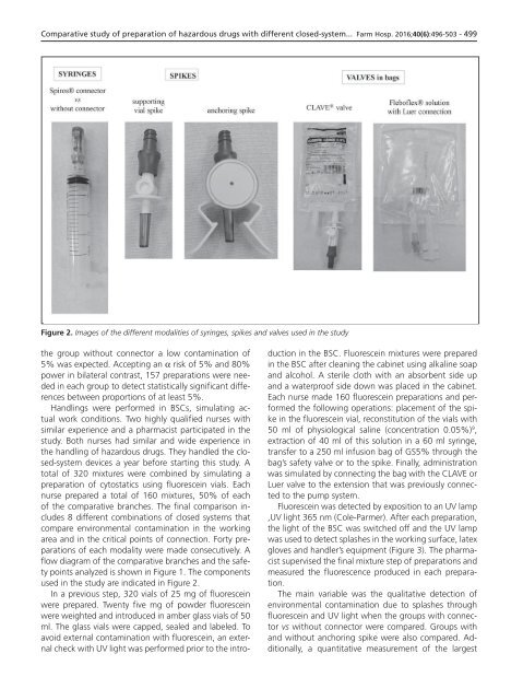
Figure 2. Images of the different modalities of syringes, spikes and valves used in the study<br />
the group without connector a low contamination of<br />
5% was expected. Accepting an α risk of 5% and 80%<br />
power in bilateral contrast, 157 preparations were needed<br />
in each group to detect statistically significant differences<br />
between proportions of at least 5%.<br />
Handlings were performed in BSCs, simulating actual<br />
work conditions. Two highly qualified nurses with<br />
similar experience and a pharmacist participated in the<br />
study. Both nurses had similar and wide experience in<br />
the handling of hazardous drugs. They handled the closed-system<br />
devices a year before starting this study. A<br />
total of 320 mixtures were combined by simulating a<br />
preparation of cytostatics using fluorescein vials. Each<br />
nurse prepared a total of 160 mixtures, 50% of each<br />
of the comparative branches. The final comparison includes<br />
8 different combinations of closed systems that<br />
compare environmental contamination in the working<br />
area and in the critical points of connection. Forty preparations<br />
of each modality were made consecutively. A<br />
flow diagram of the comparative branches and the safety<br />
points analyzed is shown in Figure 1. The components<br />
used in the study are indicated in Figure 2.<br />
In a previous step, 320 vials of 25 mg of fluorescein<br />
were prepared. Twenty five mg of powder fluorescein<br />
were weighted and introduced in amber glass vials of 50<br />
ml. The glass vials were capped, sealed and labeled. To<br />
avoid external contamination with fluorescein, an external<br />
check with UV light was performed prior to the introduction<br />
in the BSC. Fluorescein mixtures were prepared<br />
in the BSC after cleaning the cabinet using alkaline soap<br />
and alcohol. A sterile cloth with an absorbent side up<br />
and a waterproof side down was placed in the cabinet.<br />
Each nurse made 160 fluorescein preparations and performed<br />
the following operations: placement of the spike<br />
in the fluorescein vial, reconstitution of the vials with<br />
50 ml of physiological saline (concentration 0.05%) 9 ,<br />
extraction of 40 ml of this solution in a 60 ml syringe,<br />
transfer to a 250 ml infusion bag of GS5% through the<br />
bag’s safety valve or to the spike. Finally, administration<br />
was simulated by connecting the bag with the CLAVE or<br />
Luer valve to the extension that was previously connected<br />
to the pump system.<br />
Fluorescein was detected by exposition to an UV lamp<br />
,UV light 365 nm (Cole-Parmer). After each preparation,<br />
the light of the BSC was switched off and the UV lamp<br />
was used to detect splashes in the working surface, latex<br />
gloves and handler’s equipment (Figure 3). The pharmacist<br />
supervised the final mixture step of preparations and<br />
measured the fluorescence produced in each preparation.<br />
The main variable was the qualitative detection of<br />
environmental contamination due to splashes through<br />
fluorescein and UV light when the groups with connector<br />
vs without connector were compared. Groups with<br />
and without anchoring spike were also compared. Additionally,<br />
a quantitative measurement of the largest