Cancer du sein et micro-environnement tumoral: rôle de la protéase ...
Cancer du sein et micro-environnement tumoral: rôle de la protéase ...
Cancer du sein et micro-environnement tumoral: rôle de la protéase ...

Create successful ePaper yourself
Turn your PDF publications into a flip-book with our unique Google optimized e-Paper software.
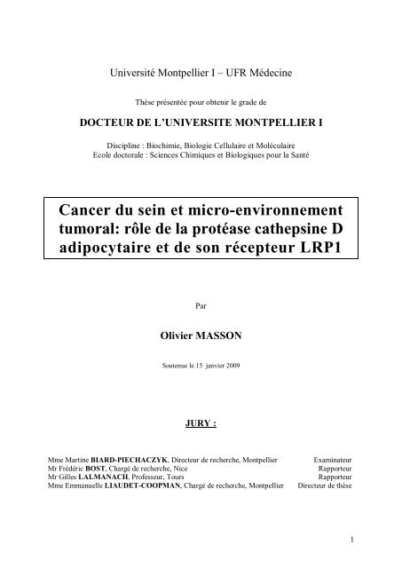
Université Montpellier I UFR Mé<strong>de</strong>cineThèse présentée pour obtenir le gra<strong>de</strong> <strong>de</strong>DOCTEUR DE LUNIVERSITE MONTPELLIER IDiscipline : Biochimie, Biologie Cellu<strong>la</strong>ire <strong>et</strong> Molécu<strong>la</strong>ireEcole doctorale : Sciences Chimiques <strong>et</strong> Biologiques pour <strong>la</strong> Santé<strong>Cancer</strong> <strong>du</strong> <strong>sein</strong> <strong>et</strong> <strong>micro</strong>-<strong>environnement</strong><strong>tumoral</strong>: <strong>rôle</strong> <strong>de</strong> <strong>la</strong> <strong>protéase</strong> cathepsine Dadipocytaire <strong>et</strong> <strong>de</strong> son récepteur LRP1ParOlivier MASSONSoutenue le 15 janvier 2009JURY :Mme Martine BIARD-PIECHACZYK, Directeur <strong>de</strong> recherche, MontpellierMr Frédéric BOST, Chargé <strong>de</strong> recherche, NiceMr Gilles LALMANACH, Professeur, ToursMme Emmanuelle LIAUDET-COOPMAN, Chargé <strong>de</strong> recherche, MontpellierExaminateurRapporteurRapporteurDirecteur <strong>de</strong> thèse1
RemerciementsJe voudrais en premier lieu madresser aux membres <strong>du</strong> jury : Martine Biard-Piechaczyk, Gilles Lalmanach <strong>et</strong> Frédéric Bost pour les remercier davoir accepté <strong>de</strong> lire <strong>et</strong>dévaluer ce travail.Je tiens à remercier chaleureusement le Dr Emmanuelle Liaud<strong>et</strong>-Coopman pourmavoir accueilli dans son <strong>la</strong>boratoire <strong>et</strong> sans qui ce travail naurait pu voir le jour. Merci pourton encadrement, ton engouement, ta disponibilité, ta joie <strong>de</strong> vivre <strong>et</strong> ta gentillesse. Grâce àtes gran<strong>de</strong>s qualités <strong>de</strong> scientifique <strong>et</strong> <strong>de</strong> pédagogue tu mas amené à toujours plus <strong>de</strong> rigueur<strong>et</strong> <strong>de</strong> précision dans mon travail. Tu mas formé, appris à mener à bien un proj<strong>et</strong>. Tu maségalement offert <strong>la</strong> chance <strong>de</strong> partir 3 mois à Vancouver ; c<strong>et</strong>te expérience fut trèsenrichissante tant sur le p<strong>la</strong>n professionnel que personnel. Je te remercie enfin <strong>de</strong> mavoirpermis <strong>de</strong> travailler dans <strong>de</strong>s conditions matérielles <strong>et</strong> humaines optimales.Bien-sûr, je remercie lensemble <strong>de</strong> léquipe. Durant ces 3 années <strong>de</strong> thèse, jai eu <strong>la</strong>chance <strong>de</strong> travailler avec <strong>de</strong>s personnes merveilleuses. Dès mon arrivée dans léquipe, vousmavez accueilli à bras ouvert <strong>et</strong> avez tout fait pour que c<strong>et</strong>te thèse se passe dans lesmeilleures conditions.Christine, tu mas beaucoup appris en ce qui concerne <strong>la</strong> culture cellu<strong>la</strong>ire <strong>et</strong> <strong>la</strong> biochimie,mais je noublierai pas non plus ta gentillesse <strong>et</strong> tes talents <strong>de</strong> cuisinière (sacrée mousse auchoco<strong>la</strong>t !) ainsi que nos discutions sur les expressions en patois (à bisto <strong>de</strong> nas)!Danielle, je te remercie pour tout ce que tu mas appris dans le domaine <strong>de</strong> <strong>la</strong> biologiemolécu<strong>la</strong>ire, mais je gar<strong>de</strong>rai également en mémoire tes encouragements, tes conseils toujoursplein <strong>de</strong> bons sens <strong>et</strong> nos discutions touchant à tous les suj<strong>et</strong>s Valérie, merci davoir toujours prêté une oreille attentive à mes questions, <strong>de</strong> têtre intéresséeà mon travail <strong>et</strong> davoir agrémenté le tout <strong>du</strong>ne bonne dose dhumour.Mé<strong>la</strong>nie, je noublierai pas ta gentillesse, ta spontanéité <strong>et</strong> ton dynamisme.Sophie, <strong>la</strong> « Top Top Woman », tu es arrivée <strong>de</strong>puis moins <strong>du</strong>n an, pourtant jai limpression<strong>de</strong> te connaître <strong>de</strong>puis le début <strong>de</strong> ma thèse. Une complicité sest rapi<strong>de</strong>ment installée entrenous, <strong>et</strong> tu as su mécouter dans les moments ou jen avais le plus besoin. Merci pour tesconseils avisés (sur le p<strong>la</strong>n professionnel mais aussi personnel), ta sympathie, ton amitié.Anne-Sophie, merci pour ta gentillesse <strong>et</strong> ta bonne humeur.Jai beaucoup appris à vos côtés, je ne vous oublierai pas, jai passé 3 années merveilleuses,on ne peut pas rêver <strong>du</strong>n meilleur <strong>environnement</strong> <strong>de</strong> travail, cest <strong>la</strong> « dream team » !
Un grand merci à tous mes col<strong>la</strong>borateurs, pour mavoir conseillé <strong>et</strong> fait partager leurconnaissances sur les adipocytes <strong>et</strong> sans qui c<strong>et</strong>te thèse ne serait pas ce quelle est, <strong>et</strong> toutparticulièrement à Carine Chavey, Catherine Mueller <strong>et</strong> Philippe Val<strong>et</strong>.Merci aussi à tous mes collègues <strong>et</strong> amis <strong>du</strong> <strong>la</strong>boratoire qui se reconnaîtront ici. Je leurexprime ma profon<strong>de</strong> sympathie <strong>et</strong> leur souhaite beaucoup <strong>de</strong> bien. Jai une pensée pour tousmes collègues <strong>et</strong> amis étudiants avec qui jai partagé <strong>de</strong> bons moments : Cycy, Iréna, Emilie,Marie, Stef, Didier, <strong>et</strong> le c<strong>la</strong>n <strong>de</strong> <strong>la</strong> cantine, Hayat, Mayssa, Aurélie (Louloute), Elsa,Laurence, Vincent (<strong>la</strong> cantine cest lécole <strong>de</strong> <strong>la</strong> vie!), ainsi que Max <strong>et</strong> Samir (même si vousen avez gros, je vous considèrerai toujours en tant que tel!).Je tiens également à remercier les personnes qui mont encadré <strong>du</strong>rant mes stages <strong>de</strong>master <strong>et</strong> qui mont donné le goût <strong>de</strong> <strong>la</strong> recherche. Un grand merci au Dr Martine Biard-Piechaczyk, pour mavoir accueilli lors <strong>de</strong> mon Master2, ainsi quà toute léquipe <strong>de</strong> <strong>la</strong> mortcellu<strong>la</strong>ire, <strong>et</strong> tout particulièrement à Lucile qui ma soutenu <strong>et</strong> poussé à faire c<strong>et</strong>te thèse aumoment où jen avais le plus besoin <strong>et</strong> où je doutais le plus <strong>de</strong> moi.David, mon premier encadrant (tu mas <strong>la</strong>issé te vouvoyer pendant 2 semaines mais je ne tentiens pas rigueur), tu occupes une p<strong>la</strong>ce particulière car tu mas permis <strong>de</strong> découvrir le mon<strong>de</strong><strong>de</strong> <strong>la</strong> recherche <strong>et</strong> si jen suis là aujourdhui, cest aussi grâce à toi. Jai également une penséepour mes anciens collègues qui sont <strong>de</strong>venus par <strong>la</strong> suite <strong>de</strong>s partenaires <strong>de</strong> jorki <strong>et</strong> enfin <strong>de</strong>samis qui me sont chers, Patrick <strong>et</strong> Stephan (mon Patou <strong>et</strong> mon Poussin).Jai également une pensée pour tous ceux que jai rencontrés <strong>et</strong> qui mont suivi <strong>du</strong>ranttoutes ces années détu<strong>de</strong>s. Je pense notamment à Ben, David, Georges, Arnaud, Olivier (leMaury, un sacré coureur <strong>de</strong> fond), Adri, Amandine, <strong>et</strong> au San<strong>de</strong>rs, une sacrée ban<strong>de</strong> <strong>de</strong>copains! Merci pour tous ces bons moments <strong>et</strong> ces soirées danthologies !!!!!Enfin, je tiens à remercier ma famille (Maman, Papa, Mamie, Juju, Tatie) qui masoutenu sans relâche <strong>du</strong>rant toutes ces années détu<strong>de</strong>s dans les bons <strong>et</strong> les mauvais moments.Merci pour <strong>la</strong>mour que vous me témoignez chaque jour, votre présence <strong>et</strong> pour mavoirtoujours encouragé dans mes choix. Merci à toute <strong>la</strong> p<strong>et</strong>ite famille, en espérant quellesagrandisse encore (Juju <strong>et</strong> Sophie...). Merci à vous tous pour mavoir offert ces momentsuniques qui nous rappellent qui on est <strong>et</strong> doù lon vient.3
RésuméLaspartyl <strong>protéase</strong> cathepsine D, surexprimée <strong>et</strong> hyper-sécrétée par les cellulesépithéliales cancéreuses mammaires est un facteur <strong>de</strong> mauvais pronostic <strong>de</strong>s cancers <strong>du</strong> <strong>sein</strong> <strong>et</strong>stimule <strong>la</strong> prolifération <strong>de</strong>s cellules cancéreuses <strong>et</strong> <strong>la</strong> formation <strong>de</strong>s métastases. Elle affecteégalement le <strong>micro</strong>-<strong>environnement</strong> <strong>tumoral</strong> en in<strong>du</strong>isant <strong>la</strong> croissance invasive <strong>de</strong>sfibrob<strong>la</strong>stes. Les travaux <strong>de</strong> léquipe ont montré que le LDL-receptor re<strong>la</strong>ted protein 1, LRP1,est le récepteur fibrob<strong>la</strong>stique <strong>de</strong> <strong>la</strong> cathepsine D. LRP1 est fortement exprimé par lesadipocytes. Les étu<strong>de</strong>s cliniques indiquent que lobésité est un facteur <strong>de</strong> risque dans <strong>de</strong>nombreux cancers, dont le cancer <strong>du</strong> <strong>sein</strong> chez <strong>la</strong> femme ménopausée.Dans c<strong>et</strong>te thèse, nous avons étudié le <strong>rôle</strong> <strong>de</strong> <strong>la</strong> cathepsine D <strong>et</strong> <strong>du</strong> LRP1 dans les adipocytes,type cellu<strong>la</strong>ire prédominant <strong>du</strong> <strong>micro</strong>-<strong>environnement</strong> <strong>tumoral</strong> mammaire. Nos résultatsindiquent une surexpression <strong>de</strong> <strong>la</strong> cathepsine D <strong>et</strong> <strong>du</strong> LRP1 dans le tissu adipeux humain <strong>et</strong>murin obèse. De plus, lexpression <strong>de</strong> <strong>la</strong> cathepsine D est augmentée pendant <strong>la</strong>différenciation adipocytaire. Finalement, lextinction <strong>de</strong> lexpression <strong>de</strong> <strong>la</strong> cathepsine D <strong>et</strong> <strong>du</strong>LRP1 inhibe <strong>la</strong>dipogenèse indiquant leurs <strong>rôle</strong>s clefs dans ce processus.Lensemble <strong>de</strong> ces résultats suggère que <strong>la</strong> cathepsine D <strong>et</strong> son récepteur LRP1 pourraient être<strong>de</strong>s cibles thérapeutiques potentielles dans le traitement <strong>de</strong> lobésité.Mots-clésAdipocytes, cancer <strong>du</strong> <strong>sein</strong>, cathepsine D, LRP1, <strong>micro</strong>-<strong>environnement</strong> <strong>tumoral</strong>, obésité.Laboratoire daccueilINSERM U896 cathepsines, autophagie <strong>et</strong> cancerInstitut <strong>de</strong> Recherche en Cancérologie <strong>de</strong> Montpellier208 rue <strong>de</strong>s apothicaires34298 Montpellier France4
TitleBreast cancer and <strong>tumoral</strong> <strong>micro</strong>-environment : Role of the cathepsin-D protease and its LRP1receptor in adipocytes.SummaryThe aspartyl protease cathepsin D, overexpressed and hyper-secr<strong>et</strong>ed by epithelialbreast cancer cells is a poor prognosis factor in breast cancers and stimu<strong>la</strong>tes cancer cellgrowth and m<strong>et</strong>astasis formation. It also affects the tumor <strong>micro</strong>environment, in<strong>du</strong>cing thefibrob<strong>la</strong>sts invasive outgrowth. Our works have shown that the LDL receptor-re<strong>la</strong>ted protein1,LRP1, is the fibrob<strong>la</strong>stic receptor for cathepsin D. LRP1 is highly expressed in adipocytes.Clinical studies indicate that obesity is a risk factor in many cancers, including breast cancerin postmenopausal women.During this thesis, we studied the role of cathepsin D and LRP1 in adipocytes, which are theprominent cell type in the tumor <strong>micro</strong>environment of breast cancers. Our results indicate thatcathepsin D and LRP1 are overexpressed in human and mouse obese adipose tissue.Furthermore, the expression of cathepsin D is increased <strong>du</strong>ring adipocyte differentiation.Finally, the inhibition of the cathepsin D and LRP1 expression inhibits adipogenesisindicating their key role in this process.All these results suggest that cathepsin D and its receptor LRP1 could be potential therapeutictarg<strong>et</strong>s in the treatment of obesity.5
PUBLICATIONS INCLUSES DANS LA THESE1. Olivier Masson, Carine Chavey, Cédric Dray, Aline Meulle, Danielle Daviaud, DidierQuilliot, Catherine Muller, Philippe Val<strong>et</strong>, Emmanuelle Liaud<strong>et</strong>-Coopman: LRP1 receptorcontrols adipogenesis and is up-regu<strong>la</strong>ted in human and mouse obese adipose tissue. (PLoSOne. 2009 Oct 12;4(10):e7422.)2. Olivier Masson, Christine Prébois, Carine Chavey, Cédric Dray, Aline Meulle, DanielleDaviaud, Didier Quilliot, Catherine Muller, Philippe Val<strong>et</strong>, Emmanuelle Liaud<strong>et</strong>-Coopman:Cathepsin D, a key factor in breast cancer, controls adipogenesis and is up-regu<strong>la</strong>ted in obeseadipose tissue. (soumis pour publication)6
<strong>Cancer</strong> <strong>du</strong> <strong>sein</strong> <strong>et</strong> <strong>micro</strong>-<strong>environnement</strong><strong>tumoral</strong>: <strong>rôle</strong> <strong>de</strong> <strong>la</strong> <strong>protéase</strong> cathepsine Dadipocytaire <strong>et</strong> <strong>de</strong> son récepteur LRP17
TABLE DES MATIERESA. BUT DE LA THESE ....................................................................................................... 10But <strong>de</strong> <strong>la</strong> thèse ............................................................................................................................. 11B. INTRODUCTION ........................................................................................................... 12I. Le tissu adipeux ....................................................................................................................... 131) Généralités ........................................................................................................................................ 132) Ladipocyte ....................................................................................................................................... 15a. Morphologie .................................................................................................................................. 15b. La lipogenèse ................................................................................................................................ 16c. La lipolyse..................................................................................................................................... 19d. Ladipocyte : une cellule sécrétrice ................................................................................................ 223) Ladipogenèse ................................................................................................................................... 24a. Les différentes étapes .................................................................................................................... 24b. Les facteurs <strong>de</strong> transcription .......................................................................................................... 26c. Rôle <strong>de</strong>s <strong>protéase</strong>s dans <strong>la</strong>dipogenèse ........................................................................................... 284) Obésité, <strong>micro</strong>-<strong>environnement</strong> <strong>et</strong> cancer ............................................................................................. 30a. Le <strong>micro</strong>-<strong>environnement</strong> ................................................................................................................ 30b. Obésité <strong>et</strong> cancer ........................................................................................................................... 32II. La cathepsine-D ..................................................................................................................... 341) Synthèse, maturation <strong>et</strong> adressage lysosomal <strong>de</strong> <strong>la</strong> cath-D .................................................................. 34a. Régu<strong>la</strong>tion <strong>de</strong> <strong>la</strong> transcription <strong>de</strong> <strong>la</strong> cath-D ..................................................................................... 34b. Structure <strong>et</strong> maturation protéolytique <strong>de</strong> <strong>la</strong> cath-D .......................................................................... 35c. Adressage lysosomal ..................................................................................................................... 372) Fonctions <strong>de</strong> <strong>la</strong> cath-D ....................................................................................................................... 40a. Dans <strong>la</strong> physiologie ....................................................................................................................... 40b. Dans les pathologies ...................................................................................................................... 423) Rôle <strong>de</strong> <strong>la</strong> cath-D dans le cancer <strong>du</strong> <strong>sein</strong> ............................................................................................. 42a. Expression dans les lignées <strong>de</strong> cancer <strong>du</strong> <strong>sein</strong> ................................................................................. 42b. Facteur pronostic ........................................................................................................................... 43c. Rôles <strong>et</strong> mécanismes daction dans le cancer .................................................................................. 444) Substrats <strong>et</strong> partenaires ...................................................................................................................... 48a. Les substrats <strong>et</strong> partenaires <strong>de</strong> <strong>la</strong> cath-D ......................................................................................... 48b. I<strong>de</strong>ntification <strong>de</strong> nouveaux substrats <strong>de</strong> <strong>la</strong> cath-D ........................................................................... 48III. Le récepteur LRP1 ............................................................................................................... 541) Organisation structurale ..................................................................................................................... 542) Trafic intra-cellu<strong>la</strong>ire ......................................................................................................................... 563) Mécanismes daction ......................................................................................................................... 56a. La phosphory<strong>la</strong>tion ........................................................................................................................ 56b. Le RIP .......................................................................................................................................... 574) Fonctions ........................................................................................................................................... 588
C. PRESENTATION DU TRAVAIL DE THESE ............................................................... 61I. Etu<strong>de</strong> <strong>du</strong> <strong>rôle</strong> <strong>de</strong> <strong>la</strong> cathepsine D dans les adipocytes ............................................................. 621) Intro<strong>du</strong>ction :..................................................................................................................................... 622) Article 1: ........................................................................................................................................... 63II. Etu<strong>de</strong> <strong>du</strong> <strong>rôle</strong> <strong>du</strong> LRP1 dans les adipocytes .......................................................................... 641) Intro<strong>du</strong>ction :..................................................................................................................................... 642) Article 2: ........................................................................................................................................... 66D. CONCLUSION ET PERSPECTIVES ............................................................................ 67CONCLUSION ........................................................................................................................... 68PERSPECTIVES ........................................................................................................................ 70E. REFERENCES ............................................................................................................... 75F. ANNEXE......................................................................................................................... 98Article 3: ............................................................................................................................................... 999
A. BUT DE LA THESE10
But <strong>de</strong> <strong>la</strong> thèseLa cathepsine-D (cath-D) est une aspartyl <strong>protéase</strong> lysosomale surexprimée <strong>et</strong> hypersécrétéepar un grand nombre <strong>de</strong> carcinomes (<strong>sein</strong>, ovaire, prostate, endomètre, colon, squameux).Cest un marqueur <strong>de</strong> mauvais pronostic <strong>de</strong>s cancers <strong>du</strong> <strong>sein</strong> associé à un risque augmenté <strong>de</strong>récidive. Nos travaux indiquent que <strong>la</strong> cath-D surexprimée par les cellules cancéreusesmammaires agit à différentes étapes <strong>de</strong> <strong>la</strong> progression <strong>tumoral</strong>e métastatique. Elle stimule <strong>de</strong>façon autocrine <strong>la</strong> prolifération <strong>de</strong>s cellules cancéreuses <strong>et</strong> <strong>la</strong> formation <strong>de</strong>s métastases. Deplus, elle joue un <strong>rôle</strong> déterminant dans le <strong>micro</strong>-<strong>environnement</strong> <strong>tumoral</strong> puisquelle agit <strong>de</strong>façon paracrine en stimu<strong>la</strong>nt <strong>la</strong> croissance invasive <strong>de</strong>s fibrob<strong>la</strong>stes. Des travaux récentsréalisés dans le <strong>la</strong>boratoire ont montré que <strong>la</strong> cath-D interagit avec le domaine extracellu<strong>la</strong>ire<strong>de</strong> <strong>la</strong> chaîne b <strong>du</strong> récepteur LRP1 (LDL receptor re<strong>la</strong>ted protein1). De façon intéressante,LRP1 est fortement exprimé par les adipocytes, un <strong>de</strong>s types cellu<strong>la</strong>ires prédominant <strong>du</strong><strong>micro</strong>-<strong>environnement</strong> <strong>tumoral</strong> dans le cancer <strong>du</strong> <strong>sein</strong>. De plus LRP1 est un récepteur<strong>de</strong>ndocytose appartenant à <strong>la</strong> famille <strong>de</strong>s LDL-récepteurs. C<strong>et</strong>te interaction est responsable<strong>de</strong> leff<strong>et</strong> mitogène <strong>de</strong> <strong>la</strong> cath-D sur <strong>la</strong> croissance invasive <strong>de</strong>s fibrob<strong>la</strong>stes. Des travauxrécents indiquent <strong>de</strong>s <strong>rôle</strong>s fondamentaux <strong>de</strong>s cystéines cathepsines dans <strong>la</strong> biologie <strong>de</strong><strong>la</strong>dipocyte. Par ailleurs, les enquêtes épidémiologiques révèlent que lobésité est un facteur<strong>de</strong> mauvais pronostic dans <strong>de</strong> nombreux cancers dont le cancer <strong>du</strong> <strong>sein</strong> chez <strong>la</strong> femmeménopausée. De nombreuses étu<strong>de</strong>s suggèrent que les adipocytes, via leur capacité à sécréter<strong>de</strong>s facteurs <strong>de</strong> croissance, <strong>de</strong> nombreuses cytokines <strong>et</strong>/ou <strong>de</strong>s composés <strong>de</strong> <strong>la</strong> matrice extracellu<strong>la</strong>ire,favorisent <strong>la</strong> progression <strong>tumoral</strong>e mammaire <strong>et</strong> pourraient jouer un <strong>rôle</strong>déterminant dans les étapes précoces <strong>de</strong> <strong>la</strong> carcinogénèse mammaire.Le but <strong>de</strong> ce travail <strong>de</strong> thèse a été détudier le <strong>rôle</strong> <strong>de</strong> <strong>la</strong> cath-D <strong>et</strong> <strong>de</strong> son récepteur LRP1 dans<strong>la</strong> biologie <strong>de</strong> <strong>la</strong>dipocyte, avec un aspect orienté vers lobésité vu le <strong>rôle</strong> clef <strong>de</strong> <strong>la</strong> cath-Ddans le cancer <strong>et</strong> le lien entre obésité <strong>et</strong> cancer.11
B. INTRODUCTION12
I. Le tissu adipeux1) GénéralitésLe tissu adipeux dérive embryologiquement <strong>de</strong>s cellules souches mésenchymateuses.Ces cellules souches sont pluripotentes <strong>et</strong> possè<strong>de</strong>nt <strong>la</strong> capacité <strong>de</strong> se différencier enadipocyte, en myocyte, en osteocyte, <strong>et</strong> en chondrocyte (Figure 1) (Markert <strong>et</strong> al., 2009; Park<strong>et</strong> al., 2008).MSC(Mesenchymal Stem Cell)White adipocyteprecursorBrown adipocyte/muscleprecursorOsteob<strong>la</strong>stChondrob<strong>la</strong>stWhite adipocyte Brown adipocyte Myocyte OsteocyteChondrocyteFigure 1 : Origine <strong>du</strong> tissu adipeux (Adapté <strong>de</strong> Park <strong>et</strong> al., 2008)Les cellules souches mésenchymateuses sont pluripotentes <strong>et</strong> possè<strong>de</strong>nt <strong>la</strong> capacité <strong>de</strong> se différencieren différents types cellu<strong>la</strong>ires. Sous linfluence <strong>de</strong> différents facteurs <strong>de</strong> transcription (PPAR, Myo D,Run X2, ), ces cellules se différencieront en adipocyte b<strong>la</strong>nc ou brun, en myocyte, en ostéocyte, ouen chondrocyte.On distingue généralement 2 types <strong>de</strong> tissu adipeux qui diffèrent <strong>de</strong> par leur fonction <strong>et</strong> leurlocalisation (Figure 1). On r<strong>et</strong>rouve ainsi :1)-le tissu adipeux b<strong>la</strong>nc, qui est un organe <strong>de</strong> réserve énergétique. Lorsque les apportsénergétiques sont supérieurs aux dépenses <strong>de</strong> lorganisme, ce tissu perm<strong>et</strong> le stockage <strong>de</strong>lexcès calorique sous forme <strong>de</strong> triglycéri<strong>de</strong>s (également appelés triacylglycérols) par le13
processus <strong>de</strong> lipogenèse (Figure 2). A linverse, lorsque les apports caloriques sontinsuffisants, lorganisme mobilise ces triglycéri<strong>de</strong>s libérant ainsi <strong>de</strong>s aci<strong>de</strong>s gras, cest <strong>la</strong>lipolyse. Ces <strong>de</strong>rniers sont alors re<strong>la</strong>rgués dans <strong>la</strong> circu<strong>la</strong>tion pour être acheminés vers lestissus consommateurs en énergie (muscles, cur) afin dêtre oxydés dans les mitochondriesvia le processus <strong>de</strong> boxydation <strong>de</strong>s aci<strong>de</strong>s gras.ESTERIFICATIONGlycérol + 3 aci<strong>de</strong>s gras Triglycéri<strong>de</strong> + 3 H2OFigure 2 : Schéma <strong>de</strong> <strong>la</strong> formation <strong>du</strong>n triglycéri<strong>de</strong>Durant <strong>la</strong> formation <strong>du</strong>n triglycéri<strong>de</strong>, les trois fonctions alcools portées par le glycérol sestérifientavec <strong>la</strong> fonction aci<strong>de</strong> <strong>de</strong> trois aci<strong>de</strong>s gras. C<strong>et</strong>te réaction con<strong>du</strong>it à <strong>la</strong> formation <strong>du</strong>n triglycéri<strong>de</strong> <strong>et</strong> à<strong>la</strong> libération <strong>de</strong> trois molécules dH20.Le tissu adipeux b<strong>la</strong>nc joue également un <strong>rôle</strong> majeur au niveau <strong>de</strong> lorganisme puisquilsécrète différents facteurs (hormones) impliqués dans <strong>de</strong> nombreux processus physiologiquescomme <strong>la</strong> régu<strong>la</strong>tion <strong>de</strong> <strong>la</strong>ppétit, <strong>la</strong> réponse inf<strong>la</strong>mmatoire, <strong>la</strong>ngiogenèse (Lefterova andLazar, 2009).Chez lhomme, il est essentiellement situé sous <strong>la</strong> peau (graisse sous-cutanée) <strong>et</strong> dans <strong>la</strong> cavitéabdominale (graisse abdominale) <strong>et</strong> dans certains organes tels <strong>la</strong> prostate <strong>et</strong> le <strong>sein</strong>.2)-le tissu adipeux brun, qui cont<strong>rôle</strong> le processus <strong>de</strong> thermorégu<strong>la</strong>tion.Ce tissu contient moins <strong>de</strong> lipi<strong>de</strong>s que le tissu adipeux b<strong>la</strong>nc. Cependant les adipocytes brunssont riches en mitochondries qui possè<strong>de</strong>nt un pigment respiratoire <strong>de</strong> couleur brune quiconfère sa couleur au tissu. Ces <strong>de</strong>rnières expriment <strong>la</strong> protéine UCP1 (Uncoupling protein-1), un transporteur situé dans <strong>la</strong> membrane interne mitochondriale, qui leur perm<strong>et</strong> <strong>de</strong>pro<strong>du</strong>ire <strong>de</strong> <strong>la</strong> chaleur au lieu <strong>de</strong> fournir uniquement <strong>de</strong> lénergie sous forme dATP14
(A<strong>de</strong>nosine triphosphate). C<strong>et</strong>te protéine UCP1 perm<strong>et</strong> lentrée <strong>de</strong>s protons dans <strong>la</strong> matricemitochondriale <strong>et</strong> dissipe le gradient <strong>de</strong> protons généré par <strong>la</strong> chaîne respiratoire. L'énergielibérée par ce découp<strong>la</strong>ge entre <strong>la</strong> respiration cellu<strong>la</strong>ire <strong>et</strong> <strong>la</strong> phosphory<strong>la</strong>tion <strong>de</strong> l'ATP estdissipée sous forme <strong>de</strong> chaleur. Ce tissu est important chez le nouveau né car il perm<strong>et</strong> <strong>de</strong>résister au choc thermique dû à <strong>la</strong> naissance. Chez <strong>la</strong><strong>du</strong>lte il est très faiblement présent <strong>et</strong> sesitue autour <strong>de</strong> certains organes comme les reins, le cur ou encore le creux <strong>de</strong> <strong>la</strong>isselle.2) Ladipocytea. MorphologieLes adipocytes <strong>du</strong> tissu adipeux b<strong>la</strong>nc sont <strong>de</strong>s cellules sphériques, d'un diamètre <strong>de</strong>100 <strong>micro</strong>mètres voire plus. On r<strong>et</strong>rouve à lintérieur <strong>de</strong> ces cellules une volumineuse vacuolelipidique unique composée <strong>de</strong> triglycéri<strong>de</strong>s (Schaefer <strong>et</strong> al., 1983), repoussant à <strong>la</strong> périphérie<strong>de</strong> <strong>la</strong> cellule le noyau, le cytop<strong>la</strong>sme ainsi que certains organites cellu<strong>la</strong>ires (appareil <strong>de</strong> Golgi,réticulum endop<strong>la</strong>smique granu<strong>la</strong>ire, réticulum endop<strong>la</strong>smique lisse, mitochondries) (Figure3). Les adipocytes présentent une seule gran<strong>de</strong> goutte lipidique <strong>et</strong> <strong>de</strong> ce fait le tissu adipeuxb<strong>la</strong>nc est aussi appelé unilocu<strong>la</strong>ire à linverse <strong>du</strong> tissu adipeux brun qui est multilocu<strong>la</strong>ire.VacuolelipidiquenoyaumitochondrieFigure 3 : Schéma <strong>de</strong> <strong>micro</strong>scopie électronique représentant un adipocyte b<strong>la</strong>nc(Daprès Histologie, les tissus, Poirier <strong>et</strong> al., Masson 2006)Les adipocytes b<strong>la</strong>ncs possè<strong>de</strong>nt une volumineuse <strong>et</strong> unique vacuole lipidique qui occupe <strong>la</strong> majeurepartie <strong>du</strong> cytop<strong>la</strong>sme, repoussant à <strong>la</strong> périphérie <strong>de</strong> ces cellules le noyau ainsi que les organitescellu<strong>la</strong>ires.15
Le tissu adipeux b<strong>la</strong>nc est organisé en lobules délimités par <strong>de</strong>s septa <strong>de</strong> tissu conjonctif lâcherichement vascu<strong>la</strong>risés <strong>et</strong> innervés. Dans chaque lobule, les adipocytes sont serrés les uns auxautres <strong>et</strong> ils présentent une forme polygonale (Figure 4).Figure 4 : Coupe histologique <strong>de</strong> tissu adipeux b<strong>la</strong>ncLe tissu adipeux b<strong>la</strong>nc est divisé en lobules par <strong>de</strong> fines cloisons <strong>de</strong> tissu conjonctif fibreux lâche. Ceslobules qui contiennent les adipocytes sont richement innervés <strong>et</strong> vascu<strong>la</strong>risés. Dans le tissu adipeuxb<strong>la</strong>nc, les adipocytes, tassés les uns contre les autres, prennent une forme polyédrique.b. La lipogenèseLa lipogenèse correspond à lensemble <strong>de</strong>s voies métaboliques con<strong>du</strong>isant à <strong>la</strong>formation <strong>de</strong>s triglycéri<strong>de</strong>s dans les adipocytes (Figure 7). En eff<strong>et</strong>, les adipocytes accumulentlessentiel <strong>de</strong>s graisses sous forme <strong>de</strong> triglycéri<strong>de</strong>s (Arner, 2005; Barbaras <strong>et</strong> al., 1985). Cestriglycéri<strong>de</strong>s sont constitués <strong>de</strong> trois molécules daci<strong>de</strong>s gras reliées à un glycérol par <strong>de</strong>sliaisons esters (Figure 2). Ils constituent une forme <strong>de</strong> mise en réserve hautement concentréeen énergie. En eff<strong>et</strong>, lors <strong>de</strong> <strong>la</strong> lipolyse, le ren<strong>de</strong>ment <strong>de</strong> loxydation complète <strong>de</strong>s aci<strong>de</strong>s grasest <strong>de</strong>nviron 9 kcal/g, par comparaison au 4 kcal/g environ <strong>de</strong>s gluci<strong>de</strong>s <strong>et</strong> <strong>de</strong>s protéines.Les aci<strong>de</strong>s gras proviennent majoritairement <strong>de</strong> <strong>la</strong> circu<strong>la</strong>tion via les apports alimentaires, <strong>et</strong>dans une faible proportion <strong>de</strong> <strong>la</strong> synthèse <strong>de</strong> novo <strong>de</strong>s aci<strong>de</strong>s gras (Figure 5).16
C<strong>et</strong>te synthèse <strong>de</strong> novo a lieu dans les adipocytes <strong>et</strong> dans les hépatocytes, mais restecependant à un niveau très faible (Large <strong>et</strong> al., 2004; Sjostrom, 1972). La contribution <strong>de</strong>sadipocytes à <strong>la</strong> néosynthèse <strong>de</strong>s aci<strong>de</strong>s gras est moins bien définie chez lhomme que chez <strong>la</strong>souris (Large <strong>et</strong> al., 2004). Cependant les enzymes clés dans <strong>la</strong> synthèse <strong>de</strong>s aci<strong>de</strong>s gras, <strong>la</strong>fatty acid synthase <strong>et</strong> <strong>la</strong>c<strong>et</strong>yl coenzyme A carboxy<strong>la</strong>se 1 sont exprimés dans le tissu adipeuxhumain (Angel and Bray, 1979; L<strong>et</strong>exier <strong>et</strong> al., 2003; Shrago <strong>et</strong> al., 1971; Shrago <strong>et</strong> al., 1969).La principale source daci<strong>de</strong>s gras provient <strong>du</strong> p<strong>la</strong>sma soit à partir <strong>de</strong>s aci<strong>de</strong>s gras nonestérifiés liés à <strong>la</strong>lbumine p<strong>la</strong>smatique, soit à partir daci<strong>de</strong>s gras estérifiés incorporés dansles lipoprotéines riches en triglycéri<strong>de</strong>s (principalement les chilo<strong>micro</strong>ns <strong>et</strong> les VLDLs (VeryLow Density Lipoproteins)) <strong>du</strong>rant <strong>la</strong> pério<strong>de</strong> qui suit <strong>la</strong> prise alimentaire (Large <strong>et</strong> al., 2004)(Figure 7). Ces lipoprotéines riches en triglycéri<strong>de</strong>s vont interagir avec <strong>de</strong>s récepteurs <strong>de</strong> <strong>la</strong>famille <strong>de</strong>s LDLs, principalement le récepteur aux VLDLs qui est exprimé au niveau <strong>du</strong> tissuadipeux <strong>et</strong> qui lie les lipoprotéines riches en Apolipoprotéine E tels que les VLDL <strong>et</strong> leschylo<strong>micro</strong>ns. Les aci<strong>de</strong>s gras sont re<strong>la</strong>rgués à partir <strong>de</strong>s triglycéri<strong>de</strong>s présents dans ceslipoprotéines. Cest <strong>la</strong> lipoprotéine lipase qui hydrolyse ces triglycéri<strong>de</strong>s (Mead <strong>et</strong> al., 2002).Son expression ainsi que son activité sont augmentées après <strong>la</strong> prise alimentaire. C<strong>et</strong>teenzyme travaille <strong>de</strong> concert avec le récepteur aux VLDLs. Des étu<strong>de</strong>s chez <strong>de</strong>s sourisdéficientes en récepteur aux VLDLs présentent une masse graisseuse ré<strong>du</strong>ite, ainsi quunerésistance à lobésité, soulignant son <strong>rôle</strong> au niveau <strong>de</strong> <strong>la</strong> lipogenèse (Goudriaan <strong>et</strong> al., 2001).Le glycérol-3P (Glycérol-3Phosphate) qui sestérifie avec les aci<strong>de</strong>s gras libres pour formerles triglycéri<strong>de</strong>s provient soit <strong>de</strong> <strong>la</strong> glycérogenèse (à partir <strong>de</strong> substrat tel le pyruvate), soit <strong>du</strong>glucose après <strong>la</strong> première étape <strong>de</strong> <strong>la</strong> glycolyse (Reshef <strong>et</strong> al., 2003). Le glucose rentre dansles adipocytes grâce aux transporteurs <strong>de</strong> glucose situés au niveau <strong>de</strong> <strong>la</strong> membrane p<strong>la</strong>smique,Glut1 <strong>et</strong> Glut4 (Figure 7).La principale hormone régu<strong>la</strong>nt <strong>la</strong> lipogenèse est linsuline. Elle perm<strong>et</strong> daugmenter lentrée<strong>de</strong> glucose dans <strong>la</strong>dipocyte par le recrutement <strong>de</strong> transporteur <strong>de</strong> glucose (Glut4) au niveau<strong>de</strong> <strong>la</strong> membrane p<strong>la</strong>smique (Bryant <strong>et</strong> al., 2002; Saltiel and Kahn, 2001). Elle inhibeégalement <strong>la</strong> lipolyse en stimu<strong>la</strong>nt <strong>la</strong> phosphodiesterase 3B (PDE3B) responsable <strong>de</strong>lhydrolyse <strong>de</strong> lAMPc (A<strong>de</strong>nosine MonoPhosphate cyclique) (Fukao <strong>et</strong> al., 2004; Langin,2006b). Linsuline peut également inhiber <strong>la</strong> lipolyse en activant <strong>la</strong> protéine phosphatase-1(PP1), qui déphosphoryle <strong>la</strong> lipase hormono-sensible (HSL Hormone-Sensitive Lipase), uneprotéine qui joue un <strong>rôle</strong> clé dans <strong>la</strong> lipolyse, <strong>la</strong> rendant ainsi inactive (voir chapitre sur <strong>la</strong>lipolyse).18
c. La lipolyseLa lipolyse est le processus inverse <strong>de</strong> <strong>la</strong> lipogenèse. Elle a lieu principalement lors<strong>de</strong>s pério<strong>de</strong>s <strong>de</strong> jeûne. Elle est régulée par les hormones telles les catécho<strong>la</strong>mines <strong>et</strong> leglucagon (Large and Arner, 1998) (Figure 7). En pério<strong>de</strong> <strong>de</strong> jeûne, ce sont principalement lescatécho<strong>la</strong>mines qui sont responsables <strong>de</strong> <strong>la</strong> stimu<strong>la</strong>tion <strong>de</strong> <strong>la</strong> lipolyse (Langin, 2006a). Ceshormones atteignent les adipocytes via <strong>la</strong> circu<strong>la</strong>tion comme <strong>la</strong>drenaline (epinephrine) ou vialinnervation sympathique comme <strong>la</strong> noradrénaline (norepinephrine). Leur eff<strong>et</strong> lipolytiqueest médié par les récepteurs adrénergiques. Ces récepteurs sont couplés aux protéines G <strong>et</strong>leur activation par les catécho<strong>la</strong>mines engendre une augmentation <strong>de</strong> <strong>la</strong>ctivité a<strong>de</strong>ny<strong>la</strong>tecyc<strong>la</strong>se. C<strong>et</strong>te stimu<strong>la</strong>tion <strong>de</strong> <strong>la</strong><strong>de</strong>ny<strong>la</strong>te cyc<strong>la</strong>se entraine une augmentation <strong>de</strong> <strong>la</strong>concentration dAMPc intracellu<strong>la</strong>ire, con<strong>du</strong>isant à <strong>la</strong>ctivation <strong>de</strong> <strong>la</strong> PKA (Protéine KinaseA) dépendant <strong>de</strong> lAMPc. La PKA phosphoryle alors <strong>la</strong> périlipine, une protéine associée à <strong>la</strong>gouttel<strong>et</strong>te lipidique essentielle pour <strong>la</strong> régu<strong>la</strong>tion <strong>de</strong> <strong>la</strong> lipolyse, ainsi que <strong>la</strong> lipasehormonosensible (HSL) qui se r<strong>et</strong>rouve également activée <strong>et</strong> hydrolyse les triglycéri<strong>de</strong>s. Lestriglycéri<strong>de</strong>s sont alors hydrolysés en aci<strong>de</strong>s gras <strong>et</strong> glycérol (Bjorntorp and Ostman, 1971)puis ces <strong>de</strong>rniers sont re<strong>la</strong>rgués dans <strong>la</strong> circu<strong>la</strong>tion <strong>et</strong> transportés vers dautres tissus,principalement vers le foie pour le glycérol <strong>et</strong> vers les muscles <strong>et</strong> le cur pour les aci<strong>de</strong>s gras(Frayn <strong>et</strong> al., 2003) où ils seront dégradés dans les mitochondries via le processus <strong>de</strong> oxydation <strong>de</strong>s aci<strong>de</strong>s gras afin <strong>de</strong> fournir <strong>de</strong> lénergie sous forme dATP (Figure 6).En conclusion, une fine régu<strong>la</strong>tion <strong>de</strong> <strong>la</strong> lipogenèse <strong>et</strong> <strong>de</strong> <strong>la</strong> lipolyse est déterminante pour <strong>la</strong>maintenance <strong>de</strong> lhoméostasie énergétique.19
ABFigure 6 : Schéma <strong>de</strong> <strong>la</strong> oxydation <strong>de</strong>s aci<strong>de</strong>s grasLes aci<strong>de</strong>s gras sont dégradés par oxydation au niveau <strong>du</strong> carbone ( oxydation).(A) La première étape est <strong>la</strong>ctivation <strong>de</strong>s aci<strong>de</strong>s gras par liaison avec le Coenzyme A.Lactivation est effectuée dans <strong>la</strong> membrane mitochondriale externe, <strong>la</strong> dégradation (oxydation) dans<strong>la</strong> matrice mitochondriale.(B)Un acyl-CoA saturé est dégradé par une séquence récurrente <strong>de</strong> 4 réactions:1) Oxydation par le FAD; 2) Hydratation; 3) Oxydation par le NAD+; 4) Thiolyse par le CoA.20
glycolysisPDE3BPP1Figure 7 : Lipogenèse <strong>et</strong> lipolyse (Adapté <strong>de</strong> Vazquez-Ve<strong>la</strong> <strong>et</strong> al. 2008)Ce schéma représente les différentes voies con<strong>du</strong>isant à <strong>la</strong> lipogenèse <strong>et</strong> à <strong>la</strong> lipolyse dans lesadipocytes. Lexcès <strong>de</strong> glucose est oxydé par <strong>la</strong> glycolyse en ac<strong>et</strong>yl-CoA dans <strong>la</strong>dipocyte, puisconverti en aci<strong>de</strong> gras pour être estérifié dans le réticulum endop<strong>la</strong>smique en triglycéri<strong>de</strong> (TG). Cestriglycéri<strong>de</strong>s sont ensuite dirigés dans <strong>la</strong> gouttel<strong>et</strong>te lipidique. Les aci<strong>de</strong>s gras captés à partir <strong>de</strong>slipoprotéines sont également estérifiés en TG <strong>et</strong> stockés dans <strong>la</strong> gouttel<strong>et</strong>te lipidique. En condition <strong>de</strong>jeûne, <strong>la</strong> lipolyse est activée par <strong>de</strong>s récepteurs couplés aux protéines G, ce qui entraîne uneaugmentation <strong>du</strong> taux dAMPc, con<strong>du</strong>isant à <strong>la</strong>ctivation <strong>de</strong> <strong>la</strong> Protéine Kinase A (PKA). C<strong>et</strong>te PKAphosphoryle <strong>la</strong> périlipine située dans <strong>la</strong> membrane <strong>de</strong> <strong>la</strong> gouttel<strong>et</strong>te lipidique, <strong>et</strong> également lHormoneSensitive Lipase (HSL), perm<strong>et</strong>tant son activation <strong>et</strong> son transfert à lintérieur <strong>de</strong> <strong>la</strong> gouttel<strong>et</strong>telipidique où elle hydrolysera les diglycéri<strong>de</strong>s (DG) (pro<strong>du</strong>its par <strong>la</strong>dipocyte triglyceri<strong>de</strong> lipase(ATGL)) en monoglycéri<strong>de</strong>s (MG). Ces MG seront hydrolysés en aci<strong>de</strong> gras <strong>et</strong> glycérol à lextérieur<strong>de</strong> <strong>la</strong> gouttel<strong>et</strong>te lipidique avant dêtre re<strong>la</strong>rgués dans <strong>la</strong> circu<strong>la</strong>tion <strong>et</strong> acheminés vers les tissusconsommateurs en énergie comme le foie, les muscles, le cur.21
d. Ladipocyte : une cellule sécrétriceLes adipocytes ont longtemps été considérés comme <strong>de</strong>s cellules <strong>de</strong> réserveénergétique re<strong>la</strong>tivement inertes. Cependant, les adipocytes sécrètent <strong>de</strong> multiples facteursappelés adipokines. Parmi ces adipokines, on r<strong>et</strong>rouve <strong>de</strong>s hormones, <strong>de</strong>s cytokines, <strong>et</strong>dautres protéines ayant <strong>de</strong>s fonctions biologiques spécifiques (Ahima, 2006; Vazquez-Ve<strong>la</strong> <strong>et</strong>al., 2008) (voir tableau1).Tableau 1 : Facteurs pro<strong>du</strong>its par le tissu adipeux b<strong>la</strong>nc(Daprès Ahima RS, 2006)Parmi les adipokines les mieux caractérisées, nous r<strong>et</strong>rouvons <strong>la</strong> leptine, <strong>la</strong>diponectine,<strong>la</strong>ngiotensinogène. Certaines <strong>de</strong> ces protéines ont <strong>de</strong>s eff<strong>et</strong>s bénéfiques (leptine <strong>et</strong>adiponectine) ou délétères (angiotensinogène) pour lorganisme.La première adipokine à avoir été caractérisée est <strong>la</strong> leptine par léquipe <strong>de</strong> Friedman <strong>et</strong> al. en1994 (Zhang <strong>et</strong> al., 1994). C<strong>et</strong>te protéine principalement sécrétée par les adipocytes, agit auniveau <strong>de</strong> lhypotha<strong>la</strong>mus <strong>et</strong> mo<strong>du</strong>le le poids corporel, <strong>la</strong> prise alimentaire, ainsi que le22
stockage <strong>de</strong>s graisses (Wozniak <strong>et</strong> al., 2009). La fonction <strong>de</strong> <strong>la</strong> leptine a été déterminée par<strong>de</strong>s étu<strong>de</strong>s sur <strong>de</strong>s souris obèses ayant <strong>de</strong>s dommages au niveau <strong>de</strong> lhypotha<strong>la</strong>mus. Cestravaux ont permis <strong>de</strong> montrer que <strong>la</strong> leptine cont<strong>rôle</strong> <strong>la</strong> croissance <strong>du</strong> tissu adipeux via sonaction au niveau <strong>du</strong> système nerveux central (Maffei <strong>et</strong> al., 1995). De plus, <strong>la</strong>dministrationlocale <strong>de</strong> leptine au niveau <strong>de</strong>s régions hypotha<strong>la</strong>miques ré<strong>du</strong>it <strong>la</strong> prise alimentaire <strong>et</strong> le poidscorporel chez les animaux (Campfield <strong>et</strong> al., 1995; Vazquez-Ve<strong>la</strong> <strong>et</strong> al., 2008). Cependant, leshauts niveaux <strong>de</strong> leptine r<strong>et</strong>rouvés chez les patients obèses naffectent pas <strong>la</strong> suppression <strong>de</strong><strong>la</strong>ppétit à cause <strong>de</strong> <strong>la</strong> résistance <strong>de</strong> lhormone dûe à une absence <strong>de</strong> signalisation <strong>du</strong> récepteurà <strong>la</strong> leptine ou un défaut <strong>de</strong> transport <strong>de</strong> <strong>la</strong> leptine au niveau <strong>de</strong> <strong>la</strong> barrière hématoencéphalique(Flier, 2004). En plus <strong>de</strong> son <strong>rôle</strong> majeur dans le métabolisme, elle joue un <strong>rôle</strong>dans dautres processus physiologiques comme limmunité, ou <strong>la</strong> fertilité (Huang and Li,2000).Une autre adipokine bien caractérisée est <strong>la</strong>diponectine. Cest une protéine <strong>de</strong> 30 kDasécrétée par le tissu adipeux qui a été i<strong>de</strong>ntifiée en 1995 (Scherer <strong>et</strong> al., 1995). Son expression<strong>et</strong> celle <strong>de</strong> ses récepteurs sont fortement diminuées chez <strong>la</strong> souris obèse, <strong>et</strong> son tauxp<strong>la</strong>smatique est inversement corrélé à <strong>la</strong> résistance à linsuline <strong>et</strong> au syndrôme métabolique(Yatagai <strong>et</strong> al., 2003). Ladiponectine augmente <strong>la</strong> sensibilité à linsuline, diminue linfluxdaci<strong>de</strong>s gras non estérifiés, <strong>et</strong> augmente loxydation <strong>de</strong>s aci<strong>de</strong>s gras par le foie <strong>et</strong> les muscles.De nombreuses étu<strong>de</strong>s ont également souligné son <strong>rôle</strong> protecteur dans <strong>la</strong>thérosclérose(Antonia<strong>de</strong>s <strong>et</strong> al., 2009; Han <strong>et</strong> al., 2009b; Kim <strong>et</strong> al., 2008).Les adipocytes <strong>du</strong> tissu adipeux b<strong>la</strong>nc représentent une source extra-hépatique majeuredangiotensinogène (AGT) (Ailhaud, 2006). Lhypertension est connue comme étant unecomplication fréquente liée à lobésité. Bien que les mécanismes par lesquels lexcès <strong>de</strong>graisse entraîne c<strong>et</strong>te hypertension soient mal connus, <strong>la</strong>ugmentation <strong>de</strong> <strong>la</strong> pro<strong>du</strong>ction dAGTpourrait contribuer à lélévation <strong>de</strong> <strong>la</strong> pression artérielle chez les patients obèses. De plus, <strong>de</strong>sétu<strong>de</strong>s menées chez <strong>de</strong>s souris déficientes en AGT ont montré que sa ré-expression dans l<strong>et</strong>issu adipeux in<strong>du</strong>it <strong>la</strong> présence dAGT dans le sang <strong>et</strong> <strong>la</strong> restauration <strong>du</strong>ne pression sanguinenormale (Massiera <strong>et</strong> al., 2001).23
3) Ladipogenèsea. Les différentes étapesLadipogenèse correspond à <strong>la</strong> formation <strong>du</strong>n adipocyte mature à partir <strong>du</strong>ne celluleprécurseur, <strong>la</strong>dipob<strong>la</strong>ste (Vazquez-Ve<strong>la</strong> <strong>et</strong> al., 2008) (Figure 8).Tout dabord, les adipob<strong>la</strong>stes, qui présentent un morphotype fibrob<strong>la</strong>stique, prolifèrentjusquà atteindre un sta<strong>de</strong> <strong>de</strong> confluence marqué par un arrêt <strong>de</strong> <strong>la</strong> multiplication cellu<strong>la</strong>ire(Avram <strong>et</strong> al., 2007; Reichert and Eick, 1999). C<strong>et</strong>te interruption <strong>de</strong> croissance est nécessairepour lengagement vers <strong>la</strong> différenciation adipocytaire <strong>de</strong>s cellules qui <strong>de</strong>viennent alors <strong>de</strong>spréadipocytes. Les cellules sont alors engagées (commises) dans le processus <strong>de</strong>différenciation qui peut être divisé en évènements précoces <strong>et</strong> tardifs (Figure 8). Parmi lesévènements précoces, une phase <strong>de</strong>xpansion clonale, qui se caractérise par plusieurs mitosessuccessives fait suite à c<strong>et</strong>te phase darrêt. Ce r<strong>et</strong>our dans le cycle cellu<strong>la</strong>ire est indispensablepuisque <strong>de</strong>s inhibiteurs <strong>du</strong> cycle cellu<strong>la</strong>ire (Tang <strong>et</strong> al., 2003) ou <strong>de</strong>s inhibiteurs <strong>de</strong> <strong>la</strong> synthèse<strong>de</strong> lADN bloquent le processus adipogenique (Richon <strong>et</strong> al., 1997). On assiste égalementlors <strong>de</strong> c<strong>et</strong>te phase précoce à <strong>de</strong>s remo<strong>de</strong><strong>la</strong>ges <strong>de</strong> <strong>la</strong> matrice extracellu<strong>la</strong>ire (Gregoire <strong>et</strong> al.,1998; Kubo <strong>et</strong> al., 2000), ainsi que <strong>du</strong> cytosquel<strong>et</strong>te ce qui se tra<strong>du</strong>it par une diminutionimportante <strong>de</strong> lexpression dactine, tubuline, vimentine, entraînant le changementmorphologique <strong>de</strong> <strong>la</strong> cellule (Smas and Sul, 1995; Spiegelman and Farmer, 1982). Lors <strong>de</strong>c<strong>et</strong>te phase, on observe également lexpression <strong>de</strong> marqueurs précoces, tel que PPAR(peroxisome proliferator-activated receptor gamma). Durant <strong>la</strong> phase terminale <strong>de</strong> <strong>la</strong>différenciation, lexpression <strong>de</strong>s enzymes impliquées dans le métabolisme <strong>du</strong> glucose <strong>et</strong> <strong>de</strong>slipi<strong>de</strong>s augmente très fortement (<strong>de</strong> 10 à 100 fois) (Avram <strong>et</strong> al., 2007; Gregoire <strong>et</strong> al., 1998).De plus, les cellules acquièrent <strong>la</strong> sensibilité à linsuline (avec une augmentation <strong>de</strong>stransporteurs <strong>du</strong> glucose <strong>et</strong> <strong>du</strong> nombre <strong>de</strong>s récepteurs à linsuline) <strong>et</strong> accumulent <strong>de</strong>striglycéri<strong>de</strong>s. Les cellules différenciées expriment aussi <strong>de</strong>s marqueurs spécifiques <strong>du</strong> tissuadipeux tel que <strong>la</strong> protéine aP2 (qui est un transporteur <strong>de</strong>s aci<strong>de</strong>s gras), ou <strong>la</strong> périlipine.Enfin, les adipocytes pro<strong>du</strong>isent <strong>et</strong> sécrètent <strong>de</strong> nombreuses protéines qui leur sont spécifiquescomme <strong>la</strong> leptine, <strong>la</strong>ngiotensinogène, <strong>la</strong>dipsine, conférant à ce tissus sa fonction endocrine(Avram <strong>et</strong> al., 2007) (Figure 8).24
Type cellu<strong>la</strong>ireEtapeCellule souchemésenchymateuseDéterminationRemo<strong>de</strong><strong>la</strong>ge <strong>de</strong> <strong>la</strong> MEC <strong>et</strong><strong>du</strong> cytosquel<strong>et</strong>teC/EBP, -PPARγC/EBPαPrécoceAdipob<strong>la</strong>steArrêt <strong>de</strong> croissancePréadipocyteExpansion clonaleGènesadipocytairesDifférenciationTardifEnzymes lipogéniquesEnzymes lipolytiquesFacteurs sécrétésAdipocytematureFigure 8 : Schéma représentant les différentes étapes <strong>de</strong> <strong>la</strong>dipogenèseLes cellules souches mésenchymateuses, sous linfluence <strong>de</strong> certains facteurs sengagent dans leprocessus adipogénique <strong>et</strong> <strong>de</strong>veniennent <strong>de</strong>s adipob<strong>la</strong>stes. Ces cellules vont proliférer jusquàatteindre un état <strong>de</strong> confluence, marqué par un arrêt <strong>de</strong> croissance cellu<strong>la</strong>ire. Les cellules vont alors<strong>de</strong>venir <strong>de</strong>s préadipocytes. C<strong>et</strong>te étape est suivie <strong>du</strong>ne expansion clonale qui se tra<strong>du</strong>it par plusieursmitoses successives. Au cours <strong>du</strong> processus adipogenique, les cellules vont également changer <strong>de</strong>morphologie grâce à une réorganisation <strong>de</strong>s protéines <strong>du</strong> cytosquel<strong>et</strong>te ainsi que <strong>de</strong> <strong>la</strong> matrice extracellu<strong>la</strong>ire.Enfin les cellules entrent dans le processus <strong>de</strong> différenciation terminal <strong>et</strong> expriment lesenzymes nécessaires à <strong>la</strong> lipogenèse <strong>et</strong> à <strong>la</strong> lipolyse <strong>et</strong> acquièrent <strong>la</strong> capacité à synthétiser <strong>et</strong> sécréterles adipokines.25
. Les facteurs <strong>de</strong> transcriptionLa différenciation <strong>de</strong>s préadipocytes en adipocytes est régulée par différents facteurs<strong>de</strong> transcription qui vont engendrer lexpression coordonnée <strong>de</strong> centaines <strong>de</strong> protéines quiperm<strong>et</strong>tront lengagement <strong>et</strong> le maintien <strong>du</strong> phénotype terminal <strong>de</strong>s adipocytes (Farmer, 2006)(Figure 8). C<strong>et</strong>te reprogrammation génétique complexe est contrôlée par <strong>de</strong>s hormones, <strong>de</strong>scytokines, <strong>de</strong>s nutriments, <strong>de</strong>s molécules <strong>de</strong> signalisation qui vont changer le niveau<strong>de</strong>xpression ou <strong>la</strong>ctivité <strong>de</strong> facteurs <strong>de</strong> transcription qui en r<strong>et</strong>our réguleront le processus <strong>de</strong>différenciation <strong>de</strong>s adipocytes. Parmi les principaux facteurs <strong>de</strong> transcription régu<strong>la</strong>nt<strong>la</strong>dipogenèse, on r<strong>et</strong>rouve notamment PPAR (peroxisome proliferator-activated receptorgamma), <strong>et</strong> <strong>de</strong>s membres <strong>de</strong> <strong>la</strong> famille <strong>de</strong>s C/EBPs (CCAAT/enhancer-binding proteins)(Figure 9).PPAR appartient à <strong>la</strong> superfamille <strong>de</strong>s récepteurs hormonaux nucléaires ; il est exprimé dans<strong>de</strong> nombreux tissus <strong>et</strong> joue un <strong>rôle</strong> majeur dans <strong>la</strong> différenciation adipocytaire. Il existe 2isoformes <strong>de</strong> PPAR (PPAR1, PPAR2,). PPAR1 est exprimé dans <strong>de</strong> nombreux typescellu<strong>la</strong>ires à <strong>de</strong>s niveaux assez faibles, alors que PPAR2 est principalement exprimé dans lesadipocytes à <strong>de</strong>s taux élevés (Feve, 2005). PPAR shétérodimérise avec le récepteur RXR(R<strong>et</strong>inoid X Receptor) <strong>et</strong> lie lADN au niveau <strong>de</strong> séquences consensus composées <strong>du</strong>ne seulerépétition <strong>de</strong> lhexamère AGGTCA séparée par un nucléoti<strong>de</strong>. C<strong>et</strong>te séquence consensus estappelée élément <strong>de</strong> réponse à PPAR (PPAR reponsive element). Récemment, les basesmolécu<strong>la</strong>ires <strong>de</strong> <strong>la</strong> liaison <strong>de</strong> lhétérodimère à c<strong>et</strong>te séquence consensus ont été élucidéesgrâce à <strong>de</strong>s étu<strong>de</strong>s cristallographiques (Chandra <strong>et</strong> al., 2008). Bien que <strong>la</strong> nature précise <strong>de</strong>sligands endogènes <strong>de</strong> PPAR ne soit pas encore bien établie, il a été décrit que certains aci<strong>de</strong>sgras ainsi que leurs métabolites, telle <strong>la</strong> prostag<strong>la</strong>ndine D2 qui dérive <strong>du</strong> métabolisme <strong>de</strong><strong>la</strong>ci<strong>de</strong> arachidonique (Forman <strong>et</strong> al., 1995; Kliewer <strong>et</strong> al., 1995) sont capables dactiverPPAR (Tsai and Maeda, 2005). La plupart <strong>de</strong>s gènes cibles <strong>de</strong> PPAR sont impliqués dans lemétabolisme <strong>de</strong>s lipi<strong>de</strong>s <strong>et</strong> <strong>du</strong> glucose.De façon intéressante, <strong>de</strong>s étu<strong>de</strong>s ont démontré que lexpression <strong>de</strong> PPAR est nécessaire <strong>et</strong>suffisante au déclenchement <strong>du</strong> processus adipogénique. En eff<strong>et</strong>, lexpression ectopique <strong>de</strong>PPAR dans les fibrob<strong>la</strong>stes (Tontonoz <strong>et</strong> al., 1994; Tontonoz <strong>et</strong> al., 1995) ou les myob<strong>la</strong>stes(Hu <strong>et</strong> al., 1995) in<strong>du</strong>it <strong>la</strong>dipogenèse. Limportance <strong>de</strong> PPAR dans le développement <strong>du</strong>tissu adipeux a également été démontrée grâce aux modèles murins. Labsence <strong>de</strong> c<strong>et</strong>teprotéine chez <strong>la</strong> souris savère létal au sta<strong>de</strong> embryonnaire. Cependant, une délétion <strong>de</strong>PPAR au niveau <strong>du</strong> tissu adipeux inhibe le développement <strong>du</strong> tissu adipeux ainsi quunstockage ectopique <strong>de</strong>s graisses au niveau <strong>du</strong> foie (hépatostéatose) (Jones <strong>et</strong> al., 2005). Il a26
également été décrit dans <strong>de</strong>s modèles <strong>de</strong> souris déficientes en PPAR2 que PPAR1 peutcompenser c<strong>et</strong>te absence <strong>et</strong> perm<strong>et</strong>tre le développement <strong>du</strong> tissu adipeux bien que<strong>la</strong>dipogenèse ex-vivo <strong>de</strong> MEFs ou <strong>de</strong> pré-adipocytes déficients en PPAR2 soit fortementdiminuée (Medina-Gomez <strong>et</strong> al., 2005; Zhang <strong>et</strong> al., 2004b). Enfin, PPAR est égalementnécessaire à <strong>la</strong> survie <strong>de</strong>s adipocytes matures puisque son extinction con<strong>du</strong>it à <strong>la</strong> mortcellu<strong>la</strong>ire (Imai <strong>et</strong> al., 2004). A lheure actuelle, aucun facteur pouvant perm<strong>et</strong>tre<strong>la</strong>dipogenèse en absence <strong>de</strong> PPAR na été décrit soulignant son <strong>rôle</strong> capital dans ceprocessus (Lefterova <strong>et</strong> al., 2008).Lautre gran<strong>de</strong> famille <strong>de</strong>s facteurs <strong>de</strong> transcription impliqués dans <strong>la</strong>dipogenèse est <strong>la</strong>famille <strong>de</strong>s C/EBPs, avec notamment C/EBP, , . C/EBP <strong>et</strong> sont les premiers à êtreexprimés au cours <strong>de</strong> <strong>la</strong>dipogenèse <strong>et</strong> in<strong>du</strong>isent en r<strong>et</strong>our lexpression <strong>de</strong> PPAR <strong>et</strong> <strong>de</strong>C/EBP (Figure 9).PPARγResponseElementC/EBP-ResponseElementC/EBP-ResponseElementPPARγResponseElementFigure 9 : Modèle <strong>de</strong> <strong>la</strong>ction transcriptionelle <strong>de</strong>s facteurs <strong>de</strong> transcriptionPPARγ <strong>et</strong> C/EBP (Adapté <strong>de</strong> Lefterova <strong>et</strong> al., 2009)Durant les étapes précoces <strong>de</strong> <strong>la</strong>dipogenèse, C/EBP <strong>et</strong> activent lexpression <strong>de</strong> PPAR, C/EBP <strong>et</strong>dautres gènes adipogéniques en se fixant sur leur élément <strong>de</strong> réponse (C/EBP Response Element).Les gènes caractéristiques <strong>de</strong>s adipocytes matures sont quant à eux régulés par PPAR (après sonhétérodimérisation avec RXR) <strong>et</strong> par C/EBP-α.La surexpression <strong>de</strong>s facteurs C/EBP <strong>et</strong> dans les préadipocytes augmente <strong>la</strong> différenciationadipocytaire. Par contre, <strong>de</strong>s MEFs déficientes en C/EBP, ou C/EBP présentent un niveau<strong>de</strong> différenciation diminué par rapport aux souris sauvages (Feve, 2005). Ils sont aussiresponsables <strong>de</strong> <strong>la</strong>rrêt <strong>du</strong> cycle cellu<strong>la</strong>ire via <strong>la</strong> surexpression <strong>de</strong> p21 (un inhibiteur <strong>de</strong>s CDKs(Cyclin Dependant Kinase) générant une diminution <strong>de</strong> <strong>la</strong> phosphory<strong>la</strong>tion <strong>de</strong> <strong>la</strong> protéine <strong>du</strong>rétinob<strong>la</strong>stome (protéine Rb). C<strong>et</strong>te hypo-phosphory<strong>la</strong>tion <strong>de</strong> <strong>la</strong> protéine Rb stimule <strong>la</strong>rrêt <strong>du</strong>cycle cellu<strong>la</strong>ire.27
Lexpression <strong>de</strong> C/EBP est quant à elle plus tardive <strong>et</strong> survient après lexpression <strong>de</strong> PPAR(Figure 9). C/EBP est fortement exprimé dans les adipocytes matures où il joue un <strong>rôle</strong>crucial dans <strong>la</strong> prise <strong>de</strong> glucose dépendant <strong>de</strong> linsuline (Wu <strong>et</strong> al., 1999). Il a été décrit que<strong>de</strong>s MEFs déficientes en C/EBP sont incapables <strong>de</strong> se différencier en adipocyte, mais que cedéfaut est compensé en sur-exprimant PPAR2 (Wu <strong>et</strong> al., 1999). En revanche, <strong>la</strong>surexpression <strong>de</strong> C/EBP dans <strong>de</strong>s MEFs déficientes en PPAR ne perm<strong>et</strong> pas <strong>de</strong> rétablir leprocessus adipogénique (Rosen <strong>et</strong> al., 2002). Lensemble <strong>de</strong> ces travaux montre que PPAR2est le régu<strong>la</strong>teur clé <strong>de</strong> <strong>la</strong>dipogenèse <strong>et</strong> que C/EBP perm<strong>et</strong> <strong>de</strong> maintenir le taux élevé <strong>de</strong>PPAR2.En conclusion, il est important <strong>de</strong> souligner que PPAR <strong>et</strong> les C/EBPs sont les facteurs d<strong>et</strong>ranscription essentiels à <strong>la</strong> différenciation adipocytaire <strong>et</strong> quils agissent <strong>de</strong> concert afin <strong>de</strong>générer <strong>la</strong>dipocyte mature (Feve, 2005). Le mécanisme molécu<strong>la</strong>ire précis par lequel ilscoopèrent reste encore à être élucidé (Lefterova and Lazar, 2009).c. Rôle <strong>de</strong>s <strong>protéase</strong>s dans <strong>la</strong>dipogenèseDes travaux réalisés au cours <strong>de</strong>s 10 <strong>de</strong>rnières années montrent limplication <strong>de</strong>certaines <strong>protéase</strong>s dans le processus adipogénique. Leur action se ferait en gran<strong>de</strong> partie viale remo<strong>de</strong><strong>la</strong>ge <strong>de</strong> <strong>la</strong> matrice extra-cellu<strong>la</strong>ire (MEC). En eff<strong>et</strong>, au cours <strong>de</strong> <strong>la</strong> différenciationadipocytaire, on observe un profond changement <strong>de</strong>s composés <strong>de</strong> <strong>la</strong> MEC, <strong>et</strong> ce remo<strong>de</strong><strong>la</strong>geest indispensable à <strong>la</strong> suite <strong>du</strong> processus adipogénique. Lexpression <strong>de</strong> certains composéscomme <strong>la</strong> fibronectine, lintégrine , le col<strong>la</strong>gène <strong>de</strong> type I <strong>et</strong> III est régulée négativementalors que dautres sont surexprimés comme le col<strong>la</strong>gène <strong>de</strong> type IV ou lentactine (nidogène)(Lil<strong>la</strong> <strong>et</strong> al., 2002; Smas and Sul, 1995). On sait aujourdhui que ces composants jouent un<strong>rôle</strong> crucial dans ce processus puisque lorsque <strong>de</strong>s préadipocytes sont cultivés sur une matrice<strong>de</strong> fibronectine, ils sont incapables <strong>de</strong> se différencier, suggérant que <strong>la</strong> fibronectine pourraitréguler lexpression <strong>de</strong> certains gènes ou <strong>de</strong> certaines enzymes lipogéniques (Spiegelman andGinty, 1983). Elle pourrait également interférer avec les protéines <strong>du</strong> cytosquel<strong>et</strong>te empêchantles changements morphologiques nécessaires à lexpression <strong>de</strong> nouveaux gènes. Il a été décritque les MMPs <strong>et</strong> plus particulièrement les MMP2 <strong>et</strong> 9 sont impliquées dans le processus <strong>de</strong>différenciation adipocytaire. En eff<strong>et</strong>, lorsque les préadipocytes sont traités à <strong>la</strong>i<strong>de</strong>dinhibiteurs <strong>de</strong>s MMPs ou danticorps neutralisants anti-MMP2 <strong>et</strong> 9, on observe une fortediminution <strong>de</strong> <strong>la</strong> différenciation adipocytaire (Bouloumie <strong>et</strong> al., 2001; Chavey <strong>et</strong> al., 2003;Croissan<strong>de</strong>au <strong>et</strong> al., 2002). Ceci se tra<strong>du</strong>it par une absence din<strong>du</strong>ction <strong>de</strong> marqueurs <strong>de</strong> <strong>la</strong>différenciation adipocytaire (PPAR <strong>et</strong> adipsine), une baisse <strong>de</strong> <strong>la</strong>ccumu<strong>la</strong>tion <strong>de</strong>s28
triglycéri<strong>de</strong>s dans les cellules, ainsi quune absence <strong>de</strong> dégradation <strong>du</strong> réseau <strong>de</strong> fibronectine(Croissan<strong>de</strong>au <strong>et</strong> al., 2002). De plus, <strong>de</strong>s cultures primaires <strong>de</strong> préadipocytes humains traitéespar un inhibiteur <strong>de</strong>s MMP-2 <strong>et</strong> MMP-9 (Batimastat) présentent une très forte diminution <strong>de</strong><strong>la</strong> différenciation adipocytaire (Bourlier <strong>et</strong> al., 2005).Il a également été décrit que <strong>de</strong>s souris traitées par <strong>du</strong> ga<strong>la</strong>rdin, un inhibiteur à <strong>la</strong>rge spectre<strong>de</strong>s MMPs, <strong>et</strong> soumises à un régime hyper-lipidique, présentent <strong>de</strong>s dépôts graisseuxgonadiques <strong>et</strong> sous-cutanés significativement diminués par rapport au groupe non traité(Lijnen <strong>et</strong> al., 2002). Il semblerait que <strong>la</strong> croissance <strong>et</strong> le développement <strong>du</strong> tissu adipeuxsoient limités par <strong>la</strong> formation <strong>du</strong>ne matrice <strong>de</strong> col<strong>la</strong>gène entourant les cellules, suggérant un<strong>rôle</strong> fonctionnel pour les MMPs dans le développement <strong>du</strong> tissu adipeux.Plus récemment, <strong>de</strong>s travaux sur les cystéines cathepsines K (cath-K) (Funicello <strong>et</strong> al., 2007;Han <strong>et</strong> al., 2009a; Xiao <strong>et</strong> al., 2006; Yang <strong>et</strong> al., 2008), L (cath-L) (Yang <strong>et</strong> al., 2007) <strong>et</strong> S(cath-S) (Taleb <strong>et</strong> al., 2006) m<strong>et</strong>tent en exergue leur implication dans <strong>la</strong>dipogenèse.Lutilisation dinhibiteurs spécifiques <strong>de</strong> ces <strong>protéase</strong>s suggère également que leur action seferait via le remo<strong>de</strong><strong>la</strong>ge <strong>de</strong> <strong>la</strong> matrice extracellu<strong>la</strong>ire, plus précisément via <strong>la</strong> dégradation <strong>du</strong>réseau <strong>de</strong> fibronectine (Taleb <strong>et</strong> al., 2006; Yang <strong>et</strong> al., 2008; Yang <strong>et</strong> al., 2007). Les travauxplus récents <strong>de</strong> Han <strong>et</strong> al. suggèrent que <strong>la</strong> cath-K pourrait aussi réguler <strong>la</strong> différenciation <strong>de</strong>sadipocytes en dégradant le col<strong>la</strong>gène <strong>de</strong> type I (Han <strong>et</strong> al., 2009a).De plus, <strong>de</strong>s étu<strong>de</strong>s in vivo utilisant <strong>de</strong>s souris déficientes en cath-L ou en cath-K ont montréque ces souris <strong>de</strong>viennent partiellement résistantes à lobésité in<strong>du</strong>ite par un régime hyperlipidiqueen comparaison avec le groupe <strong>de</strong> souris sauvages, soulignant le <strong>rôle</strong> potentiel <strong>de</strong> ces<strong>protéase</strong>s dans le cont<strong>rôle</strong> <strong>de</strong> <strong>la</strong>ccumu<strong>la</strong>tion <strong>du</strong> tissu adipeux (Funicello <strong>et</strong> al., 2007; Yang <strong>et</strong>al., 2007).Pour finir, <strong>la</strong> cath-S a récemment été i<strong>de</strong>ntifiée comme étant un nouveau bio-marqueur sécrétépar le tissu adipeux (Taleb <strong>et</strong> al., 2006). Lexpression <strong>de</strong> son ARN messager dans le tissuadipeux sous-cutané ainsi que ses taux circu<strong>la</strong>nts sont positivement corrélés avec lindice <strong>de</strong>masse corporel (IMC), faisant <strong>de</strong> c<strong>et</strong>te protéine un nouveau marqueur potentiel <strong>de</strong> lobésité.De façon paradoxale, il a été décrit que <strong>la</strong> MMP11 (stromélysine3) est puissant régu<strong>la</strong>teurnégatif <strong>de</strong> <strong>la</strong>dipogenèse (Andarawewa <strong>et</strong> al., 2005). Ces travaux réalisés dans le cadre <strong>du</strong>neétu<strong>de</strong> sur le cancer <strong>du</strong> <strong>sein</strong>, suggèrent que les cellules cancéreuses in<strong>du</strong>isent <strong>la</strong> sécrétion <strong>de</strong>stromélysine3 par les adipocytes situés au niveau <strong>du</strong> front invasif <strong>de</strong>s tumeurs, ce qui enr<strong>et</strong>our entraînerait une « dédifférenciation » <strong>de</strong> ces adipocytes, aboutissant à <strong>la</strong>ccumu<strong>la</strong>tion<strong>du</strong>ne popu<strong>la</strong>tion <strong>de</strong> fibrob<strong>la</strong>stes péri-tumoraux particuliers. De façon intéressante, les sourisdéficientes en stromélysine3 présentent un poids corporel plus élevé ainsi quun excès <strong>de</strong> tissu29
adipeux. Les analyses histologiques ont révélé que le diamètre <strong>de</strong>s adipocytes <strong>de</strong>s sourisdéficientes en stromélysine3 est plus grand que celui <strong>de</strong>s souris sauvages, indiquant unehypertrophie <strong>de</strong> ces adipocytes. On r<strong>et</strong>rouve également une augmentation <strong>de</strong> lexpression <strong>de</strong>lARN messager <strong>de</strong> marqueurs adipogéniques tel que PPAR ou <strong>la</strong> protéine aP2 au niveau <strong>du</strong>tissu adipeux <strong>de</strong>s souris déficientes en stromélysine3.Lensemble <strong>de</strong> ces travaux souligne le <strong>rôle</strong> primordial <strong>de</strong>s <strong>protéase</strong>s dans le processusadipogenique, via le cont<strong>rôle</strong> <strong>de</strong> <strong>la</strong> composition <strong>et</strong>/ou <strong>de</strong> lorganisation <strong>de</strong> <strong>la</strong> MEC.Cependant, c<strong>et</strong> eff<strong>et</strong> pourrait également être in<strong>du</strong>it par <strong>la</strong> libération <strong>et</strong> <strong>la</strong>ctivation <strong>de</strong> facteurs<strong>de</strong> croissance emprisonnés dans <strong>la</strong> MEC, ou par <strong>la</strong> dégradation dinhibiteurs <strong>de</strong> croissance.4) Obésité, <strong>micro</strong>-<strong>environnement</strong> <strong>et</strong> cancera. Le <strong>micro</strong>-<strong>environnement</strong>Les cellules épithéliales évoluent dans un contexte tissu<strong>la</strong>ire dynamique, appelé <strong>micro</strong><strong>environnement</strong>,contenant <strong>de</strong>s cellules stromales (fibrob<strong>la</strong>stes, macrophages, lymphocytes,adipocytes <strong>et</strong> cellules endothéliales) entourées <strong>de</strong> matrice extra-cellu<strong>la</strong>ire (Mueller andFusenig, 2004) (Figure 10).De nombreux travaux ont souligné limportance <strong>du</strong> stroma dans <strong>la</strong> progression <strong>tumoral</strong>e. Lespremiers travaux ont permis détablir le <strong>rôle</strong> majeur <strong>de</strong> <strong>la</strong> néo-angiogenèse <strong>tumoral</strong>e dans <strong>la</strong>progression <strong>et</strong> <strong>la</strong> dissémination <strong>de</strong>s tumeurs (Folkman and Kalluri, 2004). En eff<strong>et</strong>,<strong>la</strong>ngiogénèse joue un <strong>rôle</strong> important dans <strong>la</strong> progression <strong>du</strong> cancer car les tumeurs soli<strong>de</strong>s, au<strong>de</strong>là <strong>du</strong>ne certaine taille (1 à 2 mm <strong>de</strong> diamètre) ne peuvent plus grandir ni sétendre car ellesne sont pas vascu<strong>la</strong>risées (Folkman, 2003). Les cellules au centre <strong>de</strong> <strong>la</strong> tumeur soli<strong>de</strong> ontbesoin doxygène (O 2 ) <strong>et</strong> <strong>de</strong> nutriments mais également doivent pouvoir se débarrasser <strong>de</strong>sdéch<strong>et</strong>s métaboliques. Dautres composants <strong>du</strong> stroma ont été i<strong>de</strong>ntifiés comme <strong>de</strong>s acteursimportants <strong>de</strong> <strong>la</strong> progression <strong>tumoral</strong>e comme les myo-fibrob<strong>la</strong>stes ou les macrophages(Mueller and Fusenig, 2004). Leur implication dans <strong>la</strong> progression <strong>tumoral</strong>e dépendprincipalement <strong>de</strong> leur capacité à sécréter <strong>de</strong>s facteurs <strong>de</strong> croissance, <strong>de</strong>s composants <strong>de</strong> <strong>la</strong>matrice extra-cellu<strong>la</strong>ire perm<strong>et</strong>tant <strong>la</strong>ugmentation <strong>de</strong> <strong>la</strong> migration <strong>de</strong>s cellules <strong>tumoral</strong>es ainsique <strong>de</strong>s sérines <strong>protéase</strong>s <strong>et</strong> <strong>de</strong>s métallo-<strong>protéase</strong>s perm<strong>et</strong>tant <strong>la</strong> dégradation <strong>et</strong> le remo<strong>de</strong><strong>la</strong>ge<strong>de</strong> <strong>la</strong> matrice extra-cellu<strong>la</strong>ire (De Wever <strong>et</strong> al., 2004; Mueller and Fusenig, 2004; Sato <strong>et</strong> al.,2004; Sieuwerts <strong>et</strong> al., 1998).30
Cependant, le <strong>rôle</strong> <strong>de</strong> ce <strong>micro</strong>-<strong>environnement</strong> <strong>tumoral</strong> ne semble pas limité à un soutien à <strong>la</strong>progression <strong>tumoral</strong>e dans le cadre <strong>du</strong>ne tumeur établie mais pourrait activement participeraux étapes précoces <strong>du</strong> processus <strong>de</strong> carcinogenèse (Bissell and Labarge, 2005; Elenbaas andWeinberg, 2001; Tlsty, 2001; Wiseman and Werb, 2002). Les premiers travaux montrent que<strong>de</strong>s myofibrob<strong>la</strong>stes obtenus à partir <strong>de</strong> tumeurs prostatiques stimulent <strong>la</strong> transformation<strong>tumoral</strong>e <strong>de</strong>s cellules épithéliales (Olumi <strong>et</strong> al., 1999).Parmi les cellules qui constituent le stroma <strong>tumoral</strong>, les adipocytes sont probablement cellesdonc le <strong>rôle</strong> a été le moins bien caractérisé malgré le fait quils représentent un <strong>de</strong>s typescellu<strong>la</strong>ires prédominant <strong>du</strong> <strong>micro</strong>-<strong>environnement</strong> <strong>de</strong> certaines tumeurs comme le cancer <strong>du</strong><strong>sein</strong> (Wiseman and Werb, 2002). Des étu<strong>de</strong>s récentes suggèrent que les adipocytesfavoriseraient <strong>la</strong> progression <strong>tumoral</strong>e mammaire (Iyengar <strong>et</strong> al., 2003) <strong>et</strong> pourraient jouer un<strong>rôle</strong> dans les étapes précoces <strong>de</strong> <strong>la</strong> carcinogenèse mammaire (Iyengar <strong>et</strong> al., 2005). Cesrésultats pourraient sexpliquer par <strong>la</strong> capacité <strong>de</strong>s adipocytes à sécréter <strong>de</strong> nombreux facteursincluant <strong>de</strong>s facteurs <strong>de</strong> croissance, <strong>de</strong> nombreuses cytokines ou adipokines, <strong>de</strong>s <strong>protéase</strong>sainsi que certains composés <strong>de</strong> <strong>la</strong> matrice extra-cellu<strong>la</strong>ire comme le col<strong>la</strong>gène VI (Iyengar <strong>et</strong>al., 2005; Raja<strong>la</strong> and Scherer, 2003).31
adipocyteFigure 10 : Schéma représentant les différents types cellu<strong>la</strong>ires qui composent le<strong>micro</strong><strong>environnement</strong> <strong>tumoral</strong> (Daprès (Mohamed and Sloane, 2006)).b. Obésité <strong>et</strong> cancerLe tissu adipeux nest pas seulement un organe <strong>de</strong> réserve. En eff<strong>et</strong>, il intervient dansdivers processus physiologiques (métabolisme <strong>du</strong> glucose, appétit, réponse inf<strong>la</strong>mmatoire,angiogenèse, pression sanguine, fonction repro<strong>du</strong>ctive) via <strong>la</strong> sécrétion <strong>de</strong> nombreusesadipokines. La dérégu<strong>la</strong>tion <strong>de</strong> <strong>la</strong> sécrétion <strong>de</strong> ces molécules, constatée en cas dobésité,pourrait être responsable <strong>de</strong> certaines pathologies. En eff<strong>et</strong>, lobésité, qui se caractérise parune hypertrophie <strong>et</strong> une hyperp<strong>la</strong>sie <strong>de</strong>s adipocytes, est fréquemment associée à certainespathologies comme le diabète <strong>de</strong> type 2, lhypertension, <strong>la</strong>thérosclérose (Bluher, 2009). Deplus, lobésité <strong>et</strong> les désordres métaboliques qui en découlent sont associés à un risque accru<strong>de</strong> développer certains types <strong>de</strong> cancer comme le cancer <strong>du</strong> colon, <strong>de</strong> <strong>la</strong> vésicule biliaire, <strong>du</strong>rein, <strong>du</strong> pancréas, ainsi que le cancer <strong>du</strong> <strong>sein</strong> chez <strong>la</strong> femme ménopausée (Asseryanis <strong>et</strong> al.,2004; Reeves <strong>et</strong> al., 2007; Renehan <strong>et</strong> al., 2008; Rose <strong>et</strong> al., 2004; van Kruijsdijk <strong>et</strong> al., 2009).32
De récentes enquêtes épidémiologiques appuient le fait que lobésité est un facteur <strong>de</strong> risquepour ces cancers (Pan and DesMeules, 2009; Renehan <strong>et</strong> al., 2008).Ces <strong>de</strong>rnières années, <strong>de</strong> nombreux travaux ont été réalisés afin <strong>de</strong> comprendre le <strong>rôle</strong> <strong>de</strong>sadipocytes dans <strong>la</strong> progression <strong>tumoral</strong>e. Il a été décrit que <strong>de</strong>s milieux conditionnés issus <strong>de</strong>culture dadipocytes sont capables <strong>de</strong> stimuler <strong>la</strong> croissance <strong>de</strong>s cellules cancéreusesmammaires MCF7 <strong>et</strong> également din<strong>du</strong>ire <strong>de</strong>s programmes transcriptionnels anti-apoptotiques(Iyengar <strong>et</strong> al., 2003). Très récemment, il a été rapporté que le milieu conditionnédadipocytes stimule <strong>la</strong> migration <strong>et</strong> <strong>la</strong> capacité invasive <strong>de</strong>s cellules <strong>de</strong> cancer <strong>du</strong> <strong>sein</strong> MDA-MB-231 via <strong>la</strong> pro<strong>du</strong>ction <strong>de</strong> <strong>la</strong> chimiokine CCL20 (Kim <strong>et</strong> al., 2009). Dautre part, <strong>de</strong>sexpériences <strong>de</strong> coculture dans <strong>de</strong>s matrices <strong>de</strong> col<strong>la</strong>gène entre <strong>de</strong>s adipocytes <strong>et</strong> <strong>de</strong>s cellules<strong>de</strong> cancer <strong>du</strong> <strong>sein</strong> (Manabe <strong>et</strong> al., 2003) ou <strong>de</strong> cancer <strong>de</strong> colon (Amemori <strong>et</strong> al., 2007) ontmontré que les adipocytes favorisent <strong>la</strong> prolifération <strong>de</strong> ces cellules cancéreuses.Les mécanismes par lesquels lobésité participe au processus carcinogénique sont encore malconnus. Cependant, <strong>de</strong> nombreuses étu<strong>de</strong>s indiquent <strong>la</strong> participation <strong>de</strong> certaines adipokinesdans ce processus. Ainsi <strong>la</strong> leptine, dont le taux p<strong>la</strong>smatique est augmenté chez les patientsobèses (Munzberg and Myers, 2005), stimule (Vona-Davis and Rose, 2007) <strong>la</strong> croissance <strong>de</strong>scellules <strong>de</strong> cancer <strong>du</strong> <strong>sein</strong>, <strong>de</strong> lsophage, <strong>de</strong> <strong>la</strong> prostate, mais inhibe <strong>la</strong> croissance <strong>de</strong>s cellules<strong>de</strong> cancer <strong>du</strong> pancréas (Housa <strong>et</strong> al., 2006; Somasundar <strong>et</strong> al., 2003). Son eff<strong>et</strong> antiapoptotiquea également été décrit dans les cellules <strong>de</strong> cancer <strong>de</strong> colon <strong>et</strong> <strong>de</strong> prostate (vanKruijsdijk <strong>et</strong> al., 2009). A linverse, <strong>la</strong>diponectine, dont le taux p<strong>la</strong>smatique est inversementcorrélé avec lindice <strong>de</strong> masse corporelle (IMC), a un eff<strong>et</strong> anti-mitogène (Vona-Davis andRose, 2007) en diminuant <strong>la</strong> prolifération cellu<strong>la</strong>ire <strong>et</strong> en augmentant <strong>la</strong>poptose (Dieudonne<strong>et</strong> al., 2006). Dans le cadre <strong>du</strong> cancer <strong>du</strong> <strong>sein</strong>, il a été suggéré quune autre adipokine,<strong>la</strong>romatase, serait impliquée dans le processus carcinogénique. C<strong>et</strong>te enzyme catalyse <strong>la</strong>biosynthèse <strong>de</strong>s strogènes à partir <strong>de</strong>s androgènes. La biosynthèse <strong>de</strong>s strogènes diffèreentre <strong>la</strong> femme pré-ménopausée <strong>et</strong> <strong>la</strong> femme ménopausée (Cleary and Grossmann, 2009).Avant <strong>la</strong> ménopause, <strong>la</strong> principale source dstrogènes provient <strong>de</strong>s ovaires. Par contre, chez<strong>la</strong> femme ménopausée, c<strong>et</strong>te biosynthèse se fait à partir <strong>de</strong> tissus périphériques tel le tissuadipeux. Les enquêtes épidémiologiques ont souligné quun taux élevé dstrogènep<strong>la</strong>smatique augmente le risque <strong>de</strong> développer un cancer <strong>du</strong> <strong>sein</strong> chez <strong>la</strong> femme ménopausée(Kaaks <strong>et</strong> al., 2005; Kendall <strong>et</strong> al., 2007; Missmer <strong>et</strong> al., 2004). Chez <strong>la</strong> femme ménopausée <strong>et</strong>obèse, le tissu adipeux représente <strong>la</strong> principale source dstrogène, cest pourquoi <strong>la</strong>romataseserait un bon candidat pour comprendre <strong>la</strong> re<strong>la</strong>tion entre lobésité <strong>et</strong> le risque <strong>de</strong> développerun cancer <strong>du</strong> <strong>sein</strong> chez <strong>la</strong> femme ménopausée.33
II. La cathepsine-D1) Synthèse, maturation <strong>et</strong> adressage lysosomal <strong>de</strong> <strong>la</strong> cath-Da. Régu<strong>la</strong>tion <strong>de</strong> <strong>la</strong> transcription <strong>de</strong> <strong>la</strong> cath-DLa cathepsine-D (cath-D) est une aspartyl-endopeptidase lysosomale ubiquitaire activeà pH aci<strong>de</strong>. Le gène <strong>de</strong> <strong>la</strong> cath-D contient 9 exons <strong>et</strong> est localisé sur le bras court <strong>du</strong> 11 èmechromosome, en 11p15 (Augereau <strong>et</strong> al., 1988; Re<strong>de</strong>cker <strong>et</strong> al., 1991). La transcription <strong>de</strong> cegène con<strong>du</strong>it à <strong>la</strong> formation <strong>du</strong>n ARNm <strong>de</strong> 1988 bases (Minarowska <strong>et</strong> al., 2008). Il a étédécrit que le promoteur <strong>de</strong> <strong>la</strong> cath-D est un promoteur mixte (Cavailles <strong>et</strong> al., 1993; Zaidi <strong>et</strong>al., 2008). En eff<strong>et</strong> comme <strong>de</strong> nombreux gènes eucaryotes régulés (appelés regu<strong>la</strong>ted genes),il comporte une boite TATA (TATA box) (séquence riche en adénine <strong>et</strong> thymine), qui perm<strong>et</strong><strong>la</strong> liaison au facteur <strong>de</strong> transcription IID (Transcription Factor II-D TFII-D) <strong>et</strong> linitiation <strong>de</strong><strong>la</strong> transcription. C<strong>et</strong>te région promotrice contient également <strong>de</strong> multiples boites GC (GC box)(séquence riche en guanine <strong>et</strong> cytosine), comme <strong>de</strong> nombreux gènes <strong>de</strong> ménage (appeléshouse-keeping genes), tels les gènes codants pour les enzymes lysosomales. Ces GC box sont<strong>de</strong>s sites putatifs <strong>de</strong> liaison à certains facteurs <strong>de</strong> transcription comme Sp1. Il a été décrit que<strong>la</strong> transcription <strong>de</strong> <strong>la</strong> cath-D est initiée à partir <strong>de</strong> cinq sites principaux dinitiation <strong>de</strong> <strong>la</strong>transcription (transcription starting site TSS). Lexpression ubiquitaire <strong>de</strong> cath-D, chezlhomme, est indépendante <strong>de</strong> <strong>la</strong> TATA box (TATA-in<strong>de</strong>pen<strong>de</strong>nt) <strong>et</strong> débute sur lun <strong>de</strong>s TSS(TSS-II à -V) en amont <strong>de</strong> <strong>la</strong> TATA box <strong>et</strong> est probablement contrôlée par les GC box <strong>et</strong> lefacteur Sp1 comme pour <strong>de</strong> nombreux gènes <strong>de</strong> ménage (Cavailles <strong>et</strong> al., 1993; Zaidi <strong>et</strong> al.,2008). Par contre, dans les lignées cellu<strong>la</strong>ires <strong>de</strong> cancer <strong>du</strong> <strong>sein</strong> MCF7 qui expriment lerécepteur aux strogènes (RE), il a été décrit un élément <strong>de</strong> réponse aux strogènes (ERE) auniveau <strong>du</strong> promoteur (fragment situé <strong>de</strong> -365 à -122) <strong>de</strong> <strong>la</strong> cath-D (Augereau <strong>et</strong> al., 1994;Cavailles <strong>et</strong> al., 1991). Lin<strong>du</strong>ction <strong>de</strong> <strong>la</strong> cath-D par les oestrogènes est réalisée parlinteraction <strong>du</strong> RE sur son ERE.Ceci <strong>de</strong>man<strong>de</strong> également <strong>la</strong>ction dautres éléments situés en amont <strong>et</strong> en aval <strong>de</strong> c<strong>et</strong> ERE(Augereau <strong>et</strong> al., 1994). Dans ce cas <strong>la</strong> transcription dépend <strong>de</strong> <strong>la</strong> TATA box (TATA<strong>de</strong>pen<strong>de</strong>nt) <strong>et</strong> débute 28 paires <strong>de</strong> bases en aval <strong>de</strong> <strong>la</strong> TATA box (TSS-I) (Figure 11).34
Figure 11 : Schéma représentant <strong>la</strong> région promotrice <strong>de</strong> <strong>la</strong> cath-D (daprès Zaidi 2008)En condition basale, lexpression <strong>de</strong> <strong>la</strong> cath-D humaine est TATA-in<strong>de</strong>pen<strong>de</strong>nt <strong>et</strong> débute sur lun <strong>de</strong>sTSS (transcription stating site) (TSS-II à V) <strong>et</strong> est dirigée par les GC box <strong>et</strong> le facteur Sp1. Dans lescellules <strong>de</strong> cancer <strong>de</strong> <strong>sein</strong> stimulées par les oestrogènes, <strong>la</strong> transcription est TATA-<strong>de</strong>pen<strong>de</strong>nt <strong>et</strong>débute à partir <strong>du</strong> TSS-I. La partie inférieure <strong>de</strong> <strong>la</strong> figure représente une comparaison schématique <strong>de</strong><strong>la</strong> longueur <strong>de</strong>s différents ARN messagers <strong>de</strong> <strong>la</strong> cath-D.En résumé, le promoteur <strong>de</strong> <strong>la</strong> cath-D présente une structure mixte ayant les caractéristiques à<strong>la</strong> fois <strong>de</strong>s gènes <strong>de</strong> ménages (GC box) <strong>et</strong> <strong>de</strong>s gènes régulés (TATA box). Le gène <strong>de</strong> <strong>la</strong> cath-Dpeut ainsi à <strong>la</strong> fois être exprimé constitutivement à partir <strong>de</strong> sites dinitiation <strong>de</strong> <strong>la</strong>transcription indépendants <strong>de</strong> <strong>la</strong> TATA box, <strong>et</strong> aussi être surexprimé dans certaines conditionsphysiologiques tel le développement ou le remo<strong>de</strong><strong>la</strong>ge <strong>de</strong> certains tissus via une transcriptiondépendante <strong>de</strong> <strong>la</strong> TATA box.b. Structure <strong>et</strong> maturation protéolytique <strong>de</strong> <strong>la</strong> cath-DLa cath-D est synthétisée dans le réticulum endop<strong>la</strong>smique rugueux (RER) sous forme<strong>du</strong>ne pré-pro-enzyme <strong>de</strong> 412 aci<strong>de</strong>s aminés <strong>et</strong> va subir <strong>de</strong>s glycosy<strong>la</strong>tions <strong>et</strong> <strong>de</strong> nombreuxclivages protéolytiques au cours <strong>de</strong> son routage lysosomal. Après synthèse <strong>et</strong> protéolyse <strong>du</strong>pepti<strong>de</strong> signal (20 aci<strong>de</strong>s aminés N-terminaux), <strong>la</strong> cath-D est sous <strong>la</strong> forme <strong>du</strong>ne pro-enzymeprotéolytiquement inactive <strong>de</strong> 52 kDa. Sous <strong>la</strong>ction <strong>de</strong>s cystéines-<strong>protéase</strong>s (autres que lescath-B <strong>et</strong> -L) <strong>et</strong> non par auto-activation, elle est maturée en plusieurs formes intermédiaires52-48 kDa, pour aboutir à une forme intermédiaire active simple chaîne <strong>de</strong> 48 kDa (Laurent-Matha <strong>et</strong> al., 2006). C<strong>et</strong> intermédiaire est ensuite clivé, par <strong>de</strong>s cystéines-<strong>protéase</strong>s telles que35
les cath-B <strong>et</strong> L, en une forme mature à <strong>de</strong>ux chaînes <strong>de</strong> 34 kDa, en position C-terminale, <strong>et</strong>14 kDa en position N-terminale (Laurent-Matha <strong>et</strong> al., 2006). Les <strong>de</strong>ux chaînes sassocient <strong>de</strong>façon non covalente via <strong>de</strong>s liaisons <strong>de</strong> types hydrophobes. La maturation <strong>de</strong>s <strong>de</strong>ux chaînesest complétée par <strong>de</strong>s amino-peptidases <strong>et</strong> <strong>de</strong>s carboxy-peptidases (Faust <strong>et</strong> al., 1985) (Figure12).Figure 12 : Maturation intracellu<strong>la</strong>ire <strong>de</strong> <strong>la</strong> cath-D (daprès Laurent-Matha <strong>et</strong> al.,2006)La cath-D est synthétisée dans le RER sous forme <strong>du</strong>ne pré-pro-enzyme <strong>de</strong> 412 aci<strong>de</strong>s aminés qui vasubir plusieurs clivages protéolytiques lors <strong>de</strong> sa maturation. Ainsi, après <strong>la</strong> perte <strong>du</strong> pepti<strong>de</strong> signal,<strong>la</strong> pro-cath-D est maturée en plusieurs formes intermédiaires 52-48 kDa, pour aboutir à une formesimple chaîne active <strong>de</strong> 48 kDa. C<strong>et</strong> intermédiaire est ensuite clivé par <strong>de</strong>s cystéines <strong>protéase</strong>s, tellesque les cath-B <strong>et</strong> L, en une forme mature à <strong>de</strong>ux chaînes <strong>de</strong> 14 kDa en position N-terminale, <strong>et</strong> <strong>de</strong> 34kDa en position C-terminale. La maturation <strong>de</strong>s <strong>de</strong>ux chaînes est complétée par <strong>de</strong>s amino- <strong>et</strong>carboxy-peptidases.De plus, il est décrit quà pH aci<strong>de</strong>, in vitro, <strong>la</strong> procath-D sécrétée sautoactive en pseudocath-Dprésentant une activité protéolytique. Elle présente un poids molécu<strong>la</strong>ire apparent <strong>de</strong>36
51 kDa, <strong>et</strong> possè<strong>de</strong> 18 rési<strong>du</strong>s (27-44) <strong>du</strong> pro-fragment résultant <strong>du</strong> clivage <strong>de</strong> ce pro-pepti<strong>de</strong>entre <strong>la</strong> Leucine 26 <strong>et</strong> lIsoleucine 27 (Capony <strong>et</strong> al., 1987; Richo and Conner, 1994).Cependant, c<strong>et</strong> intermédiaire <strong>de</strong> 51 kDa na pas encore été observé in vivo (Laurent-Matha <strong>et</strong>al., 2006; Richo and Conner, 1994).Le site catalytique <strong>de</strong> <strong>la</strong> cath-D contient <strong>de</strong>ux aci<strong>de</strong>s aspartiques (33 <strong>et</strong> 231) situés dans uneséquence bien conservée <strong>de</strong> type Asp-Thr-Gly <strong>et</strong> localisés respectivement sur les chaînes 14 <strong>et</strong>34 kDa. Daprès létu<strong>de</strong> <strong>de</strong> <strong>la</strong> structure tridimensionnelle <strong>de</strong> <strong>la</strong> cath-D <strong>de</strong> 48 kDa parcristallisation, ces <strong>de</strong>ux aci<strong>de</strong>s aspartiques situés au niveau <strong>du</strong> site catalytique se font face(M<strong>et</strong>calf and Fusek, 1993; M<strong>et</strong>calf and Fusek, 1995).A lheure actuelle, aucun inhibiteur endogène <strong>de</strong> <strong>la</strong>ctivité protéolytique <strong>de</strong> <strong>la</strong> cath-D na étéencore mis en évi<strong>de</strong>nce dans les cellules mammifères. Cependant, une étu<strong>de</strong> récente à révéléque <strong>la</strong> maspine, une protéine sécrétée inhibe <strong>la</strong> dégradation <strong>de</strong> <strong>la</strong> matrice extra-cellu<strong>la</strong>ire par<strong>de</strong> <strong>la</strong> cath-D (Khalkhali-Ellis and Hendrix, 2007). Toutefois, le mécanisme précis <strong>de</strong> c<strong>et</strong>teinhibition reste à être déterminé car aucun eff<strong>et</strong> direct na été démontré. Le seul inhibiteurnaturel <strong>de</strong>s aspartyl-<strong>protéase</strong>s, <strong>la</strong> pepstatine A, a été isolé à partir dactinomycètes (Umezawa<strong>et</strong> al., 1970). Il est utilisé en routine pour étudier <strong>la</strong> fonction <strong>de</strong> <strong>la</strong> cath-D in vitro <strong>et</strong> pour sapurification par chromatographie daffinité (M<strong>et</strong>calf and Fusek, 1993). La cristallisation <strong>de</strong> <strong>la</strong>forme mature active (Baldwin <strong>et</strong> al., 1993) <strong>et</strong> <strong>de</strong> <strong>la</strong> forme inhibée par <strong>la</strong> pepstatine A <strong>de</strong> <strong>la</strong>cath-D humaine (M<strong>et</strong>calf and Fusek, 1993) montre lexistence <strong>de</strong> similitu<strong>de</strong>s entre <strong>la</strong> structure3D <strong>de</strong> <strong>la</strong> cath-D <strong>et</strong> celle <strong>de</strong>s autres aspartyl-<strong>protéase</strong>s (<strong>la</strong> pepsine, <strong>la</strong> rénine <strong>et</strong> <strong>la</strong> <strong>protéase</strong> <strong>du</strong>virus HIV, par exemple). Ces enzymes adoptent une structure bilobu<strong>la</strong>ire. A lheure actuelle,il nexiste pas <strong>de</strong> structure 3D <strong>de</strong> <strong>la</strong> forme précurseur <strong>de</strong> <strong>la</strong> cath-D.c. Adressage lysosomalLa maturation intracellu<strong>la</strong>ire <strong>de</strong> <strong>la</strong> pro-cath-D se déroule lors <strong>de</strong> son routage vers leslysosomes <strong>et</strong> comprend plusieurs étapes con<strong>du</strong>isant à <strong>la</strong> synthèse <strong>du</strong>ne enzyme active(Erickson, 1989). Lors <strong>de</strong> son transit dans <strong>la</strong>ppareil <strong>de</strong> golgi, <strong>la</strong> pro-enzyme est glycosyléesur certains rési<strong>du</strong>s asparagine par addition doligosacchari<strong>de</strong>s <strong>de</strong> type oligomannosique. Elleest ensuite reconnue <strong>de</strong> façon concertée par <strong>de</strong>ux enzymes qui assurent <strong>la</strong> formation <strong>du</strong> signalMannose-6-Phosphate (M6P) (Baranski <strong>et</strong> al., 1992; Cantor <strong>et</strong> al., 1992) qui perm<strong>et</strong>tra <strong>la</strong>liaison ultérieure <strong>de</strong>s hydro<strong>la</strong>ses à <strong>de</strong>s récepteurs <strong>du</strong> Mannose-6-Phosphate (RM6P)spécifiques dans le réseau trans-Golgien qui assurent son transport vers les lysosomes(Kornfeld, 1990) (Figure 13).37
Il existe <strong>de</strong>ux RM6P : le RM6P/IGFII, ou grand récepteur ou récepteur cation indépendant, aun poids molécu<strong>la</strong>ire <strong>de</strong> 275 kDa. Le second <strong>de</strong> 46 kDa se nomme le p<strong>et</strong>it récepteur, ou lerécepteur cation dépendant. Ces récepteurs sont <strong>de</strong>s glycoprotéines trans-membranairescomprenant une séquence signal, un domaine extra-cytop<strong>la</strong>smique N-terminal, une régiontransmembranaire hydrophobe <strong>et</strong> un p<strong>et</strong>it domaine cytop<strong>la</strong>smique C-terminal. Cest leRM6P/IGFII qui réalise en majorité <strong>la</strong>dressage lysosomal <strong>de</strong> <strong>la</strong> cath-D car son affinité pour<strong>la</strong> <strong>protéase</strong> est supérieure à celle <strong>du</strong> p<strong>et</strong>it récepteur (Ludwig <strong>et</strong> al., 1994; Ludwig <strong>et</strong> al., 1993).En eff<strong>et</strong>, <strong>de</strong>s fibrob<strong>la</strong>stes nexprimant plus le récepteur RM6P/IGFII hypersécrètent <strong>la</strong> procath-D(Ludwig <strong>et</strong> al., 1994). Ce récepteur est aussi responsable <strong>de</strong> lendocytose <strong>de</strong> <strong>la</strong> procath-Dsécrétée (Stein <strong>et</strong> al., 1987) (Figure 13).Bien que les récepteurs <strong>du</strong> M6P soient les partenaires les mieux connus pour <strong>la</strong>dressagelysosomal, il existe dautres systèmes transportant les hydro<strong>la</strong>ses aux lysosomes (Rijnboutt <strong>et</strong>al., 1991).En eff<strong>et</strong>, plusieurs étu<strong>de</strong>s ont décrit une association membranaire <strong>de</strong> <strong>la</strong> pro-cath-D <strong>et</strong> untransport lysosomal indépendant <strong>de</strong>s RM6P (Diment <strong>et</strong> al., 1988; McIntyre and Erickson,1991; McIntyre and Erickson, 1993). Dans <strong>la</strong> lignée cellu<strong>la</strong>ire HepG2, il existe uneassociation membranaire <strong>de</strong> <strong>la</strong> pro-cath-D indépendante <strong>de</strong>s RM6P <strong>et</strong> une interaction avec <strong>la</strong>pro-saposine (Zhu and Conner, 1994).Dans le cancer <strong>du</strong> <strong>sein</strong>, <strong>la</strong> surexpression <strong>de</strong> <strong>la</strong> cath-D in<strong>du</strong>it lhypersécrétion <strong>de</strong> <strong>la</strong> pro-enzyme<strong>et</strong> un ralentissement <strong>de</strong> son routage intra-cellu<strong>la</strong>ire, avec une accumu<strong>la</strong>tion <strong>de</strong>s formes activesintracellu<strong>la</strong>ires <strong>de</strong> <strong>la</strong> cath-D dans les endosomes <strong>et</strong> les lysosomes (Capony <strong>et</strong> al., 1989).Lendocytose <strong>de</strong> <strong>la</strong> pro-cath-D sécrétée dans les cellules cancéreuses mammaires se fait par <strong>la</strong>voie c<strong>la</strong>ssique <strong>du</strong> RM6P/IGFII, mais également par un récepteur alternatif encore noni<strong>de</strong>ntifié (Laurent-Matha <strong>et</strong> al., 1998; Vignon and Rochefort, 1992). Récemment, notre<strong>la</strong>boratoire a montré que linternalisation <strong>de</strong> <strong>la</strong> pro-cath-D par les fibrob<strong>la</strong>stes serait réaliséepar interaction avec <strong>la</strong> sous unité <strong>du</strong> LRP1 (LDL Receptor Re<strong>la</strong>ted Protein 1) (manuscritsoumis pour publication).38
Activationat acidic pH ?52KSecr<strong>et</strong>ion51KTumor <strong>micro</strong>-environment52KEndocytosisM6P receptors an<strong>du</strong>nknown receptorsRER Golgi EndosomesLysosomes52K48K34+14K34+14KNucleusCytop<strong>la</strong>smFigure 13 : Schéma <strong>de</strong> <strong>la</strong> localisation <strong>de</strong> <strong>la</strong> cath-Ddans les cellules cancéreuses mammairesLa cath-D, synthétisée sur le réticulum endop<strong>la</strong>smique rugueux (RER), transite par le réseautrans-Golgi avant dêtre adressée aux lysosomes via son interaction avec les récepteurs aumannose-6-phosphate (RM6P). Dans les cellules cancéreuses mammaires, <strong>la</strong> cath-D estsurexprimée <strong>et</strong> saccumule dans <strong>la</strong> cellule. Son routage lysosomal est ainsi perturbé,con<strong>du</strong>isant à lhypersécrétion <strong>de</strong> <strong>la</strong> cath-D. Durant <strong>la</strong> mort cellu<strong>la</strong>ire programmée <strong>de</strong> type Iou apoptose, <strong>la</strong> cath-D est re<strong>la</strong>rguée dans le cytop<strong>la</strong>sme suite à <strong>la</strong> perméabilisation <strong>de</strong>slysosomes.39
2) Fonctions <strong>de</strong> <strong>la</strong> cath-Da. Dans <strong>la</strong> physiologieLa cath-D est une aspartyl <strong>protéase</strong> majeure <strong>de</strong>s lysosomes <strong>et</strong> endosomes (Braulke <strong>et</strong> al.,1995). Cest une endo-protéinase clivant préférentiellement les liaisons peptidiques entre <strong>de</strong>uxaci<strong>de</strong>s aminés hydrophobes (e.g. -Phe-Phe-, -Leu-Tyr-, -Tyr-Leu-, and Phe-Tyr-) à pH aci<strong>de</strong>(pH optimum 3,5 (Barr<strong>et</strong>t, 1970)). Elle joue un <strong>rôle</strong> crucial dans le catabolisme intracellu<strong>la</strong>ire<strong>de</strong>s protéines dans les lysosomes. De nombreuses fonctions physiologiques <strong>de</strong> <strong>la</strong> cath-D ontété suggérées, basées sur sa capacité à cliver <strong>de</strong>s protéines <strong>et</strong> <strong>de</strong>s pepti<strong>de</strong>s (voir tableau 2).Ainsi il a été montré que <strong>la</strong> cath-D active <strong>de</strong>s précurseurs <strong>de</strong> protéines biologiquement activescomme lhormone parathyroi<strong>de</strong> (Diment <strong>et</strong> al., 1989) ou <strong>la</strong> pro<strong>la</strong>ctine (Piwnica <strong>et</strong> al., 2006;Piwnica <strong>et</strong> al., 2004) dans les compartiments pré-lysosomaux dans <strong>de</strong>s cellules spécialisées.Dans les cellules spécialisées, elle participe à <strong>la</strong> maturation <strong>de</strong>s antigènes (Baechle <strong>et</strong> al.,2006; Barrera <strong>et</strong> al., 2001; Mohamadza<strong>de</strong>h <strong>et</strong> al., 2004; Zaidi <strong>et</strong> al., 2007) <strong>et</strong> à <strong>la</strong> dégradationdhormone comme linsuline ou le glucagon (Authier <strong>et</strong> al., 2002; Authier <strong>et</strong> al., 1995).Dans <strong>la</strong> mort cellu<strong>la</strong>ire programmée <strong>de</strong> type I, ou apoptose, le <strong>rôle</strong> <strong>de</strong> <strong>la</strong> cath-D est en coursdétu<strong>de</strong> <strong>et</strong> semble être complexe. En eff<strong>et</strong>, <strong>la</strong> cath-D peut soit prévenir <strong>la</strong>poptose comme ce<strong>la</strong>a été décrit en condition physiologique avec les expériences utilisant les souris déficientes encath-D (Koike <strong>et</strong> al., 2003; Saftig <strong>et</strong> al., 1995), soit favoriser <strong>la</strong>poptose in<strong>du</strong>ite par <strong>de</strong>s agentscytotoxiques (Deiss <strong>et</strong> al., 1996; Emert-Sed<strong>la</strong>k <strong>et</strong> al., 2005) ou chimiothérapeutiques(Beaujouin <strong>et</strong> al., 2006).Par ailleurs, <strong>la</strong> génération <strong>de</strong> souris déficientes en cath-D a permis <strong>de</strong> montrer que <strong>la</strong> cath-Dnest pas indispensable au développement embryonnaire, mais est nécessaire au maintien <strong>de</strong>lhoméostasie <strong>de</strong> certains tissus. Elle participe au renouvellement <strong>et</strong> au remo<strong>de</strong><strong>la</strong>ge <strong>de</strong> certainsépithéliums comme le thymus <strong>et</strong> lintestin grêle. En eff<strong>et</strong>, les souris déficientes en cath-Dmeurent quatre semaines (26 jours ±1) après leur naissance. On r<strong>et</strong>rouve chez ces souris unenécrose intestinale massive, ainsi quune <strong>de</strong>struction importante <strong>de</strong>s organes lymphoï<strong>de</strong>sentraînant une chute importante <strong>de</strong>s lymphocytes B <strong>et</strong> T (Saftig <strong>et</strong> al., 1995). On r<strong>et</strong>rouveégalement chez ces souris une atrophie rétinienne, qui fait que ces souris <strong>de</strong>viennent aveuglesdans les <strong>de</strong>rniers jours <strong>de</strong> leur vie (Steinfeld <strong>et</strong> al., 2006).40
Tableau 2 : Tableau référençant les possibles fonctions <strong>et</strong> substrats biologiques <strong>de</strong> <strong>la</strong> cath-D(daprès Benes <strong>et</strong> al 2008)Le système nerveux central est également affecté, on note une neurodégénération dans lecerveau <strong>de</strong> ces souris. Dans les neurones <strong>de</strong> ces souris, on r<strong>et</strong>rouve une accumu<strong>la</strong>tion dans leslysosomes <strong>du</strong>n matériel autofluorescent, <strong>la</strong> lipofuscine céroi<strong>de</strong>. Ce phénotype est r<strong>et</strong>rouvénaturellement chez <strong>de</strong>s animaux (mouton, bulldog américain) ayant une mutation <strong>du</strong> gène <strong>de</strong><strong>la</strong> cath-D (Awano <strong>et</strong> al., 2006; Tyyne<strong>la</strong> <strong>et</strong> al., 2000).Chez lhomme, <strong>la</strong> ma<strong>la</strong>die <strong>de</strong> Batten (ou neuronal ceroid lipofuscinosis NCL) présente lesmêmes caractéristiques. Des étu<strong>de</strong>s ont montré que <strong>de</strong>s patients atteints <strong>de</strong> c<strong>et</strong>te pathologieprésentent une mutation <strong>du</strong> gène <strong>de</strong> <strong>la</strong> cath-D (Kuronen <strong>et</strong> al., 2009; Siinto<strong>la</strong> <strong>et</strong> al., 2006;Steinfeld <strong>et</strong> al., 2006).Des travaux récents suggèrent que <strong>la</strong> cath-D joue un <strong>rôle</strong> dans le transport intracellu<strong>la</strong>ire <strong>du</strong>cholestérol <strong>et</strong> <strong>de</strong>s sphingolipi<strong>de</strong>s ainsi que dans <strong>la</strong> régu<strong>la</strong>tion <strong>du</strong> flux lipidique contrôlé par <strong>la</strong>protéine ABCA-1 (ATP-binding cass<strong>et</strong>te protein A1) (Haidar <strong>et</strong> al., 2006; Mutka <strong>et</strong> al., 2009).41
. Dans les pathologiesEn plus <strong>de</strong> ses fonctions physiologiques, <strong>de</strong> nombreuses étu<strong>de</strong>s ont suggérélimportance <strong>de</strong> <strong>la</strong> cath-D dans certains processus pathologiques.En eff<strong>et</strong>, comme ce<strong>la</strong> a été décrit dans le chapitre précé<strong>de</strong>nt, <strong>la</strong> cath-D participe à <strong>la</strong> ma<strong>la</strong>die<strong>de</strong> Batten. Des mutations <strong>de</strong> <strong>la</strong> cath-D sont responsables <strong>de</strong>s formes les plus sévères <strong>de</strong> NCLs(Kuronen <strong>et</strong> al., 2009; Siinto<strong>la</strong> <strong>et</strong> al., 2006; Steinfeld <strong>et</strong> al., 2006).Dans <strong>la</strong> ma<strong>la</strong>die dAlzheimer, où son expression est augmentée (Cataldo <strong>et</strong> al., 1995; Haque<strong>et</strong> al., 2008), elle serait impliquée dans <strong>la</strong> maturation <strong>de</strong> APP (Amyloid Precursor Protein),apoE (apolipoprotein E) <strong>et</strong> <strong>de</strong> <strong>la</strong> protéine Tau, trois <strong>de</strong>s facteurs importants <strong>de</strong> c<strong>et</strong>te pathologie(Kenessey <strong>et</strong> al., 1997; Sadik <strong>et</strong> al., 1999; Zhou <strong>et</strong> al., 2006). De plus, une variation génétique<strong>de</strong> <strong>la</strong> cath-D constituerait un facteur <strong>de</strong> risque majeur pour le développement <strong>de</strong> c<strong>et</strong>te ma<strong>la</strong>die(Papassotiropoulos <strong>et</strong> al., 2002).Des étu<strong>de</strong>s récentes suggèrent que <strong>la</strong> cath-D serait impliquée dans <strong>la</strong> ma<strong>la</strong>die <strong>de</strong> Parkinson viasa capacité à cliver <strong>la</strong> synucléine (une protéine r<strong>et</strong>rouvée sous forme dagrégats dans lesneurones <strong>de</strong> patients atteints <strong>de</strong> ma<strong>la</strong>die <strong>de</strong> Parkinson) (Cullen <strong>et</strong> al., 2009; Qiao <strong>et</strong> al., 2008;Sevlever <strong>et</strong> al., 2008).Dernièrement, une étu<strong>de</strong> a montré que lexpression <strong>de</strong> <strong>la</strong> cath-D est augmentée dans lescerveaux <strong>de</strong> patients atteints dautisme. Les auteurs suggèrent que <strong>la</strong> cath-D jouerait un <strong>rôle</strong>dans le développement <strong>de</strong> <strong>la</strong> ma<strong>la</strong>die en régu<strong>la</strong>nt le processus apoptotique (Sheikh <strong>et</strong> al.,2009).Chez les patientes atteintes <strong>de</strong> cardiomyopathie <strong>du</strong> post-partum, le taux <strong>de</strong> cath-D activéedans le sérum est élevé <strong>et</strong> serait impliqué dans le clivage <strong>de</strong> <strong>la</strong> pro<strong>la</strong>ctine en multiplefragments <strong>de</strong> 16 kDA anti-angiogéniques <strong>et</strong> pro-apoptotiques qui seraient responsables <strong>de</strong> <strong>la</strong>ma<strong>la</strong>die (Hilfiker-Kleiner <strong>et</strong> al., 2007).Enfin, dans le cancer, <strong>la</strong> cath-D a été <strong>la</strong>rgement décrite comme étant une composante active<strong>du</strong> processus métastatique (voir revue (Benes <strong>et</strong> al., 2008; Liaud<strong>et</strong>-Coopman <strong>et</strong> al., 2006)).3) Rôle <strong>de</strong> <strong>la</strong> cath-D dans le cancer <strong>du</strong> <strong>sein</strong>a. Expression dans les lignées <strong>de</strong> cancer <strong>du</strong> <strong>sein</strong>Le cancer <strong>du</strong> <strong>sein</strong> est <strong>la</strong> première cause <strong>de</strong> mortalité par cancer chez <strong>la</strong> femme jeune <strong>et</strong>touche une femme sur dix. Lexpression <strong>de</strong> <strong>la</strong> cath-D dans les lignées cancéreusesmammaires, est fortement augmentée en comparaison avec les cellules mammaires normales.42
En eff<strong>et</strong>, les cellules cancéreuses mammaires humaines pro<strong>du</strong>isent un taux dARNm codantpour <strong>la</strong> cath-D <strong>de</strong> 8 à 50 fois supérieur à celui pro<strong>du</strong>it par les cellules épithéliales mammairesnormales <strong>et</strong> <strong>la</strong> concentration cytosolique totale <strong>de</strong> <strong>la</strong> cath-D (précurseur <strong>et</strong> enzyme mature) est8 fois plus élevée (Capony <strong>et</strong> al., 1989). De plus, son expression dépend <strong>de</strong> <strong>la</strong> présence ou non<strong>de</strong> récepteurs aux strogènes (estrogen receptor RE). En eff<strong>et</strong>, dans les lignées hormonosensibles<strong>de</strong> cancer <strong>du</strong> <strong>sein</strong> (RE+) telles les cellules MCF7 ou T47D, lexpression <strong>de</strong> <strong>la</strong> cath-Dest stimulée par les strogènes <strong>et</strong> les facteurs <strong>de</strong> croissance (Cavailles <strong>et</strong> al., 1988). Parcontre, dans les lignées hormono-indépendantes <strong>de</strong> cancer <strong>du</strong> <strong>sein</strong> (RE-) telles les cellulesMDA-MB231 ou BT20, <strong>la</strong> cath-D est surexprimée <strong>de</strong> façon constitutive. De plus, <strong>la</strong>surexpression <strong>de</strong> <strong>la</strong> cath-D in<strong>du</strong>it lhypersécrétion <strong>de</strong> <strong>la</strong> forme pro-enzyme ainsi quunealtération <strong>de</strong> son adressage lysosomal avec une accumu<strong>la</strong>tion <strong>de</strong>s formes intracellu<strong>la</strong>ires(Capony <strong>et</strong> al., 1989).De façon intéressante, lexpression <strong>du</strong> récepteur aux strogènes perm<strong>et</strong> <strong>de</strong> traiter lespatientes atteintes <strong>du</strong>n cancer <strong>du</strong> <strong>sein</strong> avec un traitement hormonal. Environ 80% <strong>de</strong>spatientes dont les tumeurs expriment le RE répon<strong>de</strong>nt à une thérapie anti-oestrogénique(Tamoxifène), alors que seulement 10% <strong>de</strong>s patientes ayant <strong>de</strong>s tumeurs RE négativesrépon<strong>de</strong>nt favorablement au traitement.b. Facteur pronosticSur le p<strong>la</strong>n clinique, l'augmentation <strong>de</strong> <strong>la</strong> concentration <strong>de</strong> cath-D dans les cytosols <strong>de</strong>cancers <strong>du</strong> <strong>sein</strong> est associée à un mauvais pronostic avec un risque accru <strong>de</strong> disséminationmétastatique (Fitzgibbons <strong>et</strong> al., 2000) <strong>et</strong> ai<strong>de</strong> à prédire le risque <strong>de</strong> rechute le plus souventindépendamment <strong>de</strong>s autres paramètres cliniques <strong>et</strong> biologiques (pour revue : (Rochefort,1996)). Une étu<strong>de</strong> regroupant 2810 patients indique <strong>de</strong> façon irrévocable <strong>la</strong> valeur <strong>de</strong> mauvaispronostic <strong>de</strong> <strong>la</strong> cath-D quantifiée dans les cytosols <strong>de</strong> tumeurs primaires <strong>du</strong> <strong>sein</strong> dans tous lesgroupes <strong>de</strong> patientes <strong>de</strong> cancer <strong>du</strong> <strong>sein</strong> (Foekens <strong>et</strong> al., 1999). De plus, une méta-analyseregroupant 11 étu<strong>de</strong>s avec un total <strong>de</strong> 2690 patientes confirme <strong>la</strong> valeur <strong>de</strong> mauvais pronostic<strong>de</strong> <strong>la</strong> cath-D dans le cancer <strong>du</strong> <strong>sein</strong> chez les patientes sans métastases ganglionnaireslymphatique (N-) (Ferrandina <strong>et</strong> al., 1997). Une étu<strong>de</strong> pilote indique <strong>la</strong> valeur <strong>de</strong> mauvaispronostic <strong>de</strong> <strong>la</strong> cath-D dans les lésions précoces pT1, suggérant un <strong>rôle</strong> potentiel <strong>de</strong> <strong>la</strong> cath-Ddans les étapes précoces <strong>de</strong> <strong>la</strong> progression <strong>tumoral</strong>e (Nikolic-Vukosavljevic <strong>et</strong> al., 2005).43
c. Rôles <strong>et</strong> mécanismes daction dans le cancerDe nombreuses étu<strong>de</strong>s sur le cancer <strong>du</strong> <strong>sein</strong> ont montré que <strong>la</strong> cath-D stimule <strong>la</strong>prolifération <strong>de</strong>s cellules cancéreuses in vitro <strong>et</strong> <strong>la</strong> progression métastatique in vivo. La cath-Da été décrite comme étant mitogène pour les cellules <strong>de</strong> cancer <strong>du</strong> <strong>sein</strong> ou <strong>de</strong> prostate (Fusekand V<strong>et</strong>vicka, 1994; V<strong>et</strong>vicka <strong>et</strong> al., 1994; V<strong>et</strong>vicka <strong>et</strong> al., 1998). La pro-cathD, purifiée àpartir <strong>de</strong>s milieux <strong>de</strong> sécrétion <strong>de</strong> cellules cancéreuses mammaires MCF7 <strong>et</strong> MDA-MB-231,stimule leur prolifération (V<strong>et</strong>vicka <strong>et</strong> al., 1999; Vignon <strong>et</strong> al., 1986). De plus, <strong>la</strong> cath-Dhumaine, surexprimée par transfection stable, augmente <strong>la</strong> prolifération cellu<strong>la</strong>ire <strong>et</strong> lepotentiel métastatique <strong>de</strong> cellules <strong>tumoral</strong>es <strong>de</strong> rat 3Y1-Ad12 chez <strong>la</strong> souris athymique(Garcia <strong>et</strong> al., 1990; Liaud<strong>et</strong> <strong>et</strong> al., 1995; Liaud<strong>et</strong> <strong>et</strong> al., 1994). Il a été montré que, dans <strong>la</strong>lignée cancéreuse mammaire humaine métastatique MDA-MB-231, linhibition <strong>de</strong>lexpression endogène <strong>de</strong> <strong>la</strong> cath-D par <strong>de</strong>s ARNs antisens (Glon<strong>du</strong> <strong>et</strong> al., 2002), <strong>de</strong>sribozymes (Vashishta <strong>et</strong> al., 2007) <strong>et</strong> <strong>de</strong>s shRNA (Ohri <strong>et</strong> al., 2007), inhibe <strong>la</strong> proliférationcellu<strong>la</strong>ire in vitro, <strong>la</strong> croissance <strong>tumoral</strong>e <strong>et</strong> le potentiel métastatique chez <strong>la</strong> souris athymiquein vivo. De façon intéressante, il a été rapporté que lexpression <strong>de</strong> NFkappaB2, qui joue un<strong>rôle</strong> important dans <strong>la</strong> tumorigenèse (Baldwin, 2001; Garg and Aggarwal, 2002; Takata <strong>et</strong> al.,2004), est diminuée dans ces cellules (Ohri <strong>et</strong> al., 2007).Plusieurs mécanismes ont été proposés pour expliquer leff<strong>et</strong> mitogène <strong>de</strong> <strong>la</strong> cath-D :1) <strong>la</strong> cath-D pourrait agir en tant que <strong>protéase</strong>. En eff<strong>et</strong>, son activité catalytique intra-cellu<strong>la</strong>irepourrait être impliquée dans linactivation <strong>de</strong> <strong>la</strong> sécrétion dinhibiteurs <strong>de</strong> croissance (Liaud<strong>et</strong><strong>et</strong> al., 1995), telle <strong>la</strong> protéine heat shock cognate 70 (hsc70) (Nir<strong>de</strong> <strong>et</strong> al., 2009). C<strong>et</strong>teprotéine appartient à <strong>la</strong> famille <strong>de</strong>s hsp70, qui sont <strong>de</strong>s protéines chaperone intracellu<strong>la</strong>ires.De plus, son activité protéolytique extra-cellu<strong>la</strong>ire pourrait intervenir dans <strong>la</strong>ctivation <strong>de</strong>facteurs <strong>de</strong> croissance (Briozzo <strong>et</strong> al., 1991). On sait <strong>de</strong>puis longtemps que le pH extracellu<strong>la</strong>ire<strong>de</strong>s tumeurs est plus aci<strong>de</strong> que celui <strong>de</strong>s tissus normaux correspondants (Griffiths,1991). Toutefois, <strong>la</strong>ctivation extra-cellu<strong>la</strong>ire <strong>de</strong> <strong>la</strong> pro-cath-D na jamais pu être démontrée,suggérant quelle pourrait agir par un autre mécanisme.2) elle pourrait aussi agir par interaction avec dautres protéines. En eff<strong>et</strong>, notre <strong>la</strong>boratoire amontré quun mutant catalytiquement inactif <strong>de</strong> <strong>la</strong> cath-D stimule toujours <strong>la</strong> prolifération <strong>de</strong>scellules <strong>tumoral</strong>es in vitro <strong>et</strong> in vivo, indiquant lexistence <strong>de</strong> mécanismes alternatifsindépendants <strong>de</strong> son activité protéolytique (Berchem <strong>et</strong> al., 2002; Glon<strong>du</strong> <strong>et</strong> al., 2001;Laurent-Matha <strong>et</strong> al., 2005). De plus, puisque <strong>la</strong> pro-cath-D sécrétée mime en partie <strong>la</strong>ction<strong>de</strong> <strong>la</strong> cath-D transfectée, il est envisageable que c<strong>et</strong>te <strong>protéase</strong> agisse comme un ligand, en44
interagissant avec un récepteur couplé à une voie <strong>de</strong> signalisation. Des étu<strong>de</strong>s <strong>du</strong> <strong>la</strong>boratoireont montré que <strong>la</strong>ction mitogène <strong>de</strong> <strong>la</strong> cath-D est indépendante <strong>de</strong>s récepteurs RM6P (Glon<strong>du</strong><strong>et</strong> al., 2001), suggérant lexistence <strong>du</strong>n récepteur membranaire mitogène (Glon<strong>du</strong> <strong>et</strong> al.,2001; V<strong>et</strong>vicka <strong>et</strong> al., 1999). Les travaux plus récents <strong>de</strong> léquipe ont découvert un nouveaurécepteur <strong>de</strong> <strong>la</strong> cath-D, LRP1 (LDL receptor-re<strong>la</strong>ted protein1), responsable <strong>de</strong> son eff<strong>et</strong>mitogène dans les fibrob<strong>la</strong>stes (manuscrit soumis pour publication) (Figure 14). Des travauxsont en cours pour déterminer si le récepteur LRP1 est impliqué dans leff<strong>et</strong> mitogèneautocrine <strong>de</strong> <strong>la</strong> cath-D sur les cellules cancéreuses.Il est c<strong>la</strong>ir aujourdhui quil existe <strong>de</strong>s inter-connexions, entre cellules cancéreuses <strong>et</strong>stromales, responsables <strong>de</strong> <strong>la</strong> croissance invasive <strong>de</strong>s tumeurs, dans lesquelles les <strong>protéase</strong>ssemblent être impliquées. La pro-cath-D sécrétée agit également au niveau <strong>du</strong> <strong>micro</strong><strong>environnement</strong><strong>tumoral</strong>, en affectant les cellules stromales tels les fibrob<strong>la</strong>stes, les cellulesendothéliales (Figure 14) (Laurent-Matha <strong>et</strong> al., 2005).On sait que les tumeurs soli<strong>de</strong>s, au <strong>de</strong>là <strong>du</strong>ne certaine taille (1 à 2 mm 3 ) ne peuvent plusgrandir ni sétendre car elles ne sont pas vascu<strong>la</strong>risées. Les cellules au centre <strong>de</strong> <strong>la</strong> tumeursoli<strong>de</strong> ont besoin doxygène (O 2 ) <strong>et</strong> <strong>de</strong> nutriments mais également doivent pouvoir sedébarrasser <strong>de</strong>s déch<strong>et</strong>s métaboliques. Cest pourquoi <strong>la</strong>ngiogenèse (formation <strong>de</strong> nouveauxvaisseaux) joue un <strong>rôle</strong> important dans <strong>la</strong> progression <strong>du</strong> cancer. Une étu<strong>de</strong> clinique portantsur 102 carcinomes invasifs <strong>du</strong> <strong>sein</strong> a révélé une association statistiquement significative entrelexpression <strong>de</strong> cath-D <strong>et</strong> <strong>la</strong> <strong>de</strong>nsité vascu<strong>la</strong>ire (Gonzalez-Ve<strong>la</strong> <strong>et</strong> al., 1999). Dautres travauxont montré que <strong>la</strong> cath-D stimule <strong>la</strong>ngiogenèse <strong>tumoral</strong>e dans <strong>de</strong>s xénogreffes <strong>de</strong> tumeursdans <strong>de</strong>s souris athymiques <strong>et</strong> que c<strong>et</strong> eff<strong>et</strong> est indépendant <strong>de</strong> <strong>la</strong>ctivité catalytique <strong>de</strong> <strong>la</strong> cath-D (Berchem <strong>et</strong> al., 2002). Il est donc possible que son action passe par <strong>de</strong>s voies <strong>de</strong>signalisations activées par <strong>la</strong> liaison <strong>de</strong> <strong>la</strong> cath-D à un récepteur <strong>de</strong> surface qui na pas encoreété i<strong>de</strong>ntifié (voir figure 15). Langiogenèse étant contrôlée par un équilibre entre les facteurspro- <strong>et</strong> anti angiogéniques, <strong>la</strong> cath-D pourrait libérer <strong>et</strong> activer <strong>de</strong>s facteurs <strong>de</strong> croissance oubien dégra<strong>de</strong>r <strong>de</strong>s inhibiteurs <strong>de</strong> croissance présents dans <strong>la</strong> matrice extra-cellu<strong>la</strong>ire. Dans cesens, les travaux <strong>de</strong> Briozzo <strong>et</strong> al. ont montré que <strong>la</strong> cath-D facilite le re<strong>la</strong>rgage <strong>de</strong> facteurspro-angiogéniques à partir <strong>de</strong> <strong>la</strong> matrice extra-cellu<strong>la</strong>ire (Briozzo <strong>et</strong> al., 1991). Des travauxplus récents ont montré que <strong>la</strong> thrombine augmente lexpression <strong>et</strong> <strong>la</strong> sécrétion <strong>de</strong> <strong>la</strong> cath-Dqui en r<strong>et</strong>our in<strong>du</strong>it <strong>la</strong>ngiogenèse (Hu <strong>et</strong> al., 2008).Cependant, <strong>de</strong> façon paradoxale, <strong>de</strong>s étu<strong>de</strong>s in vitro indiquent que <strong>la</strong> cath-D clive <strong>la</strong> pro<strong>la</strong>ctineen multiples fragments <strong>de</strong> 16kDa anti-angiogéniques (Hu <strong>et</strong> al., 2008; Piwnica <strong>et</strong> al., 2006;Piwnica <strong>et</strong> al., 2004). Il a également été rapporté que <strong>la</strong> pro-cath-D sécrétée par les cellules <strong>de</strong>45
carcinome <strong>de</strong> prostate serait responsable <strong>de</strong> <strong>la</strong> génération dangiostatine, un puissantinhibiteur <strong>de</strong> <strong>la</strong>ngiogenèse (Morikawa <strong>et</strong> al., 2000). A lheure actuelle, le <strong>rôle</strong> <strong>de</strong> <strong>la</strong> cath-Ddans <strong>la</strong>ngiogenèse <strong>de</strong>meure complexe <strong>et</strong> doit encore être élucidé car c<strong>et</strong>te <strong>protéase</strong> sembleprésenter à <strong>la</strong> fois <strong>de</strong>s fonctions pro- ou anti-angiogéniques suivant les modèlesexpérimentaux.Des étu<strong>de</strong>s <strong>du</strong> <strong>la</strong>boratoire ont également montré que <strong>la</strong> cath-D sécrétée stimule <strong>de</strong> façonparacrine <strong>la</strong> croissance invasive <strong>de</strong>s fibrob<strong>la</strong>stes <strong>du</strong> stroma mammaire (Laurent-Matha <strong>et</strong> al.,2005). De plus, lexpression ectopique <strong>de</strong> <strong>la</strong> cath-D humaine dans <strong>de</strong>s fibrob<strong>la</strong>stesembryonnaires <strong>de</strong> souris (MEF) déficients en cath-D stimule <strong>la</strong> croissance tridimensionnelledans le matrigel <strong>et</strong> est associée à une augmentation significative <strong>de</strong> <strong>la</strong> prolifération <strong>de</strong> cesfibrob<strong>la</strong>stes, <strong>de</strong> <strong>la</strong> survie, <strong>de</strong> <strong>la</strong> motilité, <strong>et</strong> <strong>de</strong> <strong>la</strong> capacité invasive par activation <strong>de</strong> <strong>la</strong> voie <strong>de</strong>sMAP kinases (Laurent-Matha <strong>et</strong> al., 2005). Tous ces eff<strong>et</strong>s sur les fibrob<strong>la</strong>stes sontindépendants <strong>de</strong> <strong>la</strong>ctivité protéolytique, puisque les MEFs exprimant <strong>la</strong> cath-Dprotéolytiquement inactive présentent les mêmes caractéristiques. De plus, les fibrob<strong>la</strong>stes ont<strong>la</strong> capacité <strong>de</strong> capturer <strong>la</strong> pro-cath-D sécrétée par les cellules <strong>tumoral</strong>es par un processusdépendant <strong>du</strong> RM6P (Heylen <strong>et</strong> al., 2002) ou indépendant <strong>du</strong> RM6P (Laurent-Matha <strong>et</strong> al.,2005). Les résultats <strong>du</strong> <strong>la</strong>boratoire ont montré que c<strong>et</strong> eff<strong>et</strong> mitogène <strong>de</strong> <strong>la</strong> cath-D sur lesfibrob<strong>la</strong>stes est médié par le récepteur LRP1 (Figure 14) (manuscrit soumis pour publication).cancer cellsecr<strong>et</strong>edpro-cath-DLRP1fibrob<strong>la</strong>st+ Invasive growthFigure 14 : Schéma représentant linteraction <strong>de</strong> <strong>la</strong> cath-D <strong>et</strong> <strong>de</strong> son récepteur LRP1La pro-cath-D hyper-sécrétée par les cellules épithéliales cancéreuses mammaires interagit avec lerécepteur fibrob<strong>la</strong>stique LRP1. Linteraction <strong>de</strong> <strong>la</strong> pro-cath-D sur son récepteur est responsable <strong>de</strong>leff<strong>et</strong> mitogène observé sur les fibrob<strong>la</strong>stes.46
Figure 15 : Schéma hypothétique <strong>de</strong>s <strong>rôle</strong>s <strong>de</strong> <strong>la</strong> cath-D dans <strong>la</strong> progression <strong>du</strong> cancer(daprès Benes P., 2008)La pro-cath-D hyper-sécrétée par les cellules épithéliales cancéreuses mammaires stimule <strong>la</strong>prolifération <strong>de</strong>s cellules cancéreuses <strong>de</strong> façon autocrine, en ce fixant à un récepteur <strong>de</strong> surface quina pas encore été i<strong>de</strong>ntifié. C<strong>et</strong>te interaction active <strong>la</strong> voie <strong>de</strong>s MAPKinases qui va promouvoirlexpression <strong>de</strong> certains gènes dont certaines cytokines ou NFKB2 qui seront ensuite sécrétées. Enr<strong>et</strong>our ces protéines vont stimuler <strong>la</strong> croissance <strong>de</strong> <strong>la</strong> tumeur <strong>et</strong> linvasion.C<strong>et</strong>te pro-cath-D va également agir <strong>de</strong> façon paracrine en stimu<strong>la</strong>nt <strong>la</strong> croissance invasive <strong>de</strong>sfibrob<strong>la</strong>stes. De plus, c<strong>et</strong>te pro-cath-D sécrétée va favoriser <strong>la</strong>ngiogenèse en se liant sur un récepteurprésent sur <strong>la</strong> membrane <strong>de</strong> ces cellules, <strong>et</strong>/ou en libérant <strong>et</strong> activant <strong>de</strong>s facteurs angiogéniques (unefois que <strong>la</strong> pro-cath-D est activée dans <strong>de</strong>s zones aci<strong>de</strong>s <strong>du</strong> milieu extra-cellu<strong>la</strong>ire).47
La cath-D joue donc un <strong>rôle</strong> important dans <strong>la</strong> progression <strong>du</strong> cancer, <strong>et</strong> affecte plusieurstypes cellu<strong>la</strong>ires <strong>du</strong> stroma <strong>tumoral</strong> (cellules endothéliales, fibrob<strong>la</strong>stes). De façonsurprenante, bien que les adipocytes représentent un <strong>de</strong>s types cellu<strong>la</strong>ires prédominant <strong>du</strong><strong>micro</strong><strong>environnement</strong> <strong>tumoral</strong> dans le cancer <strong>du</strong> <strong>sein</strong>, <strong>et</strong> que <strong>de</strong> nombreuses étu<strong>de</strong>s suggèrentlimplication <strong>de</strong> lobésité dans le cancer, aucun travail concernant le <strong>rôle</strong> <strong>de</strong> <strong>la</strong> cath-D dans lesadipocytes na encore été publié. Récemment, létu<strong>de</strong> <strong>du</strong> sécrétome <strong>de</strong>s adipocytes 3T3-L1montre une sécrétion augmentée <strong>de</strong> cath-D en réponse à linsuline (Zhou <strong>et</strong> al., 2009a).4) Substrats <strong>et</strong> partenairesa. Les substrats <strong>et</strong> partenaires <strong>de</strong> <strong>la</strong> cath-DLa cath-D est une aspartyl <strong>protéase</strong> qui dégra<strong>de</strong> les protéines à pH aci<strong>de</strong> dans leslysosomes. De nombreux substrats <strong>de</strong> <strong>la</strong> cath-D ont été décrits in vitro (voir tableau 2)suggérant son implication dans divers processus physiologiques. Toutefois, ses substratsendogènes sont encore mal connus <strong>et</strong> restent à être i<strong>de</strong>ntifiés. De plus, le <strong>rôle</strong> <strong>de</strong> <strong>la</strong> cath-D nese limite pas à <strong>la</strong> protéolyse <strong>de</strong>s protéines dans les lysosomes <strong>et</strong> les endosomes. La cath-Dpeut se lier à dautres protéines. En eff<strong>et</strong> <strong>de</strong> récentes étu<strong>de</strong>s suggèrent que son eff<strong>et</strong> mitogènedans le cancer pourrait être médié par une liaison à dautres protéines, certains récepteursmembranaires non i<strong>de</strong>ntifiés à ce jour (Fusek and V<strong>et</strong>vicka, 2005). Linteraction avec lesRM6P via les motifs M6P portée par <strong>la</strong> cath-D a été décrite lors <strong>de</strong> son adressage lysosomal(Ludwig <strong>et</strong> al., 1994) <strong>et</strong> <strong>de</strong> son endocytose (Stein <strong>et</strong> al., 1987).Différents travaux ont montré que <strong>la</strong> pro-cath-D interagit avec <strong>la</strong> pro-saposine dans lescellules HepG2 (Zhu and Conner, 1994), <strong>et</strong> dans les cellules <strong>de</strong> cancer <strong>du</strong> <strong>sein</strong> en extra- <strong>et</strong>intra-cellu<strong>la</strong>ire (Laurent-Matha <strong>et</strong> al., 2002) <strong>et</strong> c<strong>et</strong>te interaction stimulerait <strong>la</strong>uto-activation <strong>de</strong><strong>la</strong> pro-cath-D (Gopa<strong>la</strong>krishnan <strong>et</strong> al., 2004). Des travaux récents réalisés au <strong>la</strong>boratoire ontmis en exergue que linteraction <strong>de</strong> <strong>la</strong> cath-D avec <strong>la</strong> sous unité <strong>du</strong> LRP1 est responsable<strong>de</strong>s eff<strong>et</strong>s engendrés par <strong>la</strong> cath-D sur les fibrob<strong>la</strong>stes (manuscrit soumis pour publication).b. I<strong>de</strong>ntification <strong>de</strong> nouveaux substrats <strong>de</strong> <strong>la</strong> cath-DLi<strong>de</strong>ntification <strong>de</strong>s substrats <strong>du</strong>ne <strong>protéase</strong> est essentielle pour <strong>la</strong> compréhension <strong>de</strong>svoies protéolytiques qui vont perm<strong>et</strong>tre <strong>de</strong> réguler <strong>la</strong> fonction <strong>du</strong>ne cellule. Durant ma thèse,nous avons col<strong>la</strong>boré avec le <strong>la</strong>boratoire <strong>du</strong> Professeur Christopher Overall afin di<strong>de</strong>ntifierles substrats naturels <strong>de</strong> <strong>la</strong> cath-D en utilisant une approche protéomique récemment48
développée dans ce <strong>la</strong>boratoire <strong>et</strong> dénommé TAILS (Terminal Amine Isotopic Labeling ofSubstrates).C<strong>et</strong>te technique repose sur <strong>la</strong> comparaison <strong>de</strong>s protéines présentes dans le milieu <strong>de</strong> culture(sécrétome) provenant <strong>de</strong> cellules exprimant ou non <strong>la</strong> <strong>protéase</strong> dintérêt.En présence <strong>de</strong> <strong>la</strong> <strong>protéase</strong>, les protéines vont être coupées <strong>et</strong> générer une nouvelle extrêmitéNH 2 spécifique <strong>de</strong> ce clivage. La première étape consiste à récupérer les milieux <strong>de</strong> culturesoù se trouvent les protéines sécrétées (ainsi que les pepti<strong>de</strong>s générés par le clivage <strong>de</strong>ssubstrats par <strong>la</strong> <strong>protéase</strong> dintérêt), puis à les marquer au niveau <strong>de</strong>s extrêmités NH 2 libres(extrêmité N-terminale <strong>et</strong> Lysine) à <strong>la</strong>i<strong>de</strong> <strong>de</strong> marqueurs isotopiques <strong>de</strong> type iTRAQ (isobaricTag for Re<strong>la</strong>tive and Absolute Quantitation) (un marqueur différent pour chaque condition)(voir Figure 16).PRGFigure 16 : Représentation schématique <strong>de</strong>s réactifs iTRAQLes réactifs iTRAQ sont dits isobariques car ils possè<strong>de</strong>nt tous une masse molécu<strong>la</strong>ire <strong>de</strong> 145 Da.Chaque réactif iTRAQ est constitué <strong>de</strong> trois parties :- un groupement reporteur qui diffère entre les quatre formes <strong>du</strong> réactif utilisé par sa masse (m=114,115, 116 <strong>et</strong> 117 Da)- un groupement ba<strong>la</strong>nce qui sert <strong>de</strong> contre poids afin que <strong>la</strong> masse globale <strong>du</strong> marqueur soit <strong>de</strong> 145Da. Sa masse varie <strong>de</strong> 28 à 31 Da.- un groupement fonctionnel qui lie <strong>de</strong> façon covalente le réactif iTRAQ aux amines primaires libres<strong>du</strong> pepti<strong>de</strong> (les lysines (K) qui portent un NH 2 libre au niveau <strong>de</strong> leur chaîne <strong>la</strong>térale seront égalementmarquées).49
Une fois le marquage <strong>de</strong>s pepti<strong>de</strong>s <strong>et</strong> protéines effectué, les <strong>de</strong>ux conditions (avec <strong>et</strong> sans<strong>protéase</strong>) sont mé<strong>la</strong>ngées <strong>de</strong> façon équimo<strong>la</strong>ire puis clivées par <strong>la</strong> trypsine. C<strong>et</strong>te digestionperm<strong>et</strong> dobtenir un mé<strong>la</strong>nge <strong>de</strong> pepti<strong>de</strong>s qui facilitera <strong>la</strong>nalyse en spectrométrie <strong>de</strong> masse <strong>et</strong>va générer <strong>de</strong> nouvelles extrêmités NH 2 libres (Figure 17). Afin <strong>de</strong> sélectionnerspécifiquement les sites <strong>de</strong> coupures générés par <strong>la</strong> <strong>protéase</strong> dintérêt, une sélection négativepar un polymère aldéhy<strong>de</strong> va être employée. C<strong>et</strong>te étape va perm<strong>et</strong>tre <strong>de</strong> simplifierléchantillon <strong>et</strong> <strong>de</strong> récupérer uniquement les pepti<strong>de</strong>s marqués <strong>et</strong> protégés à leur extrêmité N-terminale. Léchantillon contenant les pepti<strong>de</strong>s marqués est ensuite fractionné parchromatographie liqui<strong>de</strong> haute performance (HPLC), puis les différentes fractions sontanalysées par spectrométrie <strong>de</strong> masse en tan<strong>de</strong>m (MS/MS).Lors <strong>de</strong> <strong>la</strong>nalyse en spectrométrie <strong>de</strong> masse (MS), les pepti<strong>de</strong>s marqués seront donc détectésavec <strong>la</strong> masse intrinsèque <strong>du</strong> pepti<strong>de</strong> plus 145 Da provenant <strong>du</strong> marqueur. Ce nest que lors <strong>de</strong>létape <strong>de</strong> fragmentation <strong>du</strong> pepti<strong>de</strong> (MS/MS) que <strong>la</strong> contribution <strong>de</strong> chaque pepti<strong>de</strong> pourraêtre appréciée, ce qui se tra<strong>du</strong>ira par <strong>la</strong> libération dions reporteurs ayant <strong>de</strong>s masses <strong>de</strong> 114,115, 116, ou 117 Da, suivant le marquage utilisé. Enfin, <strong>la</strong>nalyse bioinformatique <strong>de</strong>spepti<strong>de</strong>s i<strong>de</strong>ntifiés par spectrométrie <strong>de</strong> masse perm<strong>et</strong>tra <strong>de</strong> déterminer <strong>la</strong> proportion <strong>de</strong>pepti<strong>de</strong>s <strong>du</strong>n milieu <strong>de</strong> culture à <strong>la</strong>utre <strong>et</strong> di<strong>de</strong>ntifier les protéines qui leur sont associées.En résumé, lorsquune protéine est clivée dans léchantillon où <strong>la</strong> <strong>protéase</strong> est présente, cecigénère une nouvelle extrêmité NH 2 qui réagit avec le marqueur. Cest ce pepti<strong>de</strong> marqué dansune seule condition qui sera i<strong>de</strong>ntifiée par <strong>la</strong> spectrométrie <strong>de</strong> masse. C<strong>et</strong>te technique perm<strong>et</strong>donc di<strong>de</strong>ntifier <strong>de</strong>s substrats naturels qui <strong>de</strong>vront être validés par <strong>de</strong>s étu<strong>de</strong>s biochimiques.En eff<strong>et</strong>, <strong>la</strong> présence <strong>du</strong>n pepti<strong>de</strong> clivé dans le milieu <strong>de</strong> culture ou <strong>la</strong> <strong>protéase</strong> dintérêt y aété ajoutée ou exprimée, peut être le résultat <strong>du</strong>n eff<strong>et</strong> indirect <strong>de</strong> c<strong>et</strong>te <strong>protéase</strong> (via<strong>la</strong>ctivation dautre <strong>protéase</strong> par exemple). C<strong>et</strong>te technique peut être également utilisée pourli<strong>de</strong>ntification <strong>de</strong> substrats membranaires (Butler <strong>et</strong> al., 2009).50
Figure 17 : Schéma représentant <strong>la</strong> technique <strong>du</strong> TAILSLes milieux <strong>de</strong> culture sont collectés puis marqués avec les réactifs iTRAQ. Puis, les <strong>de</strong>ux conditionssont mé<strong>la</strong>ngées <strong>et</strong> clivées par <strong>la</strong> trypsine, ce qui pro<strong>du</strong>it <strong>de</strong> nouvelles extrêmités Nter.Un polymère chimique est ajouté au mé<strong>la</strong>nge <strong>et</strong> va perm<strong>et</strong>tre <strong>de</strong> le simplifier en liant les extrêmitésNH 2 libres nouvellement générées. Les pepti<strong>de</strong>s marqués sont ensuite fractionnés parchromatographie liqui<strong>de</strong> haute performance <strong>et</strong> analysés par spectrométrie <strong>de</strong> masse en tan<strong>de</strong>m.Enfin, une analyse bioinformatique perm<strong>et</strong> <strong>de</strong> traiter les résultats.51
Afin <strong>de</strong> réaliser ce proj<strong>et</strong>, nous avons utilisé <strong>de</strong>ux modèles cellu<strong>la</strong>ires développés au<strong>la</strong>boratoire (Figure 18) :1- <strong>de</strong>s fibrob<strong>la</strong>stes embryonnaires <strong>de</strong> souris (MEF) immortalisés exprimant ou pas <strong>la</strong> cath-Dhumaine ou <strong>la</strong> cath-D humaine mutée au niveau <strong>du</strong> site catalytique <strong>et</strong> donc protéolytiquementinactive. Ces trois lignées cellu<strong>la</strong>ires ont déjà été publiées (Laurent-Matha <strong>et</strong> al., 2005).2- <strong>de</strong>s cellules <strong>de</strong> cancer <strong>du</strong> <strong>sein</strong> (MDA-MB-231) transfectées <strong>de</strong> façon stable avec un shRNAcib<strong>la</strong>nt le gène <strong>de</strong> <strong>la</strong> luciférase ou celui <strong>de</strong> <strong>la</strong> cath-D. Ces cellules <strong>de</strong> cancer <strong>du</strong> <strong>sein</strong> exprimentou pas <strong>la</strong> cath-D.Lutilisation <strong>de</strong> c<strong>et</strong>te technique peut sembler ne pas être appropriée à létu<strong>de</strong> <strong>de</strong>s substratsendogènes <strong>de</strong> <strong>la</strong> cath-D car c<strong>et</strong>te <strong>de</strong>rnière a besoin <strong>du</strong>n pH aci<strong>de</strong> pour cliver ses substrats.Pourtant, il a été décrit lexistence <strong>de</strong> formes actives <strong>de</strong> <strong>la</strong> cath-D dans le milieu extracellu<strong>la</strong>irecapables <strong>de</strong> cliver <strong>la</strong> pro<strong>la</strong>ctine <strong>du</strong> côté basal <strong>de</strong>s acini <strong>de</strong> <strong>la</strong> g<strong>la</strong>n<strong>de</strong> mammaire(Castino <strong>et</strong> al., 2008). De plus, les travaux <strong>de</strong> Piwnica en 2006 suggèrent que <strong>la</strong> cath-Dsécrétée cliverait <strong>la</strong> pro<strong>la</strong>ctine à lextérieur <strong>de</strong> <strong>la</strong> cellule, mais que ce clivage nécessite uneacidification locale qui passerait par <strong>de</strong>s pompes à protons <strong>et</strong> <strong>de</strong>s échangeurs <strong>de</strong> cationsNa + /H + . Par ailleur, il est possible que le clivage <strong>de</strong> protéines par <strong>la</strong> cath-D se fasse àlintérieur <strong>de</strong> <strong>la</strong> cellule <strong>et</strong> que les pro<strong>du</strong>its <strong>de</strong> dégradations soient re<strong>la</strong>rgués par exocytose. Une<strong>de</strong>s hypothèses serait que <strong>la</strong> pro-cath-D sécrétée lie son substrat <strong>et</strong> que ce complexe soitinternalisé. La vésicule <strong>de</strong>ndocytose <strong>de</strong>venant aci<strong>de</strong> <strong>la</strong> cath-D cliverait son substrat <strong>et</strong> sespro<strong>du</strong>its <strong>de</strong> dégradation seraient re<strong>la</strong>rgués dans le milieu extra-cellu<strong>la</strong>ire. Nous avons doncappliqué c<strong>et</strong>te technique à nos <strong>de</strong>ux modèles cellu<strong>la</strong>ires, les travaux sont en cours.52
AB cathD-/- MEFcath-D cath-D-/- MEF D231N cath-D MDA-MB-231shLuc shcath-D1 shcath-D2P2 P3 P6 P2 P3 P6 P2 P3 P6cath-Dbactincath-D D231N cath-DFigure 18 : Expression <strong>de</strong> <strong>la</strong> cath-D dans différents modèles cellu<strong>la</strong>iresA) Des fibrob<strong>la</strong>stes embryonnaires <strong>de</strong> souris immortalisés (MEF) déficientes en cath-D ont ététransfectés avec <strong>de</strong> <strong>la</strong> cath-D humaine ou <strong>de</strong> <strong>la</strong> cath-D humaine mutée au niveau <strong>de</strong> son sitecatalytique. Il existe une différence <strong>du</strong>n 1 kDa au niveau <strong>de</strong> <strong>la</strong> masse molécu<strong>la</strong>ire apparente <strong>de</strong> <strong>la</strong>cath-D mutée qui est <strong>du</strong>e à une augmentation <strong>de</strong> <strong>la</strong> mobilité électrophorétique <strong>de</strong> <strong>la</strong> protéine mutée.B) Des cellules <strong>de</strong> cancer <strong>du</strong> <strong>sein</strong>, les MDA-MB-231, ont été transfectées à <strong>la</strong>i<strong>de</strong> <strong>de</strong> shRNA cib<strong>la</strong>nt legène <strong>de</strong> <strong>la</strong> luciférase (shLuc) ou le gène <strong>de</strong> <strong>la</strong> cath-D (shcath-D1 <strong>et</strong> shcath-D2). Dans les cellulestransfectées avec le shRNA cib<strong>la</strong>nt le gène <strong>de</strong> <strong>la</strong> cath-D, lexpression <strong>de</strong> <strong>la</strong> cath-D est très fortementinhibée.53
III. Le récepteur LRP11) Organisation structuraleLRP1 (LDL receptor-re<strong>la</strong>ted protein1) est un récepteur <strong>de</strong>ndocytose qui appartient à<strong>la</strong> famille <strong>de</strong>s LDL-récepteurs qui compte sept membres (Figure 19). Chaque membre <strong>de</strong> c<strong>et</strong>tefamille est composé <strong>du</strong>n domaine extra-cellu<strong>la</strong>ire (composé <strong>de</strong> régions <strong>de</strong> liaison <strong>du</strong> ligand <strong>et</strong><strong>de</strong> répétitions EGF), <strong>du</strong>n domaine trans-membranaire, <strong>et</strong> <strong>du</strong>ne queue cytop<strong>la</strong>smiquecontenant au moins un motif NPxY. Ce récepteur est capable <strong>de</strong> lier <strong>et</strong> internaliser unequarantaine <strong>de</strong> ligands différents <strong>et</strong> est impliqué dans divers processus biologiques dont lemétabolisme <strong>de</strong>s lipoprotéines, lélimination <strong>de</strong> <strong>protéase</strong>s, le développement embryonnaire, <strong>la</strong>fonction neuronale, lentrée <strong>de</strong> toxines bactériennes <strong>et</strong> <strong>de</strong> virus à lintérieur <strong>de</strong>s cellules. Il secompose <strong>du</strong>ne chaîne α extra-cellu<strong>la</strong>ire <strong>de</strong> 515 kDa, qui contient différents domaines <strong>de</strong>liaison aux ligands, <strong>et</strong> <strong>du</strong>ne chaîne trans-membranaire <strong>de</strong> 85 kDa (pour revue (Boucher andGotthardt, 2004; Lillis <strong>et</strong> al., 2008; Strick<strong>la</strong>nd and Ranganathan, 2003)). LRP1 a été localisédans les râ<strong>de</strong>aux lipidiques en réponse au PDGF-BB <strong>et</strong> à l'insuline (Boucher <strong>et</strong> al., 2002; Wuand Gonias, 2005; Zhang <strong>et</strong> al., 2004a).On r<strong>et</strong>rouve dans <strong>la</strong> partie extra-cellu<strong>la</strong>ire <strong>de</strong> <strong>la</strong> chaîne , <strong>de</strong>s répétitions <strong>de</strong> type complémentriches en cystéines (CR) (cysteine-rich complement-type repeats) qui sont généralementappelées répétitions <strong>de</strong> liaison au ligand (ligand-binding repeats) puisque <strong>la</strong> plupart <strong>de</strong>sligands se lient au niveau <strong>de</strong> ces répétitions. Concernant <strong>la</strong> chaîne <strong>du</strong> récepteur LRP1, cesCRs sont localisées dans <strong>de</strong>s régions précises appelées clusters (<strong>de</strong> I à IV), chacune contenantun nombre variable <strong>de</strong> CR. Les expériences <strong>de</strong> liaison <strong>de</strong> ligand indiquent que <strong>la</strong> plupart <strong>de</strong>sligands interagissent avec <strong>la</strong> chaîne <strong>du</strong> récepteur au niveau <strong>de</strong>s clusters II <strong>et</strong> IV (Neels <strong>et</strong> al.,1999; Willnow <strong>et</strong> al., 1994). On r<strong>et</strong>rouve également dans c<strong>et</strong>te partie extracellu<strong>la</strong>ire <strong>de</strong>srépétitions EGF <strong>et</strong> <strong>de</strong>s domaines -propeller.Tous les membres <strong>de</strong> c<strong>et</strong>te famille contiennent une chaîne qui comporte une région transmembranaireainsi quun domaine cytop<strong>la</strong>smique <strong>de</strong> longueur variable. Concernant <strong>la</strong> chaîne <strong>du</strong> LRP1, son domaine cytop<strong>la</strong>smique est constitué <strong>de</strong> 100 rési<strong>du</strong>s daci<strong>de</strong>s aminés, <strong>et</strong>comprend 2 motifs di-leucines (LL) <strong>et</strong> 2 motifs NPxY phosphory<strong>la</strong>bles par v-Src (Barnes <strong>et</strong>al., 2001) <strong>et</strong> en réponse au PDGFR- (Boucher and Gotthardt, 2004; Boucher <strong>et</strong> al., 2002;Loukinova <strong>et</strong> al., 2002; Newton <strong>et</strong> al., 2005), <strong>et</strong> CTGF (connective tissue growth factor)(Yang <strong>et</strong> al., 2004). C<strong>et</strong>te région cytop<strong>la</strong>smique peut également interagir avec <strong>de</strong>s moléculesadaptatrices telles Shc (van <strong>de</strong>r Geer, 2002), disabled (Zhang <strong>et</strong> al., 2008), Fe65 (Kinoshita <strong>et</strong>54
al., 2003) impliquées dans le trafic cellu<strong>la</strong>ire <strong>et</strong> <strong>la</strong> signalisation cellu<strong>la</strong>ire. Il a également étédécrit que chaîne <strong>du</strong> LRP1 pouvait subir une protéolyse intra-membranaire régulée(regu<strong>la</strong>ted intramembrane proteolysis) appelée RIP, qui aboutira à <strong>la</strong> libération <strong>du</strong>n domaineintra-cellu<strong>la</strong>ire (LRP1-ICD) qui, in vitro, a été impliqué dans <strong>la</strong> régu<strong>la</strong>tion <strong>de</strong> <strong>la</strong> transcription<strong>de</strong> certains gènes (Zurhove <strong>et</strong> al., 2008).Figure 19 : Organisation structurale <strong>de</strong>s membres <strong>de</strong> <strong>la</strong> famille <strong>de</strong>s LDL-récepteurs(Daprès Lillis 2008)Chaque membre <strong>de</strong> <strong>la</strong> famille <strong>de</strong>s LDL-récepteurs est composé <strong>du</strong>n domaine extracellu<strong>la</strong>ire(présentant <strong>de</strong>s régions <strong>de</strong> liaison au ligand, <strong>et</strong> <strong>de</strong>s répétitions EGF), <strong>du</strong>n domaine transmembranaire,<strong>et</strong> <strong>du</strong>ne queue cytop<strong>la</strong>smique contenant au moins un motif NPxY.55
2) Trafic intra-cellu<strong>la</strong>ireComme le récepteur LRP1 reconnaît <strong>de</strong> nombreux ligands différents, son trafic vers <strong>la</strong>membrane p<strong>la</strong>smique est contrôlé afin <strong>de</strong> prévenir toute interaction précoce avec certains <strong>de</strong>ses ligands dans le réticulum endop<strong>la</strong>smique. La protéine RAP (receptor associated protein),une protéine chaperone, interagit fortement avec LRP1 <strong>et</strong> empêche toute autre liaisonprématurée <strong>du</strong> récepteur avec ses ligands. C<strong>et</strong>te protéine a été découverte comme étant copurifiéeavec LRP1 par chromatographie daffinité <strong>de</strong> ligand (Ashcom <strong>et</strong> al., 1990; Strick<strong>la</strong>nd<strong>et</strong> al., 1990). La protéine RAP interagit avec <strong>de</strong> multiples sites <strong>de</strong> LRP1 <strong>et</strong> lescorte <strong>du</strong>réticulum endop<strong>la</strong>smique jusquà <strong>la</strong>ppareil <strong>de</strong> Golgi. C<strong>et</strong>te interaction est régulée par le pH<strong>de</strong>s différents compartiments cytop<strong>la</strong>smiques. Dans le réticulum endop<strong>la</strong>smique, linteractionse fait à pH neutre (pH 7.2) <strong>et</strong> <strong>la</strong> dissociation a lieu à pH plus aci<strong>de</strong> (pH
trans<strong>du</strong>ction <strong>du</strong> signal par <strong>la</strong> phosphory<strong>la</strong>tion <strong>de</strong>s motifs NPxY cytop<strong>la</strong>smiques sur rési<strong>du</strong>styrosine dans les fibrob<strong>la</strong>stes transformés par vSrc (Barnes <strong>et</strong> al., 2001), en réponse au PDGF-BB (Boucher <strong>et</strong> al., 2002; Loukinova <strong>et</strong> al., 2002; Newton <strong>et</strong> al., 2005), au CTGF (Yang <strong>et</strong> al.,2004) <strong>et</strong> au tPA (Hu <strong>et</strong> al., 2006).b. Le RIPDe façon intéressante, LRP1 est aussi détecté sous forme soluble dans <strong>la</strong> circu<strong>la</strong>tion,dû au clivage in vivo <strong>de</strong> son domaine extracellu<strong>la</strong>ire (ecto-domain shedding) (Quinn <strong>et</strong> al.,1997). Le fragment pro<strong>du</strong>it comporte <strong>la</strong> chaîne alpha (sous-unité <strong>de</strong> 515 kDa) <strong>et</strong> un fragment<strong>de</strong> 55 kDa <strong>de</strong> <strong>la</strong> chaîne , indiquant une protéolyse proche <strong>de</strong> <strong>la</strong> membrane p<strong>la</strong>smique (Quinn<strong>et</strong> al., 1999). Le clivage <strong>de</strong> lecto-domaine <strong>de</strong> <strong>la</strong> chaîne <strong>du</strong> LRP1 est effectué par <strong>de</strong>s<strong>protéase</strong>s associées à <strong>la</strong> membrane p<strong>la</strong>smique tel <strong>de</strong>s métallo<strong>protéase</strong>s (Quinn <strong>et</strong> al., 1999;Rozanov <strong>et</strong> al., 2004; Liu <strong>et</strong> al., 2009), ou <strong>la</strong> sécrétase BACE 1 (von Arnim <strong>et</strong> al., 2005)(Figure 20).Le clivage <strong>de</strong> lecto-domaine <strong>de</strong>s récepteurs est parfois suivi <strong>du</strong>n clivage intracellu<strong>la</strong>ire, parun mécanisme appelé RIP (Regu<strong>la</strong>ted intra-membrane proteolysis). Le premier clivage libèrele domaine extracellu<strong>la</strong>ire <strong>et</strong> pro<strong>du</strong>it une protéine membranaire comportant un domaineextracellu<strong>la</strong>ire très court appelé CTF (Carboxy Terminal Fragment). C<strong>et</strong>te protéine <strong>de</strong>vientalors substrat pour un clivage intra-membranaire généré par différentes enzymes incluant les-sécrétases. Ce clivage aboutit à <strong>la</strong> formation <strong>du</strong>n fragment intracellu<strong>la</strong>ire appelé LRP1-ICD (LRP1- intracellu<strong>la</strong>r domain). La fonction <strong>de</strong> LRP1-ICD dans <strong>la</strong> signalisation estencore mal caractérisée mais implique certainement son association avec <strong>de</strong>s protéinesadaptatrices. Ainsi, LRP1-ICD interagit avec <strong>la</strong> protéine adaptatrice Fe65 <strong>et</strong> lhistoneacétyltransférase Tip60 au noyau (Kinoshita <strong>et</strong> al., 2003). Des travaux ont suggéré quelinteraction entre LRP1 <strong>et</strong> BACE1 aurait lieu dans une zone intracellu<strong>la</strong>ire <strong>du</strong> LRP1 <strong>et</strong>également que le domaine cytop<strong>la</strong>smique libéré <strong>du</strong> LRP1 activerait <strong>la</strong> transcription <strong>de</strong> gènescibles (von Arnim <strong>et</strong> al., 2005). La fonction <strong>de</strong> LRP1-ICD comme répresseur transcriptionel<strong>du</strong> promoteur <strong>de</strong> linterferon- ainsi que ses gènes cibles viennent dêtre caractérisés (Zurhove<strong>et</strong> al., 2008).57
LRP1αLRP1Membrane associatedproteases1LRP1-CTF2γ-secr<strong>et</strong>asesLRP1-ICDtranscriptionFigure 20 : Représentation schématique <strong>du</strong> RIP (Regu<strong>la</strong>ted intra-membrane proteolysis)Le mécanisme <strong>du</strong> RIP consiste en <strong>de</strong>ux clivages successifs : le premier clivage est initié par une<strong>protéase</strong> <strong>et</strong> libère lecto-domaine <strong>du</strong> récepteur. La protéine membranaire restante, le LRP1-CTF,<strong>de</strong>vient substrat <strong>du</strong>n 2 ème clivage intra-membranaire réalisé par <strong>de</strong>s -sécrétases. Le fragment intracellu<strong>la</strong>irelibéré, appelé ICD (Intra-cellu<strong>la</strong>r domain), diffuse alors au noyau afin <strong>de</strong> réguler <strong>la</strong>transcription <strong>de</strong> gènes cibles.4) FonctionsLe LRP1 reconnaît une quarantaine <strong>de</strong> ligands différents <strong>et</strong> est impliqué dans diversprocessus physiologiques (Tableau 3). La délétion <strong>du</strong> gène <strong>du</strong> LRP1 chez <strong>la</strong> souris savèrelétale au sta<strong>de</strong> embryonnaire, soulignant un <strong>rôle</strong> déterminant mais encore mal caractérisé <strong>de</strong>ce récepteur dans le développement (Herz <strong>et</strong> al., 1992). LRP1 est fortement exprimé danscertains types cellu<strong>la</strong>ires tels que les hépatocytes, les neurones, <strong>de</strong>s cellules muscu<strong>la</strong>ires lisses,<strong>et</strong> les adipocytes. Des mutants tissus-spécifiques ont permis une meilleure compréhension <strong>de</strong>sdivers <strong>rôle</strong>s <strong>de</strong> ce récepteur.Au niveau <strong>du</strong> foie, ce récepteur perm<strong>et</strong> déliminer <strong>de</strong> <strong>la</strong> circu<strong>la</strong>tion sanguine <strong>de</strong> multiplesmolécules tels <strong>de</strong>s cofacteurs impliqués dans <strong>la</strong> coagu<strong>la</strong>tion (Facteur VIII), <strong>de</strong>s complexesenzyme-inhibiteur (complexe α2-Macroglobulin-<strong>protéase</strong> ou encore complexe Serpine<strong>protéase</strong>),ainsi que <strong>de</strong>s particules lipoprotéiques (chylo<strong>micro</strong>n remnants) grâce à soninteraction avec <strong>la</strong> protéine ApoE (Lillis <strong>et</strong> al., 2008). Linteraction <strong>du</strong> récepteur LRP1 avecces molécules perm<strong>et</strong> leur endocytose <strong>et</strong> leur dégradation intra-cellu<strong>la</strong>ire. Au niveau <strong>du</strong> foie, iljoue un <strong>rôle</strong> dans lélimination <strong>de</strong>s protéines p<strong>la</strong>smatiques.58
Il a été démontré, par <strong>de</strong>s étu<strong>de</strong>s <strong>de</strong> délétion <strong>du</strong> gène <strong>du</strong> LRP1 dans les cellules muscu<strong>la</strong>ireslisses, que ce récepteur joue un <strong>rôle</strong> protecteur <strong>de</strong> <strong>la</strong>thérosclérose en empêchant <strong>la</strong> voie <strong>de</strong>signalisation <strong>du</strong> PDGF (Boucher <strong>et</strong> al., 2003). Il perm<strong>et</strong> le maintient <strong>de</strong> lintégrité <strong>de</strong> <strong>la</strong> paroivascu<strong>la</strong>ire en supprimant <strong>la</strong>ctivation <strong>du</strong> récepteur au PDGF (PDGFR) (Zhou <strong>et</strong> al., 2009b).Au niveau <strong>de</strong>s neurones, LRP1 est abondamment exprimé, cependant sa fonction est encoremal caractérisée. Les souris déficientes en LRP1 dans les neurones développent <strong>de</strong>s troublesmoteurs <strong>et</strong> <strong>du</strong> comportement. Il semblerait que le LRP1 soit impliqué dans <strong>la</strong> transmissionsynaptique (May <strong>et</strong> al., 2004).Lexpression <strong>de</strong> ce récepteur est également élevée dans les adipocytes. La stimu<strong>la</strong>tion <strong>du</strong>LRP1 par linsuline, augmente <strong>la</strong>ssimi<strong>la</strong>tion <strong>de</strong>s triglycéri<strong>de</strong>s <strong>et</strong> <strong>de</strong>s esters <strong>de</strong> cholestérol àpartir <strong>de</strong>s lipoprotéines circu<strong>la</strong>ntes, en agissant <strong>de</strong> concert avec <strong>la</strong> lipoprotéine lipase(Beisiegel <strong>et</strong> al., 1996). Il a été récemment décrit que <strong>de</strong>s souris déficientes en LRP1 auniveau <strong>du</strong> tissu adipeux, présentent <strong>de</strong> plus p<strong>et</strong>ites réserves lipidiques, un poids corporel plusfaible, un r<strong>et</strong>ard dans lélimination <strong>de</strong>s lipi<strong>de</strong>s après <strong>la</strong> prise alimentaire (Hofmann <strong>et</strong> al.,2007) suggérant un <strong>rôle</strong> dans lhoméostasie <strong>de</strong>s lipi<strong>de</strong>s. De plus LRP1 semble jouer un <strong>rôle</strong>dans les voies <strong>de</strong> signalisation con<strong>du</strong>isant à <strong>la</strong> synthèse <strong>de</strong>s aci<strong>de</strong>s gras, <strong>et</strong> pourrait égalementréguler lhoméostasie <strong>du</strong> cholestérol via <strong>la</strong> voie <strong>de</strong> signalisation Wnt5a (Terrand <strong>et</strong> al., 2009).Son <strong>rôle</strong> dans linvasion, <strong>la</strong> migration <strong>et</strong> <strong>la</strong> prolifération <strong>de</strong>s cellules cancéreuses est enémergence. De façon intéressante, une étu<strong>de</strong> décrit que le polymorphisme C766T dans le gène<strong>du</strong> LRP1 est associé à un risque augmenté <strong>de</strong> développer un cancer <strong>du</strong> <strong>sein</strong> (Benes <strong>et</strong> al.,2003). De plus, il est exprimé dans les fibrob<strong>la</strong>stes sur le front invasif dans les carcinomescolorectaux (Obermeyer <strong>et</strong> al., 2007). Des étu<strong>de</strong>s décrivent que LRP1 stimule <strong>la</strong> prolifération<strong>de</strong>s cellules cancéreuses, <strong>la</strong> motilité <strong>et</strong> linvasion (Dedieu <strong>et</strong> al., 2008; Li <strong>et</strong> al., 2003; Song <strong>et</strong>al., 2009).59
Tableau 3 : Ligands connus <strong>du</strong> LRP1(daprès Lillis <strong>et</strong> al., 2008)60
C. PRESENTATION DUTRAVAIL DE THESE61
I. Etu<strong>de</strong> <strong>du</strong> <strong>rôle</strong> <strong>de</strong> <strong>la</strong> cathepsine D dans les adipocytes1) Intro<strong>du</strong>ction :La cathepsine-D (cath-D) est une aspartyl <strong>protéase</strong> lysosomale qui est surexprimée <strong>et</strong>fortement sécrétée par les cellules épithéliales cancéreuses mammaires. Cest un facteur <strong>de</strong>mauvais pronostic dans le cancer <strong>du</strong> <strong>sein</strong> associé à un risque plus élevé <strong>de</strong> récidives. De plus,c<strong>et</strong>te <strong>protéase</strong> stimule <strong>la</strong> croissance <strong>de</strong>s cellules cancéreuses, <strong>la</strong> croissance invasive <strong>de</strong>sfibrob<strong>la</strong>stes <strong>et</strong> <strong>la</strong> formation <strong>de</strong>s métastases.Les cellules cancéreuses interagissent <strong>de</strong> façon dynamique avec les cellules normales <strong>de</strong>sdifférents types cellu<strong>la</strong>ires présents dans le tissu <strong>de</strong> support environnant appelé <strong>micro</strong><strong>environnement</strong><strong>tumoral</strong>. Le <strong>micro</strong>-<strong>environnement</strong> <strong>tumoral</strong> est composé <strong>de</strong> cellules stromales<strong>et</strong> <strong>de</strong> matrice extra-cellu<strong>la</strong>ire. Les cellules stromales jouent un <strong>rôle</strong> important dans <strong>la</strong>progression <strong>du</strong> cancer, notamment via <strong>la</strong> sécrétion <strong>de</strong> certains facteurs tels <strong>de</strong>s facteurs <strong>de</strong>croissance ou <strong>de</strong>s <strong>protéase</strong>s. Parmi les cellules présentes dans le <strong>micro</strong>-<strong>environnement</strong><strong>tumoral</strong>, <strong>la</strong>dipocyte est probablement celui qui a été le moins bien étudié alors quilreprésente un <strong>de</strong>s types cellu<strong>la</strong>ires prédominants <strong>du</strong> <strong>micro</strong>-<strong>environnement</strong> <strong>de</strong> certains cancerscomme le cancer <strong>du</strong> <strong>sein</strong>. On sait aujourdhui que <strong>la</strong>dipocyte nest pas uniquement unecellule <strong>de</strong> réserve énergétique, mais quil sécrète <strong>de</strong> nombreuses molécules, appeléesadipokines, dont certaines hormones, <strong>de</strong>s cytokines, <strong>de</strong>s facteurs <strong>de</strong> croissance qui pourraientinfluencer le comportement <strong>de</strong>s cellules <strong>tumoral</strong>es. Ces <strong>de</strong>rnières années, <strong>de</strong>s étu<strong>de</strong>s cliniquesont mis en évi<strong>de</strong>nce que lobésité est un facteur <strong>de</strong> risque majeur ainsi quun facteur <strong>de</strong>mauvais pronostic pour <strong>de</strong> nombreux cancers, dont le cancer <strong>du</strong> <strong>sein</strong> chez <strong>la</strong> femmeménopausée. De façon intéressante, <strong>de</strong>s travaux récents indiquent que les cystéinescathepsines mo<strong>du</strong>lent <strong>la</strong> biologie <strong>de</strong> <strong>la</strong>dipocyte <strong>et</strong> jouent un <strong>rôle</strong> important dans lobésité. Ace jour, le <strong>rôle</strong> <strong>de</strong> <strong>la</strong> cath-D adipocytaire <strong>de</strong>meure inexploré. Dans c<strong>et</strong>te étu<strong>de</strong>, nous avonsquantifié le taux <strong>de</strong>xpression <strong>de</strong> <strong>la</strong> cath-D chez lhomme <strong>et</strong> <strong>la</strong> souris obèse. Nous avonségalement analysé lexpression <strong>de</strong> <strong>la</strong> cath-D au cours <strong>de</strong> <strong>la</strong> différenciation <strong>de</strong>s adipocytes dans<strong>de</strong>s modèles humain <strong>et</strong> murin. Finalement, afin <strong>de</strong> déterminer le <strong>rôle</strong> <strong>de</strong> c<strong>et</strong>te <strong>protéase</strong> aucours <strong>de</strong> <strong>la</strong>dipogenèse, nous avons inhibé <strong>de</strong> façon stable son expression dans les préadipocytespar lutilisation <strong>de</strong> shRNA anti-cath-D <strong>et</strong> nous avons étudié les conséquences surlexpression <strong>de</strong>s marqueurs <strong>de</strong> <strong>la</strong> différenciation adipocytaire, PPARg, aP2 <strong>et</strong> HSL <strong>et</strong> sur<strong>la</strong>ccumu<strong>la</strong>tion <strong>de</strong>s lipi<strong>de</strong>s pendant lin<strong>du</strong>ction <strong>de</strong> <strong>la</strong>dipogenèse.62
2) Article 1:Cathepsin-D, a key protease in breast cancer,is up-regu<strong>la</strong>ted in obese tissue and controls adipogenesis63
Cathepsin-D, a key protease in breast cancer, is up-regu<strong>la</strong>ted in obese adipos<strong>et</strong>issue and controls adipogenesisOlivier Masson 1 , Christine Prébois 1 , Aline Meulle 2,3 , Cédric Dray 2,4 , DanielleDaviaud 2,4 , Didier Quilliot 5 , Philippe Val<strong>et</strong> 2,4 , Catherine Muller 3 , EmmanuelleLiaud<strong>et</strong>-Coopman 11 IRCM, Institut <strong>de</strong> Recherche en Cancérologie <strong>de</strong> Montpellier, Montpellier, F-34298,France; INSERM, U896, Montpellier, F-34298, France; Université Montpellier1,Montpellier, F-34298, France; CRLC Val dAurelle Paul Lamarque, Montpellier, F-34298, France. 2 Université <strong>de</strong> Toulouse, UPS, Institut <strong>de</strong> Mé<strong>de</strong>cine Molécu<strong>la</strong>ire <strong>de</strong>Rangueil, Equipe n°3, IFR31, Toulouse, France. 3 Institute of Pharmacology andStructural Biology CNRS UMR 5089, Toulouse, France; Université <strong>de</strong> Toulouse,Toulouse, France. 4 Institut National <strong>de</strong> <strong>la</strong> Santé <strong>et</strong> <strong>de</strong> <strong>la</strong> Recherche Médicale(INSERM), U858, Toulouse, France.5 Service <strong>de</strong> diabétologie, Ma<strong>la</strong>diesmétaboliques <strong>et</strong> nutrition, CHU <strong>de</strong> Nancy, Hôpital J. dArc, 54201 Nancy Ce<strong>de</strong>x,France.*Corresponding author: E Liaud<strong>et</strong>-Coopman, Inserm U896, IRCM Val d'Aurelle-Paul Lamarque, 34298 Montpellier Ce<strong>de</strong>x 5, France ; Tel (33) 467 61 24 23 ; FAX(33) 467 31 37 87 ; E-mail: e.liaud<strong>et</strong>@valdorel.fnclcc.frRunning title: Role of cathepsin D in obesity and adipogenesisKeywords: cathepsin D, adipocyte, obesity, adipogenesis, differentiation, cancer1
ABSTRACTThe aspartic protease cathepsin-D (cath-D) is overexpressed by human epithelialbreast cancer cells and is closely corre<strong>la</strong>ted with poor prognosis in breast cancer.Adipocyte is one of the most prominent cell types in the tumor-<strong>micro</strong>environment ofbreast cancers and clinical studies pointed out that obesity increases the inci<strong>de</strong>nce ofbreast cancer. Here, we provi<strong>de</strong> the first evi<strong>de</strong>nce that cath-D expression is upregu<strong>la</strong>tedin adipose tissue of obese human as well as in adipocytes of obeseC57BI6/J mouse mo<strong>de</strong>l. Cath-D expression is also increased <strong>du</strong>ring human andmouse adipocyte differentiation. We <strong>de</strong>monstrate that cath-D silencing in NIH-3T3F442A murine preadipocytes significantly inhibits the expression of PPARg, HSLand aP2 adipocyte differentiation markers after adipogenesis in<strong>du</strong>ction, and leads tolipid-<strong>de</strong>pl<strong>et</strong>ed cells. This study highlights the key role of cath-D in the control ofadipogenesis, and suggests that this protease may be a novel targ<strong>et</strong> in obesity.Since cath-D is implicated in breast cancer progression, we propose that cath-D mayrepresent a molecu<strong>la</strong>r link b<strong>et</strong>ween cancer and obesity.2
Intro<strong>du</strong>ctionConsumption of meals rich in fat and carbohydrates is a major causative factor ofobesity, resulting in excessive white adipose tissue. An increase of adipose tissuemass results from combined hypertrophy of existing adipocytes (hypertrophicadipocytes) and adipogenic differentiation of precursor cells (adipocyte hyperp<strong>la</strong>sia).A <strong>la</strong>rge amount of adipose tissues has been associated with poor prognosis forbreast cancer in obese postmenauposal women (Calle & Kaaks, 2004). Recently,clinical studies pointed out that obesity is a major risk factor for cancer (Renehan <strong>et</strong>al., 2008; van Kruijsdijk <strong>et</strong> al., 2009; Wright <strong>et</strong> al., 2007).Tumor progression has been recently recognized as the pro<strong>du</strong>ct of an evolvingcross talk b<strong>et</strong>ween tumor cells and its surrounding supportive tissue, the tumorstroma (Mueller & Fusenig, 2004). <strong>Cancer</strong> cells interact dynamically with multiplenormal cell types such as fibrob<strong>la</strong>st, infiltrating immune cells, endothelial cells andadipocytes within the context of extra-cellu<strong>la</strong>r matrix (Mueller & Fusenig, 2004). Of allthe cells present in the<strong>micro</strong>environment, adipocyte is probably the least wellstudied <strong>de</strong>spite the fact that it corresponds to one of the most prominent cell type intissues such as breast and bone marrow (Wiseman & Werb, 2002). Until recently,adipocytes were mainly consi<strong>de</strong>red as an energy storage <strong>de</strong>pot, but there is nowclear evi<strong>de</strong>nce pointing to the fat tissue as an endocrine organ that pro<strong>du</strong>ceshormones,growth factors, cytokines, proteases and other molecules, anh<strong>et</strong>erogeneous group of molecules inclu<strong>de</strong>d un<strong>de</strong>r the term of adipokines (Raja<strong>la</strong> &Scherer, 2003). Accordingly, the adipocyte is therefore an excellent candidate toinfluence tumor behaviour through h<strong>et</strong>erotypic signalling processes and may prove tobe critical for tumor survival, growth, and m<strong>et</strong>astasis.3
The aspartic protease cathepsin D (cath-D), a marker of poor prognosis in breastcancer (Ferrandina <strong>et</strong> al., 1997; Foekens <strong>et</strong> al., 1999; Rochefort, 1992; Rodriguez <strong>et</strong>al., 2005; Westley & May, 1999), is overexpressed and secr<strong>et</strong>ed at high levels byhuman epithelial breast cancer cells (Capony <strong>et</strong> al., 1987; Capony <strong>et</strong> al., 1989;Liaud<strong>et</strong>-Coopman <strong>et</strong> al., 2006; Rochefort & Liaud<strong>et</strong>-Coopman, 1999; Westley &Rochefort, 1980). Cath-D stimu<strong>la</strong>tes cancer cell proliferation, fibrob<strong>la</strong>st outgrowth,angiogenesis and m<strong>et</strong>astasis (Berchem <strong>et</strong> al., 2002; Fusek & V<strong>et</strong>vicka, 1994; Garcia<strong>et</strong> al., 1990; Glon<strong>du</strong> <strong>et</strong> al., 2001; Glon<strong>du</strong> <strong>et</strong> al., 2002; Hu <strong>et</strong> al., 2008; Laurent-Matha<strong>et</strong> al., 2005; Liaud<strong>et</strong> <strong>et</strong> al., 1995; Liaud<strong>et</strong> <strong>et</strong> al., 1994). Here, we investigated theexpression of cath-D in obese adipocytes and its role in the control of adipogenesis.We show that cath-D expression is up-regu<strong>la</strong>ted in human and mouse obese adipos<strong>et</strong>issues as well as <strong>du</strong>ring adipogenesis. Moreover, we <strong>de</strong>monstrate that cath-Dpositively controls the adipogenic process.ResultsCath-D transcription is up-regu<strong>la</strong>ted in human and mouse obese adipos<strong>et</strong>issuesBecause of the recently established re<strong>la</strong>tionship b<strong>et</strong>ween obesity and cancerinci<strong>de</strong>nce (Renehan <strong>et</strong> al., 2008; van Kruijsdijk <strong>et</strong> al., 2009; Wright <strong>et</strong> al., 2007) andof the <strong>de</strong>monstrated role of cath-D in both cancer cells and stromal cells (Liaud<strong>et</strong>-Coopman <strong>et</strong> al., 2006), we investigated cath-D expression in human and mouseadipose tissues.Cath-D mRNA expression was examined in human intra-abdominal viscera<strong>la</strong>dipose tissue (VAT) from lean and obese human (Fig. 1A, panel a). Interestingly,cath-D mRNA was significantly increased in human obese visceral adipose tissue4
(Fig. 1A, panel a). This differential expression of cath-D was also observed insubcutaneous adipose tissue (SAT) from lean and obese human (Fig. 1A, panel b).To assess wh<strong>et</strong>her this cath-D up-regu<strong>la</strong>tion was a general characteristic ofobese adipocytes, we next analysed cath-D mRNA levels in adipocytes iso<strong>la</strong>ted fromC57BI6/J mice either fed a HFD or ND (Fig. 1B). As attempted HFD-fed C57BI6/Jmice exhibited a significant increase in body mass (47.6 ± 1.4 g) when compared totheir control littermates (31.1 ± 1.2 g) (data not shown). Cath-D expression wassignificantly enhanced in adipocytes of HFD obese mice when compared to NDcontrol mice (Fig. 1B). Altog<strong>et</strong>her, our results indicate that cath-D expression is upregu<strong>la</strong>tedin human and mouse obese adipose tissues.Cath-D expression is increased <strong>du</strong>ring mouse and human adipocytedifferentiationSince no report <strong>de</strong>scribed cath-D expression in adipocyte cells, we analysed cath-D expression in the well-established mouse adipocyte cell lines (NIH-3T3F442A andNIH-3T3L1), and compared it to that of mouse fibrob<strong>la</strong>sts (NIH-3T3), human breastfibrob<strong>la</strong>sts (HMF), and in epithelial breast cancer (MCF-7) cells. As shown in Figure2, mouse cells expressed prepon<strong>de</strong>rantly the single intermediate cath-D chain of 48kDa whereas human cells pro<strong>du</strong>ced the mature cath-D double chain of 34 +14 kDa,as previously <strong>de</strong>scribed (Felbor <strong>et</strong> al., 2002). As attempted, up-regu<strong>la</strong>tion of cath-Dwas observed in MCF7 breast cancer cells when compared to HMF fibrob<strong>la</strong>sts.Interestingly, cath-D expression was increased in mature adipocytes in both NIH-3T3F442A and NIH-3T3L1 cell lines (Fig. 2).Given that cath-D transcription is up-regu<strong>la</strong>ted in obese tissues and as obesity ischaracterized by the increase of intracellu<strong>la</strong>r lipid accumu<strong>la</strong>tion, a characteristic of5
adipocyte differentiation which shows a significant corre<strong>la</strong>tion with adipocytedifferentiation, we next investigated wh<strong>et</strong>her cath-D may p<strong>la</strong>y a role in adipogenesis.We first examined the regu<strong>la</strong>tion of cath-D expression <strong>du</strong>ring the course ofdifferentiation of NIH-3T3F442A preadipocyte cell line, a valuable mo<strong>de</strong>l ofadipogenesis (Maquoi Neese <strong>et</strong> al., 2002) (Fig. 3). Interestingly, cath-D mRNA (Fig.3A) and protein (Fig. 3B) expression was progressively up-regu<strong>la</strong>ted <strong>du</strong>ringadipogenesis. Secr<strong>et</strong>ed cath-D was only d<strong>et</strong>ected in fully-differentiated adipocytesfrom day 10 of differentiation (Fig. 3B). To validate our experimental conditions, westudied in parallel the expression of PPARg (Fig. 3A), HSL (Fig. 3B) and aP2 (Fig.3A) adipocyte markers of differentiation. As expected, the level of these markers wasprogressively increased <strong>du</strong>ring acquisition of the adipocyte phenotype (Fig. 3A-B). Inaddition, the cytoskel<strong>et</strong>al actin protein amount was diminished <strong>du</strong>ring adipocytedifferentiation reflecting the change in cellu<strong>la</strong>r morphology (Fig. 3B), as <strong>de</strong>scribedbefore (Spiegelman & Farmer, 1982). To further assess that insulin treatment wasnot specifically involved in the observed effect, we used fully-differentiated NIH-3T3F442A adipocytes. As shown in Fig. 3C, the levels of cath-D protein remaine<strong>du</strong>naffected.Although NIH-3T3F442A cells are a valuable experimental mo<strong>de</strong>l, thesepreadipocytes have distinct attributes compared with human cells in primary culturebeyond the obvious species differences. Therefore, we next examined the regu<strong>la</strong>tionof cath-D expression <strong>du</strong>ring adipogenesis in human preadipocytes purified fromabdominal subcutaneous adipose tissue (Bour <strong>et</strong> al., 2007) (Fig. 4). These primaryhuman cells were differentiated in an efficient manner since about 75 % ofpreadipocytes were converted to the adipocyte phenotype (Fig. 4A). As observed formouse adipocyte, expression of cath-D protein was also increased in human6
differentiated adipocyte (Fig. 4B). Taken tog<strong>et</strong>her, these findings indicate that cath-Dexpression is up-regu<strong>la</strong>ted <strong>du</strong>ring mouse and human preadipocyte differentiation.Silencing of cath-D inhibits adipogenesisSince cath-D was up-regu<strong>la</strong>ted in obese tissue and as its expression was increased<strong>du</strong>ring the adipocytic process, we subsequently investigated wh<strong>et</strong>her preadipocyterequires cath-D expression in or<strong>de</strong>r to differentiate into mature adipocyte. Cath-Dexpression was stably silenced in NIH-F442A preadipocytes with cath-D shRNA1 andshRNA2 generating, respectively, the D10 and A4 clones (Fig. 5A). Control C34 andC37 clones were obtained using Luc shRNA stably transfected in NIH-F442Apreadipocytes (Fig. 5A).To evaluate the consequences of cath-D silencing in adipocyte differentiation, wequantified the expression of PPARg, HSL and aP2 adipocyte differentiation markersin Luc and cath-D shRNA transfected NIH-3T3F442A clones at day 7 ofdifferentiation (Fig. 5B). Cath-D silencing (Fig. 5B, panel a) significantly inhibitedPPARg (Fig. 5B, panel b), HSL (Fig. 5B panel c) and aP2 (Fig. 5B, panel d) mRNAlevels. These findings indicate that cath-D silencing inhibits adipogenic expressionmarkers.Then, we analysed the cellu<strong>la</strong>r lipid levels in adipocytes silenced or not for cath-D (Fig. 6). In<strong>de</strong>ed, the most obvious feature of adipocytes is the synthesis andstorage of triglyceri<strong>de</strong>s in lipid dropl<strong>et</strong>s and therefore the gra<strong>du</strong>al appearance andgrowth of lipid dropl<strong>et</strong>s are characteristic for adipocyte precursor cells un<strong>de</strong>rgoingadipogenic differentiation. The presence of these neutral lipids was d<strong>et</strong>ected by oilred O staining (Fig. 6A-B). As illustrated in Fig. 6A, oil red O staining after 7 days ofdifferentiation revealed that extinction of cath-D expression in A4 and D10 clones7
strongly <strong>de</strong>creased neutral lipid dropl<strong>et</strong> formation and/or accumu<strong>la</strong>tion as comparedto control C34 and C37 clones and to parental NIH-3T3F442A cells. Microscopicanalysis at day 7 of differentiation illustrated that, as attempted, C34 and C37 clonesaccumu<strong>la</strong>ted numerous <strong>la</strong>rge lipid dropl<strong>et</strong>s and adopted a non adherent roundmorphology characteristic of mature NIH-3T3F442A adipocytes (Fig. 6B). Bycontrast, in A4 and D10 clones silenced for cath-D, the lipid dropl<strong>et</strong> size and numberwere strongly re<strong>du</strong>ced (Fig. 6B). Moreover, cells kept the morphological feature ofadherent fibrob<strong>la</strong>stic cells, suggesting that preadipocyte differentiation process didnot occur in the absence of cath-D. Quantification of lipids afterwards <strong>de</strong>monstratedthat silencing of cath-D in A4 and D10 clones significantly <strong>de</strong>creased lipid contentlevels at day 7 of differentiation as compared to C34 and C37 control clones (Fig.6C). Tog<strong>et</strong>her, these findings reveal that cath-D silencing in preadipocytes inhibitsadipogenesis, leading to lipid-<strong>de</strong>pl<strong>et</strong>ed cells.8
DiscussionOur results <strong>de</strong>monstrate that cath-D expression is up-regu<strong>la</strong>ted in human obes<strong>et</strong>issue. This up-regu<strong>la</strong>tion of cath-D expression in the VAT and SAT ofoverweight/obese patients is in consensus with our results in obese mice. Obesity ischaracterized by the increase of intracellu<strong>la</strong>r lipid accumu<strong>la</strong>tion which shows asignificant corre<strong>la</strong>tion with adipocyte differentiation. Terminally differentiatedadipocytes cannot divi<strong>de</strong>. Hence, alterations in the number of fat cells within the bodymust be accomplished by the differentiation of preadipocytes, which act as arenewable source of adipocytes.Our data point out that cath-D expression increased gra<strong>du</strong>ally along with thedifferentiation of NIH-3T3F442A cells into mature adipocytes. The comparableincrease b<strong>et</strong>ween mRNA and protein levels observed in NIH-3T3F442A cellssuggests that the overall increase in cath-D expression following differentiation maybe primarily <strong>du</strong>e to an increase in transcription with little or no post-transcriptionalregu<strong>la</strong>tion. Interestingly, our findings further revealed that fully-differentiated NIH-3T3F442A adipocytes secr<strong>et</strong>e pro-cath-D, suggesting its potential new function as anadipokine. A recent study analysing the secr<strong>et</strong>ome of adipocytes reported that cath-D is a secr<strong>et</strong>ory protein in<strong>du</strong>ced by insulin (Zhou <strong>et</strong> al., 2009). Simi<strong>la</strong>r up-regu<strong>la</strong>tion ofcath-D was observed <strong>du</strong>ring adipocyte conversion of primary culture of preadipocytesiso<strong>la</strong>ted from human sub-cutaneous adipose tissue. Most functional studies onadipocyte differentiation and function have been performed in the murine adipogenicNIH-3T3L1 and NIH-F442A cell lines and in gen<strong>et</strong>ically modified mice. However,there are fundamental differences in the lipoprotein m<strong>et</strong>abolism of mouse and human(Prawitt <strong>et</strong> al., 2008). Therefore, it is important to investigate the regu<strong>la</strong>tion cath-Dexpression in human adipocytes as presented in this report. Our report highlights that9
cath-D silencing by shRNAs in NIH-3T3F442A preadipocytes leads to lipid-<strong>de</strong>pl<strong>et</strong>edcells and to a significant re<strong>du</strong>ction of the expression of PPARg, HSL and aP2adipocyte markers of differentiation, indicating that cath-D acts as a key positiveregu<strong>la</strong>tor of adipogenesis. Given that cath-D protein is required for adipocytedifferentiation and as it is abundantly expressed in fully-differentiated adipocytes, wepropose that cath-D may participate in the ons<strong>et</strong> of obesity. Interestingly, murinecath-D <strong>de</strong>ficiency led to accumu<strong>la</strong>tion of cholesteryl esters in the brain, suggesting akey role of cath-D in lipid m<strong>et</strong>abolism (Mutka <strong>et</strong> al., 2009).Experimental studies revealed that adipocytes support breast growth (Iyengar <strong>et</strong> al.,2005; Iyengar & Scherer, 2003; Manabe <strong>et</strong> al., 2003). A supportive role of adipocytesfor breast growth has been <strong>de</strong>monstrated both in vivo and ex vivo (Iyengar <strong>et</strong> al.,2005; Iyengar & Scherer, 2003; Manabe <strong>et</strong> al., 2003). Different proteases, <strong>de</strong>scribedto promote cancer and m<strong>et</strong>astasis, have been shown to affect the biology of theadipocyte. The m<strong>et</strong>alloproteinases (Bouloumie <strong>et</strong> al., 2001; Maquoi <strong>et</strong> al., 2002) andthe cysteine cathepsins -K, -S and L (Taleb <strong>et</strong> al., 2006; Xiao <strong>et</strong> al., 2006; Yang <strong>et</strong>al., 2007) stimu<strong>la</strong>te adipogenesis and are up-regu<strong>la</strong>ted in obesity. By contrast,Stromelysin 3 inhibits adipogenesis and in<strong>du</strong>ces <strong>de</strong>-differentiation of adipocyte,generating a popu<strong>la</strong>tion of fibrob<strong>la</strong>st-like cells supporting the <strong>de</strong>smop<strong>la</strong>stic reactionandprogression (Andarawewa <strong>et</strong> al., 2005). The role of cath-D up-regu<strong>la</strong>ted inmature adipocyte regarding cancer remains unknown. Preliminary experimentsindicate that breast cancer cells secr<strong>et</strong>e soluble factors that <strong>de</strong>crease cath-Dexpression in adipocytes (data not shown), leading possibly to adipocyte <strong>de</strong>differentiationconversion, as <strong>de</strong>scribed for Stromelysin 3 (Andarawewa <strong>et</strong> al., 2005).In the near future, it will be important to investigate cath-D expression in biopsies10
from normal and peri-al breast adipocytes to assess its possible role in theadipocyte/cancer cells interface.In conclusion, our findings highlight that cath-D p<strong>la</strong>ys a crucial role in the control ofadipogenesis. Moreover, our results indicate that cath-D is up-regu<strong>la</strong>ted in humanand mouse obese tissues. We propose that cath-D targ<strong>et</strong>ing in obesity may lead to a<strong>de</strong>crease of hypertrophic adipocytes and of adipocyte hyperp<strong>la</strong>sia. Since cath-D isimplicated in breast cancer progression, it may represent a molecu<strong>la</strong>r link b<strong>et</strong>weencancer and obesity. In<strong>de</strong>ed, cath-D secr<strong>et</strong>ed by fully-differentiated adipocytes in <strong>la</strong>rgeamounts in obesity, may stimu<strong>la</strong>te proliferation of breast cancer epithelial cells andstromal fibrob<strong>la</strong>sts, participating in the homeostasis of progression and m<strong>et</strong>astasis.11
Materials and M<strong>et</strong>hodsEthics StatementAll subjects gave their informed written consent to participate to the study, andinvestigations were performed in accordance with the <strong>de</strong>c<strong>la</strong>ration of Helsinki asrevised in 2000 (http://www.wma.n<strong>et</strong>/e/policy/b3.htm).Cells and cell cultureCell lines were cultured in DMEM (Invitrogen) supplemented with 10% f<strong>et</strong>al calfserum (FCS). Differentiation was in<strong>du</strong>ced by incubating 3T3-F442A confluent cells indifferentiation medium (DMEM supplemented with 10% FCS and 50 nM insulin) as<strong>de</strong>scribed (Prawitt <strong>et</strong> al., 2008). Differentiation was in<strong>du</strong>ced by incubating 3T3-L1confluent cells in differentiation medium (DMEM supplemented with 10% FCS and10µg/ml insulin, 250 µM isobutylm<strong>et</strong>hylxanthine, 1 µM rosiglitazone, 1 µM<strong>de</strong>xam<strong>et</strong>hasone).RNA extraction and analysisTotal RNA was extracted using the Reasy minikit (QIAGEN Sciences, Mary<strong>la</strong>nd)according to the manufacturer's instructions. Reverse transcription of total RNA wasperformed at 37°C using Moloney murine leukemia virus reverse transcriptaseenzyme (Invitrogen, Carlsbad, CA) and random hexanucleoti<strong>de</strong> primers (Promega,Madison, WI). Quantitative PCR was carried out by real-time PCR using aLightCycler and the DNA double-strand-specific SYBR green I dye for d<strong>et</strong>ection(Roche, Basel, Switzer<strong>la</strong>nd). Results were normalized to RS9 levels. Sequences ofprimers are:12
mouse RS9 (sens 5CGGCCCGGGAGCTGTTGACG3, reverse5CTGCTTGCGGACCCTAATGTGACG3),mouse aP2 (sens 5AACACCGAGATTTCCTTCAA3, reverse5AGTCACGCCTTTCATAACACA3),mouse cath-D (sens 5TTCGTCCTCCTTCGCGATT3; reverse5TCCGTCATAGTCCGACGGATA3)mouse HSL (sens 5CTGAAGGCTCTGAGTTGGTCAA3, reverse5GGCTTACTGGGCACAGATACCT3),mouse PPAR (sens 5 AGGCCGAGAAGGAGAAGCTGTTG3, reverse5TGGCCACCTCTTTGCTCTGCTC3),human cath-D (5TTGCTGTTTTGTTCTGTGGTTTTC, reverse5CAGACAGGCAGGCAGCATT3).Stable transfection of shRNAs in NIH-3T3F442A cellsNIH-3T3F442A cells were transfected with 1 µg of shLuc, anti-cath-D shRNA1 orshRNA2 expression vectors (Invivogen) using Nucleofector Technology (Amaxabiosystems) according to the manufacturers instructions and 40 clones resistant toB<strong>la</strong>sticidin (4 µg/ml) were iso<strong>la</strong>ted. Clone D10 transfected with anti-cath-D shRNA1and clone A4 transfected with anti-cath-D shRNA2 were the best clones selected forcath-D silencing.sh1 5GGTTCCATGTAAGTCTGACCATCAAGAGTGGTCAGACTTACATGGACCC3sh2 5GACCAGTCAAAGGCAAGAGGTTCAAGAGACCTCTTGCCTTTGACTGGTC3Oil Red O staining13
NIH-3T3F442A adipocytes were washed with phosphate-buffered saline (pH 7.4) andthen fixed with Antigenfix (Diapath, Italy). Cells were stained with Oil Red O dye(saturated Oil Red O dye in six parts of isopropanol and four parts of water), anindicator of cell lipid content, and then exhaustively rinsed with water.Spectrophotom<strong>et</strong>ric quantification of the stain was performed by dissolving thestained oil dropl<strong>et</strong>s in the cell in isopropanol and measuring absorbance at 540 nm.Human samplesHuman adipose tissue was collected according to the gui<strong>de</strong>lines of the EthicalCommittee of Toulouse-Rangueil and Nancy J. dArc Hospitals and received full<strong>et</strong>hical approval from the Ethical Committee of Toulouse-Rangueil and Nancy J. d'ArcHospitals. All subjects gave their informed written consent to participate to the study.Human abdominal visceral (VAT) adipose tissue and human subcutaneous adipos<strong>et</strong>issue (SAT) samples were obtained from 9 patients healthy volunteers (42.7 +/- 4.5yr old, BMI: 23.1 +/- 3.3 kg/m 2 ) un<strong>de</strong>rgoing abdominal lipectomy for p<strong>la</strong>stic surgery.No clinical data from these patients were avai<strong>la</strong>ble. Human VAT adipose tissuesamples were obtained from 27 morbidly (gra<strong>de</strong> III) obese subjects (44.5+/-1.8 yr old,BMI: 47.6 +/- 1.3 kg/m 2 ) before a bariatric surgery.All subjects were drug-free and besi<strong>de</strong>s obesity they did not suffer of any disease.Tissue samples were immediately frozen in liquid nitrogen and stored at -80°C. TotalRNAs of iso<strong>la</strong>ted adipocytes were extracted and cath-D expression analysed by RT-PCR.For in vitro differentiation, human preadipocytes were iso<strong>la</strong>ted from humansubcutaneous adipose tissue obtained from patients un<strong>de</strong>rgoing abdominal lipectomyat the p<strong>la</strong>stic surgery <strong>de</strong>partment of Rangueil Hospital (Toulouse, France) un<strong>de</strong>r the14
agreement of local <strong>et</strong>hic committee. All subjects gave their informed written consentto participate to the study. Adipose tissue pieces were immediately used forcol<strong>la</strong>genase digestion as previously <strong>de</strong>scribed (Bour <strong>et</strong> al., 2007). The digestate wascentrifuged to separate adipocytes from the stroma-vascu<strong>la</strong>r fraction, containingpreadipocytes (pell<strong>et</strong>). Cells iso<strong>la</strong>ted from the SVF fraction were in<strong>du</strong>ced todifferentiate into adipocytes as previously <strong>de</strong>scribed (Gesta <strong>et</strong> al., 2003). Briefly,confluent cells (day 0) were in<strong>du</strong>ced to differentiate in DMEM/Hams F12 (1:1)medium containing 0.01 mg/ml transferrin, 100 nM cortisol, 0.2 nM triiodothyronine,and 20 nM insulin. To trigger differentiation, 25 nM <strong>de</strong>xam<strong>et</strong>hasone, 500 mM IBMXand 2 mM rosiglitazone were present from day 0 to day 4. Intracellu<strong>la</strong>r accumu<strong>la</strong>tionof lipid dropl<strong>et</strong>s became clearly evi<strong>de</strong>nt at day 10 (Bour <strong>et</strong> al., 2007).MiceMice were handled in accordance with the principles and gui<strong>de</strong>lines established bythe National Institute of Medical Research (INSERM). C57Bl6/J female mice wereobtained from Charles River <strong>la</strong>boratory (lArbresle, France). Mice were housedconventionally in a constant temperature (20-22°C) and humidity (5060%) animalroom and with a 12 h lightdark cycle. All mice had free access to food and waterthroughout the experiment. C57Bl6/J mice were assigned to normal-fat di<strong>et</strong> (ND) orhigh-fat di<strong>et</strong> (HFD) (SAFE, France). Energy contents of the specific di<strong>et</strong>s were (%kcals): 20% protein, 70% carbohydrate, and 10% fat for ND; 20% protein, 35%carbohydrate, and 45% fat for HFD. The main source of fat in HFD was <strong>la</strong>rd (20g/100g of food). C57Bl6/J (10 week old) mice were fed a ND or HFD for 20 weeks. Allmice were sacrificed at 30 weeks of age.15
Iso<strong>la</strong>tion of adipocytes from mouse adipose tissueMouse intra-abdominal adipose tissues were dissected immediately after sacrifice,minced in 5 ml of Dulbeccos modified Eagles medium (DMEM; Life Technologies,Inc., Invitrogen, Paisley, UK) supplemented with 1 mg/ml col<strong>la</strong>genase (SIGMA) and1% BSA for 30 min at 37°C un<strong>de</strong>r shaking. Digestion was followed by filtrationthrough a 150 µm screen, and the floating adipocytes were separated from themedium containing the stroma-vascu<strong>la</strong>r fraction (SVF). Adipocytes were washedtwice in DMEM and further processed for RNA extraction using the RNeasy mini kit(Qiagen, Germany).ImmunoblotsCells were lysed in lysis buffer (50 mM HEPES pH 7.5, 150 mM NaCl, 10% glycerol,1 % Triton X100, 1.5 mM MgCl 2 , 1 mM EGTA, 100 mM NaF, 10 mM NaPPI, 500 µmNa-Vanadate, 1 mM PMSF, 10µM Aprotinine, and a protease inhibitor cocktail). Aftergentle shaking for 20 min at 4°C, cell extracts were obtained by centrifugation in a<strong>micro</strong>fuge at 13,000 rpm for 15 min at 4°C. Equal amounts of protein (100 g) fromcell extracts, quantitated by the Bradford assay, were separated on a 7% gel by SDS-PAGE. Proteins were electro-transferred to PVDF membrane and incubated with 1µg/ml anti-mouse cath-D (Santa Cruz Biotechnology), 1 g/ml anti- tubulin (LabVision Corporation), 1 µg/ml anti-b actin (Sigma), 1 µg/ml ERK2 (Santa CruzBiotechnology), or 0.4 µg/ml anti-HSL (Santa Cruz Biotechnology). Proteins werevisualized with horseradish peroxidase-conjugated sheep anti-mouseimmunoglobulin (ECL Amersham) or horseradish peroxidase-conjugated rabbit antigoatimmunoglobulin (ECL Amersham) followed by the Renaissancechemiluminescence system (Perkin Life Sciences).16
Statistical analysis. Results are expressed as means ± SEM. Statistical differencesb<strong>et</strong>ween two groups were evaluated using Stu<strong>de</strong>nts t tests. The level of significancewas s<strong>et</strong> at P < 0.05.ACKNOWLEDGEMENTSThis work was supported by the Institut National <strong>de</strong> <strong>la</strong> Santé <strong>et</strong> <strong>de</strong> <strong>la</strong> RechercheMédicale, the University of Montpellier I, the <strong>Cancer</strong>opole Grand Sud-Ouest and theInstitut National <strong>du</strong> <strong>Cancer</strong> (INCA grants PL2006_035).The authors <strong>de</strong>c<strong>la</strong>re no conflict of interest.CONFLICT OF INTERESTREFERENCESAndarawewa KL, Motrescu ER, Chenard MP, Gansmuller A, Stoll I, Tomas<strong>et</strong>to C andRio MC. (2005). <strong>Cancer</strong> Res, 65, 10862-71.Berchem G, Glon<strong>du</strong> M, Gleizes M, Brouill<strong>et</strong> JP, Vignon F, Garcia M and Liaud<strong>et</strong>-Coopman E. (2002). Oncogene, 21, 5951-5.Bouloumie A, Sengenes C, Porto<strong>la</strong>n G, Galitzky J and Lafontan M. (2001). Diab<strong>et</strong>es,50, 2080-6.Bour S, Daviaud D, Gres S, Lefort C, Prevot D, Zorzano A, Wabitsch M, Saulnier-B<strong>la</strong>che JS, Val<strong>et</strong> P and Carpene C. (2007). Biochimie, 89, 916-25.Calle EE and Kaaks R. (2004). Nat Rev <strong>Cancer</strong>, 4, 579-91.Capony F, Moriss<strong>et</strong> M, Barr<strong>et</strong>t AJ, Capony JP, Broqu<strong>et</strong> P, Vignon F, Chambon M,Louisot P and Rochefort H. (1987). J Cell Biol, 104, 253-62.17
Capony F, Rougeot C, Montcourrier P, Cavailles V, Sa<strong>la</strong>zar G and Rochefort H.(1989). <strong>Cancer</strong> Res, 49, 3904-9.Felbor U, Kessler B, Mothes W, Goebel HH, Ploegh HL, Bronson RT and Olsen BR.(2002). Proc Natl Acad Sci U S A, 99, 7883-8.Ferrandina G, Scambia G, Bar<strong>de</strong>lli F, Bened<strong>et</strong>ti Panici P, Mancuso S and Messori A.(1997). Br J <strong>Cancer</strong>, 76, 661-6.Foekens JA, Look MP, Bolt-<strong>de</strong> Vries J, Meijer-van Gel<strong>de</strong>r ME, van Putten WL andKlijn JG. (1999). Br J <strong>Cancer</strong>, 79, 300-7.Fusek M and V<strong>et</strong>vicka V. (1994). Biochem J, 303 ( Pt 3), 775-80.Garcia M, Derocq D, Pujol P and Rochefort H. (1990). Oncogene, 5, 1809-14.Gesta S, Lolme<strong>de</strong> K, Daviaud D, Ber<strong>la</strong>n M, Bouloumie A, Lafontan M, Val<strong>et</strong> P andSaulnier-B<strong>la</strong>che JS. (2003). Horm M<strong>et</strong>ab Res, 35, 158-63.Glon<strong>du</strong> M, Coopman P, Laurent-Matha V, Garcia M, Rochefort H and Liaud<strong>et</strong>-Coopman E. (2001). Oncogene, 20, 6920-9.Glon<strong>du</strong> M, Liaud<strong>et</strong>-Coopman E, Derocq D, P<strong>la</strong>t<strong>et</strong> N, Rochefort H and Garcia M.(2002). Oncogene, 21, 5127-34.Hu L, Roth JM, Brooks P, Luty J and Karpatkin S. (2008). <strong>Cancer</strong> Res, 68, 4666-73.Iyengar P, Espina V, Williams TW, Lin Y, Berry D, Jelicks LA, Lee H, Temple K,Graves R, Pol<strong>la</strong>rd J, Chopra N, Russell RG, Sasisekharan R, Trock BJ,Lippman M, Calvert VS, P<strong>et</strong>ricoin EF, 3rd, Liotta L, Dadachova E, Pestell RG,Lisanti MP, Bonaldo P and Scherer PE. (2005). J Clin Invest, 115, 1163-76.Iyengar P and Scherer PE. (2003). Pediatr Diab<strong>et</strong>es, 4, 32-7.Laurent-Matha V, Maruani-Herrmann S, Prebois C, Beaujouin M, Glon<strong>du</strong> M, Noel A,Alvarez-Gonzalez ML, B<strong>la</strong>cher S, Coopman P, Baghdiguian S, Gilles C,18
Loncarek J, Freiss G, Vignon F and Liaud<strong>et</strong>-Coopman E. (2005). J Cell Biol,168, 489-99.Liaud<strong>et</strong>-Coopman E, Beaujouin M, Derocq D, Garcia M, Glon<strong>du</strong>-Lassis M, Laurent-Matha V, Prebois C, Rochefort H and Vignon F. (2006). <strong>Cancer</strong> L<strong>et</strong>t, 237, 167-79.Liaud<strong>et</strong> E, Derocq D, Rochefort H and Garcia M. (1995). Cell Growth Differ, 6, 1045-52.Liaud<strong>et</strong> E, Garcia M and Rochefort H. (1994). Oncogene, 9, 1145-54.Manabe Y, Toda S, Miyazaki K and Sugihara H. (2003). J Pathol, 201, 221-8.Maquoi E, Munaut C, Colige A, Collen D and Lijnen HR. (2002). Diab<strong>et</strong>es, 51, 1093-101.Maquoi Neese RA, Misell LM, Turner S, Chu A, Kim J, Cesar D, Hoh R, Antelo F,Strawford A, McCune JM, Christiansen M and Hellerstein MK. (2002). ProcNatl Acad Sci U S A, 99, 15345-50.Mueller MM and Fusenig NE. (2004). Nat Rev <strong>Cancer</strong>, 4, 839-49.Mutka AL, Haapanen A, Kake<strong>la</strong> R, Lindfors M, Wright AK, Inkinen T, Hermansson M,Rokka A, Corthals G, Jauhiainen M, Gillingwater TH, Ikonen E and Tyyne<strong>la</strong> J.(2009). J Neurochem.Prawitt J, Niemeier A, Kassem M, Beisiegel U and Heeren J. (2008). Exp Cell Res,314, 814-24.Raja<strong>la</strong> MW and Scherer PE. (2003). Endocrinology, 144, 3765-73.Renehan AG, Tyson M, Egger M, Heller RF and Zwahlen M. (2008). Lanc<strong>et</strong>, 371,569-78.Rochefort H. (1992). Eur J <strong>Cancer</strong>, 28A, 1780-3.Rochefort H and Liaud<strong>et</strong>-Coopman E. (1999). Apmis, 107, 86-95.19
Rodriguez J, Vazquez J, Corte MD, Lame<strong>la</strong>s M, Bongera M, Corte MG, Alvarez A,Allen<strong>de</strong> M, Gonzalez L, Sanchez M, Vijan<strong>de</strong> M, Garcia Muniz J and Vizoso F.(2005). Int J Biol Markers, 20, 103-11.Spiegelman BM and Farmer SR. (1982). Cell, 29, 53-60.Taleb S, Cancello R, Clement K and Lacasa D. (2006). Endocrinology, 147, 4950-9.van Kruijsdijk RC, van <strong>de</strong>r Wall E and Visseren FL. (2009). <strong>Cancer</strong> Epi<strong>de</strong>miolBiomarkers Prev, 18, 2569-78.Westley B and Rochefort H. (1980). Cell, 20, 353-62.Westley BR and May FE. (1999). Br J <strong>Cancer</strong>, 79, 189-90.Wiseman BS and Werb Z. (2002). Science, 296, 1046-9.Wright ME, Chang SC, Schatzkin A, Albanes D, Kipnis V, Mouw T, Hurwitz P,Hollenbeck A and Leitzmann MF. (2007). <strong>Cancer</strong>, 109, 675-84.Xiao Y, Junfeng H, Tianhong L, Lu W, Shulin C, Yu Z, Xiaohua L, Weixia J, Sheng Z,Yanyun G, Guo L and Min L. (2006). J Clin Endocrinol M<strong>et</strong>ab, 91, 4520-7.Yang M, Zhang Y, Pan J, Sun J, Liu J, Libby P, Sukhova GK, Doria A, Katunuma N,Peroni OD, Guerre-Millo M, Kahn BB, Clement K and Shi GP. (2007). Nat CellBiol, 9, 970-7.Zhou H, Xiao Y, Li R, Hong S, Li S, Wang L, Zeng R and Liao K. (2009). ActaBiochim Biophys Sin (Shanghai), 41, 910-21.20
Aa3000 *b4000*B8000**cath-D mRNA(arbitrary unit)20001000300020001000cath-D mRNA(arbitrary unit)6000400020000normalVATobeseVAT0normalSATobeseSAT0NDHFDFigure 1Figure 1. Cath-D expression is up-regu<strong>la</strong>ted in human and mouse obese adipos<strong>et</strong>issues(A) Cath-D expression in adipose tissue from lean and obese human. Cath-D mRNAlevel was quantified in human intra-abdominal visceral adipose tissue samples (VAT)(panel a) and in human subcutaneous adipose tissue samples (SAT) (panel b) obtainedfrom 27 morbi<strong>de</strong> gra<strong>de</strong> III obese patients (44.5+/-1.8 year old, BMI: 47.6 +/- 1.3 kg/m2)before a bariatric surgery and from 9 control patients un<strong>de</strong>rgoing abdominal lipectomy forp<strong>la</strong>stic surgery (42.7 +/- 4.5 year old, BMI: 23.1 +/- 3.3 kg/m2). Results are means +/-SEM, *P
3T3-F442A3T3-L1Co D7 Co D7NIH-3T3HMFMCF-7cath-D52K -48K -34K -a-tubulinFigure 2Figure 2. Cath-D expression in pre-adipocytes, adipocytes, fibrob<strong>la</strong>sts and breastepithelial cancer cellsCath-D expression was analysed by immunoblotting in confluent preadipocytes (Co) andadipocytes differentiated for 7 days (D7) in NIH-3T3F442A and NIH-3T3-L1 cell lines, inNIH-3T3 mouse fibrob<strong>la</strong>sts, HMF human breast fibrob<strong>la</strong>sts, and MCF-7 human epithelialbreast cancer cells . a-tubulin served as a loading control.
ABcath-D mRNA(ratio RS9)PPARg mRNA(ratio RS9)aP2 mRNA(ratio RS9)864202520151050100806040200cath-DPPARgaP2Ex Co D1 D2 D3 D7 D10 D14 D17days re<strong>la</strong>tive to confluenceEx Co D1 D 2 D3 D7 D10 D14 D17** **cellu<strong>la</strong>r cath-Dsecr<strong>et</strong>ed cath-DHSLb actinERK2C8h 24h 48hInsulin - + - + - +cath-DERK2Figure 3
Figure 3. Cath-D expression increases <strong>du</strong>ring adipogenesis of NIH-3T3F442Apreadipocytes(A)Cath-D mRNA expression <strong>du</strong>ring adipogenesis. RNA expression of cath-D,PPARg and aP2 were analysed in exponentially growing NIH-3T3F442Apreadipocytes (Ex), in NIH-3T3F442A grown to confluence (Co) and after theindicated time of culture in adipogenic differentiation medium by real-timequantitative RT-PCR. Mean ± SD of 4 in<strong>de</strong>pen<strong>de</strong>nt experiments is shown. P
A D3 D7 D14D0BaD0 D3 D7 D14D0 D3 D7 D14cath-DNScath-DNSHSLb400 Exp 1cath-D protein(% D0)300200100Exp20D0 D7 D14Figure 4
Figure 4. Expression of cath-D <strong>du</strong>ring adipogenesis in human(A) Micrographs of human adipocytes. Human preadipocytes iso<strong>la</strong>ted fromsubcutaneous adipose tissue digested with col<strong>la</strong>genase and separated from thestromal vascu<strong>la</strong>r fraction were grown for 0, 3, 7 and 14 days in the presence of theadipogenic medium as illustrated in the <strong>micro</strong>graphs. A representative experiment isshown.(B) Cath-D protein expression <strong>du</strong>ring adipogenesis. Protein expression of cath-D andHSL was analysed by immunoblotting after the indicated time following in<strong>du</strong>ction ofthe differentiation process (panel a). Two in<strong>de</strong>pen<strong>de</strong>nt experiments (panel a, left andright panels) are presented. NS, non specific band showing sample loading.Quantification of cath-D expression in non-differentiated adipocytes at day 0 (D0)and in differentiated adipocytes at days 7 and 14 (D7, D14) is shown in panel b.Exp; experiment.
AshLucshcath-DF442A C34 C37 A4 D10cath-D tubulinBBcath-D mRNA (% F442A cells)a140120100806040200ccath-D**F442A C34 C37 A4 D10PPARg mRNA (% F442A cells)b4003002001000dPPARg**F442A C34 C37 A4 D10HSL mRNA (% F442A cells)3002001000HSL**F442A C34 C37 A4 D10ap2 mRNA (% F442A cells)250200150100500aP2* *F442A C34 C37 A4 D10Figure 5
Figure 5. Silencing of cath-D inhibits expression of adipocyte differentiation markers.(A)Silencing of cath-D by shRNAs. NIH-3T3F442A preadipocytes were stably transfectedwith Luc shRNA (C34 and C37 clones), cath-D shRNA1 (D10 clone) and cath-D shRNA2(A4 clone). Cath-D protein expression was monitored by immunoblotting in C34, C37, A4,D10 clones and in parental F442A cells. a tubulin was used as a loading control. Arepresentative experiment out of 3 is shown.(B) mRNA expression of cath-D, PPARg, HSL and aP2 in cath-D-silenced preadipocytes.RNA expression of cath-D, PPARg, HSL and aP2 was analysed in Luc shRNA clones(C34 and C37) or cath-D shRNA clones (A4 and D10) after 7 day in the adipogenicdifferentiation medium. Mean ± SD of 3 in<strong>de</strong>pen<strong>de</strong>nt experiments is shown. *P< 0.005when compared with C37 clone.
AF442-AC34C37A4D10BF442-AC34C37A4D10C200lipid content (% F442-A cells)15010050**0F442A C34 C37 A4 D10Figure 6
Figure 6. Cath-D expression is required for adipogenesis.(A)Staining of neutral lipids. P<strong>la</strong>tes of parental NIH-3T3F442A cells, Luc shRNA(C34 and C37) and cath-D shRNA (A4 and D10) clones are presented at day 7 ofdifferentiation. The extent of cellu<strong>la</strong>r lipid accumu<strong>la</strong>tion was revealed by oil Red Ostaining. A representative experiment out of 3 is shown.(B) Micrographs. Micrographs of parental NIH-3T3F442A cells, Luc shRNA (C34 andC37) and cath-D shRNA (A4 and D10) clones were performed at 7 of differentiation.A representative experiment out of 3 is shown.(C) Quantification of neutral lipids. Lipid content was quantified at day 7 ofdifferentiation in parental NIH-3T3F442A cells, Luc shRNA (C34 and C37) and cath-D shRNA (A4 and D10) clones. Mean ± SD of triplicate of is shown. *P< 0.025 whencompared with C37 clone. A representative experiment out of 2 is shown.
II. Etu<strong>de</strong> <strong>du</strong> <strong>rôle</strong> <strong>du</strong> LRP1 dans les adipocytes1) Intro<strong>du</strong>ction :Lobésité qui se caractérise par <strong>la</strong>ugmentation <strong>de</strong> <strong>la</strong> taille (hypertrophie) <strong>et</strong> <strong>du</strong> nombre(hyperp<strong>la</strong>sie) <strong>de</strong>s adipocytes est fréquemment associée à certaines pathologies comme lediabète <strong>de</strong> type 2, <strong>de</strong>s troubles cardio-vascu<strong>la</strong>ires, lhypertension <strong>et</strong> certains cancers. Lenombre <strong>de</strong>s adipocytes <strong>du</strong>n organisme est déterminé par un processus finement régulé <strong>de</strong>différenciation <strong>de</strong> cellules précurseur. Ce processus est régulé par différents facteurs d<strong>et</strong>ranscription, qui vont engendrer lexpression coordonnée <strong>de</strong> centaines <strong>de</strong> protéines quiperm<strong>et</strong>tront lengagement <strong>et</strong> le maintien <strong>du</strong> phénotype terminal <strong>de</strong>s adipocytes. Parmi eux,PPAR (peroxisome proliferator-activated receptor gamma) joue un <strong>rôle</strong> capital, car lessouris déficientes en PPAR au niveau <strong>de</strong>s adipocytes présentent un tissu adipeux très peudéveloppé. PPAR in<strong>du</strong>it lexpression <strong>de</strong> gènes lipogéniques tel aP2 (fatty acid bindingprotein), ou <strong>de</strong> gènes lipolytiques tel <strong>la</strong> lipase hormono-sensibles. De façon très intéressante,ce récepteur nucléaire up-régule également <strong>la</strong> transcription <strong>du</strong> gène <strong>du</strong> LRP1 (LDL receptorre<strong>la</strong>tedprotein1), récemment i<strong>de</strong>ntifié par léquipe comme étant un nouveau récepteur <strong>de</strong> <strong>la</strong>cath-D (publication soumise). LRP1 est un récepteur <strong>de</strong>ndocytose composé <strong>du</strong>ne sous-unité <strong>et</strong> <strong>du</strong>ne sous-unité . Ce récepteur reconnaît une quarantaine <strong>de</strong> ligands différents, <strong>et</strong> estimpliqué dans divers processus physiologique. LRP1 est fortement exprimé dans leshépatocytes, les neurones, <strong>et</strong> les cellules muscu<strong>la</strong>ires lisses. Il participe à lélimination <strong>de</strong>nombreux complexes protéiques dans le foie, est impliqué dans <strong>la</strong> transmission synaptique <strong>et</strong>joue un <strong>rôle</strong> protecteur <strong>de</strong> <strong>la</strong>thérosclérose. Lexpression <strong>de</strong> LRP1 est particulièrement élevéedans les adipocytes où il participerait à lhoméostasie <strong>de</strong>s lipi<strong>de</strong>s. Ce récepteur interagit avec<strong>la</strong> protéine ApoE présente dans les lipoprotéines circu<strong>la</strong>ntes <strong>et</strong> perm<strong>et</strong> <strong>la</strong>ssimi<strong>la</strong>tion <strong>de</strong>striglycéri<strong>de</strong>s <strong>et</strong> <strong>de</strong>s esters <strong>de</strong> cholestérol en agissant <strong>de</strong> concert avec <strong>la</strong> lipoprotéine lipase.Une étu<strong>de</strong> récente montre que les souris déficientes en LRP1 dans les adipocytes maturesprésentent un poids corporel plus faible, <strong>de</strong>s réserves lipidiques ré<strong>du</strong>ites, ainsi que élimination<strong>de</strong>s lipi<strong>de</strong>s après <strong>la</strong> prise alimentaire. De plus, il a été suggéré que ce récepteur serait impliquédans les voies <strong>de</strong> signalisation con<strong>du</strong>isant à <strong>la</strong> synthèse <strong>de</strong>s aci<strong>de</strong>s gras <strong>et</strong> quil réguleraitégalement lhoméostasie <strong>du</strong> cholestérol. Cependant, <strong>la</strong> fonction <strong>de</strong> LRP1 dans <strong>la</strong> biologie <strong>du</strong>pré-adipocyte <strong>de</strong>meure encore inconnue. De plus, lexpression <strong>de</strong> LRP1 chez lindivi<strong>du</strong> obèsena pas été rapportée. Dans c<strong>et</strong>te étu<strong>de</strong>, nous avons quantifié le taux <strong>de</strong>xpression <strong>du</strong> LRP1chez lhomme <strong>et</strong> <strong>la</strong> souris obèse. Nous avons également analysé lexpression <strong>du</strong> LRP1 aucours <strong>de</strong> <strong>la</strong> différenciation <strong>de</strong>s adipocytes dans <strong>de</strong>s modèles humain <strong>et</strong> murin. De plus, afin <strong>de</strong>64
déterminer le <strong>rôle</strong> <strong>de</strong> ce récepteur au cours <strong>de</strong> <strong>la</strong>dipogenèse, nous avons inhibé <strong>de</strong> façonstable son expression dans les pré-adipocytes par lutilisation <strong>de</strong> siRNA anti-LRP1 <strong>et</strong> nousavons étudié les conséquences sur lexpression <strong>de</strong>s marqueurs <strong>de</strong> <strong>la</strong> différenciationadipocytaire, PPARg, aP2 <strong>et</strong> HSL <strong>et</strong> sur <strong>la</strong>ccumu<strong>la</strong>tion <strong>de</strong>s lipi<strong>de</strong>s pendant lin<strong>du</strong>ction <strong>de</strong><strong>la</strong>dipogenèse. Finalement, lexpression <strong>du</strong> LRP1 a été inhibée dans <strong>la</strong>dipocyte mature <strong>et</strong> lesconséquences sur <strong>la</strong>ccumu<strong>la</strong>tion a été étudiée.65
2) Article 2:LRP1 receptor controls adipogenesis and is up-regu<strong>la</strong>tedin human and mouse obese adipose tissue66
LRP1 Receptor Controls Adipogenesis and Is Up-Regu<strong>la</strong>ted In Human and Mouse Obese Adipose TissueOlivier Masson 1 , Carine Chavey 1 ,Cédric Dray 2,3 , Aline Meulle 3,4 , Danielle Daviaud 2,3 , Didier Quilliot 5 ,Catherine Muller 4 , Philippe Val<strong>et</strong> 2,3 , Emmanuelle Liaud<strong>et</strong>-Coopman 1 *1 IRCM, Institut <strong>de</strong> Recherche en Cancérologie <strong>de</strong> Montpellier, INSERM, U896, Université Montpellier1, CRLC Val d’Aurelle Paul Lamarque, Montpellier, France, 2 InstitutNational <strong>de</strong> <strong>la</strong> Santé <strong>et</strong> <strong>de</strong> <strong>la</strong> Recherche Médicale (INSERM), U858, Toulouse, France, 3 Université <strong>de</strong> Toulouse, UPS, Institut <strong>de</strong> Mé<strong>de</strong>cine Molécu<strong>la</strong>ire <strong>de</strong> Rangueil, Equipenu3, IFR31, Toulouse, France, 4 Institute of Pharmacology and Structural Biology CNRS UMR 5089, Université <strong>de</strong> Toulouse, Toulouse, France, 5 Service <strong>de</strong> diabétologie,Ma<strong>la</strong>dies métaboliques <strong>et</strong> nutrition, CHU <strong>de</strong> Nancy, Hôpital J. d’Arc, Nancy FranceAbstractThe cell surface low-<strong>de</strong>nsity lipoprotein receptor-re<strong>la</strong>ted protein 1, LRP1, p<strong>la</strong>ys a major role in lipid m<strong>et</strong>abolism. Thequestion that remains open concerns the function of LRP1 in adipogenesis. Here, we show that LRP1 is highly expressed inmurine preadipocytes as well as in primary culture of human adipocytes. Moreover, LRP1 remains abundantly synthesised<strong>du</strong>ring mouse and human adipocyte differentiation. We <strong>de</strong>monstrate that LRP1 silencing in 3T3F442A murine preadipocytessignificantly inhibits the expression of PPARc, HSL and aP2 adipocyte differentiation markers after adipogenesis in<strong>du</strong>ction,and leads to lipid-<strong>de</strong>pl<strong>et</strong>ed cells. We further show that the absence of lipids in LRP1-silenced preadipocytes is not caused bylipolysis in<strong>du</strong>ction. In addition, we provi<strong>de</strong> the first evi<strong>de</strong>nces that LRP1 is significantly up-regu<strong>la</strong>ted in obese C57BI6/Jmouse adipocytes and obese human adipose tissues. Interestingly, silencing of LRP1 in fully-differentiated adipocytes alsore<strong>du</strong>ces cellu<strong>la</strong>r lipid level and is associated with an increase of basal lipolysis. However, the ability of mature adipocytes toin<strong>du</strong>ce lipolysis is in<strong>de</strong>pen<strong>de</strong>nt of LRP1 expression. Altog<strong>et</strong>her, our findings highlight the <strong>du</strong>al role of LRP1 in the control ofadipogenesis and lipid homeostasis, and suggest that LRP1 may be an important therapeutic targ<strong>et</strong> in obesity.Citation: Masson O, Chavey C, Dray C, Meulle A, Daviaud D, <strong>et</strong> al. (2009) LRP1 Receptor Controls Adipogenesis and Is Up-Regu<strong>la</strong>ted In Human and Mouse ObeseAdipose Tissue. PLoS ONE 4(10): e7422. doi:10.1371/journal.pone.0007422Editor: Yihai Cao, Karolinska Institut<strong>et</strong>, Swe<strong>de</strong>nReceived March 9, 2009; Accepted September 22, 2009; Published October 12, 2009Copyright: ß 2009 Masson <strong>et</strong> al. This is an open-access article distributed un<strong>de</strong>r the terms of the Creative Commons Attribution License, which permitsunrestricted use, distribution, and repro<strong>du</strong>ction in any medium, provi<strong>de</strong>d the original author and source are credited.Funding: Institut National <strong>de</strong> <strong>la</strong> Santé <strong>et</strong> <strong>de</strong> <strong>la</strong> Recherche Médicale’, the University of Montpellier I, the <strong>Cancer</strong>opole Grand Sud-Ouest and the Institut National <strong>du</strong><strong>Cancer</strong> (INCA grants PL2006_035). The fun<strong>de</strong>rs had no role in study <strong>de</strong>sign, data collection and analysis, <strong>de</strong>cision to publish, or preparation of the manuscript.Comp<strong>et</strong>ing Interests: The authors have <strong>de</strong>c<strong>la</strong>red that no comp<strong>et</strong>ing interests exist.* E-mail: e.liaud<strong>et</strong>@valdorel.fnclcc.frIntro<strong>du</strong>ctionConsumption of meals rich in fat and carbohydrates is a majorcausative factor of obesity, resulting in excessive white adipose tissue.Adipos<strong>et</strong>issueservesasanenergyreservoirandasanendocrineorgan.An increase of adipose tissue mass results from combined hypertrophyof existing adipocytes (hypertrophic adipocytes) and adipogenicdifferentiation of precursor cells (hyperp<strong>la</strong>sic adipocytes) [1]. Expressionof the nuclear peroxisome proliferator-activated receptor c(PPARc) is known to be crucial for the initiation of adipocytedifferentiation. In<strong>de</strong>ed, mice with a targ<strong>et</strong>ed adipocyte-specific <strong>de</strong>l<strong>et</strong>ionof the PPARc gene disp<strong>la</strong>y a <strong>de</strong>creased adipose tissue mass [2].Activation of PPARc in<strong>du</strong>ces the expression of lipogenic genes, such asadipocyte fatty acid binding protein (aP2), and of lipolytic genes, suchas hormone-sensitive lipase (HSL) [3]. Interestingly, activated PPARcalso stimu<strong>la</strong>tes the transcription of the low-<strong>de</strong>nsity lipoprotein receptorre<strong>la</strong>tedprotein 1 (LRP1) in adipocytes [4].LRP1 is a 600-kDa multifunctional endocytic receptor that bindsand internalizes a broad range of biologically diverse ligands includingproteins important in lipoprotein m<strong>et</strong>abolism [5]. LRP1 mediates theendocytotic internalization of di<strong>et</strong>ary lipids carried in postprandialchylo<strong>micro</strong>n remnants into hepatocytes by binding to ApolipoproteinE (ApoE) [6,7], particle–bound lipoprotein lipase (LpL) [8] and hepaticlipase [9]. Interestingly, LRP1 is expressed in adipocytes [4,10,11] andinsulin stimu<strong>la</strong>tion of LRP1 increases the endocytic uptake oftriglyceri<strong>de</strong>s and cholesteryl esters from remnant lipoproteins inpostprandial adipocytes in a synergistic action with lipoprotein lipase[10]. Adipose-specific LRP1-knockout mice generated by crossingLRP1 flox/flox mice with aP2-Cre transgenic mice recently revealed itsprominent role in lipid assimi<strong>la</strong>tion affecting energy m<strong>et</strong>abolism anddi<strong>et</strong>-in<strong>du</strong>ced obesity in mature adipocytes [12]. Even through thefundamental function of LRP1 in lipid homeostasis was recentlyrevealed in mouse mo<strong>de</strong>l [12], its role in adipogenesis remains to beelucidated. Here, we report that LRP1 expression is necessary foradipocyte differentiation. Silencing of LRP1 in preadipocytes by theuse of siRNAs significantly inhibits the expression of PPARc,HSLandaP2 adipocyte differentiation markers, and leads to lipid-<strong>de</strong>pl<strong>et</strong>ed cellsinept to in<strong>du</strong>ce lipolysis. Moreover, we corroborated the key functionof LRP1 in maintaining the lipid levels in mature adipocytes. Untilnow, the implication of LRP1 in obesity has not been reported y<strong>et</strong> inhuman. Our study highlights, for the first time, that LRP1 expression isup-regu<strong>la</strong>ted in obese human tissue, and suggests that this receptor maybe an interesting therapeutic targ<strong>et</strong> in obesity.ResultsLRP1 is highly expressed in adipocytes <strong>du</strong>ringadipogenesis in mouse and humanIn or<strong>de</strong>r to explore the function of adipocytic LRP1, we firstinvestigated the level of LRP1 protein expression in preadipocytesPLoS ONE | www.plosone.org 1 October 2009 | Volume 4 | Issue 10 | e7422
LRP1, Adipogenesis, Obesityre<strong>la</strong>tive to fibrob<strong>la</strong>sts and epithelial cancer cells. As illustrated inFigure 1A, LRP1 was abundantly expressed in preadipocytes (3T3-F442A, 3T3-L1) and in fibrob<strong>la</strong>sts (NIH-3T3, LRP1+/2 MEFs, HMF),when compared to epithelial mammary immortalized (HMT3522-S1) or cancer (MCF7, MDA-MB231) cells. As expected, no LRP1expression was d<strong>et</strong>ected in LRP12/2 MEF cells (Fig. 1A).To assess the role of LRP1 in preadipocytes, we next examinedthe regu<strong>la</strong>tion of its expression <strong>du</strong>ring the course of adipogenesisin the well-established murine 3T3F442A preadipocyte cell line,which has been validated as a valuable mo<strong>de</strong>l of adipogenesis [13](Fig. 1B–C). LRP1 mRNA (Fig. 1B) and LRP1b protein (Fig. 1C)were expressed at high levels along adipogenesis. Even through, aten<strong>de</strong>ncy of LRP1 up-regu<strong>la</strong>tion was observed, no statisticalsignificance was reached (Fig. 1B–C). To validate our experimentalconditions, we studied in parallel the expression of PPARc (Fig. 1B),HSL (Fig. 1C) and aP2 (Fig. 1B) adipocyte markers of differentiation.As attempted, the level of these markers was progressivelyincreased <strong>du</strong>ring acquisition of the adipocyte phenotype (Fig. 1B–C). In addition, the cytoskel<strong>et</strong>al bactin protein amount wasdiminished <strong>du</strong>ring adipocyte differentiation reflecting the changein cellu<strong>la</strong>r morphology (Fig. 1C), as <strong>de</strong>scribed before [14].Although 3T3F442A cells are a valuable experimental mo<strong>de</strong>l,these preadipocytes have distinct attributes compared with humancells in primary culture beyond the obvious species differences.Therefore, we also analysed LRP1 expression <strong>du</strong>ring adipogenesisin human preadipocytes purified from abdominal subcutaneousadipose tissue using a recent approach [15] (Fig. 2). These primaryhuman cells were differentiated in an efficient manner since about75% of preadipocytes were converted to the adipocyte phenotype(Fig. 2A) and as reflected by HSL in<strong>du</strong>ction (Fig. 2B). As observedfor the mouse adipocyte, LRP1b protein remained abundantlyexpressed along human adipocyte differentiation (Fig. 2B). Takentog<strong>et</strong>her, these findings report the presence of high LRP1 levels<strong>du</strong>ring adipocyte differentiation in both mouse and human.Silencing of LRP1 expression inhibits adipogenesisTo d<strong>et</strong>ermine wh<strong>et</strong>her preadipocyte requires LRP1 to differentiateinto mature adipocyte, LRP1 expression was silenced in3T3F442A preadipocytes using 3 LRP1 siRNAs. As shown inFigure 3A, a robust LRP1b extinction was observed two days posttransfectionwith LRP1 siRNAs as compared to control Luc siRNA.Consequently, we in<strong>du</strong>ced adipogenesis in control (Luc siRNA) andLRP1-silenced 3T3F442A preadipocytes two days post-transfection(Fig. 3B). As shown in Fig. 3B, LRP1b protein expression wasinhibited by the 3 LRP1 siRNAs as compared to Luc siRNA <strong>du</strong>ringdifferentiation. The most efficient siRNA, LRP1 siRNA1, inhibitedLRP1b protein still after 7 days of differentiation (Fig. 3B). Agra<strong>du</strong>al lost of efficiency was observed with LRP1 siRNA2 andsiRNA3 at days 3, 5 and 7 days of differentiation (Fig. 3B). Simi<strong>la</strong>rLRP1 silencing was observed at the mRNA level (Fig. 4A, panel a;Fig. 4B, panel a), with a significant re<strong>du</strong>ction of 74% and 50% forLRP1 siRNA1 after 3 and 7 days of differentiation.In or<strong>de</strong>r to evaluate the consequences of LRP1 silencing inadipocyte differentiation, we first quantified the expression ofPPARc, HSL and aP2 adipocyte differentiation markers in Luc andLRP1 siRNA transfected 3T3F442A cells (Fig. 4). LRP1 siRNA1silencing (Fig. 4A, panel a) significantly inhibited PPARc (Fig. 4A,panel b), HSL (Fig. 4A, panel c) and aP2 (Fig. 4A, panel d) mRNAlevels at days 3 and 7 of differentiation. Extinction of LRP1expression by LRP1 siRNA2 and siRNA3 (Fig. 4B, panel a) alsore<strong>du</strong>ced PPARc (Fig. 4B, panel b), HSL (Fig. 4B, panel c) and aP2(Fig. 4B, panel d) mRNA levels at days 3 and 7 of differentiation butto a lesser extend as compared to LRP1 siRNA1. Hence, our resultsindicate that the gra<strong>du</strong>al lost of LRP1 expression by LRP1 siRNAsFigure 1. Expression of LRP1 in mouse adipocytes <strong>du</strong>ringadipogenesis. (A) LRP1b protein expression. Expression of LRP1b,and ERK2 were analysed by immunoblotting in preadipocytes (3T3-F442A, 3T3-L1), fibrob<strong>la</strong>sts (NIH-3T3, LRP12/2 MEF, LRP1+/2 MEF, HMF), an<strong>de</strong>pithelial (HMT3522-S1, MCF7, MDA-MB-231) cells. COS cells transfectedwith LRP1b serve as a positive control for LRP1b expression. ERK2 wasused as a loading control. (B) LRP1 mRNA expression <strong>du</strong>ringadipogenesis. RNA expression of LRP1, PPARc and aP2 were analysedin exponentially growing 3T3F442A preadipocytes (Ex), in 3T3F442Agrown to confluence (Co) and after the indicated time of culture inadipogenic differentiation medium by real-time quantitative RT-PCR.Mean 6 SD of 5 in<strong>de</strong>pen<strong>de</strong>nt experiments quantified in <strong>du</strong>plicate isshown. LRP1 mRNA expression was not statistically different <strong>du</strong>ringadipogenesis. (C) LRP1b protein expression <strong>du</strong>ring adipogenesis.Protein expression of LRP1b, HSL, bactin and ERK2 were analysed byimmunoblotting in exponentially growing 3T3F442A preadipocytes (Ex)and after the indicated time following in<strong>du</strong>ction of the differentiationprocess. ERK2 was used as loading control. Simi<strong>la</strong>r results wereobserved in 3 in<strong>de</strong>pen<strong>de</strong>nt experiments. LRP1b protein expressionwas not statistically different <strong>du</strong>ring adipogenesis.doi:10.1371/journal.pone.0007422.g001(Fig. 4B, panel a) led to a concomitant <strong>de</strong>crease of adipocytedifferentiation marker expression (Fig. 4B, panels b-d). Altog<strong>et</strong>her,these findings highlight that LRP1 tightly controls adipogenicexpression markers in a dose-<strong>de</strong>pen<strong>de</strong>nt manner.PLoS ONE | www.plosone.org 2 October 2009 | Volume 4 | Issue 10 | e7422
LRP1, Adipogenesis, ObesityFigure 2. Expression of LRP1 <strong>du</strong>ring adipogenesis in human. (A) Micrographs of human adipocytes. Human preadipocytes iso<strong>la</strong>ted fromsubcutaneous adipose tissue digested with col<strong>la</strong>genase and separated from the stromal vascu<strong>la</strong>r fraction were grown for 0, 3, 7 and 14 days in thepresence of the adipogenic medium as illustrated in the <strong>micro</strong>graphs. A representative experiment is shown. (B) LRP1b protein expression<strong>du</strong>ring adipogenesis. Protein expression of LRP1b and HSL was analysed by immunoblotting after the indicated time following in<strong>du</strong>ction of thedifferentiation process. NS, non specific band showing sample loading. Two in<strong>de</strong>pen<strong>de</strong>nt experiments (left and right panels) are presented.doi:10.1371/journal.pone.0007422.g002Then, we analysed the cellu<strong>la</strong>r lipid levels in adipocytes silencedor not for LRP1 (Fig. 5). In<strong>de</strong>ed, the most obvious feature ofadipocytes is the synthesis and storage of triglyceri<strong>de</strong>s in lipiddropl<strong>et</strong>s and therefore the gra<strong>du</strong>al appearance and growth of lipiddropl<strong>et</strong>s are characteristic for adipocyte precursor cells un<strong>de</strong>rgoingadipogenic differentiation. The presence of these neutral lipids wasd<strong>et</strong>ected by oil red O staining (Fig. 5A–B). As illustrated in Fig. 5A,oil red O staining after 7 days of differentiation revealed thatextinction of LRP1 expression by siRNA1 strongly <strong>de</strong>creasedneutral lipid dropl<strong>et</strong> formation and/or accumu<strong>la</strong>tion. Microscopicanalysis at day 3 and 7 of differentiation illustrated that, asattempted, Luc siRNA-transfected 3T3F442A cells accumu<strong>la</strong>tednumerous <strong>la</strong>rge lipid dropl<strong>et</strong>s and adopted a non adherent roundmorphology characteristic of mature adipocytes (Fig. 5B, panels aand c). By contrast, in 3T3F442A cells silenced with LRP1siRNA1, the lipid dropl<strong>et</strong> size and number were strongly re<strong>du</strong>ced(Fig. 5B, panels b and d). Moreover, cells kept the morphologicalfeature of adherent fibrob<strong>la</strong>stic cells, suggesting that preadipocytedifferentiation process did not occur in the absence of LRP1.Quantification of lipids afterwards <strong>de</strong>monstrated that silencing ofLRP1 with LRP1 siRNA1 significantly <strong>de</strong>creased lipid contentlevels by 7 fold at day 7 of differentiation (Fig. 5C, panel a). LRP1siRNA2 and siRNA3 also lowered the lipid content but to a lesserextend as compared to LRP1 siRNA1 (Fig. 5C, panel a). Simi<strong>la</strong>rresults were obtained by quantifying triglyceri<strong>de</strong>s (Fig. 5C, panelb). These results reveal that LRP1 silencing in preadipocytesinhibits adipogenesis, leading to lipid-<strong>de</strong>pl<strong>et</strong>ed cells.We finally studied the effects of silencing LRP1 in preadipocyteon lipolysis. In<strong>de</strong>ed, only mature adipocytes possess the compl<strong>et</strong>eapparatus for lipolysis. Our results indicating a significant <strong>de</strong>creaseof HSL expression in LRP1 silenced cells (Fig. 4), we, therefore,advocate that lipolysis should be absent in LRP1-silencedpreadipocytes. An alternative could be that LRP1 silenced cellshave an increased lipolysis, leading to lipid <strong>de</strong>pl<strong>et</strong>ed cells.However, our results revealed that glycerol release over 18 hwas significantly re<strong>du</strong>ced in LRP1 silenced cells, indicatinginhibition of basal lipolysis (Fig. 6A). Moreover, glycerol andNEFA released in the media after isoproterenol treatment werealso significantly <strong>de</strong>creased in LRP1 silenced cells, evi<strong>de</strong>ncinginhibition of in<strong>du</strong>ced-lipolysis. Therefore, extinction of LRP1 inpreadipocytes abolishes expression of adipocyte differentiationmarkers and leads to lipid-<strong>de</strong>pl<strong>et</strong>ed cells inept to in<strong>du</strong>ce lipolysis.LRP1 expression is up-regu<strong>la</strong>ted in human and mouseobese adipose tissuesObesity is characterized by the increase of intracellu<strong>la</strong>r lipidaccumu<strong>la</strong>tion which shows a significant corre<strong>la</strong>tion with adipocytedifferentiation. LRP1 mRNA expression was investigated in humanintra-abdominal visceral adipose tissue (VAT) from lean (42.764.5year old, BMI: 23.163.3 kg/m 2 ) and obese (44.561.8 year old,BMI: 47.661.3 kg/m 2 ) human (Fig. 7). Interestingly, LRP1 mRNAwas significantly increased in human obese adipose tissue (Fig. 7A).To assess wh<strong>et</strong>her this LRP1 up-regu<strong>la</strong>tion was a generalcharacteristic of obese adipocytes, we then analysed LRP1 mRNAPLoS ONE | www.plosone.org 3 October 2009 | Volume 4 | Issue 10 | e7422
LRP1, Adipogenesis, ObesityFigure 3. Inhibition of LRP1 expression by silencing <strong>du</strong>ring adipocyte differentiation. (A) Silencing of LRP1 by siRNAs. 3T3F442Apreadipocytes were transiently transfected with Luc siRNA or LRP1 siRNAs. LRP1b protein expression was monitored by immunoblotting 2 days posttransfection.a tubulin was used as a loading control. A representative experiment out of 3 is shown. (B) Time-course of LRP1b proteinexpression after Luc siRNA or LRP1 siRNAs transfection <strong>du</strong>ring adipogenesis. 3T3F442A preadipocytes were transiently transfected withLuc siRNA or LRP1 siRNAs. Two-days post-transfection, LRP1b protein expression was monitored by immunoblotting 0, 3, 5 or 7 days afterdifferentiation in<strong>du</strong>ction. ERK2 was used as a loading control.doi:10.1371/journal.pone.0007422.g003expression in adipocytes iso<strong>la</strong>ted from C57BI6/J mice either fed aHFD or ND (Fig. 7B). As attempted HFD-fed C57BI6/J miceexhibited a significant increase in body mass (47.661.4 g) whencompared to their control littermates (31.161.2 g) (data not shown).LRP1 expression was significantly enhanced in adipocytes of HFDobese mice when compared to ND control mice (Fig. 7B).Altog<strong>et</strong>her, our results indicate that LRP1 expression is upregu<strong>la</strong>tedin human and mouse obese adipose tissues.Silencing of LRP1 expression inhibits the cellu<strong>la</strong>r lipidcontent of fully-differentiated adipocytesSince we observed that LRP1 expression was up-regu<strong>la</strong>ted inobese adipose tissues, we finally investigated wh<strong>et</strong>her extinction ofLRP1 expression in fully-differentiated adipocytes could diminishtheir lipid content (Fig. 8), as recently suggested in adipose-specificLRP12/2 mouse mo<strong>de</strong>l [12]. 3T3F442A preadipocytes werecultured for 10 days in adipogenic differentiation medium (Fig. 8A,panel a) and fully-differentiated adipocytes were transientlytransfected with Luc siRNA or LRP1 siRNA1 (Fig. 8A, panel b).Oil red O staining revealed that, after 7 days of culture, the lipiddropl<strong>et</strong> size and number were <strong>de</strong>creased in LRP1 siRNA1transfected adipocytes (Fig. 8B–C). Quantification of lipids atdays 2 and 7 of culture revealed that, in Luc siRNA transfectedadipocytes, the level of cellu<strong>la</strong>r lipid remained unchanged whereas,in LRP1 siRNA1 transfected cells, the amount of lipid wassignificantly diminished by 27.8% (Fig. 8D). Interestingly, basallipolysis was significantly stimu<strong>la</strong>ted in LRP1 siRNA1 transfectedadipocytes (Fig. 9, panel a). We postu<strong>la</strong>te that, since cells could notinternalized triglyceri<strong>de</strong>s in the absence of LRP1 as previouslyshown [12], they were m<strong>et</strong>abolizing their intracellu<strong>la</strong>r lipid stock.However, in<strong>du</strong>ced lipolysis was not different in LRP1 and LucsiRNA transfected cells (Fig. 9, panel b). Altog<strong>et</strong>her, these findingshighlight, for the first time, the crucial role of LRP1 in controllingadipogenesis and maintaining the lipid content in fully-differen-PLoS ONE | www.plosone.org 4 October 2009 | Volume 4 | Issue 10 | e7422
LRP1, Adipogenesis, ObesityFigure 4. Silencing of LRP1 inhibits expression of adipocyte differentiation markers in a LRP1 dose-<strong>de</strong>pen<strong>de</strong>nt manner. (A) mRNAexpression of LRP1, PPARc, HSL and aP2 in LRP1 siRNA1-silenced preadipocytes. RNA expression of LRP1, PPARc, HSL and aP2 was analysedin 3T3F442A preadipocytes transfected with Luc siRNA or LRP1 siRNA1. RNA was quantified by RT-PCR in 3T3F442A cells after 3 and 7 days in adipogenicdifferentiation medium. Mean 6 SD of 4 in<strong>de</strong>pen<strong>de</strong>nt experiments is shown. *P,0.025 when compared with Luc siRNA transfected cells. (B) Inhibitionof PPARc, HSL and aP2 mRNA expression in LRP1-silenced preadipocytes in a LRP1 dose-<strong>de</strong>pen<strong>de</strong>nt manner. RNA levels of LRP1, PPARc,HSL and aP2 in 3T3F442A preadipocytes transfected with Luc siRNA or the 3 LRP1 siRNA1-3 were performed as <strong>de</strong>scribed in panel A.doi:10.1371/journal.pone.0007422.g004tiated adipocytes, and suggest that LRP1 may be an importanttherapeutic targ<strong>et</strong> in obesity.DiscussionHere, we show that LRP1 is highly expressed in 3T3F442Amurine preadipocyte cell line, as previously <strong>de</strong>scribed for 3T3L1preadipocytes and embryonic fibrob<strong>la</strong>sts [16,17]. Since LRP1 isexpressed at high levels in preadipocytes, it therefore can beinvolved in the early steps of adipocyte differentiation. In<strong>de</strong>ed, ourreport <strong>de</strong>monstrate that LRP1 silencing by siRNAs in 3T3F442Apreadipocytes significantly inhibits the expression of PPARc, HSLand aP2 adipocyte markers of differentiation and leads to lipid<strong>de</strong>pl<strong>et</strong>edcells, indicating that LRP1 is a key regu<strong>la</strong>tor of thePLoS ONE | www.plosone.org 5 October 2009 | Volume 4 | Issue 10 | e7422
LRP1, Adipogenesis, ObesityFigure 5. LRP1 expression is required for adipogenesis. (A)Staining of neutral lipids. P<strong>la</strong>tes of 3T3F442A transfected with LucsiRNA (panel a) or LRP1 siRNA1 (panel b) at day 7 are presented. Twodayspost Luc or LRP1 siRNA transfection, confluent 3T3F442A cellswere grown in the presence of the adipogenic differentiation medium.After 7 days of differentiation, the extent of cellu<strong>la</strong>r lipid accumu<strong>la</strong>tionwas revealed by oil Red O staining. A representative experiment out of3 is shown. (B) Micrographs of 3T3F442A preadipocytes. Cellswere transfected with Luc siRNA (panels a and c) or LRP1 siRNA1 (panelsb and d) and <strong>micro</strong>graphs were performed after 3 (panels a and b) and7 (panels c and d) days of differentiation. A representative experimentout of 3 is shown. (C) Quantification of neutral lipids andtriglyceri<strong>de</strong>s. In panel a, lipid content was quantified at 3, 5 and 7days of differentiation in 3T3F442A transfected with Luc or LRP1 siRNAs.Mean 6 SD of 3 in<strong>de</strong>pen<strong>de</strong>nt experiments is shown. *P,0.0025 whencompared with Luc siRNA transfected cells. In panel b, triglyceri<strong>de</strong>swere quantified at day 5 and 7 of differentiation in 3T3F442Atransfected with Luc or LRP1 siRNAs. Results were normalised per mgof DNA. Data are presented as mean 6 SD of an experiment performedin triplicate. *P,0.0025 when compared with Luc siRNA transfectedcells.doi:10.1371/journal.pone.0007422.g005Figure 6. Silencing of LRP1 in pre-adipocytes inhibits lipolysis.(A) Basal lipolysis. Glycerol release in the medium after 18 h wasquantified in 3T3F442A transfected with Luc or LRP1 siRNA1 after 7 daysof differentiation. Results were normalised with mg of DNA. Data arepresented as mean 6 SD of one experiment performed in quadruplicate.P,0.025 when compared with Luc siRNA transfected cells. (B)In<strong>du</strong>ced lipolysis. Glycerol and non esterified fatty acid (NAFA)releases were quantified after 7 days of differentiation in 3T3F442A cellstransfected with Luc or LRP1 siRNA1 and treated with increasingconcentrations of isoproterenol. Results were normalised per mg ofDNA. Mean 6 SD of an experiment performed in quadruplicate ispresented. P,0.025 when compared with Luc siRNA transfected cells.doi:10.1371/journal.pone.0007422.g006adipogenic process. Our experiments using 3 LRP1 siRNAs showthat LRP1 extinction abrogates adipocyte differentiation in aLRP1 dose-<strong>de</strong>pen<strong>de</strong>nt manner. Our results also pinpoint thatextinction of LRP1 expression in pre-adipocytes prevents theirlipolytic response both at a basal level and after isoproterenolin<strong>du</strong>ction. Therefore the inhibition of lipid accumu<strong>la</strong>tion andtriglyceri<strong>de</strong> levels observed in LRP1-silenced preadipocytes is notthe consequence of a stimu<strong>la</strong>tion of lipolysis, indicating a directeffect of LRP1 on the control of the adipogenic process. PPARc isknown to be crucial for the initiation of adipocyte differention [2].Interestingly, PPARc has been <strong>de</strong>scribed to in<strong>du</strong>ce LRP1transcription [4] and our results indicate that LRP1 silencinginhibits PPARc expression. This suggests the existence of aregu<strong>la</strong>tory loop b<strong>et</strong>ween these two factors. Our findings reveal thatLRP1 silencing inhibits the expression of PPARc by 80% at day 3of differentiation, indicating an early effect of LRP1 at thecommencement of adipogenesis. It is therefore important tod<strong>et</strong>ermine how LRP1 lost down-regu<strong>la</strong>tes PPARc expression. Arecent study <strong>de</strong>monstrates that LRP1 controls gene transcriptionvia Regu<strong>la</strong>ted Intramembrane Proteolysis through the cytop<strong>la</strong>smicPLoS ONE | www.plosone.org 6 October 2009 | Volume 4 | Issue 10 | e7422
LRP1, Adipogenesis, ObesityFigure 7. LRP1 expression is up-regu<strong>la</strong>ted in human and mouseobese adipose tissues. (A) LRP1 expression in adipose tissuefrom lean and obese human. LRP1 mRNA level was quantified inhuman intra-abdominal visceral adipose tissue samples (VAT) obtainedfrom 27 morbi<strong>de</strong> gra<strong>de</strong> III obese patients (44.5+/21.8 year old, BMI: 47.6+/2 1.3 kg/m 2 ) before a bariatric surgery and from 10 control patientsun<strong>de</strong>rgoing abdominal lipectomy for p<strong>la</strong>stic surgery (42.7 +/2 4.5 yearold, BMI: 23.1 +/2 3.3 kg/m 2 ). Results are mean values +/2 SEM,*P,0.01 when compared with controls (normal VAT). (B) LRP1expression in adipocytes from obese mouse. LRP1 mRNA levelwas quantified in adipocytes iso<strong>la</strong>ted from intra-abdominal adipos<strong>et</strong>issues from 30-week-old overweight C57Bl6/J mice fed in high-fat di<strong>et</strong>(HFD) and from C57Bl6/J control mice fed in normal di<strong>et</strong> (ND). Resultsare mean value +/2 SEM from 5 mice for ND group and 4 mice for HFDgroup. *P,0.05 when compared with ND controls.doi:10.1371/journal.pone.0007422.g007release of LRP1 intracellu<strong>la</strong>r domain, LRP1-ICD [18]. Studies inthe future will investigate wh<strong>et</strong>her LRP1-ICD regu<strong>la</strong>tes PPARcexpression in preadipocytes.Our results also indicate, for the first time, that LRP1 expressionis up-regu<strong>la</strong>ted in human obese tissue. This up-regu<strong>la</strong>tion of LRP1expression in the VAT of overweight/obese patients is inconsensus with our results in obese mice. Obesity is characterizedby the increase of intracellu<strong>la</strong>r lipid accumu<strong>la</strong>tion which shows asignificant corre<strong>la</strong>tion with adipocyte differentiation. Terminallydifferentiated adipocytes cannot divi<strong>de</strong>. Hence, alterations in thenumber of fat cells within the body must be accomplished by thedifferentiation of preadipocytes, which act as a renewable source ofadipocytes. Interestingly, our in vitro study in 3T3F442A cellsindicates that LRP1 expression is required for adipocytedifferentiation and since LRP1 is abundantly expressed inadipocytes, we propose that LRP1 may participate in the ons<strong>et</strong>Figure 8. LRP1 silencing in fully-differentiated adipocytes leadsto lipid-<strong>de</strong>pl<strong>et</strong>ed cells. (A) Silencing of LRP1 in fully-differentiatedadipocyte. 3T3F442A pre-adipocytes were maintained for 10 daysin adipogenic differentiation medium (panel a) and were transfected withLuc siRNA or LRP1 siRNA1. LRP1b protein expression was monitored byimmunoblotting 2 days post-transfection (panel b). ERK2 was used as aloading control. (B) Staining of neutral lipids. P<strong>la</strong>tes of differentiated3T3F442A transfected with Luc siRNA (panel a) or LRP1 siRNA1 (panel b)are shown. After siRNA transfection, 3T3F442A mature adipocytes weregrown in DMEM + 10% FCS. The extent of cellu<strong>la</strong>r lipid accumu<strong>la</strong>tion wasd<strong>et</strong>ermined at day 2 and 7 of culture by oil Red O staining. Arepresentative experiment out of 3 is shown. (C) Micrographs of3T3F442A adipocytes. Micrographs of differentiated adipocytestransfected with Luc siRNA (panel a) or LRP1 siRNA1 (panel b) wereperformed 7 days post-transfection. A representative experiment out of 3is shown. (D) Quantification of lipid content. Lipid content wasquantified at days 2 and 7 post-tranfection of differentiated adipocytestransfected with Luc or LRP1 siRNA1. Mean 6 SD of 3 in<strong>de</strong>pen<strong>de</strong>ntexperiments is shown. *P,0.005 when compared with Luc siRNAtransfected cells.doi:10.1371/journal.pone.0007422.g008of obesity. The crucial role of LRP1 in obesity is also supported bya recent study showing that adipocyte LRP12/2 mice have anoverall <strong>de</strong>crease in fat mass and are protected from high-fat di<strong>et</strong>in<strong>du</strong>cedobesity [12]. Our experiments further show that silencingPLoS ONE | www.plosone.org 7 October 2009 | Volume 4 | Issue 10 | e7422
LRP1, Adipogenesis, ObesitySome insight into LRP1 function in mature adipocyte wasobtained by generating mice with adipocyte-specific inactivation ofthe LRP1 gene [12]. Adipocyte LRP1 knockout mice disp<strong>la</strong>yed<strong>de</strong><strong>la</strong>yed postprandial lipid clearance, smaller fat stores, and lipid<strong>de</strong>pl<strong>et</strong>edadipocytes which resulted in re<strong>du</strong>ced body weight <strong>du</strong>e tooverall <strong>de</strong>crease in fat mass. This work highlights the importanceof adipocyte LRP1 in postprandial triglyceri<strong>de</strong> m<strong>et</strong>abolism, whereLRP1 in col<strong>la</strong>boration with LpL mediates both the endocytic andlipolytic processes responsible for triglyceri<strong>de</strong> catabolism[23,28,29]. Our results are in accordance with this study andindicate that extinction of LRP1 expression in mature adipocytesleads to increased basal lipolysis. In addition, we show that thecapacity of mature adipocytes to in<strong>du</strong>ce lipolysis in response toisoproterenol is not modified by LRP1 silencing. This suggests thatLRP1 does not directly control lipolysis. We propose thatinhibition of LRP1 expression stimu<strong>la</strong>tes basal lipolysis in or<strong>de</strong>rto compensate the <strong>la</strong>ck of lipid intake in the absence of LRP1.Recently, it was shown that LRP1 is also required for lipolysis andthe control of intracellu<strong>la</strong>r cholesterol storage and fatty acidsynthesis via the Wnt5a signaling pathway in LRP12/2 MEF cells[30].In conclusion, our findings highlight that LRP1 p<strong>la</strong>ys a crucialrole in the control of adipogenesis and lipid homeostasis in matureadipocytes. Moreover, our results indicate that LRP1 is upregu<strong>la</strong>tedin human obese tissues and mouse adipocytes.Therefore, we propose that LRP1 targ<strong>et</strong>ing in obesity may leadto a re<strong>du</strong>ction of hypertrophic and hyperp<strong>la</strong>sic adipocytes.Materials and M<strong>et</strong>hodsFigure 9. Analysis of lipolysis in fully-differentiated adipocytessilenced for LRP1. Adipocytes differentiated for 10 days wer<strong>et</strong>ransfected with Luc siRNA or LRP1 siRNA1. Two days post-transfection,glycerol release over 18 h (panel a) and glycerol and NEFA release afterisoproterenol stimu<strong>la</strong>tion (panel b) were quantified. Results werenormalised per mg of DNA. Mean 6 SD of an experiment inquadruplicate is shown. P,0.025.doi:10.1371/journal.pone.0007422.g009of LRP1 in fully-differentiated adipocytes significantly re<strong>du</strong>cescellu<strong>la</strong>r lipid content, suggesting that LRP1 may be an importanttherapeutic targ<strong>et</strong> in obesity. Most functional studies on adipocytedifferentiation and function have been performed in the murineadipogenic 3T3L1 cell line and in gen<strong>et</strong>ically modified mice.However, there are fundamental differences in the lipoproteinm<strong>et</strong>abolism of mouse and human [19]. Therefore, it is importantto investigate the LRP1 expression in human adipocytes aspresented in this report.Results from the literature have suggested that LRP1 is a likelycontributor to adipogenesis, adipocyte homeostasis and obesitygiven its high expression in adipocytes [4,10,11], that it bindsApoE [7,8], LpL [20,21] and hepatic lipase [9], and that itcol<strong>la</strong>borates with heparin sulfate proteoglycans to mediate theinternalization of ApoE and LpL [22,23]. The di<strong>et</strong>ary lipids arecarried in chylo<strong>micro</strong>n remnants (CR) which are taken up into theliver mainly via LRP1. LRP1 interacts with CR via ApoE [6,7] andLpL in vitro and in vivo [8,24]. Interestingly, ApoE has beenreported to be crucial for the accumu<strong>la</strong>tion of triglyceri<strong>de</strong>s inmouse adipocytes [25] and also appears to have an importantfunction in adipocyte differentiation [26] and obesity [27].Altog<strong>et</strong>her, these observations strongly suggest that a <strong>de</strong>regu<strong>la</strong>tionof LRP1 expression may have important consequences inadipocytes and obesity.Ethics StatementAll subjects gave their informed written consent to participate tothe study, and investigations were performed in accordance withthe <strong>de</strong>c<strong>la</strong>ration of Helsinki as revised in 2000 (http://www.wma.n<strong>et</strong>/e/policy/b3.htm).Cells and cell cultureCell lines were cultured in DMEM (Invitrogen) supplementedwith 10% f<strong>et</strong>al calf serum (FCS). Differentiation was in<strong>du</strong>ced byincubating 3T3F442A confluent cells in differentiation medium(DMEM supplemented with 10% FCS and 50 nM insulin) as<strong>de</strong>scribed previously for up to 7 days [31]. After 7 days ofdifferentiation, cells were maintained in DMEM with 10% FCS.Human samplesHuman adipose tissue was collected according to the gui<strong>de</strong>linesof the Ethical Committee of Toulouse-Rangueil and Nancy J.d’Arc Hospitals and received full <strong>et</strong>hical approval from the EthicalCommittee of Toulouse-Rangueil and Nancy J. d’Arc Hospitals.All subjects gave their informed written consent to participate tothe study. Human abdominal visceral (VAT) adipose tissuesamples were obtained from 10 patients healthy volunteers (42.7+/2 4.5 yr old, BMI: 23.1 +/2 3.3 kg/m 2 ) un<strong>de</strong>rgoingabdominal lipectomy for p<strong>la</strong>stic surgery. No clinical data fromthese patients were avai<strong>la</strong>ble. Human abdominal visceral adipos<strong>et</strong>issue samples were obtained from 27 morbidly (gra<strong>de</strong> III) obesesubjects (44.5+/21.8 yr old, BMI: 47.6 +/2 1.3 kg/m 2 ) before abariatric surgery. All subjects were drug-free and besi<strong>de</strong>s obesitythey did not suffer of any disease. Tissue samples wereimmediately frozen in liquid nitrogen and stored at 280uC. TotalRNAs of iso<strong>la</strong>ted adipocytes were extracted and LRP1 expressionanalysed by RT-PCR. For in vitro differentiation, humanpreadipocytes were iso<strong>la</strong>ted from human subcutaneous adipos<strong>et</strong>issue obtained from patients un<strong>de</strong>rgoing abdominal lipectomy atPLoS ONE | www.plosone.org 8 October 2009 | Volume 4 | Issue 10 | e7422
LRP1, Adipogenesis, Obesitythe p<strong>la</strong>stic surgery <strong>de</strong>partment of Rangueil Hospital (Toulouse,France) un<strong>de</strong>r the agreement of local <strong>et</strong>hic committee. All subjectsgave their informed written consent to participate to the study.Adipose tissue pieces were immediately used for col<strong>la</strong>genasedigestion as previously <strong>de</strong>scribed [15]. The digestate wascentrifuged to separate adipocytes from the stroma-vascu<strong>la</strong>rfraction, containing preadipocytes (pell<strong>et</strong>). Cells iso<strong>la</strong>ted from theSVF fraction were in<strong>du</strong>ced to differentiate into adipocytes aspreviously <strong>de</strong>scribed [31]. Briefly, confluent cells (day 0) werein<strong>du</strong>ced to differentiate in DMEM/Ham’s F12 (1:1) mediumcontaining 0.01 mg/ml transferrin, 100 nM cortisol, 0.2 nMtriiodothyronine, and 20 nM insulin. To trigger differentiation,25 nM <strong>de</strong>xam<strong>et</strong>hasone, 500 mM IBMX and 2 mM rosiglitazonewere present from day 0 to day 4. Intracellu<strong>la</strong>r accumu<strong>la</strong>tion oflipid dropl<strong>et</strong>s became clearly evi<strong>de</strong>nt at day 10 [15].MiceMice were handled in accordance with the principles andgui<strong>de</strong>lines established by the National Institute of MedicalResearch (INSERM). C57Bl6/J female mice were obtained fromCharles River <strong>la</strong>boratory (l’Arbresle, France). Mice were housedconventionally in a constant temperature (20–22uC) and humidity(50–60%) animal room and with a 12 h light–dark cycle. All micehad free access to food and water throughout the experiment.C57Bl6/J mice were assigned to normal-fat di<strong>et</strong> (ND) or high-fatdi<strong>et</strong> (HFD) (SAFE, France). Energy contents of the specific di<strong>et</strong>swere (% kcals): 20% protein, 70% carbohydrate, and 10% fat forND; 20% protein, 35% carbohydrate, and 45% fat for HFD. Themain source of fat in HFD was <strong>la</strong>rd (20 g/100 g of food). C57Bl6/J (10 week old) mice were fed a ND or HFD for 20 weeks. All micewere sacrificed at 30 weeks of age.Iso<strong>la</strong>tion of adipocytes from mouse adipose tissueMouse intra-abdominal adipose tissues were dissected immediatelyafter sacrifice, minced in 5 ml of Dulbecco’s modified Eagle’smedium (DMEM; Life Technologies, Inc., Invitrogen, Paisley,UK) supplemented with 1 mg/ml col<strong>la</strong>genase (SIGMA) and 1%BSA for 30 min at 37uC un<strong>de</strong>r shaking. Digestion was followed byfiltration through a 150 mm screen, and the floating adipocyteswere separated from the medium containing the stroma-vascu<strong>la</strong>rfraction (SVF). Adipocytes were washed twice in DMEM andfurther processed for RNA extraction using the RNeasy mini kit(Qiagen, Germany).ImmunoblotsCells were lysed in lysis buffer (50 mM HEPES pH 7.5,150 mM NaCl, 10% glycerol, 1% Triton X100, 1.5 mM MgCl 2 ,1 mM EGTA, 100 mM NaF, 10 mM NaPPI, 500 mm Na-Vanadate, 1 mM PMSF, 10 mM Aprotinine, and a protea<strong>sein</strong>hibitor cocktail). After gentle shaking for 20 min at 4uC, cellextracts were obtained by centrifugation in a <strong>micro</strong>fuge at13,000 rpm for 15 min at 4uC. Equal amounts of protein(100 mg) from cell extracts, quantitated by the Bradford assay,were separated on a 7% gel by SDS-PAGE. Proteins were electrotransferredto PVDF membrane and incubated with 1 mg/ml antiatubulin (NeoMarkers), 1 mg/ml anti-b2actin (Sigma), 1 mg/mlERK2 (D-2, Santa Cruz Biotechnology), 11H4 anti-LRP1bhybridoma (1/10 dilution, ATCC) or 0.4 mg/ml anti-HSL (SantaCruz). Proteins were then visualized with horseradish peroxidaseconjugatedsheep anti-mouse immunoglobulin (ECL Amersham)or horseradish peroxidase-conjugated donkey anti-rabbit immunoglobulin(ECL Amersham) followed by the Renaissancechemiluminescence system (Perkin Life Sciences).RNA extraction and analysisTotal RNA was extracted using the RNeasy minikit (QIAGENSciences, Mary<strong>la</strong>nd) according to the manufacturer’s instructions.Reverse transcription of total RNA was performed at 37uC usingMoloney murine leukemia virus reverse transcriptase enzyme(Invitrogen, Carlsbad, CA) and random hexanucleoti<strong>de</strong> primers(Promega, Madison, WI). Quantitative PCR was carried out byreal-time PCR using a LightCycler and the DNA double-strandspecificSYBR green I dye for d<strong>et</strong>ection (Roche, Basel,Switzer<strong>la</strong>nd). Results were normalized to RS9 levels. Sequencesof primers are:mouse RS9 (sens 59CGGCCCGGGAGCTGTTGACG39,reverse 59CTGCTTGCGGACCCTAATGTGACG39),mouse aP2 (sens 59AACACCGAGATTTCCTTCAA39, reverse59AGTCACGCCTTTCATAACACA39),mouse LRP1 (sens 59GACCAGGTGTTGGACACAGATG39,reverse 59AGTCGTTGTCTCCGTCACACTTC39),mouse HSL (sens 59CTGAAGGCTCTGAGTTGGTCAA39,reverse 59GGCTTACTGGGCACAGATACCT39),mouse PPARc (sens 59 AGGCCGAGAAGGAGAAGCTG-TTG39, reverse 59TGGCCACCTCTTTGCTCTGCTC39),human LRP1 (sens 59TAGACCGGCCCCCTGTGCTGTTG-A39, reverse 59GGTCTGCCGCGTGCTCGTAGGTGT39).siRNAs and 3T3F442A transfectionSilencing of LRP1 gene expression in adipocytes was achieved bythe use of siRNAs. Duplexes of 21-nucleoti<strong>de</strong> mouse LRP1 siRNA1(targ<strong>et</strong> sequence AAGCATCTCAGTAGACTATCA) [32], mouseLRP1 siRNA2 (targ<strong>et</strong> sequence AACTTCTTAAACTCATAGCTT)(Dharmacon), mouse LRP1 siRNA3 (targ<strong>et</strong> sequence AAG-CAGTTTGCCTGCAGAGAC) or firefly luciferase (Luc) siRNA(targ<strong>et</strong> sequence AACGTACGCGGAATACTTCGA) were synthesizedby MWG Biotech S.A. (France). 2 10 6 3T3F442A matureadipocytes were transiently transfected with 2 mg of siRNA usingNucleofector Technology (Amaxa biosystems) according to themanufacturer’s instructions using kit L (# VCA-1005) and wererep<strong>la</strong>ted in 2 wells of 6-well p<strong>la</strong>tes. After 48 h of transfection, cells haverecovered and were maintained in DEM 10% FCS for further analysis.Analysis of LDH release monitored at 48 h revealed no toxicity oftransfecting mature adipocytes with LRP1 or Luc siRNAs (CytoTox96Non-Radioactive Cytotoxicity Assay, Promega). For preadipocytes,10 6 cells were transfected and were treated with insulin 48 h posttransfection.Oil Red O staining3T3F442A adipocytes were washed with phosphate-bufferedsaline (pH 7.4) and then fixed with Antigenfix (Diapath, Italy).Cells were stained with Oil Red O dye (saturated Oil Red O dyein six parts of isopropanol and four parts of water), an indicator ofcell lipid content, and then exhaustively rinsed with water.Spectrophotom<strong>et</strong>ric quantification of lipids was performed bydissolving the stained oil dropl<strong>et</strong>s with isopropanol and measuringabsorbance at 540 nm as previously <strong>de</strong>scribed [33].Quantification of triglyceri<strong>de</strong>sCells scraped in PBS were centrifugated for 5 min at 1200 rpmat 4uC. Cell pell<strong>et</strong> was dissolved in 40 ml isopropanol andcentrifugated at 13 000 rpm for 5 min at 4uC. The supernatantwas used to d<strong>et</strong>ermine the triglyceri<strong>de</strong> content using theTriglyceri<strong>de</strong> FS kit (Diasys Diagnostic Systems, Germany)according to the manufacturer’s instructions. The pell<strong>et</strong> was usedto quantify DNA concentration by a diaminobenzoic acifluorescence assay [34].PLoS ONE | www.plosone.org 9 October 2009 | Volume 4 | Issue 10 | e7422
LRP1, Adipogenesis, ObesityLipolysis AssayCells were incubated in DMEM without serum in the presenceof 2% fatty acid-free bovine serum albumin (A7030, Sigma)overnight. Fresh medium containing isoproterenol was ad<strong>de</strong>d for90 min. Glycerol and NEFA contents in the medium weremeasured with free glycerol reagent (F6428 Sigma) and NEFA Ckit (Wako Chemicals, Germany).References1. Kahn SE, Hull RL, Utzschnei<strong>de</strong>r KM (2006) Mechanisms linking obesity toinsulin resistance and type 2 diab<strong>et</strong>es. Nature 444(7121): 840–846.2. He W, Barak Y, Hevener A, Olson P, Liao D, <strong>et</strong> al. (2003) Adipose-specificperoxisome proliferator-activated receptor gamma knockout causes insulinresistance in fat and liver but not in muscle. Proc Natl Acad Sci U S A 100(26):15712–15717.3. Tontonoz P, Hu E, Graves RA, Budavari AI, Spiegelman BM (1994) mPPARgamma 2: tissue-specific regu<strong>la</strong>tor of an adipocyte enhancer. Genes Dev 8(10):1224–1234.4. Gauthier A, Vassiliou G, Benoist F, McPherson R (2003) Adipocyte low <strong>de</strong>nsitylipoprotein receptor-re<strong>la</strong>ted protein gene expression and function is regu<strong>la</strong>ted byperoxisome proliferator-activated receptor gamma. J Biol Chem 278(14):11945–11953.5. Lillis AP, Van Duyn LB, Murphy-Ullrich JE, Strick<strong>la</strong>nd DK (2008) LDLreceptor-re<strong>la</strong>ted protein 1: unique tissue-specific functions revealed by selectivegene knockout studies. Physiol Rev 88(3): 887–918.6. Beisiegel U, Weber W, Ihrke G, Herz J, Stanley KK (1989) The LDL-receptorre<strong>la</strong>tedprotein, LRP, is an apolipoprotein E-binding protein. Nature 341(6238):162–164.7. Kowal RC, Herz J, Goldstein JL, Esser V, Brown MS (1989) Low <strong>de</strong>nsitylipoprotein receptor-re<strong>la</strong>ted protein mediates uptake of cholesteryl esters <strong>de</strong>rivedfrom apoprotein E-enriched lipoproteins. Proc Natl Acad Sci U S A 86(15):5810–5814.8. Beisiegel U, Weber W, Bengtsson-Olivecrona G (1991) Lipoprotein lipaseenhances the binding of chylo<strong>micro</strong>ns to low <strong>de</strong>nsity lipoprotein receptor-re<strong>la</strong>tedprotein. Proc Natl Acad Sci U S A 88(19): 8342–8346.9. Kounnas MZ, Chappell DA, Wong H, Argraves WS, Strick<strong>la</strong>nd DK (1995) Thecellu<strong>la</strong>r internalization and <strong>de</strong>gradation of hepatic lipase is mediated by low<strong>de</strong>nsity lipoprotein receptor-re<strong>la</strong>ted protein and requires cell surface proteoglycans.J Biol Chem 270(16): 9307–9312.10. Descamps O, Bilheimer D, Herz J (1993) Insulin stimu<strong>la</strong>tes receptor-mediate<strong>du</strong>ptake of apoE-enriched lipoproteins and activated alpha 2-macroglobulin inadipocytes. J Biol Chem 268(2): 974–981.11. Vassiliou G, Benoist F, Lau P, Kavas<strong>la</strong>r GN, McPherson R (2001) The low<strong>de</strong>nsity lipoprotein receptor-re<strong>la</strong>ted protein contributes to selective uptake ofhigh <strong>de</strong>nsity lipoprotein cholesteryl esters by SW872 liposarcoma cells andprimary human adipocytes. J Biol Chem 276(52): 48823–48830.12. Hofmann SM, Zhou L, Perez-Tilve D, Greer T, Grant E, <strong>et</strong> al. (2007) AdipocyteLDL receptor-re<strong>la</strong>ted protein-1 expression mo<strong>du</strong><strong>la</strong>tes postprandial lipidtransport and glucose homeostasis in mice. J Clin Invest 117(11): 3271–3282.13. Neese RA, Misell LM, Turner S, Chu A, Kim J, <strong>et</strong> al. (2002) Measurement invivo of proliferation rates of slow turnover cells by 2H2O <strong>la</strong>beling of the<strong>de</strong>oxyribose moi<strong>et</strong>y of DNA. Proc Natl Acad Sci U S A 99(24): 15345–15350.14. Spiegelman BM, Farmer SR (1982) Decreases in tubulin and actin geneexpression prior to morphological differentiation of 3T3 adipocytes. Cell 29(1):53–60.15. Bour S, Daviaud D, Gres S, Lefort C, Prevot D, <strong>et</strong> al. (2007) Adipogenesisre<strong>la</strong>tedincrease of semicarbazi<strong>de</strong>-sensitive amine oxidase and monoamineoxidase in human adipocytes. Biochimie 89(8): 916–925.16. Zhang H, Links PH, Ngsee JK, Tran K, Cui Z, <strong>et</strong> al. (2004) Localization of low<strong>de</strong>nsity lipoprotein receptor-re<strong>la</strong>ted protein 1 to caveo<strong>la</strong>e in 3T3-L1 adipocytesin response to insulin treatment. J Biol Chem 279(3): 2221–2230.17. Willnow TE, Herz J (1994) Gen<strong>et</strong>ic <strong>de</strong>ficiency in low <strong>de</strong>nsity lipoproteinreceptor-re<strong>la</strong>ted protein confers cellu<strong>la</strong>r resistance to Pseudomonas exotoxin A.Evi<strong>de</strong>nce that this protein is required for uptake and <strong>de</strong>gradation of multipleligands. J Cell Sci 107 (Pt 3): 719–726.18. Zurhove K, Nakajima C, Herz J, Bock HH, May P (2008) Gamma-secr<strong>et</strong>aselimits the inf<strong>la</strong>mmatory response through the processing of LRP1. Sci Signal1(47): ra15.Statistical analysisResults are expressed as means 6 SEM. Statistical differencesb<strong>et</strong>ween two groups were evaluated using Stu<strong>de</strong>nt’s t tests. Thelevel of significance was s<strong>et</strong> at P,0.05.Author ContributionsConceived and <strong>de</strong>signed the experiments: OM ELC. Performed theexperiments: OM CD AM DD. Analyzed the data: OM CC CM PV ELC.Contributed reagents/materials/analysis tools: CC DQ CM PV. Wrote thepaper: OM ELC.19. Prawitt J, Niemeier A, Kassem M, Beisiegel U, Heeren J (2008) Characterizationof lipid m<strong>et</strong>abolism in insulin-sensitive adipocytes differentiated from immortalizedhuman mesenchymal stem cells. Exp Cell Res 314(4): 814–824.20. Chappell DA, Fry GL, Waknitz MA, Iverius PH, Williams SE, <strong>et</strong> al. (1992) Thelow <strong>de</strong>nsity lipoprotein receptor-re<strong>la</strong>ted protein/alpha 2-macroglobulin receptorbinds and mediates catabolism of bovine milk lipoprotein lipase. J Biol Chem267(36): 25764–25767.21. Willnow TE, Goldstein JL, Orth K, Brown MS, Herz J (1992) Low <strong>de</strong>nsitylipoprotein receptor-re<strong>la</strong>ted protein and gp330 bind simi<strong>la</strong>r ligands, includingp<strong>la</strong>sminogen activator-inhibitor complexes and <strong>la</strong>ctoferrin, an inhibitor ofchylo<strong>micro</strong>n remnant clearance. J Biol Chem 267(36): 26172–26180.22. Ji ZS, Brecht WJ, Miranda RD, Hussain MM, Innerarity TL, <strong>et</strong> al. (1993) Roleof heparan sulfate proteoglycans in the binding and uptake of apolipoprotein E-enriched remnant lipoproteins by cultured cells. J Biol Chem 268(14):10160–10167.23. Chappell DA, Fry GL, Waknitz MA, Muhonen LE, P<strong>la</strong>d<strong>et</strong> MW, <strong>et</strong> al. (1993)Lipoprotein lipase in<strong>du</strong>ces catabolism of normal triglyceri<strong>de</strong>-rich lipoproteins viathe low <strong>de</strong>nsity lipoprotein receptor-re<strong>la</strong>ted protein/alpha 2-macroglobulinreceptor in vitro. A process facilitated by cell-surface proteoglycans. J Biol Chem268(19): 14168–14175.24. Heeren J, Niemeier A, Merkel M, Beisiegel U (2002) Endothelial-<strong>de</strong>rivedlipoprotein lipase is bound to postprandial triglyceri<strong>de</strong>-rich lipoproteins andmediates their hepatic clearance in vivo. J Mol Med 80(9): 576–584.25. Huang ZH, Reardon CA, Mazzone T (2006) Endogenous ApoE expressionmo<strong>du</strong><strong>la</strong>tes adipocyte triglyceri<strong>de</strong> content and turnover. Diab<strong>et</strong>es 55(12):3394–3402.26. Yue L, Rasouli N, Ranganathan G, Kern PA, Mazzone T (2004) Divergenteffects of peroxisome proliferator-activated receptor gamma agonists and tumornecrosis factor alpha on adipocyte ApoE expression. J Biol Chem 279(46):47626–47632.27. Gao J, Katagiri H, Ishigaki Y, Yamada T, Ogihara T, <strong>et</strong> al. (2007) Involvementof apolipoprotein E in excess fat accumu<strong>la</strong>tion and insulin resistance. Diab<strong>et</strong>es56(1): 24–33.28. Chappell DA, Inoue I, Fry GL, P<strong>la</strong>d<strong>et</strong> MW, Bowen SL, <strong>et</strong> al. (1994) Cellu<strong>la</strong>rcatabolism of normal very low <strong>de</strong>nsity lipoproteins via the low <strong>de</strong>nsitylipoprotein receptor-re<strong>la</strong>ted protein/alpha 2-macroglobulin receptor is in<strong>du</strong>cedby the C-terminal domain of lipoprotein lipase. J Biol Chem 269(27):18001–18006.29. Nykjaer A, Bengtsson-Olivecrona G, Lookene A, Moestrup SK, P<strong>et</strong>ersen CM,<strong>et</strong> al. (1993) The alpha 2-macroglobulin receptor/low <strong>de</strong>nsity lipoproteinreceptor-re<strong>la</strong>ted protein binds lipoprotein lipase and b<strong>et</strong>a-migrating very low<strong>de</strong>nsity lipoprotein associated with the lipase. J Biol Chem 268(20):15048–15055.30. Terrand J, Bruban V, Zhou L, Gong W, El Asmar Z, <strong>et</strong> al. (2009) LRP1Controls Intracellu<strong>la</strong>r Cholesterol Storage and Fatty Acid Synthesis throughMo<strong>du</strong><strong>la</strong>tion of Wnt Signaling. J Biol Chem 284(1): 381–388.31. Daviaud D, Boucher J, Gesta S, Dray C, Guigne C, <strong>et</strong> al. (2006) TNFalpha upregu<strong>la</strong>tesapelin expression in human and mouse adipose tissue. Faseb J 20(9):1528–1530.32. Fears CY, Grammer JR, Stewart JE, Jr., Annis DS, Mosher DF, <strong>et</strong> al. (2005)Low-<strong>de</strong>nsity lipoprotein receptor-re<strong>la</strong>ted protein contributes to the antiangiogenicactivity of thrombospondin-2 in a murine glioma mo<strong>de</strong>l. <strong>Cancer</strong> Res65(20): 9338–9346.33. Ramirez-Zacarias JL, Castro-Munozledo F, Kuri-Harcuch W (1992) Quantitationof adipose conversion and triglyceri<strong>de</strong>s by staining intracytop<strong>la</strong>smic lipidswith Oil red O. Histochemistry 97(6): 493–497.34. Vignon F, Capony F, Chambon M, Freiss G, Garcia M, <strong>et</strong> al. (1986) Autocrinegrowth stimu<strong>la</strong>tion of the MCF 7 breast cancer cells by the estrogen-regu<strong>la</strong>ted 52K protein. Endocrinology 118: 1537–1545.PLoS ONE | www.plosone.org 10 October 2009 | Volume 4 | Issue 10 | e7422
D. CONCLUSION ETPERSPECTIVES67
CONCLUSIONLa cathepsine D (cath-D) est une aspartyl <strong>protéase</strong> lysosomale surexprimée <strong>et</strong>hypersécrétée par un grand nombre <strong>de</strong> carcinomes (<strong>sein</strong>, ovaire, foie, colon). Cest unmarqueur reconnu <strong>de</strong> mauvais pronostic dans le cancer <strong>du</strong> <strong>sein</strong> associé a un risque plus élevé<strong>de</strong> rechute (Ferrandina <strong>et</strong> al., 1997; Foekens <strong>et</strong> al., 1999; Liaud<strong>et</strong>-Coopman <strong>et</strong> al., 2006). C<strong>et</strong>te<strong>protéase</strong> stimule <strong>la</strong> prolifération <strong>de</strong>s cellules cancéreuses (Fusek and V<strong>et</strong>vicka, 1994; Glon<strong>du</strong><strong>et</strong> al., 2001; Vignon <strong>et</strong> al., 1986) <strong>et</strong> <strong>la</strong> formation <strong>de</strong>s métastases (Garcia <strong>et</strong> al., 1990; Glon<strong>du</strong> <strong>et</strong>al., 2002). Elle stimule aussi <strong>la</strong> croissance invasive <strong>de</strong>s fibrob<strong>la</strong>stes <strong>et</strong> l'angiogenèse <strong>tumoral</strong>e,indiquant son <strong>rôle</strong> clef dans le <strong>micro</strong>-<strong>environnement</strong> <strong>tumoral</strong> (Berchem <strong>et</strong> al., 2002; Garcia <strong>et</strong>al., 1990; Glon<strong>du</strong> <strong>et</strong> al., 2002; Laurent-Matha <strong>et</strong> al., 2005). Les travaux récents <strong>du</strong> <strong>la</strong>boratoireont mis en évi<strong>de</strong>nce linteraction <strong>de</strong> <strong>la</strong> cath-D avec le domaine extracellu<strong>la</strong>ire <strong>de</strong> <strong>la</strong> chaîne <strong>du</strong> récepteur LRP1 (LDL receptor-re<strong>la</strong>ted protein1) <strong>et</strong> ont montré que c<strong>et</strong>te liaison estresponsable <strong>de</strong> <strong>la</strong> stimu<strong>la</strong>tion <strong>de</strong> <strong>la</strong> croissance <strong>de</strong>s fibrob<strong>la</strong>stes (manuscrit soumis pourpublication). LRP1 est un récepteur <strong>de</strong>ndocytose reconnu par une quarantaine <strong>de</strong> ligandsdifférents <strong>et</strong> impliqué dans divers processus physiologiques tels lélimination <strong>du</strong> cholestérolpar le foie, <strong>la</strong> transmission synaptique ou encore <strong>la</strong> protection contre <strong>la</strong>thérosclérose (Lillis <strong>et</strong>al., 2008). Des étu<strong>de</strong>s récentes indiquent que le récepteur LRP1 joue aussi un <strong>rôle</strong> biologiqueimportant chez <strong>la</strong>dipocyte mature qui représente un <strong>de</strong>s types cellu<strong>la</strong>ire prédominant <strong>du</strong><strong>micro</strong>-<strong>environnement</strong> <strong>du</strong> cancer <strong>du</strong> <strong>sein</strong> (Hofmann <strong>et</strong> al., 2007; Terrand <strong>et</strong> al., 2009).Les étu<strong>de</strong>s épidémiologiques récentes indiquent que lobésité est un facteur <strong>de</strong> risque <strong>de</strong>cancer ainsi quun facteur <strong>de</strong> mauvais pronostic dans <strong>de</strong> nombreux cancers dont le cancer <strong>du</strong><strong>sein</strong> chez <strong>la</strong> femme ménopausée (Pan and DesMeules, 2009; Renehan <strong>et</strong> al., 2008). Lesadipocytes sécrètent <strong>de</strong> nombreuses molécules appelées adipokines, ainsi que <strong>de</strong>s constituants<strong>de</strong> <strong>la</strong> matrice extra-cellu<strong>la</strong>ire. Ils participent à <strong>la</strong> progression <strong>tumoral</strong>e mammaire en sécrétantdifférentes adipokines telles <strong>la</strong> leptine, le TNFα, <strong>de</strong>s métallo<strong>protéase</strong>s (MMP2, MMP9). Ces<strong>de</strong>rnières années, plusieurs étu<strong>de</strong>s décrivent également que certaines <strong>protéase</strong>s, connues pourfavoriser <strong>la</strong> progression <strong>de</strong>s cancers, affectent le comportement <strong>de</strong>s adipocytes. Ainsi, lesmétallo<strong>protéase</strong>s MMP2 <strong>et</strong> MMP9 <strong>et</strong> les cystéines cathepsines K, S, L cont<strong>rôle</strong>nt le processus<strong>de</strong> différenciation adipocytaire (Bouloumie <strong>et</strong> al., 2001; Chavey <strong>et</strong> al., 2003; Croissan<strong>de</strong>au <strong>et</strong>al., 2002; Funicello <strong>et</strong> al., 2007; Pan and DesMeules, 2009; Renehan <strong>et</strong> al., 2008; Taleb <strong>et</strong> al.,2006; Xiao <strong>et</strong> al., 2006; Yang <strong>et</strong> al., 2007). A ce jour, <strong>la</strong> participation <strong>de</strong> <strong>la</strong> cath-D dans <strong>la</strong>biologie <strong>de</strong> <strong>la</strong>dipocyte na pas encore été étudiée.68
Principaux résultats <strong>de</strong> <strong>la</strong>rticle 1La première partie <strong>de</strong> ce travail <strong>de</strong> thèse a mis en exergue limplication <strong>de</strong> <strong>la</strong> cath-Ddans <strong>la</strong> biologie <strong>de</strong> <strong>la</strong>dipocyte. Notre étu<strong>de</strong> montre que lexpression <strong>de</strong> <strong>la</strong> cath-D estsignificativement augmentée au cours <strong>de</strong> <strong>la</strong>dipogenèse dans <strong>de</strong>s modèles murin (lignée préadipocytairemurine 3T3-F442A) <strong>et</strong> humain (pré-adipocyte sous-cutané). De plus, nous avonsmis en évi<strong>de</strong>nce une sécrétion <strong>de</strong> pro-cath-D par les adipocytes murins matures, confirmantune étu<strong>de</strong> protéomique indiquant que les adipocytes matures sécrètent <strong>la</strong> pro-cath-D enréponse à linsuline (Zhou <strong>et</strong> al., 2009a). Pour étudier le <strong>rôle</strong> <strong>de</strong> <strong>la</strong> cath-D dans <strong>la</strong>dipogenèse,nous avons généré <strong>de</strong>s pré-adipocytes (3T3-F442A) déficients en cath-D en utilisant unestratégie shRNA anti-cath-D. Nos résultats indiquent que lextinction <strong>de</strong> lexpression <strong>de</strong> <strong>la</strong>cath-D inhibe significativement lexpression <strong>de</strong>s marqueurs <strong>de</strong> <strong>la</strong> différenciation adipocytaire,PPAR (Peroxisome Proliferator-Activated Receptor gamma), HSL (Hormone SensitiveLipase) <strong>et</strong> aP2 (Adipocyte lipid binding Protein2). De plus, les préadipocytes déficients encath-D accumulent significativement moins <strong>de</strong> triglycéri<strong>de</strong>s indiquant que le processusdadipogenèse est inhibé en <strong>la</strong>bsence <strong>de</strong> cath-D. Ces travaux sont les premiers à démontrer le<strong>rôle</strong> clef positif <strong>de</strong> <strong>la</strong> cath-D dans <strong>la</strong> différenciation <strong>du</strong> tissu adipeux <strong>et</strong> son implicationpossible dans lobésité.Principaux résultats <strong>de</strong> <strong>la</strong>rticle 2Dans <strong>la</strong> <strong>de</strong>uxième partie <strong>de</strong> ce travail <strong>de</strong> thèse, nous avons étudié le <strong>rôle</strong> <strong>du</strong> LRP1,récepteur <strong>de</strong> <strong>la</strong> cath-D i<strong>de</strong>ntifié récemment dans notre équipe, dans les pré-adipocytes <strong>et</strong> lesadipocytes. Des étu<strong>de</strong>s sur <strong>de</strong>s souris déficientes en LRP1 au niveau <strong>de</strong>s adipocytes ontmontré que ce récepteur joue un <strong>rôle</strong> majeur dans lhoméostasie <strong>du</strong> tissu adipeux (Hofmann <strong>et</strong>al., 2007). En eff<strong>et</strong>, ces souris présentent un poids corporel plus faible, <strong>de</strong>s troubles <strong>de</strong><strong>la</strong>ssimi<strong>la</strong>tion <strong>de</strong>s lipi<strong>de</strong>s après <strong>la</strong> prise alimentaire ainsi quun tissu adipeux extrêmementré<strong>du</strong>it. De plus il a été décrit que ce récepteur interagit avec <strong>la</strong> protéine apoE présente auniveau <strong>de</strong>s lipoprotéines <strong>et</strong> perm<strong>et</strong> <strong>la</strong>ssimi<strong>la</strong>tion <strong>de</strong>s lipi<strong>de</strong>s après <strong>la</strong> prise alimentaire. Uneétu<strong>de</strong> récente indique son implication dans le stockage <strong>du</strong> cholestérol, <strong>et</strong> également dans <strong>la</strong>synthèse <strong>de</strong>s aci<strong>de</strong>s gras via <strong>la</strong> mo<strong>du</strong><strong>la</strong>tion <strong>de</strong> <strong>la</strong> voie Wnt (Terrand <strong>et</strong> al., 2009). Nos travauxmontrent que les pré-adipocytes expriment <strong>de</strong>s taux élevés <strong>de</strong> LRP1, comparativement auxcellules épithéliales mammaires. De plus lexpression <strong>de</strong> LRP1 <strong>de</strong>meure élevée au cours <strong>de</strong> <strong>la</strong>différenciation adipocytaire chez lhomme <strong>et</strong> <strong>la</strong> souris. De plus, nos résultats indiquent quelexpression <strong>du</strong> LRP1 est significativement augmentée dans le tissu adipeux chez lhomme <strong>et</strong>69
<strong>la</strong> souris obèses. Enfin, linhibition <strong>de</strong> lexpression <strong>du</strong> LRP1 par <strong>la</strong> technique <strong>de</strong> lARNinterférence, a mis en évi<strong>de</strong>nce le <strong>rôle</strong> majeur <strong>de</strong> ce récepteur dans <strong>la</strong>dipogenèse. En absence<strong>de</strong> LRP1, les cellules ne se différencient plus, naccumulent plus <strong>de</strong> triglycéri<strong>de</strong>s <strong>et</strong>nexpriment plus les marqueurs adipocytaires PPAR, aP2 <strong>et</strong> HSL. Nos étu<strong>de</strong>s suggèrent quec<strong>et</strong>te absence daccumu<strong>la</strong>tion <strong>de</strong> triglycéri<strong>de</strong>s nest pas <strong>du</strong>e à une augmentation <strong>de</strong> <strong>la</strong> lipolysemais à linhibition <strong>de</strong> <strong>la</strong>dipogenèse. Pour compléter notre étu<strong>de</strong>, nous avons inhibélexpression <strong>du</strong> LRP1 dans les adipocytes matures. En absence <strong>de</strong> LRP1, le contenu entriglycéri<strong>de</strong>s est significativement diminué <strong>et</strong> <strong>la</strong> lipolyse basale est augmentée. Sachant queLRP1 participe à lincorporation daci<strong>de</strong>s gras précurseurs <strong>de</strong>s triglycéri<strong>de</strong>s, il est possibleque lextinction <strong>de</strong> lexpression <strong>du</strong> LRP1 dans <strong>la</strong>dipocyte mature in<strong>du</strong>ise une stimu<strong>la</strong>tion <strong>de</strong><strong>la</strong> lipolyse <strong>de</strong>s triglycéri<strong>de</strong>s pour le maintien énergétique <strong>de</strong> <strong>la</strong>dipocyte. Ces <strong>de</strong>rniers résultatsconfirment le <strong>rôle</strong> <strong>du</strong> LRP1 dans le maintien <strong>du</strong> contenu en triglycéri<strong>de</strong>s <strong>de</strong>s adipocytes,comme suggéré par les étu<strong>de</strong>s réalisées sur les souris avec invalidation <strong>de</strong> LRP1 dans lesadipocytes matures (Hofmann <strong>et</strong> al., 2007; Terrand <strong>et</strong> al., 2009).PERSPECTIVESLensemble <strong>de</strong> ces travaux apporte <strong>de</strong> nouvelles informations quant à limplication <strong>de</strong><strong>la</strong> cath-D <strong>et</strong> <strong>de</strong> son récepteur LRP1 dans <strong>la</strong> différenciation adipocytaire <strong>et</strong> dans <strong>la</strong> biologie <strong>de</strong><strong>la</strong>dipocyte normal <strong>et</strong> obèse. Ces données suggèrent que ces <strong>de</strong>ux protéines seraient <strong>de</strong>s ciblespotentiellement intéressantes dans le traitement <strong>de</strong> lobésité. Cependant, <strong>de</strong>s étu<strong>de</strong>scomplémentaires <strong>de</strong>vront être réalisées afin <strong>de</strong> mieux comprendre leur fonction dans <strong>la</strong>biologie <strong>de</strong> <strong>la</strong>dipocyte ainsi que dans le <strong>micro</strong>-<strong>environnement</strong> <strong>tumoral</strong>.I Rôle <strong>de</strong> <strong>la</strong> cath-D <strong>et</strong> <strong>du</strong> LRP1 dans <strong>la</strong> biologie <strong>de</strong> <strong>la</strong>dipocytePour compléter ce travail, il serait intéressant <strong>de</strong> déterminer si leff<strong>et</strong> <strong>de</strong> <strong>la</strong> cath-D surle processus adipogénique est contrôlé par son activité protéolytique. Les travaux impliquant<strong>de</strong>s <strong>protéase</strong>s dans <strong>la</strong> différenciation <strong>de</strong>s adipocytes indiquent une action extra-cellu<strong>la</strong>ire via leremo<strong>de</strong><strong>la</strong>ge <strong>et</strong> <strong>la</strong> dégradation <strong>de</strong> <strong>la</strong> matrice extra-cellu<strong>la</strong>ire, mais également une action intracellu<strong>la</strong>ire.Pour ce<strong>la</strong>, <strong>de</strong>s expériences <strong>de</strong> ré-expression <strong>de</strong> cath-D pourraient être réalisées dans les clonesshRNA cath-D que nous avons générés dans <strong>la</strong> lignée adipocytaire F442A. Nous avons ànotre disposition au <strong>la</strong>boratoire, <strong>de</strong>s vecteurs codants pour <strong>la</strong> cath-D sauvage ou une forme70
mutée dépourvue dactivité catalytique. La ré-expression <strong>de</strong> ces <strong>de</strong>ux formes <strong>de</strong> <strong>la</strong> <strong>protéase</strong>dans nos clones perm<strong>et</strong>trait <strong>de</strong> déterminer si les cellules sont à nouveau capables <strong>de</strong> sedifférencier, <strong>et</strong> si <strong>la</strong>ctivité <strong>de</strong> <strong>la</strong> cath-D est nécessaire à <strong>la</strong> différenciation <strong>de</strong>s adipocytes.Lajout direct dans le milieu <strong>de</strong> culture <strong>de</strong> cath-D recombinante inactive (forme 52 kDa) ouactive (forme 51 kDa) pourrait apporter <strong>de</strong>s informations supplémentaires sur leff<strong>et</strong> <strong>de</strong> <strong>la</strong>cath-D sécrétée dans <strong>la</strong> différenciation <strong>de</strong>s adipocytes. Même si <strong>la</strong> cath-D nécessite un<strong>environnement</strong> aci<strong>de</strong> pour cliver ses substrats, il est décrit que <strong>la</strong> cath-D sécrétée clive <strong>la</strong>pro<strong>la</strong>ctine à pH physiologique <strong>et</strong> ce clivage est dépendant <strong>de</strong>s pompes à protons <strong>et</strong> <strong>de</strong>séchangeurs <strong>de</strong> cations présents sur <strong>la</strong> membrane p<strong>la</strong>smique (Piwnica <strong>et</strong> al., 2006). Dans c<strong>et</strong>temême étu<strong>de</strong>, il a été rapporté que le proN pepti<strong>de</strong>, qui correspond à <strong>la</strong> partie N-terminale <strong>du</strong>propepti<strong>de</strong> <strong>de</strong> <strong>la</strong> cath-D, lie le site actif <strong>de</strong> <strong>la</strong> cath-D à pH neutre ou légèrement aci<strong>de</strong> (Fusek<strong>et</strong> al., 1991; Lee <strong>et</strong> al., 1998; Piwnica <strong>et</strong> al., 2006; Wittlin <strong>et</strong> al., 1999). Il en résulte uneinhibition <strong>de</strong> <strong>la</strong>ctivité catalytique <strong>de</strong> <strong>la</strong> cath-D. Lutilisation <strong>de</strong> pepstatine A <strong>et</strong> <strong>du</strong> proNpepti<strong>de</strong> dans le milieu <strong>de</strong> culture <strong>de</strong>s pré-adipocytes perm<strong>et</strong>trait <strong>de</strong> déterminer si <strong>la</strong>ctivitécatalytique <strong>de</strong> <strong>la</strong> cath-D est nécessaire à <strong>la</strong>dipogenèse.Enfin, le développement <strong>de</strong> souris knock-out pour <strong>la</strong> cath-D dans les adipocytes perm<strong>et</strong>traitdétudier le <strong>rôle</strong> <strong>de</strong> c<strong>et</strong>te <strong>protéase</strong> dans le tissu adipeux in vivo. Une étu<strong>de</strong> morphologique <strong>du</strong>tissu adipeux ainsi que <strong>de</strong>s analyses portant sur lhoméostasie énergétique (assimi<strong>la</strong>tion <strong>de</strong>slipi<strong>de</strong>s, tolérance au glucose, ) <strong>de</strong> ces souris pourrait être réalisées.Concernant le LRP1, son mo<strong>de</strong> daction est complexe <strong>et</strong> peut passer par :1- lendocytose <strong>de</strong> certaines molécules,2- <strong>la</strong>ctivation <strong>de</strong>s voies <strong>de</strong> signalisation par phosphory<strong>la</strong>tion <strong>de</strong> rési<strong>du</strong>s tyrosines présents sursa queue cytop<strong>la</strong>smique,3- <strong>la</strong> protéolyse intra-membranaire régulée (RIP) qui génère un fragment intra-cellu<strong>la</strong>ire quiira au noyau pour réguler <strong>la</strong> transcription génique.Il sera important <strong>de</strong> déterminer le mécanisme daction <strong>du</strong> LRP1 impliqué dans <strong>la</strong>différenciation <strong>de</strong>s adipocytes en présence ou en absence <strong>de</strong> cath-D.II Rôle <strong>de</strong> <strong>la</strong> cath-D <strong>et</strong> <strong>du</strong> LRP1 adipocytaires dans le <strong>micro</strong>-<strong>environnement</strong> <strong>tumoral</strong>Les travaux accomplis <strong>du</strong>rant c<strong>et</strong>te thèse m<strong>et</strong>tent en évi<strong>de</strong>nce le <strong>rôle</strong> <strong>de</strong> <strong>la</strong> cath-D <strong>et</strong> <strong>de</strong>son récepteur LRP1 dans les adipocytes <strong>et</strong> leur implication dans <strong>la</strong>dipogenèse. Toutefois, ilserait intéressant dapprofondir ces recherches en axant nos travaux futurs en cancérologie.Des étu<strong>de</strong>s épidémiologiques indiquent que lobésité est un facteur <strong>de</strong> risque pour <strong>de</strong>nombreux cancer. Cependant les mécanismes par lesquels lobésité participe au processus71
carcinogénique sont encore mal connus. Des étu<strong>de</strong>s suggèrent <strong>la</strong> participation <strong>de</strong> certainesadipokines dans ce processus <strong>et</strong> les données <strong>de</strong> <strong>la</strong> littérature indiquent que <strong>la</strong> cath-D participeà <strong>la</strong> progression <strong>tumoral</strong>e.Durant c<strong>et</strong>te thèse, nous avons montré que lexpression <strong>de</strong> <strong>la</strong> cath-D est augmentée au cours<strong>de</strong> <strong>la</strong> différenciation <strong>de</strong>s adipocytes <strong>et</strong> également que les adipocytes matures sécrètent <strong>la</strong> cath-D, suggérant que <strong>la</strong> cath-D est une nouvelle adipokine. De plus, les indivi<strong>du</strong>s obèsesexpriment davantage <strong>de</strong> cath-D <strong>et</strong> lon sait aujourdhui que lobésité est un facteur <strong>de</strong> risquepour certains cancers. Il est possible que <strong>la</strong> cath-D sécrétée chez lindivi<strong>du</strong> obèse participe à <strong>la</strong>progression <strong>de</strong> tumeurs. En eff<strong>et</strong> il est connu que <strong>la</strong> cath-D est mitogène sur les cellulescancéreuses (Vignon <strong>et</strong> al., 1986). La cath-D sécrétée par les adipocytes en surnombrepourrait favoriser les étapes précoces <strong>de</strong> prolifération <strong>de</strong> p<strong>et</strong>ites tumeurs initiées nécessitant<strong>de</strong>s facteurs <strong>de</strong> croissance pour leur survie.Tout dabord, <strong>la</strong>nalyse <strong>du</strong> niveau <strong>de</strong>xpression <strong>de</strong> <strong>la</strong> cath-D <strong>et</strong> <strong>du</strong> LRP1 dans <strong>de</strong>s adipocytesmammaires purifiés à partir <strong>de</strong> patientes atteintes <strong>de</strong> cancer <strong>du</strong> <strong>sein</strong> ou <strong>de</strong> ré<strong>du</strong>ctionmammop<strong>la</strong>stique, perm<strong>et</strong>trait <strong>de</strong> déterminer si lexpression <strong>de</strong> ces <strong>de</strong>ux protéines estperturbée dans les adipocytes au cours <strong>de</strong> <strong>la</strong> pathologie.Par <strong>la</strong> suite, une analyse immunohistochimique <strong>de</strong> lexpression adipocytaire <strong>de</strong> <strong>la</strong> cath-D <strong>et</strong> <strong>du</strong>LRP1 dans <strong>de</strong>s biopsies <strong>de</strong> cancer <strong>du</strong> <strong>sein</strong> <strong>et</strong> <strong>de</strong> ré<strong>du</strong>ction mammop<strong>la</strong>stique apporteraitégalement <strong>de</strong>s informations quant à <strong>la</strong> localisation <strong>de</strong> ces protéines en conditionphysiologique <strong>et</strong> pathologique.Par ailleurs, en plus <strong>du</strong>n <strong>rôle</strong> <strong>de</strong> charpente <strong>du</strong> tissu cancéreux, les cellules stromalesparticipent activement à <strong>la</strong> nutrition <strong>et</strong> à <strong>la</strong> progression <strong>tumoral</strong>e. Les cellules cancéreuses <strong>et</strong>stromales interagissent <strong>et</strong> plusieurs <strong>protéase</strong>s notamment sont impliquées dans c<strong>et</strong> échange.Dans ce sens, les travaux réalisés par léquipe <strong>du</strong> Dr Marie-Christine Rio, révèlent que lescellules cancéreuses in<strong>du</strong>isent <strong>la</strong> sécrétion <strong>de</strong> stromélysine 3 par les adipocytes, <strong>et</strong> quenr<strong>et</strong>our c<strong>et</strong>te stromélysine 3 entraîne <strong>la</strong> dé-différenciation <strong>de</strong>s adipocytes, générant unepopu<strong>la</strong>tion <strong>de</strong> fibrob<strong>la</strong>stes péritumoraux particuliers (Andarawewa <strong>et</strong> al., 2005). Il serait doncintéressant <strong>de</strong> déterminer <strong>la</strong> régu<strong>la</strong>tion <strong>de</strong> lexpression <strong>de</strong> <strong>la</strong> cath-D <strong>et</strong> <strong>du</strong> LRP1 adipocytairespar les cellules épithéliales mammaires cancéreuses. Pour ce<strong>la</strong>, il faudrait réaliser <strong>de</strong>sexpériences <strong>de</strong> co-culture entre <strong>de</strong>s adipocytes cultivés seuls ou co-cultivés avec <strong>de</strong>s lignéescancéreuses mammaires ou normales immortalisées.Enfin, le croisement <strong>de</strong> souris déficientes en cath-D ou LRP1 dans les adipocytes avec <strong>de</strong>ssouris transgéniques capables <strong>de</strong> développer « spontanément » <strong>de</strong>s tumeurs <strong>de</strong> <strong>la</strong> g<strong>la</strong>n<strong>de</strong>72
mammaire, perm<strong>et</strong>trait <strong>de</strong> déterminer in vivo, le <strong>rôle</strong> <strong>de</strong> ces <strong>de</strong>ux protéines dans linitiation <strong>et</strong><strong>la</strong> progression <strong>du</strong> cancer.73
Ce travail <strong>de</strong> thèse a étudié, pour <strong>la</strong> première fois, le <strong>rôle</strong> <strong>de</strong> <strong>la</strong> cathepsine D dansles adipocytes. Nous avons montré que lexpression <strong>de</strong> <strong>la</strong> cathepsine D est augmentée aucours <strong>de</strong> <strong>la</strong> différenciation adipocytaire dans <strong>de</strong>s modèles humain <strong>et</strong> murin. De plus, nosrésultats indiquent que les adipocytes matures sécrètent <strong>la</strong> pro-cathepsine D. De façonintéressante, nos travaux révèlent que lexpression <strong>de</strong> <strong>la</strong> cathepsine D <strong>et</strong> <strong>du</strong> récepteurLRP1 sont nécessaires pour <strong>la</strong> différenciation adipocytaire. Par ailleurs, nos travauxindiquent que le LRP1 participe également au maintien <strong>du</strong> contenu en triglycéri<strong>de</strong>s <strong>de</strong>sadipocytes différenciés. Enfin, nous avons montré que les taux <strong>de</strong>xpression <strong>de</strong> <strong>la</strong>cathepsine D <strong>et</strong> <strong>de</strong> son récepteur LRP1 sont augmentés chez lhomme <strong>et</strong> <strong>la</strong> souris obèse.Lensemble <strong>de</strong> ces résultats suggère que <strong>la</strong> cathepsine D <strong>et</strong> le LRP1 seraient <strong>de</strong>s ciblesthérapeutiques potentielles dans le traitement <strong>de</strong> lobésité.74
E. REFERENCES75
REFERENCESAhima, R. S. (2006). Adipose tissue as an endocrine organ. Obesity (Silver Spring) 14 Suppl5, 242S-249S.Ailhaud, G. (2006). Adipose tissue as a secr<strong>et</strong>ory organ: from adipogenesis to the m<strong>et</strong>abolicsyndrome. C R Biol 329, 570-577; discussion 653-575.Amemori, S., Ootani, A., Aoki, S., Fujise, T., Shimoda, R., Kakimoto, T., Shiraishi, R.,Sakata, Y., Tsunada, S., Iwakiri, R., and Fujimoto, K. (2007). Adipocytes and preadipocytespromote the proliferation of colon cancer cells in vitro. Am J Physiol Gastrointest LiverPhysiol 292, G923-929.Andarawewa, K. L., Motrescu, E. R., Chenard, M. P., Gansmuller, A., Stoll, I., Tomas<strong>et</strong>to, C.,and Rio, M. C. (2005). Stromelysin-3 is a potent negative regu<strong>la</strong>tor of adipogenesisparticipating to cancer cell-adipocyte interaction/crosstalk at the tumor invasive front. <strong>Cancer</strong>Res 65, 10862-10871.Angel, A., and Bray, G. A. (1979). Synthesis of fatty acids and cholesterol by liver, adipos<strong>et</strong>issue and intestinal mucosa from obese and control patients. Eur J Clin Invest 9, 355-362.Antonia<strong>de</strong>s, C., Antonopoulos, A. S., Tousoulis, D., and Stefanadis, C. (2009). Adiponectin:from obesity to cardiovascu<strong>la</strong>r disease. Obes Rev 10, 269-279.Arner, P. (2005). Human fat cell lipolysis: biochemistry, regu<strong>la</strong>tion and clinical role. BestPract Res Clin Endocrinol M<strong>et</strong>ab 19, 471-482.Ashcom, J. D., Tiller, S. E., Dickerson, K., Cravens, J. L., Argraves, W. S., and Strick<strong>la</strong>nd, D.K. (1990). The human alpha 2-macroglobulin receptor: i<strong>de</strong>ntification of a 420-kD cell surfaceglycoprotein specific for the activated conformation of alpha 2-macroglobulin. J Cell Biol110, 1041-1048.Asseryanis, E., Ruecklinger, E., Hel<strong>la</strong>n, M., Kubista, E., and Singer, C. F. (2004). Breastcancer size in postmenopausal women is corre<strong>la</strong>ted with body mass in<strong>de</strong>x and androgenserum levels. Gynecol Endocrinol 18, 29-36.Augereau, P., Garcia, M., Mattei, M. G., Cavailles, V., Depadova, F., Derocq, D., Capony, F.,Ferrara, P., and Rochefort, H. (1988). Cloning and sequencing of the 52K cathepsin Dcomplementary <strong>de</strong>oxyribonucleic acid of MCF7 breast cancer cells and mapping onchromosome 11. Mol Endocrinol 2, 186-192.Augereau, P., Miralles, F., Cavailles, V., Gau<strong>de</strong>l<strong>et</strong>, C., Parker, M., and Rochefort, H. (1994).Characterization of the proximal estrogen-responsive element of human cathepsin D gene.Mol Endocrinol 8, 693-703.76
Authier, F., M<strong>et</strong>ioui, M., Fabrega, S., Kouach, M., and Briand, G. (2002). Endosomalproteolysis of internalized insulin at the C-terminal region of the B chain by cathepsin D. JBiol Chem 277, 9437-9446.Authier, F., Mort, J. S., Bell, A. W., Posner, B. I., and Bergeron, J. J. (1995). Proteolysis ofglucagon within hepatic endosomes by membrane-associated cathepsins B and D. J BiolChem 270, 15798-15807.Avram, M. M., Avram, A. S., and James, W. D. (2007). Subcutaneous fat in normal anddiseased states 3. Adipogenesis: from stem cell to fat cell. J Am Acad Dermatol 56, 472-492.Awano, T., Katz, M. L., O'Brien, D. P., Taylor, J. F., Evans, J., Khan, S., Sohar, I., Lobel, P.,and Johnson, G. S. (2006). A mutation in the cathepsin D gene (CTSD) in American Bulldogswith neuronal ceroid lipofuscinosis. Mol Gen<strong>et</strong> M<strong>et</strong>ab 87, 341-348.Baechle, D., F<strong>la</strong>d, T., Cansier, A., Steffen, H., Schittek, B., Tolson, J., Herrmann, T., Dihazi,H., Beck, A., Mueller, G. A., <strong>et</strong> al. (2006). Cathepsin D is present in human eccrine sweat andinvolved in the postsecr<strong>et</strong>ory processing of the anti<strong>micro</strong>bial pepti<strong>de</strong> DCD-1L. J Biol Chem281, 5406-5415.Baldwin, A. S. (2001). Control of oncogenesis and cancer therapy resistance by th<strong>et</strong>ranscription factor NF-kappaB. J Clin Invest 107, 241-246.Baldwin, E. T., Bhat, T. N., Gulnik, S., Hosur, M. V., Sow<strong>de</strong>r, R. C., 2nd, Cachau, R. E.,Collins, J., Silva, A. M., and Erickson, J. W. (1993). Crystal structures of native and inhibitedforms of human cathepsin D: implications for lysosomal targ<strong>et</strong>ing and drug <strong>de</strong>sign. Proc NatlAcad Sci U S A 90, 6796-6800.Baranski, T. J., Cantor, A. B., and Kornfeld, S. (1992). Lysosomal enzyme phosphory<strong>la</strong>tion. I.Protein recognition d<strong>et</strong>erminants in both lobes of procathepsin D mediate its interaction withUDP-GlcNAc:lysosomal enzyme N-ac<strong>et</strong>ylglucosamine-1-phosphotransferase. J Biol Chem267, 23342-23348.Barbaras, R., Grimaldi, P., Negrel, R., and Ailhaud, G. (1985). Binding of lipoproteins andregu<strong>la</strong>tion of cholesterol synthesis in cultured mouse adipose cells. Biochim Biophys Acta845, 492-501.Barnes, H., Larsen, B., Tyers, M., and van Der Geer, P. (2001). Tyrosine-phosphory<strong>la</strong>ted low<strong>de</strong>nsity lipoprotein receptor-re<strong>la</strong>ted protein 1 (Lrp1) associates with the adaptor protein SHCin SRC-transformed cells. J Biol Chem 276, 19119-19125.Barrera, C., Ye, G., Espejo, R., Gunasena, S., Almanza, R., Leary, J., Crowe, S., Ernst, P., andReyes, V. E. (2001). Expression of cathepsins B, L, S, and D by gastric epithelial cells77
implicates them as antigen presenting cells in local immune responses. Hum Immunol 62,1081-1091.Barr<strong>et</strong>t, A. J. (1970). Cathepsin D. Purification of isoenzymes from human and chicken liver.Biochem J 117, 601-607.Beaujouin, M., Baghdiguian, S., Glon<strong>du</strong>-Lassis, M., Berchem, G., and Liaud<strong>et</strong>-Coopman, E.(2006). Overexpression of both catalytically active and -inactive cathepsin D by cancer cellsenhances apoptosis-<strong>de</strong>pen<strong>de</strong>nt chemo-sensitivity. Oncogene 25, 1967-1973.Beisiegel, U., Krapp, A., Weber, W., Olivecrona, G., and Gliemann, J. (1996). The role oflipases and LRP in the catabolism of triglyceri<strong>de</strong>-rich lipoproteins. Z Gastroenterol 34 Suppl3, 108-109.Benes, P., Jurajda, M., Zaloudik, J., Izakovicova-Hol<strong>la</strong>, L., and Vacha, J. (2003). C766T low<strong>de</strong>nsitylipoprotein receptor-re<strong>la</strong>ted protein 1 (LRP1) gene polymorphism and susceptibility tobreast cancer. Breast <strong>Cancer</strong> Res 5, R77-81.Benes, P., V<strong>et</strong>vicka, V., and Fusek, M. (2008). Cathepsin D--many functions of one asparticprotease. Crit Rev Oncol Hematol 68, 12-28.Berchem, G., Glon<strong>du</strong>, M., Gleizes, M., Brouill<strong>et</strong>, J. P., Vignon, F., Garcia, M., and Liaud<strong>et</strong>-Coopman, E. (2002). Cathepsin-D affects multiple tumor progression steps in vivo:proliferation, angiogenesis and apoptosis. Oncogene 21, 5951-5955.Bissell, M. J., and Labarge, M. A. (2005). Context, tissue p<strong>la</strong>sticity, and cancer: are tumorstem cells also regu<strong>la</strong>ted by the <strong>micro</strong>environment? <strong>Cancer</strong> Cell 7, 17-23.Bjorntorp, P., and Ostman, J. (1971). Human adipose tissue dynamics and regu<strong>la</strong>tion. AdvM<strong>et</strong>ab Disord 5, 277-327.Bluher, M. (2009). The distinction of m<strong>et</strong>abolically 'healthy' from 'unhealthy' obe<strong>sein</strong>divi<strong>du</strong>als. Curr Opin Lipidol.Boucher, P., and Gotthardt, M. (2004). LRP and PDGF signaling: a pathway toatherosclerosis. Trends Cardiovasc Med 14, 55-60.Boucher, P., Gotthardt, M., Li, W. P., An<strong>de</strong>rson, R. G., and Herz, J. (2003). LRP: role invascu<strong>la</strong>r wall integrity and protection from atherosclerosis. Science 300, 329-332.Boucher, P., Liu, P., Gotthardt, M., Hiesberger, T., An<strong>de</strong>rson, R. G., and Herz, J. (2002).P<strong>la</strong>tel<strong>et</strong>-<strong>de</strong>rived growth factor mediates tyrosine phosphory<strong>la</strong>tion of the cytop<strong>la</strong>smic domainof the low Density lipoprotein receptor-re<strong>la</strong>ted protein in caveo<strong>la</strong>e. J Biol Chem 277, 15507-15513.78
Bouloumie, A., Sengenes, C., Porto<strong>la</strong>n, G., Galitzky, J., and Lafontan, M. (2001). Adipocytepro<strong>du</strong>ces matrix m<strong>et</strong>alloproteinases 2 and 9: involvement in adipose differentiation. Diab<strong>et</strong>es50, 2080-2086.Bourlier, V., Zakaroff-Girard, A., De Barros, S., Pizzacal<strong>la</strong>, C., <strong>de</strong> Saint Front, V. D.,Lafontan, M., Bouloumie, A., and Galitzky, J. (2005). Protease inhibitor treatments revealspecific involvement of matrix m<strong>et</strong>alloproteinase-9 in human adipocyte differentiation. JPharmacol Exp Ther 312, 1272-1279.Braulke, T., C<strong>la</strong>ussen, M., Saftig, P., Wend<strong>la</strong>nd, M., Neifer, K., Schmidt, B., Zapf, J., vonFigura, K., and P<strong>et</strong>ers, C. (1995). Proteolysis of IGFBPs by cathepsin D in vitro and incathepsin D-<strong>de</strong>ficient mice. Prog Growth Factor Res 6, 265-271.Briozzo, P., Bad<strong>et</strong>, J., Capony, F., Pieri, I., Montcourrier, P., Barritault, D., and Rochefort, H.(1991). MCF7 mammary cancer cells respond to bFGF and internalize it following its releasefrom extracellu<strong>la</strong>r matrix: a permissive role of cathepsin D. Exp Cell Res 194, 252-259.Bryant, N. J., Govers, R., and James, D. E. (2002). Regu<strong>la</strong>ted transport of the glucos<strong>et</strong>ransporter GLUT4. Nat Rev Mol Cell Biol 3, 267-277.Butler, G. S., Dean, R. A., Smith, D., and Overall, C. M. (2009). Membrane protease<strong>de</strong>gradomics: proteomic i<strong>de</strong>ntification and quantification of cell surface protease substrates.M<strong>et</strong>hods Mol Biol 528, 159-176.Campfield, L. A., Smith, F. J., Guisez, Y., Devos, R., and Burn, P. (1995). Recombinantmouse OB protein: evi<strong>de</strong>nce for a peripheral signal linking adiposity and central neuraln<strong>et</strong>works. Science 269, 546-549.Cantor, A. B., Baranski, T. J., and Kornfeld, S. (1992). Lysosomal enzyme phosphory<strong>la</strong>tion.II. Protein recognition d<strong>et</strong>erminants in either lobe of procathepsin D are sufficient forphosphory<strong>la</strong>tion of both the amino and carboxyl lobe oligosacchari<strong>de</strong>s. J Biol Chem 267,23349-23356.Capony, F., Moriss<strong>et</strong>, M., Barr<strong>et</strong>t, A. J., Capony, J. P., Broqu<strong>et</strong>, P., Vignon, F., Chambon, M.,Louisot, P., and Rochefort, H. (1987). Phosphory<strong>la</strong>tion, glycosy<strong>la</strong>tion, and proteolytic activityof the 52-kD estrogen-in<strong>du</strong>ced protein secr<strong>et</strong>ed by MCF7 cells. J Cell Biol 104, 253-262.Capony, F., Rougeot, C., Montcourrier, P., Cavailles, V., Sa<strong>la</strong>zar, G., and Rochefort, H.(1989). Increased secr<strong>et</strong>ion, altered processing, and glycosy<strong>la</strong>tion of pro-cathepsin D inhuman mammary cancer cells. <strong>Cancer</strong> Res 49, 3904-3909.Castino, R., Delpal, S., Bouguyon, E., Demoz, M., Isidoro, C., and Ollivier-Bousqu<strong>et</strong>, M.(2008). Pro<strong>la</strong>ctin promotes the secr<strong>et</strong>ion of active cathepsin D at the basal si<strong>de</strong> of ratmammary acini. Endocrinology 149, 4095-4105.79
Cataldo, A. M., Barn<strong>et</strong>t, J. L., Berman, S. A., Li, J., Quarless, S., Bursztajn, S., Lippa, C., andNixon, R. A. (1995). Gene expression and cellu<strong>la</strong>r content of cathepsin D in Alzheimer'sdisease brain: evi<strong>de</strong>nce for early up-regu<strong>la</strong>tion of the endosomal-lysosomal system. Neuron14, 671-680.Cavailles, V., Augereau, P., Garcia, M., and Rochefort, H. (1988). Estrogens and growthfactors in<strong>du</strong>ce the mRNA of the 52K-pro-cathepsin-D secr<strong>et</strong>ed by breast cancer cells. NucleicAcids Res 16, 1903-1919.Cavailles, V., Augereau, P., and Rochefort, H. (1991). Cathepsin D gene of human MCF7cells contains estrogen-responsive sequences in its 5' proximal f<strong>la</strong>nking region. BiochemBiophys Res Commun 174, 816-824.Cavailles, V., Augereau, P., and Rochefort, H. (1993). Cathepsin D gene is controlled by amixed promoter, and estrogens stimu<strong>la</strong>te only TATA-<strong>de</strong>pen<strong>de</strong>nt transcription in breast cancercells. Proc Natl Acad Sci U S A 90, 203-207.Chandra, V., Huang, P., Hamuro, Y., Raghuram, S., Wang, Y., Burris, T. P., and Rastinejad,F. (2008). Structure of the intact PPAR-gamma-RXR- nuclear receptor complex on DNA.Nature 456, 350-356.Chavey, C., Mari, B., Monthouel, M. N., Bonnafous, S., Ang<strong>la</strong>rd, P., Van Obberghen, E., andTartare-Deckert, S. (2003). Matrix m<strong>et</strong>alloproteinases are differentially expressed in adipos<strong>et</strong>issue <strong>du</strong>ring obesity and mo<strong>du</strong><strong>la</strong>te adipocyte differentiation. J Biol Chem 278, 11888-11896.Cleary, M. P., and Grossmann, M. E. (2009). Minireview: Obesity and breast cancer: theestrogen connection. Endocrinology 150, 2537-2542.Croissan<strong>de</strong>au, G., Chr<strong>et</strong>ien, M., and Mbikay, M. (2002). Involvement of matrixm<strong>et</strong>alloproteinases in the adipose conversion of 3T3-L1 preadipocytes. Biochem J 364, 739-746.Cullen, V., Lindfors, M., Ng, J., Pa<strong>et</strong>au, A., Swinton, E., Kolodziej, P., Boston, H., Saftig, P.,Woulfe, J., Feany, M. B., <strong>et</strong> al. (2009). Cathepsin D expression level affects alpha-synucleinprocessing, aggregation, and toxicity in vivo. Mol Brain 2, 5.De Wever, O., Nguyen, Q. D., Van Hoor<strong>de</strong>, L., Bracke, M., Bruyneel, E., Gespach, C., andMareel, M. (2004). Tenascin-C and SF/HGF pro<strong>du</strong>ced by myofibrob<strong>la</strong>sts in vitro provi<strong>de</strong>convergent pro-invasive signals to human colon cancer cells through RhoA and Rac. Faseb J18, 1016-1018.Dedieu, S., Langlois, B., Devy, J., Sid, B., Henri<strong>et</strong>, P., Sartel<strong>et</strong>, H., Bellon, G., Emonard, H.,and Martiny, L. (2008). LRP-1 silencing prevents malignant cell invasion <strong>de</strong>spite increasedpericellu<strong>la</strong>r proteolytic activities. Mol Cell Biol 28, 2980-2995.80
Deiss, L. P., Galinka, H., Berissi, H., Cohen, O., and Kimchi, A. (1996). Cathepsin D proteasemediates programmed cell <strong>de</strong>ath in<strong>du</strong>ced by interferon-gamma, Fas/APO-1 and TNF-alpha.Embo J 15, 3861-3870.Dieudonne, M. N., Bussiere, M., Dos Santos, E., Leneveu, M. C., Giudicelli, Y., andPecquery, R. (2006). Adiponectin mediates antiproliferative and apoptotic responses in humanMCF7 breast cancer cells. Biochem Biophys Res Commun 345, 271-279.Diment, S., Leech, M. S., and Stahl, P. D. (1988). Cathepsin D is membrane-associated inmacrophage endosomes. J Biol Chem 263, 6901-6907.Diment, S., Martin, K. J., and Stahl, P. D. (1989). Cleavage of parathyroid hormone inmacrophage endosomes illustrates a novel pathway for intracellu<strong>la</strong>r processing of proteins. JBiol Chem 264, 13403-13406.Elenbaas, B., and Weinberg, R. A. (2001). H<strong>et</strong>erotypic signaling b<strong>et</strong>ween epithelial tumorcells and fibrob<strong>la</strong>sts in carcinoma formation. Exp Cell Res 264, 169-184.Emert-Sed<strong>la</strong>k, L., Shangary, S., Rabinovitz, A., Miranda, M. B., De<strong>la</strong>ch, S. M., and Johnson,D. E. (2005). Involvement of cathepsin D in chemotherapy-in<strong>du</strong>ced cytochrome c release,caspase activation, and cell <strong>de</strong>ath. Mol <strong>Cancer</strong> Ther 4, 733-742.Erickson, A. H. (1989). Biosynthesis of lysosomal endopeptidases. J Cell Biochem 40, 31-41.Farmer, S. R. (2006). Transcriptional control of adipocyte formation. Cell M<strong>et</strong>ab 4, 263-273.Faust, P. L., Kornfeld, S., and Chirgwin, J. M. (1985). Cloning and sequence analysis ofcDNA for human cathepsin D. Proc Natl Acad Sci U S A 82, 4910-4914.Ferrandina, G., Scambia, G., Bar<strong>de</strong>lli, F., Bened<strong>et</strong>ti Panici, P., Mancuso, S., and Messori, A.(1997). Re<strong>la</strong>tionship b<strong>et</strong>ween cathepsin-D content and disease-free survival in no<strong>de</strong>-negativebreast cancer patients: a m<strong>et</strong>a-analysis. Br J <strong>Cancer</strong> 76, 661-666.Feve, B. (2005). Adipogenesis: cellu<strong>la</strong>r and molecu<strong>la</strong>r aspects. Best Pract Res ClinEndocrinol M<strong>et</strong>ab 19, 483-499.Fitzgibbons, P. L., Page, D. L., Weaver, D., Thor, A. D., Allred, D. C., C<strong>la</strong>rk, G. M., Ruby, S.G., O'Malley, F., Simpson, J. F., Connolly, J. L., <strong>et</strong> al. (2000). Prognostic factors in breastcancer. College of American Pathologists Consensus Statement 1999. Arch Pathol Lab Med124, 966-978.Flier, J. S. (2004). Obesity wars: molecu<strong>la</strong>r progress confronts an expanding epi<strong>de</strong>mic. Cell116, 337-350.Foekens, J. A., Look, M. P., Bolt-<strong>de</strong> Vries, J., Meijer-van Gel<strong>de</strong>r, M. E., van Putten, W. L.,and Klijn, J. G. (1999). Cathepsin-D in primary breast cancer: prognostic evaluation involving2810 patients. Br J <strong>Cancer</strong> 79, 300-307.81
Folkman, J. (2003). Fundamental concepts of the angiogenic process. Curr Mol Med 3, 643-651.Folkman, J., and Kalluri, R. (2004). <strong>Cancer</strong> without disease. Nature 427, 787.Forman, B. M., Tontonoz, P., Chen, J., Brun, R. P., Spiegelman, B. M., and Evans, R. M.(1995). 15-Deoxy-<strong>de</strong>lta 12, 14-prostag<strong>la</strong>ndin J2 is a ligand for the adipocyte d<strong>et</strong>erminationfactor PPAR gamma. Cell 83, 803-812.Frayn, K. N., Karpe, F., Fielding, B. A., Macdonald, I. A., and Coppack, S. W. (2003).Integrative physiology of human adipose tissue. Int J Obes Re<strong>la</strong>t M<strong>et</strong>ab Disord 27, 875-888.Fukao, T., Lopaschuk, G. D., and Mitchell, G. A. (2004). Pathways and control of k<strong>et</strong>onebody m<strong>et</strong>abolism: on the fringe of lipid biochemistry. Prostag<strong>la</strong>ndins Leukot Essent FattyAcids 70, 243-251.Funicello, M., Novelli, M., Ragni, M., Vottari, T., Cocuzza, C., Soriano-Lopez, J., Chiellini,C., Boschi, F., Marzo<strong>la</strong>, P., Masiello, P., <strong>et</strong> al. (2007). Cathepsin K null mice show re<strong>du</strong>cedadiposity <strong>du</strong>ring the rapid accumu<strong>la</strong>tion of fat stores. PLoS One 2, e683.Fusek, M., Mares, M., Vagner, J., Voburka, Z., and Baudys, M. (1991). Inhibition of asparticproteinases by propart pepti<strong>de</strong>s of human procathepsin D and chicken pepsinogen. FEBS L<strong>et</strong>t287, 160-162.Fusek, M., and V<strong>et</strong>vicka, V. (1994). Mitogenic function of human procathepsin D: the role ofthe propepti<strong>de</strong>. Biochem J 303 ( Pt 3), 775-780.Fusek, M., and V<strong>et</strong>vicka, V. (2005). Dual role of cathepsin D: ligand and protease. BiomedPap Med Fac Univ Pa<strong>la</strong>cky Olomouc Czech Repub 149, 43-50.Garcia, M., Derocq, D., Pujol, P., and Rochefort, H. (1990). Overexpression of transfectedcathepsin D in transformed cells increases their malignant phenotype and m<strong>et</strong>astatic potency.Oncogene 5, 1809-1814.Garg, A., and Aggarwal, B. B. (2002). Nuclear transcription factor-kappaB as a targ<strong>et</strong> forcancer drug <strong>de</strong>velopment. Leukemia 16, 1053-1068.Glon<strong>du</strong>, M., Coopman, P., Laurent-Matha, V., Garcia, M., Rochefort, H., and Liaud<strong>et</strong>-Coopman, E. (2001). A mutated cathepsin-D <strong>de</strong>void of its catalytic activity stimu<strong>la</strong>tes thegrowth of cancer cells. Oncogene 20, 6920-6929.Glon<strong>du</strong>, M., Liaud<strong>et</strong>-Coopman, E., Derocq, D., P<strong>la</strong>t<strong>et</strong>, N., Rochefort, H., and Garcia, M.(2002). Down-regu<strong>la</strong>tion of cathepsin-D expression by antisense gene transfer inhibits tumorgrowth and experimental lung m<strong>et</strong>astasis of human breast cancer cells. Oncogene 21, 5127-5134.82
Gonzalez-Ve<strong>la</strong>, M. C., Garijo, M. F., Fernan<strong>de</strong>z, F., Buelta, L., and Val-Bernal, J. F. (1999).Cathepsin D in host stromal cells is associated with more highly vascu<strong>la</strong>r and aggressiveinvasive breast carcinoma. Histopathology 34, 35-42.Gopa<strong>la</strong>krishnan, M. M., Grosch, H. W., Locatelli-Hoops, S., Werth, N., Smolenova, E.,N<strong>et</strong>tersheim, M., Sandhoff, K., and Hasilik, A. (2004). Purified recombinant humanprosaposin forms oligomers that bind procathepsin D and affect its autoactivation. Biochem J383, 507-515.Goudriaan, J. R., Tacken, P. J., Dahlmans, V. E., Gijbels, M. J., van Dijk, K. W., Havekes, L.M., and Jong, M. C. (2001). Protection from obesity in mice <strong>la</strong>cking the VLDL receptor.Arterioscler Thromb Vasc Biol 21, 1488-1493.Gregoire, F. M., Smas, C. M., and Sul, H. S. (1998). Un<strong>de</strong>rstanding adipocyte differentiation.Physiol Rev 78, 783-809.Griffiths, J. R. (1991). Are cancer cells acidic? Br J <strong>Cancer</strong> 64, 425-427.Haidar, B., Kiss, R. S., Sarov-B<strong>la</strong>t, L., Brun<strong>et</strong>, R., Har<strong>de</strong>r, C., McPherson, R., and Marcel, Y.L. (2006). Cathepsin D, a lysosomal protease, regu<strong>la</strong>tes ABCA1-mediated lipid efflux. J BiolChem 281, 39971-39981.Han, J., Luo, T., Gu, Y., Li, G., Jia, W., and Luo, M. (2009a). Cathepsin K regu<strong>la</strong>tesadipocyte differentiation: possible involvement of type I col<strong>la</strong>gen <strong>de</strong>gradation. Endocr J 56,55-63.Han, S. H., Sakuma, I., Shin, E. K., and Koh, K. K. (2009b). Antiatherosclerotic and antiinsulinresistance effects of adiponectin: basic and clinical studies. Prog Cardiovasc Dis 52,126-140.Haque, A., Banik, N. L., and Ray, S. K. (2008). New insights into the roles of endolysosomalcathepsins in the pathogenesis of Alzheimer's disease: cathepsin inhibitors as potentialtherapeutics. CNS Neurol Disord Drug Targ<strong>et</strong>s 7, 270-277.Herz, J., Clouthier, D. E., and Hammer, R. E. (1992). LDL receptor-re<strong>la</strong>ted proteininternalizes and <strong>de</strong>gra<strong>de</strong>s uPA-PAI-1 complexes and is essential for embryo imp<strong>la</strong>ntation.Cell 71, 411-421.Heylen, N., Vincent, L. M., Devos, V., Dubois, V., Remacle, C., and Trou<strong>et</strong>, A. (2002).Fibrob<strong>la</strong>sts capture cathepsin D secr<strong>et</strong>ed by breast cancer cells: possible role in the regu<strong>la</strong>tionof the invasive process. Int J Oncol 20, 761-767.Hilfiker-Kleiner, D., Kaminski, K., Po<strong>de</strong>wski, E., Bonda, T., Schaefer, A., Sliwa, K., Forster,O., Quint, A., Landmesser, U., Doerries, C., <strong>et</strong> al. (2007). A cathepsin D-cleaved 16 kDa formof pro<strong>la</strong>ctin mediates postpartum cardiomyopathy. Cell 128, 589-600.83
Hofmann, S. M., Zhou, L., Perez-Tilve, D., Greer, T., Grant, E., Wancata, L., Thomas, A.,Pfluger, P. T., Basford, J. E., Gilham, D., <strong>et</strong> al. (2007). Adipocyte LDL receptor-re<strong>la</strong>tedprotein-1 expression mo<strong>du</strong><strong>la</strong>tes postprandial lipid transport and glucose homeostasis in mice.J Clin Invest 117, 3271-3282.Housa, D., Housova, J., Vernerova, Z., and Haluzik, M. (2006). Adipocytokines and cancer.Physiol Res 55, 233-244.Hu, E., Tontonoz, P., and Spiegelman, B. M. (1995). Transdifferentiation of myob<strong>la</strong>sts by theadipogenic transcription factors PPAR gamma and C/EBP alpha. Proc Natl Acad Sci U S A92, 9856-9860.Hu, K., Yang, J., Tanaka, S., Gonias, S. L., Mars, W. M., and Liu, Y. (2006). Tissue-typep<strong>la</strong>sminogen activator acts as a cytokine that triggers intracellu<strong>la</strong>r signal trans<strong>du</strong>ction andin<strong>du</strong>ces matrix m<strong>et</strong>alloproteinase-9 gene expression. J Biol Chem 281, 2120-2127.Hu, L., Roth, J. M., Brooks, P., Luty, J., and Karpatkin, S. (2008). Thrombin up-regu<strong>la</strong>tescathepsin D which enhances angiogenesis, growth, and m<strong>et</strong>astasis. <strong>Cancer</strong> Res 68, 4666-4673.Huang, L., and Li, C. (2000). Leptin: a multifunctional hormone. Cell Res 10, 81-92.Imai, T., Takakuwa, R., Marchand, S., Dentz, E., Bornert, J. M., Messad<strong>de</strong>q, N., Wendling,O., Mark, M., Desvergne, B., Wahli, W., <strong>et</strong> al. (2004). Peroxisome proliferator-activatedreceptor gamma is required in mature white and brown adipocytes for their survival in themouse. Proc Natl Acad Sci U S A 101, 4543-4547.Iyengar, P., Combs, T. P., Shah, S. J., Gouon-Evans, V., Pol<strong>la</strong>rd, J. W., Albanese, C.,F<strong>la</strong>nagan, L., Tenniswood, M. P., Guha, C., Lisanti, M. P., <strong>et</strong> al. (2003). Adipocyte-secr<strong>et</strong>edfactors synergistically promote mammary tumorigenesis through in<strong>du</strong>ction of anti-apoptotictranscriptional programs and proto-oncogene stabilization. Oncogene 22, 6408-6423.Iyengar, P., Espina, V., Williams, T. W., Lin, Y., Berry, D., Jelicks, L. A., Lee, H., Temple,K., Graves, R., Pol<strong>la</strong>rd, J., <strong>et</strong> al. (2005). Adipocyte-<strong>de</strong>rived col<strong>la</strong>gen VI affects earlymammary tumor progression in vivo, <strong>de</strong>monstrating a critical interaction in the tumor/stroma<strong>micro</strong>environment. J Clin Invest 115, 1163-1176.Jones, J. R., Barrick, C., Kim, K. A., Lindner, J., Blon<strong>de</strong>au, B., Fujimoto, Y., Shiota, M.,Kesterson, R. A., Kahn, B. B., and Magnuson, M. A. (2005). Del<strong>et</strong>ion of PPARgamma inadipose tissues of mice protects against high fat di<strong>et</strong>-in<strong>du</strong>ced obesity and insulin resistance.Proc Natl Acad Sci U S A 102, 6207-6212.Kaaks, R., Rinaldi, S., Key, T. J., Berrino, F., Pe<strong>et</strong>ers, P. H., Biessy, C., Dossus, L.,Lukanova, A., Bingham, S., Khaw, K. T., <strong>et</strong> al. (2005). Postmenopausal serum androgens,84
oestrogens and breast cancer risk: the European prospective investigation into cancer andnutrition. Endocr Re<strong>la</strong>t <strong>Cancer</strong> 12, 1071-1082.Kendall, A., Folkerd, E. J., and Dows<strong>et</strong>t, M. (2007). Influences on circu<strong>la</strong>ting oestrogens inpostmenopausal women: re<strong>la</strong>tionship with breast cancer. J Steroid Biochem Mol Biol 103, 99-109.Kenessey, A., Nacharaju, P., Ko, L. W., and Yen, S. H. (1997). Degradation of tau bylysosomal enzyme cathepsin D: implication for Alzheimer neurofibril<strong>la</strong>ry <strong>de</strong>generation. JNeurochem 69, 2026-2038.Khalkhali-Ellis, Z., and Hendrix, M. J. (2007). Elucidating the function of secr<strong>et</strong>ed maspin:inhibiting cathepsin D-mediated matrix <strong>de</strong>gradation. <strong>Cancer</strong> Res 67, 3535-3539.Kim, K. Y., Baek, A., Park, Y. S., Park, M. Y., Kim, J. H., Lim, J. S., Lee, M. S., Yoon, S. R.,Lee, H. G., Yoon, Y., <strong>et</strong> al. (2009). Adipocyte culture medium stimu<strong>la</strong>tes invasiveness ofMDA-MB-231 cell via CCL20 pro<strong>du</strong>ction. Oncol Rep 22, 1497-1504.Kim, S. H., Kang, E. S., Hur, K. Y., Lee, H. J., Han, S. J., Kwak, J. Y., Nam, C. M., Ahn, C.W., Cha, B. S., and Lee, H. C. (2008). Adiponectin gene polymorphism 45T>G is associatedwith carotid artery p<strong>la</strong>ques in patients with type 2 diab<strong>et</strong>es mellitus. M<strong>et</strong>abolism 57, 274-279.Kinoshita, A., Shah, T., Tangredi, M. M., Strick<strong>la</strong>nd, D. K., and Hyman, B. T. (2003). Theintracellu<strong>la</strong>r domain of the low <strong>de</strong>nsity lipoprotein receptor-re<strong>la</strong>ted protein mo<strong>du</strong><strong>la</strong>testransactivation mediated by amyloid precursor protein and Fe65. J Biol Chem 278, 41182-41188.Kliewer, S. A., Lenhard, J. M., Willson, T. M., Patel, I., Morris, D. C., and Lehmann, J. M.(1995). A prostag<strong>la</strong>ndin J2 m<strong>et</strong>abolite binds peroxisome proliferator-activated receptorgamma and promotes adipocyte differentiation. Cell 83, 813-819.Koike, M., Shibata, M., Ohsawa, Y., Nakanishi, H., Koga, T., Kam<strong>et</strong>aka, S., Waguri, S.,Momoi, T., Kominami, E., P<strong>et</strong>ers, C., <strong>et</strong> al. (2003). Involvement of two different cell <strong>de</strong>athpathways in r<strong>et</strong>inal atrophy of cathepsin D-<strong>de</strong>ficient mice. Mol Cell Neurosci 22, 146-161.Kornfeld, S. (1990). Lysosomal enzyme targ<strong>et</strong>ing. Biochem Soc Trans 18, 367-374.Kubo, Y., Kaidzu, S., Nakajima, I., Takenouchi, K., and Nakamura, F. (2000). Organizationof extracellu<strong>la</strong>r matrix components <strong>du</strong>ring differentiation of adipocytes in long-term culture.In Vitro Cell Dev Biol Anim 36, 38-44.Kuronen, M., Talvitie, M., Lehesjoki, A. E., and Myllykangas, L. (2009). Gen<strong>et</strong>ic modifiersof <strong>de</strong>generation in the cathepsin D <strong>de</strong>ficient Drosophi<strong>la</strong> mo<strong>de</strong>l for neuronal ceroidlipofuscinosis. Neurobiol Dis 36, 488-493.85
Langin, D. (2006a). Adipose tissue lipolysis as a m<strong>et</strong>abolic pathway to <strong>de</strong>finepharmacological strategies against obesity and the m<strong>et</strong>abolic syndrome. Pharmacol Res 53,482-491.Langin, D. (2006b). Control of fatty acid and glycerol release in adipose tissue lipolysis. C RBiol 329, 598-607; discussion 653-595.Large, V., and Arner, P. (1998). Regu<strong>la</strong>tion of lipolysis in humans. Pathophysiologicalmo<strong>du</strong><strong>la</strong>tion in obesity, diab<strong>et</strong>es, and hyperlipidaemia. Diab<strong>et</strong>es M<strong>et</strong>ab 24, 409-418.Large, V., Peroni, O., L<strong>et</strong>exier, D., Ray, H., and Beylot, M. (2004). M<strong>et</strong>abolism of lipids inhuman white adipocyte. Diab<strong>et</strong>es M<strong>et</strong>ab 30, 294-309.Laurent-Matha, V., Derocq, D., Prebois, C., Katunuma, N., and Liaud<strong>et</strong>-Coopman, E. (2006).Processing of human cathepsin D is in<strong>de</strong>pen<strong>de</strong>nt of its catalytic function and auto-activation:involvement of cathepsins L and B. J Biochem 139, 363-371.Laurent-Matha, V., Farnoud, M. R., Lucas, A., Rougeot, C., Garcia, M., and Rochefort, H.(1998). Endocytosis of pro-cathepsin D into breast cancer cells is mostly in<strong>de</strong>pen<strong>de</strong>nt ofmannose-6-phosphate receptors. J Cell Sci 111 ( Pt 17), 2539-2549.Laurent-Matha, V., Lucas, A., Huttler, S., Sandhoff, K., Garcia, M., and Rochefort, H. (2002).Procathepsin D interacts with prosaposin in cancer cells but its internalization is not mediatedby LDL receptor-re<strong>la</strong>ted protein. Exp Cell Res 277, 210-219.Laurent-Matha, V., Maruani-Herrmann, S., Prebois, C., Beaujouin, M., Glon<strong>du</strong>, M., Noel, A.,Alvarez-Gonzalez, M. L., B<strong>la</strong>cher, S., Coopman, P., Baghdiguian, S., <strong>et</strong> al. (2005).Catalytically inactive human cathepsin D triggers fibrob<strong>la</strong>st invasive growth. J Cell Biol 168,489-499.Lee, A. Y., Gulnik, S. V., and Erickson, J. W. (1998). Conformational switching in an asparticproteinase. Nat Struct Biol 5, 866-871.Lee, D., Walsh, J. D., Mikhailenko, I., Yu, P., Migliorini, M., Wu, Y., Krueger, S., Curtis, J.E., Harris, B., Lock<strong>et</strong>t, S., <strong>et</strong> al. (2006). RAP uses a histidine switch to regu<strong>la</strong>te its interactionwith LRP in the ER and Golgi. Mol Cell 22, 423-430.Lefterova, M. I., and Lazar, M. A. (2009). New <strong>de</strong>velopments in adipogenesis. TrendsEndocrinol M<strong>et</strong>ab 20, 107-114.Lefterova, M. I., Zhang, Y., Steger, D. J., Schupp, M., Schug, J., Cristancho, A., Feng, D.,Zhuo, D., Stoeckert, C. J., Jr., Liu, X. S., and Lazar, M. A. (2008). PPARgamma and C/EBPfactors orchestrate adipocyte biology via adjacent binding on a genome-wi<strong>de</strong> scale. GenesDev 22, 2941-2952.86
L<strong>et</strong>exier, D., Pinteur, C., Large, V., Frering, V., and Beylot, M. (2003). Comparison of theexpression and activity of the lipogenic pathway in human and rat adipose tissue. J Lipid Res44, 2127-2134.Li, Y., Lu, W., and Bu, G. (2003). Essential role of the low <strong>de</strong>nsity lipoprotein receptorre<strong>la</strong>tedprotein in vascu<strong>la</strong>r smooth muscle cell migration. FEBS L<strong>et</strong>t 555, 346-350.Liaud<strong>et</strong>-Coopman, E., Beaujouin, M., Derocq, D., Garcia, M., Glon<strong>du</strong>-Lassis, M., Laurent-Matha, V., Prebois, C., Rochefort, H., and Vignon, F. (2006). Cathepsin D: newly discoveredfunctions of a long-standing aspartic protease in cancer and apoptosis. <strong>Cancer</strong> L<strong>et</strong>t 237, 167-179.Liaud<strong>et</strong>, E., Derocq, D., Rochefort, H., and Garcia, M. (1995). Transfected cathepsin Dstimu<strong>la</strong>tes high <strong>de</strong>nsity cancer cell growth by inactivating secr<strong>et</strong>ed growth inhibitors. CellGrowth Differ 6, 1045-1052.Liaud<strong>et</strong>, E., Garcia, M., and Rochefort, H. (1994). Cathepsin D maturation and its stimu<strong>la</strong>toryeffect on m<strong>et</strong>astasis are prevented by addition of KDEL r<strong>et</strong>ention signal. Oncogene 9, 1145-1154.Lijnen, H. R., Maquoi, E., Hansen, L. B., Van Hoef, B., Fre<strong>de</strong>rix, L., and Collen, D. (2002).Matrix m<strong>et</strong>alloproteinase inhibition impairs adipose tissue <strong>de</strong>velopment in mice. ArteriosclerThromb Vasc Biol 22, 374-379.Lil<strong>la</strong>, J., Stickens, D., and Werb, Z. (2002). M<strong>et</strong>alloproteases and adipogenesis: a weightysubject. Am J Pathol 160, 1551-1554.Lillis, A. P., Van Duyn, L. B., Murphy-Ullrich, J. E., and Strick<strong>la</strong>nd, D. K. (2008). LDLreceptor-re<strong>la</strong>ted protein 1: unique tissue-specific functions revealed by selective geneknockout studies. Physiol Rev 88, 887-918.Liu, Q., Zhang, J., Tran, H., Verbeek, M. M., Reiss, K., Estus, S., and Bu, G. (2009). LRP1shedding in human brain: roles of ADAM10 and ADAM17. Mol Neuro<strong>de</strong>gener 4, 17.Loukinova, E., Ranganathan, S., Kuzn<strong>et</strong>sov, S., Gor<strong>la</strong>tova, N., Migliorini, M. M., Loukinov,D., Ulery, P. G., Mikhailenko, I., Lawrence, D. A., and Strick<strong>la</strong>nd, D. K. (2002). P<strong>la</strong>tel<strong>et</strong><strong>de</strong>rivedgrowth factor (PDGF)-in<strong>du</strong>ced tyrosine phosphory<strong>la</strong>tion of the low <strong>de</strong>nsitylipoprotein receptor-re<strong>la</strong>ted protein (LRP). Evi<strong>de</strong>nce for integrated co-receptor functionb<strong>et</strong>wenn LRP and the PDGF. J Biol Chem 277, 15499-15506.Ludwig, T., Munier-Lehmann, H., Bauer, U., Hollinshead, M., Ovitt, C., Lobel, P., andHof<strong>la</strong>ck, B. (1994). Differential sorting of lysosomal enzymes in mannose 6-phosphatereceptor-<strong>de</strong>ficient fibrob<strong>la</strong>sts. Embo J 13, 3430-3437.87
Ludwig, T., Ovitt, C. E., Bauer, U., Hollinshead, M., Remmler, J., Lobel, P., Ruther, U., andHof<strong>la</strong>ck, B. (1993). Targ<strong>et</strong>ed disruption of the mouse cation-<strong>de</strong>pen<strong>de</strong>nt mannose 6-phosphatereceptor results in partial missorting of multiple lysosomal enzymes. Embo J 12, 5225-5235.Maffei, M., Fei, H., Lee, G. H., Dani, C., Leroy, P., Zhang, Y., Proenca, R., Negrel, R.,Ailhaud, G., and Friedman, J. M. (1995). Increased expression in adipocytes of ob RNA inmice with lesions of the hypotha<strong>la</strong>mus and with mutations at the db locus. Proc Natl Acad SciU S A 92, 6957-6960.Manabe, Y., Toda, S., Miyazaki, K., and Sugihara, H. (2003). Mature adipocytes, but notpreadipocytes, promote the growth of breast carcinoma cells in col<strong>la</strong>gen gel matrix cultur<strong>et</strong>hrough cancer-stromal cell interactions. J Pathol 201, 221-228.Markert, C. D., Ata<strong>la</strong>, A., Cann, J. K., Christ, G., Furth, M., Ambrosio, F., and Chil<strong>de</strong>rs, M.K. (2009). Mesenchymal stem cells: emerging therapy for Duchenne muscu<strong>la</strong>r dystrophy. PmR 1, 547-559.Massiera, F., Bloch-Faure, M., Ceiler, D., Murakami, K., Fukamizu, A., Gasc, J. M.,Quignard-Bou<strong>la</strong>nge, A., Negrel, R., Ailhaud, G., Seydoux, J., <strong>et</strong> al. (2001). Adiposeangiotensinogen is involved in adipose tissue growth and blood pressure regu<strong>la</strong>tion. Faseb J15, 2727-2729.May, P., Rohlmann, A., Bock, H. H., Zurhove, K., Marth, J. D., Schomburg, E. D., Noebels,J. L., Beffert, U., Sweatt, J. D., Weeber, E. J., and Herz, J. (2004). Neuronal LRP1functionally associates with postsynaptic proteins and is required for normal motor function inmice. Mol Cell Biol 24, 8872-8883.McIntyre, G. F., and Erickson, A. H. (1991). Procathepsins L and D are membrane-bound inacidic <strong>micro</strong>somal vesicles. J Biol Chem 266, 15438-15445.McIntyre, G. F., and Erickson, A. H. (1993). The lysosomal proenzyme receptor that bindsprocathepsin L to <strong>micro</strong>somal membranes at pH 5 is a 43-kDa integral membrane protein.Proc Natl Acad Sci U S A 90, 10588-10592.Mead, J. R., Irvine, S. A., and Ramji, D. P. (2002). Lipoprotein lipase: structure, function,regu<strong>la</strong>tion, and role in disease. J Mol Med 80, 753-769.Medina-Gomez, G., Virtue, S., Lelliott, C., Boiani, R., Campbell, M., Christodouli<strong>de</strong>s, C.,Perrin, C., Jimenez-Linan, M., Blount, M., Dixon, J., <strong>et</strong> al. (2005). The link b<strong>et</strong>weennutritional status and insulin sensitivity is <strong>de</strong>pen<strong>de</strong>nt on the adipocyte-specific peroxisomeproliferator-activated receptor-gamma2 isoform. Diab<strong>et</strong>es 54, 1706-1716.M<strong>et</strong>calf, P., and Fusek, M. (1993). Two crystal structures for cathepsin D: the lysosomaltarg<strong>et</strong>ing signal and active site. Embo J 12, 1293-1302.88
M<strong>et</strong>calf, P., and Fusek, M. (1995). Cathepsin D crystal structures and lysosomal sorting. AdvExp Med Biol 362, 193-200.Minarowska, A., Gacko, M., Karwowska, A., and Minarowski, L. (2008). Human cathepsinD. Folia Histochem Cytobiol 46, 23-38.Missmer, S. A., Eliassen, A. H., Barbieri, R. L., and Hankinson, S. E. (2004). Endogenousestrogen, androgen, and progesterone concentrations and breast cancer risk amongpostmenopausal women. J Natl <strong>Cancer</strong> Inst 96, 1856-1865.Mohamadza<strong>de</strong>h, M., Mohamadza<strong>de</strong>h, H., Brammer, M., Sestak, K., and Luftig, R. B. (2004).I<strong>de</strong>ntification of proteases employed by <strong>de</strong>ndritic cells in the processing of protein purified<strong>de</strong>rivative (PPD). J Immune Based Ther Vaccines 2, 8.Mohamed, M. M., and Sloane, B. F. (2006). Cysteine cathepsins: multifunctional enzymes incancer. Nat Rev <strong>Cancer</strong> 6, 764-775.Morikawa, W., Yamamoto, K., Ishikawa, S., Takemoto, S., Ono, M., Fukushi, J., Naito, S.,Nozaki, C., Iwanaga, S., and Kuwano, M. (2000). Angiostatin generation by cathepsin Dsecr<strong>et</strong>ed by human prostate carcinoma cells. J Biol Chem 275, 38912-38920.Mueller, M. M., and Fusenig, N. E. (2004). Friends or foes - bipo<strong>la</strong>r effects of the tumourstroma in cancer. Nat Rev <strong>Cancer</strong> 4, 839-849.Munzberg, H., and Myers, M. G., Jr. (2005). Molecu<strong>la</strong>r and anatomical d<strong>et</strong>erminants ofcentral leptin resistance. Nat Neurosci 8, 566-570.Mutka, A. L., Haapanen, A., Kake<strong>la</strong>, R., Lindfors, M., Wright, A. K., Inkinen, T.,Hermansson, M., Rokka, A., Corthals, G., Jauhiainen, M., <strong>et</strong> al. (2009). Murine cathepsin D<strong>de</strong>ficiency is associated with dysmyelination/myelin disruption and accumu<strong>la</strong>tion ofcholesteryl esters in the brain. J Neurochem.Neels, J. G., van Den Berg, B. M., Lookene, A., Olivecrona, G., Pannekoek, H., and vanZonneveld, A. J. (1999). The second and fourth cluster of c<strong>la</strong>ss A cysteine-rich repeats of thelow <strong>de</strong>nsity lipoprotein receptor-re<strong>la</strong>ted protein share ligand-binding properties. J Biol Chem274, 31305-31311.Newton, C. S., Loukinova, E., Mikhailenko, I., Ranganathan, S., Gao, Y., Hau<strong>de</strong>nschild, C.,and Strick<strong>la</strong>nd, D. K. (2005). P<strong>la</strong>tel<strong>et</strong>-<strong>de</strong>rived growth factor receptor-b<strong>et</strong>a (PDGFR-b<strong>et</strong>a)activation promotes its association with the low <strong>de</strong>nsity lipoprotein receptor-re<strong>la</strong>ted protein(LRP). Evi<strong>de</strong>nce for co-receptor function. J Biol Chem 280, 27872-27878.Nikolic-Vukosavljevic, D., Markicevic, M., Grujic-Adanja, G., P<strong>et</strong>rovic, A., Kanjer, K., andNeskovic-Konstantinovic, Z. (2005). Cathepsin D-re<strong>la</strong>ted disease-free interval in pT1 primarybreast carcinomas: a pilot study. Clin Exp M<strong>et</strong>astasis 22, 363-368.89
Nir<strong>de</strong>, P., Derocq, D., Maynadier, M., Chambon, M., Basile, I., Gary-Bobo, M., and Garcia,M. (2009). Heat shock cognate 70 protein secr<strong>et</strong>ion as a new growth arrest signal for cancercells. Oncogene.Obermeyer, K., Krueger, S., P<strong>et</strong>ers, B., Falkenberg, B., Roessner, A., and Rocken, C. (2007).The expression of low <strong>de</strong>nsity lipoprotein receptor-re<strong>la</strong>ted protein in colorectal carcinoma.Oncol Rep 17, 361-367.Ohri, S. S., Vashishta, A., Proctor, M., Fusek, M., and V<strong>et</strong>vicka, V. (2007). Depl<strong>et</strong>ion ofprocathepsin D gene expression by RNA interference: a potential therapeutic targ<strong>et</strong> for breastcancer. <strong>Cancer</strong> Biol Ther 6, 1081-1087.Olumi, A. F., Grossfeld, G. D., Hayward, S. W., Carroll, P. R., Tlsty, T. D., and Cunha, G. R.(1999). Carcinoma-associated fibrob<strong>la</strong>sts direct tumor progression of initiated humanprostatic epithelium. <strong>Cancer</strong> Res 59, 5002-5011.Pan, S. Y., and DesMeules, M. (2009). Energy intake, physical activity, energy ba<strong>la</strong>nce, andcancer: epi<strong>de</strong>miologic evi<strong>de</strong>nce. M<strong>et</strong>hods Mol Biol 472, 191-215.Papassotiropoulos, A., Lewis, H. D., Bagli, M., Jessen, F., Ptok, U., Schulte, A., Shearman,M. S., and Heun, R. (2002). Cerebrospinal fluid levels of b<strong>et</strong>a-amyloid(42) in patients withAlzheimer's disease are re<strong>la</strong>ted to the exon 2 polymorphism of the cathepsin D gene.Neuroreport 13, 1291-1294.Park, K. W., Halperin, D. S., and Tontonoz, P. (2008). Before they were fat: adipocyteprogenitors. Cell M<strong>et</strong>ab 8, 454-457.Piwnica, D., Fernan<strong>de</strong>z, I., Binart, N., Touraine, P., Kelly, P. A., and Goffin, V. (2006). Anew mechanism for pro<strong>la</strong>ctin processing into 16K PRL by secr<strong>et</strong>ed cathepsin D. MolEndocrinol 20, 3263-3278.Piwnica, D., Touraine, P., Struman, I., Tabruyn, S., Bolbach, G., C<strong>la</strong>pp, C., Martial, J. A.,Kelly, P. A., and Goffin, V. (2004). Cathepsin D processes human pro<strong>la</strong>ctin into multiple16K-like N-terminal fragments: study of their antiangiogenic properties and physiologicalrelevance. Mol Endocrinol 18, 2522-2542.Qiao, L., Hamamichi, S., Caldwell, K. A., Caldwell, G. A., Yacoubian, T. A., Wilson, S., Xie,Z. L., Speake, L. D., Parks, R., Crabtree, D., <strong>et</strong> al. (2008). Lysosomal enzyme cathepsin Dprotects against alpha-synuclein aggregation and toxicity. Mol Brain 1, 17.Quinn, K. A., Grimsley, P. G., Dai, Y. P., Tapner, M., Chesterman, C. N., and Owensby, D.A. (1997). Soluble low <strong>de</strong>nsity lipoprotein receptor-re<strong>la</strong>ted protein (LRP) circu<strong>la</strong>tes in humanp<strong>la</strong>sma. J Biol Chem 272, 23946-23951.90
Quinn, K. A., Pye, V. J., Dai, Y. P., Chesterman, C. N., and Owensby, D. A. (1999).Characterization of the soluble form of the low <strong>de</strong>nsity lipoprotein receptor-re<strong>la</strong>ted protein(LRP). Exp Cell Res 251, 433-441.Raja<strong>la</strong>, M. W., and Scherer, P. E. (2003). Minireview: The adipocyte--at the crossroads ofenergy homeostasis, inf<strong>la</strong>mmation, and atherosclerosis. Endocrinology 144, 3765-3773.Re<strong>de</strong>cker, B., Heckendorf, B., Grosch, H. W., Mersmann, G., and Hasilik, A. (1991).Molecu<strong>la</strong>r organization of the human cathepsin D gene. DNA Cell Biol 10, 423-431.Reeves, G. K., Pirie, K., Beral, V., Green, J., Spencer, E., and Bull, D. (2007). <strong>Cancer</strong>inci<strong>de</strong>nce and mortality in re<strong>la</strong>tion to body mass in<strong>de</strong>x in the Million Women Study: cohortstudy. Bmj 335, 1134.Reichert, M., and Eick, D. (1999). Analysis of cell cycle arrest in adipocyte differentiation.Oncogene 18, 459-466.Renehan, A. G., Tyson, M., Egger, M., Heller, R. F., and Zwahlen, M. (2008). Body-massin<strong>de</strong>x and inci<strong>de</strong>nce of cancer: a systematic review and m<strong>et</strong>a-analysis of prospectiveobservational studies. Lanc<strong>et</strong> 371, 569-578.Reshef, L., Olswang, Y., Cassuto, H., Blum, B., Croniger, C. M., Kalhan, S. C., Tilghman, S.M., and Hanson, R. W. (2003). Glyceroneogenesis and the triglyceri<strong>de</strong>/fatty acid cycle. J BiolChem 278, 30413-30416.Richo, G. R., and Conner, G. E. (1994). Structural requirements of procathepsin D activationand maturation. J Biol Chem 269, 14806-14812.Richon, V. M., Lyle, R. E., and McGehee, R. E., Jr. (1997). Regu<strong>la</strong>tion and expression ofr<strong>et</strong>inob<strong>la</strong>stoma proteins p107 and p130 <strong>du</strong>ring 3T3-L1 adipocyte differentiation. J Biol Chem272, 10117-10124.Rijnboutt, S., Kal, A. J., Geuze, H. J., Aerts, H., and Strous, G. J. (1991). Mannose 6-phosphate-in<strong>de</strong>pen<strong>de</strong>nt targ<strong>et</strong>ing of cathepsin D to lysosomes in HepG2 cells. J Biol Chem266, 23586-23592.Rochefort, H. (1996). The prognostic value of cathepsin D in breast cancer. A long road to theclinic. Eur J <strong>Cancer</strong> 32A, 7-8.Rose, D. P., Komninou, D., and Stephenson, G. D. (2004). Obesity, adipocytokines, andinsulin resistance in breast cancer. Obes Rev 5, 153-165.Rosen, E. D., Hsu, C. H., Wang, X., Sakai, S., Freeman, M. W., Gonzalez, F. J., andSpiegelman, B. M. (2002). C/EBPalpha in<strong>du</strong>ces adipogenesis through PPARgamma: a unifiedpathway. Genes Dev 16, 22-26.91
Rozanov, D. V., Hahn-Dantona, E., Strick<strong>la</strong>nd, D. K., and Strongin, A. Y. (2004). The low<strong>de</strong>nsity lipoprotein receptor-re<strong>la</strong>ted protein LRP is regu<strong>la</strong>ted by membrane type-1 matrixm<strong>et</strong>alloproteinase (MT1-MMP) proteolysis in malignant cells. J Biol Chem 279, 4260-4268.Sadik, G., Kaji, H., Takeda, K., Yamagata, F., Kameoka, Y., Hashimoto, K., Miyanaga, K.,and Shinoda, T. (1999). In vitro processing of amyloid precursor protein by cathepsin D. Int JBiochem Cell Biol 31, 1327-1337.Saftig, P., H<strong>et</strong>man, M., Schmahl, W., Weber, K., Heine, L., Mossmann, H., Koster, A., Hess,B., Evers, M., von Figura, K., and <strong>et</strong> al. (1995). Mice <strong>de</strong>ficient for the lysosomal proteinasecathepsin D exhibit progressive atrophy of the intestinal mucosa and profound <strong>de</strong>struction oflymphoid cells. Embo J 14, 3599-3608.Saltiel, A. R., and Kahn, C. R. (2001). Insulin signalling and the regu<strong>la</strong>tion of glucose andlipid m<strong>et</strong>abolism. Nature 414, 799-806.Sato, T., Sakai, T., Noguchi, Y., Takita, M., Hirakawa, S., and Ito, A. (2004). Tumor-stromalcell contact promotes invasion of human uterine cervical carcinoma cells by augmenting theexpression and activation of stromal matrix m<strong>et</strong>alloproteinases. Gynecol Oncol 92, 47-56.Schaefer, E. J., Woo, R., Kibata, M., Bjornsen, L., and Schreibman, P. H. (1983).Mobilization of triglyceri<strong>de</strong> but not cholesterol or tocopherol from human adipocytes <strong>du</strong>ringweight re<strong>du</strong>ction. Am J Clin Nutr 37, 749-754.Scherer, P. E., Williams, S., Fogliano, M., Baldini, G., and Lodish, H. F. (1995). A novelserum protein simi<strong>la</strong>r to C1q, pro<strong>du</strong>ced exclusively in adipocytes. J Biol Chem 270, 26746-26749.Sevlever, D., Jiang, P., and Yen, S. H. (2008). Cathepsin D is the main lysosomal enzymeinvolved in the <strong>de</strong>gradation of alpha-synuclein and generation of its carboxy-terminallytruncated species. Biochemistry 47, 9678-9687.Sheikh, A. M., Li, X., Wen, G., Tauqeer, Z., Brown, W. T., and Malik, M. (2009). CathepsinD and apoptosis re<strong>la</strong>ted proteins are elevated in the brain of autistic subjects. Neuroscience.Shrago, E., Glennon, J. A., and Gordon, E. S. (1971). Comparative aspects of lipogenesis inmammalian tissues. M<strong>et</strong>abolism 20, 54-62.Shrago, E., Spenn<strong>et</strong>ta, T., and Gordon, E. (1969). Fatty acid synthesis in human adipos<strong>et</strong>issue. J Biol Chem 244, 2761-2766.Sieuwerts, A. M., Klijn, J. G., Henzen-Logmand, S. C., Bouwman, I., Van Roozendaal, K. E.,P<strong>et</strong>ers, H. A., S<strong>et</strong>yono-Han, B., and Foekens, J. A. (1998). Urokinase-type-p<strong>la</strong>sminogenactivator(uPA) pro<strong>du</strong>ction by human breast (myo) fibrob<strong>la</strong>sts in vitro: influence of92
transforming growth factor-b<strong>et</strong>a(1) (TGF b<strong>et</strong>a(1)) compared with factor(s) released by humanepithelial-carcinoma cells. Int J <strong>Cancer</strong> 76, 829-835.Siinto<strong>la</strong>, E., Partanen, S., Stromme, P., Haapanen, A., Haltia, M., Maehlen, J., Lehesjoki, A.E., and Tyyne<strong>la</strong>, J. (2006). Cathepsin D <strong>de</strong>ficiency un<strong>de</strong>rlies congenital human neuronalceroid-lipofuscinosis. Brain 129, 1438-1445.Sjostrom, L. (1972). A<strong>du</strong>lt human adipose tissue cellu<strong>la</strong>rity and m<strong>et</strong>abolism with specialreference to obesity and fatty acid synthesis <strong>de</strong> novo. Acta Med Scand Suppl 544, 1-52.Smas, C. M., and Sul, H. S. (1995). Control of adipocyte differentiation. Biochem J 309 ( Pt3), 697-710.Somasundar, P., Yu, A. K., Vona-Davis, L., and McFad<strong>de</strong>n, D. W. (2003). Differential effectsof leptin on cancer in vitro. J Surg Res 113, 50-55.Song, H., Li, Y., Lee, J., Schwartz, A. L., and Bu, G. (2009). Low-<strong>de</strong>nsity lipoproteinreceptor-re<strong>la</strong>ted protein 1 promotes cancer cell migration and invasion by in<strong>du</strong>cing theexpression of matrix m<strong>et</strong>alloproteinases 2 and 9. <strong>Cancer</strong> Res 69, 879-886.Spiegelman, B. M., and Farmer, S. R. (1982). Decreases in tubulin and actin gene expressionprior to morphological differentiation of 3T3 adipocytes. Cell 29, 53-60.Spiegelman, B. M., and Ginty, C. A. (1983). Fibronectin mo<strong>du</strong><strong>la</strong>tion of cell shape andlipogenic gene expression in 3T3-adipocytes. Cell 35, 657-666.Stein, M., Zij<strong>de</strong>rhand-Bleekemolen, J. E., Geuze, H., Hasilik, A., and von Figura, K. (1987).Mr 46,000 mannose 6-phosphate specific receptor: its role in targ<strong>et</strong>ing of lysosomal enzymes.Embo J 6, 2677-2681.Steinfeld, R., Reinhardt, K., Schreiber, K., Hillebrand, M., Kra<strong>et</strong>zner, R., Bruck, W., Saftig,P., and Gartner, J. (2006). Cathepsin D <strong>de</strong>ficiency is associated with a humanneuro<strong>de</strong>generative disor<strong>de</strong>r. Am J Hum Gen<strong>et</strong> 78, 988-998.Strick<strong>la</strong>nd, D. K., Ashcom, J. D., Williams, S., Burgess, W. H., Migliorini, M., and Argraves,W. S. (1990). Sequence i<strong>de</strong>ntity b<strong>et</strong>ween the alpha 2-macroglobulin receptor and low <strong>de</strong>nsitylipoprotein receptor-re<strong>la</strong>ted protein suggests that this molecule is a multifunctional receptor. JBiol Chem 265, 17401-17404.Strick<strong>la</strong>nd, D. K., and Ranganathan, S. (2003). Diverse role of LDL receptor-re<strong>la</strong>ted proteinin the clearance of proteases and in signaling. J Thromb Haemost 1, 1663-1670.Takata, H., Tomizawa, K., Matsushita, M., and Matsui, H. (2004). 2-m<strong>et</strong>hoxyestradiolenhances p53 protein trans<strong>du</strong>ction therapy-associated inhibition of the proliferation of oralcancer cells through the suppression of NFkappaB activity. Acta Med Okayama 58, 181-187.93
Taleb, S., Cancello, R., Clement, K., and Lacasa, D. (2006). Cathepsin s promotes humanpreadipocyte differentiation: possible involvement of fibronectin <strong>de</strong>gradation. Endocrinology147, 4950-4959.Tang, Q. Q., Otto, T. C., and Lane, M. D. (2003). Mitotic clonal expansion: a synchronousprocess required for adipogenesis. Proc Natl Acad Sci U S A 100, 44-49.Terrand, J., Bruban, V., Zhou, L., Gong, W., El Asmar, Z., May, P., Zurhove, K., Haffner, P.,Philippe, C., Woldt, E., <strong>et</strong> al. (2009). LRP1 controls intracellu<strong>la</strong>r cholesterol storage and fattyacid synthesis through mo<strong>du</strong><strong>la</strong>tion of Wnt signaling. J Biol Chem 284, 381-388.Tlsty, T. D. (2001). Stromal cells can contribute oncogenic signals. Semin <strong>Cancer</strong> Biol 11,97-104.Tontonoz, P., Hu, E., and Spiegelman, B. M. (1994). Stimu<strong>la</strong>tion of adipogenesis infibrob<strong>la</strong>sts by PPAR gamma 2, a lipid-activated transcription factor. Cell 79, 1147-1156.Tontonoz, P., Hu, E., and Spiegelman, B. M. (1995). Regu<strong>la</strong>tion of adipocyte gene expressionand differentiation by peroxisome proliferator activated receptor gamma. Curr Opin Gen<strong>et</strong>Dev 5, 571-576.Tsai, Y. S., and Maeda, N. (2005). PPARgamma: a critical d<strong>et</strong>erminant of body fatdistribution in humans and mice. Trends Cardiovasc Med 15, 81-85.Tyyne<strong>la</strong>, J., Sohar, I., Sleat, D. E., Gin, R. M., Donnelly, R. J., Baumann, M., Haltia, M., andLobel, P. (2000). A mutation in the ovine cathepsin D gene causes a congenital lysosomalstorage disease with profound neuro<strong>de</strong>generation. Embo J 19, 2786-2792.Umezawa, H., Aoyagi, T., Morishima, H., Matsuzaki, M., and Hamada, M. (1970). Pepstatin,a new pepsin inhibitor pro<strong>du</strong>ced by Actinomyc<strong>et</strong>es. J Antibiot (Tokyo) 23, 259-262.van <strong>de</strong>r Geer, P. (2002). Phosphory<strong>la</strong>tion of LRP1: regu<strong>la</strong>tion of transport and signaltrans<strong>du</strong>ction. Trends Cardiovasc Med 12, 160-165.van Kruijsdijk, R. C., van <strong>de</strong>r Wall, E., and Visseren, F. L. (2009). Obesity and cancer: therole of dysfunctional adipose tissue. <strong>Cancer</strong> Epi<strong>de</strong>miol Biomarkers Prev 18, 2569-2578.Vashishta, A., Ohri, S. S., Proctor, M., Fusek, M., and V<strong>et</strong>vicka, V. (2007). Ribozym<strong>et</strong>arg<strong>et</strong>ingprocathepsin D and its effect on invasion and growth of breast cancer cells: animplication in breast cancer therapy. Int J Oncol 30, 1223-1230.Vazquez-Ve<strong>la</strong>, M. E., Torres, N., and Tovar, A. R. (2008). White adipose tissue as endocrineorgan and its role in obesity. Arch Med Res 39, 715-728.V<strong>et</strong>vicka, V., Vektvickova, J., and Fusek, M. (1994). Effect of human procathepsin D onproliferation of human cell lines. <strong>Cancer</strong> L<strong>et</strong>t 79, 131-135.94
V<strong>et</strong>vicka, V., V<strong>et</strong>vickova, J., and Fusek, M. (1998). Effect of procathepsin D and itsactivation pepti<strong>de</strong> on prostate cancer cells. <strong>Cancer</strong> L<strong>et</strong>t 129, 55-59.V<strong>et</strong>vicka, V., V<strong>et</strong>vickova, J., and Fusek, M. (1999). Anti-human procathepsin D activationpepti<strong>de</strong> antibodies inhibit breast cancer <strong>de</strong>velopment. Breast <strong>Cancer</strong> Res Treat 57, 261-269.Vignon, F., Capony, F., Chambon, M., Freiss, G., Garcia, M., and Rochefort, H. (1986).Autocrine growth stimu<strong>la</strong>tion of the MCF 7 breast cancer cells by the estrogen-regu<strong>la</strong>ted 52 Kprotein. Endocrinology 118, 1537-1545.Vignon, F., and Rochefort, H. (1992). Interactions of pro-cathepsin D and IGF-II on themannose-6-phosphate/IGF-II receptor. Breast <strong>Cancer</strong> Res Treat 22, 47-57.von Arnim, C. A., Kinoshita, A., Peltan, I. D., Tangredi, M. M., Herl, L., Lee, B. M.,Spoelgen, R., Hshieh, T. T., Ranganathan, S., Battey, F. D., <strong>et</strong> al. (2005). The low <strong>de</strong>nsitylipoprotein receptor-re<strong>la</strong>ted protein (LRP) is a novel b<strong>et</strong>a-secr<strong>et</strong>ase (BACE1) substrate. J BiolChem 280, 17777-17785.Vona-Davis, L., and Rose, D. P. (2007). Adipokines as endocrine, paracrine, and autocrinefactors in breast cancer risk and progression. Endocr Re<strong>la</strong>t <strong>Cancer</strong> 14, 189-206.Willnow, T. E., Armstrong, S. A., Hammer, R. E., and Herz, J. (1995). Functional expressionof low <strong>de</strong>nsity lipoprotein receptor-re<strong>la</strong>ted protein is controlled by receptor-associated proteinin vivo. Proc Natl Acad Sci U S A 92, 4537-4541.Willnow, T. E., Orth, K., and Herz, J. (1994). Molecu<strong>la</strong>r dissection of ligand binding sites onthe low <strong>de</strong>nsity lipoprotein receptor-re<strong>la</strong>ted protein. J Biol Chem 269, 15827-15832.Wiseman, B. S., and Werb, Z. (2002). Stromal effects on mammary g<strong>la</strong>nd <strong>de</strong>velopment andbreast cancer. Science 296, 1046-1049.Wittlin, S., Rosel, J., Hofmann, F., and Stover, D. R. (1999). Mechanisms and kin<strong>et</strong>ics ofprocathepsin D activation. Eur J Biochem 265, 384-393.Wozniak, S. E., Gee, L. L., Wachtel, M. S., and Frezza, E. E. (2009). Adipose tissue: the newendocrine organ? A review article. Dig Dis Sci 54, 1847-1856.Wu, L., and Gonias, S. L. (2005). The low-<strong>de</strong>nsity lipoprotein receptor-re<strong>la</strong>ted protein-1associates transiently with lipid rafts. J Cell Biochem 96, 1021-1033.Wu, Z., Rosen, E. D., Brun, R., Hauser, S., A<strong>de</strong>lmant, G., Troy, A. E., McKeon, C.,Darlington, G. J., and Spiegelman, B. M. (1999). Cross-regu<strong>la</strong>tion of C/EBP alpha and PPARgamma controls the transcriptional pathway of adipogenesis and insulin sensitivity. Mol Cell3, 151-158.95
Xiao, Y., Junfeng, H., Tianhong, L., Lu, W., Shulin, C., Yu, Z., Xiaohua, L., Weixia, J.,Sheng, Z., Yanyun, G., <strong>et</strong> al. (2006). Cathepsin K in adipocyte differentiation and its potentialrole in the pathogenesis of obesity. J Clin Endocrinol M<strong>et</strong>ab 91, 4520-4527.Yang, M., Huang, H., Li, J., Li, D., and Wang, H. (2004). Tyrosine phosphory<strong>la</strong>tion of theLDL receptor-re<strong>la</strong>ted protein (LRP) and activation of the ERK pathway are required forconnective tissue growth factor to potentiate myofibrob<strong>la</strong>st differentiation. Faseb J 18, 1920-1921.Yang, M., Sun, J., Zhang, T., Liu, J., Zhang, J., Shi, M. A., Darakhshan, F., Guerre-Millo, M.,Clement, K., Gelb, B. D., <strong>et</strong> al. (2008). Deficiency and inhibition of cathepsin K re<strong>du</strong>ce bodyweight gain and increase glucose m<strong>et</strong>abolism in mice. Arterioscler Thromb Vasc Biol 28,2202-2208.Yang, M., Zhang, Y., Pan, J., Sun, J., Liu, J., Libby, P., Sukhova, G. K., Doria, A., Katunuma,N., Peroni, O. D., <strong>et</strong> al. (2007). Cathepsin L activity controls adipogenesis and glucos<strong>et</strong>olerance. Nat Cell Biol 9, 970-977.Yatagai, T., Nagasaka, S., Taniguchi, A., Fukushima, M., Nakamura, T., Kuroe, A., Nakai,Y., and Ishibashi, S. (2003). Hypoadiponectinemia is associated with visceral fataccumu<strong>la</strong>tion and insulin resistance in Japanese men with type 2 diab<strong>et</strong>es mellitus.M<strong>et</strong>abolism 52, 1274-1278.Zaidi, N., Burster, T., Sommandas, V., Herrmann, T., Boehm, B. O., Driessen, C., Voelter,W., and Kalbacher, H. (2007). A novel cell pen<strong>et</strong>rating aspartic protease inhibitor blocksprocessing and presentation of t<strong>et</strong>anus toxoid more efficiently than pepstatin A. BiochemBiophys Res Commun 364, 243-249.Zaidi, N., Maurer, A., Nieke, S., and Kalbacher, H. (2008). Cathepsin D: a cellu<strong>la</strong>r roadmap.Biochem Biophys Res Commun 376, 5-9.Zhang, H., Lee, J. M., Wang, Y., Dong, L., Ko, K. W., Pell<strong>et</strong>ier, L., and Yao, Z. (2008).Mutational analysis of the FXNPXY motif within LDL receptor-re<strong>la</strong>ted protein 1 (LRP1)reveals the functional importance of the tyrosine resi<strong>du</strong>es in cell growth regu<strong>la</strong>tion and signaltrans<strong>du</strong>ction. Biochem J 409, 53-64.Zhang, H., Links, P. H., Ngsee, J. K., Tran, K., Cui, Z., Ko, K. W., and Yao, Z. (2004a).Localization of low <strong>de</strong>nsity lipoprotein receptor-re<strong>la</strong>ted protein 1 to caveo<strong>la</strong>e in 3T3-L1adipocytes in response to insulin treatment. J Biol Chem 279, 2221-2230.Zhang, J., Fu, M., Cui, T., Xiong, C., Xu, K., Zhong, W., Xiao, Y., Floyd, D., Liang, J., Li,E., <strong>et</strong> al. (2004b). Selective disruption of PPARgamma 2 impairs the <strong>de</strong>velopment of adipos<strong>et</strong>issue and insulin sensitivity. Proc Natl Acad Sci U S A 101, 10703-10708.96
Zhang, Y., Proenca, R., Maffei, M., Barone, M., Leopold, L., and Friedman, J. M. (1994).Positional cloning of the mouse obese gene and its human homologue. Nature 372, 425-432.Zhou, H., Xiao, Y., Li, R., Hong, S., Li, S., Wang, L., Zeng, R., and Liao, K. (2009a).Quantitative analysis of secr<strong>et</strong>ome from adipocytes regu<strong>la</strong>ted by insulin. Acta BiochimBiophys Sin (Shanghai) 41, 910-921.Zhou, L., Takayama, Y., Boucher, P., Tallquist, M. D., and Herz, J. (2009b). LRP1 regu<strong>la</strong>tesarchitecture of the vascu<strong>la</strong>r wall by controlling PDGFRb<strong>et</strong>a-<strong>de</strong>pen<strong>de</strong>nt phosphatidylinositol 3-kinase activation. PLoS One 4, e6922.Zhou, W., Scott, S. A., Shelton, S. B., and Crutcher, K. A. (2006). Cathepsin D-mediatedproteolysis of apolipoprotein E: possible role in Alzheimer's disease. Neuroscience 143, 689-701.Zhu, Y., and Conner, G. E. (1994). Intermolecu<strong>la</strong>r association of lysosomal protein precursors<strong>du</strong>ring biosynthesis. J Biol Chem 269, 3846-3851.Zilberberg, A., Yaniv, A., and Gazit, A. (2004). The low <strong>de</strong>nsity lipoprotein receptor-1,LRP1, interacts with the human frizzled-1 (HFz1) and down-regu<strong>la</strong>tes the canonical Wntsignaling pathway. J Biol Chem 279, 17535-17542.Zurhove, K., Nakajima, C., Herz, J., Bock, H. H., and May, P. (2008). Gamma-secr<strong>et</strong>aselimits the inf<strong>la</strong>mmatory response through the processing of LRP1. Sci Signal 1, ra15.97
F. ANNEXE98
Article 3:Cathepsin D is a new ligand for extracellu<strong>la</strong>r domain of LRP1 chain and promotesLRP1-<strong>de</strong>pen<strong>de</strong>nt fibrob<strong>la</strong>st outgrowth99
Cathepsin D is a new ligand for extracellu<strong>la</strong>r domain of LRP1 b chain andpromotes LRP1-<strong>de</strong>pen<strong>de</strong>nt fibrob<strong>la</strong>st outgrowthMé<strong>la</strong>nie Beaujouin 1 , Christine Prébois 1 , Danielle Derocq 1 , Valérie Laurent-Matha 1 ,Olivier Masson 1 , Sophie Pattingre 1 , P<strong>et</strong>er Coopman 2 , Nadir B<strong>et</strong>tache 2 , JamiGrossfield 3 , Robert E. Hollingsworth 3 , Hongyu Zhang 4 , Zemin Yao 4 , Bradley THyman 5 , P<strong>et</strong>er van <strong>de</strong>r Geer 6 , Gary K Smith 7 , and Emmanuelle Liaud<strong>et</strong>-Coopman 1* .1 IRCM, Institut <strong>de</strong> Recherche en Cancérologie <strong>de</strong> Montpellier, Montpellier, F-34298,France; INSERM, U896, Montpellier, F-34298, France; Université Montpellier1,Montpellier, F-34298, France; CRLC Val dAurelle Paul Lamarque, Montpellier, F-34298, France. 2 Centre <strong>de</strong> Recherche <strong>de</strong> Biochimie Macromolécu<strong>la</strong>ire, CNRS UMR5237, Université Montpellier 2, 34293 Montpellier Ce<strong>de</strong>x 5, France. 3 Gen<strong>et</strong>icsResearch, G<strong>la</strong>xoSmithKline, Inc. Five Moore Drive, Research Triangle Park, NorthCarolina, 27709, USA. 4 Department of Biochemistry, Microbiology and Immunology,University of Ottawa, Ottawa K1Y4W7, Canada. 5 Alzheimer Disease ResearchLaboratory, Massachus<strong>et</strong>ts General Hospital, Harvard Medical School, Charlestown,Massachus<strong>et</strong>ts 02129, USA. 6 Department of Chemistry and Biochemistry, San DiegoState University, 5500 Campanile Drive, MC 1030, San Diego, CA 92182-1030, USA.7 Screening and Compound Profiling, G<strong>la</strong>xoSmithKline, Inc. Five Moore Drive,Research Triangle Park, North Carolina, 27709, USA.*Corresponding author: E Liaud<strong>et</strong>-Coopman, Inserm U896, IRCM Val d'Aurelle-Paul Lamarque, 34298 Montpellier Ce<strong>de</strong>x 5, France ; Tel (33) 467 61 24 23 ; FAX(33) 467 31 37 87 ; E-mail: emmanuelle.liaud<strong>et</strong>-coopman@inserm.frRunning title: cath-D promotes LRP1-<strong>de</strong>pen<strong>de</strong>nt fibrob<strong>la</strong>st outgrowthKey words: cancer, fibrob<strong>la</strong>st, cathepsin, LRP1, tumor <strong>micro</strong>-environment1
ABSTRACTThe aspartic protease cathepsin-D (cath-D) is highly secr<strong>et</strong>ed by breast cancer cellsand triggers fibrob<strong>la</strong>st outgrowth via a paracrine loop. Here, we <strong>de</strong>monstrate that theLDL receptor-re<strong>la</strong>ted protein-1, LRP1, is the cath-D fibrob<strong>la</strong>stic receptor. Cath-Dbinds to resi<strong>du</strong>es 349-394 of LRP1b and is therefore the first i<strong>de</strong>ntified ligand of theextra-cellu<strong>la</strong>r domain of the b chain of LRP1. Interaction occurs at the cell surfaceand in vesicle-like structures. Cath-D is partially endocytosed by LRP1 in fibrob<strong>la</strong>sts.Our results revealed that the ability of secr<strong>et</strong>ed cath-D to promote fibrob<strong>la</strong>stoutgrowth <strong>de</strong>pends on LRP1 expression. These observations <strong>de</strong>monstrate that procath-Dsecr<strong>et</strong>ed by epithelial cancer cells promotes fibrob<strong>la</strong>st outgrowth in aparacrine LRP1-<strong>de</strong>pen<strong>de</strong>nt manner in the breast tumour <strong>micro</strong>-environment.2
INTRODUCTIONLysosomal aspartic protease cathepsin-D (cath-D) is overexpressed and highlysecr<strong>et</strong>ed by human epithelial breast cancer cells (1-4). Its overexpression in breastcancer is corre<strong>la</strong>ted with poor prognosis (5-7). Cath-D is synthesized as a 52-kDa,catalytically-inactive precursor. It is present in endosomes as an active 48-kDa,single-chain intermediate that is subsequently converted in the lysosomes into thefully active mature protease, composed of a 34-kDa heavy chain, and a 14-kDa lightchain. The 52-kDa pro-cath-D hypersecr<strong>et</strong>ed by breast cancer cells into theextracellu<strong>la</strong>r environment is endocytosed by both cancer cells and fibrob<strong>la</strong>sts (1, 8,9). While cath-D endocytosis is mainly performed by the mannose-6-phosphatereceptors (M6PR), the existence of alternative cath-D receptors has been postu<strong>la</strong>ted(10, 11). Cath-D affects both cancer cells and stromal cells in the tumor <strong>micro</strong>environmentby increasing cell proliferation, m<strong>et</strong>astasis, angiogenesis and fibrob<strong>la</strong>stoutgrowth (1, 12-16). We previously observed that a mutated catalytically-inactiveversion of cath-D (D231N) remains mitogenic for tumor cells and fibrob<strong>la</strong>sts (12, 14,15), suggesting that cath-D acts as an extracellu<strong>la</strong>r messenger that interacts eitherdirectly or indirectly with an as-y<strong>et</strong> uni<strong>de</strong>ntified cell surface receptor.Here, we i<strong>de</strong>ntify the fibrob<strong>la</strong>st receptor that mediates the cath-D-in<strong>du</strong>cedparacrine fibrob<strong>la</strong>st outgrowth, LDL receptor-re<strong>la</strong>ted protein-1 (LRP1). LRP1 iscomposed of a 515-kDa extracellu<strong>la</strong>r a chain, and an 85-kDa b chain (17, 18). Theb chain contains an extracellu<strong>la</strong>r domain, a trans-membrane region, and acytop<strong>la</strong>smic tail. The extracellu<strong>la</strong>r a chain contains binding-sites for many structurallyunre<strong>la</strong>ted ligands, including lipoprotein particles, proteases and protease-inhibitorcomplexes. At present, the ligands of the extracellu<strong>la</strong>r domain of the LRP1b chain are3
still unknown. In this study, we report for the first time the interaction b<strong>et</strong>ween cath-Dand the extracellu<strong>la</strong>r domain of the b chain of LRP1.MATERIALS AND METHODSMaterials. Human mammary fibrob<strong>la</strong>sts (HMFs), kindly provi<strong>de</strong>d by J. Pi<strong>et</strong>te (IGM,Montpellier, France), were obtained from re<strong>du</strong>ction mammop<strong>la</strong>sty tissues from apatient without cancer.cath-D-/- MEF-cath-D,cath-D-/- MEF- D231N cath-D and cath-Dtransfected 3Y1-Ad12 cells were previously <strong>de</strong>scribed (15). LRP1-/- MEF and LRP1+/-MEFs were purchased from ATCC. B-41 cells, LRP1-/- MEF cells stably transfected withthe full-length human LRP1, were kindly provi<strong>de</strong>d by D. Strick<strong>la</strong>nd (UniversityMary<strong>la</strong>nd School of Medicine, Baltimore, USA). Cells were cultured in DMEMmedium with 10% f<strong>et</strong>al calf serum (FCS, GibcoBRL). The 11H4 hybridoma directedagainst the C-terminal part of LRP1b chain was purchased at ATCC. Polyclonal antihumanLRP1b-chain antiserum was previously <strong>de</strong>scribed (19). The anti-human cath-D monoclonal antibody (BD Biosciences) used for immuno-blotting recognized 52-,48- and 34-kDa forms of cath-D. The anti-human cath-D M1G8 monoclonal antibodyused for immuno-precipitation and purification recognized 52-, 48- and 34-kDa formsof cath-D. The anti-human, cath-D monoclonal antibody M2E8, used for immunofluorescence,interacts only with 52-kDa pro-cath-D. Anti-b actin polyclonal antibodywas purchased from Sigma.P<strong>la</strong>smids. pcDNA3.1(+)Myc-tagged LRP1b chain, pcDNA3.1(-)cath-D andpcDNA3.1(-) D231N cath-D expression p<strong>la</strong>smids have previously been <strong>de</strong>scribed (12).The pcDNA3.1(+)LRP1b extracellu<strong>la</strong>r domain (1-476) expression p<strong>la</strong>smid wascreated by inserting the PCR-amplified cDNA encoding LRP1b (1-476) from4
pcDNA3.1(+)Myc-tagged LRP1b into pcDNA3.1(+) previously digested by NheI andXbaI. pGEX-4T-1 LRP1b (307-479) was generated by inserting PCR-amplified cDNAencoding LRP1b (307-479) from pYESTrp2-LRP1b (307-479) i<strong>de</strong>ntified by a twoyeasthybrid assay into pGEX-4T-1 previously digested by EcoRI. pGEX-2TK -34, -14and -4 kDa cath-D domains were obtained by inserting PCR-amplified cDNAencoding cath-D fragments into pGEX-4T-1 previously digested by EcoRI. pSG5-APP695 p<strong>la</strong>smid was kindly provi<strong>de</strong>d by B. De Strooper (Laboratory of Neuronal CellBiology, University of Leuven, Belgium).Yeast two-hybrid assay. Human cath-D was fused with the LexA DNA-bindingdomain in the pMW101 vector. A cDNA library <strong>de</strong>rived from normal breast tissue wascloned into the ga<strong>la</strong>ctosidase-in<strong>du</strong>cible pYESTrp2 vector (Invitrogen) containing aB42 activating domain. The yeast two-hybrid screen was performed as <strong>de</strong>scribedpreviously (20).siRNA transfections and outgrowth assays. 30,000 HMF cells were transientlytransfected with 10-µg human LRP1 or Luc siRNAs using Oligofectamine(Invitrogen). 50,000cath-D-/- MEF-cath-Dorcath-D-/- MEF- D231N cath-D cells wer<strong>et</strong>ransiently transfected with 2.5 µg mouse LRP1 or Luc siRNAs. For outgrowthassays, 50,000 cath-D-/- MEF-cath-D , cath-D-/- MEF- D231N cath-D and HMF cells cells werere-suspen<strong>de</strong>d at 4°C in Matrigel (BD Biosciences) 24 or 48 h post-transfection withLRP or Luc siRNAs, and ad<strong>de</strong>d to a pre-s<strong>et</strong> <strong>la</strong>yer of Matrigel (12). In co-cultureoutgrowth assays, 100,000 cells from mock- or cath-D-transfected 3Y1-Ad12 celllines were p<strong>la</strong>ted and then covered with a <strong>la</strong>yer of Matrigel, and then with a <strong>la</strong>yer ofMatrigel containing 50,000 HMFs 48h post-transfection with LRP or Luc siRNAs (15).5
siRNAs. Duplexes of 21-nucleoti<strong>de</strong> human LRP1 siRNA1 (targ<strong>et</strong> sequenceAAGCAGTTTGCCTGCAGAGAT, resi<strong>du</strong>es 666-684) (21), human LRP1 siRNA2(targ<strong>et</strong> sequence AAGCTATGAGTTTAAGAAGTT) (Dharmacon), mouse LRP1siRNA3 (targ<strong>et</strong> sequence AAGCAUCUCAGUAGACUAUCA) (22), mouse LRP1siRNA4 (targ<strong>et</strong> sequence AAGCAGTTTGCCTGCAGAGAC) or firefly luciferase (Luc)siRNA (targ<strong>et</strong> sequence AACGTACGCGGAATACTTCGA) were synthesized byMWG Biotech S.A. (France) or Dharmacon.GST pull-down assays[ 35 S]m<strong>et</strong>hionine-<strong>la</strong>beled pro-cath-D, LRP1 (1-476) or APP695 were obtained bytranscription and trans<strong>la</strong>tion using the TNT T7 -coupled r<strong>et</strong>iculocyte lysate system(Promega). GST, GST-LRP1b (307479), GST-LRP1b shorter fragments, GST-cath-D-52K, -34K, -14K and -4K fusion proteins were pro<strong>du</strong>ced in Escherichia coli B strainBL21 using isopropyl-1-thio-b-D-ga<strong>la</strong>ctopyranosi<strong>de</strong> (1 mM) for 3 h at 37°C. GSTfusion proteins were purified on glutathione-Sepharose beads (AmershamBiosciences). For pull-down assays, 20-µl bed volume of glutathione-Sepharosebeads with immobilized GST fusion proteins were incubated overnight at 4°C witheither [ 35 S]m<strong>et</strong>hionine-<strong>la</strong>beled proteins in 500 µl PDB buffer (20 mM HEPES-KOH[pH 7.9], 10% glycerol, 100 mM KCl, 5 mM MgCl 2 , 0.2 mM EDTA, 1 mM DTT, 0.2 mMphenylm<strong>et</strong>hylsulfonyl fluori<strong>de</strong>) containing 15 mg/ml BSA and 0.1% Tween 20. Thebeads were washed with 500 µl PDB buffer, and bound proteins were resolved by15% SDS-PAGE, stained with Coomassie blue, and exposed to autoradiographicfilm. The refolding of GST proteins was performed using a multi-step dialysis protocol6
(Protein refolding kit, Novagen) followed by disulfi<strong>de</strong> bond formation using a redoxsystem (cystein/cystin).Co-transfection, co-immunoprecipitation and co-purification. COS cells wer<strong>et</strong>ransfected with 10 µg of pcDNA3-Myc-LRP1b and 10 µg of pcDNA3.1, pcDNA3.1-cath-D, or pcDNA3.1- D231N cath-D vectors. Transient transfections were carried outusing Lipofectamine and Opti-MEM (Gibco-BRL). Two days post-transfection, cellswere lysed in 50 mM Hepes [pH 7.5], 150 mM NaCl, 10% glycerol, 1% Triton X-100,1.5 mM MgCl 2 1 mM EGTA, 100 mM NaF, 10 mM sodium pyrophosphate, 500 µMsodium vanadate, 1 mM phenylm<strong>et</strong>hylsulfonyl fluori<strong>de</strong> and a protease inhibitorcocktail (PLC lysis buffer). Lysates were incubated with 3 µg of anti-cath-D M1G8, oranti-IgG monoclonal antibodies, or 40 µlof the anti-LRP1b 11H4 hybridomaovernight at 4°C, and subsequently with 25 µl of 10% protein A- or G-Sepharose, for2 h at 4°C on a shaker. Sepharose beads were washed 4 times with PLC buffer,boiled for 3 min in SDS sample buffer, and resolvedby SDS-PAGE andimmunoblotting. For cath-D purification, cells lysed in PLC buffer were passed overan anti-cath-D M1G8-coupled agarose column. The column was washed with 20 mlof phosphate buffer (0.5 M NaPO 4 , 150 mM NaCl, 0.01% Tween 80, 5 mM b-glycerophosphate) containing protease inhibitors, and then eluted in differentfractions with 20 mM lysine, pH 11.Immunoblotting. Cell extracts (100 µg) or conditioned media (80 µl) were submittedto SDS-PAGE and anti-LRP1b, anti-cath-D or anti-b-actin immuno-blotting.7
Cath-D endocytosis. Human cath-D transfected 3Y1-Ad12 cells (12) were <strong>la</strong>beledwith 175 µCi/ml [ 35 S]Trans<strong>la</strong>bel (MP Biomedicals, Inc.) for 24 h in serum-,m<strong>et</strong>hionine- and cysteine-free DMEM medium, and the <strong>la</strong>beled conditioned culturemedium containing secr<strong>et</strong>ed <strong>la</strong>beled-pro-cath-D was used for internalization. Inparallel, 100,000 HMF cells were transiently transfected with 10 µg human LRP1 orLuc siRNAs using Oligofectamine (Invitrogen). Two days post-transfection, theexpression of human LRP1 (100 µg cellu<strong>la</strong>r protein) was monitored byimmunoblotting using the 11H4 hybridoma. For endocytosis experiments, siRNAtransfectedHMFs were incubated for 18h in DMEM medium supplemented with theconditioned medium prepared from 3Y1-Ad12-cath-D cells corresponding to 3 x10 6 cpm of total TCA-precipitable proteins, 10% FCS, 10 mM of m<strong>et</strong>hionine and5 mM of cysteine in the absence or presence of 10 mM mannose-6-phosphate(M6P). Cell extracts were prepared in PLC buffer. Simi<strong>la</strong>r amounts of total proteinwere obtained in LRP1- and Luc siRNA-transfected HMFs, indicating that cell viabilitywas not affected by LRP1 silencing. [ 35 S]cath-D endocytosed into cells wasimmunoprecipitated with M1G8 anti-cath-D antibody, and analyzed by 15% SDS-PAGE and autoradiography (23). LRP1-/- MEF-LRP1 cells (clone B-41) and LRP1-/- MEFcells were tested for cath-D endocytosis, as <strong>de</strong>scribed above for HMF fibrob<strong>la</strong>sts inthe presence of 10 mM M6P.Immunogold. Cells were fixed with 3% paraformal<strong>de</strong>hy<strong>de</strong> and 0.1% glutaral<strong>de</strong>hy<strong>de</strong>in a phosphate buffer (pH 7.2) overnight at 4°C, and then cryoprotected. Ultra-thinsections (85 nm) were incubated in PBS with 0.1 % cold water fish ge<strong>la</strong>tin, 1% BSA,and 0.05% Tween 20, and then with rabbit anti-LRP1b (1/20) and mouse anti-cath-DM2E8 (4 µg/ml), and subsequently with 15-nm goat anti-rabbit-gold and 6-nm goat8
anti-mouse-gold (Aurion). Sections were observed using a Hitachi 7100 transmissionelectron <strong>micro</strong>scope.RESULTSI<strong>de</strong>ntification of LRP1 as the cath-D receptor in fibrob<strong>la</strong>stsSince cath-D hypersecr<strong>et</strong>ed by cancer cells is able to stimu<strong>la</strong>te fibrob<strong>la</strong>st outgrowthin a paracrine manner (15), we postu<strong>la</strong>te that cath-D acts via a cell surface receptor.To i<strong>de</strong>ntify potential cath-D receptors, we performed a yeast two-hybrid screen usingLexA DNA-binding domain fused to cath-D as bait (Fig. 1A, panel a), and a cDNAlibrary iso<strong>la</strong>ted from normal breast. The clone iso<strong>la</strong>ted in our screen was 100%i<strong>de</strong>ntical to amino acids 307-479 of the extracellu<strong>la</strong>r domain of LRP1 b chain (Fig. 1A,panel b).To validate the cath-D/LRP1 interaction, we performed GST pull-down experimentsby using GST-fusion proteins containing the 52-, 34-, 14- or 4-kDa cath-D fragments(Fig. 1B, panels a and c). The full-length extracellu<strong>la</strong>r LRP1b domain, LRP1b (1-476),was shown to bind to 52-, 34- and 14-kDa cath-D fragments (Fig. 1B, panels b andd). However, LRP1b (1-476) was shown to bind poorly to GST-RAP, a ligand of theLRP1a chain (Fig. 1B, panel f). Subsequently, a polypepti<strong>de</strong> containing amino acids307-479 of the extracellu<strong>la</strong>r domain of the LRP1b chain, GST-LRP1b (307-479), wasexpressed as a GST fusion protein (Fig. 1C, panel a), and tested for its ability to bindpro-cath-D. Our results show that pro-cath-D bound to GST-LRP1b (307-479) (Fig.1C, panel b). In contrast, APP, a ligand of the LRP1a chain, did not bind to GST-LRP1b (307-479) (Fig. 1C, panel d). The LRP1b (307-479) domain contains fourjuxtaposed compl<strong>et</strong>e EGF-like repeats (19-22) (Fig. 2A, panel a). In or<strong>de</strong>r to i<strong>de</strong>ntifythe LRP1 cath-D-binding domain, GST-fusion proteins containing various fragments9
of LRP1b (307-479) were tested for their ability to bind to pro-cath-D (Fig. 2A, panelb). We i<strong>de</strong>ntified the fragment of LRP1b (349-394) that contains the EGF-like repeat20, as the shortest fragment able to bind cath-D (Fig. 2B, panels b and d). Becauseof the <strong>la</strong>ck of disulfi<strong>de</strong> linkage in E. coli, we verified that binding of pro-cath-D wascomparable after GST-LRP1b refolding (Fig. 2B, panel f). Our results provi<strong>de</strong>in<strong>de</strong>pen<strong>de</strong>nt confirmation of the yeast two-hybrid screen, and reveal direct interactionb<strong>et</strong>ween cath-D and resi<strong>du</strong>es 349-394 of the extracellu<strong>la</strong>r domain of the LRP1b chain.To find out wh<strong>et</strong>her cath-D interacts with LRP1b chain in cellulo, LRP1b wasexpressed in COS cells in presence of wild-type cath-D or of a cath-D mutant <strong>de</strong>voidof proteolytic activity ( D231N cath-D). Our results show that both wild-type and D231Npro-cath-D precursor co-immunoprecipitated with the b chain of LRP1 either byperforming the immunoprecipitation using anti-LRP1b antibody (Fig. 3A, panel a) oranti-cath-D antibody (Fig. 3A, panel b). D231N cath-D disp<strong>la</strong>yed greater electrophor<strong>et</strong>icmobility, corresponding to an apparent molecu<strong>la</strong>r mass shift of ~1 kDa (Fig. 3A, pane<strong>la</strong>), as previously reported (24). We, therefore, conclu<strong>de</strong> that co-transfected cath-Dand LRP1b interact in cells.To study endogenous interaction, we screened a vari<strong>et</strong>y of cell lines for LRP1expression (Fig. 3B). It is known that cath-D is overexpressed and hypersecr<strong>et</strong>ed bybreast cancer cells (2) but our survey revealed that breast cancer cells (MCF-7 andMDA-MB-231) express low levels of LRP1 (Fig. 3B). By contrast, LRP1 was highlyexpressed in all the fibrob<strong>la</strong>stic cell lines tested ( LRP1-/+ MEF, cath-D-/- MEF-cath-D andCCL146 mouse fibrob<strong>la</strong>sts, HMF human mammary fibrob<strong>la</strong>sts and CCD45K skinfibrob<strong>la</strong>sts) (Fig. 3B), as observed in cancer biopsies (25, 26) whereas fibrob<strong>la</strong>sts do10
not secr<strong>et</strong>e d<strong>et</strong>ectable levels of pro-cath-D (data not shown) as previously reported(15).Since the cells that express high LRP1 levels and those that secr<strong>et</strong>e pro-cath-Dwere distinct, we studied the interaction of cath-D with endogenous LRP1 using cath-D-/- MEF cells stably transfected with human wild-type or mutated D231N cath-D thathad previously been shown to secr<strong>et</strong>e pro-cath-D (15). Cath-D immuno-affinitypurification revealed that LRP1b was co-eluted with both wild-type (Fig. 3C panel a)and D231N cath-D (Fig. Sup1). Our results show that endogenous LRP1 interactswith transfected cath-D. We next studied the paracrine interaction of endogenousLRP1 with cath-D. Interestingly, LRP1 was co-purified with cath-D in humanmammary fibrob<strong>la</strong>sts (HMFs) incubated with 15 nM cath-D (Fig. 3C, panel b). Weconclu<strong>de</strong> that cath-D interacts with intact LRP1 in fibrob<strong>la</strong>sts.To investigate LRP1/cath-D subcellu<strong>la</strong>r co-localization, HMF cells incubatedwith 15 nM of pro-cath-D were analyzed by immunogold electron <strong>micro</strong>scopy afterdouble staining with a monoclonal antibody anti-pro-cath-D and a polyclonal antibodydirected against the cytop<strong>la</strong>smic tail of LRP1b (Fig. 4A, panels a-c). It has beenreported that LRP1 is localized both at the cell surface and in early endosomes (27).Our data revealed that, in HMF fibrob<strong>la</strong>sts, pro-cath-D and LRP1 b co-localized at thecell surface (Fig. 4A, panel a) and in vesicle-like structures proximal to the p<strong>la</strong>smamembrane (Fig. 4A, panels b and c). These findings indicate that, in intact cells, procath-Dand LRP1b co-localized at the cell surface and in vesicle-like structures.Given that we observed the co-localization of cath-D and LRP1 in vesicle-likestructures proximal to the p<strong>la</strong>sma membrane (Fig. 4A), and as LRP1 is an endocyticreceptor, we hypothesized that pro-cath-D internalization may alternatively bemediated through its interaction with LRP1 (Fig. 4B). The endocytosis of secr<strong>et</strong>ed11
pro-cath-D was analyzed in HMF fibrob<strong>la</strong>sts in which endogenous LRP1 expressionhad or had not been silenced. As previously observed (23), <strong>la</strong>beled 52-kDa proenzymewas transformed into a 48-kDa intermediate, and a 34-kDa mature enzymeafter binding and internalization into cells (Fig. 4B, panel a). Moreover, as expected,mannose-6-phosphate, that neutralizes the cath-D primary receptor (e.g. M6Preceptors) (23), strongly re<strong>du</strong>ced pro-cath-D internalization (Fig. 4B, panel a,compare <strong>la</strong>nes 1 and 3). Interestingly, in the presence of M6P, siRNA-mediatedinhibition of LRP1 expression (Fig. 4B, panel b) led to a significant re<strong>du</strong>ction in theuptake of pro-cath-D in<strong>de</strong>pen<strong>de</strong>nt of the M6P receptors (Fig. 4B, panel a, compare<strong>la</strong>nes 3 and 4; panel c for quantification). These findings strongly suggest that LRP1is a cath-D alternative endocytosis receptor. To support these results, additionalexperiments were performed using LRP1-/- MEF fibrob<strong>la</strong>sts transfected or not with fulllengthLRP1. LRP1-<strong>de</strong>pen<strong>de</strong>nt cath-D endocytosis was also observed in LRP1-/-fibrob<strong>la</strong>sts transfected with LRP1 (MEF-B41) (Fig. 4C, panels a-b). Altog<strong>et</strong>her, thesefindings indicate that pro-cath-D is partially endocytosed by LRP1 in fibrob<strong>la</strong>sts. We,therefore, propose that LRP1 is an internalization pathway of pro-cath-D alternativ<strong>et</strong>o the M6P receptors.LRP1 is the receptor that mediates the cath-D-in<strong>du</strong>ced stimu<strong>la</strong>tion of fibrob<strong>la</strong>stoutgrowthWe previously observed that pro-cath-D secr<strong>et</strong>ed by cancer cells mediates fibrob<strong>la</strong>stoutgrowth in a paracrine manner (15). As we i<strong>de</strong>ntified LRP1 as the novel receptorfor cath-D on fibrob<strong>la</strong>sts, we further checked wh<strong>et</strong>her paracrine cath-D would in<strong>du</strong>cefibrob<strong>la</strong>st outgrowth via its interaction with LRP1. To find out wh<strong>et</strong>her cath-D requiresLRP1 expression to trigger fibrob<strong>la</strong>st outgrowth, we performed 3D culture assays12
using cath-D-/- MEF transfected with human cath-D ( cath-D-/- MEF-cath-D), cath-D-/- MEFtransfected with proteolytically-inactive cath-D ( cath-D-/- MEF- D231N cath-D), and HMFcells, which express LRP1 (Fig. 5 and Fig. 6).Our findings <strong>de</strong>monstrate that LRP1 silencing in cath-D-/- MEF-cath-D cells usingsiRNA3 (Fig. 5A, panel e) and siRNA4 (Fig. Sup. 2, panel e) re<strong>du</strong>ced their cath-D<strong>de</strong>pen<strong>de</strong>ntoutgrowth (Fig. 5A, panels a and c; Fig. Sup. 2, panels a and c),compared to cells transfected with Luc siRNA (Fig. 5A, panels b and d; Fig. Sup. 2,panels b and d). Simi<strong>la</strong>r results were observed when LRP1 expression was inhibitedin cath-D-/- MEF- D231N cath-D cells, indicating a mechanism in<strong>de</strong>pen<strong>de</strong>nt of cath-Dproteolytic activity (Fig. 5B). These findings indicate that cath-D does in<strong>de</strong>ed requireLRP1 expression to promote fibrob<strong>la</strong>st invasive outgrowth.To gain further insight into wh<strong>et</strong>her LRP1 mediates the paracrine action ofsecr<strong>et</strong>ed pro-cath-D on fibrob<strong>la</strong>st outgrowth, we performed 3D co-culture assays ofLuc- and LRP1-silenced HMF fibrob<strong>la</strong>sts (Fig. 6, panel e) and control- or cath-Dsecr<strong>et</strong>ing3Y1-Ad12 cancer cell lines (Fig. 6, panel f). The co-culture assay revealedthat only pro-cath-D-secr<strong>et</strong>ing 3Y1-Ad12 cells stimu<strong>la</strong>ted the outgrowth of HMFfibrob<strong>la</strong>sts (Fig. 6, panel b), in contrast to 3Y1-Ad12 control cells (Fig. 6, panel a).Our results further showed that silencing LRP1 expression in HMF fibrob<strong>la</strong>sts usingsiRNA1 (Fig. 6, panel e) blocked their mitogenic response to secr<strong>et</strong>ed pro-cath-D(Fig. 6, panel d). Simi<strong>la</strong>r results were obtained with LRP1 siRNA2 (Fig. Sup. 3). Incontrol experiments, we verified that the viability of HMF cells remained unaffected byLRP1-silencing (data not shown). These experiments <strong>de</strong>monstrate that the paracrinestimu<strong>la</strong>tion of cath-D-in<strong>du</strong>ced fibrob<strong>la</strong>st outgrowth requires LRP1 expression.DISCUSSION13
In this report, we i<strong>de</strong>ntified LRP1 as the cath-D fibrob<strong>la</strong>stic receptor. At present,no ligand of the extracellu<strong>la</strong>r domain of the LRP1b chain has been discovered. Ourresults <strong>de</strong>monstrate that in fibrob<strong>la</strong>sts the pro-cath-D protease interacts with theextracellu<strong>la</strong>r domain of the b chain of LRP1 and establishe cath-D as the firstdiscovered ligand of the extracellu<strong>la</strong>r LRP1 b chain. We i<strong>de</strong>ntified the LRP1b (349-394) fragment, that contains the EGF-like repeat 20, as the shortest fragment able tobind cath-D. A previous work exclu<strong>de</strong>d LRP1 as a cath-D receptor in cancer cells(28). These observations were, however, based on the use of the RAP chaperoneprotein (29) that comp<strong>et</strong>es with ligands that bind to the LRP1a chain. Consequently,our findings showing that cath-D binds to the LRP1b chain, exp<strong>la</strong>ins why RAP did notcomp<strong>et</strong>e with cath-D for binding to LRP1.LRP1 was originally i<strong>de</strong>ntified as an endocytosis receptor that shuttles b<strong>et</strong>weenthe cell surface and early endosomes (27). Here, we showed that cath-D interactswith LRP1 at the cell surface, as well as in vesicle-like structures resemblingendosomes. Our results also indicate that pro-cath-D is partially endocytosed byLRP1 in fibrob<strong>la</strong>sts and show that LRP1 is a novel cath-D endocytic receptoralternative to the M6P receptor. Since pro-cath-D is secr<strong>et</strong>ed by cancer cells in thenanomo<strong>la</strong>r range, LRP1-<strong>de</strong>pen<strong>de</strong>nt endocytosis should result in the internalization ofsignificant levels of pro-cath-D by fibrob<strong>la</strong>sts.We previously observed that pro-cath-D secr<strong>et</strong>ed by cancer cells mediatesfibrob<strong>la</strong>st outgrowth in a paracrine manner (15). In this study, we reported that LRP1is the fibrob<strong>la</strong>st receptor responsive for the cath-D-in<strong>du</strong>ced outgrowth stimu<strong>la</strong>tion. Ascreen for cell types that express LRP1 and secr<strong>et</strong>e cath-D revealed that fibrob<strong>la</strong>stsexpress high LRP1 levels whereas breast cancer cells secr<strong>et</strong>e pro-cath-D. Outgrowthassays using cath-D-transfected MEFs, silenced or not silenced for LRP1, and 3D14
co-culture outgrowth assays with cancer cells, secr<strong>et</strong>ing or not cath-D, and HMFs,silenced or not silenced for LRP1, <strong>de</strong>monstrated that cath-D requires LRP1expression to promote fibrob<strong>la</strong>st invasive outgrowth. These findings support a mo<strong>de</strong>lin which pro-cath-D hypersecr<strong>et</strong>ed by cancer cells stimu<strong>la</strong>tes the outgrowth ofsurrounding fibrob<strong>la</strong>sts in a paracrine and LRP1-<strong>de</strong>pen<strong>de</strong>nt manner. Other studieshave reported the stimu<strong>la</strong>tory role of LRP1 in cancer cell proliferation, motility andinvasion (21, 30, 31). LRP1 may also be involved in the ons<strong>et</strong> and progression ofvarious human malignancies. In<strong>de</strong>ed, LRP1 is expressed by fibrob<strong>la</strong>sts in breastcancer biopsies and at the invasive front in colon cancer biopsies (25, 26). Inaddition, the C766T LRP1 gene polymorphism is associated with an increased risk ofbreast cancer <strong>de</strong>velopment (32). Although breast cancer cells express LRP1 at amuch lower level than fibrob<strong>la</strong>sts, several studies reported that hypoxia upregu<strong>la</strong>tesLRP1 expression in breast cancer cells (33-35). Therefore, it is possible that cathDenhancedbreast cancer cell proliferation and/or migration may be also <strong>de</strong>pen<strong>de</strong>nt onLRP1 expression.The mechanism by which LRP1 controls the cath-D-<strong>de</strong>pen<strong>de</strong>nt stimu<strong>la</strong>tion offibrob<strong>la</strong>st outgrowth implies an action of cath-D in<strong>de</strong>pen<strong>de</strong>nt of its catalytic activity.The serine protease tPA has also been shown to act as a cytokine via LRP1 (36).LRP1 is known to mo<strong>du</strong><strong>la</strong>te signal trans<strong>du</strong>ction via cytop<strong>la</strong>smic LRP1b tyrosinephosphory<strong>la</strong>tion (36-40), and gene transcription by RIP (41-44). Future studies willinvestigate the mechanism by which cath-D promotes fibrob<strong>la</strong>st outgrowthin<strong>de</strong>pen<strong>de</strong>ntly of its proteolytic activity.In conclusion, we report, for the first time, that cath-D interacts with theextracellu<strong>la</strong>r domain of LRP1 b chain and that it promotes an LRP1-<strong>de</strong>pen<strong>de</strong>ntfibrob<strong>la</strong>st outgrowth.15
ACKNOWLEDGEMENTSWe would like to thank Nadia Kerdjadj for secr<strong>et</strong>arial assistance, Jean-YvesCance for the photographs, and Chantal Cazevieille (Centre <strong>de</strong> Ressources enImagerie Cellu<strong>la</strong>ire, Montpellier) for the immunogold <strong>micro</strong>scopy. We would alsothank Stephan Ja<strong>la</strong>guier for helpful discussions regarding the GST pull-downexperiments. This work was supported by the Institut National <strong>de</strong> <strong>la</strong> Santé <strong>et</strong> <strong>de</strong> <strong>la</strong>Recherche Médicale, University of Montpellier I, ANR Jeunes Chercheuses, JeunesChercheurs and the Ligue Nationale contre le <strong>Cancer</strong>, and the Association pour <strong>la</strong>Recherche sur le <strong>Cancer</strong>, which provi<strong>de</strong>d a fellowship for Mé<strong>la</strong>nie Beaujouin.16
REFERENCES1. Vignon, F., Capony, F., Chambon, M., Freiss, G., Garcia, M., and Rochefort,H. Autocrine growth stimu<strong>la</strong>tion of the MCF 7 breast cancer cells by theestrogen-regu<strong>la</strong>ted 52 K protein. Endocrinology, 118: 1537-1545, 1986.2. Capony, F., Rougeot, C., Montcourrier, P., Cavailles, V., Sa<strong>la</strong>zar, G., andRochefort, H. Increased secr<strong>et</strong>ion, altered processing, and glycosy<strong>la</strong>tion ofpro-cathepsin D in human mammary cancer cells. <strong>Cancer</strong> Res, 49: 3904-3909, 1989.3. Rochefort, H. and Liaud<strong>et</strong>-Coopman, E. Cathepsin D in cancer m<strong>et</strong>astasis: aprotease and a ligand. Apmis, 107: 86-95, 1999.4. Liaud<strong>et</strong>-Coopman, E., Beaujouin, M., Derocq, D., Garcia, M., Glon<strong>du</strong>-Lassis,M., Laurent-Matha, V., Prebois, C., Rochefort, H., and Vignon, F. Cathepsin D:newly discovered functions of a long-standing aspartic protease in cancer andapoptosis. <strong>Cancer</strong> L<strong>et</strong>t, 237: 167-179, 2006.5. Ferrandina, G., Scambia, G., Bar<strong>de</strong>lli, F., Bened<strong>et</strong>ti Panici, P., Mancuso, S.,and Messori, A. Re<strong>la</strong>tionship b<strong>et</strong>ween cathepsin-D content and disease-freesurvival in no<strong>de</strong>-negative breast cancer patients: a m<strong>et</strong>a-analysis. Br J<strong>Cancer</strong>, 76: 661-666, 1997.6. Foekens, J. A., Dall, P., Klijn, J. G., Skroch-Angel, P., C<strong>la</strong>assen, C. J., Look,M. P., Ponta, H., Van Putten, W. L., Herrlich, P., and Henzen-Logmans, S. C.Prognostic value of CD44 variant expression in primary breast cancer. Int J<strong>Cancer</strong>, 84: 209-215, 1999.7. Rodriguez, J., Vazquez, J., Corte, M. D., Lame<strong>la</strong>s, M., Bongera, M., Corte, M.G., Alvarez, A., Allen<strong>de</strong>, M., Gonzalez, L., Sanchez, M., Vijan<strong>de</strong>, M., Garcia17
Muniz, J., and Vizoso, F. Clinical significance of cathepsin D concentration intumor cytosol of primary breast cancer. Int J Biol Markers, 20: 103-111, 2005.8. Laurent-Matha, V., Farnoud, M. R., Lucas, A., Rougeot, C., Garcia, M., andRochefort, H. Endocytosis of pro-cathepsin D into breast cancer cells is mostlyin<strong>de</strong>pen<strong>de</strong>nt of mannose-6-phosphate receptors. J Cell Sci, 111 ( Pt 17):2539-2549, 1998.9. Heylen, N., Vincent, L. M., Devos, V., Dubois, V., Remacle, C., and Trou<strong>et</strong>, A.Fibrob<strong>la</strong>sts capture cathepsin D secr<strong>et</strong>ed by breast cancer cells: possible rolein the regu<strong>la</strong>tion of the invasive process. Int J Oncol, 20: 761-767, 2002.10. Rijnboutt, S., Kal, A. J., Geuze, H. J., Aerts, H., and Strous, G. J. Mannose 6-phosphate-in<strong>de</strong>pen<strong>de</strong>nt targ<strong>et</strong>ing of cathepsin D to lysosomes in HepG2 cells.J Biol Chem, 266: 23586-23592, 1991.11. Capony, F., Braulke, T., Rougeot, C., Roux, S., Montcourrier, P., andRochefort, H. Specific mannose-6-phosphate receptor-in<strong>de</strong>pen<strong>de</strong>nt sorting ofpro-cathepsin D in breast cancer cells. Exp Cell Res, 215: 154-163, 1994.12. Glon<strong>du</strong>, M., Coopman, P., Laurent-Matha, V., Garcia, M., Rochefort, H., andLiaud<strong>et</strong>-Coopman, E. A mutated cathepsin-D <strong>de</strong>void of its catalytic activitystimu<strong>la</strong>tes the growth of cancer cells. Oncogene, 20: 6920-6929, 2001.13. Glon<strong>du</strong>, M., Liaud<strong>et</strong>-Coopman, E., Derocq, D., P<strong>la</strong>t<strong>et</strong>, N., Rochefort, H., andGarcia, M. Down-regu<strong>la</strong>tion of cathepsin-D expression by antisense gen<strong>et</strong>ransfer inhibits tumor growth and experimental lung m<strong>et</strong>astasis of humanbreast cancer cells. Oncogene, 21: 5127-5134, 2002.14. Berchem, G., Glon<strong>du</strong>, M., Gleizes, M., Brouill<strong>et</strong>, J. P., Vignon, F., Garcia, M.,and Liaud<strong>et</strong>-Coopman, E. Cathepsin-D affects multiple tumor progression18
steps in vivo: proliferation, angiogenesis and apoptosis. Oncogene, 21: 5951-5955, 2002.15. Laurent-Matha, V., Maruani-Herrmann, S., Prebois, C., Beaujouin, M., Glon<strong>du</strong>,M., Noel, A., Alvarez-Gonzalez, M. L., B<strong>la</strong>cher, S., Coopman, P., Baghdiguian,S., Gilles, C., Loncarek, J., Freiss, G., Vignon, F., and Liaud<strong>et</strong>-Coopman, E.Catalytically inactive human cathepsin D triggers fibrob<strong>la</strong>st invasive growth. JCell Biol, 168: 489-499, 2005.16. Hu, L., Roth, J. M., Brooks, P., Luty, J., and Karpatkin, S. Thrombin upregu<strong>la</strong>tescathepsin D which enhances angiogenesis, growth, and m<strong>et</strong>astasis.<strong>Cancer</strong> Res, 68: 4666-4673, 2008.17. Strick<strong>la</strong>nd, D. K. and Ranganathan, S. Diverse role of LDL receptor-re<strong>la</strong>tedprotein in the clearance of proteases and in signaling. J Thromb Haemost, 1:1663-1670, 2003.18. Lillis, A. P., Mikhailenko, I., and Strick<strong>la</strong>nd, D. K. Beyond endocytosis: LRPfunction in cell migration, proliferation and vascu<strong>la</strong>r permeability. J ThrombHaemost, 3: 1884-1893, 2005.19. Zhang, H., Links, P. H., Ngsee, J. K., Tran, K., Cui, Z., Ko, K. W., and Yao, Z.Localization of low <strong>de</strong>nsity lipoprotein receptor-re<strong>la</strong>ted protein 1 to caveo<strong>la</strong>e in3T3-L1 adipocytes in response to insulin treatment. J Biol Chem, 279: 2221-2230, 2004.20. Slentz-Kesler, K., Moore, J. T., Lombard, M., Zhang, J., Hollingsworth, R., andWeiner, M. P. I<strong>de</strong>ntification of the human Mnk2 gene (MKNK2) through proteininteraction with estrogen receptor b<strong>et</strong>a. Genomics, 69: 63-71, 2000.19
21. Li, Y., Lu, W., and Bu, G. Essential role of the low <strong>de</strong>nsity lipoprotein receptorre<strong>la</strong>tedprotein in vascu<strong>la</strong>r smooth muscle cell migration. FEBS L<strong>et</strong>t, 555: 346-350, 2003.22. Fears, C. Y., Grammer, J. R., Stewart, J. E., Jr., Annis, D. S., Mosher, D. F.,Bornstein, P., and G<strong>la</strong>dson, C. L. Low-<strong>de</strong>nsity lipoprotein receptor-re<strong>la</strong>tedprotein contributes to the antiangiogenic activity of thrombospondin-2 in amurine glioma mo<strong>de</strong>l. <strong>Cancer</strong> Res, 65: 9338-9346, 2005.23. Capony, F., Moriss<strong>et</strong>, M., Barr<strong>et</strong>t, A. J., Capony, J. P., Broqu<strong>et</strong>, P., Vignon, F.,Chambon, M., Louisot, P., and Rochefort, H. Phosphory<strong>la</strong>tion, glycosy<strong>la</strong>tion,and proteolytic activity of the 52-kD estrogen-in<strong>du</strong>ced protein secr<strong>et</strong>ed byMCF7 cells. J Cell Biol, 104: 253-262, 1987.24. Laurent-Matha, V., Derocq, D., Prebois, C., Katunuma, N., and Liaud<strong>et</strong>-Coopman, E. Processing of human cathepsin D is in<strong>de</strong>pen<strong>de</strong>nt of its catalyticfunction and auto-activation: involvement of cathepsins L and B. J Biochem,139: 363-371, 2006.25. Christensen, L., Wiborg Simonsen, A. C., Heegaard, C. W., Moestrup, S. K.,An<strong>de</strong>rsen, J. A., and Andreasen, P. A. Immunohistochemical localization ofurokinase-type p<strong>la</strong>sminogen activator, type-1 p<strong>la</strong>sminogen-activator inhibitor,urokinase receptor and alpha(2)-macroglobulin receptor in human breastcarcinomas. Int J <strong>Cancer</strong>, 66: 441-452, 1996.26. Obermeyer, K., Krueger, S., P<strong>et</strong>ers, B., Falkenberg, B., Roessner, A., andRocken, C. The expression of low <strong>de</strong>nsity lipoprotein receptor-re<strong>la</strong>ted proteinin colorectal carcinoma. Oncol Rep, 17: 361-367, 2007.27. Herz, J. and Strick<strong>la</strong>nd, D. K. LRP: a multifunctional scavenger and signalingreceptor. J Clin Invest, 108: 779-784, 2001.20
28. Laurent-Matha, V., Lucas, A., Huttler, S., Sandhoff, K., Garcia, M., andRochefort, H. Procathepsin D interacts with prosaposin in cancer cells but itsinternalization is not mediated by LDL receptor-re<strong>la</strong>ted protein. Exp Cell Res,277: 210-219, 2002.29. Herz, J. The switch on the RAPper's neck<strong>la</strong>ce. Mol Cell, 23: 451-455, 2006.30. Dedieu, S., Langlois, B., Devy, J., Sid, B., Henri<strong>et</strong>, P., Sartel<strong>et</strong>, H., Bellon, G.,Emonard, H., and Martiny, L. LRP-1 silencing prevents malignant cell invasion<strong>de</strong>spite increased pericellu<strong>la</strong>r proteolytic activities. Mol Cell Biol, 28: 2980-2995, 2008.31. Song, H., Li, Y., Lee, J., Schwartz, A. L., and Bu, G. Low-<strong>de</strong>nsity lipoproteinreceptor-re<strong>la</strong>ted protein 1 promotes cancer cell migration and invasion byin<strong>du</strong>cing the expression of matrix m<strong>et</strong>alloproteinases 2 and 9. <strong>Cancer</strong> Res, 69:879-886, 2009.32. Benes, P., Jurajda, M., Zaloudik, J., Izakovicova-Hol<strong>la</strong>, L., and Vacha, J.C766T low-<strong>de</strong>nsity lipoprotein receptor-re<strong>la</strong>ted protein 1 (LRP1) genepolymorphism and susceptibility to breast cancer. Breast <strong>Cancer</strong> Res, 5: R77-81, 2003.33. Montel, V., Gaultier, A., Lester, R. D., Campana, W. M., and Gonias, S. L. Thelow-<strong>de</strong>nsity lipoprotein receptor-re<strong>la</strong>ted protein regu<strong>la</strong>tes cancer cell surviva<strong>la</strong>nd m<strong>et</strong>astasis <strong>de</strong>velopment. <strong>Cancer</strong> Res, 67: 9817-9824, 2007.34. Koong, A. C., Denko, N. C., Hudson, K. M., Schindler, C., Swiersz, L., Koch,C., Evans, S., Ibrahim, H., Le, Q. T., Terris, D. J., and Giaccia, A. J. Candidategenes for the hypoxic tumor phenotype. <strong>Cancer</strong> Res, 60: 883-887, 2000.21
35. Bando, H., Toi, M., Kitada, K., and Koike, M. Genes commonly upregu<strong>la</strong>ted byhypoxia in human breast cancer cells MCF-7 and MDA-MB-231. BiomedPharmacother, 57: 333-340, 2003.36. Hu, K., Yang, J., Tanaka, S., Gonias, S. L., Mars, W. M., and Liu, Y. Tissu<strong>et</strong>ypep<strong>la</strong>sminogen activator acts as a cytokine that triggers intracellu<strong>la</strong>r signaltrans<strong>du</strong>ction and in<strong>du</strong>ces matrix m<strong>et</strong>alloproteinase-9 gene expression. J BiolChem, 281: 2120-2127, 2006.37. Boucher, P. and Gotthardt, M. LRP and PDGF signaling: a pathway toatherosclerosis. Trends Cardiovasc Med, 14: 55-60, 2004.38. Boucher, P., Liu, P., Gotthardt, M., Hiesberger, T., An<strong>de</strong>rson, R. G., and Herz,J. P<strong>la</strong>tel<strong>et</strong>-<strong>de</strong>rived growth factor mediates tyrosine phosphory<strong>la</strong>tion of thecytop<strong>la</strong>smic domain of the low Density lipoprotein receptor-re<strong>la</strong>ted protein incaveo<strong>la</strong>e. J Biol Chem, 277: 15507-15513, 2002.39. Yang, M., Huang, H., Li, J., Li, D., and Wang, H. Tyrosine phosphory<strong>la</strong>tion ofthe LDL receptor-re<strong>la</strong>ted protein (LRP) and activation of the ERK pathway arerequired for connective tissue growth factor to potentiate myofibrob<strong>la</strong>stdifferentiation. Faseb J, 18: 1920-1921, 2004.40. Barnes, H., Ackermann, E. J., and van <strong>de</strong>r Geer, P. v-Src in<strong>du</strong>ces Shc bindingto tyrosine 63 in the cytop<strong>la</strong>smic domain of the LDL receptor-re<strong>la</strong>ted protein 1.Oncogene, 22: 3589-3597, 2003.41. May, P., Reddy, Y. K., and Herz, J. Proteolytic processing of low <strong>de</strong>nsitylipoprotein receptor-re<strong>la</strong>ted protein mediates regu<strong>la</strong>ted release of itsintracellu<strong>la</strong>r domain. J Biol Chem, 277: 18736-18743, 2002.22
42. Zurhove, K., Nakajima, C., Herz, J., Bock, H. H., and May, P. Gammasecr<strong>et</strong>aselimits the inf<strong>la</strong>mmatory response through the processing of LRP1.Sci Signal, 1: ra15, 2008.43. von Arnim, C. A., Kinoshita, A., Peltan, I. D., Tangredi, M. M., Herl, L., Lee, B.M., Spoelgen, R., Hshieh, T. T., Ranganathan, S., Battey, F. D., Liu, C. X.,Bacskai, B. J., Sever, S., Irizarry, M. C., Strick<strong>la</strong>nd, D. K., and Hyman, B. T.The low <strong>de</strong>nsity lipoprotein receptor-re<strong>la</strong>ted protein (LRP) is a novel b<strong>et</strong>asecr<strong>et</strong>ase(BACE1) substrate. J Biol Chem, 280: 17777-17785, 2005.44. Kinoshita, A., Shah, T., Tangredi, M. M., Strick<strong>la</strong>nd, D. K., and Hyman, B. T.The intracellu<strong>la</strong>r domain of the low <strong>de</strong>nsity lipoprotein receptor-re<strong>la</strong>ted proteinmo<strong>du</strong><strong>la</strong>tes transactivation mediated by amyloid precursor protein and Fe65. JBiol Chem, 278: 41182-41188, 2003.23
A abNH2Beaujouin, Figure 14K 14K 34KAsp33Asp231-44 13481 QIDRGVTHLN ISGLKMPRGI AIDWVAGNVY WTDSGRDVIE VAQMKGENRK51 TLISGMIDEP HAIVVDPLRG TMYWSDWGNH PKIETAAMDG TLRETLVQDN101 IQWPTGLAVD YHNERLYWAD AKLSVIGSIR LNGTDPIVAA DSKRGLSHPF151 SIDVFEDYIY GVTYINNRVF KIHKFGHSPL VNLTGGLSHA SDVVLYHQHK201 QPEVTNPCDR KKCEWLCLLS PSGPVCTCPN GKRLDNGTCV PVPSPTPPPD251 APRPGTCNLQ CFNGGSCFLN ARRQPKCRCQ PRYTGDKCEL DQCWEHCRNG301 GTCAASPSGM PTCRCPTGFT GPKCTQQVCA GYCANNSTCT VNQGNQPQCR351 CLPGFLG DRC QYRQCSGYCE NFGTCQMAAD GSRQCRCTAY FEGSRCEVNK401 CSRCLEGACV VNKQSGDVTC NCTDGRVAPS CLTCVGHCSN GGSCTMNSKM451 MPECQCPPHM TGPRCEEHVF SQQQPGHIAS ILIPLLLLLL LVLVAGVVFW501 YKRRVQGAKG FQHQRMTNGA MNVEIGNPTY KMYEGGE PDD VGGLLDADFA551 LDPDKPTNFT NPVYATLYMG GHGSRHSLAS TDEKRELLGR GPEDEIGDPL601 ACOOHCoomassieAutoradiographyBab79K -27K -LRP1b [1-476]cd61K -27K -LRP1b [1-476]e66K -fLRP1b [1-476]27K -Cab43K -27K -52K pro-cath-Dcd43K -27K -APP
Figure 1. Cath-D interacts with the b chain of LRP1 in vitro(A) Sequence of the LRP1b chain interacting with cath-D. Panel a shows aschematic representation of human cath-D. The locations of 4-kDa cath-D profragment,14-kDa light and 34-kDa heavy mature chains are indicated. Theintermediate 48-kDa form (not shown) corresponds to non-cleaved 14 + 34-kDachains. Number 1 corresponds to the first amino acid of the mature cath-D. Theposition of the catalytic aspartic acid is shown.The sequence of the LRP1 b chain is shown in panel b. LRP1 is synthesized as a4544-amino acid precursor that is cleaved into a a chain and a b chain. Thesequence of the b chain is shown, and the amino acids are numbered starting atthe first resi<strong>du</strong>e of the b chain. Resi<strong>du</strong>es 307-479, which form the cath-D bindingsite, are shown in bold. The trans-membrane sequence is un<strong>de</strong>rlined, and th<strong>et</strong>wo NPXY motifs are in italics.(B) Binding of the full-length extracellu<strong>la</strong>r domain of LRP1b to cath-D. The radio<strong>la</strong>beled,full-length extracellu<strong>la</strong>r domain of LRP1b (1-476) was incubated withglutathione-Sepharose beads containing GST-cath-D/52K, GST-cath-D/34K, GSTcath-D/14K,GST-cath-D/4K, GST-RAP and GST. GST proteins were stained byCoomassie (panels a, c and e). Bound LRP1b (1-476) was d<strong>et</strong>ected byautoradiography (panels b, d and f). The input corresponds to the lysate used forthe binding reaction.(C) Binding of pro-cath-D to LRP1b (307-479). Radio-<strong>la</strong>beled pro-cath-D wasincubated with beads containing GST-LRP1b (307-479) or GST. GST proteinsstained by Coomassie are shown in panels a and c. Bound pro-cath-D (panel b)was d<strong>et</strong>ected by autoradiography. APP was used a negative control (panel d).
Beaujouin, Figure 2ABaabF01Coomassie203 239 252 288 324 360 396 431 46516b1718307 348 349 394432 433479F7F419F5383 394F9 349382F10 349 363F11 356 372F12 364 382F13 357 36320F6Autoradiography2122membraneF843K -pro-cath-D27K -cd43K -27K -pro-cath-Derefoldingfrefoldingpro-cath-D
Figure 2. Cath-D binds to resi<strong>du</strong>es (349-394) of LRP1b in vitro(A)Schematic representation of LRP1b extracellu<strong>la</strong>r domain and GST-LRP1bfragments. In panel a, schematic representation and numbering of EGF-likerepeats 16 to 22 located in the extracellu<strong>la</strong>r domain of LRP1b is illustrated. Inpanel b, shorter GST-LRP1b fragments (F4, F6-F8) were <strong>de</strong>rived from GST-LRP1b (307-479) (F0). The F4 (349-394) GST-LRP1b fragment was then subdivi<strong>de</strong>dinto shorter fragments (F5, F9-13).(B) Binding of pro-cath-D to GST-LRP1b fragments. Radio-<strong>la</strong>beled pro-cath-D wasincubated with beads containing GST-LRP1b fragments or GST. GST proteinsbound to beads were stained by Coomassie (panels a, c and e) and bound procath-Dwas d<strong>et</strong>ected by autoradiography (panels b, d and f).
Beaujouin, Figure 3Aa52K -48K -CE IP cath-D IP LRP1bIP IgG- Abpro-cath-D34K ---AbbWB cath-D85K -WB LRP1bfibrob<strong>la</strong>stsbreast cancer cellsBaLRP1bbactinCacath-D-/-MEF-cath-D (N° fractions)3 4 5 6 7 8 9 10bHMF (N° fractions)2 3 4 5 6 7 8 9 10cath-D52K -48K -52K -48K -34K -34K -85K -LRP1b85K -
Figure 3. Cath-D interacts with LRP1b in cellulo(A)Co-transfected cath-D and LRP1b co-immunoprecipitate. COS cells wer<strong>et</strong>ransiently co-transfected with LRP1b in the presence or the absence of cath-D orD231Ncath-D. Cath-D, LRP1b, and non-immune IgG (control)immunoprecipitations (IP) and cell extracts (CE) were analyzed by anti-cath-D(panel a) and anti-LRP1b (panel b) immunoblotting. Arrows show coimmunoprecipitatedcath-D.(B) Expression of LRP1 in fibrob<strong>la</strong>sts and breast cancer cells. Protein expressionof LRP1b chain is shown in panel a. Whole cell extracts were subjected to SDS-PAGE and immunoblotting with the anti-LRP1b hybridoma. LRP1b transfectedCOS lysate was used as a positive control, LRP1-<strong>de</strong>ficient MEF cells as anegative control, and b actin as a loading control.(C) Co-purification of endogenous LRP1 with cath-D. Panel a shows the copurificationof endogenous LRP1 with cath-D in cath-D-transfected MEFs. Elutedfractions were subjected to SDS-PAGE and immunoblotting with the anti-cath-Dantibody (top panels) and anti-LRP1b hybridoma (bottom panels). Panel b showsthe co-purification of endogenous LRP1 with cath-D in HMF cells treated withcath-D. HMF fibrob<strong>la</strong>sts were incubated for 48h with conditioned mediumcontaining 15 nM of pro-cath-D. HMF cell extracts were purified on an M1G8-coupled column and analyzed by immunoblotting as <strong>de</strong>scribed in panel a.
Beaujouin, Figure 4Aa b cPMPMPMBa - M6P + M6P bsiRNA Luc LRP1 Luc LRP1C52K48K34K1 2 3 4a52K48KMEF+ M6PLRP1bbactinLRP1-/-LRP1-/-LRP1-/-MEF-B-41siRNALucbsiRNA1LRP1LRP1bMEFccath-D uptake+ M6P (% siLuc)LRP1-/-MEF-B41100806040200*****52K 48K 34Kcath-D forms34K
Figure 5. Silencing LRP1 in cath-D and D231Ncath-D expressing MEFs inhibitsinvasive growth capacities(A) Silencing LRP1 in cath-D expressing MEFs inhibits outgrowth. cath-D-/-MEFcath-Dcells transfected with LRP1 siRNA3 (panels a and c) or Luc siRNA (panelsb and d) were embed<strong>de</strong>d in Matrigel 24 h post-transfection. Phase contrast opticalphoto<strong>micro</strong>graphs (panels a and b), and p-nitrot<strong>et</strong>razolium viol<strong>et</strong> stained cells(panels c and d) are shown after 4 days of culture. Data from one representativeexperiment out of 2 are shown. LRP1b expression was monitored 24h posttransfectionof cath-D-/-MEF-cath-D cells with Luc siRNA or LRP1 siRNA3(panel e). Bars, 75 µm.(B) Silencing LRP1 in D231Ncath-D expressing MEFs inhibits outgrowth. cath-D-/-MEF-D231Ncath-D cells transfected with LRP1 siRNA3 (panels a and c) or LucsiRNA (panels b and d) were embed<strong>de</strong>d in Matrigel 24 h post-transfection andanalyzed as <strong>de</strong>scribed in A.
Beaujouin, Figure 5Aacath-D-/-MEF-cath-DLRP1 siRNA3Luc siRNAbesiRNALucsiRNA3LRP1LRP1bbactincdBacath-D-/-MEF- D231N cath-DLRP1 siRNA3Luc siRNAbesiRNALucsiRNA3LRP1LRP1bbactincd
Figure 1. Cath-D interacts with the b chain of LRP1 in vitro(A) Sequence of the LRP1b chain interacting with cath-D. Panel a shows aschematic representation of human cath-D. The locations of 4-kDa cath-D profragment,14-kDa light and 34-kDa heavy mature chains are indicated. Theintermediate 48-kDa form (not shown) corresponds to non-cleaved 14 + 34-kDachains. Number 1 corresponds to the first amino acid of the mature cath-D. Theposition of the catalytic aspartic acid is shown.The sequence of the LRP1 b chain is shown in panel b. LRP1 is synthesized as a4544-amino acid precursor that is cleaved into a a chain and a b chain. Thesequence of the b chain is shown, and the amino acids are numbered starting atthe first resi<strong>du</strong>e of the b chain. Resi<strong>du</strong>es 307-479, which form the cath-D bindingsite, are shown in bold. The trans-membrane sequence is un<strong>de</strong>rlined, and th<strong>et</strong>wo NPXY motifs are in italics.(B) Binding of the full-length extracellu<strong>la</strong>r domain of LRP1b to cath-D. The radio<strong>la</strong>beled,full-length extracellu<strong>la</strong>r domain of LRP1b (1-476) was incubated withglutathione-Sepharose beads containing GST-cath-D/52K, GST-cath-D/34K, GSTcath-D/14K,GST-cath-D/4K, GST-RAP and GST. GST proteins were stained byCoomassie (panels a, c and e). Bound LRP1b (1-476) was d<strong>et</strong>ected byautoradiography (panels b, d and f). The input corresponds to the lysate used forthe binding reaction.(C) Binding of pro-cath-D to LRP1b (307-479). Radio-<strong>la</strong>beled pro-cath-D wasincubated with beads containing GST-LRP1b (307-479) or GST. GST proteinsstained by Coomassie are shown in panels a and c. Bound pro-cath-D (panel b)was d<strong>et</strong>ected by autoradiography. APP was used a negative control (panel d).
Beaujouin, Figure 6CaLuc siRNA HMF co-cultured on3Y1Ad12/control 3Y1Ad12/cath-DbesiRNALucHMFsiRNA1LRP1LRP1bbactincLRP1 siRNA1 HMF co-cultured on3Y1Ad12/control 3Y1Ad12/cath-DdfLuc siRNAHMFLRP1 siRNA1HMF- NS52Kpro-cath-D
Figure 6. LRP1 is the receptor mediating the cath-D-in<strong>du</strong>ced paracrinestimu<strong>la</strong>tion of fibrob<strong>la</strong>st outgrowthHMFs transfected with Luc siRNA (panels a, b) or LRP1 siRNA1 (panels c, d) wereembed<strong>de</strong>d 48 h post-transfection in the presence of a bottom <strong>la</strong>yer of 3Y1-Ad12cancer cell lines that did not secr<strong>et</strong>e cath-D (control, panels a and c) or didsecr<strong>et</strong>e human cath-D (cath-D, panels b and d). Panels (a-d) correspond to phasecontrast optical photo<strong>micro</strong>graphs of HMFs after 3 days of co-culture. Data fromone representative experiment out of 3 is shown. LRP1b expression wasmonitored 48h post-transfection and before the co-culture assays (panel e). Procath-Dsecr<strong>et</strong>ion was analyzed by immunoblotting after 3 days of co-culture of LucsiRNA (panel f, left panel) or LRP1 siRNA1 (panel f, right panel) transfected HMFswith 3Y1Ad12 control or cath-D-transfected cells. NS = non-specific contaminantprotein. Bars, 75 µm.
Beaujouin, Figure Sup.1cath-D-/-MEF- D231N cath-D (N° fractions)3 4 5 6 7 8 9 1052K -48K -cath-D34K -85K -LRP1bFigure Sup. 1. Co-purification of endogenous LRP1 with D231Ncath-DEluted fractions were subjected to SDS-PAGE and immunoblotting with theanti-cath-D antibody (top panels) and anti-LRP1b hybridoma (bottom panels).
Beaujouin, Figure Sup. 2cath-D-/-MEF-cath-DLRP1 siRNA4aLuc siRNAbesiRNALucsiRNA4LRP1LRP1bbactincdFigure Sup. 2. Silencing LRP1 in cath-D expressing MEFs inhibitsinvasive growthcath-D-/-MEF-cath-D cells transfected with LRP1 siRNA4 (panels a andc) or Luc siRNA (panels b and d) were embed<strong>de</strong>d in Matrigel 24 h posttransfection,and analyzed as <strong>de</strong>scribed in the legend to Figure 6A.
Beaujouin, Figure Sup. 3Luc siRNA HMF co-cultured ona3Y1Ad12/controlb3Y1Ad12/cath-DeHMFsiRNALucsiRNA2LRP1LRP1bbactincLRP1 siRNA2 HMF co-cultured on3Y1Ad12/control 3Y1Ad12/cath-DdfLuc siRNAHMFLRP1 siRNA2HMF- NS52Kpro-cath-DFigure Sup. 3. LRP1 silencing in HMF fibrob<strong>la</strong>sts prevents cath-D-in<strong>du</strong>ced outgrowthHMF fibrob<strong>la</strong>sts transfected with Luc siRNA (panels a, b) or LRP1 siRNA2 (panels c, d)were embed<strong>de</strong>d 48h post-transfection in the presence of a bottom <strong>la</strong>yer of 3Y1-Ad12cancer cell lines secr<strong>et</strong>ing either no cath-D (control, panels a and c) or human cath-D (cath-D, panels b and d). Phase contrast optical photo<strong>micro</strong>graphs of HMF fibrob<strong>la</strong>sts after 3days of co-culture are shown in panels a-d. HMF cells were transfected with Luc siRNA orLRP1 siRNA2 and LRP1b expression was monitored 48h post-transfection and before theco-culture assays (panel e). Pro-cath-D secr<strong>et</strong>ion was analyzed after 3 days of co-cultureof Luc (panel f, left panel) or LRP1 (panel f, right panel) siRNA HMF fibrob<strong>la</strong>sts with3Y1Ad12/control or 3Y1Ad12/cath-D cells by immunoblotting. NS = non-specificcontaminant protein. Bars, 75 µm.
RésuméLaspartyl <strong>protéase</strong> cathepsine D, surexprimée <strong>et</strong> hyper-sécrétée par les cellulesépithéliales cancéreuses mammaires est un facteur <strong>de</strong> mauvais pronostic <strong>de</strong>s cancers <strong>du</strong> <strong>sein</strong> <strong>et</strong>stimule <strong>la</strong> prolifération <strong>de</strong>s cellules cancéreuses <strong>et</strong> <strong>la</strong> formation <strong>de</strong>s métastases. Elle affecteégalement le <strong>micro</strong>-<strong>environnement</strong> <strong>tumoral</strong> en in<strong>du</strong>isant <strong>la</strong> croissance invasive <strong>de</strong>sfibrob<strong>la</strong>stes. Les travaux <strong>de</strong> léquipe ont montré que le LDL-receptor re<strong>la</strong>ted protein 1, LRP1,est le récepteur fibrob<strong>la</strong>stique <strong>de</strong> <strong>la</strong> cathepsine D. LRP1 est fortement exprimé par lesadipocytes. Les étu<strong>de</strong>s cliniques indiquent que lobésité est un facteur <strong>de</strong> risque dans <strong>de</strong>nombreux cancers, dont le cancer <strong>du</strong> <strong>sein</strong> chez <strong>la</strong> femme ménopausée.Dans c<strong>et</strong>te thèse, nous avons étudié le <strong>rôle</strong> <strong>de</strong> <strong>la</strong> cathepsine D <strong>et</strong> <strong>du</strong> LRP1 dans les adipocytes,type cellu<strong>la</strong>ire prédominant <strong>du</strong> <strong>micro</strong>-<strong>environnement</strong> <strong>tumoral</strong> mammaire. Nos résultatsindiquent une surexpression <strong>de</strong> <strong>la</strong> cathepsine D <strong>et</strong> <strong>du</strong> LRP1 dans le tissu adipeux humain <strong>et</strong>murin obèse. De plus, lexpression <strong>de</strong> <strong>la</strong> cathepsine D est augmentée pendant <strong>la</strong>différenciation adipocytaire. Finalement, lextinction <strong>de</strong> lexpression <strong>de</strong> <strong>la</strong> cathepsine D <strong>et</strong> <strong>du</strong>LRP1 inhibe <strong>la</strong>dipogenèse indiquant leurs <strong>rôle</strong>s clefs dans ce processus.Lensemble <strong>de</strong> ces résultats suggère que <strong>la</strong> cathepsine D <strong>et</strong> son récepteur LRP1 pourraient être<strong>de</strong>s cibles thérapeutiques potentielles dans le traitement <strong>de</strong> lobésité.Mots-clésAdipocytes, cancer <strong>du</strong> <strong>sein</strong>, cathepsine D, LRP1, <strong>micro</strong>-<strong>environnement</strong> <strong>tumoral</strong>, obésité.Laboratoire daccueilINSERM U896 cathepsines, autophagie <strong>et</strong> cancerInstitut <strong>de</strong> Recherche en Cancérologie <strong>de</strong> Montpellier208 rue <strong>de</strong>s apothicaires34298 Montpellier France100