

kwartalnik polskiego towarzystwa ultrasonograficznego
kwartalnik polskiego towarzystwa ultrasonograficznego
kwartalnik polskiego towarzystwa ultrasonograficznego

You also want an ePaper? Increase the reach of your titles
YUMPU automatically turns print PDFs into web optimized ePapers that Google loves.
Characterization of focal liver lesions in non-cirrhotic liver<br />
Table 5. Demographic characteristics and indications for biopsy in 58 histological proven haemangiomas.<br />
Characteristics<br />
Demography<br />
no (M/f) 58 (29/29)<br />
Mean age (year)<br />
indication for biopsy<br />
56 ± 13 [24 – 77]<br />
Underlying malignant disease 41/58 (81%)<br />
growing tumour* 2/58 (3%)<br />
(suspected) liver cirrhosis (HcV/PBc/cf*) 15/58 (19%) (10/4/1)<br />
m = male, f = female, HcV = hepatitis-c-virus infection, PBc = primary biliary cirrhosis, cf = cystic fibrosis<br />
*an impressive growth pattern from less than 20 mm to more than 50 mm within 2 years was seen in a patient with a malignant<br />
ovarian tumour.<br />
Table 6. B-mode and Colour Doppler imaging characteristics of 58 histological proven lesions.<br />
number of lesions<br />
One lesion 21/58 (36%)<br />
Multiple lesions 37/58 (64%)<br />
size [mm]+<br />
Echogenicity<br />
35 ± 28 [6 – 130]<br />
Hyperechoic 45/58 (78%)<br />
Isoechoic 4/58 (7%)<br />
Hypoechoic 9/58 (15%)<br />
Halo<br />
colour Doppler imaging<br />
0/58 (0%)<br />
feeding and draining vessels* 25/58 (43%)<br />
Homogenous hypervascularity 4/58 (7%)<br />
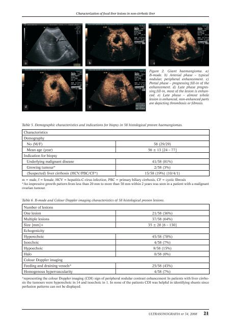
Figure 2. Giant haemangioma. a)<br />
B-mode. b) Arterial phase – typical<br />
nodular, peripheral enhancement. c)<br />
Portal phase – progressing fill-in of the<br />
enhancement. d) Late phase progressing<br />
fill-in, most of the lesion is enhanced.<br />
e) Late phase – almost whole<br />
lesion is enhanced, non-enhanced parts<br />
are depicting thrombosis or fibrosis.<br />
*representing the colour Doppler imaging (cDi) sign of peripheral nodular contrast enhancement in patients with liver cirrhosis<br />
the tumours were hyperechoic in 14 and isoechoic in 1. in none of the patients cDi was helpful in identifying shunts since<br />
perfusion patterns can not be displayed.<br />
ULTRASONOGRAFIA nr 34, 2008 21
![Ultrasonografia nr21 [4.58 Mb] - Kwartalnik](https://img.yumpu.com/51838921/1/184x260/ultrasonografia-nr21-458-mb-kwartalnik.jpg?quality=85)
![Ultrasonografia nr35 [13.00 MB] - Kwartalnik](https://img.yumpu.com/10640460/1/188x260/ultrasonografia-nr35-1300-mb-kwartalnik.jpg?quality=85)
![Ultrasonografia nr39 [8.00 MB] - Kwartalnik](https://img.yumpu.com/10637726/1/188x260/ultrasonografia-nr39-800-mb-kwartalnik.jpg?quality=85)
![Ultrasonografia nr46 [3.17 Mb] - Kwartalnik](https://img.yumpu.com/6154909/1/188x260/ultrasonografia-nr46-317-mb-kwartalnik.jpg?quality=85)