

kwartalnik polskiego towarzystwa ultrasonograficznego
kwartalnik polskiego towarzystwa ultrasonograficznego
kwartalnik polskiego towarzystwa ultrasonograficznego

Create successful ePaper yourself
Turn your PDF publications into a flip-book with our unique Google optimized e-Paper software.
shunt haemangiomas are typically surrounded by<br />
focal hypoechoic areas representing less fat content in<br />
comparison with the surrounding liver parenchyma.<br />
the metabolic changes are explained by a mainly arterial<br />
blood supply of the hypoechoic areas whereas blood<br />
supply of the surrounding liver parenchyma is mainly by<br />
portal venous vessels containing more lipids and insulin.<br />
arterioportal shunts have been observed in hepatic haemangiomas<br />
not only using cEUs but also on multiphase<br />
helical ct and magnetic resonance imaging (MRi) in<br />
22 ULTRASONOGRAFIA nr 34, 2008<br />
Dietrich C.F., Jędrzejczyk M.<br />
Table 7. Contrast enhancing pattern of 58 histologically proven lesions.<br />
Characteristics<br />
Contrast enhancement, arterial<br />
Peripheral nodular arterial enhancement 43/58 (74%)<br />
Peripheral rim like enhancement 0/58 (0%)<br />
not determinable (e.g., due to size of the lesion, solitary fibrotic nodule) 6/58 (10%)<br />
strong homogenous arterial enhancement*<br />
Centripetal filling<br />
9/58 (16%)<br />
complete (homogenous) fill-in 30 seconds and £ 60 seconds 6 (10%)<br />
> 60 seconds and £ 180 seconds 27 (47%)<br />
incomplete (inhomogenous, incomplete iris diaphragm sign, including one with<br />
non-enhancing solitary necrotic nodule)<br />
Sensitivity<br />
13 /58 (22%)<br />
Peripheral nodular arterial enhancement 43/58 (74%)<br />
complete portal venous fill-in 45/58 (78%)<br />
combination of both<br />
*in two patients with liver cirrhosis shunt-haemangiomas were found.<br />
57/58 (98%)<br />
a significant percentage of haemangiomas (19%–26%)<br />
[(10)] which is in the same range.<br />
Focal nodular hyperplasia (FNH)<br />
fnH is typically an isoechoic tumour of variable<br />
size, with a central scar and calcifications (about 70%).<br />
focal nodular hyperplasia and the important differential<br />
diagnosis of hepatocellular adenoma (Hca) are<br />
two benign, mostly incidentally discovered hepatic neoplasia.<br />
Differentiation is essential because of different<br />
therapeutic approaches. the examination of the hepatic<br />
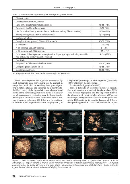
Figure 3. FNH. a) Power Doppler shows central vessel and smaller radiating vessels – “spoke wheel” pattern. b) Early<br />
arterial phase – regular pattern of arteries within the lesion are visible. c) Following seconds of arterial phase – centrifugal<br />
enhancement of the lesion. d) The end of arterial phase – whole lesion is hyperenhanced. e) Portal phase – lesion is slightly<br />
hyperenhanced. f) Late phase – lesion is still slightly hyperenhanced.
![Ultrasonografia nr21 [4.58 Mb] - Kwartalnik](https://img.yumpu.com/51838921/1/184x260/ultrasonografia-nr21-458-mb-kwartalnik.jpg?quality=85)
![Ultrasonografia nr35 [13.00 MB] - Kwartalnik](https://img.yumpu.com/10640460/1/188x260/ultrasonografia-nr35-1300-mb-kwartalnik.jpg?quality=85)
![Ultrasonografia nr39 [8.00 MB] - Kwartalnik](https://img.yumpu.com/10637726/1/188x260/ultrasonografia-nr39-800-mb-kwartalnik.jpg?quality=85)
![Ultrasonografia nr46 [3.17 Mb] - Kwartalnik](https://img.yumpu.com/6154909/1/188x260/ultrasonografia-nr46-317-mb-kwartalnik.jpg?quality=85)