90-269-01 MicroStop dt.qxd - KLS Martin
90-269-01 MicroStop dt.qxd - KLS Martin
90-269-01 MicroStop dt.qxd - KLS Martin

Erfolgreiche ePaper selbst erstellen
Machen Sie aus Ihren PDF Publikationen ein blätterbares Flipbook mit unserer einzigartigen Google optimierten e-Paper Software.
Fall 4<br />
Case 4<br />
Posttraumatisches Genu valgum<br />
et recurvatum rechts<br />
bei anterolateraler Überbrückung der distalen<br />
Femurepiphysenfuge und ventraler Überbrückung<br />
der proximalen Tibiaepiphysenfuge. Deutliches<br />
Kniebeugedefizit.<br />
14-jährige Jugendliche.<br />
56 kg Körpergewicht.<br />
Post-traumatic valgus and extension<br />
deformity of the right knee<br />
due to anterolateral bridging of the distal femoral<br />
growth plate and ventral bridging of the proximal<br />
tibial growth plate. Significant knee flexion deficit.<br />
14-year-old girl.<br />
56 kg body weight.<br />
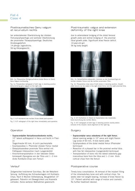
Abb. 1a: Präoperative Röntgenaufnahme beider Beine im Stand.<br />
Genu Valgum rechtsseitig.<br />
Fig. 1a: Preoperative radiograph of both legs in standing position<br />
demonstrating genu valgum on the right side.<br />
Abb. 1b: Nahaufnahme präoperativ. Lesionen an der Epiphysenfuge am<br />
rechten distalen Femur und der rechten proximalen Tibia.<br />
Fig. 1b: Preoperative close view of both knees in standing position. Growth<br />
plate lesions can be noted in the right distal femur and the right proximal tibia.<br />
Fig. 2: A.P.-Aufnahme des rechten Knies direkt post-operativ.<br />
Fig. 2: A.P. radiograph of the right knee immediately post-operative.<br />
Fig. 3: A.P.-Aufnahme im Stand vor Explantation des Implantats.<br />
Gute Ausrichtung des rechten Beins.<br />
Fig. 3: A.P. radiograph of both legs in standing position shortly before<br />
removal of the implant. Good alignment of the right leg.<br />
Operation<br />
•<br />
•<br />
•<br />
•<br />
Supracondyläre Varisationsosteotomie rechts,<br />
10° lateral aufklappend in Varus und leicht in Flexion<br />
Tragschraube 60 mm, 4-Loch-Laschenplatte<br />
Epiphyseodese n. Phemister distaler Femur medial<br />
Desepiphyseodese proximale Tibia ventral mit<br />
Fettlappen-Interposition (nach Langenskiöld)<br />
Knochentransplantation in den lateralen Osteotomiespalt<br />
(Spongiosa von der Tibia und 1–2 mm<br />
dicke Kortikalis-Chips vom Femur)<br />
Surgery<br />
• Supracondylar varus osteotomy of the right femur,<br />
lateral opening wedge in 10° varus and slight flexion<br />
Lag screw of 60 mm, 4-hole barrel plate<br />
• Epiphysiodesis of the distal medial femur (Phemister<br />
technique)<br />
• Resection of a physeal bar in the proximal ventral tibia<br />
with free fat interposition (Langenskiöld technique)<br />
• Transplantation of bone into the lateral osteotomy gap<br />
(cancellous bone from the tibia and 1–2 mm thick<br />
cortical chips from the femur)<br />
Verlauf<br />
Zeitgerechter knöcherner Durchbau. Bei der Metallentfernung<br />
Auffüllung des Schraubenlagers mit Kortikalis-<br />
Chips. Nach 6 Wochen Vollbelastung. Beugedefizit 15°<br />
gebessert. Patientin mit Beweglichkeit und Kosmetik<br />
zufrieden. Keine weiteren Maßnahmen gewünscht.<br />
Postoperative course<br />
Timely bony consolidation. At removal of the implant filling<br />
of the intramedullary bone void with cortical chips. Six<br />
weeks later full weight bearing. Increase of knee flexion by<br />
15°. Patient satisfied with range of motion and cosmetic.<br />
No further treatment desired.