DJD External Fixator, Stryker - ShoulderDoc.co.uk
DJD External Fixator, Stryker - ShoulderDoc.co.uk
DJD External Fixator, Stryker - ShoulderDoc.co.uk

Create successful ePaper yourself
Turn your PDF publications into a flip-book with our unique Google optimized e-Paper software.
14<br />
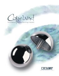
Dynamic Joint Distractor - Medial Frame Option<br />
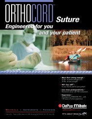
Pronator teres m.<br />
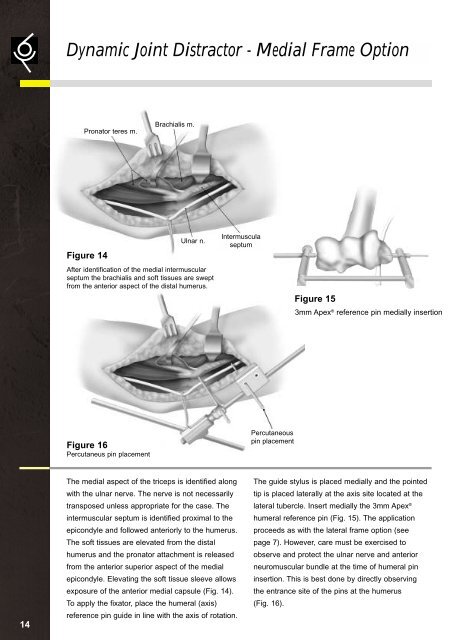
Figure 14<br />
After identification of the medial intermuscular<br />
septum the brachialis and soft tissues are swept<br />
from the anterior aspect of the distal humerus.<br />
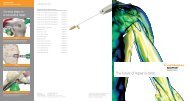
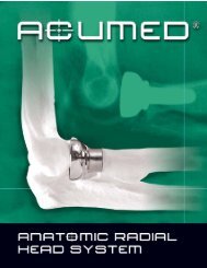
Figure 16<br />
Percutaneus pin placement<br />
Brachialis m.<br />
Ulnar n.<br />
The medial aspect of the triceps is identified along<br />
with the ulnar nerve. The nerve is not necessarily<br />
transposed unless appropriate for the case. The<br />
intermuscular septum is identified proximal to the<br />
epi<strong>co</strong>ndyle and followed anteriorly to the humerus.<br />
The soft tissues are elevated from the distal<br />
humerus and the pronator attachment is released<br />
from the anterior superior aspect of the medial<br />
epi<strong>co</strong>ndyle. Elevating the soft tissue sleeve allows<br />
exposure of the anterior medial capsule (Fig. 14).<br />
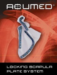
To apply the fixator, place the humeral (axis)<br />
reference pin guide in line with the axis of rotation.<br />
Intermuscula<br />
septum<br />
Percutaneous<br />
pin placement<br />
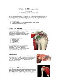
Figure 15<br />
3mm Apex ® reference pin medially insertion<br />
The guide stylus is placed medially and the pointed<br />
tip is placed laterally at the axis site located at the<br />
lateral tubercle. Insert medially the 3mm Apex ®<br />
humeral reference pin (Fig. 15). The application<br />
proceeds as with the lateral frame option (see<br />
page 7). However, care must be exercised to<br />
observe and protect the ulnar nerve and anterior<br />
neuromuscular bundle at the time of humeral pin<br />
insertion. This is best done by directly observing<br />
the entrance site of the pins at the humerus<br />
(Fig. 16).