DJD External Fixator, Stryker - ShoulderDoc.co.uk
DJD External Fixator, Stryker - ShoulderDoc.co.uk
DJD External Fixator, Stryker - ShoulderDoc.co.uk

Create successful ePaper yourself
Turn your PDF publications into a flip-book with our unique Google optimized e-Paper software.
Dynamic Joint Distractor - Unilateral Frame<br />
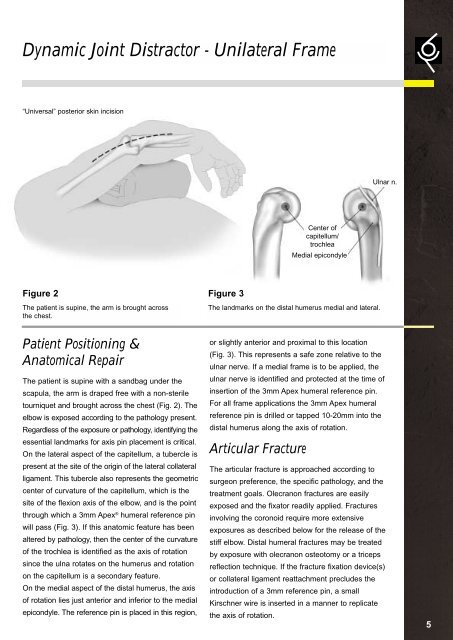
“Universal” posterior skin incision<br />
Figure 2 Figure 3<br />
The patient is supine, the arm is brought across<br />
the chest.<br />
Patient Positioning &<br />
Anatomical Repair<br />
The patient is supine with a sandbag under the<br />
scapula, the arm is draped free with a non-sterile<br />
tourniquet and brought across the chest (Fig. 2). The<br />
elbow is exposed ac<strong>co</strong>rding to the pathology present.<br />
Regardless of the exposure or pathology, identifying the<br />
essential landmarks for axis pin placement is critical.<br />
On the lateral aspect of the capitellum, a tubercle is<br />
present at the site of the origin of the lateral <strong>co</strong>llateral<br />
ligament. This tubercle also represents the geometric<br />
center of curvature of the capitellum, which is the<br />
site of the flexion axis of the elbow, and is the point<br />
through which a 3mm Apex ® humeral reference pin<br />
will pass (Fig. 3). If this anatomic feature has been<br />
altered by pathology, then the center of the curvature<br />
of the trochlea is identified as the axis of rotation<br />
since the ulna rotates on the humerus and rotation<br />
on the capitellum is a se<strong>co</strong>ndary feature.<br />
On the medial aspect of the distal humerus, the axis<br />
of rotation lies just anterior and inferior to the medial<br />
epi<strong>co</strong>ndyle. The reference pin is placed in this region,<br />
or slightly anterior and proximal to this location<br />
(Fig. 3). This represents a safe zone relative to the<br />
ulnar nerve. If a medial frame is to be applied, the<br />
ulnar nerve is identified and protected at the time of<br />
insertion of the 3mm Apex humeral reference pin.<br />
For all frame applications the 3mm Apex humeral<br />
reference pin is drilled or tapped 10-20mm into the<br />
distal humerus along the axis of rotation.<br />
Articular Fracture<br />
Center of<br />
capitellum/<br />
trochlea<br />
Medial epi<strong>co</strong>ndyle<br />
The landmarks on the distal humerus medial and lateral.<br />
Ulnar n.<br />
The articular fracture is approached ac<strong>co</strong>rding to<br />
surgeon preference, the specific pathology, and the<br />
treatment goals. Olecranon fractures are easily<br />
exposed and the fixator readily applied. Fractures<br />
involving the <strong>co</strong>ronoid require more extensive<br />
exposures as described below for the release of the<br />
stiff elbow. Distal humeral fractures may be treated<br />
by exposure with olecranon osteotomy or a triceps<br />
reflection technique. If the fracture fixation device(s)<br />
or <strong>co</strong>llateral ligament reattachment precludes the<br />
introduction of a 3mm reference pin, a small<br />
Kirschner wire is inserted in a manner to replicate<br />
the axis of rotation.<br />
5