DJD External Fixator, Stryker - ShoulderDoc.co.uk
DJD External Fixator, Stryker - ShoulderDoc.co.uk
DJD External Fixator, Stryker - ShoulderDoc.co.uk

You also want an ePaper? Increase the reach of your titles
YUMPU automatically turns print PDFs into web optimized ePapers that Google loves.
6<br />
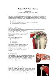
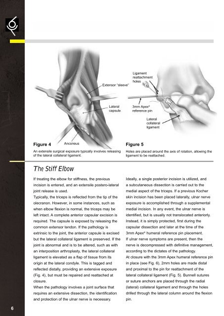
The Stiff Elbow<br />
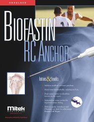
Extensor “sleeve”<br />
Lateral<br />
capsule<br />
Figure 4 An<strong>co</strong>neus<br />
Figure 5<br />
An extensile surgical exposure typically involves releasing<br />
of the lateral <strong>co</strong>llateral ligament.<br />
If treating the elbow for stiffness, the previous<br />
incision is entered, and an extensile postero-lateral<br />
joint release is used.<br />
Typically, the triceps is reflected from the tip of the<br />
olecranon. However, in some instances, such as<br />
when elbow flexion is normal, the triceps may be<br />
left intact. A <strong>co</strong>mplete anterior capsular excision is<br />
required. The capsule is exposed by releasing the<br />
<strong>co</strong>mmon extensor tendon. If the pathology is<br />
extrinsic to the joint, the anterior capsule is excised<br />
but the lateral <strong>co</strong>llateral ligament is preserved. If the<br />
joint is abnormal and is to be altered, such as with<br />
an interposition arthroplasty, the lateral <strong>co</strong>llateral<br />
ligament is elevated as a flap of tissue from its<br />
origin at the lateral <strong>co</strong>ndyle. This is tagged and<br />
reflected distally, providing an extensive exposure<br />
(Fig. 4), but must be repaired and reattached at<br />
closure.<br />
When the pathology involves a joint surface that<br />
requires an extensive dissection, the identification<br />
and protection of the ulnar nerve is necessary.<br />
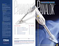
Ligament<br />
reattachment<br />
holes<br />
3mm Apex ®<br />
reference pin<br />
Lateral<br />
<strong>co</strong>llateral<br />
ligament<br />
Holes are placed around the axis of rotation, allowing the<br />
ligament to be reattached.<br />
Ideally, a single posterior incision is utilized, and<br />
a subcutaneous dissection is carried out to the<br />
medial aspect of the triceps. If a previous Kocher<br />
skin incision has been placed laterally, ulnar nerve<br />
exposure is ac<strong>co</strong>mplished through a supplemental<br />
medial incision. In any event, the ulnar nerve is<br />
identified, but is usually not translocated anteriorly.<br />
Instead, it is simply protected, first during the<br />
capsular dissection and later at the time of the<br />
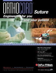
3mm Apex ® humeral reference pin placement.<br />
If ulnar nerve symptoms are present, then the<br />
nerve is de<strong>co</strong>mpressed with definitive management,<br />
ac<strong>co</strong>rding to the dictates of the pathology.<br />
At closure with the 3mm Apex humeral reference pin<br />
in place (see Fig. 6), 2mm holes are made distal<br />
and proximal to the pin for reattachment of the<br />
lateral <strong>co</strong>llateral ligament (Fig. 5). Bunnell sutures<br />
or suture anchors are placed through the radial<br />
(lateral) <strong>co</strong>llateral ligament and through the holes<br />
drilled through the lateral <strong>co</strong>lumn around the flexion<br />
pin.