Arūnas Diškus - VPU biblioteka - Vilniaus pedagoginis universitetas
Arūnas Diškus - VPU biblioteka - Vilniaus pedagoginis universitetas
Arūnas Diškus - VPU biblioteka - Vilniaus pedagoginis universitetas

Create successful ePaper yourself
Turn your PDF publications into a flip-book with our unique Google optimized e-Paper software.
According to updated and newly reviewed host-plant data, the world<br />
Nepticuloidea & Tischerioidea are trophically associated with 66 plant families:<br />
most species occur on Rosaceae (123 species) and Fagaceae (93 species); to a<br />
lesser extent, but still widely utilised, are such families as: Rhamnaceae (32 species),<br />
Asteraceae (31 species), Fabaceae (31 species), Salicaceae (28 species) and<br />
Betulaceae (27 species). In the light of host-plant data, the evolution of the<br />
Nepticuloidea or Tischerioidea probably occurred by way of trophic adaptation to<br />
7 plant subclasses from the 12 recognized; four of these host-plant subclasses are<br />
common to all three studied moth families: Hamamelididae, Dilleniidae, Rosidae<br />
and Lamiidae. Detailed review is given in the thesis or in published version in<br />
Puplesis, Diðkus, 2003.<br />
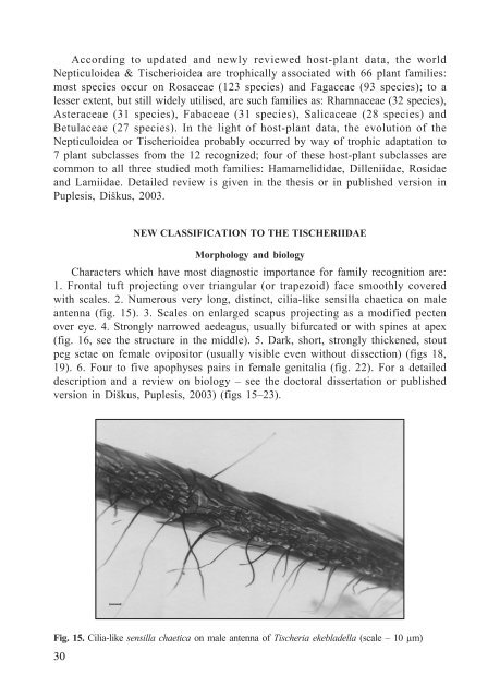
Fig. 15. Cilia-like sensilla chaetica on male antenna of Tischeria ekebladella (scale – 10 µm)<br />
30<br />
NEW CLASSIFICATION TO THE TISCHERIIDAE<br />
Morphology and biology<br />
Characters which have most diagnostic importance for family recognition are:<br />
1. Frontal tuft projecting over triangular (or trapezoid) face smoothly covered<br />
with scales. 2. Numerous very long, distinct, cilia-like sensilla chaetica on male<br />
antenna (fig. 15). 3. Scales on enlarged scapus projecting as a modified pecten<br />
over eye. 4. Strongly narrowed aedeagus, usually bifurcated or with spines at apex<br />
(fig. 16, see the structure in the middle). 5. Dark, short, strongly thickened, stout<br />
peg setae on female ovipositor (usually visible even without dissection) (figs 18,<br />
19). 6. Four to five apophyses pairs in female genitalia (fig. 22). For a detailed<br />
description and a review on biology – see the doctoral dissertation or published<br />
version in Diðkus, Puplesis, 2003) (figs 15–23).