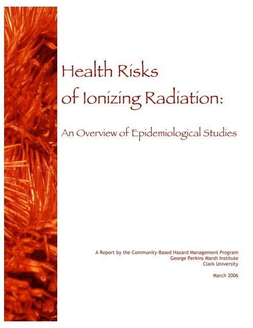
Health Risks of Ionizing Radiation: - Clark University
Health Risks of Ionizing Radiation: - Clark University
Health Risks of Ionizing Radiation: - Clark University

You also want an ePaper? Increase the reach of your titles
YUMPU automatically turns print PDFs into web optimized ePapers that Google loves.
<strong>Health</strong> <strong>Risks</strong><br />
<strong>of</strong> <strong>Ionizing</strong> <strong>Radiation</strong>:<br />
An Overview <strong>of</strong> Epidemiological Studies<br />
A Report by the Community-Based Hazard Management Program<br />
George Perkins Marsh Institute<br />
<strong>Clark</strong> <strong>University</strong><br />
March 2006
<strong>Health</strong> <strong>Risks</strong><br />
<strong>of</strong> <strong>Ionizing</strong> <strong>Radiation</strong>:<br />
An Overview <strong>of</strong> Epidemiological Studies<br />
by<br />
Abel Russ, Casey Burns, Seth Tuler, and Octavia Taylor<br />
Community-Based Hazard Management<br />
The George Perkins Marsh Institute<br />
<strong>Clark</strong> <strong>University</strong><br />
Worcester, MA 01610-1477<br />
USA<br />
March 2006
Contents<br />
List <strong>of</strong> Tables ............................................................................................................................... vi<br />
List <strong>of</strong> Figures ............................................................................................................................. vii<br />
Acknowledgments ........................................................................................................................ x<br />
1. INTRODUCTION ................................................................................................................ 1<br />
1.1 Goals ............................................................................................................................. 1<br />
<strong>Ionizing</strong> radiation ................................................................................................... 1<br />
Low dose ................................................................................................................ 1<br />
1.2 A brief history <strong>of</strong> radiation ........................................................................................... 1<br />
1.3 Exposure ....................................................................................................................... 2<br />
1.4 Standards ...................................................................................................................... 3<br />
1.5 <strong>Radiation</strong> basics ............................................................................................................ 3<br />
1.6 Epidemiological methods ............................................................................................. 7<br />
Statistical significance ............................................................................................ 7<br />
Study design ........................................................................................................... 8<br />
1.7 Risk terminology .......................................................................................................... 9<br />
Relative risk ........................................................................................................... 9<br />
Excess relative risk (ERR) ..................................................................................... 9<br />
Odds ratio (OR) ...................................................................................................... 9<br />
Excessive absolute risk (EAR) ............................................................................... 9<br />
Standardized incidence ratio (SIR) ........................................................................ 9<br />
Standardized mortality ratio (SMR) ....................................................................... 9<br />
P value .................................................................................................................. 10<br />
Confidence interval .............................................................................................. 10<br />
1.8 Dose-response curves ................................................................................................. 10<br />
1.9 Overview structure ..................................................................................................... 11<br />
2. BACKGROUND RADIATION ......................................................................................... 13<br />
2.1 Introduction ................................................................................................................ 13<br />
2.2 Summary <strong>of</strong> studies .................................................................................................... 13<br />
External exposure ................................................................................................. 14<br />
High exposure areas ............................................................................................. 14<br />
Radon ................................................................................................................... 15<br />
2.3 Discussion ................................................................................................................... 17<br />
3. MEDICAL EXPOSURES .................................................................................................. 19<br />
3.1 Introduction ................................................................................................................ 19<br />
3.2 Diagnostic exposures .................................................................................................. 21<br />
Diagnostic x-rays ................................................................................................. 21<br />
Diagnostic iodine-131 .......................................................................................... 23<br />
i
ii Contents<br />
Thorotrast ............................................................................................................. 24<br />
3.3 Radiotherapy for non-cancer disease .......................................................................... 24<br />
Tuberculosis ......................................................................................................... 25<br />
Ankylosingspondylitis ......................................................................................... 25<br />
Hyperthyroidism .................................................................................................. 26<br />
Radiotherapy for other non-cancer diseases ........................................................ 26<br />
Radiotherapy for non-cancer disease in children ................................................. 26<br />
3.4 Radiotherapy for cancer ............................................................................................. 28<br />
Radiotherapy for childhood cancer ...................................................................... 29<br />
3.5 In utero exposures ....................................................................................................... 29<br />
3.6 Parental exposures ...................................................................................................... 30<br />
3.7 Radiologists’ occupational exposures ......................................................................... 31<br />
3.8 Conclusions ................................................................................................................ 31<br />
4. ATOMIC BOMB SURVIVORS ........................................................................................ 43<br />
4.1 Introduction ................................................................................................................ 43<br />
RERF research ..................................................................................................... 43<br />
Doses .................................................................................................................... 44<br />
Uncertainties ........................................................................................................ 45<br />
4.2 Cancer mortality ......................................................................................................... 45<br />
4.3 Noncancer mortality ................................................................................................... 48<br />
4.4.1 Solid cancer incidence ................................................................................................ 48<br />
4.4.2 Incidence <strong>of</strong> leukemia and related cancers ................................................................. 49<br />
4.5 Incidence <strong>of</strong> noncancer disease .................................................................................. 50<br />
4.6 Preconception and prenatal exposures ........................................................................ 51<br />
4.7 Discussion ................................................................................................................... 52<br />
Age, time and gender ........................................................................................... 52<br />
Dose-response curves ........................................................................................... 52<br />
Applicability to other contexts ............................................................................. 53<br />
5. NUCLEAR WEAPONS TESTING .................................................................................. 58<br />
5.1 Military personnel ...................................................................................................... 58<br />
5.2 Semipalatinsk Test Site downwinders ........................................................................ 59<br />
5.3 Nevada Test Site downwinders ................................................................................... 60<br />
Leukemia .............................................................................................................. 60<br />
Thyroid disease .................................................................................................... 61<br />
5.4 South Pacific testing ................................................................................................... 63<br />
5.5 <strong>Health</strong> effects in Scandinavia ..................................................................................... 63<br />
5.6 Discussion ................................................................................................................... 63<br />
6. RADIATION WORKERS ................................................................................................. 70<br />
6.1 Introduction ................................................................................................................ 70<br />
Exposure limits .................................................................................................... 70<br />
Assessing risks ..................................................................................................... 72<br />
6.2 US facilities ................................................................................................................ 73<br />
Hanford ................................................................................................................ 73<br />
Oak Ridge ............................................................................................................ 75<br />
Oak Ridge and age at exposure ............................................................................ 76<br />
Rocketdyne .......................................................................................................... 77
Contents iii<br />
Rocky Flats .......................................................................................................... 78<br />
Lawrence Livermore National Laboratory .......................................................... 78<br />
Savannah River Site ............................................................................................. 79<br />
Los Alamos National Laboratory ......................................................................... 80<br />
Portsmouth Uranium Enrichment Plant ............................................................... 80<br />
The Mound Facility .............................................................................................. 81<br />
Mallinckrodt Chemical Works ............................................................................. 81<br />
Linde Air Products Company Ceramics Plant ..................................................... 81<br />
Feed Material Production Center (Fernald site) ................................................... 82<br />
6.3 UK facilities ................................................................................................................ 82<br />
Sellafield .............................................................................................................. 82<br />
Other UK facilities ............................................................................................... 83<br />
6.4 Multi-site studies ........................................................................................................ 83<br />
Pooled UK data .................................................................................................... 83<br />
Pooled Canadian data ........................................................................................... 84<br />
Pooled US data ..................................................................................................... 85<br />
Pooled international data ...................................................................................... 86<br />
6.5 Discussion ................................................................................................................... 86<br />
Leukemia and multiple myeloma ......................................................................... 87<br />
Solid cancer .......................................................................................................... 88<br />
Female workers .................................................................................................... 89<br />
Age at exposure .................................................................................................... 89<br />
7. MAYAK WORKERS ......................................................................................................... 97<br />
7.1 Introduction ................................................................................................................ 97<br />
7.2 Helath effects .............................................................................................................. 97<br />
Lung cancer .......................................................................................................... 97<br />
Bone cancer and liver cancer ............................................................................... 98<br />
Leukemia and related cancers .............................................................................. 99<br />
Non-cancer disease .............................................................................................. 99<br />
7.3 Summary ..................................................................................................................... 99<br />
8. URANIUM MINERS ....................................................................................................... 103<br />
8.1 Introduction .............................................................................................................. 103<br />
8.2 US studies ................................................................................................................. 104<br />
Navajo miners .................................................................................................... 106<br />
Pathological observations .................................................................................. 106<br />
8.3 Studies <strong>of</strong> miners in other countries ......................................................................... 106<br />
Australia ............................................................................................................. 106<br />
Canada ................................................................................................................ 106<br />
China .................................................................................................................. 107<br />
Czechoslovakia .................................................................................................. 107<br />
France ................................................................................................................. 107<br />
Germany ............................................................................................................. 107<br />
Sweden ............................................................................................................... 107<br />
8.4 Combined estimates <strong>of</strong> risk ...................................................................................... 107<br />
8.5 Discussion ................................................................................................................. 108<br />
Comparisons with residential radon studies ...................................................... 108
iv Contents<br />
9. RADIATION EXPOSURE IN FLIGHT .........................................................................113<br />
9.1 Epidemiological studies ............................................................................................ 113<br />
9.2 Cell studies ............................................................................................................... 113<br />
9.3 Discussion ................................................................................................................. 113<br />
10. PRECONCEPTION EXPOSURES .................................................................................117<br />
10.1 The Seascale leukemia cluster and the Gardner hypothesis ..................................... 117<br />
10.2 Further studies <strong>of</strong> the Gardner hypothesis in the UK ............................................... 118<br />
10.3 Preconception exposure in other settings ................................................................. 119<br />
10.4 Solid cancers in association with preconception expsoures ..................................... 120<br />
10.5 Adverse birth outcomes in association with preconception exposures .................... 121<br />
10.6 Cell studies ............................................................................................................... 121<br />
10.7 Animal evidence ....................................................................................................... 122<br />
10.8 Discussion ................................................................................................................. 122<br />
11. NUCLEAR POWER ACCIDENTS ................................................................................ 130<br />
11.1 Three Mile Island ..................................................................................................... 130<br />
11.2 Chernobyl ................................................................................................................. 131<br />
11.2.1 Dose reconstruction for Chernobyl downwinders .................................................. 131<br />
11.2.2 Chernobyl emergency workers ................................................................................ 132<br />
11.2.3 Childhood thyroid cancer ................................................................................... 133<br />
11.2.4 Childhood leukemia ........................................................................................... 135<br />
11.2.5 Non-cancer effects in children ........................................................................... 136<br />
11.2.6 Chernobyl discussion ......................................................................................... 137<br />
12. COMMUNITIES NEAR NUCLEAR FACILITIES ..................................................... 145<br />
12.1 Introduction .............................................................................................................. 145<br />
Types <strong>of</strong> studies .................................................................................................. 146<br />
12.2 US studies ................................................................................................................. 146<br />
Hanford .............................................................................................................. 146<br />
Other US facilities .............................................................................................. 147<br />
Multi-site studies in the US ................................................................................ 148<br />
12.3 UK studies ................................................................................................................ 148<br />
Sellafield ............................................................................................................ 149<br />
Dounreay ............................................................................................................ 149<br />
Other UK facilities and multi-site studies .......................................................... 150<br />
12.4 Nuclear facilities outside the US and UK ................................................................. 151<br />
12.5 Discussion ................................................................................................................. 153<br />
13. DISCUSSION .................................................................................................................... 167<br />
The idea <strong>of</strong> a threshold dose .............................................................................. 167<br />
Positive resluts at low doses .............................................................................. 167<br />
Uncertainty and judgement ................................................................................ 171<br />
Synthesis <strong>of</strong> information .................................................................................... 171<br />
What if there is a threshold dose? ...................................................................... 173<br />
Conclusion ......................................................................................................... 174
Contents v<br />
APPENDIX A .......................................................................................................................... 175<br />
Leukemia ............................................................................................................................ 175<br />
A.1 About the Disease ..................................................................................................... 175<br />
A.2 Atomic Bomb Survivors ........................................................................................... 176<br />
A.3 Medical Exposures ................................................................................................... 176<br />
Adult medical exposures .................................................................................... 176<br />
Childhood medical exposures ............................................................................ 177<br />
Medical exposure in utero .................................................................................. 177<br />
Radiology occupation ........................................................................................ 177<br />
A.4 Worders ..................................................................................................................... 177<br />
A.5 Fallout ....................................................................................................................... 178<br />
Nuclear Weapons Testing ................................................................................... 178<br />
Chernobyl ........................................................................................................... 178<br />
A.6 Discussion ................................................................................................................. 178<br />
APPENDIX B .......................................................................................................................... 181<br />
Thyroid disease ......................................................................................................................... 181<br />
B.1 Introduction to the Thyroid ....................................................................................... 181<br />
B.2 External <strong>Radiation</strong> and Thyroid Cancer ................................................................... 182<br />
B.3 Internal <strong>Radiation</strong> and Thyroid Cancer .................................................................... 183<br />
Fallout ................................................................................................................ 183<br />
Chernobyl ........................................................................................................... 183<br />
Medical exposure ............................................................................................... 184<br />
B.4 Non-cancer Thyroid Disease .................................................................................... 184<br />
B.5 Discussion ................................................................................................................. 185<br />
APPENDIX C .......................................................................................................................... 187<br />
Analysis <strong>of</strong> preconception exposure studies ............................................................................. 187<br />
APPENDIX D .......................................................................................................................... 194<br />
Recent information .................................................................................................................... 194<br />
Fifteen-country worker study .................................................................................................... 194<br />
Techa River cohort .................................................................................................................... 194<br />
BEIR VII ............................................................................................................................ 195<br />
Acronyms & Abbreviations ...................................................................................................... 196<br />
Glossary <strong>of</strong> Terms for Epidemiology and <strong>Radiation</strong> ................................................................ 197<br />
Bibliography ............................................................................................................................ 211<br />
Index ............................................................................................................................ 243
List <strong>of</strong> Tables<br />
Table 2-1 Studies <strong>of</strong> background radiation exposure ............................................................18<br />
Table 3-1 Studies <strong>of</strong> diagnostic x-ray exposure (including fluoroscopies and<br />
radiologists’ exposures) ........................................................................................33<br />
Table 3-2 Studies <strong>of</strong> external radiation therapy for benign disease ......................................36<br />
Table 3-3 Studies <strong>of</strong> internal radiation exposure in diagnosis or treatment <strong>of</strong><br />
benign disease .......................................................................................................39<br />
Table 3-4 Studies <strong>of</strong> radiation therapy for cancer .................................................................41<br />
Table 4-1 ERR estimates (Sv -1 ) from the RERF analyses <strong>of</strong> the atomic bomb survivors .....46<br />
Table 4-2 Studies <strong>of</strong> the atomic bomb survivors ..................................................................54<br />
Table 5-1 Nuclear weapons test participants ........................................................................65<br />
Table 5-2 Fallout from the Semipalatinsk test site in Kazakhstan ........................................66<br />
Table 5-3 Fallout from the Nevada test site ..........................................................................67<br />
Table 5-4 South Pacific exposures to fallout from nuclear weapons testing ........................69<br />
Table 6-1 Leukemia in nuclear workers and atomic bomb survivors. ..................................88<br />
Table 6-2 Facility-specific studies <strong>of</strong> occupational exposure ...............................................90<br />
Table 6-3 Studies <strong>of</strong> occupational exposure using combined datasets .................................95<br />
Table 7.1 Dose (Gy) received by Mayak workers at each <strong>of</strong> the three main facilities<br />
and auxiliary plants ...............................................................................................98<br />
Table 7-2 Studies <strong>of</strong> Mayak workers ..................................................................................101<br />
Table 8-1 Comparison <strong>of</strong> miner data with residential radon studies ..................................109<br />
Table 8-2 Studies <strong>of</strong> radon exposure in underground miners .............................................110<br />
Table 9-1 Studies <strong>of</strong> cancer in flight personnel ...................................................................116<br />
Table 10-1 Preconception irradiation and leukemia and non-Hodgkin’s lymphoma<br />
(LNHL) ...............................................................................................................124<br />
Table 10-2 Preconception irradiation and solid cancers .......................................................126<br />
Table 10-3 Preconception irradiation and stillbirths and congenital malformations ............128<br />
Table 11-1 Studies <strong>of</strong> health effects around Three Mile Island ............................................138<br />
Table 11-2 <strong>Health</strong> effects in Chernobyl cleanup workers .....................................................139<br />
Table 11-3 Childhood thyroid cancer in Chernobyl downwinders .......................................141<br />
Table 11-4 Childhood leukemia in Chernobyl downwinders ...............................................143<br />
Table 11-5 Noncancer disease in Chernobyl downwinders ..................................................144<br />
Table 12-1 Cancer incidence (adult or all ages) near nuclear facilities ................................156<br />
Table 12-2 Childhood cancer incidence near nuclear facilities ............................................160<br />
Table 12-3 Childhood cancer mortality near nuclear facilities .............................................164<br />
Table 12-4 Adverse birth outcomes near nuclear facilities ...................................................166<br />
Table 13-1 Significantly positive risk estimates at low doses ...............................................170<br />
Table 13-2 Estimated risk <strong>of</strong> non-CLL leukemia in adults ...................................................172<br />
Table B-1 Thyroid cancer risk estimates for various exposures ..........................................186<br />
Table C-1 Candidate studies for inclusion ...........................................................................188<br />
Table C-2 Pooled result for paternal preconception x-rays .................................................190<br />
Table C-3 Pooled result for paternal radiation occupation ..................................................191<br />
Table C-4 Exposure to a total preconception dose > 50 mSv ..............................................192<br />
Table C-5 Exposure to a total preconception dose in the 6 months before conception<br />
<strong>of</strong> > 5 mSv .........................................................................................................192<br />
vi
List <strong>of</strong> Figures<br />
Figure 1-1 Average worldwide exposure to radiation sources; total exposure averages<br />
2.8 mSv per year .....................................................................................................2<br />
Figure 1-2 The evolution <strong>of</strong> health protection standards for nuclear workers ........................4<br />
Figure 1-3 The atom .................................................................................................................5<br />
Figure 1-4 Types <strong>of</strong> radiation ...................................................................................................5<br />
Figure 1-5(a) Alpha particle radiation occurs when an unstable nucleus (the parent nucleus)<br />
releases a particle equivalent to the nucleus <strong>of</strong> a helium atom (2 neutrons<br />
and 2 protons) thus leaving a nucleus with 2 less protons and neutrons (the<br />
daughter nucleus) ....................................................................................................6<br />
Figure 1-5(b) Beta particle radiation occurs when a parent nucleus releases an electron<br />
(this is called a beta particle to differentiate it from the electrons that orbit<br />
the nucleus) .............................................................................................................6<br />
Figure 1-5(c) After a decay reaction, the nucleus is <strong>of</strong>ten in an “excited” state. ..........................6<br />
Figure 1-6 Examples <strong>of</strong> dose-response relationships .............................................................11<br />
Figure 2-1 A bar graph showing average annual natural radiation doses worldwide ............14<br />
Figure 2-2. High background radiation areas around the world ..............................................15<br />
Figure 2-3 A map created by the EPA showing average indoor radon levels by county ........16<br />
Figure 3-1 Average doses from diagnostic radiation exposures .............................................20<br />
Figure 3-2 In the 1950s the panoramic X-ray machine was invented to take a picture<br />
<strong>of</strong> the whole mouth with just one exposure ..........................................................21<br />
Figure 3-3 Relative risk <strong>of</strong> myeloid leukemia among adults according to the number<br />
<strong>of</strong> x-ray exams <strong>of</strong> the trunk (torso) in the period up to six months before<br />
leukemia diagnosis ................................................................................................22<br />
Figure 3-4 Thyroid cancer SIRs for patients who had been diagnosed for reasons<br />
other than suspicion <strong>of</strong> a thyroid tumor ................................................................22<br />
Figure 3-5 CAT scans deliver some <strong>of</strong> the highest doses <strong>of</strong> medical radiation to<br />
patients. .................................................................................................................23<br />
Figure 3-6 In the late 1940s chest x-rays were used to screen for tuberculosis .....................24<br />
Figure 4-1 The bomb dropped over Nagasaki on August 9, 1945 was called Fat Man .........43<br />
Figure 4-2 A view <strong>of</strong> the destruction <strong>of</strong> the city <strong>of</strong> Hiroshima ..............................................44<br />
Figure 4-3 A view from the air <strong>of</strong> the bombing <strong>of</strong> Nagasaki .................................................44<br />
Figure 4-4a These figures show how the radiation exposure <strong>of</strong> the cities <strong>of</strong> Hiroshima<br />
and Nagasaki were studied ...................................................................................47<br />
Figure 4-4b These figures show how the radiation exposure <strong>of</strong> the cities <strong>of</strong> Hiroshima<br />
and Nagasaki were studied ...................................................................................47<br />
Figure 4-5 ERR estimates for total solid cancer incidence by gender and age at<br />
exposure ................................................................................................................48<br />
Figure 4-6 Incidence ERR by age at exposure for selected cancer sites ................................49<br />
Figure 5-1 A map showing the approximate locations <strong>of</strong> worldwide nuclear bomb<br />
test sites .................................................................................................................58<br />
Figure 5-2 Military observers shield their faces from the Teapot Test in 1955 ......................59<br />
Figure 5-3 Atmospheric test Grable conducted in 1953 at the Nevada Test Site ..................60<br />
Figure 5-4 Per capita thyroid doses resulting from all exposure routes from all test .............62<br />
Figure 5-5 A nuclear test conducted as part <strong>of</strong> Operation Crossroads <strong>of</strong>f Bikini atoll<br />
in the Marshall Islands in 1946 .............................................................................63<br />
vii
viii List <strong>of</strong> Figures<br />
Figure 6-1 The United States nuclear weapons complex comprised dozens <strong>of</strong> industrial<br />
facilities and laboratories across the country .......................................................71<br />
Figure 6-2(a) The first external monitoring devices were pocket ionization chambers and<br />
pocket dosimeters (basically tubes the size <strong>of</strong> pens containing a wire with<br />
an electrical charge that decreased upon exposure to radiation) ..........................72<br />
Figure 6-2(b) Pocket dosimeters were replaced by film badges. A worker here is pictured<br />
checking film badges in 1974 ...............................................................................72<br />
Figure 6-3 Located at the Hanford site in Washington and completed in September<br />
1944, the B Reactor was the world’s first large-scale plutonium production<br />
reactor ...................................................................................................................74<br />
Figure 6-4 Plutonium is handled through a glovebox at the Plutonium Finishing Plant<br />
at the Hanford site .................................................................................................75<br />
Figure 6-5 A low-level solid waste burial ground at Oak Ridge National Laboratory ..........76<br />
Figure 6-6 The effect <strong>of</strong> exposure age on the risk <strong>of</strong> cancer mortality in Oak Ridge<br />
workers .................................................................................................................77<br />
Figure 6-7 <strong>Radiation</strong>-safety technicians check workers for contamination before<br />
they exit a Rocky Flats facility .............................................................................77<br />
Figure 6-8 Today the X-Y Retriever Room at Rocky Flats is used to store surplus<br />
plutonium ..............................................................................................................78<br />
Figure 6-9 Association between lung dose and lung cancer mortality among Rocky<br />
Flats workers employed for 15-25 years ..............................................................79<br />
Figure 6-10 A pool type reactor at the Livermore facility is pictured here ..............................80<br />
Figure 6-11 Dr. John G<strong>of</strong>man (second from left) at the time that this photo was taken<br />
(1960s) was the first Associate Director for the Biomedical Program at<br />
Lawrence Livermore National Laboratory ...........................................................80<br />
Figure 6-12 Barrels <strong>of</strong> transuranic waste site on a concrete pad at the Savannah<br />
River Site ..............................................................................................................81<br />
Figure 6-13 A worker holding uranium metal product at Fernald ............................................82<br />
Figure 6-14 A facility at Hanford for treating persons injured by embedded radioactive<br />
particles (circa 1967) ............................................................................................86<br />
Figure 8-1 The process <strong>of</strong> uranium mining and milling from ore to its reactor useable<br />
form is depicted here .........................................................................................103<br />
Figure 8-2 Uranium mines in the US are predominantly located in the southwest and<br />
central parts <strong>of</strong> the country .................................................................................105<br />
Figure 8-3 Many uranium mines were located on Native American reservations ...............107<br />
Figure 8-4 Dependence <strong>of</strong> lung cancer risk estimate on dose based on a combined<br />
analysis <strong>of</strong> the lung cancer mortality risk in 11 cohorts <strong>of</strong> miners .....................108<br />
Figure 11-1 <strong>Radiation</strong> emissions and incidence <strong>of</strong> lung cancer, 1981-1985, in the TMI<br />
10-mile area ........................................................................................................130<br />
Figure 11-2. Researchers at the Lawrence Livermore National Laboratory map the<br />
plumes <strong>of</strong> radiation originating from the Chernobyl accident ............................132<br />
Figure 12-1 A map showing the approximate location <strong>of</strong> worldwide commercial nuclear plants<br />
145<br />
Figure 12-2 A leaky drum at the 903 pad at Rocky Flats. Photo courtesy <strong>of</strong> the U.S.<br />
Department <strong>of</strong> Energy ........................................................................................148<br />
Figure 12-3 Native Americans from San Ildefonso Pueblo and Los Alamos residents .........149<br />
Figure 13-1 Estimated solid cancer mortality risk coefficient over increasing ranges<br />
<strong>of</strong> dose .................................................................................................................174<br />
Figure A-1 Leukemia risk across exposure ages in populations downwind <strong>of</strong> Chernobyl<br />
and the Nevada Test Site .....................................................................................180
We are indeed grateful to those who have helped bring about this document, assisting us in the<br />
research, writing, and compilation <strong>of</strong> figures, tables, and images. Without Abel Russ, Research<br />
Associate at the George Perkins Marsh Institute, <strong>Clark</strong> <strong>University</strong>, this project would not<br />
have been realized. He was the primary researcher and writer <strong>of</strong> this document. Casey Burns<br />
provided thoughtful research and lead writing on major sections <strong>of</strong> the manuscript. Several<br />
research assistants, Jessica Cook, Rose Heil, and Katie Scott explored some <strong>of</strong> the many studies<br />
included in this document.<br />
Our discussions with Rob Goble, Research Pr<strong>of</strong>essor at the Marsh Institute and Physics Pr<strong>of</strong>essor<br />
at <strong>Clark</strong> <strong>University</strong>, in the early conceptual stages <strong>of</strong> the book, helped us get <strong>of</strong>f to a good start.<br />
Seth Tuler, Research Fellow at the Marsh Institute, encouraged us and made suggestions along<br />
the way. Lu Ann Pacenka, Desktop Publisher at the Marsh Institute, played a key role in the<br />
layout and design <strong>of</strong> this document.<br />
We are grateful also to the members <strong>of</strong> the Western Shoshone tribes in Nevada and California,<br />
Southern Paiutes bands in Utah, members <strong>of</strong> the Laguna and Acoma Pueblos in New Mexico and<br />
organizational members <strong>of</strong> the Alliance for Nuclear Accountability with whom we have worked<br />
over the years and all <strong>of</strong> whom have inspired us to produce this manuscript.<br />
The production <strong>of</strong> this resource was made possible through a grant from RESOLVE, Inc. on<br />
behalf <strong>of</strong> the Citizens’ Monitoring and Technical Assessment Fund (CMTA). The Fund was<br />
created as part <strong>of</strong> a 1998 court settlement between the U.S. Department <strong>of</strong> Energy and 39<br />
nonpr<strong>of</strong>it peace and environmental groups around the country. The Fund provided financial<br />
support to organizations, such as ours, to conduct technical and scientific reviews and analyses<br />
<strong>of</strong> environmental management activities at DOE sites and to disseminate those reviews and<br />
analyses.<br />
My thanks and appreciation to all.<br />
Acknowledgments<br />
ix<br />
Octavia Taylor, Program Manager<br />
Community-Based Hazard Management<br />
George Perkins Marsh Institute<br />
<strong>Clark</strong> <strong>University</strong><br />
Worcester, MA
1<br />
INTRODUCTION<br />
1.1 Goals<br />
The health risks <strong>of</strong> exposure to low levels <strong>of</strong><br />
ionizing radiation are disputed within the scientific<br />
community. <strong>Risks</strong> associated with exposure to high<br />
levels <strong>of</strong> radiation are widely accepted and well<br />
documented based primarily on the studies <strong>of</strong> the<br />
atomic bomb survivors in Nagasaki and Hiroshima.<br />
Some feel that the best way to estimate risk for lowlevel<br />
exposures is to extrapolate from higher doses,<br />
although there is some clear evidence <strong>of</strong> low-dose<br />
risk. In this overview we have attempted to give an<br />
unbiased summary <strong>of</strong> the available research with<br />
an emphasis on the lower doses. The strengths and<br />
weaknesses <strong>of</strong> the studies are explained in order<br />
to help assess the variety <strong>of</strong> sometimes conflicting<br />
evidence.<br />
Not every epidemiological study that has ever<br />
been done on the risks <strong>of</strong> radiation is included in<br />
this overview. The body <strong>of</strong> research is simply too<br />
great for us to have collected and read every study.<br />
We attempted to collect studies generally considered<br />
to be key for the various sources <strong>of</strong> exposure and<br />
to exhaust our own capabilities to collect as many<br />
studies as possible. Our search methodology<br />
included the use <strong>of</strong> the PubMed search engine<br />
and smaller follow-up searches through the end <strong>of</strong><br />
2003. Another source <strong>of</strong> epidemiologic data is the<br />
DOE Office <strong>of</strong> Environment, Safety and <strong>Health</strong>’s<br />
“Comprehensive Epidemiologic Data Resource” 1 ,<br />
which provides data sets and studies relating to<br />
worker health at DOE facilities, populations residing<br />
near DOE sites, atomic bomb survivors, and radium<br />
1 http://cedr.lbl.gov<br />
2 www.lowdose.org<br />
1<br />
dial painters. In light <strong>of</strong> the ongoing generation <strong>of</strong><br />
relevant information we have decided to consider<br />
our overview a work in progress and plan to release<br />
supplements <strong>of</strong> this initial review in the future.<br />
Our intended audience is not necessarily one that<br />
has been scientifically trained and so scientific terms<br />
will be discussed and defined in a glossary found at<br />
the end <strong>of</strong> the overview. The two following terms<br />
from our title defined the scope <strong>of</strong> our overview:<br />
<strong>Ionizing</strong> radiation refers to radiation that has<br />
enough energy to remove an electron from a neutral<br />
atom or molecule, creating a free radical. <strong>Ionizing</strong><br />
radiation is capable <strong>of</strong> creating DNA damage that<br />
can lead to cancer. <strong>Radiation</strong>s from sources such as<br />
power lines, cell phones, and traffic radars are all<br />
classified as non-ionizing radiation because they<br />
are not capable <strong>of</strong> removing an electron. There is<br />
ongoing research concerning health effects <strong>of</strong> nonionizing<br />
radiation but it will not be covered in this<br />
overview.<br />
Low dose. Although we have collected studies<br />
<strong>of</strong> a wide range <strong>of</strong> exposures we should define a low<br />
dose as a reference point for the reader. Generally<br />
speaking, a dose is low relative to doses where<br />
the evidence <strong>of</strong> a health risk is more robust. The<br />
Department <strong>of</strong> Energy’s (DOE) Low Dose <strong>Radiation</strong><br />
Research Program 2 has the fuzzy definition <strong>of</strong> any<br />
dose not documented to show significant health risks;<br />
they generally consider a low dose to be below 10<br />
rem or 0.1 Sieverts (Sv). The <strong>Health</strong> Physics Society<br />
recommends that exposures below 0.1 Sv only be<br />
evaluated qualitatively as the risks are too small to<br />
be observed. For the purposes <strong>of</strong> our overview we<br />
have considered doses below 0.1 Sv to be low.
2 Introduction<br />
1.2 A brief history <strong>of</strong> radiation<br />
The history <strong>of</strong> the relationship between science,<br />
radiation and health goes back to the late 19 th<br />
century. X-rays were first discovered in late 1895<br />
and dangers associated with exposure became<br />
apparent very quickly. In 1896 the first injuries<br />
due to x-ray exposure were recorded and in 1904<br />
Thomas Edison’s assistant Clarence Dally was the<br />
first person recorded to have died as a result <strong>of</strong> x-ray<br />
exposure. Despite the risk, the use <strong>of</strong> x-rays for a<br />
variety <strong>of</strong> applications rapidly caught on. During the<br />
First World War portable x-ray machines were used<br />
on both sides to locate shrapnel and to set broken<br />
bones.<br />
Radium was discovered in 1898 and its use in<br />
medicine also spread very quickly. In the 1920s,<br />
sickness and death in watch dial painters, who<br />
ingested small amounts <strong>of</strong> radium in their work,<br />
taught scientists and doctors that internal exposure<br />
to radium could be harmful. It was also during the<br />
1920s that the cancer risks <strong>of</strong> radiology became<br />
apparent. Some awareness was beginning to spread<br />
that radiation exposure from the new technologies<br />
could be harmful and in 1928 the first internationally<br />
recognized radiation safety guidelines were<br />
published.<br />
There was still a popular enthusiasm about<br />
radiation during this time. Between 1920 and 1950<br />
patents were registered for radium toothpaste, radium<br />
hearing aids and radium tonic water. As World War<br />
II emerged in the 1930s plans for building a nuclear<br />
bomb were made. Plutonium was discovered in<br />
1940 and the Manhattan Project, with its goal to<br />
“make the bomb”, was initiated in 1942. On July 16,<br />
1945 the first nuclear weapon was detonated in New<br />
Mexico. At the time <strong>of</strong> the test the type <strong>of</strong> fallout that<br />
would result was something <strong>of</strong> a mystery. In August<br />
<strong>of</strong> 1945 two nuclear bombs were dropped on Japan<br />
in Nagasaki and Hiroshima. In the weeks after the<br />
bombs, radiation exposure was not discussed in the<br />
US press.<br />
In the 1950s the US government began a<br />
campaign for a more friendly attitude toward<br />
nuclear science and promoted civil nuclear power.<br />
There were hundreds <strong>of</strong> uranium mines across the<br />
country serving both the nuclear power industry<br />
and the nuclear weapons industry. In 1951 the US<br />
established the Nevada Test Site in order to have a<br />
domestic testing location that could ease logistical<br />
and security concerns. Prior to this tests had been<br />
conducted in the South Pacific. The fallout from<br />
the test sites and the occupational hazards <strong>of</strong> the<br />
workers in the mines and in the weapons factories<br />
were adding to the range <strong>of</strong> radiation exposures that<br />
we experience.<br />
1.3 Exposure<br />
People are exposed to ionizing radiation from many<br />
different sources. The exact amount depends on where<br />
they live, what their jobs are, what their lifestyles are<br />
like and so on. Over 80% <strong>of</strong> total average exposure<br />
comes from natural sources. Manmade sources such<br />
as consumer products that emit radiation, fallout<br />
from the US and global nuclear tests, diagnostic and<br />
therapeutic medicine, radiation from nuclear plants<br />
and occupational exposures make up the rest. The<br />
following pie chart gives the estimated worldwide<br />
average exposure spectrum.<br />
It should be noted that illustrations such as the<br />
pie chart (Figure 1-1) presented here can be used<br />
to minimize the importance <strong>of</strong> manmade sources <strong>of</strong><br />
radiation. For example, one may argue that fallout<br />
from test sites contributes a mere fraction, less than<br />
1%, <strong>of</strong> the total exposure to ionizing radiation. On<br />
the other hand this is an involuntary exposure that<br />
<br />
<br />
<br />
<br />
<br />
<br />
<br />
<br />
<br />
<br />
<br />
<br />
<br />
<br />
<br />
<br />
<br />
<br />
Figure 1-1. Average worldwide exposure to radiation<br />
sources; total exposure averages 2.8 mSv per year<br />
(UNSCEAR 2000).
may be <strong>of</strong> little benefit to those who bear the burden<br />
<strong>of</strong> risk.<br />
A figure such as this can also be misleading<br />
because <strong>of</strong> the variability <strong>of</strong> individual exposure<br />
histories. For example, a person who works in a<br />
nuclear facility will probably be exposed to more<br />
manmade radiation than a person who does not; this<br />
will alter the distribution <strong>of</strong> the pie chart and the<br />
overall amount <strong>of</strong> exposure. A person who lives in<br />
an area <strong>of</strong> particularly high fallout from the Nevada<br />
Test Site will also have a different exposure pattern<br />
than the average American, and so will someone<br />
with a history <strong>of</strong> extensive diagnostic radiation for a<br />
medical condition such as scoliosis.<br />
1.4 Standards<br />
In 1946 the Atomic Energy Act established the<br />
Atomic Energy Commission (AEC) to maintain<br />
control <strong>of</strong> atomic technology and to further its use<br />
for military purposes. In 1954 Congress passed new<br />
legislation outlining three roles for the AEC: 1)<br />
continue its weapons program, 2) promote the private<br />
use <strong>of</strong> atomic energy for peaceful application, and 3)<br />
protect public health and safety from the hazards <strong>of</strong><br />
commercial nuclear power. It soon became apparent<br />
that there was a conflict <strong>of</strong> interest with the same<br />
agency promoting the use <strong>of</strong> nuclear power and<br />
setting the standards <strong>of</strong> safety for this use. In 1974<br />
Congress divided the AEC into the Energy Research<br />
and Development Administration (ERDA) and<br />
the Nuclear Regulatory Commission (NRC). The<br />
NRC is not the sole agency with regulatory power<br />
regarding nuclear safety. The Federal <strong>Radiation</strong><br />
Council, established in 1959 and consolidated into<br />
the Environmental Protection Agency (EPA) in<br />
1970, advises the President regarding radiation and<br />
health. The EPA’s radiation protection guides (RPG)<br />
are approved by the President and have the force and<br />
effect <strong>of</strong> law as all federal agencies are required to<br />
follow them. NRC regulations, which nuclear power<br />
facilities must follow, must be consistent with the<br />
RPGs. Other agencies responsible for regulating<br />
aspects <strong>of</strong> radiation exposure include the Food<br />
and Drug Administration (FDA), the Occupational<br />
Safety and <strong>Health</strong> Administration (OSHA), the<br />
Introduction 3<br />
Postal Service (USPS), and the Department<br />
<strong>of</strong> Transportation (DOT). Naturally occurring<br />
radionuclides are normally regulated by state<br />
governments. Twenty nine states regulate byproduct<br />
radioactive material and radiation devices within the<br />
state. 3<br />
Two series <strong>of</strong> reports provide much <strong>of</strong> the data<br />
used in radiation standard setting. The reports are<br />
produced by the National Academy <strong>of</strong> Sciences’<br />
Committee on the Biological Effects <strong>of</strong> <strong>Ionizing</strong><br />
<strong>Radiation</strong> (NAS/BEIR) and the United Nations<br />
Scientific Committee on the Effects <strong>of</strong> Atomic<br />
<strong>Radiation</strong> (UNSCEAR). Both committees prepare<br />
reports as new data or analyses are made available<br />
indicating that previous risk estimates need to be<br />
revised either up or down.<br />
A relatively new radiation protection advisory<br />
committee was formed in 1997, the European<br />
Committee on <strong>Radiation</strong> Risk (ECRR). This<br />
committee was formed by the Green Group in the<br />
European Parliament to discuss details <strong>of</strong> recent<br />
progress in radiological protection standards.<br />
The ECRR strives to make no assumptions about<br />
radiation safety based on preceding analyses and<br />
to remain independent <strong>of</strong> previous risk assessment<br />
committees such as the ICRP and the UNSCEAR. A<br />
report from the ECRR was released in 2003.<br />
Over the years, as the general consciousness<br />
has evolved about the risks <strong>of</strong> radiation exposure,<br />
the international and national radiation protection<br />
standards have dramatically changed. Figure 1-<br />
2 depicts the change in protection standards for<br />
nuclear workers. Because there is no one standard<br />
that all facilities and workplaces follow, the figure<br />
is based on recommendations <strong>of</strong> a variety <strong>of</strong> sources<br />
and summarizes the overall trend <strong>of</strong> standards.<br />
1.5 <strong>Radiation</strong> basics<br />
The physics and chemistry <strong>of</strong> radiation can be<br />
confusing, and the extensive terminology associated<br />
with radiation makes the problem much worse.<br />
We present below a minimum <strong>of</strong> information to<br />
avoid clouding our focus on epidemiology. Much<br />
more information is available online in the form <strong>of</strong><br />
tutorials and factsheets 4 .<br />
3 www.fpm.wisc.edu/safety/<strong>Radiation</strong>/2000%20Manual/chapter3.pdf<br />
4 For example: www.physics.isu.edu/radinf/cover.htm or www.nirs.org/factsheets/whatisradiation.htm
4 Introduction<br />
Figure 1-2. The evolution <strong>of</strong> health protection standards for nuclear workers (Department <strong>of</strong> Energy, Office <strong>of</strong> Environmental<br />
Management 1996).
Figure 1-3. The atom. A simple depiction <strong>of</strong> a helium atom<br />
with approximate locations <strong>of</strong> the positively charged<br />
protons and the neutrally charged neutrons in the atom’s<br />
nucleus and the negatively charged electrons orbiting<br />
outside the nucleus (www.ocrwm.doe.gov/.../images/<br />
ymp0403graphics.htm).<br />
<strong>Ionizing</strong> radiation is typically emitted when a<br />
radioactive, unstable nucleus rearranges itself into a<br />
more stable state. A stable atom has an equal number<br />
<strong>of</strong> protons and neutrons in its nucleus (Figure 1-<br />
3). Stable iodine, for example, has 53 protons and<br />
53 neutrons. An atom is defined by the number <strong>of</strong><br />
protons it has; iodine is iodine because it has 53<br />
protons. An atom can have a different number <strong>of</strong><br />
neutrons, however, and in this case it is an unstable<br />
isotope <strong>of</strong> the atom. Iodine-131, for example, an<br />
important isotope in nuclear fallout, has 53 protons<br />
and 78 neutrons. We call it Iodine-131 because<br />
the combined number <strong>of</strong> protons and neutrons is<br />
131. This number is also commonly written as a<br />
superscript prefix, for example “ 131 I”. This isotope<br />
is radioactive, meaning that it will spontaneously<br />
rearrange itself at some point in the future. When<br />
it does it will emit one or two forms <strong>of</strong> ionizing<br />
radiation.<br />
The three major types <strong>of</strong> ionizing radiation that<br />
radioactive substances emit are alpha particles, beta<br />
particles and gamma rays (Figures 1-4, 1-5). Alpha<br />
particles consist <strong>of</strong> two protons and two neutrons,<br />
making them identical to the nucleus <strong>of</strong> a helium<br />
atom (Figure 1-3). These particles are relatively<br />
large and can only travel short distances. They are<br />
also easily stopped by skin. Beta particles are high-<br />
Introduction 5<br />
speed electrons that are emitted from an unstable<br />
nucleus. Since these are smaller than alpha particles<br />
they can penetrate deeper, roughly a centimeter, into<br />
tissue. In order for alpha and beta particles to cause<br />
biological damage they must enter the body (through<br />
inhalation or ingestion).<br />
Gamma rays are photons, or packets <strong>of</strong> light<br />
energy. Gamma radiation represents the energy lost<br />
when the particles within the nucleus reorganize<br />
into more stable arrangements and they accompany<br />
other forms <strong>of</strong> radiation. X-rays, like gamma rays,<br />
are photons. Unlike gamma rays, x-rays originate<br />
in electron fields around the nucleus 5 . Gamma and<br />
x-rays can pass through the body and are thus are<br />
the most important source <strong>of</strong> external radiation<br />
exposure.<br />
Different types <strong>of</strong> radiation have distinct damage<br />
potential described by their Linear Energy Transfer<br />
(LET). LET refers to the energy deposited in the<br />
surrounding medium, and thus potential damage,<br />
per unit <strong>of</strong> distance traveled. Alpha radiation is high-<br />
LET because it deposits a relatively large amount <strong>of</strong><br />
energy in a small area before it stops. Beta, gamma<br />
and x-radiation are low-LET because they deposit<br />
energy in a more diffuse pattern.<br />
Figure 1-4. Types <strong>of</strong> radiation. Alpha particles are<br />
comparatively large and are not able to pass through<br />
skin or a sheet <strong>of</strong> paper. A beta particle is smaller and can<br />
pass through a piece <strong>of</strong> paper but is stopped by a sheet <strong>of</strong><br />
aluminum foil. A gamma ray can pass through both paper<br />
and aluminum foil (and the human body) but is stopped<br />
by lead or concrete (www.ocrwm.doe.gov/.../images/<br />
ymp0403graphics.htm).<br />
5 The generation <strong>of</strong> x-rays is largely artificial. A good, simple, online illustration is available at http://www.colorado.<br />
edu/physics/2000/xray/making_x-rays.html
6 Introduction<br />
Figure 1-5(a). Alpha particle radiation occurs when an<br />
unstable nucleus (the parent nucleus) releases a particle<br />
equivalent to the nucleus <strong>of</strong> a helium atom (2 neutrons and<br />
2 protons) thus leaving a nucleus with 2 less protons and<br />
neutrons (the daughter nucleus).<br />
Figure 1-5(b). Beta particle radiation occurs when a parent<br />
nucleus releases an electron (this is called a beta particle to<br />
differentiate it from the electrons that orbit the nucleus).<br />
Figure 1-5(c). After a decay reaction, the nucleus is <strong>of</strong>ten<br />
in an “excited” state. This means that the decay has resulted<br />
in a nucleus which still has excess energy to get<br />
rid <strong>of</strong>. Rather than emitting another beta or alpha particle,<br />
this energy is lost by emitting a pulse <strong>of</strong> electromagnetic<br />
radiation called a gamma ray (www.doh.wa.gov/.../<br />
Fact%20Sheet%203.htm).
The terminology associated with radiation and<br />
radiation dose is very confusing. There are units <strong>of</strong><br />
radioactivity, <strong>of</strong> energy deposited in matter, and <strong>of</strong><br />
biologically relevant dose. In addition, two common<br />
units (rad and rem) have been replaced with larger<br />
units for the same things (grays and sieverts). The<br />
units are:<br />
• Curie (Ci). The curie is a unit used to<br />
measure radioactivity. One curie is a quantity<br />
<strong>of</strong> a radioactive material that will have<br />
37,000,000,000 transformations, or nuclear<br />
decays, in one second. Often radioactivity is<br />
expressed in smaller units like a thousandth <strong>of</strong> a<br />
curie (mCi), a millionth <strong>of</strong> a curie (uCi) or even<br />
a billionth <strong>of</strong> a curie (nCi).<br />
• Becquerel (Bq). A Becquerel is a unit that<br />
describes one radioactive disintegration per<br />
second and is therefore a much smaller version<br />
<strong>of</strong> the curie. There are 37,000,000,000 Bq in one<br />
Ci.<br />
• Gray (Gy). The gray is a unit <strong>of</strong> absorbed dose.<br />
This relates to the amount <strong>of</strong> energy actually<br />
deposited in some material, and is used for any<br />
type <strong>of</strong> radiation and any material.<br />
• Rad. The rad (radiation absorbed dose) is the<br />
older unit <strong>of</strong> absorbed dose. One rad is equal to<br />
0.01 Gy.<br />
• Sievert (Sv). The sievert is used to express<br />
effective dose, or the biological damage<br />
potential <strong>of</strong> some amount <strong>of</strong> radiation. Effective<br />
dose is typically calculated by multiplying the<br />
absorbed dose by a factor specific to the type <strong>of</strong><br />
radiation. This is usually called a quality factor<br />
or relative biological effectiveness factor. For<br />
low-LET radiations this factor is typically close<br />
to or equal to one so that one Sv is approximately<br />
equal to one Gy. For high-LET radiations like<br />
alpha particles the factor might be as high as<br />
twenty.<br />
• Rem. The rem (roentgen equivalent in man) is<br />
the older unit <strong>of</strong> effective dose. One rem is equal<br />
to 0.01 Sv.<br />
For this overview we have chosen to use units <strong>of</strong> Gy<br />
or Sv for dose. Where the primary source used rad<br />
or rem we have made the appropriate conversion.<br />
1.6 Epidemiological methods<br />
Introduction 7<br />
Epidemiology is the statistical study <strong>of</strong> disease in<br />
human populations. In epidemiological studies<br />
researchers attempt to identify and analyze<br />
relationships between health effects and possible<br />
causes. This is difficult, in general, because there are<br />
many confounding factors in any study that make a<br />
simple cause-and-effect relationship hard to isolate.<br />
In studies <strong>of</strong> cancer these confounding factors might<br />
include, among other things, genetic predisposition<br />
or exposure to carcinogens other than the one<br />
being studied. If a study population demonstrates<br />
an elevated cancer rate these confounding factors<br />
make it hard to determine the cause. There is also<br />
considerable uncertainty in any epidemiological<br />
study. Researchers can never exactly quantify<br />
exposure or the true background rate <strong>of</strong> a disease,<br />
for example. Uncertainties in studies <strong>of</strong> cancer are<br />
compounded by the random nature <strong>of</strong> cancer: Out<br />
<strong>of</strong> a group <strong>of</strong> people exposed to a carcinogen some<br />
might get cancer and some might not; this is partly<br />
determined by chance.<br />
Epidemiological studies <strong>of</strong> low-dose radiation<br />
present several unique challenges. People are exposed<br />
to radiation from a variety <strong>of</strong> sources, both natural<br />
and manmade, and as we discussed above there is<br />
considerable variability in this exposure. Although<br />
we can estimate average exposures we never know<br />
exactly how much radiation an individual has been<br />
exposed to or from what sources this radiation<br />
came. When we consider that people are always<br />
experiencing some background radiation exposure<br />
(and exposure to other carcinogens), and that there<br />
is always a background cancer rate, the effects <strong>of</strong><br />
small additional doses can be very hard to detect.<br />
In some cases, such as workers at nuclear weapons<br />
facilities, researchers have some idea <strong>of</strong> individual<br />
exposures from measurements that have been made.<br />
In other cases, for example communities around<br />
nuclear facilities, there are no such measurements.<br />
Statistical significance. Because <strong>of</strong> these<br />
difficulties epidemiological studies cannot prove<br />
or disprove causation. Instead epidemiological<br />
studies can suggest associations; these associations<br />
carry statistical power based on how strong the<br />
relationship is and how well the study was designed.
8 Introduction<br />
In epidemiological studies researchers <strong>of</strong>ten look<br />
for and highlight “significant results”. This is<br />
confusing because the word ‘significant’ has a<br />
statistical meaning that is different from its everyday<br />
meaning. To a person concerned about health risks<br />
from an exposure, any disease, risk or exposure<br />
is significant. When epidemiologists speak <strong>of</strong><br />
significance, however, they are talking about a very<br />
specific definition. Statistical significance typically<br />
refers to the circumstance where the results <strong>of</strong> a<br />
study have less than a 5% likelihood <strong>of</strong> occurring by<br />
chance. The standard for calling a result significant<br />
can vary, but in any credible scientific report a study<br />
author will always provide the criteria that they use<br />
for significance. In this overview we will frequently<br />
refer to the significance <strong>of</strong> results; we will use the<br />
scientific definition <strong>of</strong> the word.<br />
Statistical power depends on large numbers.<br />
Imagine a very small community <strong>of</strong> two people.<br />
Each person has a small chance <strong>of</strong> getting cancer and<br />
when they are exposed to a carcinogen this chance<br />
increases. Imagine that these people are exposed to<br />
a small amount <strong>of</strong> a carcinogen and ten years later<br />
one <strong>of</strong> them develops cancer. You could fairly say<br />
that half <strong>of</strong> the community developed cancer. On the<br />
other hand it would be very difficult to show that it<br />
wasn’t just by chance. As a more realistic example<br />
consider a community <strong>of</strong> several thousand people. In<br />
this community, based on national rates, we expect<br />
that 80 people will get cancer during our study. This<br />
is an uncertain rate, however, varying from place to<br />
place, and so 75 or 85 cases <strong>of</strong> cancer might not be<br />
unusual. But what if this community is exposed to<br />
radiation from a small nuclear release and in our study<br />
period 87 people get cancer? These are 7 cases more<br />
than we expected, but there is still some possibility<br />
that this increase was just bad luck. Epidemiology<br />
is based on estimating the likelihood that the result<br />
occurred by chance and calculating the significance<br />
<strong>of</strong> the result. In our example an epidemiologist<br />
might say that there was a 12% chance <strong>of</strong> getting 87<br />
cancer cases without any exposure. In this case the<br />
result would not be significant. If the epidemiologist<br />
said that there was only a 2% chance <strong>of</strong> getting this<br />
result, however, it would be significant.<br />
The 5% level is a widely accepted convention<br />
for significance but it is <strong>of</strong> course an arbitrary cut<strong>of</strong>f.<br />
Statistical significance can introduce a bias when<br />
the scientific community is less enthusiastic about<br />
publishing statistically inconclusive findings. There<br />
is an even stronger potential bias when a statistically<br />
inconclusive result is held up as evidence that “there<br />
was no effect”. In cases where conditions prevent a<br />
robust epidemiological design, for example a small<br />
community exposed to a small amount <strong>of</strong> radiation, a<br />
real effect might not be detected and the community<br />
will be faced with a scientific publication implying<br />
that there was no risk from the exposure. In 1991<br />
the National Research Council’s Committee on<br />
Environmental Epidemiology went so far as to<br />
say that conventional approaches to environmental<br />
epidemiology may not only place an unfair burden<br />
on communities for proving causation but may<br />
actually be promoting bad science. The report stated<br />
that “under some circumstances this stipulation<br />
[the high threshold for statistical significance] can<br />
stifle innovation in research when studies that fail to<br />
meet the conventional criteria for a positive finding<br />
are prematurely dismissed”. There is a classic and<br />
important phrase relating to this issue: the absence <strong>of</strong><br />
evidence is not evidence <strong>of</strong> absence. In other words,<br />
it can never be proven that there was “no risk”, even<br />
when we can’t detect a significant increase. We<br />
discuss further the notation <strong>of</strong> significance in the<br />
‘risk terminology’ section below.<br />
Study designs. There are two categories <strong>of</strong><br />
epidemiological study design, descriptive and<br />
analytical. Descriptive studies explore associations<br />
between exposure and disease and sometimes precede<br />
more expensive and time-consuming analytical<br />
studies. Ecologic studies, for example, compare<br />
disease rates between populations based on public<br />
records and use data at the group level (county, town,<br />
etc.) and not the individual level. Descriptive studies<br />
are capable <strong>of</strong> generating suggestive evidence <strong>of</strong> a<br />
cause-and-effect relationship but are not very good<br />
at ruling out alternative explanations for an observed<br />
effect.<br />
Analytical studies are usually considered to be<br />
stronger and more reliable than descriptive studies<br />
because they can address confounding variables<br />
and help rule out alternative explanations. The two<br />
analytical study designs are known as case-control<br />
studies and cohort studies. In a case-control design<br />
people who have a particular disease (cases) are<br />
matched with people who do not (controls). The cases
and controls are matched according to potentially<br />
confounding variables (age, smoking, gender, etc.).<br />
The cases and controls are assessed to determine if<br />
a certain risk factor like radiation is more prominent<br />
among the cases.<br />
Cohort studies look at things the other way<br />
around, comparing populations based on known<br />
exposures rather than known disease outcomes. In a<br />
typical cohort study design an exposed population is<br />
followed through time to see if they develop diseases<br />
more than a non-exposed population. Cohort studies<br />
can either be done retrospectively (looking back in<br />
time) or prospectively (following a population into<br />
the future).<br />
The result <strong>of</strong> any <strong>of</strong> these epidemiological<br />
designs is an expression <strong>of</strong> the rate <strong>of</strong> a disease in the<br />
study population compared to some reference value,<br />
and this could be matched controls from within the<br />
study, national disease rates, or some other standard.<br />
This is discussed more below.<br />
1.7 Risk terminology<br />
Researchers use different terms when quantifying<br />
risk depending on study design and their own<br />
goals and preferences. Some <strong>of</strong> these concepts can<br />
be applied to either disease incidence or disease<br />
mortality. The following list scratches the surface <strong>of</strong><br />
an epidemiologist’s glossary but should be sufficient<br />
for our purposes:<br />
Relative Risk (RR). The relative risk is a ratio<br />
<strong>of</strong> disease rates in exposed and unexposed groups<br />
without units <strong>of</strong> dimension. If, for example, the<br />
cancer rate in an exposed population is 5 out <strong>of</strong><br />
10,000, and in the unexposed population the rate<br />
is 2 out <strong>of</strong> 10,000, the relative risk would be 5<br />
divided by 2, or 2.5. Relative risks are sometimes<br />
given in percentages. An author may explain the<br />
above example by saying that the RR equals 250%,<br />
meaning that the risk in the study population is<br />
250% <strong>of</strong> the risk in the unexposed population. A<br />
Rate Ratio is typically equivalent to a relative risk,<br />
particularly at low disease rates. Both incidence and<br />
mortality can be described with a relative risk.<br />
Excess Relative Risk (ERR) is simply the<br />
relative risk minus 1. This number refers to the<br />
additional risk <strong>of</strong> a disease that can be associated<br />
with an exposure. In the example given above the<br />
ERR would be equal to the RR (2.5) minus one, or<br />
Introduction 9<br />
1.5. This unit becomes more important in describing<br />
dose-response relationships, described below.<br />
Odds Ratio (OR). An odds ratio compares the<br />
odds <strong>of</strong> a disease among an exposed population to the<br />
odds <strong>of</strong> a disease among an unexposed population.<br />
“The odds” in this case means the number <strong>of</strong> times<br />
an event happens versus the number <strong>of</strong> times it does<br />
not happen. In the example given above the odds<br />
<strong>of</strong> an exposed person getting cancer are 5/9,995 =<br />
0.00050025, because 5 out <strong>of</strong> the 10,000 had cancer<br />
and 9,995 did not. The odds <strong>of</strong> an unexposed person<br />
getting cancer are 2/9,998, or 0.00020004. In order<br />
to find the odds ratio, you would divide the odds <strong>of</strong><br />
the exposed person by the odds <strong>of</strong> the unexposed<br />
person. The odds ratio in this example would be<br />
2.50075. For rare outcomes like cancer the odds<br />
ratio is very similar to the relative risk. Odds ratios<br />
can sometimes be difficult to interpret intuitively<br />
although they can be mathematically beneficial.<br />
Odds ratios are <strong>of</strong>ten used in case-control studies<br />
where disease prevalence is unknown among the<br />
general population.<br />
Excess Absolute Risk (EAR). The excess<br />
absolute risk describes the exact number <strong>of</strong> cases <strong>of</strong><br />
a disease that we should expect, without reference<br />
to the background rate. In our example we have 5<br />
cancer deaths, which are two more than we expected.<br />
The EAR in this case is 2/10,000 or 0.0002. EAR,<br />
like ERR, is <strong>of</strong>ten used to describe dose-response<br />
relationships (discussed below).<br />
Standardized Incidence Ratio (SIR) is a ratio<br />
<strong>of</strong> the number <strong>of</strong> cases <strong>of</strong> a disease observed in the<br />
study population to the number <strong>of</strong> cases that would<br />
be expected in the study population. The expected<br />
number is calculated by recreating a hypothetical<br />
study population out <strong>of</strong> a standard reference group.<br />
This standardization helps to eliminate differences<br />
between the study population and the reference<br />
group. For example, if our study population were<br />
comprised <strong>of</strong> 70 children and 8 adults we would not<br />
want to base our expected number on the average<br />
US. Instead we would calculate the US incidence<br />
among children and the US incidence among adults<br />
and combine these rates in proportions that are<br />
appropriate for our study group.<br />
Standardized Mortality Ratio (SMR) is<br />
similar to SIR but measures mortality rather than<br />
incidence. Studies using SMRs and SIRs are usually<br />
ecologic studies and are rarely done with enough
10 Introduction<br />
precision to detect low-dose effects.<br />
P-value. As we discussed above, there is<br />
considerable uncertainty in any study <strong>of</strong> low-dose<br />
radiation and in epidemiologic studies generally. To<br />
deal with this problem there are statistical ways <strong>of</strong><br />
describing how well we know what we know. The<br />
simplest way is to say whether a result is significant<br />
or not. This is accomplished with the p-value, which<br />
gives an estimate <strong>of</strong> the probability that the result<br />
occurred by chance. A p-value <strong>of</strong> 0.01, for example,<br />
indicates that the result could have happened by<br />
chance only 1 out <strong>of</strong> 100 times. The results <strong>of</strong> a<br />
study are <strong>of</strong>ten given with an indirect reference to<br />
the p-value by saying that the p-value is less than<br />
some threshold (0.1, 0.01, 0.05, etc.). This is done<br />
to demonstrate that the result passes the significance<br />
test. The threshold for significance, as we mentioned<br />
above, is usually a 5% probability <strong>of</strong> the result<br />
occurring by chance, and so a p-value less than 0.05<br />
is generally indicative <strong>of</strong> a significant result.<br />
Confidence interval. A confidence interval<br />
gives a range <strong>of</strong> possible results, and for some<br />
purposes this is more informative than the ‘best<br />
guess’. This range <strong>of</strong> possibilities could theoretically<br />
extend into infinity and so it is usually truncated<br />
at some predetermined level <strong>of</strong> certainty. A 95%<br />
confidence interval, for example, will include the<br />
range within which we are 95% sure the true answer<br />
lies. The confidence interval typically follows a risk<br />
estimate in the following manner: RR 2.13 (95% CI<br />
2.00-2.26). This means that the true relative risk is<br />
probably between 2.00 and 2.26, the author is 95%<br />
sure that it is, and the most likely estimate is 2.13.<br />
The confidence interval gives us a shortcut to<br />
assessing the significance <strong>of</strong> a result. Consider a<br />
relative risk. A relative risk <strong>of</strong> 1 signifies that an<br />
exposed group has the same risk as a control group<br />
and there is thus no evidence <strong>of</strong> an additional risk. If<br />
we have a relative risk <strong>of</strong> 1.01, and a 95% confidence<br />
interval <strong>of</strong> 0.98-1.08, then we have a positive<br />
result but not a significantly positive result. This is<br />
because our range <strong>of</strong> possibilities, indicated by our<br />
confidence interval, includes the possibility that there<br />
is no additional risk (RR=1) and even the possibility<br />
that the exposure decreased our risk (RR
in a linear-quadratic curve (Figure 1-6, curve C).<br />
In this case the linear term predominates at lower<br />
doses and the quadratic term predominates at higher<br />
doses. This model has been applied to some types <strong>of</strong><br />
leukemia in the atomic bomb survivors, for example 6 .<br />
It seems to fit the data at relatively low doses but<br />
is less appropriate for high doses. A sigmoid curve<br />
(Figure 1-6, curve D) is concave-up at low doses and<br />
concave-down at higher doses. This is not commonly<br />
used although it is good at describing the response<br />
pattern at higher doses where cell-killing becomes<br />
an important consideration. The supralinear model<br />
(Figure 1-6, curve E) has been found to fit some data<br />
sets well, especially those focusing on low doses, and<br />
there are biological reasons why we might expect<br />
this kind <strong>of</strong> curve in some cases. In this model, the<br />
line is steeper at lower doses (the effect per dose is<br />
greater at low doses).<br />
<br />
<br />
Figure 1-6. Examples <strong>of</strong> dose-response relationships.<br />
<br />
<br />
Introduction 11<br />
The standard model used by agencies responsible<br />
for radiation protection is the linear no-threshold<br />
model. As we mentioned, this model assumes that<br />
risk is directly proportional to dose and that every<br />
dose carries some amount <strong>of</strong> risk. This is the simplest<br />
model, and although there is evidence for departure<br />
from the model in some cases it tends to fit the data<br />
reasonably well.<br />
1.9 Overview structure<br />
Epidemiological studies based on low-dose radiation<br />
exposure are <strong>of</strong>ten inconclusive and it is difficult<br />
or impossible to show links <strong>of</strong> causation with<br />
certainty. The best way to assess the reality <strong>of</strong> risk<br />
is to examine the overall body <strong>of</strong> studies, pool the<br />
many years <strong>of</strong> research, and weigh the evidence. We<br />
have tried to make this task easier by collecting the<br />
6 There are biological reasons why this shape would be chosen, aside from the fact that it tends to fit the data. Little<br />
(2000), for example, suggested that the shape <strong>of</strong> the dose-response curve for leukemia in the atomic bomb survivors<br />
is similar to that for radiation-induced chromosome damage in blood cells (although the comparison was highly<br />
uncertain).
12 Introduction<br />
available information and presenting the essence <strong>of</strong><br />
each study with a minimum <strong>of</strong> interpretation.<br />
The optimal organization scheme depends on the<br />
intent and interest <strong>of</strong> the reader; some readers may<br />
be interested in a particular disease, others may be<br />
interested in a particular type <strong>of</strong> radiation, and others<br />
may be interested in specific sources <strong>of</strong> radiation. We<br />
have organized the overview primarily by radiation<br />
source. Natural background exposure and medical<br />
exposure are covered in sections 2 and 3, exposure<br />
to fallout from the atomic bombs and from weapons<br />
testing are covered in sections 4 and 5, and sections<br />
6-9 deal with various occupational exposures.<br />
Section 10 addresses the risks <strong>of</strong> exposure prior<br />
to conception <strong>of</strong> a child (also mainly occupational<br />
exposures). Section 11 deals with the accidents at<br />
Chernobyl and Three Mile Island and Section 12<br />
covers studies <strong>of</strong> communities near nuclear power<br />
and nuclear weapons facilities. We have also included<br />
appendices that deal with specific diseases, leukemia<br />
and thyroid cancer, and an appendix that presents an<br />
analysis <strong>of</strong> the risks associated with preconceptional<br />
exposure (following up on section 10). Each section<br />
includes a brief introduction followed by a review <strong>of</strong><br />
the available epidemiological studies for that source<br />
<strong>of</strong> exposure. At the end <strong>of</strong> each section we have<br />
included a table where we list both quantitative and<br />
qualitative information about each study. We have<br />
not attempted any kind <strong>of</strong> comprehensive summary;<br />
the effects <strong>of</strong> radiation vary by dose, by age at<br />
exposure, and so on, and so such a simple answer<br />
to the question at hand would be misleading. Our<br />
concluding section (section 13) is a discussion that<br />
returns the focus to low doses and considers some<br />
possible interpretations <strong>of</strong> the data.<br />
A few notes on the conventions we adopted for<br />
writing this overview:<br />
• Several acronyms appear regularly throughout<br />
the overview. We have supplied a list <strong>of</strong> acronyms<br />
along with the glossary. The most common will<br />
be the risk units described above (RR, ERR,<br />
OR) and the names <strong>of</strong> certain agencies (National<br />
Cancer Institute, NCI, and Department <strong>of</strong><br />
Energy, DOE). Types <strong>of</strong> leukemia are <strong>of</strong>ten<br />
refered to by their common acronyms (Acute<br />
Lymphocytic Leukemia, ALL, Chronic Myeloid<br />
Leukemia, CML, etc.) and we also periodically<br />
adopt a convention for grouping leukemia and<br />
non-Hodgkin’s lymphoma as LNHL.<br />
• The difference between absolute and relative risk<br />
rests on important biological assumptions. If we<br />
assume that radiation increases an underlying<br />
probability <strong>of</strong> cancer then we use the relative<br />
risk. For example, the number <strong>of</strong> stomach<br />
cancer cases that we expect might depend on the<br />
underlying stomach cancer risk. This is different<br />
between the US and Japan because <strong>of</strong> diet and<br />
maybe other factors.<br />
If, on the other hand, we assume that<br />
radiation confers an independent risk, not<br />
affected by the underlying risk, then we use<br />
the absolute risk concept. Under absolute risk<br />
assumptions a certain dose would produce the<br />
same number <strong>of</strong> cancers in any population.<br />
We have kept our emphasis on the relative risk<br />
model, rather than the absolute risk model,<br />
based on a belief that the relative risk model is<br />
more plausible biologically and in order to keep<br />
the presentation <strong>of</strong> results less cluttered.<br />
• The 95% confidence level is by far the most<br />
common degree <strong>of</strong> confidence; we have<br />
simplified the notation by dropping the ‘95%<br />
CI’ when the confidence interval is 95%, writing<br />
out the confidence level only in those cases<br />
where the author uses another percentage. For<br />
example ‘RR 2.13 (2.00-2.26)’ should be read<br />
as ‘a relative risk <strong>of</strong> 2.13 with a 95% confidence<br />
interval <strong>of</strong> 2.00 to 2.26’.<br />
• We have chosen to use units <strong>of</strong> Gy or Sv for dose.<br />
Where the primary source used rad or rem we<br />
have made the appropriate conversion. We have<br />
also frequently used units <strong>of</strong> mGy (thousandth<br />
<strong>of</strong> a Gray) or mSv (thousandth <strong>of</strong> a Sievert).<br />
If you read through the overview in order you might<br />
notice small differences from section to section. This<br />
is because we wrote this as a large and revolving team<br />
and authorship varied by section. We have done our<br />
best to preserve the document’s fluidity and intent<br />
throughout. Any questions, suggestions for updates,<br />
or comments can be addressed to Octavia Taylor at<br />
the George Perkins Marsh Institute.
2<br />
BACKGROUND RADIATION<br />
2.1 Introduction<br />
Exposures to ionizing radiation that result from<br />
human activities such as nuclear testing <strong>of</strong>ten receive<br />
significant popular attention. However, the largest<br />
proportion <strong>of</strong> radiation around the world is emitted<br />
by natural sources. Most <strong>of</strong> the exposure typically<br />
received by the public is produced by cosmic rays,<br />
terrestrial radiation, and internally deposited natural<br />
radionuclides (Samet 1997). According to Hoel<br />
(1995), radon alone, one source <strong>of</strong> background<br />
radiation that enters indoor environments from<br />
the soil and irradiates the lung through inhalation,<br />
accounts for over fifty percent <strong>of</strong> the world’s total<br />
estimated effective dose <strong>of</strong> radiation.<br />
While these exposures are termed “natural<br />
radiation” this does not indicate an inherently<br />
benign nature. It is necessary to look further into<br />
the health effects <strong>of</strong> background radiation because<br />
“there is a mistaken tendency to assume that natural<br />
radiation is harmless” (Caufield 1989). Claims that<br />
man-made exposures are the same or only a fraction<br />
higher than natural radiation levels imply that the<br />
effects are insignificant, and this is a false assurance.<br />
A substantial body <strong>of</strong> research suggests that natural<br />
radiation can be harmful, and as we increase<br />
our exposure through intensified dependence on<br />
mineral processing, airplane flights, phosphate<br />
and potassium fertilizers and fossil fuels, we also<br />
increase our exposure and related health risks.<br />
Outcomes associated with background radiation<br />
include chromosomal aberrations and childhood and<br />
adult cancers including leukemia, osteosarcoma, and<br />
melanoma (Henshaw et al. 1990). Indeed, as <strong>of</strong> 1989<br />
according to Caufield, “most scientists believe[d]<br />
that natural radiation cause[d] about one per cent <strong>of</strong><br />
all fatal cancers”.<br />
Estimates <strong>of</strong> average annual background<br />
radiation exposure center around 0.003 Sv (3 mSv),<br />
about two-thirds <strong>of</strong> which comes from radon. There<br />
is considerable variability in individual annual<br />
exposure according to geology, elevation, and<br />
other factors. Smokers, for example, are exposed<br />
to roughly twice as much radiation as nonsmokers<br />
due to radionuclides in tobacco smoke 1 . These are<br />
within the range <strong>of</strong> doses that would be considered<br />
low, although a lifetime dose can exceed our cut<strong>of</strong>f<br />
(as we have defined it,
14 Background <strong>Radiation</strong><br />
Figure 2-1. A bar graph showing average annual natural radiation doses worldwide. The radiation is measured in mSv and<br />
shows the approximate distribution <strong>of</strong> natural radiation doses from radon, indoor gamma, outdoor gamma and cosmic<br />
rays (http://www.uic.com.au/ral.htm).<br />
or multiplicative in its effect combined with other<br />
doses (Samet 1997). Due to these weaknesses and<br />
constraints, most researchers advocate for more<br />
research to be dedicated to understanding the<br />
impacts <strong>of</strong> background radiation and other chronic<br />
low-dose exposures (Hoel 1995, Ron 1998).<br />
2.2 Summary <strong>of</strong> studies<br />
External exposure. Two ecologic studies<br />
conducted in Great Britain and the US found<br />
positive correlations between childhood cancers<br />
and external gamma exposure. In Great Britain a<br />
positive correlation was found for fetal exposures<br />
and childhood cancers (Knox et al. 1988). This<br />
study used gamma exposure estimates for Great<br />
Britain in a grid <strong>of</strong> 10-km squares; the average dose<br />
accumulated by a fetus during gestation was estimated<br />
to be ~0.2 mGy. After controlling for a variety <strong>of</strong><br />
factors including maternal age and prenatal x-rays<br />
this study estimated that prenatal gamma exposures<br />
were about 3.6 times as effective, per Gy, as prenatal<br />
x-rays in the induction <strong>of</strong> childhood cancer. The US<br />
study analyzed the area within 10 miles <strong>of</strong> the Three<br />
Mile Island nuclear power plant in Pennsylvania,<br />
with a population <strong>of</strong> about 160,000 (Hatch and<br />
Susser 1990). Annual estimated gamma exposure in<br />
the area ranged from 0.5-0.9 mGy. When comparing<br />
childhood cancers in areas with 0.8-0.9 mGy against<br />
areas with 0.5-0.6 mGy the authors found an OR <strong>of</strong><br />
2.4 (1.2-4.6).<br />
A highly questionable ecologic experiment<br />
which assessed the cancer death rates <strong>of</strong> the Rocky<br />
Mountain states compared with the Gulf Coast states<br />
did not find any correlation between background<br />
radiation and cancer mortality (Jagger 1998).<br />
Jagger did not address any confounding factors in<br />
his analysis, used speculative estimates <strong>of</strong> radon<br />
exposure levels, and his approach was inherently<br />
incapable <strong>of</strong> generating meaningful results; this was<br />
effectively demonstrated in a response by Archer<br />
(1999).<br />
High exposure areas. Several locations have<br />
become notorious for their high levels <strong>of</strong> background
adiation due primarily to radioactive elements in the<br />
soil. These include Kerala, South India; Yangjiang,<br />
China; Guarapari, Brazil; and Ramsar, Iran (Figure<br />
2-2). Chromosome aberrations, evidence <strong>of</strong> DNA<br />
damage that could also lead to cancer, are <strong>of</strong>ten<br />
measured to assess the potential risk associated with<br />
an exposure, and several rudimentary explorations<br />
<strong>of</strong> chromosome aberrations in these areas have been<br />
made. In Guarapari, an area with 10-100 times the<br />
normal background exposure, researchers found<br />
an increase <strong>of</strong> about 30% in chromosome breaks<br />
compared to a control area (Barcinski et al. 1975).<br />
In Kerala exposure has been estimated to be 15-30<br />
times higher than surrounding areas. Kochupillai<br />
et al. (1976) found a significant increase in some<br />
types <strong>of</strong> aberrations but not others 2 . Preliminary<br />
assessments <strong>of</strong> Ramsar and Yangjiang have not<br />
detected increases in aberrations compared to local<br />
control areas (Ghiassi-nejad et al. 2002, Hayata et<br />
al. 2000). A series <strong>of</strong> papers published following<br />
a collaboration between Chinese and Japanese<br />
researchers assessed possible health effects<br />
Background <strong>Radiation</strong> 15<br />
Figure 2-2. Areas around the world having high external exposure to terrestrial radiation (mSv/yr) (http://www.angelfire.<br />
com/mo/radioadaptive/ramsar.html).<br />
attributable to high background radiation rates in<br />
Yangjiang. This study did not detect an elevated<br />
cancer risk (Sun et al. 2000, Tao et al. 2000, Zou et<br />
al. 2000). A simple cross-sectional survey conducted<br />
in Kerala, in panchayats with background exposures<br />
as high as 70 mGy/yr, did not find a correlation<br />
between cancer incidence and exposure (Nair et al.<br />
1999). Although radiation measurements were made<br />
in over 60,000 houses, the correlation analysis was<br />
made on the panchayat level and did not address any<br />
potential confounding factors.<br />
Radon. As we mentioned above, radon is the<br />
major source <strong>of</strong> background radiation exposure.<br />
Studies <strong>of</strong> radon in homes and in mines are hard to<br />
compare with other studies <strong>of</strong> radiation because <strong>of</strong><br />
the nature <strong>of</strong> radon and the exposure terminology<br />
associated with it. Radon exposure is in truth<br />
exposure to both radon and airborne decay products<br />
<strong>of</strong> radon, or radon daughters. Radon in mines is<br />
typically measured in working level months (WLM),<br />
a unit that is described in more detail in the section<br />
on miners (section 8). Radon in homes is commonly<br />
2 There was a roughly 2-fold increase in chromatid-type aberrations (not significant) and a significant 10-fold increase<br />
in chromosome-type aberrations.
16 Background <strong>Radiation</strong><br />
Figure 2-3. A map created by the EPA showing average indoor radon levels by county (ehpnet1.<br />
niehs.nih.gov/docs/1994/102-10/focus.html).<br />
measured in Bq/m 3 or pCi/L, units <strong>of</strong> concentration<br />
in air (Figure 2-3).<br />
In a broad ecologic survey Henshaw et al. (1990)<br />
found a several significantly positive correlations<br />
with radon. National rates <strong>of</strong> myeloid leukemia<br />
and childhood cancer (including leukemia) were<br />
correlated with national mean radon exposure<br />
levels. Acute myeloid leukemia was also correlated<br />
with radon in Canadian provinces. These authors<br />
estimated that 13-25% <strong>of</strong> myeloid leukemia<br />
worldwide might be attributable to radon exposure.<br />
Although these are interesting results, it is important<br />
to keep the limitations <strong>of</strong> ecologic studies in mind,<br />
particularly the within-group variations in exposure<br />
and response and the inability to account for potential<br />
confounding factors. Results from miners exposed<br />
to radon are not directly comparable since there are<br />
no children in miner cohorts, but Darby et al. (1995)<br />
did detect a significant excess <strong>of</strong> leukemia mortality<br />
in miners 3 .<br />
Most studies <strong>of</strong> radon focus on lung cancer, and<br />
residential lung cancer risks were for a long time<br />
estimated based on the experience <strong>of</strong> miners. Casecontrol<br />
studies <strong>of</strong> indoor radon have been carried out<br />
around the world and these have had mixed results.<br />
Recently, however, researchers have conducted metaanalyses<br />
or pooled the data from various studies and<br />
produced relatively stable estimates <strong>of</strong> the residential<br />
radon risk 4 . Lubin and Boice (1997) combined the<br />
results <strong>of</strong> 8 case-control studies on radon exposure<br />
3 This study found a significant excess in the observation window defined as the first ten years after first exposure (21<br />
observed, 11 expected, p
and lung cancer incidence from around the world.<br />
If these studies were assessed individually it would<br />
be hard to draw a conclusion; four studies indicated<br />
a significantly positive risk and four did not. When<br />
Lubin and Boice combined the results, however, a<br />
significantly positive risk estimate was obtained for<br />
an exposure level <strong>of</strong> 150 Bq/m 3 (RR 1.14, 1.0-1.3).<br />
This estimate is in agreement with the estimated<br />
risk from radon in mines at the same concentration<br />
(RR 1.13, 1.0-1.2). The authors also detected a<br />
significant log-linear 5 dose-response relationship.<br />
Recently, Lubin et al. (2004) have pooled the results<br />
<strong>of</strong> two case-control studies in China, one urban and<br />
one rural. The estimated RR at 150 Bq/m 3 based<br />
on this analysis would be 1.2-1.5 6 . Krewski et al.<br />
(2005) pooled the data <strong>of</strong> seven North American<br />
case-control studies. This analysis would estimate<br />
a RR at 150 Bq/m3 <strong>of</strong> 1.17 7 . Finally, Darby et al.<br />
(2005) pooled the data <strong>of</strong> 13 European case-control<br />
studies. The RR at 150 Bq/m3 based on this analysis<br />
would be 1.24 8 . All <strong>of</strong> these estimates are similar and<br />
all are compatible with miner-based risk estimates<br />
(see section 8); these data therefore lend support<br />
to the conclusion <strong>of</strong> the BEIR VI committee that<br />
residential radon may account for 10-15% <strong>of</strong> lung<br />
cancer in the US (NRC 1999).<br />
2.3 Discussion<br />
Background <strong>Radiation</strong> 17<br />
Studies <strong>of</strong> background radiation have been largely<br />
<strong>of</strong> ecologic design and this type <strong>of</strong> study is limited<br />
in its informative ability. Based on the linear<br />
no-threshold model we can expect some cancer<br />
risk to be associated with background radiation.<br />
Detecting this risk is very challenging because <strong>of</strong><br />
uncertainty and variability in individual exposure<br />
to both radiation and the variety <strong>of</strong> other factors<br />
contributing to background cancer risk. Studies like<br />
the meta-analysis <strong>of</strong> Lubin and Boice (1997) are<br />
useful in demonstrating that background radiation<br />
risk estimates are consistent with other estimates, in<br />
this case miner data. Knox et al. (1988) and Hatch<br />
and Susser (1990) have generated childhood cancer<br />
risk estimates that are larger than what we might<br />
expect based on studies <strong>of</strong> in utero x-ray exposures<br />
or risk estimates from larger exposures. Although<br />
the exact magnitude <strong>of</strong> the background risk from<br />
radiation exposure is unclear we can see that there<br />
is likely to be some risk; natural radiation is not<br />
harmless. This is an important context in which to<br />
consider the additional exposures that we discuss in<br />
the rest <strong>of</strong> the overview.<br />
5 In a log-linear model the logarithm <strong>of</strong> the relative risk is directly proportional to exposure level; at low levels <strong>of</strong> risk<br />
the differences between log-linear and linear models are minimal.<br />
6 3 Lubin et al. (2004) used a linear excess odds ratio model and calculated excess odds ratios at 100 Bq/m <strong>of</strong> 0.133<br />
(0.01-0.36) and 0.315 (0.07-0.91), the latter estimate for a more restricted cohort with better dose estimates.<br />
7 Like Lubin et al. (2004), Krewski et al. (2005) used a linear excess odds ratio model and presented the risk estimate<br />
at 100 Bq/m3 (RR 1.11, 1.00-1.28).<br />
8 Darby et al. (2005) used a linear excess relative risk model. At 100 Bq/m3 the ERR was reported to be 0.16 (0.05-<br />
0.31) after correction for random errors in dose estimation.
18 Background <strong>Radiation</strong><br />
<br />
<br />
Significant chromosome break increase in<br />
Guarapari relative to a control area<br />
640 mR/yr<br />
(6.4 mGy/yr)<br />
Cross sectional survey <strong>of</strong> chromosome<br />
aberrations in Guarapari, Brazil<br />
Barcinski<br />
et al. 1975<br />
Maximum 260 mGy/yr No significant difference between exposed and<br />
control areas<br />
Preliminary survey <strong>of</strong> chromosome aberrations<br />
in Ramsar, Iran<br />
Ghiassi-nejad<br />
et al. 2002<br />
0.5-0.9 mGy/yr Significantly greater childhood cancer incidence in<br />
high vs. low exposure areas 1 (OR 2.4, 1.2-4.6)<br />
Ecologic study <strong>of</strong> childhood cancer within 10<br />
miles <strong>of</strong> Three Mile Island plant 1975-1985<br />
Hatch and<br />
Susser 1990<br />
2-4 mGy/year No significant difference between exposed and<br />
control areas<br />
Cross sectional survey <strong>of</strong> chromosome<br />
aberrations in Yangjiang, China<br />
Hayata<br />
et al. 2000<br />
Significant correlations between domestic radon<br />
and both myeloid leukemia and childhood cancers.<br />
Mean worldwide concentration ~50<br />
Bq/m 3<br />
A preliminary international ecologic survey <strong>of</strong><br />
myeloid leukemia and other cancers<br />
Henshaw<br />
et al. 1990<br />
0.1-0.5 mGy/yr 0.0034 change in ln(RR) per nGy hr -1 (~5-23%<br />
increase in childhood cancer risk over exposure<br />
range)<br />
Ecologic study <strong>of</strong> childhood cancer in Great<br />
Britain 1953-1979<br />
Knox<br />
et al. 1988<br />
15-30 mGy/yr Significantly increase in chromsome-type<br />
aberrations and Down’s syndrome in study area<br />
relative to control area<br />
Cross sectional survey <strong>of</strong> Down’s syndrome<br />
and chromosome aberrations in Kerala, South<br />
India<br />
Kochupillai<br />
et al. 1976<br />
RR 1.14 (1.0-1.3) at 150 Bq/m 3<br />
Mean US concentration <strong>of</strong> 46 Bq/m 3<br />
Meta-analysis <strong>of</strong> 8 case-control studies <strong>of</strong> lung<br />
cancer and domestic radon<br />
Lubin and<br />
Boice 1997<br />
No observed correlation<br />
Panchayat median exposure rates <strong>of</strong><br />
1-5 mGy/yr, up to 76 mGy/yr in<br />
some houses<br />
Cross sectional survey <strong>of</strong> cancer incidence in<br />
Kerala, South India<br />
Nair<br />
et al. 1999<br />
6.4 mSv/year (internal and external) Cancer mortality RR 0.96 (0.80-1.15) relative to a<br />
control area<br />
Cohort study <strong>of</strong> cancer mortality in Yangjiang,<br />
China 1979-1995<br />
Sun et al. 2000,<br />
Tao et al. 2000<br />
6.4mSv/year (internal and external) OR 0.87 (0.45-1.67) relative to a control area<br />
A case-control study <strong>of</strong> nasopharyngeal<br />
carcinoma in Yangjiang, China 1987-1995<br />
Zou<br />
et al. 2000<br />
1 High exposure was defined by the fourth exposure quartile (0.8-0.9 mGy/yr); low exposure was defined by the first exposure quartile (0.5-0.6 mGy/yr)
3<br />
MEDICAL EXPOSURES<br />
3.1 Introduction<br />
Approximately fifteen percent <strong>of</strong> the public’s total<br />
ionizing radiation exposure is artificial and much <strong>of</strong><br />
this comes from routine diagnostic or therapeutic<br />
medical procedures (Ron 2003). In the early<br />
years <strong>of</strong> nuclear medicine, radiation was used at<br />
dangerously high doses putting patients and medical<br />
workers at risk. <strong>Radiation</strong> protection has improved<br />
dramatically over the past century, and medical<br />
pr<strong>of</strong>essionals still find many useful applications for<br />
radiation. Gamma radiation, applied at doses <strong>of</strong> 450<br />
to 500 Sv in the 1940s and 50s, is still used today<br />
at more conservative doses to identify and target<br />
malignant growths. Iodine-131, the most common<br />
beta emitter used for nuclear medicine, has been<br />
used with doses <strong>of</strong> up to 1,000 Gy in the past. It is<br />
still used to diagnose and treat thyroid cancers and<br />
hyperthyroidism. X-rays are also, <strong>of</strong> course, a very<br />
common diagnostic tool.<br />
John G<strong>of</strong>man was the first director <strong>of</strong> the<br />
Biomedical Research Division at the Atomic Energy<br />
Commission’s (now DOE) Lawrence Livermore<br />
National Laboratory. G<strong>of</strong>man (1996) stirred up a lot<br />
<strong>of</strong> controversy when he conducted a review <strong>of</strong> all<br />
major sources, mostly medical sources, <strong>of</strong> women’s<br />
exposure to ionizing radiation in the US and related<br />
them to breast cancer risk. Based on his analysis<br />
<strong>of</strong> the atomic bomb survivor data he estimated that<br />
approximately 75% <strong>of</strong> all breast cancers in the US<br />
were caused by medical exposure. G<strong>of</strong>man is a strong<br />
advocate <strong>of</strong> the position that there is no such thing as<br />
a “safe dose” and that the doses routinely received<br />
by patients confer some cancer risk. Clearly it is this<br />
type <strong>of</strong> concern that has led to changing medical<br />
practice (for example more judicious and sparing<br />
use <strong>of</strong> x-rays). On the other side <strong>of</strong> the coin, many<br />
authors say that emphasizing the potential harmful<br />
effects <strong>of</strong> therapeutic radiation is counter-productive.<br />
For example, Schaefer et al. (2002) question this<br />
type <strong>of</strong> inquiry noting that many patients would die<br />
without the treatment that might eventually lead<br />
to cancer later in life. Skolnick (1995) asserts that<br />
any claims <strong>of</strong> carcinogenic effects <strong>of</strong> x-rays might<br />
discourage women from getting mammograms, and<br />
ultimately lead to higher mortality rates. Regardless<br />
<strong>of</strong> any controversy we are still in the possession <strong>of</strong><br />
a large body <strong>of</strong> research that can help the decisionmaking<br />
process.<br />
Medical exposures can be divided into<br />
characteristically different groups. Diagnostic<br />
exposures are generally much lower than therapeutic<br />
exposures. Exposures received by patients are<br />
also different than exposures received by medical<br />
pr<strong>of</strong>essionals. Radiologists were one <strong>of</strong> the earliest<br />
occupational groups to be exposed to ionizing<br />
radiation. Their particular type <strong>of</strong> exposure is<br />
characterized by cumulative, fractionated 1 , and<br />
external low doses. Patients, on the other hand,<br />
receive a wider range <strong>of</strong> doses depending on the<br />
procedure, are exposed both internally and externally,<br />
and receive exposures over a shorter time period<br />
(including one-time doses). Another categorization<br />
involves age at exposure; childhood exposures are<br />
likely to carry a higher risk than adult exposures.<br />
While precautions in the use <strong>of</strong> radiation for<br />
medical purposes are becoming increasingly strict,<br />
the possible long-term effects <strong>of</strong> any exposure are<br />
a topic <strong>of</strong> intense debate and concern. The studies<br />
reviewed here vary from those that focus on a few<br />
1 A fractionated dose is one that is received as a series <strong>of</strong> small doses rather than one large dose.<br />
19
20 Medical Exposures<br />
<br />
<br />
<br />
<br />
<br />
<br />
<br />
Figure 3-1. Average doses from diagnostic radiation exposures (values from http://www.ans.org/pi/resources/<br />
dosechart/).<br />
large doses to many small doses. Because most <strong>of</strong><br />
the doses have been administered in a controlled<br />
way we have very good dose information for these<br />
patients and they can provide good insight into<br />
the patterns <strong>of</strong> radiation risk. Studies <strong>of</strong> the health<br />
effects <strong>of</strong> medical radiation also provide important<br />
insights into risks <strong>of</strong> a variety <strong>of</strong> exposure patterns,<br />
into risks <strong>of</strong> cancers with long latency, and into the<br />
relevance <strong>of</strong> in utero risk estimates to adult cancers.<br />
These studies can also organize unique sets <strong>of</strong> data<br />
about the effect <strong>of</strong> fractionated doses due to the<br />
scheduled dose patterns that many patients receive.<br />
Despite having good exposure information,<br />
medical radiation studies, like other radiation<br />
studies, are uncertain. Study results and estimates<br />
<strong>of</strong> risk vary 2 . Uncertainty in this area comes from a<br />
number <strong>of</strong> factors:<br />
• Dose distributions are <strong>of</strong>ten anatomically<br />
heterogeneous; administration <strong>of</strong> iodine-131 for<br />
hyperthyroidism will result in a huge dose to the<br />
thyroid but a negligible dose to the brain or the<br />
bones.<br />
<br />
<br />
<br />
<br />
• Patients undergoing radiotherapy, particularly<br />
cancer therapy, may have a predisposition to<br />
the same effects that the therapy can be causing;<br />
distinguishing the underlying risk from the<br />
radiation-induced risk can be difficult.<br />
• Exposure is <strong>of</strong>ten received in adulthood and<br />
many radiotherapy-induced cancers, with long<br />
latent periods, may go unnoticed.<br />
• The healthy worker effect is an issue in studies<br />
<strong>of</strong> radiologists and technicians. It has <strong>of</strong>ten been<br />
observed that health care pr<strong>of</strong>essionals have<br />
lower mortality and morbidity rates than the<br />
general population and this can complicate the<br />
interpretation <strong>of</strong> results.<br />
• Retrospective studies <strong>of</strong>ten lack individual dose<br />
information and therefore researchers are unable<br />
to make dose-response relationship estimates<br />
or comparisons with other studies <strong>of</strong> radiation<br />
exposure (Berrington 2001). Although it is<br />
possible to estimate doses based on the number<br />
<strong>of</strong> years a worker was certified or the number <strong>of</strong><br />
x-rays a person thinks she had over the years,<br />
this can lead to misclassification errors (Doody<br />
1998).<br />
2 This variability is exemplified in the difference between G<strong>of</strong>man’s (1996) estimate that medical x-rays are responsible<br />
for 62-90% <strong>of</strong> breast cancer and Evan’s (1986) estimate <strong>of</strong> fewer than 1%.
Figure 3-2. In the 1950s the panoramic X-ray machine was<br />
invented to take a picture <strong>of</strong> the whole mouth with just<br />
one exposure (www.nist.gov/public_affairs/licweb/dental.<br />
htm).<br />
There is a vast and growing literature on<br />
this topic and we have not included all <strong>of</strong> the<br />
available information; the studies reviewed here<br />
are representative. Included in this section are<br />
studies focusing on diagnostic exposure, treatment<br />
for non-cancerous diseases, treatment for cancer,<br />
irradiation <strong>of</strong> adults and <strong>of</strong> children, in-utero<br />
exposure, and radiologists’ occupational exposure.<br />
As a practical matter an individuals’ decision to<br />
accept medical radiation exposure will always be<br />
a matter <strong>of</strong> balancing risks; hopefully this section<br />
can help inform that decision. Tables 3-1 through<br />
3-4 summarize the studies discussed below. Table<br />
3-1 includes diagnostic exposures to x-rays, tables<br />
3-2 and 3-3 include other exposures for diagnosis<br />
or treatment <strong>of</strong> benign disease, and Table 3-4 covers<br />
cancer survivors who received radiation therapy.<br />
3.2 Diagnostic exposures<br />
Diagnostic radiation, commonly used in modern<br />
medicine to detect the presence <strong>of</strong> health problems<br />
such as a bone break or a tumor, is the largest manmade<br />
source <strong>of</strong> radiation exposure to the general<br />
public. The doses that patients have received from<br />
this type <strong>of</strong> exposure have varied greatly over the<br />
Medical Exposures 21<br />
years and for the most part have been strategically<br />
reduced over time. Doses currently vary by procedure<br />
and by organ. A dental x-ray, for example, might<br />
result in a thyroid dose <strong>of</strong> 0.1 mGy while a CT chest<br />
scan 3 could result in doses <strong>of</strong> over 20 mGy to the<br />
breast, lung and esophagus. Figure 3-1 shows one set<br />
<strong>of</strong> “average” dose estimates for various procedures.<br />
A good source <strong>of</strong> organ-specific dose estimates is<br />
Berrington de Gonzalez and Darby 2004. Because<br />
these exposures are so common and are so carefully<br />
controlled (especially in recent years), they are a<br />
good source <strong>of</strong> data for epidemiologic research.<br />
Diagnostic x-rays. There is some evidence<br />
linking diagnostic x-rays with leukemia. Gibson et<br />
al. (1972) conducted a case-control study <strong>of</strong> leukemia<br />
in adults who had been exposed to diagnostic xrays<br />
in upstate New York, in Baltimore, and in<br />
Minneapolis-St.Paul. This study found significant<br />
risks <strong>of</strong> acute myeloid leukemia (AML) and chronic<br />
myeloid leukemia (CML) in males. The RRs for the<br />
lowest category <strong>of</strong> exposure, 11 or more x-rays, were<br />
1.61 (p
22 Medical Exposures<br />
<br />
<br />
<br />
<br />
<br />
<br />
<br />
<br />
<br />
<br />
<br />
<br />
<br />
Figure 3-3. Relative risk <strong>of</strong> myeloid leukemia among adults according to the number <strong>of</strong> x-ray exams <strong>of</strong> the trunk (torso)<br />
in the period up to six months before leukemia diagnosis. Data from Gibson et al. (1972). “*” indicates significant risk<br />
(p
<strong>of</strong> acute lymphocytic leukemia and found that<br />
diagnostic x-rays (postnatal) increased the risk <strong>of</strong><br />
ALL by roughly 50% (OR 1.48, 1.11-1.97 5 ).<br />
Other studies have dealt with solid cancers.<br />
Preston-Martin et al. (1988) conducted a case-control<br />
analysis <strong>of</strong> tumors <strong>of</strong> the parotid gland 6 in patients<br />
who had received medical and dental x-rays. There<br />
was a significant risk with cumulative doses over<br />
0.5 Gy in this cohort (RR 3.4, 1.02-11.46). Doody<br />
et al. (2000) found that women who had received<br />
diagnostic radiographs for scoliosis during childhood<br />
had an increased risk <strong>of</strong> breast cancer. These women<br />
received an average <strong>of</strong> 25 radiographic exams each,<br />
with a mean dose to the breast <strong>of</strong> 10.8 cGy. It was<br />
found that breast cancer risk was significantly related<br />
to both number <strong>of</strong> exams and cumulative dose, and<br />
an ERR <strong>of</strong> 2.5/Gy (-0.3-8.9) was calculated 7 . These<br />
women were exposed at a mean age <strong>of</strong> 10 years; this<br />
risk estimate compares with an estimated ERR <strong>of</strong><br />
2.77/Sv (90% CI 1.70-4.26) for women exposed to<br />
the atomic bombs between ages 5 and 14 (Land et<br />
al. 2003).<br />
A recent paper by Berrington de Gonzalez and<br />
Darby (2004), while not an epidemiologic study,<br />
is an interesting paper to consider. These authors<br />
estimated the risk <strong>of</strong> cancer from diagnostic x-rays<br />
for the UK and 14 other countries using recent<br />
estimates <strong>of</strong> dose and risk estimates from the atomic<br />
bomb survivor data. They calculated that 0.6%<br />
<strong>of</strong> cumulative cancer risk to the age <strong>of</strong> 75 (in the<br />
UK) could be attributable to diagnostic x-rays; this<br />
is approximately equivalent to 700 cases per year.<br />
In 13 other countries investigated the attributable<br />
percentage <strong>of</strong> cancer cases ranged from 0.6-1.8%<br />
while in Japan the percentage was >3%.<br />
Diagnostic iodine-131. Iodine-131 has been<br />
used to help with the diagnosis <strong>of</strong> thyroid disease at<br />
Medical Exposures 23<br />
doses on the order <strong>of</strong> 1 Gy. Studying these patients<br />
is complicated by the fact that they have already<br />
demonstrated a potential thyroid cancer risk. One<br />
Swedish cohort that was originally established<br />
in the late 1980s to assess thyroid cancer risk has<br />
been revisited twice. Holm et al. (1988) found a<br />
thyroid cancer SIR <strong>of</strong> 1.27 (0.94-1.67), although<br />
the comparison with national rates was potentially<br />
misleading, as pointed out by Archer (1989) 8 . After<br />
six more years <strong>of</strong> follow-up this cohort showed a<br />
significant excess (SIR 1.35, 1.05-1.71) although<br />
risk was not significantly related to dose and the<br />
excess was concentrated in those patients who had<br />
been examined based on the suspicion <strong>of</strong> a thyroid<br />
tumor. The most recent follow-up (Dickman et<br />
al. 2003), which continued to make comparisons<br />
Figure 3-5. CAT scans deliver some <strong>of</strong> the highest doses<br />
<strong>of</strong> medical radiation to patients. CAT scans work by taking<br />
x-ray pictures <strong>of</strong> the patient in slices to generate a threedimensional<br />
image <strong>of</strong> a person’s body from two-dimensional<br />
x-ray pictures (www.health.gov.mt/.../epilessija/Epilessija.<br />
htm).<br />
5 Doses were not estimated; this OR is for children receiving 2 or more x-rays vs. none.<br />
6 The parotid gland, located below and in front <strong>of</strong> the ear, is the largest salivary gland.<br />
7 Some women (11% <strong>of</strong> the cohort) had scoliosis but never received diagnostic radiographs indicating less severe<br />
disease. It was suggested that the degree <strong>of</strong> scoliosis might be correlated with reproductive history, a potentially<br />
confounding breast cancer risk factor, and when these women were excluded the unadjusted ERR was 2.7 (-0.2-9.3).<br />
The unadjusted ERR for the full cohort was 5.4 (1.2-14.1). These differences apparently disappeared, however, when<br />
the ERR was adjusted for age at first exposure, and both groups showed estimates <strong>of</strong> 2.5/Gy (-0.3-8.9).<br />
8 Archer suggested that a dose-response analysis within the cohort would find much stronger evidence <strong>of</strong> a significant<br />
trend; although the low-dose groups within the cohort had lower than expected rates <strong>of</strong> thyroid cancer, this may be<br />
due to the screening effect <strong>of</strong> the original diagnosis. Figure 3-4 shows a dose-response graph from the most recent<br />
follow-up <strong>of</strong> this cohort.
24 Medical Exposures<br />
with national rates, continued to be inconclusive.<br />
Although an increased risk persisted in the cohort<br />
as a whole (SIR 1.77, 1.45-2.14), a risk among<br />
patients who had not been examined on suspicion<br />
<strong>of</strong> a thyroid tumor was not evident (SIR 0.91, 0.64-<br />
1.26). Risk in this cohort appeared to decline over<br />
time 9 and dose-response analysis was inconclusive<br />
(see Figure 3-4).<br />
The radiation-induced risk <strong>of</strong> thyroid cancer is<br />
known to be much greater in childhood, particularly<br />
early childhood (Ron et al. 1995). The Swedish cohort<br />
was unfortunately not very informative regarding<br />
children since only 7% <strong>of</strong> the subjects were under<br />
age 20 at the time <strong>of</strong> exposure. Hahn et al. (2001)<br />
focused on exposures to diagnostic iodine-131 in<br />
a German cohort <strong>of</strong> adolescent children (median<br />
age 14 years). This study, with only five observed<br />
cases <strong>of</strong> thyroid cancer, was inconclusive (RR 0.9,<br />
0.1-5.1). Thyroid cancer observations are discussed<br />
further in appendix B.<br />
Thorotrast. Thorotrast was a commercially<br />
prepared alpha-emitting solution that was used for a<br />
variety <strong>of</strong> different diagnostic procedures 10 from the<br />
late 1920s into the early 1950s; at this time the toxic<br />
properties <strong>of</strong> the treatments were becoming clear<br />
and its use was discontinued. Thorotrast remains in<br />
the patient’s body for life, thus giving a lifetime <strong>of</strong><br />
alpha radiation exposure and high cumulative doses,<br />
particularly to the liver. Although several studies<br />
have addressed these patients 11 we only present two<br />
representative studies here; since these exposures<br />
were unusually high they are <strong>of</strong> limited applicability<br />
to our low-dose area <strong>of</strong> interest.<br />
Nyberg et al. (2002) investigated cancer<br />
incidence among patients who had been exposed<br />
to Thorotrast in Sweden and found elevated SIRs<br />
<strong>of</strong> various cancers 12 . Liver and gallbladder cancer<br />
showed a dramatic 40-fold increase (SIR 39.2, 30.2-<br />
49.9). The dose rates estimated by these authors were<br />
220 mGy per year for the liver and 1-5 mGy per<br />
year for other organs. Dos Santos Silva et al. (2003)<br />
studied Portuguese patients who received estimated<br />
liver doses <strong>of</strong> 400 mGy per year. In addition to a<br />
high incidence <strong>of</strong> liver cancer (RR 42.4, 13.9-210)<br />
there was a notable increase in non-CLL leukemia<br />
(RR 10, 1.24-471). The estimated bone marrow<br />
dose rate in this study was 100 mGy per year and the<br />
leukemia risk estimate was compatible with those <strong>of</strong><br />
other Thorotrast cohorts.<br />
3.3 Radiotherapy for non-cancer disease<br />
<strong>Radiation</strong> is still used in some circumstances to<br />
treat non-cancer disease, although not nearly as<br />
Figure 3-6. In the late 1940s chest x-rays were used to<br />
screen for tuberculosis (http://pr<strong>of</strong>iles.nlm.nih.gov/RR/B/<br />
B/B/Z/_/rrbbbz.jpg).<br />
9 For the full cohort the central estimates <strong>of</strong> SIR were 3.07, 2.53, 1.18 and 1.70 for 2-5, 5-10, 10-20 and >20 years<br />
after exposure.<br />
10 For example, cerebral angiography, a process in which arterial pulse movement is tracked through the vascular<br />
system <strong>of</strong> the brain.<br />
11 See, for example, LB Travis et al. (2001). Mortality following cerebral angiography with or without radioactive<br />
Thorotrast: An international cohort <strong>of</strong> 3143 two-year survivors. Radiat Res 156:136-50.<br />
12 Again, doses and consequent cancer risks were quite high. SIRs for various cancer sites were: stomach 10.5 (3.9-23),<br />
small intestine 12 (2.4-35.2), pancreas 2.9 (1.1-6.4), kidney 3.4 (1.4-7.0), brain and CNS 3.1 (1.0-7.4), connective<br />
tissue 8.3 (1.7-24.4), leukemia 6.1 (2.9-11.2) and all sites combined 3.0 (2.8-3.7)
frequently or as intensively as it was in the early and<br />
middle part <strong>of</strong> the 20 th century. This section reviews<br />
studies <strong>of</strong> treatment <strong>of</strong> various diseases including<br />
tuberculosis, ankylosing spondylitis, and childhood<br />
skin conditions (hemangiomas and tinea capitis).<br />
Tuberculosis. Fluoroscopy, a form <strong>of</strong> x-ray<br />
examination, was frequently used in the past to<br />
both diagnosis and treat tuberculosis. Although<br />
exposures varied, this procedure resulted in average<br />
doses on the order <strong>of</strong> 1 Gy to the chest (lungs, breast,<br />
esophagus), and ~0.1 Gy to the bone marrow and<br />
stomach organs.<br />
Davis et al. (1989) looked at a cohort <strong>of</strong><br />
Massachusetts fluoroscopy patients and found<br />
evidence for an increased risk <strong>of</strong> mortality from<br />
cancer <strong>of</strong> the breast (SMR 1.4) and esophagus (SMR<br />
2.1). A largely overlapping cohort was revisited<br />
by Boice et al. (1991) to assess the risk <strong>of</strong> breast<br />
cancer in more detail. This study found a significant<br />
dose-response relationship that depended on age<br />
at exposure; the estimated relative risk for women<br />
exposed to 1 Gy at age 15 was 2.0; corresponding<br />
estimates for exposure at ages 20, 35 and 45 were 1.7,<br />
1.2 and 1.1, respectively. Howe (1995) investigated<br />
the lung cancer risk associated with fluoroscopies<br />
administered in Canada. There was no evidence for<br />
such a risk in this cohort, and the estimated RR at<br />
1 Gy was 1.00 (0.94-1.07), significantly lower than<br />
the estimate from the atomic bomb survivors <strong>of</strong> 1.60<br />
(1.27-1.99).<br />
The fact that these observations differ markedly<br />
from atomic bomb survivor data has been explained<br />
as an effect <strong>of</strong> dose fractionation. Although<br />
cumulative doses averaged ~1 Gy, the fluoroscopic<br />
regimen included an average <strong>of</strong> 90 separate exposures<br />
received at a rate <strong>of</strong> two per month. Regarding breast<br />
cancer, Little and Boice (1999) showed that although<br />
Medical Exposures 25<br />
the ERR estimate from the fluoroscopy group was<br />
roughly half that from the atomic bomb survivors,<br />
the absolute risks were essentially the same 13 .<br />
This implies that the induction <strong>of</strong> breast cancer by<br />
radiation is independent <strong>of</strong> the underlying breast<br />
cancer risk, which is much lower in Japan. Brenner<br />
(1999), however, observed that fluoroscopy radiation<br />
should be more biologically effective (damaging)<br />
than the atomic bomb radiation 14 . He concluded that<br />
the dose fractionation <strong>of</strong> the fluoroscopic treatment<br />
probably reduced the risk per dose.<br />
Ankylosing spondylitis. Ankylosing spondylitis<br />
is a chronic, progressive inflammation <strong>of</strong> the<br />
vertebrae. Over 14,000 patients in the UK, between<br />
1935 and 1954, were treated for this disease with<br />
high doses <strong>of</strong> x-rays 15 . Weiss et al. (1994, 1995)<br />
studied the cancer mortality in this cohort through<br />
1991 16 . The researchers found that there was<br />
increased risk <strong>of</strong> cancer mortality (RR 1.30, 1.24-<br />
1.35), particularly non-CLL leukemia mortality (RR<br />
3.11, 2.37-4.07), which peaked in the 1-5 years after<br />
first treatment. The dose-response relationship for<br />
cancer mortality was best described with an ERR<br />
<strong>of</strong> 0.28/Gy 17 (the atomic bomb survivors estimate is<br />
0.47/Gy; Preston et al. 2003).<br />
Leukemia results were analyzed by Weiss et<br />
al. in a 1995 paper. The dose-response relationship<br />
for leukemia showed a maximum risk in the 0.01-<br />
1.0 Gy dose range (RR 6.58, 2.22-15.98). This is<br />
likely to be a result <strong>of</strong> cell killing at higher doses- if<br />
cells that might otherwise develop into a cancer are<br />
killed then the risk per unit dose can be less 18 . Weiss<br />
et al. (1995) modeled leukemia mortality with a<br />
complicated linear-exponential model that allowed<br />
for this effect and also calculated total risk as the<br />
sum <strong>of</strong> the risks in different bone marrow regions.<br />
This compartmental component <strong>of</strong> the model took<br />
13 The EAR estimates were 5.48/10,000 PY-Sv (0.90-10.43) for the fluoroscopy cohort and 4.95/10,000 PY-Sv (3.37-<br />
6.71) for the atomic bomb survivors (Little and Boice 1999).<br />
14 Fluoroscopic x-rays and gamma radiation from the atomic bombs are types <strong>of</strong> photons that have different energies.<br />
Fluoroscopic x-rays have a lower energy but a higher linear energy transfer, or LET. At low doses these photons are<br />
expected to be 1.6 to 1.9 times more damaging than the gamma radiation from the atomic bombs (Brenner 1999).<br />
15 Weiss et al. (1994) report mean total body and bone marrow doses <strong>of</strong> 2.64 and 5.10 Gy.<br />
16 An earlier analysis through 1982 was published by Darby et al. (1987).<br />
17 Although an estimated ERR <strong>of</strong> 0.1/Gy (0.04-0.18; all cancers except leukemia) was derived with a simple linear<br />
model, the linear term in a linear-quadratic model was reported to be 0.28/Gy (0.07-0.48); this would be a more<br />
appropriate value to use when considering low doses.<br />
18 It was estimated that the ERR at 1 Gy was reduced by 47% (17-79%) due to the effect <strong>of</strong> cell killing.
26 Medical Exposures<br />
localized exposures into account; some regions might<br />
have been exposed with doses in the cell killing range<br />
while others received more carcinogenic exposures.<br />
This type <strong>of</strong> model fit the data better than more<br />
conventional models and the linear term, which we<br />
are interested in because it applies directly to low<br />
doses, described an ERR <strong>of</strong> 12.37/Gy (2.25-52.07)<br />
for 10 years after first treatment. The risk declined<br />
with time so that the ERR was 5.18/Gy (0.81-23.63)<br />
25 years after first treatment.<br />
Damber et al. (1995, 2002) studied the risks <strong>of</strong><br />
x-ray treatment <strong>of</strong> spondylitis and related diseases in<br />
Sweden. The 1995 study found increased incidence<br />
<strong>of</strong> leukemia (SIR 1.18, 0.98-1.42; SMR 1.25, 0.99-<br />
1.45); bone marrow doses were estimated to have<br />
been about 0.4 Gy. The 2002 study demonstrated<br />
an increased risk <strong>of</strong> thyroid cancer (SIR 1.60, 1.00-<br />
2.42); thyroid doses were estimated to have been<br />
about 1 Gy.<br />
Hyperthyroidism. Hyperthyroid patients who<br />
were treated with iodine-131 have been studied in<br />
the US (Ron et al. 1998), the UK (Franklyn et al.<br />
1999) and Sweden (Hall et al. 1992). This procedure<br />
involves very high thyroid doses (50-100 Gy);<br />
although this is intended to kill the thyroid there<br />
are significant risks <strong>of</strong> cancer developing in the<br />
remaining tissue. The SIR for thyroid cancer was<br />
estimated to be 3.25 (1.69-6.25) in the UK cohort,<br />
and SMRs <strong>of</strong> 3.94 (2.52-5.86) and 1.95 (1.01-3.41)<br />
were reported for the US and Sweden cohorts.<br />
Although the thyroid doses in these cohorts were<br />
high, the doses received by other parts <strong>of</strong> the body<br />
were relatively low 19 . Cancer sites with elevated<br />
mortality in the Swedish cohort included the digestive<br />
tract (SMR 1.14, 1.03-1.25) and the respiratory tract<br />
(SMR 1.26, 1.06-1.49). The US cohort exhibited<br />
increased mortality from lung cancer (SMR 1.1, 1.0-<br />
1.2), kidney cancer (SMR 1.3, 1.0-1.6) and breast<br />
cancer (SMR 1.2, 1.1-1.3). The UK cohort showed<br />
increased incidence <strong>of</strong> cancer <strong>of</strong> the small intestine<br />
(SIR 4.81, 2.16-10.72).<br />
Radiotherapy for other non-cancer diseases.<br />
Inskip et al. (1990) found that patients treated for<br />
uterine bleeding with radiotherapy were at an<br />
elevated risk for cancer, specifically leukemia. This<br />
procedure, which was used mainly in the 1930s and<br />
40s, involved either radium implants or x-rays and<br />
resulted in median bone marrow doses <strong>of</strong> 0.5 Gy<br />
(radium) and 2.5 Gy (x-rays). The SMR for cancer<br />
was 1.3 (1.2-1.4) and for leukemia was 2.0 (1.4-2.8).<br />
Leukemia risk was dependent on dose with an ERR<br />
<strong>of</strong> 1.9/Gy (0.8-3.2). In a larger cohort 20 Inskip et al.<br />
(1993) analyzed leukemia, lymphoma and multiple<br />
myeloma mortality. The mean bone marrow dose in<br />
this cohort was 1.2 Gy; non-CLL leukemia mortality<br />
was significantly elevated (SMR 1.7, 1.3-2.3)<br />
although a dose-response trend was not evident.<br />
X-rays were used to treat peptic ulcers at the<br />
<strong>University</strong> <strong>of</strong> Chicago between 1937 and 1965; doses<br />
from this procedure were very high (mean stomach<br />
dose 14.8 Gy). Carr et al. (2002) analyzed the<br />
cancer mortality patterns in 3,719 exposed patients.<br />
Observed increases in mortality included stomach<br />
cancer (RR 2.6, 1.33-5.09), lung cancer (RR 1.50,<br />
1.08-2.08) and all cancers combined (RR 1.41, 1.18-<br />
1.67). Leukemia risk was also elevated (RR 2.46,<br />
0.75-8.01); the estimated mean bone marrow dose<br />
was 1.6 Gy. Analysis <strong>of</strong> dose-response was not very<br />
informative owing largely to a narrow distribution<br />
<strong>of</strong> relatively high doses.<br />
Radiotherapy for non-cancer disease in<br />
children. Hemangiomas <strong>of</strong> the skin are benign<br />
lesions that have been treated in infancy with xrays<br />
or radium 21 . Lindberg et al. (1995) investigated<br />
a cohort that had been treated at Sahlgrenska<br />
<strong>University</strong> Hospital in Gothenburg, Sweden.<br />
This study found significantly positive SIRs for<br />
all malignancies (1.21, 1.06-1.37), cancers <strong>of</strong> the<br />
central nervous system (1.85, 1.28-2.59), thyroid<br />
cancer (1.88, 1.05-3.09), and other endocrine gland<br />
cancers (2.58, 1.64-3.87). Lundell and Holm (1995)<br />
assessed the cancer risk in people who had been<br />
19 In the Swedish cohort, for example, estimated doses to the stomach wall, small intestine, and bone marrow were<br />
0.25, 0.14 and 0.06 Gy.<br />
20 Inskip et al. (1990) studied 4,483 women treated in Massachussets. Inskip et al. (1993) studied 12,995 women<br />
treated in any <strong>of</strong> 17 hospitals in New England or New York State.<br />
21 Although radium-226 is an alpha-emitter, it was used in these cases by placing it next to the hemangiomas, exposing<br />
the skin to beta particles and gamma radiation.
treated at the Radiumhemmet in Stockholm, Sweden<br />
and found similar results. Specifically, this study<br />
found an increased cancer risk (SIR 1.11, 0.99-1.24)<br />
that was mainly attributable to breast cancer (SIR<br />
1.24 0.98-1.34 22 ) and thyroid cancer (SIR 2.28, 1.33-<br />
3.65) 23 . The dose-response trend for thyroid cancer<br />
showed an ERR <strong>of</strong> 4.9 (1.3-10.2); this is compatible<br />
with other studies (see appendix B). Karlsson et al.<br />
(1998) pooled the two Swedish skin hemangioma<br />
cohorts to assess the risks <strong>of</strong> intracranial (brain<br />
and central nervous system) tumors. Although the<br />
mean brain dose in the pooled cohort was 8 cGy,<br />
individual doses ranged as high as 11.5 Gy. The<br />
study found a significant risk (SIR 1.42, 1.13-1.75)<br />
and a significant dose-response relationship (ERR<br />
2.2/Gy, 0.2-5.9). <strong>Risks</strong> associated with low doses<br />
(
28 Medical Exposures<br />
(2.1-28.7). This paper is discussed in greater detail<br />
in appendix B.<br />
3.4 Radiotherapy for cancer<br />
<strong>Radiation</strong> is an important cancer therapy tool.<br />
Although the benefits <strong>of</strong> radiation therapy obviously<br />
outweigh the risks in many cases, researchers<br />
and doctors have long been concerned about the<br />
possibility <strong>of</strong> second cancers in cancer patients.<br />
Two types <strong>of</strong> potential risk factors converge in these<br />
patients; genetic or environmental predisposition<br />
to cancer, demonstrated by the first cancer, and the<br />
therapy, either chemotherapy or radiation. These<br />
factors make information about cancer patients a hard<br />
body <strong>of</strong> evidence to compare with other studies. The<br />
underlying genetic cancer risk can be accounted for,<br />
to some degree, by focusing exclusively on cancer<br />
patients and assessing the dose-response patterns or<br />
using cancer patients not treated with radiation as<br />
controls. Doses are typically very high, so we do not<br />
go into these studies in depth and only discuss a few<br />
representative papers. Little et al. (1999) provide<br />
a comprehensive review <strong>of</strong> the field in relation to<br />
atomic bomb survivor-based risk estimates 28 .<br />
Boice et al. (1987, 1988) have studied radiation<br />
treatment for cancer <strong>of</strong> the cervix using casecontrol<br />
methods. Doses to bone marrow were on the<br />
order <strong>of</strong> several Gy from this procedure. The 1987<br />
paper focused on leukemia and found a marginally<br />
significant doubling <strong>of</strong> risk (RR 2.0, 90% CI 1.0-<br />
4.2). The risk appeared to increase with dose up<br />
to about 4 Gy and the authors fit the data with a<br />
linear-exponential model with compartmentalized<br />
dose estimates (see Weiss et al. 1995, above). In this<br />
case the linear estimate <strong>of</strong> ERR was 0.88/Gy 29 . The<br />
1988 paper focused on second cancers generally.<br />
Tissues close to the cervix received doses ranging<br />
as high as 200 Gy or more; cell killing is an obvious<br />
consideration in these cases. The incidence <strong>of</strong><br />
cancers <strong>of</strong> the rectum, bladder, and genital organs<br />
was significantly increased. The thyroid received an<br />
average dose <strong>of</strong> 0.11 Gy and showed a nonsignificant<br />
tw<strong>of</strong>old increase in cancer risk (RR 2.35, 0.6-8.7).<br />
Travis et al. (2000) examined men who had<br />
been treated for testicular cancer with radiation to<br />
see if they had an increased incidence <strong>of</strong> leukemia.<br />
The mean bone marrow dose in this cohort was 12.6<br />
Gy. Patients who had been treated with radiation and<br />
not chemotherapy were found to have a threefold<br />
leukemia risk (RR 3.1, 0.7-22) compared to patients<br />
who had not received radiation or chemotherapy.<br />
Risk appeared to be significantly related to dose<br />
although the relationship was not quantified.<br />
Gilbert et al. (2003) investigated the lung cancer<br />
risk among Hodgkin’s disease patients. This study<br />
benefited from estimates <strong>of</strong> radiation dose for<br />
specific locations <strong>of</strong> the lung where cancer later<br />
developed. The authors were also able to control for<br />
chemotherapy treatments and smoking. There was a<br />
significant dose-response relationship in this cohort<br />
(ERR 0.15/Gy, 0.06-0.39) and even though most<br />
doses were above 30 Gy there was little evidence<br />
for departure from linearity, indicating similar doseresponse<br />
relationships for lower doses 30 . It was<br />
found that the interaction between chemotherapy<br />
and radiation was almost exactly additive while the<br />
interaction between radiotherapy and tobacco use<br />
seemed to be multiplicative. Hancock et al. (1993)<br />
found that women who were treated for Hodgkin’s<br />
disease had an increased risk <strong>of</strong> breast cancer<br />
incidence and mortality (incidence RR 4.1, 2.5-5.7);<br />
this risk was particularly evident in women who<br />
had been treated before the age <strong>of</strong> 15 (incidence RR<br />
136, 34-371). Most tumors arose in tissues receiving<br />
~44 Gy. These authors also found evidence for an<br />
increased incidence <strong>of</strong> thyroid disease in this cohort<br />
(thyroid cancer RR 15.6, 6.3-32.5; Hancock et al.<br />
1991).<br />
28 They found that in almost all cases the atomic bomb survivor-based risk estimates were higher. The authors<br />
speculated that cell sterilization from the scheduled high doses <strong>of</strong> the medical exposure were largely responsible for<br />
the difference.<br />
29 This is substantially lower than the estimated ERR <strong>of</strong> 5.18/Gy (0.81-23.63) from the ankylosing spondylitis cohort<br />
(Weiss et al. 1995).<br />
30 The lung cancer incidence pattern in the atomic bomb survivors showed a higher ERR <strong>of</strong> 0.95/Gy (0.60-1.36;<br />
Thompson et al. 1994). Doses among atomic bomb survivors were acute, single doses about 100 times lower than<br />
the fractionated doses in these Hodgkin’s disease patients.
Radiotherapy for childhood cancer. Tucker et<br />
al. (1987), investigating leukemia after childhood<br />
cancer therapy, found an excess associated with<br />
alkylating agents but not radiation therapy. Garwitz et<br />
al. (2000), on the other hand, concluded that radiation<br />
was the most important treatment-related risk factor<br />
for the development <strong>of</strong> a second malignant neoplasm<br />
in children who were exposed under the age <strong>of</strong> 20<br />
for a first malignant neoplasm. The irradiated group<br />
had a RR <strong>of</strong> 4.3. Chemotherapy appeared to play<br />
only an accessory role <strong>of</strong> potentiating the effects <strong>of</strong><br />
radiotherapy. Loning et al. (2000) found that children<br />
that received cranial radiation for the treatment <strong>of</strong><br />
acute lymphoblastic leukemia had a higher risk<br />
for developing secondary neoplasms. The risk <strong>of</strong><br />
thyroid cancer was specifically investigated by de<br />
Vathaire et al. (1999b). Cases where the first cancer<br />
had been thyroid cancer were excluded from this<br />
cohort <strong>of</strong> French and English children. The mean<br />
thyroid dose was quite high (7 Gy), and although<br />
the estimated risk was unusually high, suggesting<br />
a predisposition to cancer, the dose-response<br />
relationship within the cohort was consistent with<br />
radiation-induced thyroid cancer patterns in other<br />
cohorts 31 . Acharya et al. (2003) looked at 33 cases <strong>of</strong><br />
thyroid neoplasms in cancer survivors who had been<br />
treated with radiation. Thirteen <strong>of</strong> these neoplasms<br />
were malignant, more than would be expected based<br />
on a 5% malignancy rate in the general population.<br />
Again, doses had been quite high (10-42 Gy) and<br />
these patients were possibly predisposed to cancer.<br />
Wong et al. (1997) examined retinoblastoma 32<br />
patients; these patients are expected to have a<br />
strong genetic predisposition to cancer. The authors<br />
demonstrated a much higher risk in patients whose<br />
primary cancer was hereditary, as expected, but also<br />
demonstrated a dose-response trend for sarcomas <strong>of</strong><br />
bone and s<strong>of</strong>t tissue 33 . The OR for any second cancer<br />
Medical Exposures 29<br />
among nonhereditary retinoblastoma patients was<br />
1.6 (0.7-3.1).<br />
3.5 In utero exposures<br />
Studies <strong>of</strong> prenatal exposure to radiation, mainly<br />
through obstetric x-rays, have clearly demonstrated<br />
risks at relatively low doses. Alice Stewart and<br />
others published results linking prenatal exposure<br />
and childhood cancer in the 1950s (Stewart et al<br />
1956, 1958), and this study grew into the Oxford<br />
Survey <strong>of</strong> Childhood Cancers (OSCC). Although the<br />
OSCC contains the majority <strong>of</strong> the data pertaining<br />
to this association, other studies have generated<br />
consistent results. One <strong>of</strong> the challenges in analyzing<br />
these data is the lack <strong>of</strong> good dose information; the<br />
average dose <strong>of</strong> an x-ray exam has declined over<br />
the years and precise estimates <strong>of</strong> dose per exam<br />
are not available 34 . The studies discussed below<br />
have attempted to make reasonable inferences about<br />
dose and apply them to observed childhood cancer<br />
outcomes.<br />
The OSCC grew to include all childhood cancers<br />
in Great Britain and in 1981 consisted <strong>of</strong> 15,276 casecontrol<br />
pairs. Initial observations found increased<br />
risks <strong>of</strong> childhood leukemia (RR 1.92, 1.12-3.28)<br />
and other childhood cancers (RR 2.28, 1.31-3.97)<br />
after prenatal x-rays <strong>of</strong> the mother’s abdominal area<br />
(Stewart et al. 1956, Doll and Wakeford 1997). As<br />
prenatal exposure has decreased, so has the estimated<br />
risk. Knox et al. (1987) estimated an average relative<br />
risk <strong>of</strong> 1.94 over the period 1953-79. This analysis<br />
also suggested that the relative risk was highest for<br />
cancers at age 4-7. Gilman et al (1988) also looked<br />
at the OSCC and argued that the timing <strong>of</strong> the<br />
exposure in the pregnancy was more important than<br />
the dose in increasing cancer risk. Specifically, these<br />
authors found that x-rays taken in the first trimester<br />
31 With a dose <strong>of</strong> 0.5 Gy the SIR was 35 (90% CI 10-87). The ERR was stated to be between 4 and 8 per Gy; this can<br />
be compared to the estimate from Ron et al. (1995) <strong>of</strong> 7.7/Gy<br />
32 Retinoblastoma is a cancer <strong>of</strong> the eye that typically affects young children and is largely hereditary.<br />
33 The odds ratios for sarcomas (1.9, 3.7, 4.5, 10.7) increased with median dose (7, 20, 40, 98 Gy) compared to the 0-5<br />
Gy dose group.<br />
34 Knox et al. (1987) cite the National Radiological Protection Board estimate that the mean fetal gonadal dose per<br />
exposure in 1977 was 3.5 mGy. The United Nations Scientific Committee on the Effects <strong>of</strong> Atomic <strong>Radiation</strong><br />
estimated that the mean fetal dose per film was 18.0 mGy between 1947 and 1950 and 5.0 mGy between 1958 and<br />
1960. Mole (1990) used an estimated mean dose <strong>of</strong> 6 mGy for the period 1958-61.
30 Medical Exposures<br />
posed 2.69 times more risk than those taken in the<br />
third trimester. Mole (1990) demonstrated that the<br />
association seen in the OSCC was similar in both<br />
singleton and twin pregnancies. This finding was<br />
confirmed in the US by Harvey et al. (1985). Bithell<br />
(1993) combined the OSCC data with estimates <strong>of</strong><br />
in utero dose to generate an estimated ERR <strong>of</strong> 51/Gy<br />
(28-76).<br />
Studies in the US have produced similar results<br />
to those seen in the OSCC. MacMahon (1962) and<br />
Monson and MacMahon (1984) looked at children<br />
born and discharged alive from maternity hospitals<br />
in the northeastern states and found a RR <strong>of</strong> 1.47<br />
(1.22-1.77) with prenatal x-rays. It was found that<br />
excess cancer mortality was most marked in the<br />
ages <strong>of</strong> 5-7, where the relative risk was closer to<br />
2. With adjustments made for confounding factors,<br />
MacMahon determined a relative risk value for<br />
prenatal x-ray exposure <strong>of</strong> 1.52 for all cancers.<br />
Harvey et al. (1985), mentioned above, estimated<br />
the childhood cancer incidence RR to be 2.4 (1.0-<br />
5.9) with an average dose <strong>of</strong> 10 mGy. Bithell (1993)<br />
made a meta-analysis <strong>of</strong> all available data, including<br />
the studies mentioned above and several others. The<br />
overall RR was 1.38 (1.31-1.47) and the studies<br />
were remarkably consistent 35 .<br />
Several reviews <strong>of</strong> these data provide much<br />
more insight into the issue than we cover here 36 , but<br />
comments regarding the compatibility <strong>of</strong> atomic<br />
bomb survivor data bear repeating. Boice and<br />
Miller (1999) provide a skeptical voice, noting that<br />
the estimated risks from prenatal x-rays and in utero<br />
atomic bomb exposure are apparently not compatible<br />
(among other concerns 37 ). Wakeford and Little (2002,<br />
2003), however, decided that the atomic bomb ERR<br />
estimate for childhood cancer mortality was in fact<br />
compatible with the ERR estimate <strong>of</strong> 51/Gy (28-<br />
76) from the OSCC 38 . The atomic bomb data might<br />
not be a useful comparison for several reasons. The<br />
at-risk subgroup <strong>of</strong> the atomic bomb survivors was<br />
small (~1,200) and the expected number <strong>of</strong> childhood<br />
cancer deaths in exposed children was less than<br />
one. This statistical uncertainty is responsible for<br />
the wide confidence intervals noted above. It might<br />
also be the case that a few childhood cancer deaths<br />
occurred in the few years between the bombings and<br />
the onset <strong>of</strong> the studies; given the small numbers <strong>of</strong><br />
cancers observed any additional cases would change<br />
the risk estimates appreciably. It is also important to<br />
consider that atomic bomb exposures were over a<br />
wide range <strong>of</strong> doses. Effects such as cell sterilization<br />
or fetal death might have reduced the observed<br />
childhood cancer risk at higher doses.<br />
Wakeford and Little (2003) conclude that the<br />
atomic bomb survivor data are compatible with the<br />
prenatal x-ray exposure data and that the evidence<br />
supports a cause and effect relationship. The preferred<br />
risk estimate is an ERR <strong>of</strong> 51/Gy, equivalent to an<br />
EAR <strong>of</strong> 8%/Gy. Most importantly, these authors<br />
conclude that there is evidence <strong>of</strong> significant risk to<br />
doses as low as 10 mGy.<br />
3.6 Parental exposures<br />
Boice et al. (2003) analyzed the genetic effects <strong>of</strong><br />
radiotherapy. Specifically, they examined whether<br />
there was evidence that children <strong>of</strong> patients who<br />
received radiotherapy for cancer had an increased<br />
risk <strong>of</strong> cancer themselves. The authors found that<br />
children <strong>of</strong> radiotherapy patients did have elevated<br />
cancer risks but found that the risk was concentrated<br />
in the diseases known to have a strong hereditary<br />
component (retinoblastoma). This study is very<br />
different from other preconceptional exposure<br />
studies in that the exposures in this case occurred<br />
during childhood. Other human and animal studies<br />
suggest that risk is associated with paternal exposures<br />
that occur within a few months <strong>of</strong> conception; these<br />
childhood cancer survivors do not fit that description<br />
and may therefore be uninformative on the issue.<br />
35 The OSCC estimate was 1.39 (1.30-1.49) and the combined estimate from all other studies was 1.37 (1.22-1.53).<br />
36 Good sources <strong>of</strong> further reading include Doll and Wakeford (1997) and Wakeford and Little (2003).<br />
37 Boice and Miller (1999) also suggest that the in utero risks should not be greater than risks following exposures in<br />
infancy, that some cohort studies have been inconclusive, and that leukemia and solid cancer risks should not be so<br />
similar on biological grounds.<br />
38 The atomic bomb survivors exposed in utero produced only one childhood cancer death. An ERR <strong>of</strong> 7/Gy (-3-45)<br />
was estimated based on Japanese background rates and an estimate <strong>of</strong> 23/Gy (2-88) was based on rates among LSS<br />
subjects with little or no exposure (Delongchamp et al. 1997).
Preconceptional exposures are discussed in detail in<br />
section 10 and appendix C.<br />
3.7 Radiologists’ occupational exposures<br />
In contrast to estimates <strong>of</strong> the controlled exposure<br />
received by patients, exposure estimates for<br />
radiologists are uncertain and <strong>of</strong>ten based on the<br />
number <strong>of</strong> years a radiologist is certified. Exposures<br />
have decreased dramatically over time, so that a<br />
although radiologist in the 1920s or 30s might have<br />
been exposed to 1 Gy per year, exposures today<br />
are about a thousand times less (0.5 mGy per year;<br />
Berrington et al. 2001).<br />
Doody et al. (1998) and Mohan et al. (2003)<br />
conducted a cohort study <strong>of</strong> radiologists in the US.<br />
Mortality was lower, overall, than in the general<br />
population, indicating a healthy worker effect.<br />
Comparisons between radiologists first employed in<br />
the 1940s or 50s and radiologists entering the field<br />
more recently are useful because, as noted above,<br />
exposure has declined over time. Radiologists in<br />
the 1950s were likely exposed to 50-100 mGy per<br />
year (Berrington et al. 2001). Mohan et al. found<br />
increased risks <strong>of</strong> breast cancer (RR 2.92, 1.22-<br />
7.00) and leukemia (RR 1.64, 0.42-6.31) making<br />
such comparisons 39 . This study also found that the<br />
risk <strong>of</strong> breast cancer or leukemia was related to the<br />
number <strong>of</strong> years working in the field before 1950.<br />
Sigurdson et al. (2003) found that radiologists who<br />
were employed in the US between 1926 and 1982<br />
were at elevated risk <strong>of</strong> all solid tumors, breast<br />
cancer, melanoma, and thyroid cancer. Although<br />
the eligibility requirements for the study were that<br />
radiologists must have been certified in the US<br />
between 1926 and 1982, thus allowing for earlier<br />
employees who may have had higher exposures,<br />
78% <strong>of</strong> the cohort was first certified in 1960 or<br />
later. This implies that the doses that radiologists<br />
received were low, although there are no direct<br />
dose estimates available. Berrington et al. (2001)<br />
looked at occupational exposure in the UK and<br />
found again that there was a significant risk only for<br />
those radiologists who had been registered for over<br />
40 years. In conclusion, although risks associated<br />
Medical Exposures 31<br />
with historical exposures can be quantified, the<br />
risks associated with the low doses that radiologists<br />
currently receive would be too small to be detected<br />
by epidemiological methods (Brenner and Hall<br />
2003).<br />
3.8 Conclusions<br />
There are several areas <strong>of</strong> medical radiation<br />
epidemiology that have a particularly strong body<br />
<strong>of</strong> evidence associating low doses and risk. Several<br />
important characteristics <strong>of</strong> these risks have been<br />
described in the studies discussed above:<br />
• There is a wide body <strong>of</strong> research that shows<br />
that radiation therapy for non-cancer disease<br />
can cause cancer in patients, although these<br />
are typically high-dose exposures. <strong>Radiation</strong><br />
therapy varies according to the disease being<br />
treated, the parts <strong>of</strong> the body that are exposed,<br />
and the cancer sites that might respond.<br />
Leukemia and cancers <strong>of</strong> the pancreas, stomach,<br />
small intestine, liver, gall bladder, kidney, brain,<br />
central nervous system, connective tissue,<br />
breast, and thyroid have all been associated<br />
with therapeutic exposure. Davis et al. (1989),<br />
Boice et al. (1991) and Little and Boice (1999)<br />
show some <strong>of</strong> the most compelling evidence for<br />
the risks <strong>of</strong> low dose and fractionated exposure<br />
where fluoroscopy patients who were exposed<br />
to small doses over time show risks similar to<br />
those observed in the atomic bomb survivors.<br />
• Comparisons <strong>of</strong> medical exposure and atomic<br />
bomb studies seem to show that some cell-killing<br />
and risk attenuation effects may occur with<br />
fractionated doses (Little et al. 1999), but there<br />
is also evidence suggesting that radiosensitive<br />
tissues such as the breast may be more sensitive<br />
to fractionated doses (Boice et al. 1991, Doody<br />
et al. 2000, Davis et al. 1999).<br />
• Gibson et al. (1972), Preston Martin et al. (1988),<br />
Preston-Martin et al. (1988), and Doody et al.<br />
(2000) have found risks <strong>of</strong> leukemia, parotid<br />
gland tumors and breast cancer associated<br />
39 The breast cancer risk estimate compares first employment before 1940 to after 1960; the leukemia risk estimate<br />
compares first employment before or after 1950.
32 Medical Exposures<br />
with diagnostic exposure. Preston-Martin et al.<br />
(1989) found a significant trend associated with<br />
cumulative bone marrow doses.<br />
• Thyroid cancer is an important topic in medical<br />
radiation research because the thyroid is one<br />
<strong>of</strong> the most radiosensitive tissues in the body.<br />
Children have demonstrated particularly high<br />
risks with linear dose response relationships<br />
appearing down to doses as low as 0.1 Gy (De<br />
Vathaire et al. 1999, Modan et al. 1977). The<br />
most convincing evidence for childhood thyroid<br />
cancer risk associated with medical irradiation is<br />
the pooled analysis <strong>of</strong> Ron et al. (1995), where<br />
the ERR was found to be 7.7/Gy.<br />
• In-utero diagnostic x-ray exposure has been<br />
convincingly related to cancer risk, and<br />
although the magnitude <strong>of</strong> the risk per unit dose<br />
is uncertain, there does appear to be evidence<br />
<strong>of</strong> a significant risk at doses as low as 10 mGy<br />
(Wakeford and Little 2003).
Table 3-1. Studies <strong>of</strong> diagnostic x-ray exposure (including fluoroscopies and radiologists’ exposures)<br />
Source Study Information Exposure Results<br />
SMR 1.41 (1.03-1.90) for radiologists<br />
registered more than 40 years<br />
Annual exposures<br />
decreased over time 1<br />
Cohort study <strong>of</strong> cancer mortality in 2,698<br />
radiologists in the UK, 1897-1997<br />
Berrington<br />
et al. 2001<br />
Mean dose 0.79 Gy RR 1.29 (1.1-1.5)<br />
Cohort study <strong>of</strong> breast cancer risk in 4,940<br />
tuberculosis patients treated with fluoroscopy,<br />
1925-1954<br />
Boice<br />
et al. 1991<br />
Non-CLL leukemia RR 0.9 (0.5-1.8);<br />
significantly positive SMRs for several<br />
cancer sites 2<br />
Mean doses <strong>of</strong> 0.8 Gy<br />
(breast, lung, esophagus)<br />
and 0.09 Gy (bone marrow)<br />
Cohort study <strong>of</strong> lung cancer mortality in 13,385<br />
Massachusetts tuberculosis patients treated with<br />
fluoroscopy, 1925-1954<br />
Davis<br />
et al. 1989<br />
SMR 0.79 for all cancers; breast cancer<br />
SMR 1.5 (1.2-1.9) for those certified before<br />
1940. Strong healthy worker effect observed<br />
Exposure defined by<br />
numbers <strong>of</strong> years certified<br />
Cohort study <strong>of</strong> cancer mortality in 143,517 US<br />
radiologists, 1926-1990<br />
Doody<br />
et al. 1998 3<br />
Breast cancer SMR 1.69 (1.3-2.1)<br />
Mean breast dose 10.8 cGy<br />
(range 0-170 cGy)<br />
Cohort study <strong>of</strong> breast cancer mortality in 5,573<br />
female scoliosis patients diagnosed with<br />
radiation in the US, 1912-65<br />
Doody<br />
et al. 2000<br />
ERR 5.4/Gy (1.2-14.1)<br />
Significantly positive risk <strong>of</strong> AML and CML<br />
among males exposed to 11 or more x-ray<br />
exams 4 ; positive exposure-response trend<br />
apparent but not quantified by authors<br />
Exposure defined by<br />
numbers <strong>of</strong> exams<br />
Case-control study <strong>of</strong> leukemia after diagnostic<br />
radiation exposure (1,414 cases and 1,320<br />
controls, upstate NY, Minneapolis and<br />
Baltimore metropolitan regions, 1959-1962)<br />
Gibson<br />
et al. 1972<br />
No significant difference between cases and<br />
controls<br />
Hallquist and<br />
Näsman 2001<br />
Medical Exposures 33<br />
Mean thyroid doses 7.0<br />
mGy (cases) and 7.4 mGy<br />
(controls)<br />
Case-control study <strong>of</strong> thyroid cancer following<br />
adult exposure to diagnostic x-rays in northern<br />
Sweden, 1980-1989<br />
1<br />
Mean annual exposure 1.0 Sv (1920s-30s), 100 mSv (1940s), 50 mSv (early 1950s), 5 mSv (1964) and 0.5 mSv/yr (1993).<br />
2<br />
Significantly increased SMRs were observed for all cancer (1.5) and cancers <strong>of</strong> the larynx (3.6), esophagus (2.5), mouth (3.3), large intestine<br />
(1.7), and bladder (1.9).<br />
3<br />
This cohort was revisited by Sigurdson et al. in 2003<br />
4 Significantly positive risks <strong>of</strong> CML seen with 11 or more exams (RR 1.60) and 11 or more trunk exposures (RR 2.22). <strong>Risks</strong> <strong>of</strong> AML were<br />
significantly positive for 41 or more exams (2.34) and 41 or more trunk exposures (RR 5.06).
34 Medical Exposures<br />
Table 3-1. Studies <strong>of</strong> diagnostic x-ray exposure (including fluoroscopies and radiologists’ exposures) (continued)<br />
Source Study Information Exposure Results<br />
Average dose 10 mGy RR for childhood cancer incidence 2.4 (1.0-<br />
5.9)<br />
Case-control study <strong>of</strong> childhood cancer<br />
following prenatal x-ray exposure (over 32,000<br />
subjects born in Connecticut 1930-1969)<br />
Harvey<br />
et al. 1985<br />
Mean lung dose 1.02 Sv Lung cancer mortality RR 1.00 (0.94-1.07)<br />
at 1 Sv and 1.09 (0.80-1.50) at 2-3 Sv<br />
Howe 1995 Cohort study <strong>of</strong> lung cancer mortality in 64,172<br />
Canadian tuberculosis patients treated with<br />
fluoroscopy, 1950-1987<br />
Dose information unknown OR 1.16 (0.87-1.55) for 1 x-ray and 1.48<br />
(1.11-1.97) for 2 or more x-rays<br />
Case-control study <strong>of</strong> childhood ALL in<br />
association with postnatal x-rays (701 cases and<br />
701 controls, 1980-1993, Canada)<br />
Infante-Rivard<br />
2003<br />
RR 1.94 (p
Table 3-1. Studies <strong>of</strong> diagnostic x-ray exposure (including fluoroscopies and radiologists’ exposures) (continued)<br />
Source Study Information Exposure Results<br />
<br />
<br />
<br />
<br />
<br />
<br />
<br />
<br />
<br />
<br />
<br />
<br />
<br />
<br />
<br />
<br />
<br />
<br />
<br />
<br />
<br />
<br />
<br />
<br />
<br />
<br />
<br />
<br />
<br />
<br />
<br />
<br />
<br />
<br />
<br />
<br />
<br />
<br />
<br />
<br />
<br />
<br />
<br />
<br />
<br />
<br />
Medical Exposures 35
36 Medical Exposures
Thyroid cancer rate ratio 5.5 3 ; breast<br />
cancer RR 2.11 (90% CI 1.05-4.24)<br />
Average doses 9 cGy (thyroid)<br />
and 1.6 cGy (breast)<br />
Cohort study <strong>of</strong> thyroid and breast cancer incidence in<br />
over 10,902 children (and 10,902 matched controls)<br />
treated for tinea capiti in Israel, 1949-1960<br />
Modan et al.<br />
1977, 1989<br />
RR 4.0 (2.3-7.9); significant linear<br />
dose-response<br />
Mean thyroid dose 9.3 cGy;<br />
range 4.5-49.5 cGy<br />
Cohort study <strong>of</strong> brain and CNS tumors following<br />
childhood treatment for tinea capitis in Israel (1948-<br />
1960; 10,834 exposed and 10,834 nonexposed subjects)<br />
Ron<br />
et al. 1989<br />
ERR 7.7/Gy (2.1-28.7) for exposure<br />
age 15<br />
Mean dose in pooled cohort<br />
0.37 Gy<br />
Pooled analysis <strong>of</strong> thyroid cancer in seven external<br />
exposure cohorts (approximately 58,000 exposed and<br />
61,000 nonexposed subjects)<br />
Ron<br />
et al. 1995<br />
Sample too small to be informative; no<br />
significant difference in cancer<br />
mortality compared to general<br />
population<br />
17-24 Gy per series (some<br />
patients had 2 or 3 series)<br />
Cohort study <strong>of</strong> cancer mortality following treatment<br />
for 250 cases <strong>of</strong> Graves’ ophthalmopathy 4 in Germany,<br />
1963-1978<br />
Schaeffer<br />
et al. 2002<br />
SIRs 3.0 (1.3-5.9; brain cancer) and 3.2<br />
(1.5-6.1; leukemia)<br />
Mean doses <strong>of</strong> 4 Gy (bone<br />
marrow <strong>of</strong> the skull), 1.4 Gy<br />
(brain), and 0.06 Gy (thyroid)<br />
Cohort study <strong>of</strong> disease outcomes following treatment<br />
for tinea capitis in 5,604 children 5 (New York City,<br />
1940-1959)<br />
Shore<br />
et al. 2003<br />
Medical Exposures 37<br />
3<br />
Based on reported rates <strong>of</strong> 1.1/1,000 in exposed cases and 0.2/1,000 in unexposed controls<br />
4<br />
Grave’s opthalmopathy is an eye disorder associated with hyperthyroidism that is characterized by protrusion <strong>of</strong> the eye, inflammation and general eye pain. It<br />
has been treated in some cases with high, localized radiation exposure.<br />
5<br />
2,224 children were treated with radiation and 1,380 received non-radiation treatment
38 Medical Exposures<br />
<br />
<br />
Mean total body dose 2.6 Gy Increased risk <strong>of</strong> cancer (RR 1.30, 1.24-<br />
1.35) with a significant dose-response<br />
relationship 7 and time-dependent risks<br />
<strong>of</strong> various sites 8<br />
Cohort study <strong>of</strong> cancer mortality in 15,577 ankylosing<br />
spondylitis patients treated in the UK, 1935-1957<br />
Weiss<br />
et al. 1994<br />
Non-CLL leukemia RR 3.11 (2.37-4.07)<br />
with a strong trend over time 9 ; ERR<br />
during the first 25 years after exposure<br />
12.37/Gy (2.25-52.07) 10<br />
Mean bone marrow dose 4.4<br />
Gy<br />
Cohort study <strong>of</strong> leukemia mortality in 14,767<br />
ankylosing spondylitis patients treated in the UK,<br />
1935-1957<br />
Weiss<br />
et al. 1995<br />
Brain tumor RR 15 (0.8-286); thyroid<br />
cancer RR 4.2 (0.4-47)<br />
Typical doses <strong>of</strong> 0.78 Gy<br />
(lower brain) and 0.09 Gy<br />
(thyroid)<br />
Cohort study <strong>of</strong> cancer incidence in 2,925 people who<br />
had been exposed to nasopharyngeal radium treatment<br />
in childhood (Maryland, 1943-1960)<br />
Yeh<br />
et al. 2001<br />
The ERR for non-leukemia cancer mortality was estimated to be 0.1/Gy (0.04-0.18) in a simple linear model. A more accurate estimate for the low-dose<br />
region, however, may be 0.28/Gy (0.07-0.48), the linear coefficient in a linear-quadratic model. This compares to an ERR <strong>of</strong> 0.47/Gy in the atomic bomb<br />
7<br />
survivors (Preston et al. 2003).<br />
8<br />
In addition to leukemia (see next reference), there were significantly positive risks <strong>of</strong> non-Hodgkin’s lymphoma, multiple myeloma, and cancers <strong>of</strong> the<br />
esophagus, colon, pancreas, larynx, lung, bones, connective tissue, prostate, bladder, and kidney. For colon and lung cancers, non-Hodgkin’s lymphoma, and<br />
cancer generally the risk declined over time. For all non-leukemia cancers the RR was 1.38 over the 5-25 years after exposure, declining to 1.16 for the<br />
period more than 25 years after exposure.<br />
9<br />
The RR was 11.01 over the first five years after exposure and declined to 1.87 for the period 25 or more years after exposure<br />
10<br />
The estimated ERR <strong>of</strong> 12.37/Gy is the linear term <strong>of</strong> a compartmental linear-exponential model. The estimated ERR for 25-40 years after exposure was<br />
5.18/Gy (0.81-23.63)
Medical Exposures 39
40 Medical Exposures
39% <strong>of</strong> thyroid nodules were malignant, in<br />
contrast to 5% malignancy in the general<br />
public<br />
Median thyroid dose 24<br />
Gy (range 10-42 Gy)<br />
Cohort study <strong>of</strong> secondary thyroid neoplasms<br />
in 33 childhood cancer survivors, 1970-1998<br />
Acharya et<br />
al. 2003<br />
All leukemia RR 2.0 (90% CI 1.0-4.2)<br />
Estimated average bone<br />
marrow dose 7.1 Gy<br />
Case-control study (195 cases and 745<br />
controls) <strong>of</strong> leukemia risk following therapy<br />
for cervical cancer in Europe and North<br />
America<br />
Boice et al.<br />
1987<br />
Significantly increased risks <strong>of</strong> non-CLL<br />
leukemia and cancers <strong>of</strong> the rectum,<br />
bladder, stomach, and vagina 1<br />
Organ doses 2 Gy<br />
(stomach and kidney) and<br />
30-60 Gy (bladder and<br />
rectum)<br />
Case-control study (4188 cases and 6880<br />
controls) <strong>of</strong> second cancer risk following<br />
therapy for cervical cancer in Europe and<br />
North America<br />
Boice et al.<br />
1988<br />
Danish cohort: RR 2.80 (1.79-4.17) for all<br />
cancer, 66 (34-115) for retinoblastoma, 19<br />
(3.9-56) for cancers <strong>of</strong> connective tissue<br />
Gonad dose 1 cGy-45 Gy<br />
depending on primary<br />
cancer 2<br />
Cohort study <strong>of</strong> genetic disease in 6,847<br />
children <strong>of</strong> childhood cancer survivors in the<br />
US and Denmark 1970-86 (US) and 1943-96<br />
(Denmark)<br />
Boice et al.<br />
2003<br />
US cohort: cancer prevalence ratio 1.4<br />
Thyroid carcinoma RR 80 (50-120)<br />
Average thyroid dose 7.0<br />
Gy<br />
Cohort study <strong>of</strong> thyroid adenomas and<br />
carcinomas in 4,096 childhood cancer<br />
survivors in France and the UK, 1942-1985<br />
De Vathaire<br />
et al. 1999<br />
Doses not reported (high) RR 4.3 (3.0-6.2); risk highest in children<br />
first diagnosed before age 5<br />
Case-control study <strong>of</strong> second cancer in<br />
25,120 childhood cancer survivors (Nordic<br />
countries 1960-1991)<br />
Garwicz et<br />
al. 2000<br />
Medical Exposures 41<br />
1<br />
RR (90% CI) 2.02 (1.0-4.2; non-CLL leukemia), 1.83 (1.2-2.8; rectum), 4.05 (1.9-8.5; bladder), 2.08 (1.1-4.0; stomach), and 2.65 (1.0-6.3; vagina).<br />
2<br />
<strong>Radiation</strong> therapy for craniospinal tumors or neuroblastoma, for example, resulted in gonad doses <strong>of</strong> 2-125 cGy while treatment for Hodgkin’s disease,<br />
involving abdominal exposure, resulted in gonad doses <strong>of</strong> 2-45 Gy.
42 Medical Exposures<br />
<br />
<br />
Gilbert et Case-control study <strong>of</strong> lung cancer in Median radiotherapy dose 32 ERR 0.15/Gy (0.06-0.39)<br />
al. 2003 Hodgkin’s Disease survivors (227 cases Gy; mean dose to specific site<br />
and 455 controls, international, 1965-1994) <strong>of</strong> lung cancer diagnosis 24 Gy<br />
Median thyroid dose1.0 Gy RR 0.86 (0.14-5.13) 3<br />
Cohort study <strong>of</strong> thyroid cancer incidence in<br />
German children diagnosed with (789) or<br />
without (1,118) I-131, 1989-1997<br />
Hahn et al.<br />
2001<br />
~40 Gy to the neck area Thyroid cancer RR 15.6 (6.3-32.5);<br />
increases in hypothyroidism and<br />
hyperthyroidism<br />
Cohort study <strong>of</strong> thyroid disease in 1787<br />
Hodgkin’s disease patients treated at<br />
Stanford <strong>University</strong> 1961-1989<br />
Hancock et<br />
al. 1991<br />
~40 Gy to the neck area Incidence RR 4.1 (2.5-5.7) overall and<br />
136 (34-371) with treatment before age<br />
15<br />
Cohort study <strong>of</strong> breast cancer in 885 female<br />
Hodgkin’s disease patients treated at<br />
Stanford <strong>University</strong> 1961-1990<br />
Hancock et<br />
al. 1993<br />
12-30 Gy RR 1.7 (1.1-2.5)<br />
Cohort study <strong>of</strong> second cancers in 5,006<br />
childhood ALL survivors (Germany, 1979-<br />
1995)<br />
Loning et<br />
al. 2000<br />
RR 3.1 (0.7-22) with radiotherapy and<br />
not chemotherapy<br />
Mean bone marrow dose 12.6<br />
Gy<br />
International case-control study <strong>of</strong> leukemia<br />
in 18,567 testicular cancer survivors, 1970-<br />
1993<br />
Travis et<br />
al. 2000<br />
Local doses up to ~200 Gy RR 17 (15-19) overall and 30 (26-47) in<br />
patients with hereditary retinoblastoma.<br />
Cancer risk mainly seen as sarcomas 4<br />
Cohort and case-control study <strong>of</strong> cancer<br />
incidence in 1,604 retinoblastoma patients<br />
treated in Massachusetts and New York,<br />
1914-1984<br />
Wong et al.<br />
1997<br />
3<br />
SIRs were elevated for both exposed (SIR 4.2, 0.5-15.1) and nonexposed (SIR 5.3, 1.1-15.3) groups based on 5 total cases.<br />
4<br />
Out <strong>of</strong> 199 second cancers 70 were osteosarcomas and 44 were s<strong>of</strong>t tissue sarcomas. The ERR for sarcomas was 0.19/Gy (0.14-32).
4<br />
ATOMIC BOMB SURVIVORS<br />
4.1 Introduction<br />
The bombings <strong>of</strong> Hiroshima and Nagasaki in<br />
August <strong>of</strong> 1945 were terrible events that have<br />
come to symbolize the destructive potential <strong>of</strong> our<br />
civilization. They have also been a grim opportunity<br />
to study the health effects <strong>of</strong> radiation exposure in<br />
humans and have become the standard reference<br />
point for what radiation does to our bodies. The story<br />
<strong>of</strong> the bombs and their health effects is complicated<br />
and continually evolving. In the months immediately<br />
after the bombings there was little understanding<br />
among the general population about possible health<br />
risks. Dr. Harold Jacobson, a former Manhattan<br />
Project scientist, warned that the effects <strong>of</strong> an<br />
atomic bomb could be long lasting and was widely<br />
criticized for his comments by members <strong>of</strong> the War<br />
Department and Manhattan Project <strong>of</strong>ficials. A<br />
team <strong>of</strong> Manhattan Project doctors and technicians<br />
traveled to the two cities to prove that there was<br />
no residual radioactivity from the bombs. In the<br />
American press, radiation was rarely mentioned and<br />
President Truman was quoted as saying the bombs<br />
were “just another piece <strong>of</strong> artillery”. This ignorance<br />
was short lived, however. After two years excess<br />
cases <strong>of</strong> leukemia began to develop and in 1946 The<br />
Atomic Bomb Causality Commission (ABCC) was<br />
created to collect information about the increasingly<br />
apparent health effects in the survivors. The<br />
<strong>Radiation</strong> Effects Research Foundation (RERF) was<br />
organized in 1975 out <strong>of</strong> the ABCC as a scientific<br />
research institution financially supported by the<br />
governments <strong>of</strong> the United States and Japan. Today,<br />
many radiation protection standards are largely<br />
dependent on the data and analysis generated by the<br />
ABCC and RERF.<br />
43<br />
RERF research. Many <strong>of</strong> the RERF studies<br />
reviewed here are based on a roster including<br />
over 284,000 survivors (known as the Master<br />
Sample). Studies conducted by the ABCC/RERF<br />
have used subsets <strong>of</strong> the Master Sample including<br />
the Life Span Study (LSS) cohort, which includes<br />
all survivors whose permanent place <strong>of</strong> family<br />
registration was Hiroshima or Nagasaki. The LSS<br />
sample was divided into four groups corresponding<br />
to where people were at the time <strong>of</strong> the bombing:<br />
within 2,000 meters <strong>of</strong> the hypocenter, 2,000-2,499<br />
meters from the hypocenter, 2,500-10,000 meters<br />
from the hypocenter (the people in this group were<br />
matched with the first group by city, age, and sex),<br />
and outside <strong>of</strong> both cities (also matched with people<br />
in the first group). The LSS was expanded in the late<br />
1960s and again in 1980. Most <strong>of</strong> the LSS analyses<br />
have focused on the 93,741 cohort members who<br />
were in the cities at the time <strong>of</strong> the bombings. Other<br />
Figure 4-1. The bomb dropped over Nagasaki on<br />
August 9, 1945 was called Fat Man. It weighed 10,000<br />
lbs and had a 21,000 ton yield (http://library.thinkquest.<br />
org/20176/atomicbomb.htm&h=161&w=250&sz=31&tbnid<br />
=CHSWCuBAVGoJ:&tbnh=68&tbnw=106&start=6&prev=/<br />
images%3Fq%3Da-bomb%2B%2522fat%2Bman%2522%26<br />
hl%Den%26lr%3D).
44 Atomic Bomb Survivors<br />
Figure 4-2. A view <strong>of</strong> the destruction <strong>of</strong> the city <strong>of</strong> Hiroshima.<br />
The building in the top right corner still stands as a<br />
monument to the event (www.ww2guide.com/atombomb.<br />
shtml).<br />
sub-samples <strong>of</strong> the Master Sample include a sample<br />
<strong>of</strong> those survivors who were exposed in utero and<br />
a cohort <strong>of</strong> the first generation children born to<br />
survivors. The RERF has released the results <strong>of</strong> its<br />
LSS studies in a series <strong>of</strong> papers that describe cancer<br />
and noncancer mortality, solid cancer incidence, and<br />
leukemia incidence. These documents have provided<br />
important insights into the effects <strong>of</strong> radiation<br />
exposure and have been updated and expanded over<br />
time 1 .<br />
Over the years RERF researchers have<br />
consistently demonstrated associations between<br />
radiation exposure and many diseases, cancer<br />
and non-cancer. Thompson et al. (1994) found<br />
significant excesses <strong>of</strong> total solid tumors and<br />
tumors <strong>of</strong> the digestive system, stomach, colon,<br />
liver, respiratory system, trachea, bronchus, lung,<br />
skin (nonmelanoma), breast, ovary, kidney, urinary<br />
bladder, and thyroid. Preston et al. (1994) found<br />
positive correlations between radiation exposure and<br />
the incidence <strong>of</strong> non-solid cancers including acute<br />
lymphocytic leukemia (ALL), acute myelogenous<br />
leukemia (AML), chronic myelocytic leukemia<br />
(CML) and lymphoma in males. Shimizu et al. (1999)<br />
demonstrated excess mortality from noncancer<br />
diseases <strong>of</strong> the circulatory, respiratory, and digestive<br />
systems. Other studies have demonstrated increased<br />
risk in people who were in utero at the time <strong>of</strong> the<br />
bombings and have examined, with inconclusive<br />
results, risks in the F 1 generation (children born to<br />
survivors).<br />
Much more information is available at the<br />
RERF website 2 including periodic updates on the<br />
latest developments in the research project.<br />
Doses. Reliable estimates <strong>of</strong> exposure are<br />
critical in accurately assessing the risks associated<br />
with the atomic bombs. When the bombs were first<br />
dropped in 1945 the immediate effects <strong>of</strong> the bombs<br />
were expected but the lasting effects were unknown;<br />
at the time <strong>of</strong> the bombing there was no system <strong>of</strong><br />
measuring doses in place. It wasn’t until excess<br />
leukemia became apparent a few years later that<br />
particular attention was paid to estimating doses.<br />
During the 1950s and 1960s Japanese scientists<br />
created the T57D and then T65D dosimetry<br />
systems. Gamma and neutron doses were inferred<br />
for these dosimetry systems from calculations and<br />
experiments performed at the Nevada Test Site.<br />
Neutron doses were not considered to be <strong>of</strong> great<br />
importance in these initial systems. In 1986 the<br />
DS86 dosimetry system was developed as part <strong>of</strong> an<br />
international effort to refine dose estimates. In the<br />
new system there was a considerable reduction in<br />
the estimated neutron doses; it was concluded that<br />
neutrons were much less significant than gamma<br />
radiation in contributing to health effects. The<br />
DS86 system also takes into account the survivor<br />
distance from the epicenter, shielding (both by<br />
housing and by the posture <strong>of</strong> the person at the time<br />
Figure 4-3. A view from the air <strong>of</strong> the bombing <strong>of</strong> Nagasaki<br />
(http://www.astrosurf.com/lombry/quantique-bombesatomiques-pic.htm).<br />
1 As <strong>of</strong> January 1998, 48% <strong>of</strong> the exposed cohort was still alive; follow-up will be incomplete and ongoing into the<br />
near future.<br />
2 www.rerf.or.jp/eigo/experhp/rerfhome.htm
<strong>of</strong> the bombing), orientation (facing away from or<br />
towards the hypocenter) and the age <strong>of</strong> the person<br />
at the time <strong>of</strong> the bombing. The DS86 system will<br />
soon be replaced by a new system (DS02) that<br />
will incorporate moderate changes in neutron dose<br />
estimates, changes in methods for gamma radiation<br />
dose computation and better adjustments for external<br />
shielding by factory buildings and local terrain<br />
features (Kellerer and Rühm 2002).<br />
Some may wonder why the atomic bomb studies,<br />
which are usually considered to be high-dose studies,<br />
would be included in a low-dose overview. Although<br />
it is true that many people received high doses <strong>of</strong><br />
radiation from the detonation <strong>of</strong> the bombs, 75% <strong>of</strong><br />
the exposed survivors had doses between 0.005 and<br />
0.2 Sv (Pierce and Preston 2000). At the same time<br />
the cohort is also representative <strong>of</strong> a wide range <strong>of</strong><br />
doses, from less than 1 mSv to several Sv, making it<br />
useful for dose-response analysis.<br />
Uncertainties. As with all scientific endeavors,<br />
studies <strong>of</strong> the atomic bomb survivors are associated<br />
with various complexities and uncertainties that can<br />
impact our interpretations <strong>of</strong> the data. Stewart and<br />
Kneale (1993) and Stewart (1997) have challenged<br />
the sampling accuracy <strong>of</strong> the LSS on the grounds that<br />
the initial exposures may have caused an unequal<br />
distribution <strong>of</strong> deaths among those who had strong<br />
and weak immune defenses. If this is true then the<br />
surviving population is unusually strong and risk<br />
estimates based on the cohort may underestimate<br />
risks in the general population. Stewart and Kneale<br />
found evidence <strong>of</strong> this ‘healthy survivor’ phenomena<br />
in the data; there is a significant deficit <strong>of</strong> those who<br />
were 50 years old and those who were
46 Atomic Bomb Survivors
Atomic Bomb Survivors 47
48 Atomic Bomb Survivors<br />
approximating a supralinear curve. For example,<br />
over the dose range 5-20 mSv the ERR estimate is<br />
2.6/Sv and it declines steadily as dose increases 4 .<br />
Solid cancer mortality depends on age at exposure<br />
and gender; specifically, risks are higher for females<br />
and for childhood exposure. The 2003 analysis<br />
generated an average ERR estimate <strong>of</strong> 0.47/Sv<br />
(averaged for both sexes and assuming exposure at<br />
age 30). As in the 1996 analysis, the dose-response<br />
curve was steeper at lower doses so that the ERR<br />
estimate was 0.74/Sv (0.1-1.5) for doses less than<br />
120 mSv. The effect <strong>of</strong> age at exposure was described<br />
as a decrease in the ERR estimate by 36% per 10-yr<br />
increase in age at exposure. Exposures in infancy,<br />
for example, would be associated with a solid<br />
cancer mortality ERR <strong>of</strong> ~1.9/Sv 5 . Delongchamp<br />
et al. (1997) calculated a similar ERR estimate <strong>of</strong><br />
1.4/Sv (90% CI 0.4-3.1) for adult cancer mortality<br />
after exposures in early childhood (less than 6 years<br />
old).<br />
The dose-response pattern <strong>of</strong> leukemia mortality<br />
is best described with a nonlinear model so that<br />
one estimate <strong>of</strong> ERR/Sv would be misleading.<br />
Pierce et al. (1996) presented summary estimates<br />
<strong>of</strong> leukemia mortality risk with an ERR <strong>of</strong> 4.6/Sv<br />
(90% CI 3.3-6.4), apparently based on a linear<br />
model. The effect <strong>of</strong> age at exposure on leukemia<br />
mortality was complicated. Exposures in childhood<br />
were associated with higher risks in the first 10 or 20<br />
years after exposure. If we consider the risk over the<br />
entire follow-up period to date, however, then risks<br />
are about the same for all ages at exposure. This<br />
pattern can also be described by saying that leukemia<br />
mortality risks declined over time, and this decline<br />
was steeper for people exposed in childhood.<br />
4.3 Noncancer mortality<br />
Noncancer mortality through 1990 was assessed<br />
by Shimizu et al. (1999) and additional followup<br />
through 1997 was discussed by Preston et al.<br />
(2003). The pattern <strong>of</strong> deaths from noncancer<br />
diseases showed a strong healthy survivor effect.<br />
This was evident in a lower noncancer mortality<br />
rate among proximal survivors for a few years after<br />
the bombings; the people that were strong enough<br />
to survive the bombings were apparently healthier<br />
than average. This effect diminished over time; to<br />
account for this effect noncancer mortality risks<br />
were estimated based on data from 1968-1997<br />
(Preston et al. 2003). Generally the noncancer<br />
mortality rates were associated with dose, although<br />
the effect was smaller than that seen for cancer, and<br />
the dose-response pattern could be described as<br />
linear. Deaths from stroke, heart disease, respiratory<br />
disease, and digestive disease increased by 10-20%<br />
per Sv (ERR 0.1-0.2/Sv). Blood diseases were an<br />
exception, showing a stronger effect with an ERR<br />
<strong>of</strong> 1.9/Sv (1.2-2.9; Shimizu et al. 1999). There was<br />
some evidence, not significant, <strong>of</strong> an effect <strong>of</strong> age at<br />
exposure that was similar to that seen with cancer<br />
mortality.<br />
4.4.1 Solid cancer incidence<br />
Thompson et al. (1994) analyzed the incidence <strong>of</strong><br />
solid tumors over the period 1958-1987. Generally,<br />
there was a linear dose-response relationship for<br />
cancer incidence with an ERR <strong>of</strong> 0.63/Sv (0.52-<br />
0.74). There was a two-fold greater ERR for females<br />
(0.84/Sv) than for males (0.38/Sv), and a strong<br />
effect <strong>of</strong> age at exposure (Figure 4-5) 6 .<br />
Figure 4-5. ERR estimates for total solid cancer incidence<br />
by gender and age at exposure (based on data from<br />
Thompson et al. 1994).<br />
4 At doses <strong>of</strong> 20-50, 50-100, 100-200, and 200-500 mSv the ERR estimates are 1.6, 0.60, 0.43, and 0.38/Sv,<br />
respectively.<br />
5 Based on the following model: ERR = 0.50d{exp[-0.045(agex-30)]} (Preston et al. 2003).<br />
6 The ERR estimates were 1.90, 1.21, 0.58, and 0.37/Sv for ages 0-9, 10-19, 20-39 and 40+ at exposure.
Site-specific risks were all compatible with a<br />
linear dose-response model (see Table 4-1). Genderspecific<br />
risks were significantly different for skin<br />
cancer and cancers <strong>of</strong> the digestive, respiratory,<br />
and excretory (kidney and bladder) systems; in all<br />
cases the ERR estimates for females were 2- to 4fold<br />
higher than for males 7 . Age at exposure was<br />
a significant factor in the risks <strong>of</strong> skin, breast, and<br />
thyroid cancer and cancers <strong>of</strong> the digestive system<br />
and central nervous system (Figure 4-6). Land et<br />
al. (2003) reanalyzed the breast cancer incidence<br />
in depth and reaffirmed that risk was higher with<br />
exposure before age 20 8 .<br />
Skin cancer risk was further explored by Ron<br />
et al. (1998). This study found an overall ERR <strong>of</strong><br />
0.62/Sv (90% CI 0.23-1.3) for non-melanoma skin<br />
cancer. The risk <strong>of</strong> basal cell carcinoma, a subtype<br />
<strong>of</strong> skin cancer, was higher (ERR 1.9/Sv) and was<br />
found to depend significantly on age at exposure.<br />
Atomic Bomb Survivors 49<br />
The risk <strong>of</strong> basal cell carcinoma after exposure at<br />
ages 0-9, for example, had an ERR <strong>of</strong> 21/Sv (4-73).<br />
The liver cancer risk estimate was improved with<br />
better pathology review by Cologne et al. (1999).<br />
This study found an ERR <strong>of</strong> 0.81/Sv (0.32-1.43)<br />
and this was similar for males and females. Risk<br />
appeared to be higher after exposure at ages 10-30.<br />
Sharp et al. (2003) demonstrated that liver cancer<br />
risk was increased dramatically with combined risk<br />
factors <strong>of</strong> radiation and the hepatitis C virus 9 .<br />
4.4.2 Incidence <strong>of</strong> leukemia and related cancers<br />
Preston et al. (1994) analyzed the incidence <strong>of</strong> cancers<br />
<strong>of</strong> the blood and lymph through 1987. These include<br />
leukemias, lymphomas, and multiple myeloma,<br />
a cancer that originates in the liquid part <strong>of</strong> blood<br />
and lymph and infiltrates bone marrow. Leukemia<br />
was subdivided into subtypes: acute lymphocytic<br />
Figure 4-6. Incidence ERR by age at exposure for selected cancer sites (based on data from Thompson et al. 1994).<br />
7 Estimates <strong>of</strong> ERR/Sv (females/males) were 1.4/0.4 (nonmelanoma skin), 0.5/0.3 (digestive system), 1.7/0.4<br />
(respiratory system), and 2.3/0.7 (kidney and urinary organs)<br />
8 ERR estimates were 3.9, 2.8, 2.7, 1.3 and 0.5/Sv after exposure at ages 0-4, 5-14, 15-19, 20-39, and 40+,<br />
respectively.<br />
9 The ERR estimate was 58/Sv (2.0-∞) for HCV-positive, cirrhosis-free hepatocellular carcinoma.
50 Atomic Bomb Survivors<br />
(ALL), acute myeloid (AML), chronic myeloid<br />
(CML) and adult T-cell leukemia (ATL). A fourth<br />
subtype, chronic lymphocytic leukemia (CLL), is<br />
typically not associated with radiation. The study<br />
found strong evidence for radiation induced risks <strong>of</strong><br />
all leukemia subtypes except ATL, some evidence<br />
for a lymphoma risk, and no significant evidence for<br />
a multiple myeloma risk.<br />
The shapes <strong>of</strong> the dose-response curves and the<br />
effects <strong>of</strong> age and gender appeared to be distinctly<br />
different among these diseases. The pattern <strong>of</strong> ALL<br />
risk was consistent with a linear dose-response<br />
model having a high risk coefficient (ERR 10.3/<br />
Sv, 4.3-25). Risk appeared higher for childhood<br />
exposures and declined over time. CML risk was<br />
also consistent with a linear dose-response model<br />
and a decline in risk over time. Age at exposure was<br />
not found to have a significant effect on risk; the<br />
average ERR estimate for this subtype was 6.2/Sv.<br />
The dose-response pattern for AML was nonlinear,<br />
concave-up, and less dependent on age at exposure<br />
or time since exposure. The estimated ERR at 1 Sv<br />
was 3.3 (Preston et al. 1994).<br />
It is important to point out that the LSS followup<br />
began in 1950, five years after the bombings.<br />
Leukemia has a latent period <strong>of</strong> as few as 2 years,<br />
so a significant number <strong>of</strong> early cases were probably<br />
missed. Preston et al. (1994), after considering some<br />
information about these early cases, estimated that<br />
leukemia risk estimates might have been 10-15%<br />
higher if these early cases had been included.<br />
Results for the other diseases studied by<br />
these authors were inconclusive. ATL is endemic<br />
to Nagasaki and rare in Hiroshima and has been<br />
associated with the HTLV-1 virus. Of the 25 ATL cases<br />
in the cohort, 24 were from Nagasaki, suggesting<br />
that the virus may have independently caused the<br />
leukemia cases. Furthermore, when analysis was<br />
limited to Nagasaki there was no evidence <strong>of</strong> a doseresponse<br />
pattern. There was some evidence <strong>of</strong> an<br />
increased risk <strong>of</strong> non-Hodgkin’s lymphoma among<br />
males but there was not a significant relationship<br />
between dose and ERR. Multiple myeloma was not<br />
significantly related to dose in the main analysis,<br />
although previous analyses had shown an association<br />
with incidence and mortality. Some cases were<br />
excluded from the main analysis because they had<br />
high doses (>4 Gy) or because their first primary<br />
cancer diagnosis was not multiple myeloma. If these<br />
cases were included then a significant dose-response<br />
relationship was observed (ERR 0.9/Sv, p = 0.02).<br />
Little et al. (1999) pooled the leukemia data from<br />
the atomic bomb survivors, cervical cancer patients<br />
treated with radiation, and ankylosing spondylitis<br />
patients also treated with radiation. These medically<br />
exposed cohorts received much higher doses than the<br />
atomic bomb survivors and therefore contribute more<br />
information about high-dose dynamics and little or<br />
no information about low-dose effects. These authors<br />
used a model that included an exponential decline in<br />
risk as doses increase; this accounts for the killing <strong>of</strong><br />
precancerous cells and has been used to model the<br />
risks <strong>of</strong> the medical exposure in other contexts (see<br />
sections 3.3 and 3.4). This analysis demonstrated<br />
the differences among leukemia subtypes. When<br />
all leukemias were modeled together the cohorts<br />
were not showing compatible risk estimates; when<br />
leukemia subtypes were modeled independently the<br />
cohorts were consistent with each other.<br />
4.5 Incidence <strong>of</strong> noncancer disease<br />
Wong et al. (1993) examined noncancer disease<br />
incidence through 1986. This study found significant<br />
linear dose-response patterns for thyroid disease<br />
(ERR 0.3/Gy, 0.16-0.47), chronic liver disease and<br />
cirrhosis (ERR 0.14/Gy, 0.04-0.27), and uterine<br />
myoma 10 (ERR 0.46/Gy, 0.27-0.70). Results for<br />
Parkinson’s disease were based on 50 cases and were<br />
suggestive although not significantly positive (ERR<br />
0.44/Gy, -0.06-1.57). When analysis was restricted<br />
to the 1968-1986 time period there was an apparent<br />
risk <strong>of</strong> heart attack among those who were younger<br />
than 40 at the time <strong>of</strong> the bombings (ERR 0.57/Gy,<br />
0.26-1.76). Hayashi et al. (2003) reported a dosedependent<br />
increase <strong>of</strong> certain proteins in the blood<br />
(interleukin 6, C-reactive protein) that indicate<br />
an inflammatory response. These measurements<br />
were made in 1995-1997, so they indicate a longterm<br />
change in the status <strong>of</strong> the blood, and this<br />
10 A uterine myoma is a common benign tumor <strong>of</strong> the uterine muscle, present in about 40% <strong>of</strong> adult women and usually<br />
asymptomatic.
inflammatory condition has been associated with<br />
cardiovascular disease.<br />
Thyroid disease risk was highest for people<br />
exposed as children, and in fact the dose-response<br />
pattern was not significant for people exposed at age<br />
20 or older. For exposure before age 20 the ERR was<br />
0.38/Gy (0.21-0.60; Wong et al. 1993). Nagataki<br />
et al. (1994) specifically examined thyroid disease<br />
incidence among survivors exposed in Nagasaki.<br />
The researchers found significantly more cases <strong>of</strong><br />
thyroid nodules and spontaneous antibody-positive<br />
hypothyroidism than expected 11 . The dose-response<br />
pattern for solid nodules was apparently linear and<br />
there was a suggestion <strong>of</strong> higher risk at younger<br />
exposure ages (neither was quantified). The doseresponse<br />
relationship for hypothyroidism, on the<br />
other hand, was nonlinear (linear-quadratic) and<br />
risk peaked at a dose around 0.7 Sv with an odds<br />
ratio <strong>of</strong> around 2.6. The concave nature <strong>of</strong> the doseresponse<br />
indicates a need for further research into<br />
low-dose effects on the thyroid. Yoshimoto et al.<br />
(1995) demonstrated a similar dose-response curve<br />
for chronic thyroiditis 12 among autopsy results from<br />
Hiroshima (1951-1985), with a maximum odds ratio<br />
<strong>of</strong> ~2.4 in the dose range 0.5-1 Gy.<br />
Shintani et al. (1999) analyzed the data from the<br />
Hiroshima Tumor Registry to examine a possible<br />
correlation between brain radiation dose from the<br />
Hiroshima bomb and incidence <strong>of</strong> meningioma, a<br />
tumor <strong>of</strong> the brain membrane. The study investigated<br />
patients who had been surgically treated for<br />
meningioma between 1975 and 1992 and found that<br />
there was evidence <strong>of</strong> a correlation between brain<br />
dose and meningioma incidence; a relative risk <strong>of</strong><br />
6.5 was estimated for those who were within 1 km<br />
<strong>of</strong> the bomb (compared to the unexposed group).<br />
4.6 Preconception and prenatal exposures<br />
Exposure <strong>of</strong> parents to radiation before they conceive<br />
a child is a controversial risk factor that many<br />
scientists disregard. We discuss this issue in depth in<br />
Atomic Bomb Survivors 51<br />
section 10 and appendix C. The information on this<br />
exposure pathway from the atomic bomb survivors<br />
is limited and inconclusive. In a survey covering<br />
the period 1946-1982 it was shown that childhood<br />
cancers in the F1 generation (born to survivors)<br />
were not significantly elevated with 43 observed<br />
and 37 expected cases, including 16 observed<br />
and 13 expected childhood leukemia cases. It is<br />
important to consider, however, that only about 2%<br />
<strong>of</strong> the children in this sample were conceived within<br />
6 months <strong>of</strong> the bombings; there is strong evidence<br />
from animal studies and other human studies that<br />
this is the critical window <strong>of</strong> exposure. The RERF<br />
data are not very informative on the issue in this<br />
case (Yoshimoto 1990).<br />
The in utero atomic bomb survivors had uniquely<br />
traumatic gestations. The health status <strong>of</strong> children<br />
in the womb was already compromised by food<br />
shortages and other stress before the bombings and<br />
this problem increased after the bombings (Yamazaki<br />
and Schull 1990). The stress and direct radiation<br />
effects <strong>of</strong> the bombs compounded the problem. As<br />
an illustration, 98 pregnant women in Nagasaki were<br />
within 2 km <strong>of</strong> the bomb and 19 <strong>of</strong> these children<br />
died before birth or in infancy; this effect was<br />
more prominent among women who experienced<br />
radiation sickness. Other fetal deaths undoubtedly<br />
occurred before a woman knew she was pregnant 13 .<br />
This high rate <strong>of</strong> prenatal and infant death may have<br />
obscured other health endpoints by leaving a cohort<br />
with unusually strong immune systems. At the same<br />
time, high doses may have caused long-lasting bone<br />
marrow and immune system damage. Although<br />
these two effects may cancel each other out overall,<br />
they show that a comparison between the in utero<br />
atomic bomb survivors and other in utero cohorts is<br />
not straightforward (Stewart and Kneale 1993).<br />
Yamazaki and Schull (1990) reviewed studies<br />
<strong>of</strong> in utero exposure and reported dose-dependent<br />
increases in microcephaly (small head size) and<br />
mental retardation. The maximum period <strong>of</strong><br />
vulnerability to radiation was from the beginning<br />
11 Thyroid nodules include cancer, goiter, and benign growths on the thyroid. Antibody-positive hypothyroidism is a<br />
condition where the thyroid is not producing enough thyroid hormone in conjunction with the presence <strong>of</strong> destructive<br />
antibodies targeting the thyroid.<br />
12 Chronic thyroiditis is related to hypothyroidism and describes a condition in which the immune system is attacking<br />
the thyroid.<br />
13 There was a significant deficit <strong>of</strong> children in the in utero cohort who were
52 Atomic Bomb Survivors<br />
<strong>of</strong> the 8 th week to the end <strong>of</strong> the 15 th week after<br />
conception; within this period the dose response for<br />
mental retardation can be described as linear with<br />
an apparent ERR <strong>of</strong> 43/Gy 14 . In a later review Otake<br />
and Schull (1998) expanded these observations to<br />
include impaired school performance and decreased<br />
IQ, both outcomes related to dose and restricted to<br />
the gestational period <strong>of</strong> 8-25 weeks.<br />
Delongchamp et al. (1997) found that there was<br />
a significantly elevated risk <strong>of</strong> cancer mortality for<br />
those exposed in utero with an ERR <strong>of</strong> 3/Sv (90% CI<br />
0.6-7.2) based on 8 cases. The risk <strong>of</strong> childhood cancer<br />
could not be easily observed in this cohort; based on<br />
1 case the ERR estimate was 23/Sv (1.7-88). This is<br />
very uncertain but consistent with the estimated doseresponse<br />
pattern <strong>of</strong> childhood cancer after prenatal<br />
x-rays (51/Gy, 28-76; Wakeford and Little 2003).<br />
4.7 Discussion<br />
Age, time and gender. It is clear from the above<br />
discussion that age and gender play critical roles in<br />
the patterns <strong>of</strong> risk associated with exposure to the<br />
atomic bombs. Generally, relative risks are higher for<br />
younger ages at exposure and for females. Preston<br />
(2000) summarizes the influence <strong>of</strong> age and gender<br />
in the RERF analyses. Time can be a complicated<br />
confounding variable because it can be represented<br />
in different ways. Age at exposure is one way, and<br />
another is time since exposure. Different models<br />
make use <strong>of</strong> one or both <strong>of</strong> these factors. Pierce and<br />
Mendelsohn (1999) have argued that attained age,<br />
the age <strong>of</strong> subjects at the time <strong>of</strong> diagnosis or death,<br />
is the best way to model the effect <strong>of</strong> time on solid<br />
cancer incidence. They found that excess relative<br />
risk decreases throughout life in proportion to 1/age.<br />
Although these alternative models are interesting,<br />
it may be the case that several models fit the data<br />
equally well. Heidenreich et al. (2002) have argued<br />
that the atomic bomb survivors, despite being the<br />
standard reference cohort, may not be sufficiently<br />
large to illuminate the fine points <strong>of</strong> the cancer<br />
response to radiation.<br />
What we can say with little doubt is that<br />
childhood exposure presents more <strong>of</strong> a risk than adult<br />
exposures. This is mechanistically reasonable since<br />
the tissues in a child’s body are growing rapidly.<br />
The ERR <strong>of</strong> solid cancer mortality was ~1.9 per Sv<br />
after exposure in infancy according to Preston et al.<br />
(2003), compared to 0.47 per Sv after exposure at<br />
age 30. The risks <strong>of</strong> solid cancer incidence followed<br />
a similar pattern, and this was true <strong>of</strong> several types,<br />
notably thyroid cancer (see Figures 4-1 and 4-2).<br />
Leukemia risk follows a more complicated pattern<br />
and only ALL shows a clear effect <strong>of</strong> age at exposure,<br />
with childhood exposures carrying a higher risk.<br />
Leukemia, however, has a very short latent period<br />
<strong>of</strong> as few as 2 years and follow-up <strong>of</strong> the LSS cohort<br />
began 5 years after the bombings. A number <strong>of</strong><br />
cases are therefore not included in these analyses;<br />
inclusion <strong>of</strong> these cases might have increases the<br />
estimated leukemia risk and might have shed more<br />
light on the effects <strong>of</strong> age at exposure (Preston et al.<br />
1994).<br />
Some major confounders can be addressed in this<br />
cohort. Pierce et al. (2003), for example, considered<br />
the effect <strong>of</strong> smoking on lung cancer incidence rates<br />
in the a-bomb cohorts. The authors analyzed the<br />
smoking histories <strong>of</strong> 45,113 members <strong>of</strong> the LSS<br />
cohort and found that smoking and radiation are<br />
likely additive in effect and are almost certainly not<br />
multiplicative.<br />
Dose-response curves. The atomic bomb<br />
survivor data have been analyzed many times to<br />
examine dose-response patterns in detail. This<br />
research has consistently found no evidence <strong>of</strong> a<br />
threshold dose below which there is no cancer risk<br />
(Thompson et al. 1994, Pierce et al. 1996, Preston et<br />
al. 1994, 2003a, 2003b, Little and Muirhead 1997).<br />
The linear dose-response model has been<br />
commonly applied to solid cancer incidence and<br />
mortality data because it is simple and intuitive and<br />
because it fits the data better than a linear-quadratic<br />
or some other upward-turning curve. Some detail<br />
is obscured by a simple linear model, however. At<br />
doses above a few Gy the estimated ERR per dose<br />
begins to plateau; these doses are approaching the<br />
acutely lethal dose range and the dynamics <strong>of</strong> the<br />
biological response are very different from lowdose<br />
scenarios. At low doses, as we mentioned<br />
above, a simple linear model might underestimate<br />
the true risk. The ERR for solid cancer mortality, for<br />
14 The dose-response was described in the text as having a linear increase in frequency <strong>of</strong> 0.44 per Gy (0.26-0.62) and<br />
a background frequency <strong>of</strong> 0.01.
example, was reported to be 0.47 per Sv (Preston et<br />
al. 2003a). This would imply a RR <strong>of</strong> 1.047 at 0.1<br />
Sv, or a 5% increase in risk. For doses less than 0.12<br />
Sv, however, these authors estimate an ERR <strong>of</strong> 0.74<br />
per Sv. This estimate implies a relative risk <strong>of</strong> 1.074<br />
at 0.01 Sv, or a 7.4% increase in risk. For even lower<br />
doses, 0.005-0.02 mSv, Pierce et al. (1996) report<br />
an ERR <strong>of</strong> 2.6 per Sv. Pierce and Preston (2000)<br />
reported a similar pattern for solid cancer incidence,<br />
where the risk below 0.3 Sv is underestimated by a<br />
linear dose-response model. Although this kind <strong>of</strong><br />
supralinear effect has not been formally assessed it<br />
is important to keep in mind for low-dose exposure<br />
scenarios.<br />
The leukemia data are very different from the<br />
solid cancer data in that a linear dose-response<br />
model tends to overestimate risks at low doses. It<br />
has been shown that leukemia subtypes have unique<br />
dose-response patterns; ALL and CML have been<br />
described with linear dose-response models while<br />
AML appears linear-quadratic (Preston et al. 1994).<br />
Applicability to other contexts. The atomic<br />
bomb survivors are <strong>of</strong>ten seen as the standard<br />
reference cohort for the effects <strong>of</strong> radiation. This<br />
is true for a couple <strong>of</strong> reasons in addition to the<br />
size <strong>of</strong> the cohort. The range <strong>of</strong> doses covered the<br />
entire range <strong>of</strong> interest, from doses a little higher<br />
than background to doses that can be acutely<br />
lethal. The demographic distribution <strong>of</strong> the cohort,<br />
Atomic Bomb Survivors 53<br />
representative <strong>of</strong> a general public <strong>of</strong> all ages, also<br />
makes this cohort a useful reference point.<br />
Some other characteristics <strong>of</strong> the cohort are<br />
problematic, however. The initial trauma <strong>of</strong> the<br />
bombings may have caused the disproportionate<br />
death <strong>of</strong> weaker individuals in Hiroshima and<br />
Nagasaki, those that were very young, very old, or<br />
had compromised immune systems. This would lead<br />
to the healthy survivor effect discussed above, and<br />
there is evidence for such an effect in the patterns <strong>of</strong><br />
noncancer disease. The follow-up <strong>of</strong> the cohort didn’t<br />
begin right away and the missing data are important<br />
for leukemia, with a latent period <strong>of</strong> just a couple<br />
<strong>of</strong> years. Finally, the atomic bombs exposed people<br />
to a single, acute exposure, and most exposures <strong>of</strong><br />
concern to the general public today are chronic or<br />
in small fractions, for example a series <strong>of</strong> low-dose<br />
diagnostic medical exposures or ongoing exposure<br />
to very small doses from a nuclear facility. There<br />
are biological reasons to expect acute and chronic<br />
exposures to have very different effects on the<br />
human body; generally scientists assume that a dose<br />
spread out over time is less dangerous than a dose<br />
received all at once, but this is not necessarily true<br />
in every case. The atomic bomb survivor studies<br />
are an undeniably powerful body <strong>of</strong> evidence, but<br />
these uncertainties should be kept in mind when<br />
estimating risks in other contexts.
54 Atomic Bomb Survivors<br />
<br />
<br />
Significant dose-dependent increase in microcephaly and<br />
mental retardation; high-risk period 8-15 weeks after<br />
conception.<br />
Review <strong>of</strong> miscarriages, infant mortality, and<br />
neurological abnormalities among children exposed in<br />
utero 1949-1989<br />
Yamazaki and<br />
Schull 1990<br />
RR at 1 Gy 3.77 for all cancers after in utero exposure.<br />
Study inconclusive regarding preconception exposures<br />
(see text and footnote to Izumi et al. 2003 below).<br />
Yoshimoto 1990 Review <strong>of</strong> cancer incidence in children exposed in<br />
utero (920) or born to survivors between 1946 and<br />
1982 (31,150) with follow-up through 1984<br />
Significant linear dose-response curves were fit for<br />
thyroid disease, liver disease and uterine myoma; data for<br />
myocardial infarction and Parkinson’s disease were also<br />
suggestive <strong>of</strong> an effect 1 .<br />
Wong et al. 1993 Cohort study <strong>of</strong> noncancer disease incidence in 9,641<br />
survivors 1958-1986<br />
Hypothyroidism associated with dose in a non-linear<br />
pattern; peak hypothyroidism incidence at 0.7 Sv (OR<br />
2.6). Excess solid nodules associated with dose (see text).<br />
Nagataki et al. 1994 Cohort study <strong>of</strong> thyroid disease incidence in 2,587<br />
Nagasaki survivors 1984-1987<br />
Dose-response patterns and the effects <strong>of</strong> age, gender, and<br />
time varied by subtype (see text). Linear dose-response<br />
curves for ALL (ERR 10.3/Sv, 4.3-25) and CML (ERR<br />
6.2/Sv). Inconclusive results for lymphoma and multiple<br />
myeloma.<br />
Cohort study <strong>of</strong> the incidence <strong>of</strong> leukemia, lymphoma<br />
and multiple myeloma in 93,696 survivors 1950-1987<br />
Preston et al.<br />
1994<br />
Overall ERR estimate for incidence 40% higher than ERR<br />
estimate for mortality. Mortality data shown to be less<br />
useful for studying cancers with high survival rates<br />
including salivary gland, skin, breast and thyroid cancers.<br />
Ron et al. 1994 Comparison <strong>of</strong> cancer mortality and incidence in the<br />
LSS cohort<br />
1 RR estimates at 1 Gy were 1.30 (1.16-1.47; thyroid disease), 1.14 (1.04-1.27; liver disease and cirrhosis), 1.46 (1.27-1.70; uterine myoma), 1.44 (0.94-<br />
2.57; Parkinson’s disease) and 1.15 (0.83-1.62; myocardial infarction).
Atomic Bomb Survivors 55
56 Atomic Bomb Survivors
Atomic Bomb Survivors 57
5<br />
Nuclear Weapons Testing<br />
Over 500 nuclear weapons have been detonated<br />
above-ground since 1945 at test sites in Kazakhstan,<br />
Russia, the United States, and the South Pacific.<br />
Other tests have been conducted underground.<br />
Figure 5-1 shows nuclear test sites around the world.<br />
Some <strong>of</strong> the radioactive fallout that was generated<br />
was deposited locally, and some <strong>of</strong> it was dispersed<br />
around the globe after entering the upper atmosphere.<br />
Adverse health effects, mainly leukemia and thyroid<br />
disease, have been observed in military personnel<br />
participating in nuclear tests and in people living<br />
downwind <strong>of</strong> the test sites. This section reviews<br />
what we know about these exposures and effects.<br />
5.1 Military personnel<br />
Several cohorts <strong>of</strong> participants in U.S. and British<br />
tests have been studied. Dose estimates were<br />
provided for three <strong>of</strong> these cohorts and average<br />
doses were less than 5 mSv in all cases. Knox et<br />
al. (1983a, 1983b) assessed cancers, particularly<br />
blood and lymph cancers 1 , in British servicemen<br />
involved in nuclear tests in 1957 and 1958. After<br />
calculating the expected number <strong>of</strong> these cancers<br />
based on servicemen from the same period who<br />
were not involved in the tests, the authors began<br />
collecting cases <strong>of</strong> cancer in test veterans. With only<br />
a fraction <strong>of</strong> the veterans responding to requests for<br />
information it was clear that there was an excess<br />
Figure 5-1. A map showing the approximate locations <strong>of</strong> worldwide nuclear bomb test sites (www.atomicarchive.com/<br />
Almanac/Testing.shtml).<br />
1 Knox et al. looked at leukemia, myeloma, lymphoma, and polycythemia vera, lumping the cancers as<br />
reticuloendothelial (RES) neoplasms. Classifications are different today.<br />
58
Figure 5-2. Military observers shield their faces from the<br />
Teapot Test in 1955 (http://www.aracnet.com/~pdxavets/).<br />
<strong>of</strong> blood and lymph cancers including leukemia.<br />
Darby et al. (1988, 1993) and Muirhead et al. (2003)<br />
conducted a cohort follow-up <strong>of</strong> 20,000 British test<br />
veterans through 1998 and found that the leukemia<br />
mortality rate, while not much greater than the<br />
national rate, was significantly higher than in a<br />
control population (servicemen not participating in<br />
the tests). For the follow up through 25 years the<br />
relative risk (RR) was 3.38 (90% CI 1.45-8.25). 2<br />
New Zealand servicemen also participated in the<br />
same tests as the British servicemen and were also<br />
found to have increased leukemia mortality (RR 5.6;<br />
90% CI 1.0-41.7; Pearce et al. 1990, 1997). 3<br />
Several studies <strong>of</strong> U.S. servicemen have also<br />
found evidence <strong>of</strong> increased leukemia. Caldwell et<br />
al. (1983) followed 3,217 participants in the 1957<br />
test “Smoky” through 1979 and found an increase<br />
Nuclear Weapons Testing 59<br />
in both leukemia incidence (RR 2.50; 1.20-4.60)<br />
and leukemia mortality (RR 2.58; 1.11-5.09). The<br />
same authors, looking at the same cohort, also<br />
found four cases <strong>of</strong> polycythemia vera, another<br />
type <strong>of</strong> blood neoplasm, when 0.2 cases would be<br />
expected in this population based on national rates<br />
(Caldwell et al. 1984). This translates into a relative<br />
risk <strong>of</strong> 20.2 (5.5-51.7). Dalager et al. (2000) found<br />
statistically significant increases in mortality from<br />
lymphopoietic cancers (RR 3.72; 1.28-10.83) and<br />
all-cause mortality (RR 1.22; 1.04-1.44) when<br />
comparing U.S. test veterans with high doses (> 50<br />
mSv) to veterans with “minimal” doses.<br />
The leukemia association is less clear in some<br />
other studies <strong>of</strong> U.S. veterans. Johnson et al. (1997)<br />
studied the mortality <strong>of</strong> approximately 40,000<br />
participants in Operation Crossroads, a 1946 test<br />
series at the Bikini atoll. Leukemia mortality was not<br />
significantly elevated (RR 1.02; 0.75-1.39) although<br />
all-cause mortality was (RR 1.05; 1.02-1.07). 4<br />
Watanabe et al. (1995) also found a nonsignificant<br />
increase in leukemia mortality among the 1,094<br />
personnel exposed to more than 1 cGy <strong>of</strong> gamma<br />
radiation during the 1958 test series “Hardtack I”<br />
(RR 1.73; 0.39-7.56). This study looked at a total <strong>of</strong><br />
8,554 veterans and also found significant increases<br />
in total cancer mortality (RR 1.42; 1.03-1.96) and<br />
liver cancer mortality (RR 6.42; 1.17-3.53).<br />
5.2 Semipalatinsk Test Site downwinders<br />
There were 118 atmospheric tests at the<br />
Semipalatinsk Test Site between 1949 and 1963,<br />
and over 300 underground tests through 1989, some<br />
<strong>of</strong> them releasing radioactive material when they<br />
broke the surface (vented). 95% <strong>of</strong> the collective<br />
dose received by nearby residents is thought to<br />
have come from four tests in 1949, 1951, 1953 and<br />
1956. In 1994 Zaridze et al. published a paper that<br />
examined the relationship between childhood cancer<br />
incidence and distance from the test site. They began<br />
2 For the extended follow-up (through 1998) the RR for non-CLL leukemia mortality was significant but lower (1.83,<br />
90% CI 1.15, 2.93). Leukemia incidence risks were similar to those <strong>of</strong> mortality (Muirhead et al. 2003).<br />
3 In this cohort the RR <strong>of</strong> leukemia incidence was the same as the RR <strong>of</strong> leukemia mortality. There was also a significant<br />
excess <strong>of</strong> all hematologic cancers, with a RR <strong>of</strong> 1.9 (0.8-4.3) for incidence and 3.8 (1.4-10.8) for mortality (Pearce<br />
et al. 1997).<br />
4 The subgroup <strong>of</strong> Operation Crossroads participants having an Engineering and Hull specialty showed a<br />
leukemia risk that was close to statistical significance (RR 1.51; 0.94-2.44) (Johnson et al. 1997).
60 Nuclear Weapons Testing<br />
by distinguishing three sites where 1) above ground<br />
tests were conducted, 2) underground tests were<br />
conducted, and 3) a reservoir was created with four<br />
nuclear explosions in 1965 (Atom Lake). The three<br />
sites are relatively close to each other and located<br />
in the center <strong>of</strong> Kazakhstan, so distance-dependent<br />
risk calculations produce similar results for the three<br />
sites. The authors found that all cancers and leukemia<br />
were significantly correlated with distance; when<br />
comparing regions within 200 km <strong>of</strong> the test sites to<br />
those more than 400 km away relative risks <strong>of</strong> 1.7-<br />
2.0 were derived, suggesting that rates <strong>of</strong> cancer,<br />
and leukemia in particular, were roughly doubled in<br />
children closest to the test sites 5 .<br />
A leukemia study with dose information was<br />
conducted by Abylkassimova et al. (2000). They<br />
found a doubling <strong>of</strong> risk for people exposed to >2<br />
Sv as compared to those exposed to
Utah (thought to have had greater fallout exposure).<br />
In response to this study a group <strong>of</strong> National Cancer<br />
Institute scientists conducted their own analysis<br />
based on the Lyon study design and using data from<br />
the National Center for <strong>Health</strong> Statistics (NCHS)<br />
(Land et al. 1984). These authors eliminated the first<br />
control group, the 1944-50 birth cohort, because<br />
the NCHS only provides data since 1950. This left<br />
them with two groups to compare, the exposed birth<br />
cohort and a 1959-78 birth cohort. Since leukemia<br />
rates declined from 1950 to 1978 in the United States<br />
generally, Land et al. claimed that the evidence did<br />
not support the leukemia association; without an<br />
early birth cohort, however, this study was not very<br />
informative.<br />
Carl Johnson (1984) conducted an analysis<br />
<strong>of</strong> cancer among Utah Mormons, a group with<br />
extensive health records and low background cancer<br />
rates. Separating six towns as high fallout areas,<br />
Johnson found excess cancer, particularly leukemia<br />
and breast, bone and thyroid cancer. He also assessed<br />
cancer incidence among Mormons who remembered<br />
experiencing acute radiation effects such as skin<br />
burns, hair loss or nausea. These subjects appeared to<br />
have excess leukemia, lymphoma, and stomach and<br />
breast cancer relative to all Utah Mormons. Again<br />
the same three scientists from the NCI answered<br />
with a reanalysis using NCHS data (Machado et al.<br />
1987). Their study differed from Johnson’s in that it<br />
used mortality rather than incidence as the endpoint<br />
<strong>of</strong> concern, used county-level data for the three<br />
southwestern Utah counties <strong>of</strong> Washington, Iron<br />
and Kane, and used a slightly different definition <strong>of</strong><br />
the high-exposure time period group 8 . They failed to<br />
find the excess cancers that Johnson found, with the<br />
notable exception <strong>of</strong> leukemia.<br />
Another ambitious paper associated leukemia<br />
with fallout nationwide (Archer 1987). This simple<br />
correlation experiment used state-level leukemia<br />
data and compared them with three indexes <strong>of</strong><br />
exposure: Strontium-90 in cow milk, Stronium-<br />
90 in human bone, and Strontium-90 in diet. Five<br />
states ranked in the top five <strong>of</strong> all three indexes, and<br />
Nuclear Weapons Testing 61<br />
five states ranked in the bottom five; these became<br />
the most exposed and least exposed states, with<br />
all others classified as intermediate 9 . Leukemia<br />
mortality rates in all states were higher in the 1960s<br />
than in the 1950s or 1970s, but this by itself would<br />
be only slightly suggestive <strong>of</strong> a link with fallout.<br />
An additional piece <strong>of</strong> evidence for a possible<br />
association is that the high exposure group showed<br />
a higher mortality rate than the intermediate group,<br />
and both were higher than the low-exposure group.<br />
Finally, only the radiogenic leukemias (myeloid and<br />
acute) showed a temporal association with fallout.<br />
A couple <strong>of</strong> critical characteristics make this paper<br />
statistically weak, however, including the huge<br />
groupings <strong>of</strong> cases (states) and the lack <strong>of</strong> control<br />
for possible confounding factors. In addition, the<br />
exposure groups are not consistent with the recent<br />
NCI/CDC feasibility report bone marrow dose<br />
estimates (NCI 2001).<br />
The most rigorous study to address the leukemia<br />
issue was presented in 1990 by Walter Stevens,<br />
Joseph Lyon and others. They estimated doses for<br />
1,177 leukemia deaths and 5,330 matched controls<br />
from the deceased membership file <strong>of</strong> the Mormon<br />
Church. Both cases and controls were born before<br />
November 1958 and died in the period 1952-1981.<br />
This group also estimated bone marrow dose<br />
based on the Town and County Databases <strong>of</strong> the<br />
Department <strong>of</strong> Energy’s Offsite <strong>Radiation</strong> Exposure<br />
Review Project. This study found increasing risk<br />
with dose for all ages and all types <strong>of</strong> leukemia, but<br />
this overall excess was not significant. Focusing<br />
specifically on leukemia deaths in children, a<br />
significant RR <strong>of</strong> 5.82 (1.55-21.8) was found when<br />
comparing high doses (6-30 mGy) to low doses<br />
(0-3 mGy), and a significant trend with dose was<br />
observed. Similar results were found when limiting<br />
the response to acute lymphocytic leukemia, and a<br />
significant trend was also observed when limiting<br />
observations to deaths between 1952 and 1957 (this<br />
study is discussed further in appendix A).<br />
Thyroid disease. Two studies have looked<br />
8 Johnson defined the high exposure group as people born in 1962 or earlier and diagnosed with cancer in 1958 or<br />
later. Machado et al. defined the high exposure group as people born in 1957 or earlier and dying after 1954 (<strong>of</strong><br />
leukemia or bone cancer) or after 1963 (<strong>of</strong> other cancers).<br />
9 The high-exposure states were Georgia, Louisiana, Mississippi, North Carolina and Minnesota. The low-exposure<br />
states were California, Florida, Hawaii, New Mexico and Texas.
62 Nuclear Weapons Testing<br />
Figure 5-4. Per capita thyroid doses resulting from all exposure routes from all test (http://rex.nci.nih.gov/massmedia/<br />
fig1.html).<br />
at thyroid disease in relation to NTS fallout, one<br />
close to the test site and one nationwide in scope.<br />
The same group that found the strong evidence for<br />
a childhood leukemia excess conducted a cohort<br />
study for thyroid disease, estimating thyroid doses<br />
<strong>of</strong> radioiodine (iodine-131) and selecting subjects<br />
that had been children living in a high fallout area<br />
during the peak fallout years (Kerber et al. 1993). 10<br />
This study showed excess thyroid neoplasms,<br />
nodules, and carcinomas; it also showed a significant<br />
dose response for neoplasms similar to that found<br />
in externally exposed children and a marginally<br />
significant dose-response for carcinomas 11 .<br />
The national study was conducted by NCI staff<br />
and used county- and state- level 131 I thyroid dose<br />
estimates, also from the NCI (Gilbert et al. 1998,<br />
Figure 5-4). While they did not find any associations<br />
for all ages, they did find that exposures received<br />
in the first year <strong>of</strong> life were associated with thyroid<br />
cancer incidence and mortality; one Gray <strong>of</strong> thyroid<br />
dose was estimated to double thyroid cancer<br />
incidence and lead to even larger increases in thyroid<br />
cancer mortality 12 . A significant relationship was also<br />
found for the 1955 birth cohort. These results are<br />
more valuable than the Archer results for leukemia<br />
discussed above since they do include estimates <strong>of</strong><br />
dose at a finer level <strong>of</strong> detail. However, they still<br />
suffer the limitations inherent in an ecological study,<br />
a fact noted by the authors. For example, almost 20%<br />
<strong>of</strong> the U.S. population changes county <strong>of</strong> residence<br />
10 The 2,473 subjects were born from 1945 through 1956 and were enrolled in school in either Washington County<br />
UT, Lincoln County NV, or Graham County AZ. They were examined for thyroid abnormalities in 1965-70 and in<br />
1985-86.<br />
11 Kerber et al. found an ERR for neoplasms <strong>of</strong> 7/Gy, comparable to 7.7/Gy in the case <strong>of</strong> external exposures (Ron et<br />
al. 1995). The ERR for carcinomas was 8/Gy (Kerber et al. 1993).<br />
12 The mortality ERR for county-specific thyroid doses was 10.6 (–1.1-29) and the incidence ERR was 2.4 (-0.5-5.6).<br />
The state-specific relationship was a marginally significant ERR <strong>of</strong> 16.6 (-0.2-43).
Figure 5-5. A nuclear test conducted as part <strong>of</strong> Operation<br />
Crossroads <strong>of</strong>f Bikini atoll in the Marshall Islands in 1946<br />
((http://www.astrosurf.com/lombry/quantique-bombesatomiques-pic.htm).<br />
in any five-year period. The authors also make the<br />
important caveat that “…errors in dose estimation<br />
have almost certainly led to underestimation <strong>of</strong> risk<br />
in our study” (Gilbert et al. 1998, p. 1659).<br />
5.4 South Pacific testing<br />
South Pacific Islanders have suffered greater<br />
exposures than downwinders in the U.S. or<br />
Kazakhstan but in much smaller populations. The<br />
most intensely exposed Marshall Islanders have<br />
shown clear increases in thyroid nodules (Howard<br />
et al. 1997). 13 Two studies have correlated distance<br />
with thyroid nodules for all Marshall Islanders,<br />
predictably indicating that proximity to the tests<br />
increased the exposure to 131 I and therefore the risk<br />
<strong>of</strong> developing thyroid nodules (Hamilton et al. 1987,<br />
Takahashi et al. 1997). A study <strong>of</strong> French Polynesia<br />
residents exposed to fallout from French nuclear<br />
tests (1966-74) found that the subjects had thyroid<br />
cancer rates 2-3 times greater than those in Maoris<br />
or Hawaiians, but this was true for people exposed<br />
as adults as well as those exposed as children,<br />
indicating that the difference may not be due to<br />
radiation exposures (de Vathaire et al. 2000).<br />
5.5 <strong>Health</strong> effects in Scandinavia<br />
Nuclear Weapons Testing 63<br />
We should mention two studies dealing with leukemia<br />
and thyroid cancer in Scandinavia; doses received<br />
by these populations are unclear but researchers<br />
have attempted, as in some <strong>of</strong> the studies mentioned<br />
above, to correlate disease patterns with probable<br />
peak fallout years. Darby et al. (1992) looked at<br />
leukemia rates in Nordic countries and compared<br />
them with global fallout exposure trends in England,<br />
assuming that the time patterns <strong>of</strong> exposure should<br />
be similar. No relationships were found. Lund<br />
and Galanti (1999) compared the 1951-1962 birth<br />
cohort in Sweden and Norway to the birth cohort <strong>of</strong><br />
1963-70; they were looking for a relationship with<br />
arctic testing at Novaya Zemlya, where major tests<br />
occurred from 1957-1962. They found a significant<br />
increase in thyroid cancers diagnosed before age<br />
14 (RR 1.7; 1.0-3.0) and there was also a nonsignificant<br />
increase in thyroid cancers for the 1947-<br />
1950 cohort relative to the 1963-70 cohort (these<br />
people would have been 7-15 years old during<br />
peak fallout exposures). These authors also made<br />
estimates <strong>of</strong> thyroid dose based on measurements<br />
<strong>of</strong> radioactivity in Norwegian milk; the maximum<br />
calculated dose was 1.8 cGy for people born in 1957<br />
and 1958.<br />
5.6 Discussion<br />
In summary, the studies <strong>of</strong> atomic veterans, cohorts<br />
with average gamma doses <strong>of</strong> less than 10 mSv, have<br />
consistently found increases in leukemia. Increases<br />
in other related diseases such as multiple myeloma<br />
and polycythemia vera have been observed as well.<br />
Children downwind <strong>of</strong> the test sites in Nevada and<br />
Kazakhstan have also shown evidence <strong>of</strong> an increase<br />
in leukemia and thyroid cancer. These results<br />
are discussed in comparison with other sources<br />
<strong>of</strong> exposure in the leukemia and thyroid disease<br />
sections <strong>of</strong> this report.<br />
The studies discussed above address exposures<br />
to local fallout, meaning fallout that was generated<br />
13 Of 67 Rongelap Islanders exposed to fallout from the Bravo test (190 rads <strong>of</strong> external radiation), 24 developed<br />
thyroid nodules by 1990. 26 out <strong>of</strong> 167 exposed residents <strong>of</strong> Utirik (11 rads) developed nodules (Howard et al.<br />
1997).
64 Nuclear Weapons Testing<br />
near the place <strong>of</strong> exposure. Nuclear weapons tests<br />
also deposited large amounts <strong>of</strong> fallout in the<br />
upper atmosphere and this was dispersed globally.<br />
Exposure to global fallout is estimated to have<br />
produced a global average dose <strong>of</strong> 1 mSv before the<br />
year 2000, largely due to the radionuclide Cesium-<br />
137. 14 Certain subgroups may have received higher<br />
doses. One example is arctic people who consume<br />
reindeer meat- a simple food chain links lichen, a<br />
plant with a known ability to concentrate fallout,<br />
with humans through reindeer. Alaskan Eskimos<br />
and Russian reindeer herders have received doses at<br />
least 10 times greater than the global average in this<br />
way (Hanson 1982, Ramzaev et al. 1993).<br />
The test sites can <strong>of</strong> course be a health risk<br />
after testing stops. Marshall Islanders returning<br />
to Rongelap and Utirik have, since moving home,<br />
received external doses greater than the average<br />
Nevada Test Site downwinder and roughly equal<br />
to the average Semipalatinsk downwinder. Internal<br />
doses were slightly greater than external doses<br />
(Lessard et al. 1984). If Aboriginal Australians<br />
reoccupy the Maralinga and Emu test sites they<br />
may receive even greater internal doses than the<br />
Marshallese, mainly through the inhalation <strong>of</strong><br />
Plutonium-239 (Johnston et al. 1992, Haywood and<br />
Smith 1992).<br />
14 Exposure to global fallout residue will continue into the future with annual doses <strong>of</strong> less than 0.6 mrem, largely from<br />
Carbon-14 (Bouville et al. 2002).
Unknown Increased incidence <strong>of</strong> cancer <strong>of</strong> the reticuloendothelial<br />
system, particularly leukemia<br />
Cohort study <strong>of</strong> 594 British<br />
military personnel involved in<br />
South Pacific tests<br />
Knox et al.<br />
1983a, 1983b<br />
Significant increase in leukemia incidence and mortality and<br />
incidence <strong>of</strong> polycythemia vera. Non-significant increases in<br />
other cancers, notably melanoma and cancers <strong>of</strong> the eye and<br />
orbit.<br />
Median gamma<br />
dose <strong>of</strong> 1.6 mSv,<br />
range 0-100 mSv.<br />
Cohort study <strong>of</strong> 3,072<br />
participants in the 1957 test<br />
“Smoky”<br />
Caldwell et al.<br />
1983, 1984<br />
Significant increase in multiple myeloma mortality through<br />
1983, significant increase in leukemia mortality through<br />
1998 with a higher risk evident in the first 25 years <strong>of</strong><br />
follow-up.<br />
17 person-Sv in<br />
5,686 men (mean<br />
<strong>of</strong> roughly 3<br />
mSv).<br />
Cohort study <strong>of</strong> 21,358<br />
participants in tests in Australia<br />
(Emu and Maralinga) and<br />
Christmas Island<br />
Darby et al.<br />
1988, 1993;<br />
Muirhead et al.<br />
2003<br />
Unknown Test participants, relative to unexposed navy personnel, had<br />
significantly increased mortality from all cancer through<br />
1987 (RR 1.4). Through 1992 there was a significant<br />
increase in mortality from hematological cancers (RR 3.8)<br />
and leukemia (RR 5.6).<br />
Cohort study <strong>of</strong> 528 New<br />
Zealand Navy personnel<br />
involved in British South Pacific<br />
tests<br />
Pearce et al.<br />
1990, 1997<br />
Watanabe et al.<br />
1995<br />
Nuclear Weapons Testing 65<br />
Significant increase in cancer mortality, liver cancer<br />
mortality, and digestive cancer mortality. Nonsignificant<br />
increase in leukemia mortality (RR 1.73) among the high<br />
dose group (gamma dose > 1 rem).<br />
Median gamma<br />
dose <strong>of</strong> 3.88<br />
mSv.<br />
Cohort study <strong>of</strong> 8,554<br />
participants in the 1958 test<br />
series “Hardtack I” at the Bikini<br />
and Enewetak atolls and<br />
Johnston Island<br />
No data Significant increase in all-cause mortality (RR 1.05). Nonsignificant<br />
increases in malignancies and leukemia.<br />
Cohort study <strong>of</strong> 40,000<br />
participants in the 1946 test<br />
series “Crossroads” at the Bikini<br />
atoll<br />
Johnson et al.<br />
1997
66 Nuclear Weapons Testing
Nuclear Weapons Testing 67
68 Nuclear Weapons Testing
Nuclear Weapons Testing 69
6<br />
RADIATION WORKERS<br />
Introduction<br />
This section deals with occupational exposure.<br />
‘Occupational exposure’ specifically refers to<br />
exposure incurred by a worker, attributable to<br />
the job, and occurring during a period <strong>of</strong> work.<br />
Occupational exposure to radiation occurs in a<br />
range <strong>of</strong> industries including medicine, flight,<br />
nuclear power and nuclear weapons. This section<br />
specifically deals with the manufacture <strong>of</strong> nuclear<br />
weapons and the generation <strong>of</strong> nuclear power;<br />
these workers are similar in that they are generally<br />
exposed to low levels <strong>of</strong> radiation over a long period<br />
<strong>of</strong> time. Other types <strong>of</strong> occupational exposure are<br />
addressed in sections 3 (the medical industry), 8<br />
(uranium miners and millers), and 9 (exposure in<br />
the airline industry). Workers at the Mayak facility<br />
in Russia were exposed to particularly high levels<br />
<strong>of</strong> radiation and are addressed in a separate section<br />
(section 7). The effects <strong>of</strong> occupational exposure on<br />
the <strong>of</strong>fspring <strong>of</strong> the workers are addressed in section<br />
10.<br />
Exposure limits. The United Nations Scientific<br />
Committee on the Effects <strong>of</strong> Atomic <strong>Radiation</strong><br />
(UNSCEAR) reported that approximately 11 million<br />
workers were being monitored for exposure to<br />
ionizing radiation worldwide in 2000 (UNSCEAR<br />
2000). A variety <strong>of</strong> systems have been developed<br />
to monitor and control occupational exposure<br />
to radiation including dose reduction programs,<br />
emergency preparedness planning, routine physical<br />
examinations <strong>of</strong> workers, accident and incident<br />
investigations, and the preparation <strong>of</strong> accident<br />
70<br />
and injury reports 1 . Regulations establishing<br />
allowable levels <strong>of</strong> radiation exposure have changed<br />
dramatically over the years as the state <strong>of</strong> knowledge<br />
concerning radiation risk has evolved. Most limits<br />
for occupational exposure are higher than limits<br />
for exposure to the general public for a couple <strong>of</strong><br />
reasons. Occupational exposure is thought to be a<br />
voluntary risk and so a higher level <strong>of</strong> exposure is<br />
ethically acceptable. Public exposures potentially<br />
involve a much larger exposed population, increasing<br />
the total absolute risk at a given exposure level.<br />
Furthermore, the general public includes sensitive<br />
subgroups, for example young children, that are<br />
screened out <strong>of</strong> worker populations. The ICRP and<br />
the NCRP have recommended that the exposure<br />
level for large populations should not exceed 1/30th<br />
the occupational limit. The Nuclear Regulatory<br />
Commission (NRC) occupational dose limits for<br />
adults include:<br />
• 5 rem (0.05 Sv) per year for the total effective<br />
dose equivalent (TEDE), which is the sum <strong>of</strong><br />
the deep dose equivalent (DDE) from external<br />
exposure to the whole body and the committed<br />
effective dose equivalent (CEDE) from intakes<br />
<strong>of</strong> radioactive material.<br />
• 50 rem (0.5 Sv) per year for the total organ dose<br />
equivalent (TODE), which is the sum <strong>of</strong> the<br />
DDE from external exposure to the whole body<br />
and the committed dose equivalent (CDE) from<br />
intakes <strong>of</strong> radioactive material to any individual<br />
organ or tissue other than the lens <strong>of</strong> the eye.<br />
• 15 rem (0.15 Sv) per year for the lens dose<br />
equivalent (LDE), which is the external dose to<br />
the lens <strong>of</strong> the eye.<br />
1 http://tis.eh.doe.gov/ohre/new/findingaids/epidemiologic/rockyplant/employ/intro.html
<strong>Radiation</strong> Workers 71<br />
Figure 6-1. The United States nuclear weapons complex comprised dozens <strong>of</strong> industrial facilities and laboratories across the country. Department <strong>of</strong> Energy, Office<br />
<strong>of</strong> Environmental Management 1996
72 <strong>Radiation</strong> Workers<br />
• 50 rem (0.5 Sv) per year for the shallow dose<br />
equivalent (SDE), which is the external dose to<br />
the skin or to any extremity.<br />
• For workers under 18, the annual occupational<br />
dose limits are 10 percent <strong>of</strong> the dose limits for<br />
adult workers.<br />
• For protection <strong>of</strong> the embryo/fetus <strong>of</strong> a declared<br />
pregnant woman, the dose limit is 0.5 rem (5<br />
mSv) during the entire pregnancy 2 .<br />
In order to remain in compliance with<br />
occupational exposure regulations the owners <strong>of</strong><br />
nuclear facilities must monitor the exposure <strong>of</strong> their<br />
employees. The first external monitoring devices were<br />
pocket ionization chambers and pocket dosimeters;<br />
these were basically pen-sized tubes containing a<br />
wire with an electrical charge that decreased upon<br />
exposure to radiation (Figure 6-2a). These devices<br />
were later replaced by film dosimeters, which were<br />
less expensive, more reliable, and could be attached<br />
to a worker’s security badge. The film on the badges<br />
reacted to radiation by darkening and could later<br />
be analyzed to estimate exposure (Figure 6-2b).<br />
Some facilities switched to thermoluminescent<br />
dosimeters in the early 1970s. These dosimeters<br />
contain light-emitting crystal phosphors that can be<br />
read by a computer after they have been exposed to<br />
radioactive energy. In 1994 researchers found that<br />
Figure 6-2(a). The first external monitoring devices were<br />
pocket ionization chambers and pocket dosimeters<br />
(basically tubes the size <strong>of</strong> pens containing a wire with<br />
an electrical charge that decreased upon exposure to<br />
radiation) (http://www.ieer.org/sdafiles/vol_8/8-4/devices.<br />
html).<br />
2 www.state.ma.us/dph/rcp/faq13.htm<br />
3 tis.eh.doe.gov/ohre/new/findingaids/epidemiologic/ rockyplant/employ/intro.html<br />
many film badges worn from 1953 to 1967 had been<br />
consistently misinterpreted, causing exposures to be<br />
underestimated 3 . In the early stages <strong>of</strong> monitoring<br />
workers for radiation exposure nasal swipes were<br />
used for internal alpha radiation monitoring; this<br />
method was later found to be unreliable. Urine<br />
and blood sampling was added, and when used in<br />
conjunction with the nose swipes, provided better<br />
estimates <strong>of</strong> internal exposure 3 .<br />
Assessing risks. There are many factors to<br />
consider when determining the health risk involved<br />
with occupational exposure. Researchers can compare<br />
disease incidence among workers with national or<br />
local populations, for example using standardized<br />
mortality ratios (SMRs), but this task is complicated<br />
by the fact that workers usually exhibit lower<br />
disease rates and lower death rates than the general<br />
population. This is a predictable situation because<br />
the ill, weak and disabled are usually excluded from<br />
employment; among epidemiologists this pattern is<br />
referred to as the healthy worker effect. This can be<br />
dealt with by comparing disease rates in exposed<br />
workers to disease rates in similar groups <strong>of</strong> workers<br />
who are not exposed to radiation. In section 5, for<br />
example, we saw how military personnel who were<br />
exposed to fallout from nuclear weapons testing<br />
were compared to military personnel who were not<br />
present at the tests. This is <strong>of</strong>ten not done, however.<br />
Figure 6-2(b). Pocket dosimeters were replaced by film<br />
badges. A worker here is pictured checking film badges in<br />
1974 (imglib.lbl.gov/...index/97702936html.
When we look at an analysis <strong>of</strong> SMRs, for example,<br />
we should consider each estimate in the context<br />
<strong>of</strong> the overall SMR for the cohort. If workers at a<br />
facility have an SMR <strong>of</strong> 0.8 overall then they are a<br />
healthy cohort with mortality rate 20% lower than<br />
average. Judging SMRs for specific diseases by<br />
whether or not they are significantly greater than 1 is<br />
misleading in this case, because they should in fact<br />
be lower than 1 in a healthy cohort. Any SMRs that<br />
are significantly greater than 1 become very strong<br />
evidence <strong>of</strong> an effect.<br />
Another common factor involves time;<br />
researchers <strong>of</strong>ten represent workplace exposure with<br />
a proxy variable like years <strong>of</strong> employment. This is<br />
affected by the fact that sick people are weeded out<br />
<strong>of</strong> employment over the years. A person with lung<br />
cancer, for example, is unlikely to remain working<br />
for very long. This results in a situation where the<br />
people who have been working the longest can be<br />
expected to be unusually healthy. This is known<br />
as the healthy survivor effect. Baillargeon and<br />
Wilkinson (1999) studied the healthy survivor effect<br />
among workers at Hanford; they found that there<br />
was a stepwise decline in the standardized mortality<br />
ratios <strong>of</strong> workers with increasing employment length<br />
and that workers in the highest employment duration<br />
category showed a significant survival advantage<br />
over other workers.<br />
Other methodological limitations associated<br />
with occupational exposure studies include:<br />
• relatively low numbers <strong>of</strong> excess deaths, reducing<br />
precision in risk estimation<br />
• missing or incomplete workplace employment<br />
and exposure records (this is especially true for<br />
older data)<br />
• confounding variables such as smoking that are<br />
difficult to control<br />
• poor records <strong>of</strong> disease incidence leading to a<br />
reliance on mortality data<br />
Despite these limitations, many researchers<br />
look to studies <strong>of</strong> occupational exposure as a key<br />
resource for data on health effects <strong>of</strong> chronic, lowlevel<br />
radiation exposure. These workers constitute<br />
a large population for whom exposure monitoring,<br />
although far from ideal, is relatively thorough.<br />
Furthermore, the majority <strong>of</strong> doses received by<br />
these workers are relatively low. The mean dose<br />
<strong>Radiation</strong> Workers 73<br />
in a study <strong>of</strong> workers from three countries was 40<br />
mSv and 80% <strong>of</strong> workers had doses below 0.05<br />
Sv (Cardis et al. 1995). In a large Canadian study<br />
the mean dose was even lower (6.6 mSv) because<br />
it included exposure in medical pr<strong>of</strong>essions; in this<br />
case 97% <strong>of</strong> workers had doses below 0.05 Sv (Sont<br />
et al. 2001).<br />
Sections 6.2 and 6.3 discuss studies <strong>of</strong> worker<br />
health by facility. Examining facilities individually<br />
can reveal information and risks that “a common<br />
estimator” from a multi-site study may obscure (Ritz<br />
1999). On the other hand, many facility-specific<br />
studies have been inconclusive because the cohorts<br />
are small and because the healthy worker effect<br />
limits meaningful comparisons to analyses within<br />
the cohort. Because <strong>of</strong> this, a number <strong>of</strong> large pooled<br />
analyses have been undertaken, some general and<br />
some focused on specific disease endpoints. These<br />
are addressed in Section 6.4. Section 6.5 presents a<br />
summary and discussion <strong>of</strong> health effects in nuclear<br />
workers.<br />
6.2 US facilities<br />
Hanford. Hanford, managed by the DOE, is<br />
located in Richland, Washington and covers 560<br />
square miles. From 1944 to the late 1980s, Hanford<br />
operated as a plutonium production facility for<br />
the nuclear weapons complex. More than 100,000<br />
people have worked at Hanford over the years. In<br />
our literature review for Hanford it became clear that<br />
there had been something <strong>of</strong> an academic battle over<br />
interpretation <strong>of</strong> the occupational exposure research<br />
for this facility. Stewart and Kneale (1991) provide<br />
some background as follows: In the early years <strong>of</strong><br />
standard setting for occupational radiation exposure<br />
there was a lot <strong>of</strong> fluctuation and disagreement.<br />
The Atomic Energy Commission (AEC) lowered<br />
its exposure limits as risk awareness increased.<br />
However, after hearing that significant risk was<br />
difficult to detect in low-dose survivors <strong>of</strong> the<br />
atomic bomb, the AEC wished to relax its standards.<br />
In 1964 the AEC agreed to sponsor a lifetime health<br />
mortality study <strong>of</strong> all <strong>of</strong> its employees in order to<br />
gather more definitive information by which to set<br />
standards. They hired Thomas Mancuso, <strong>of</strong> the<br />
<strong>University</strong> <strong>of</strong> Pittsburgh School <strong>of</strong> Public <strong>Health</strong>,<br />
to lead the study and Mancuso decided to focus on<br />
data from Hanford. Shortly after Mancuso began his
74 <strong>Radiation</strong> Workers<br />
work, reports from another researcher indicated that<br />
there was significant health risk related to exposure<br />
at Hanford. The AEC contacted Mancuso and asked<br />
him to refute these claims and Mancuso responded<br />
that he would be unable to do so until he completed<br />
his own research. Mancuso was shortly thereafter<br />
told that his contract would not be renewed; with<br />
two years left on his contract he invited British<br />
epidemiologists Alice Stewart and George Kneale<br />
to help conduct the remaining research. At the end<br />
<strong>of</strong> Mancuso’s contract, the DOE appointed Ethel<br />
Gilbert as chief scientist <strong>of</strong> the Hanford study.<br />
Mancuso, Stewart and Kneale were able to continue<br />
their work with a grant from the National Institute <strong>of</strong><br />
Occupational Safety and <strong>Health</strong> (NIOSH).<br />
Gilbert et al. published an analysis <strong>of</strong> the<br />
mortality risk in Hanford employees in 1989,<br />
covering employee data from 1945-1981, and then<br />
published a follow-up analysis in 1993, covering<br />
1945-1986. The 1989 analysis found a strong healthy<br />
worker effect, with low standardized mortality ratios<br />
(SMRs), and cancer was generally not significantly<br />
related to dose. This study did find a significantly<br />
elevated risk <strong>of</strong> multiple myeloma and a significant<br />
trend with dose for this disease 4 . The 1993 analysis<br />
generally confirmed the results <strong>of</strong> the 1989 analysis<br />
and aside from the multiple myeloma association<br />
there was no significant evidence <strong>of</strong> a health risk. It<br />
was also clear, however, that the results were very<br />
uncertain and not inconsistent with atomic bomb<br />
survivor data. For example, the solid cancer ERR<br />
estimate was –0.04/Sv but had a 90% CI <strong>of</strong> –1.7<br />
to 1.25/Sv (compared to the atomic bomb survivor<br />
estimate <strong>of</strong> 0.47/Sv; Preston et al. 2003).<br />
Mancuso, Stewart and Kneale also published<br />
a series <strong>of</strong> studies examining cancer mortality at<br />
Hanford (Mancuso et al. 1977, Kneale et al. 1981,<br />
Figure 6-3. Located at the Hanford site in Washington<br />
and completed in September 1944, the B Reactor was<br />
the world’s first large-scale plutonium production reactor<br />
(http://ma.mbe.doe.gov/me70/history/b_reactor.htm).<br />
Kneale and Stewart 1993) as well as a review <strong>of</strong> the<br />
issues <strong>of</strong> age, exposure and dose recording (Stewart<br />
and Kneale 1993). In a preliminary analysis Mancuso<br />
et al. (1977) used methods involving cumulative<br />
mean doses and proportional mortality. They found<br />
that workers who died <strong>of</strong> cancers that might be<br />
related to radiation (leukemias and myelomas, lung<br />
cancer, etc.) had higher mean doses than workers<br />
who died <strong>of</strong> other cancers. They also found that the<br />
death from these cancers occurred more frequently,<br />
as a proportion <strong>of</strong> total mortality, than in the general<br />
population 5 . In addition, risk was found to be higher<br />
for those exposed at younger and older ages. This<br />
study was controversial because <strong>of</strong> the methods<br />
used and also because it estimated higher risks<br />
than would be expected based on the atomic bomb<br />
survivor data. Life tables and regression models<br />
were used by Kneale et al. (1981) and Kneale and<br />
Stewart (1993) for follow-up through 1977 and then<br />
4 In exposure categories 0-19, 20-49, 50-149 and 150+ mSv the RR estimates were 1.0, 0.0 (no deaths), 8.5 (based on<br />
two deaths) and 14.7 (based on one death). Multiple myeloma had by this time also been associated with exposure<br />
to radiation at the Sellafield facility in England (Smith and Douglas 1986).<br />
5 This proportional mortality method assumes that, for example, if 10% <strong>of</strong> cancers in the general population are<br />
prostate cancers then 10% <strong>of</strong> workers’ cancers should also be prostate cancers. This avoids the healthy worker effect<br />
because the overall mortality rate is not compared across populations. According to this method, Mancuso et al.<br />
found 11 myeloid leukemia deaths where 5.8 would be expected; similar excesses were apparent for cancers <strong>of</strong> the<br />
lung (192 vs. 144 expected), kidney (21 vs. 15) and pancreas (49 vs. 37) and for multiple myeloma (11 vs. 7.6) and<br />
lymphoma (34 vs. 27.7).<br />
6 Life tables are a way <strong>of</strong> arranging data to allow for year-to-year accounting <strong>of</strong> exposure and mortality; regression<br />
models are used to account for a variety <strong>of</strong> confounding factors.
Figure 6-4. Plutonium is handled through a glovebox at the<br />
Plutonium Finishing Plant at the Hanford site. Department<br />
<strong>of</strong> Energy, Office <strong>of</strong> Environmental Management 1996.<br />
1986 6 . In the 1981 analysis the dose-response pattern<br />
appeared supralinear, although this suggestion did<br />
not hold with further follow-up (Stewart and Kneale<br />
1993, Kneale and Stewart 1993). The 1993 analysis<br />
suggested that age at exposure was a very important<br />
factor and that exposure at older ages (60 and older)<br />
can carry a particularly high risk. The overall risk<br />
was described with a doubling dose <strong>of</strong> ~260 mSv;<br />
this is equivalent to an ERR <strong>of</strong> roughly 4/Sv (again,<br />
this can be compared to the atomic bomb survivor<br />
estimate <strong>of</strong> 0.47/Sv; Preston et al. 2003).<br />
Oak Ridge. Oak Ridge, a 58 square mile<br />
reservation located near Knoxville, Tennessee,<br />
was established in 1943 as a laboratory to produce<br />
enriched uranium for the Manhattan Project. During<br />
the 1950s and 1960s Oak Ridge was a center for<br />
the study <strong>of</strong> nuclear energy and related research. In<br />
the 1970s, with the creation <strong>of</strong> the Department <strong>of</strong><br />
Energy, Oak Ridge expanded research to include<br />
energy production, transmission and conservation.<br />
It is now the location <strong>of</strong> the K-25 and Y-12 plants as<br />
well as the Oak Ridge National Laboratory (ORNL),<br />
also known as X-10.<br />
The Oak Ridge studies are a good example <strong>of</strong><br />
how cohort selection can affect a study. Although<br />
all researchers were drawing from a list <strong>of</strong> Oak<br />
<strong>Radiation</strong> Workers 75<br />
Ridge employees, there are a variety <strong>of</strong> ways in<br />
which characteristics such as date <strong>of</strong> hire, job<br />
description, race, gender, follow-up time, and other<br />
factors were used to define cohorts. Some studies,<br />
for example, have focused specifically on the Y-12<br />
and/or X-10 plants in Oak Ridge since these had<br />
higher documented exposures than the other Oak<br />
Ridge facilities. Other studies exclude or analyze<br />
separately the years during which Y-12 was used<br />
for uranium enrichment. Researchers have also<br />
used different interpretations <strong>of</strong> cause <strong>of</strong> death. The<br />
variety <strong>of</strong> cohort definitions in Oak Ridge studies<br />
have contributed to the variety <strong>of</strong> results you will<br />
see below.<br />
A retrospective cohort study <strong>of</strong> white male<br />
employees (Checkoway et al. 1985) generally<br />
found low SMRs, predictably demonstrating the<br />
healthy worker effect. For prostate cancer, leukemia<br />
and lymphoma, however, SMRs were positive 7 .<br />
Leukemia mortality appeared to be related to dose,<br />
although this was not quantified. In a 1988 paper<br />
Checkoway et al. restricted their analysis to workers<br />
at the Y-12 plant employed during 1947-1974.<br />
Elevated SMRs were found for cancers <strong>of</strong> the lung 8<br />
(1.36; 1.09-1.67), brain and CNS (1.80; 0.98-3.02)<br />
and ‘other lymphatic cancers 9 ’ (1.86; 0.85-3.53),<br />
but not leukemia (SMR 0.50; 0.14-1.28). US rates<br />
were not available for multiple myeloma but based<br />
on Tennessee rates an SMR <strong>of</strong> 1.43 (0.39-3.66) was<br />
estimated. The authors note that although these risk<br />
estimates lack statistical power they do suggest risks<br />
at relatively low doses. A positive dose-response<br />
relationship for lung cancer was apparent, but this<br />
relationship was weakened when a 10 year latency<br />
was assumed and ten years is considered a minimum<br />
for radiation-induced lung cancer.<br />
The association with CNS cancers was revisited<br />
by Carpenter et al. (1987) in a case-control study<br />
that included white males and white females who<br />
were employed 1943-1979. This was a very small<br />
study (27 cases with external and 47 with internal<br />
dose estimates) and the results were uncertain. In<br />
a comparison <strong>of</strong> workers with doses greater or less<br />
7 Prostate cancer (1.16), Hodgkin’s disease (1.10), leukemia (1.48). These were not significantly positive, but this<br />
should be taken with a grain <strong>of</strong> salt since the overall SMR was low (0.73).<br />
8 Polednak and Frome (1981) calculated SMRs for workers employed at the Y-12 plant between 1943 and 1947 and<br />
also found excess lung cancer (SMR 1.51 95% CI 1.01-2.31).<br />
9 This category includes codes 202, 203, and 208 <strong>of</strong> the International Classification <strong>of</strong> Diseases, 8 th revision.
76 <strong>Radiation</strong> Workers<br />
Figure 6-5. A low-level solid waste burial ground at Oak<br />
Ridge National Laboratory (http://www.doedigitalarchive.<br />
doe.gov/ImageDetailView.cfm?ImageID=2001089&page=se<br />
arch&pageid=thumb).<br />
than 0.05 Sv there were no cases in the low-dose<br />
group; the OR in this case was 0, with a 95% upper<br />
confidence limit <strong>of</strong> 2.3. This study was therefore not<br />
inconsistent with the estimate <strong>of</strong> Checkoway et al.<br />
(1988) and was largely inconclusive.<br />
Loomis and Wolf (1996) performed a separate<br />
analysis <strong>of</strong> mortality in Y-12 workers, in this case<br />
defining the cohort as all workers (including women<br />
and nonwhite men) who were employed between<br />
1943 and 1974. Uranium enrichment was done at Y-<br />
12 until 1947 and workers employed in this project<br />
were assessed separately. Results for the 1947-1974<br />
employment cohort were predictably close to those<br />
<strong>of</strong> Checkoway et al. (1988) 10 .<br />
Steve Wing and colleagues (1991) published<br />
a study <strong>of</strong> a cohort defined as X-10 workers hired<br />
1943-1972 and followed through 1984. The addition<br />
<strong>of</strong> several years <strong>of</strong> follow-up produced findings that<br />
were quite different from earlier studies, particularly<br />
a significantly positive SMR for leukemia (1.63;<br />
1.08-2.35) that was even higher among workers<br />
monitored for internal deposition <strong>of</strong> radionuclides<br />
(2.23; 1.27-3.62). Dose-response curves were fit for<br />
cancers generally and for lung cancer and leukemia;<br />
lag times <strong>of</strong> 0, 10 or 20 years were assumed to test<br />
the effect <strong>of</strong> time since exposure. The cancer trend<br />
was significant at all lag times, but a 20-year lag<br />
appeared to provide the most significant result, an<br />
estimated ERR <strong>of</strong> ~5/Sv 11 . The lung cancer trend<br />
was similar, with an ERR <strong>of</strong> ~5.2/Sv, and this was<br />
unaffected by choice <strong>of</strong> lag time. The leukemia doseresponse<br />
trend was high (ERR ~9/Sv with 20-year<br />
lag) but not significant. These estimates are much<br />
higher than corresponding atomic bomb survivor<br />
estimates and were not apparent in earlier studies<br />
with fewer years <strong>of</strong> follow-up. The results were also<br />
surprising because <strong>of</strong> the low level <strong>of</strong> exposure:<br />
over 99.8% <strong>of</strong> annual reported doses were less than<br />
50 mSv.<br />
These researchers conducted another SMR<br />
analysis <strong>of</strong> all workers employed 1943-1984, in<br />
addition to cross-facility comparisons and a doseresponse<br />
analysis (Frome et al. 1997). This analysis<br />
was different in a few ways. The Wing et al. (1991)<br />
analysis included both underlying cause <strong>of</strong> death<br />
and ‘contributing cause’ <strong>of</strong> death in defining cases;<br />
the new analysis only included underlying causes <strong>of</strong><br />
death. Dose categories were defined differently in<br />
the newer study and some analytical methods were<br />
changed. Finally, the dose-response analysis was<br />
expanded to include workers at the Y-12 facility,<br />
increasing the eligible cohort from 8,318 to 28,347.<br />
This SMR analysis revealed excess lung cancer<br />
mortality among white males (SMR 1.18) and<br />
excess cancer <strong>of</strong> the large intestine among nonwhite<br />
males (SMR 1.73); the leukemia excess was no<br />
longer evident. The dose-response analysis revealed<br />
significant, although lower, ERR estimates for<br />
lung cancer (1.68/Sv; 0.03-4.94) and for all cancer<br />
(1.45/Sv; 0.15-3.48). Cross-facility comparison<br />
showed significant heterogeneity in lung cancer<br />
and leukemia mortality rates; specifically, the X-10<br />
facility showed unusually low lung cancer mortality<br />
and unusually high leukemia mortality relative to<br />
the Y-12 facility.<br />
Oak Ridge and age at exposure. In 1999,<br />
Richardson and Wing published two papers further<br />
analyzing the Oak Ridge data, followed through<br />
1990, and focusing specifically on the effect <strong>of</strong><br />
age. The first paper (Richardson and Wing 1999a)<br />
only considered white males and found a significant<br />
trend with age at exposure (see Figure 6-6).<br />
10 Elevated SMRS <strong>of</strong> lung cancer (1.17; 1.01-1.34), prostate cancer (1.31; 0.91-1.81), CNS cancers (1.29; 0.79-2.00),<br />
kidney cancer (1.30; 0.74-2.11) and ‘other lymphatic tissue’ (1.32; 0.82-1.99).<br />
11 Reported as 4.94% increase per 10 mSv based on an exponential relative risk model.
Figure 6-6 The effect <strong>of</strong> exposure age on the risk <strong>of</strong> cancer<br />
mortality in Oak Ridge workers. The estimated % increase<br />
per cSv is roughly the same as an estimate <strong>of</strong> ERR per Sv<br />
at low doses. These data assume a 10-year lag time in the<br />
development <strong>of</strong> cancer. Error bars show the standard error<br />
<strong>of</strong> the estimate (based on Richardson and Wing 1999a).<br />
Richardson and Wing (1999b) published another<br />
analysis including all workers; estimated risks were<br />
greater with the inclusion <strong>of</strong> all workers although<br />
the greater diversity in the larger cohort increased<br />
the uncertainty <strong>of</strong> the results. These results are at<br />
odds with those <strong>of</strong> the atomic bomb survivors,<br />
which generally show constant or declining risk<br />
at increasing ages at exposure. In addition, a large<br />
international pooled study <strong>of</strong> workers, discussed<br />
below, did not detect such an effect (Cardis et al.<br />
1995). At the same time, it is a plausible concept<br />
mechanistically because cellular repair mechanisms<br />
are known to become less efficient with age.<br />
Rocketdyne. The Santa Susana Field Laboratory<br />
in southern California has been the site <strong>of</strong> nuclear<br />
activities under two companies that merged in 1984<br />
(Atomics International and Rocketdyne, a division<br />
<strong>of</strong> Boeing). Since the 1950s the area has been used<br />
for various projects including rocket testing, the<br />
development and testing <strong>of</strong> nuclear reactors, the<br />
decladding <strong>of</strong> irradiated reactor fuel, and storage<br />
<strong>of</strong> radioactive material. <strong>Health</strong> impacts on workers<br />
<strong>Radiation</strong> Workers 77<br />
have been studied by Hal Morgentstern, Beate Ritz,<br />
and other researchers at UCLA (summarized by<br />
Morgenstern and Ritz 2001 12 ).<br />
Workers in this cohort have been exposed to<br />
radiation externally, they have potentially taken<br />
radionuclides into their bodies and been exposed<br />
internally, and they have also been exposed to<br />
chemical toxins. The UCLA study analyzed<br />
each <strong>of</strong> these exposure pathways for risk. As in<br />
other studies, the SMRs for this cohort were not<br />
significantly elevated, although the leukemia SMR<br />
was suggestive <strong>of</strong> an effect 13 . The authors attributed<br />
these results to the healthy worker effect.<br />
An analysis that grouped workers into dose<br />
categories found associations between high doses<br />
and mortality from leukemia/lymphoma, lung<br />
cancer, cancers <strong>of</strong> the upper aerodigestive tract,<br />
and all cancers 14 . Significant positive dose-response<br />
Figure 6-7. <strong>Radiation</strong>-safety technicians check workers<br />
for contamination before they exit a Rocky Flats<br />
facility (Department <strong>of</strong> Energy, Office <strong>of</strong> Environmental<br />
Management 1996).<br />
12 Reports from this study have been published by Morgenstern et al. (1997) and Ritz et al. (1999a and 1999b).<br />
13 The SMR for externally exposed workers was 1.60 (0.95-2.52); for internally monitored workers it was 1.46 (0.63-<br />
2.88).<br />
14 The highest category <strong>of</strong> external dose, 200 mSv or more, was significantly associated with mortality from all cancers<br />
(RR 3.10, 1.13-8.48), lymphopoietic cancers (RR 15.7, 3.33-73.5) and lung cancer (RR 4.70, 1.05-21.0). The highest<br />
category <strong>of</strong> internal dose (30 mSv or more) was also associated with mortality from all cancers (RR 2.56, 0.93-<br />
7.09) and lymphopoietic cancer (RR 44.6, 5.64-353), but there were no lung cancer deaths in this category. The<br />
highest category <strong>of</strong> internal dose was also associated with upper aerodigestive tract cancers including cancers <strong>of</strong> the<br />
pharynx, esophagus, mouth, and stomach (RR 57.2, 8.17-401).
78 <strong>Radiation</strong> Workers<br />
Figure 6-8. Today the X-Y Retriever Room at Rocky Flats<br />
is used to store surplus plutonium (Department <strong>of</strong> Energy,<br />
Office <strong>of</strong> Environmental Management 1996).<br />
trends were apparent for external radiation and<br />
lymphopoietic cancers, lung cancer, or all cancers.<br />
The RR at 100 mSv was estimated to be 1.22<br />
(0.86-1.73) for all cancers, compared to the atomic<br />
bomb survivor estimate <strong>of</strong> 1.05 (1.04-1.06). Ritz<br />
et al. (1999a) reported external exposure results<br />
for leukemia and other lymphopoietic diseases<br />
separately: the RR for leukemia at 100 mSv, based<br />
on 13 deaths, was 1.76 (0.71-4.32). The estimate<br />
for multiple myeloma and lymphoma, based on 15<br />
deaths, was very similar (RR 2.27; 1.18-4.39).<br />
This study also detected an effect <strong>of</strong> age at<br />
exposure, finding that the risk <strong>of</strong> lung cancer (and<br />
radiosensitive solid cancers generally) was higher<br />
if exposure occurred after age 50. The reverse<br />
appeared to be the case for leukemia/lymphoma,<br />
although the estimated risk at exposure age 50-66<br />
was very uncertain for this cause <strong>of</strong> death (RR 0.06,<br />
0.00-114).<br />
Rocky Flats. Rocky Flats, located 16 miles<br />
northwest <strong>of</strong> Denver, CO, was the site <strong>of</strong> plutonium<br />
pit production for nuclear weapons from 1952-1989.<br />
Over 23,000 people have worked at the plant.<br />
Wilkinson et al. (1987) analyzed mortality<br />
rates among workers who dealt with plutonium<br />
and among other workers. SMRs were generally<br />
low, reflecting the healthy worker effect. Analysis<br />
<strong>of</strong> plutonium exposure inside the body made use <strong>of</strong><br />
urine measurements; workers with more than 2 nCi<br />
<strong>of</strong> plutonium in the body were compared to workers<br />
with less than this amount 15 . Results from this<br />
analysis were largely inconclusive because <strong>of</strong> the<br />
small numbers <strong>of</strong> deaths in each disease category, but<br />
there was a very suggestive excess <strong>of</strong> lymphopoietic<br />
cancers (RR 7.69, 90% CI 0.99-72.93) based on four<br />
deaths among exposed workers and assuming a twoyear<br />
lag time 16 . Analyses <strong>of</strong> workers with more or<br />
less than 1 cSv <strong>of</strong> external dose were again largely<br />
inconclusive, although with a ten-year lag time there<br />
was some evidence <strong>of</strong> three-fold increases in the risk<br />
<strong>of</strong> mortality from CNS tumors, myeloid leukemia,<br />
or lymphosarcomas.<br />
Ruttenber et al. (2003) analyzed the health<br />
<strong>of</strong> Rocky Flats workers from 1952-1989. SMRs<br />
generally reflected a healthy worker effect,<br />
although the SMR for unspecified CNS cancers<br />
was significantly positive (2.51, 1.14-4.76) based<br />
on Colorado rates. Lung cancer was assessed in a<br />
case-control analysis within this cohort (Ruttenber<br />
et al. 2003, Brown et al. 2004). Workers with<br />
cumulative internal lung doses <strong>of</strong> over 400mSv<br />
had a statistically significantly lung cancer risk<br />
(OR roughly 2-3 depending on adjustments and lag<br />
time). When the analysis strictly examined workers<br />
who were employed 15-25 years at Rocky Flats, the<br />
odds ratio increased with increasing internal lung<br />
dose categories in a statistically significant linear<br />
trend (See Figure 6-9). The levels <strong>of</strong> risk found<br />
in this cohort are compatible with risk coefficients<br />
derived from workers at the Mayak facility who<br />
were exposed to much higher doses (see section 7).<br />
Lawrence Livermore National Laboratory.<br />
Lawrence Livermore National Laboratory (LLNL)<br />
is a high technology, high energy physics research<br />
facility located in Livermore in Alameda County,<br />
California. A physician working at LLNL first<br />
15 This cannot be easily translated into dose due to the influence <strong>of</strong> time and the variability among tissues, but it can be<br />
compared to the occupational standard maximum body burden <strong>of</strong> 40 nCi.<br />
16 These four deaths included one lymphosarcoma, one non-Hodgkin’s lymphoma, one multiple myeloma and one<br />
myeloid leukemia. The estimate was slightly higher assuming a 5-year lag time (RR 9.86, 90% CI 1.26-94.03).
Figure 6-9. Association between lung dose and lung cancer<br />
mortality among Rocky Flats workers employed for 15-25<br />
years (based on data from Ruttenber et al. 2003).<br />
brought attention to an apparent increase in<br />
malignant melanoma among the employees in the<br />
1970s. Although rates <strong>of</strong> malignant melanoma were<br />
already high in the counties surrounding the facility,<br />
it appeared that the rate among LLNL employees<br />
was even higher.<br />
A study jointly funded by LLNL and the State<br />
<strong>of</strong> California (Austin et al. 1981) used case-control<br />
methods within Alameda and Contra Costa counties<br />
and found an incidence ratio <strong>of</strong> 2.9 (1.5-5.9) based<br />
on 16 observed cases. Possible sources <strong>of</strong> bias in the<br />
data included a study group that had more access to<br />
medical care, higher probability <strong>of</strong> reporting disease,<br />
and a higher socioeconomic status. Reynolds and<br />
Austin (1985) confirmed this result using different<br />
methods (7 observed and 1.35 expected cases).<br />
Austin and Reynolds had also suggested that the risk<br />
<strong>of</strong> malignant melanoma was higher among workers<br />
who had worked around radiation. Schwartzbaum<br />
et al. (1994) confirmed that workers with recorded<br />
exposure were at a higher risk (OR 10.8, 1.4-85.1).<br />
Austin and Reynolds (1997) calculated a lower<br />
risk for work around radiation (OR 2.3, 1.0-7.6)<br />
after controlling for other potential occupational<br />
risk factors (chemical exposures). Moore et al.<br />
(1997), using different criteria for matching cases<br />
and controls, did not find an association between<br />
<strong>Radiation</strong> Workers 79<br />
melanoma and occupational exposure to radiation.<br />
As a follow-up to the melanoma study Reynolds<br />
and Austin (1985) investigated the incidence <strong>of</strong> other<br />
cancers in relation to LLNL employment. Although<br />
they did not find an excess <strong>of</strong> radiosensitive cancers<br />
as a group they did detect excess salivary gland<br />
tumors (4 observed and 0.8 expected); salivary<br />
gland tumors have been associated with radiation<br />
from the atomic bombs and from medical exposures<br />
indicating that this may be more than a chance<br />
finding.<br />
Savannah River Site. Savannah River Nuclear<br />
Fuels (also known as the Savannah River Site, or<br />
SRS) opened in 1951 near Aiken, South Carolina and<br />
has been involved in uranium processing, nuclear<br />
fuel fabrication, nuclear reactor operation, nuclear<br />
reactor refueling, and nuclear fuel reprocessing,<br />
among other activities. Seventeen thousand people<br />
have worked at the facility over the years. Employees<br />
<strong>of</strong> the plant have been exposed to both external<br />
radiation and internally deposited radionuclides<br />
including tritium, uranium and plutonium.<br />
An SMR analysis <strong>of</strong> SRS workers found<br />
generally low SMRs, consistent with a healthy<br />
worker effect, but did find an elevated leukemia<br />
mortality rate among hourly workers who were hired<br />
before 1955 and who were employed between 5 and<br />
10 years (SMR 2.75, p
80 <strong>Radiation</strong> Workers<br />
Figure 6-10. A pool type reactor at the Livermore facility is<br />
pictured here (http://www.llnl.gov/timeline50s.html).<br />
Los Alamos National Laboratory. Los Alamos<br />
National Laboratory (LANL) is located on 27,000<br />
acres <strong>of</strong> land 35 miles west <strong>of</strong> Santa Fe, NM. LANL<br />
was established in 1943 as a secret laboratory (then<br />
known as Project Y) with a mission to design,<br />
construct and test the first atomic bomb. Nuclear<br />
research has continued at the laboratory since then.<br />
Primary forms <strong>of</strong> radiation that workers at Los<br />
Alamos are exposed to are x-rays, gamma rays and<br />
neutrons (externally), and tritium and plutonium<br />
(internally).<br />
After observations at Lawrence Livermore<br />
National Laboratory <strong>of</strong> increased malignant<br />
melanoma among employees, researchers at Los<br />
Alamos were prompted to investigate disease<br />
incidence in their workforce. Acquavella et al.<br />
(1982) reported 6 cases <strong>of</strong> malignant melanoma,<br />
not significantly more than the 5.7 expected<br />
cases. In 1983, Acquavella et al. published a casecontrol<br />
analysis <strong>of</strong> LANL workers and again found<br />
no association between radiation exposure and<br />
malignant melanoma; this report suggested that<br />
melanoma incidence increased with educational level<br />
(potentially reflecting lifestyle risk factors). Wiggs<br />
et al. (1994) conducted a more general analysis <strong>of</strong><br />
mortality rates at Los Alamos. This study reported<br />
one case <strong>of</strong> osteogenic sarcoma, a notable finding<br />
since it is a rare bone cancer that has been related<br />
to plutonium exposure in animal studies. There was<br />
also an elevated risk <strong>of</strong> lung cancer among workers<br />
monitored for internal exposure to plutonium (RR<br />
1.78, 0.79-3.99). This finding is notable because<br />
lung cancer mortality was relatively low among<br />
externally exposed workers indicating a strong<br />
healthy worker effect. Significantly positive doseresponse<br />
relationships were found between external<br />
exposure and brain/CNS cancer, esophageal cancer,<br />
and Hodgkin’s disease. Hodgkin’s disease is not<br />
typically associated radiation exposure.<br />
Portsmouth Uranium Enrichment Plant. The<br />
Portsmouth Uranium Enrichment Plant site, located<br />
in Pike County, Ohio, covers approximately 4,000<br />
acres. Uranium enrichment activities began in 1954<br />
and continued through 1981. During the years <strong>of</strong><br />
operation the plant employed approximately 3,000<br />
people. Studies <strong>of</strong> this cohort have not been peerreviewed<br />
and have been largely inconclusive.<br />
Brown and Bloom (1987) investigated mortality<br />
over a relatively short study period, with a maximum<br />
observation period <strong>of</strong> only 28 years. 40% <strong>of</strong> the<br />
cohort was hired after 1965 leaving only 17 years for<br />
follow-up. These authors did find elevated SMRs for<br />
stomach cancer (1.69, 0.81-2.10) and lymphopoietic<br />
cancers (1.46, 0.92-2.18). Ahrenholz et al. (2001)<br />
also examined risks at the Portsmouth plant and<br />
again found non-significant increases in cancers <strong>of</strong><br />
Figure 6-11. Dr. John G<strong>of</strong>man (second from left) at the time<br />
that this photo was taken (1960s) was the first Associate<br />
Director for the Biomedical Program at Lawrence Livermore<br />
National Laboratory (http://www.llnl.gov/50th_anniv/<br />
decades/1960s.htm).
Figure 6-12. Barrels <strong>of</strong> transuranic waste site on a concrete<br />
pad at the Savannah River Site (Department <strong>of</strong> Energy,<br />
Office <strong>of</strong> Environmental Management 1996).<br />
the stomach and lymphopoietic system in addition<br />
to increases in cancers <strong>of</strong> female genital organs and<br />
bone 19 .<br />
The Mound Facility. The Mound Facility,<br />
located in Miamisburg, Ohio, was established as the<br />
first permanent Atomic Energy Commission facility<br />
for atomic weapons research in 1944. Activities<br />
at the facility included process development,<br />
production engineering, manufacturing surveillance<br />
and evaluation <strong>of</strong> explosive components for the<br />
nuclear arsenal until its closing in 1989.<br />
A cohort mortality study <strong>of</strong> Mound workers<br />
was published in 1991 (Wiggs et al. 1991b). SMRs<br />
were low, indicating a healthy worker effect, and<br />
comparisons between workers with more or less than<br />
10 mSv <strong>of</strong> cumulative dose were not significant due<br />
to small numbers <strong>of</strong> deaths. Dose-response trends,<br />
however, were significantly positive for leukemia 20 .<br />
Wiggs et al. (1991a) studied mortality in Mound<br />
workers with emphasis on potential exposure to the<br />
<strong>Radiation</strong> Workers 81<br />
alpha emitter polonium-210 (Po-210). This isotope<br />
was separated from lead and used to generate<br />
neutrons to trigger nuclear reactions. Po-210<br />
activities were conducted at Mound between 1944<br />
and 1972; exposure to Po-210 occurred primarily<br />
through inhalation although ingestion may also have<br />
occurred. This study did not find any significantly<br />
elevated SMRs, although more lung cancers were<br />
observed than expected (SMR 1.19, 0.98-1.43; 82<br />
deaths). The assignment <strong>of</strong> Po-210 dose estimates<br />
was done retroactively and may have been in error;<br />
no significant dose-response trends were observed.<br />
Mallinckrodt Chemical Works. Mallinckrodt<br />
Chemical Works, in St. Louis, Missouri, processed<br />
uranium ore into pure uranium tetrafluroide metal<br />
between 1943 and 1966. Daily average uranium dust<br />
concentrations were sometimes between 100 and<br />
200 times the maximum allowable concentration <strong>of</strong><br />
50 µg/m 3 in some areas <strong>of</strong> the plant. Dupree-Ellis<br />
et al. (2000) analyzed mortality rates at the facility<br />
and found that, although SMRs were not elevated,<br />
there was a significantly positive dose-response<br />
relationship for kidney cancer (ERR 10.5/Sv; 90%<br />
CI 0.6-57.4). As discussed below, this may have<br />
more to do with the chemical nature <strong>of</strong> uranium than<br />
with its radiological nature.<br />
Linde Air Products Company Ceramics<br />
Plant. The Linde Air Products Company Ceramics<br />
Plant is located in Buffalo NY and processed<br />
uranium between 1943 and 1949. Workers at the site<br />
were exposed to ionizing radiation as well as a wide<br />
variety <strong>of</strong> chemicals, and uranium contributed to<br />
both types <strong>of</strong> exposure. The site’s occupational limit<br />
for lung dose was 150 mSv/year during the time <strong>of</strong><br />
operation and the exposure levels <strong>of</strong>ten approached<br />
this limit. One intention <strong>of</strong> Dupree et al. (1987), who<br />
studied the risks <strong>of</strong> the occupational cohort, was to<br />
test the effectiveness <strong>of</strong> the limit, and this study<br />
found elevated SMRs for a number <strong>of</strong> endpoints 21 .<br />
19 Elevated SMRs, with 95% CI and number <strong>of</strong> deaths, were reported for stomach cancer (1.2, 0.7-1.9, 15 deaths),<br />
lympho-reticulosarcoma (SMR 1.4, 0.6-2.8, 7 deaths), Hodgkin’s disease (1.4, 0.5-3.2, 5 deaths), bone cancer (1.7,<br />
0.2-6.0, 2 deaths) and cancer <strong>of</strong> female genital organs (1.3, 0.5-2.8, 6 deaths).<br />
20 There were two cases <strong>of</strong> lymphocytic leukemia (including one CLL case) and two cases <strong>of</strong> myeloid leukemia in<br />
this cohort. Comparing workers with 50+ mSv to workers with
82 <strong>Radiation</strong> Workers<br />
Feed Material Production Center (Fernald<br />
site). The Fernald site is located in Fernald, Ohio. Its<br />
primary function during its operation was to produce<br />
high-purity uranium products for other sites in the<br />
nuclear weapons complex. The plant became fully<br />
operational in 1954, employment peaked in 1956<br />
with 2,879 employees, and in 1989-91 the plant was<br />
closed and remediation began.<br />
Ritz (1999) found that there were nonsignificantly<br />
raised SMRs for all cancers and cancers<br />
<strong>of</strong> the prostate, brain, bladder, lymphopoietic<br />
system, digestive organs and peritoneum. Since the<br />
number <strong>of</strong> deaths was low, dose-response analyses<br />
were limited to groups <strong>of</strong> 1) all cancers, 2) all<br />
radiosensitive solid cancers, 3) lung cancer, and 4)<br />
blood and lymph cancers. Mortality from each <strong>of</strong><br />
these causes was associated with external dose 22 ,<br />
and the effect was most pronounced in association<br />
with exposure after age 40.<br />
6.3 UK facilities<br />
In the UK, nuclear weapons development, acquisition<br />
and deployment occurs within the organizational<br />
structure <strong>of</strong> the Ministry <strong>of</strong> Defense. British Nuclear<br />
Fuels (BNFL) is the primary nuclear energy company<br />
in the UK, and some <strong>of</strong> the facilities that operate<br />
under BNFL also double as weapons production<br />
facilities.<br />
Sellafield. Sellafield is an English reprocessing<br />
plant located on the northwest coast <strong>of</strong> England,<br />
13 miles north <strong>of</strong> the town Barrow-in-Furness on<br />
the Irish Sea. The plant is owned and operated by<br />
BNFL. The site includes four nuclear reactors, two<br />
reprocessing plants and one waste management<br />
plant for handling high-level liquid waste. In the<br />
1950s and 60s, Sellafield played a crucial part in<br />
the British nuclear weapons program. The plant is<br />
one <strong>of</strong> the three remaining reprocessing plants in the<br />
world.<br />
Omar et al. (1999) assessed the mortality<br />
and morbidity <strong>of</strong> Sellafield workers following<br />
on previous analyses 23 . Cancer <strong>of</strong> the pleura, a<br />
Figure 6-13. A worker holding uranium metal product at<br />
Fernald (http://www.fernald.gov/50th/fppp.htm).<br />
membrane surrounding the lungs, was a prominent<br />
cause <strong>of</strong> death in this cohort, reflected in a<br />
significantly positive SMR (3.5, p
Tawn et al. (2000, 2003) found positive, although not<br />
significant, dose-response curves for chromosome<br />
damage and for one type <strong>of</strong> mutation; the trends<br />
suggested that chronic exposure was less effective<br />
than acute exposure at inducing these forms <strong>of</strong><br />
damage.<br />
Other UK facilities. McGeoghegan and Binks<br />
(2000a, 200b, 2001) have analyzed the health data<br />
for three UK facilities (Springfields, Capenhurst and<br />
Chapelcross, respectively) in parallel reports. In all<br />
cases SMRs were low, indicating a healthy worker<br />
effect. Although results were largely inconclusive<br />
in each case, there is general evidence <strong>of</strong> increased<br />
risks <strong>of</strong> pleural cancer, bladder cancer, and cancer<br />
<strong>of</strong> the blood and lymph when the three sites are<br />
considered together. The three reports are described<br />
briefly below.<br />
The cohort <strong>of</strong> workers from Springfields,<br />
a uranium processing plant, included 19,589<br />
employees. Dose-response trends were significantly<br />
positive for incidence <strong>of</strong> blood and lymph cancers,<br />
lung cancer, pleural cancer, and all cancer. The ERR<br />
estimate for solid cancer mortality in this cohort was<br />
0.64/Sv (-0.95-2.67). Hodgkin’s disease and bladder<br />
cancer mortality were also significantly related to<br />
dose (McGeoghegan and Binks 2000a).<br />
Capenhurst is a gaseous diffusion plant where<br />
uranium is enriched. The dose-response for total<br />
cancer incidence in this cohort <strong>of</strong> 12,540 workers was<br />
not significant (ERR –0.9/Sv,
84 <strong>Radiation</strong> Workers<br />
by Beral et al. (1985), nuclear power plant workers,<br />
and military personnel. The NRPB analyzed the<br />
NRRW in a 1992 report (Kendall et al.) and again<br />
with more follow-up in 1999 (Muirhead et al.).<br />
SMRs were predictably low in this cohort, but they<br />
were significantly positive for thyroid cancer in the<br />
first analysis (SMR 3.0, p
and Sont et al. (2001a) analyzed cancer mortality<br />
and incidence using the National Dose Registry for<br />
Canada, a database for radiation workers. This study<br />
is a useful addition to the literature for a number <strong>of</strong><br />
reasons. First, this cohort is large, including roughly<br />
200,000 workers. Second, this cohort is truly a<br />
low-dose cohort, with a mean dose <strong>of</strong> 6 mSv and<br />
a relatively large proportion <strong>of</strong> workers with dose<br />
less than 100 mSv 30 . Finally, this study included an<br />
analysis <strong>of</strong> cancer incidence, which can be more<br />
revealing than mortality studies, particularly for<br />
cancers with good survival rates. One <strong>of</strong> the reasons<br />
that doses were so low is that the cohort includes<br />
medical and dental occupations. This can be seen<br />
as a limitation because it increased the variability in<br />
exposure conditions and dose estimation methods.<br />
Incidence data were analyzed by Sont et al.<br />
(2001a). Risk estimates based on dose-response<br />
trends were very uncertain due to the relatively<br />
low number <strong>of</strong> higher doses but were significantly<br />
positive for a number <strong>of</strong> sites 31 . Leukemia had an<br />
ERR <strong>of</strong> 5.4/Sv (0.2-20). This was higher than the<br />
corresponding mortality estimate, although that<br />
estimate was very uncertain (Ashmore et al. 1998) 32 .<br />
Solid cancer (cancer other than leukemia) showed<br />
an ERR <strong>of</strong> 2.3/Sv (1.1-3.9), very similar to the<br />
mortality estimate <strong>of</strong> 3.0/Sv (1.1-4.9; Ashmore et al.<br />
1998) but much higher than in other large studies<br />
including the atomic bomb survivors (see Table 4-<br />
1).<br />
Pooled US data. Gilbert et al. (1993) conducted<br />
an analysis <strong>of</strong> mortality data for workers at the<br />
Hanford, Oak Ridge, and Rocky Flats facilities.<br />
This study focused exclusively on dose-response<br />
relationships within the cohort and found significant<br />
trends. Hodgkin’s disease and cancers <strong>of</strong> the<br />
esophagus and larynx. The dose-response trend for<br />
multiple myeloma was positive and <strong>of</strong> borderline<br />
significance while the dose-response trend for<br />
leukemia was negative. Pooled ERR estimates were<br />
–1.3/Sv (90% CI
86 <strong>Radiation</strong> Workers<br />
Figure 6-14. A facility at Hanford for treating persons<br />
injured by embedded radioactive particles (circa 1967). In<br />
this shielded operating cell, a mock patient is flanked by a<br />
surgeon (right) and a radiation monitor (left) (www.eh.doe.<br />
gov/.../photos/handord/fig1.html).<br />
internal and external radiation exposure.<br />
Pooled international data. Cardis et al. (1995)<br />
pooled data from the Canadian, UK and US cohorts.<br />
This combined cohort <strong>of</strong> 95,673 workers included<br />
fewer Canadian workers (11,355) than the analyses<br />
<strong>of</strong> Asmore et al. (1998) and Sont et al. (2001), but<br />
included virtually the entire cohorts analyzed by<br />
Gilbert et al. (1993) and Carpenter et al. (1994,<br />
1998). In the combined group there were significant<br />
trends with dose for leukemia, multiple myeloma and<br />
uterine cancer 34 . There was also a significant doseresponse<br />
trend for noncancer circulatory disease.<br />
The pooled estimates <strong>of</strong> risk for leukemia and solid<br />
cancer (all cancer except leukemia) were 1.55/Sv<br />
(90% CI –0.2-4.7) and –0.07 (90% CI –0.4-0.3). The<br />
central estimate for solid cancer risk from the threecountry<br />
cohort is negative and the upper confidence<br />
limit is less than the atomic bomb survivors estimate<br />
<strong>of</strong> 0.47/Sv (Preston et al. 2003). The leukemia risk<br />
by subtype is shown in Table 6-1 and is discussed<br />
further below.<br />
6.5 Discussion<br />
The studies reviewed in this section paint a variety<br />
34 The estimated ERR <strong>of</strong> multiple myeloma was 4.2/Sv (90% CI 0.3-14.4).<br />
<strong>of</strong> pictures, and this variety can be partly explained<br />
by differences in the analytical methods that were<br />
employed by the authors. This is particularly true<br />
in the case <strong>of</strong> Hanford; utilizing basically the same<br />
data pool Gilbert et al. (1993) found no significant<br />
evidence <strong>of</strong> heath risks aside from multiple<br />
myeloma while Kneale and Stewart (1993) found<br />
risks greater than would be expected based on the<br />
atomic bomb survivor data and also detected an<br />
increased sensitivity in association with exposure at<br />
older ages.<br />
Perhaps more important than methodological<br />
differences are differences among facilities. The<br />
range <strong>of</strong> radiological and chemical exposures that<br />
workers experience depends on the type <strong>of</strong> activity<br />
a facility is engaged in; some workers experience<br />
external exposure to gamma radiation and other<br />
workers inhale one or more radionuclides. In<br />
addition, exposure monitoring has varied by<br />
facility and over time. Interpreting the results <strong>of</strong><br />
facility-specific studies is complicated by these real<br />
differences and is further complicated by the role<br />
<strong>of</strong> chance and small numbers <strong>of</strong> workers at risk. In<br />
general, inconsistencies are expected and individual<br />
results should be judged in a broader context.<br />
The kidney cancer mortality results from a few<br />
<strong>of</strong> the studies described above provide a useful<br />
illustration. Dupree-Ellis et al. (2000) reported that<br />
there was a significantly positive dose-response trend<br />
for kidney cancer mortality among Mallinckrodt<br />
Chemical Works workers. This was based on ten<br />
deaths and had wide a confidence interval (ERR<br />
10.5/Sv, 0.6-57.4). Omar et al. (1999), on the other<br />
hand, reported the reverse relationship in Sellafield<br />
workers. This study found a significantly negative<br />
dose-response relationship based on thirteen deaths.<br />
Follow-up time was very similar in these two studies<br />
(1942-1993 and 1947-1992) and so was mean dose<br />
(48 and 130 mSv), so something else must explain<br />
the difference in the results. This could be study<br />
design, although this would probably not result in<br />
such dramatic differences. But the difference could<br />
also be chance. Although Mancuso et al. (1977) and<br />
Loomis and Wolf (1996) found evidence suggesting<br />
a kidney cancer risk at Hanford and Oak Ridge,<br />
this result has not been confirmed in other studies.<br />
The international pooled workers study found no
significant dose-response trend for kidney cancer<br />
(Cardis et al. 1995), and the atomic bomb survivors<br />
have also not shown an effect (ERR –0.02/Sv,
88 <strong>Radiation</strong> Workers<br />
Table 6-1. Leukemia in nuclear workers and atomic bomb survivors. Number <strong>of</strong> cases or deaths<br />
(%) and ERR estimate from each study where available 1 .<br />
Three-country mortality<br />
(Cardis et al. 1995)<br />
Total 146 deaths<br />
1.6/Sv<br />
ALL 11 (8%)<br />
-0.9/Sv<br />
AML 32 (22%)<br />
3.4/Sv<br />
CML 28 (19%)<br />
11/Sv<br />
CLL 27 (18%)<br />
0/Sv 2<br />
concluded that the association cannot be ruled out.<br />
Multiple myeloma has been significantly<br />
associated with radiation at Hanford, Oak Ridge,<br />
Rocketdyne, Sellafield, and in pooled cohorts. The<br />
mortality ERR estimate from the three-country<br />
study was 4.2/Sv (90% CI 0.3-14.4), very close to<br />
the estimate <strong>of</strong> the NRPB (4.1/Sv, 90% CI 0.03-14.8;<br />
Muirhead et al. 1999). This estimate is almost 4 times<br />
higher than the atomic bomb survivors estimate<br />
(1.15/Sv, 90% CI 0.30-2.7; Preston et al. 2003). In a<br />
separate analysis <strong>of</strong> workers at four US sites Wing et<br />
al. (2000) found a significant multiple myeloma risk<br />
among workers exposed to radiation after age 45,<br />
and the estimated ERR for these workers was ~7/Sv<br />
assuming a 5-year lag time 37 .<br />
Solid cancer. The estimated ERR for solid cancer<br />
in the three-country workers study was negative<br />
and the upper confidence limit was lower than the<br />
estimate from the atomic bomb survivors. There is<br />
probably some explanation for this difference, and<br />
it might include a number <strong>of</strong> factors. First, there<br />
were no children among the workers, and children<br />
are <strong>of</strong>ten more sensitive to the effects <strong>of</strong> radiation.<br />
Canadian workers<br />
incidence (Sont et al. 2001)<br />
98 cases<br />
5.4/Sv<br />
Atomic bomb survivors<br />
incidence (Preston et al. 1994)<br />
231 cases<br />
3.9/Sv<br />
- 32 (14%)<br />
10.3/Sv<br />
26 (27%)<br />
5.2/Sv<br />
25 (26%)<br />
-<br />
103 (45%)<br />
3.3/Sv<br />
57 (25%)<br />
6.2/Sv<br />
- 4 (2%)<br />
-<br />
1 Although this table is comparing mortality to incidence data, the proportions <strong>of</strong> subtypes should be<br />
relatively similar. There is some overlap between the Canadian cohort and the three-country study.<br />
2 The estimated ERR for CLL was –0.95/Sv (90% CI
Female workers. Although it is widely accepted<br />
that the magnitude <strong>of</strong> radiation-related cancer risks<br />
can be different in women than in men, occupational<br />
studies have less information to contribute to this<br />
issue. The international pooled cohort, for example,<br />
was less than 15% female, and female doses were<br />
almost ten times lower, on average, than male<br />
doses (Cardis et al. 1995). In that analysis the solid<br />
cancer risk estimate for women was 0.97/Sv but was<br />
accompanied by a high degree <strong>of</strong> uncertainty (90%<br />
CI
90 <strong>Radiation</strong> Workers<br />
<br />
<br />
Not available No evidence <strong>of</strong> risk<br />
Acquavella et al. 1983 Case-control study <strong>of</strong> melanoma<br />
incidence in LANL workers 1969-<br />
1978<br />
Not available OR for malignant melanoma in<br />
association with workplace activities<br />
around radioactive materials 2.3<br />
(1.0-7.6)<br />
Cohort study and case-control<br />
analysis <strong>of</strong> melanoma incidence in<br />
LLNL workers 1969-1980<br />
Austin and Reynolds<br />
1997, Reynolds and<br />
Austin 1985, Austin et al.<br />
1981<br />
Inconclusive study due to small size;<br />
no correlation between lung dose<br />
and CNS cancer<br />
Mean external dose ~12 mSv,<br />
mean lung dose<br />
~270 mSv<br />
Case-control study <strong>of</strong> central<br />
nervous system (CNS) cancer<br />
mortality in Oak Ridge workers<br />
1943-1979 1<br />
Carpenter et al.<br />
1987<br />
Mean external dose 17 mSv Elevated SMRs for leukemia,<br />
Hodgkin’s disease and prostate<br />
cancer (not significant)<br />
Checkoway et al. 1985 Cohort study <strong>of</strong> mortality in 8,375<br />
ORNL workers 1943-1977<br />
Lung cancer related to both internal<br />
and external dose 2 ; elevated lung<br />
and CNS cancer SMRs<br />
Mean doses <strong>of</strong> 82 mSv (alpha<br />
dose to the lung) and 10 mSv<br />
(external dose) among<br />
monitored workers<br />
Mean total exposure (external<br />
and internal) 40.5 mSv<br />
Checkoway et al. 1988 Cohort study <strong>of</strong> mortality in 6,781<br />
Y-12 workers 1947-1979<br />
Leukemia mortality ERR 13.6/Sv<br />
(90% CI 0.6-50.6)<br />
Cragle et al. 1988, 1998 Cohort study <strong>of</strong> mortality in 9,860<br />
SRS workers 1952-1986
Not available Elevated SMRs for cancers <strong>of</strong> the digestive system (1.3,<br />
0.9-1.9), stomach (1.7, 0.7-3.4) and larynx (4.5, 1.4-10.4)<br />
Cohort study <strong>of</strong> mortality in 995<br />
workers at the Linde Air Products<br />
Company 1943-1949<br />
Dupree et al.<br />
1987<br />
ERR for kidney cancer 10.5/Sv (90% CI 0.6-57.4)<br />
Mean cumulative<br />
dose 47.8 mSv<br />
Cohort study <strong>of</strong> mortality in 2,514<br />
workers at Mallinckrodt Chemical<br />
Works 1942-1966<br />
Dupree-Ellis<br />
et al. 2000<br />
Significant dose-response trends for all cancer (ERR<br />
1.5/Sv, 0.2-3.5) and lung cancer (ERR 1.7/Sv, 0.0-4.9)<br />
with significant differences among facilities 3<br />
Mean dose ~17 mSv<br />
among exposed<br />
workers<br />
Cohort study <strong>of</strong> mortality in 106,020<br />
Oak Ridge workers 1945-1985<br />
Frome et al<br />
1997<br />
Significant dose-response trends for multiple myeloma,<br />
Hodgkin’s disease, and cancers <strong>of</strong> the liver and pancreas<br />
Mean external dose<br />
23 mSv<br />
Cohort study <strong>of</strong> cancer mortality in<br />
36,971 monitored Hanford employees<br />
1945-1986<br />
Gilbert et al.<br />
1993<br />
Significant dose-response trends for multiple myeloma,<br />
female genital cancer and all female cancers<br />
Mean external dose<br />
23 mSv<br />
Cohort study <strong>of</strong> cancer mortality in<br />
36,235 monitored Hanford employees<br />
1945-1981<br />
Gilbert et al.<br />
1989<br />
ERR ~3.8/Sv for all ages and ~20/Sv for exposures after<br />
age 58, no compelling evidence for a supralinear doseresponse<br />
curve<br />
Mean external dose<br />
23 mSv<br />
Reanalysis <strong>of</strong> cancer mortality in<br />
27,395 monitored Hanford employees<br />
1945-1986<br />
Kneale and<br />
Stewart 1993<br />
<strong>Radiation</strong> Workers 91<br />
Data appeared to fit a supralinear dose-response curve<br />
(but see Kneale and Stewart 1993)<br />
Mean external dose<br />
~30 mSv<br />
Cohort study <strong>of</strong> cancer mortality in<br />
27,962 Hanford workers 1944-1977<br />
Kneale et al.<br />
1981
92 <strong>Radiation</strong> Workers<br />
<br />
<br />
Not available No evidence <strong>of</strong> risk<br />
Case-control study <strong>of</strong> melanoma<br />
incidence in LANL workers<br />
1969-1978<br />
Acquavella et al.<br />
1983<br />
Not available Elevated SMRs for cancers <strong>of</strong> the lung, prostate, CNS,<br />
and kidney.<br />
Cohort study <strong>of</strong> mortality in<br />
8,116 Y-12 workers 1947-1990<br />
Loomis and Wolf<br />
1996<br />
Data suggest that younger (45)<br />
workers may have increased sensitivity to low level<br />
exposure and that the induced cancers have long latent<br />
periods<br />
Mean external dose <strong>of</strong><br />
11 mSv among<br />
monitored deceased<br />
workers<br />
Analysis <strong>of</strong> cancer mortality in<br />
24,939 Hanford workers 1944-<br />
1972<br />
Mancuso et al.<br />
1977<br />
Significant association between dose and bladder<br />
cancer incidence with a 20-year lag. Overall ERR for<br />
cancer incidence with a 10-yr lag –0.9/Sv (
<strong>Radiation</strong> Workers 93
94 <strong>Radiation</strong> Workers<br />
<br />
<br />
Significant associations between external dose and lung<br />
cancer, blood/lymph cancers, and all cancers<br />
70% <strong>of</strong> external<br />
doses 2nCi had an<br />
elevated risk <strong>of</strong> blood and lymph cancer mortality 10<br />
Cohort study <strong>of</strong> cancer mortality in<br />
5,413 Rocky Flats workers 1952-1979<br />
Wilkinson et al<br />
1987<br />
Estimated ERR ~5/Sv for lung cancer and for all cancer<br />
with 20-year lag time 11<br />
Mean external dose<br />
17.3 mSv<br />
Cohort study <strong>of</strong> mortality in 8,318<br />
ORNL workers 1943-1984<br />
Wing et al.<br />
1991
High prostate cancer SMRs for >10 mSv external<br />
exposure (5.9, 1.6-15.3) or exposure to tritium (8.9,<br />
3.3-19.5); significant dose-response for prostate<br />
cancer<br />
Mean external dose 32.4<br />
mSv<br />
Cohort study <strong>of</strong> mortality in 39,546 employees <strong>of</strong><br />
the UK Atomic Energy Authority 1946-1979<br />
Beral et al.<br />
1985<br />
ERR estimates <strong>of</strong> 2.2/Sv (90% CI 0.1-5.7; non-CLL<br />
leukemia), 4.2/Sv (90% CI 0.3-14.4; multiple<br />
myeloma), and 0.07/Sv (90% CI -0.30-0.30; solid<br />
Mean external dose 40.2<br />
mSv<br />
Cohort study <strong>of</strong> mortality in 95,673 workers in<br />
Canada, UK, and US 1943-1988<br />
Cardis et al.<br />
1995<br />
cancer)<br />
Tritium exposure associated with cancers <strong>of</strong> the lung,<br />
prostate and testicles. Plutonium exposure associated<br />
with cancers <strong>of</strong> the lung and pleura<br />
Exposure to tritium,<br />
plutonium, or other<br />
nuclides assessed<br />
without dose<br />
Cohort study <strong>of</strong> mortality in 40,761 employees <strong>of</strong><br />
UK nuclear industry (UK Atomic Energy<br />
Authority, Atomic Weapons Establishment,<br />
Sellafield) in association with internally deposited<br />
Carpenter et<br />
al. 1998<br />
ERR estimates <strong>of</strong> 4.2/Sv (0.4-13.4; leukemia) and –<br />
0.02 (-0.5-0.6; all other cancers). Positive risks <strong>of</strong><br />
pleural cancer (RR 7.1) and uterine cancer (RR 3.0)<br />
information.<br />
Mean external dose<br />
56.5 mSv<br />
radionuclides 1946-1988<br />
Cohort study <strong>of</strong> mortality in 75,006 employees <strong>of</strong><br />
UK nuclear industry (UK Atomic Energy<br />
Authority, Atomic Weapons Establishment,<br />
Sellafield) 1946-1988<br />
Case-control study <strong>of</strong> 787 lung cancer deaths and<br />
787 controls from 4 uranium processing facilities<br />
in Missouri, Ohio and Tennessee 1943-1983<br />
Cohort study <strong>of</strong> mortality in 44,943 employees <strong>of</strong><br />
Hanford, Oak Ridge National Laboratory and<br />
Rocky Flats facilities 1945-1986<br />
Carpenter et<br />
al.1994<br />
in monitored workers<br />
Lung dose 0-1.4 Gy OR 2.0 (0.4-10.9) with radon exposure<br />
Dupree et al.<br />
1995<br />
Facility mean external<br />
doses 22-41 mSv<br />
Gilbert et al.<br />
1993<br />
<strong>Radiation</strong> Workers 95<br />
Significant dose-response trends for Hodgkin’s<br />
disease, multiple myeloma, and cancers <strong>of</strong> the<br />
esophagus and larynx. Nonsignificant ERR estimates<br />
for leukemia and solid cancer 1 . ERR for all cancer<br />
deaths after age 75 6.1/Sv (90% CI 2.2-14)<br />
ERR estimates 0.47/Sv (90% CI –0.12-1.20; all<br />
cancer), 4.3/Sv (90% CI 0.40-13.6; leukemia), and<br />
6.9/Sv (90% CI –0.03-46; multiple myeloma).<br />
Significantly positive thyroid cancer SMR (3.0,<br />
p
96 <strong>Radiation</strong> Workers<br />
<br />
ERR estimates 0.1/Sv (90% CI –0.3-0.5; solid cancer),<br />
2.6/Sv (90% CI 0.0-7.2; leukemia), and 4.1/Sv (90% CI<br />
0.0-15; multiple myeloma). Significantly positive pleural<br />
cancer SMR (2.0, 1.4-2.7)<br />
Mean external<br />
dose 31 mSv<br />
Cohort study <strong>of</strong> cancer mortality in 124,743<br />
employees on the UK National Registry for<br />
<strong>Radiation</strong> Workers 1976-1992<br />
Muirhead<br />
et al.<br />
1999<br />
Significant dose-response trends for several types <strong>of</strong><br />
cancer 2 . Significantly positive SIRs for melanoma and<br />
thyroid cancer<br />
Mean external<br />
dose 6.6 mSv<br />
Cohort study <strong>of</strong> cancer incidence in 191,333<br />
employees on the National Dose Registry <strong>of</strong><br />
Canada 1951-1988<br />
Sont et al.<br />
2001<br />
RR 1.8 (90% CI 1.2-2.7) exposure to more than 10 mSv<br />
92% <strong>of</strong> doses
7<br />
MAYAK WORKERS<br />
7.1 Introduction<br />
The cohort <strong>of</strong> Mayak workers is distinctly different<br />
from other worker cohorts in that the average<br />
exposures <strong>of</strong> these workers were much higher and<br />
women made up a greater fraction <strong>of</strong> the workforce.<br />
For this reason we have devoted a separate section to<br />
this cohort. Mayak has also made a pr<strong>of</strong>ound impact<br />
on the communities along the Techa River; this is<br />
discussed in Section 12.<br />
Mayak (the Mayak Production Association), in<br />
the southern Urals region <strong>of</strong> Chelyabinsk, was the<br />
first nuclear complex in the Soviet Union. The plant<br />
included a nuclear reactor, a plutonium production<br />
facility, and a radiochemical fuel separation facility.<br />
<strong>Radiation</strong> exposures during the first few years <strong>of</strong><br />
operation were quite high, with average annual<br />
external doses <strong>of</strong> roughly 0.5 Gy. Exposures declined<br />
following a 20-fold reduction in the maximum daily<br />
permissible dose in 1952 (Koshurnikova et al. 1999),<br />
but were still relatively high- the mean lifetime<br />
dose received by Mayak workers averaged 0.8 Gy,<br />
roughly an order <strong>of</strong> magnitude higher than mean<br />
lifetime doses in most worker cohorts (see Section<br />
6). In addition to external radiation these workers<br />
were exposed to plutonium. Inhaled plutonium<br />
is deposited most heavily in the lungs, and <strong>of</strong> the<br />
plutonium that migrates to the rest <strong>of</strong> the body about<br />
50% is deposited in the skeleton (Koshurnikova et<br />
al. 2000). Deposited plutonium remains in the body<br />
for a long time resulting in chronic exposure to alpha<br />
particles; the mean alpha lung dose in this cohort<br />
was 0.2 Gy. Exposure pr<strong>of</strong>iles varied by facility as<br />
shown in Table 1.<br />
7.2 <strong>Health</strong> effects<br />
Two approaches to this cohort have been pursued<br />
by different teams <strong>of</strong> scientists from Chelyabinsk<br />
and their colleagues- case-control studies <strong>of</strong> lung<br />
cancer (Tokarskaya et al. 1995, 1997, 2002) and<br />
cohort studies <strong>of</strong> total mortality and various causes<br />
<strong>of</strong> death. These studies have been reviewed by the<br />
United Nations Scientific Committee on the Effects<br />
<strong>of</strong> Atomic <strong>Radiation</strong> (UNSCEAR 1994, 2000), by<br />
the Radiological Assessments Corporation for an<br />
analysis <strong>of</strong> plutonium risks around Rocky Flats<br />
Plant (RAC 2000), and recently by Harrison and<br />
Muirhead for a comparison <strong>of</strong> risks from external<br />
and internal radiation (2003). A comprehensive<br />
cohort study <strong>of</strong> mortality in all 21,557 Mayak<br />
workers was presented in 2003 (Shilnikova et al.).<br />
The most apparent health effects in Mayak workers<br />
have been the cancers associated with plutonium-<br />
lung, liver, and bone cancer and leukemia.<br />
Lung cancer. Tokarskaya et al. (1995) found<br />
that lung cancer was associated with plutonium<br />
exposures, gamma exposures, and smoking. No<br />
dose-response analysis was performed for gamma<br />
exposures but the plutonium dose-response curve<br />
that they generated was nonlinear with a threshold<br />
at 0.8 Gy 1 . In 2002 Tokarskaya et al. used an<br />
1 Issues with this dose-response analysis were raised by Jan Beyea (1998); he noted that case-control studies are<br />
not able to distinguish between non-linear dose-response relationships and differences in exposure distributions<br />
between cases and controls.<br />
97
98 Mayak Workers<br />
Table 1. Dose (Gy) received by Mayak workers at each <strong>of</strong> the three main facilities and<br />
auxiliary plants (from Shilnikova et al. 2003)<br />
Nuclear<br />
reactor<br />
Plutonium<br />
production<br />
facility<br />
alternative odds ratio calculation to assess the<br />
combined effects <strong>of</strong> smoking, plutonium exposure,<br />
and gamma exposure on lung cancer risk. They<br />
suggested that the combined effects <strong>of</strong> smoking<br />
and radiation exposure can be multiplicative<br />
and that the combined effects <strong>of</strong> gamma and<br />
alpha radiation can be supramultiplicative,<br />
meaning that the combined effect is greater than<br />
the two individual effects multiplied together.<br />
The reports <strong>of</strong> the cohort studies conducted<br />
by Koshurnikova, Kreisheimer, Khokhryakov<br />
and others present a range <strong>of</strong> risk estimates for<br />
incorporated plutonium lung doses (alpha radiation)<br />
and lung cancer mortality. Stated in terms <strong>of</strong> excess<br />
relative risk, three papers report estimates <strong>of</strong> 0.3/Sv<br />
(Koshurnikova et al. 1998), 0.6/Sv (Kreisheimer et<br />
al. 2000), and 0.8/Sv (Khokhryakov et al. 1998).<br />
This range is slightly higher than the corresponding<br />
estimate from the male atomic bomb survivors <strong>of</strong><br />
0.34/Sv (Thompson et al. 1994). Differences among<br />
analyses can be partly explained by different risk<br />
models and model fitting procedures 2 and may also<br />
be partly explained by the gender composition <strong>of</strong> the<br />
study populations- Koshurnikova et al. (1998) and<br />
Kreisheimer et al. (2000) included only men while<br />
Khokhryakov et al. (1998) also included women.<br />
At the time <strong>of</strong> publication <strong>of</strong> the UNSCEAR<br />
Radiochemical<br />
plant<br />
Auxiliary<br />
plants<br />
Total (all<br />
facilities)<br />
N 4,396 7,892 6,545 2,724 21,557<br />
% female 22% 25% 27% 19% 24%<br />
Mean<br />
external dose 0.66 0.44 1.21 0.17 0.81<br />
Mean lung<br />
dose (alpha) 0.01 0.32 0.06 0 0.18<br />
2000 report it was concluded that the Mayak cohort<br />
showed no dose-reponse for gamma radiation and<br />
lung cancer. In Kreisheimer et al. (2000), however, a<br />
dose-response relationship was reported, although it<br />
had a wide confidence interval. The ERR for gamma<br />
exposures was estimated to be 0.20/Sv at age 60 (–<br />
0.04, 0.69); without adjusting for age the ERR was<br />
0.27/Sv.<br />
All <strong>of</strong> these cohort studies <strong>of</strong> lung cancer were<br />
consistent with a linear, no-threshold dose-response<br />
model. The lowest dose range showing significant<br />
excess lung cancer mortality risk was 0-0.08<br />
Gy <strong>of</strong> absorbed alpha radiation dose to the lung<br />
(Kreisheimer et al. 2000).<br />
Bone cancer and liver cancer. A pair <strong>of</strong> papers<br />
published in 2000 analyzed bone and liver cancers<br />
among the same Mayak cohort, this time followed<br />
from the beginning <strong>of</strong> employment (1948-1958)<br />
through 1996. The bone cancer analysis included<br />
27 deaths 3 <strong>of</strong> which only 7 had plutonium exposure<br />
information, yet a significant mortality trend with<br />
plutonium dose was observed (p
urden was observed (p
100 Mayak Workers<br />
workers at the radiochemical facility using gamma<br />
doses and no apparent correction for plutonium<br />
exposures (Koshurnikova et al. 1994, 1996). It is<br />
important to make note <strong>of</strong> some <strong>of</strong> the details that<br />
Shilnikova et al. (2003) present. Although the risk<br />
<strong>of</strong> lung, liver and bone cancers was higher than<br />
the risk <strong>of</strong> other cancers, the risk <strong>of</strong> other cancers<br />
was also significant, with an ERR <strong>of</strong> 0.21/Gy (90%<br />
CI 0.06-0.37). For all solid cancers it appeared<br />
that risk decreased with age at hire; those workers<br />
that were hired when they were relatively young<br />
showed a higher radiation-induced cancer risk. This<br />
is generally consistent with the experience <strong>of</strong> the<br />
atomic bomb survivors but at odds with some <strong>of</strong> the<br />
results from other occupational cohorts that suggest<br />
risks might increase with exposure age after age 50.<br />
Finally, radiation-induced leukemia in this cohort<br />
appears to have developed quickly, with an ERR <strong>of</strong><br />
~7/Gy in the 3-5 years after exposure. This result<br />
is consistent with the atomic bomb survivor studies<br />
and other studies <strong>of</strong> leukemia.
Mayak Workers 101
102 Mayak Workers<br />
<br />
<br />
<br />
<br />
<br />
<br />
<br />
<br />
<br />
<br />
<br />
<br />
<br />
<br />
<br />
<br />
<br />
<br />
<br />
<br />
<br />
<br />
<br />
<br />
<br />
<br />
<br />
<br />
<br />
<br />
<br />
<br />
<br />
<br />
<br />
<br />
<br />
<br />
<br />
<br />
<br />
<br />
<br />
<br />
<br />
<br />
<br />
<br />
<br />
<br />
<br />
<br />
<br />
<br />
<br />
<br />
<br />
<br />
<br />
<br />
<br />
<br />
<br />
<br />
<br />
<br />
<br />
<br />
<br />
<br />
<br />
<br />
<br />
<br />
<br />
<br />
2<br />
The authors’ preferred model included an age effect whereby risk decreased with attained age. The estimates presented in this table are for 60-yr old<br />
workers. Results <strong>of</strong> the model without age consideration are 0.27/Sv (gamma) and 0.61/Sv (alpha).<br />
3<br />
Relative risks were calculated based on body burden rather than dose. For the 3 bone cancer deaths with body burdens greater than 7.4 kBq the RR was<br />
7.9 (95% CI 1.6-32). For the 16 liver cancer deaths with body burdens greater than 7.4 kBq the RR was 17 (95% CI 8.0-36).<br />
4<br />
The solid cancer risk estimates presented here are the linear terms in concave-down linear-quadratic dose-response curves. The leukemia risk estimate,<br />
from a linear dose-response model, is for the entire follow up period although the risk for the period 3-5 years post-exposure was significantly higher<br />
(ERR 6.9/Gy, 90% CI 2.9-15).
8<br />
URANIUM MINERS<br />
8.1 Introduction<br />
Uranium mining has a long history going back at least<br />
to the mining <strong>of</strong> ore in Europe in the 1400s (Caufield<br />
1989). The United States started mining uranium<br />
when the Atomic Energy Act in 1946 guaranteed<br />
a price for uranium mined in the US (Brugge and<br />
Goble 2002). Mining in the US subsided after 1971<br />
when government purchase <strong>of</strong> uranium decreased,<br />
but commercial demand still drives mining in<br />
many countries 1 . The association between uranium<br />
mining and adverse health effects is well known,<br />
and associations between the mine environment<br />
and lung disease predate the classification <strong>of</strong> lung<br />
cancer in the late 19 th century (Eisenbud 1987).<br />
This connection is evidenced in many <strong>of</strong> the<br />
epidemiological studies <strong>of</strong> miners worldwide and<br />
is also reflected in compensation legislation like the<br />
<strong>Radiation</strong> Exposure Compensation Act (RECA) <strong>of</strong><br />
the US.<br />
Uranium mining and milling are two separate<br />
processes (Figure 8-1). Uranium mining is simply<br />
the removal <strong>of</strong> uranium ore from the ground. This<br />
has typically meant physical removal in underground<br />
mines or open pit mines, although newer techniques<br />
involve leaching. The milling <strong>of</strong> the ore is a more<br />
involved process that isolates the uranium. This<br />
involves crushing the ore to pebbles and sand,<br />
separating uranium from this sand, usually by<br />
leaching (a process where acid is added to take out<br />
the uranium), and then taking out the non-uranium<br />
waste products from the mixture. The milling process<br />
isolates the compound uranium oxide (U 3 O 8 ). A<br />
third stage in making the uranium useful is done in<br />
enrichment facilities. The final enriched uranium<br />
product is used for nuclear power and nuclear<br />
weapons 2 . In some cases the milling <strong>of</strong> uranium ore<br />
is done where the mining occurred, but sometimes<br />
it is conducted at separate milling facilities or at the<br />
Figure 8-1. The process <strong>of</strong> uranium mining and milling from<br />
ore to its reactor useable form is depicted here (web.ead.<br />
anl.gov/uranium/guide/uf6/index.cfm).<br />
1 Countries still actively mining uranium include but are not limited to: US, Canada, Australia, Ukraine, Bulgaria,<br />
Portugal, Romania, Russia, China, India, Kazakhstan, Mongolia, Pakistan, Uzbekistan, Namibia, South Africa,<br />
Niger and Brazil (http://www.antenna.nl/wise/uranium/#UMMSTAT).<br />
2 The depleted uranium byproduct is <strong>of</strong> course also used for various applications including munitions. The health effects<br />
<strong>of</strong> depleted uranium are the subject <strong>of</strong> intense scrutiny, and most evidence indicates that the chemical properties<br />
<strong>of</strong> uranium are responsible for the observed effects. One source for more information on this topic from an activist<br />
perspective is the Depleted Uranium Education Project: http://www.iacenter.org/depleted/du.htm.<br />
103
104 Uranium Miners<br />
enrichment facilities. The exposures <strong>of</strong> millers have<br />
not been studied as intensively as the exposures<br />
<strong>of</strong> miners or radiation workers generally. Some <strong>of</strong><br />
the papers that we discuss in this section include<br />
the exposures <strong>of</strong> millers, and some <strong>of</strong> the papers in<br />
section 6 discuss exposure at uranium processing<br />
and enrichment facilities. The bulk <strong>of</strong> this section,<br />
however, is focused on the exposures <strong>of</strong> miners.<br />
The mine environment is dangerous is many<br />
ways, and lung cancer risk factors include silica,<br />
arsenic, chromium, and nickel. Exposure to radiation<br />
in mining typically occurs through inhalation <strong>of</strong><br />
air and dust containing radon (Rn-222); the term<br />
“radon” is usually used in reference to an air<br />
mixture <strong>of</strong> alpha-emitters including radon and its<br />
radioactive decay products, or daughters. Radon<br />
daughters can be retained in the lungs for long<br />
periods, so exposure continues long after the initial<br />
inhalation. It should be noted that although Rn-222<br />
is a decay product <strong>of</strong> uranium, exposure to radon<br />
is not at all limited to uranium mining--miners <strong>of</strong><br />
other ores and residents <strong>of</strong> many homes are exposed<br />
to the naturally occurring decay products <strong>of</strong> both<br />
uranium and thorium. The exposures <strong>of</strong> uranium<br />
miners are thus comparable to exposures in some<br />
other mines and to residential radon exposures,<br />
although residential exposures are usually lower in<br />
magnitude. Comparisons with residential exposures<br />
are discussed at the end <strong>of</strong> this section, but based<br />
on the availability <strong>of</strong> exposure information and the<br />
relatively high exposures, epidemiological data on<br />
miners provides some <strong>of</strong> the best information that<br />
we have about alpha radiation exposure, especially<br />
lung exposure due to inhalation.<br />
Studies <strong>of</strong> miners and other radon-exposed<br />
cohorts are hard to compare to the rest <strong>of</strong> the<br />
radiation epidemiology field because exposure is<br />
not measured in conventional units. Exposure to<br />
radiation in mines is measured in “working levels”<br />
which correspond to 200 pCi (picocuries) <strong>of</strong> radon<br />
daughters per liter <strong>of</strong> air. Exposure to the equivalent<br />
<strong>of</strong> one working level for one month <strong>of</strong> work is called<br />
one Working Level Month (WLM). The conversion<br />
<strong>of</strong> WLM to sieverts is not straightforward because it<br />
depends on breathing rate and the size <strong>of</strong> the radon<br />
daughter aerosols. UNSCEAR (2000) suggests that<br />
the average value is roughly 5.7 mSv per WLM 3 .<br />
Although there has been an awareness <strong>of</strong> the<br />
hazards <strong>of</strong> mining for over a centrury, it was not<br />
until 1959 that the US passed a radon safety standard<br />
for workers’ health at 1 WL (or 12 WLM per year).<br />
This standard was adjusted in 1971 to an annual<br />
allowable exposure <strong>of</strong> 4 WLM (Caufield 1989).<br />
Still, by 1971 many miners in the US and elsewhere<br />
had already been exposed to much greater amounts<br />
<strong>of</strong> radiation than standards allowed, and the earlier<br />
epidemiological reports easily showed correlations<br />
between exposure to radiation in uranium mining<br />
activities and adverse health effects.<br />
8.2 US studies<br />
Early US studies looked at large groups <strong>of</strong> uranium<br />
miners from various mines in the western part <strong>of</strong><br />
the country (see Figure 8-2). Wagoner et al. (1964)<br />
showed a significant excess <strong>of</strong> lung cancer mortality<br />
among uranium miners and millers (15 observed<br />
vs. 6.9 expected, p
in this study, suggesting that a radon dose received<br />
slowly is potentially more damaging than a radon<br />
dose received quickly.<br />
The Colorado Plateau cohort was assembled<br />
by the US Public <strong>Health</strong> Service in the early years<br />
<strong>of</strong> US mining (1950) and consists <strong>of</strong> 4,126 miners<br />
with at least one month <strong>of</strong> underground experience<br />
in the mines <strong>of</strong> the Colorado Plateau (including the<br />
“four corners” states <strong>of</strong> Colorado, New Mexico,<br />
Arizona and Utah). Roscoe (1997) found elevated<br />
SMRs for several diseases in this cohort 6 . For lung<br />
cancer and pneumoconiosis standardized rate ratios<br />
increased with increasing exposure or duration <strong>of</strong><br />
employment. Stram et al. (1999) assessed the risk<br />
patterns in this cohort with adjustments for various<br />
cancer factors and additional adjustment for dose<br />
measurement error. These authors found that risks<br />
declined with exposure age (after age 54) and were<br />
higher within the 10 or 20 years after exposure. An<br />
Uranium Miners 105<br />
Figure 8-2. Uranium mines in the US are predominantly located in the southwest and central parts <strong>of</strong> the country (www.<br />
eia.doe.gov/.../page/reserves/uresarea.html).<br />
ERR estimate <strong>of</strong> 0.0082/WLM was reported based<br />
on a model that included a smoking term and used<br />
adjusted doses. Hornung et al. analyzed the cohort<br />
periodically (1987, 1998; reviewed in Hornung<br />
2001) and have consistently shown an inverse doserate<br />
effect and have also shown that the interaction<br />
between smoking and radon appears to be more<br />
than additive but less than multiplicative 7 . Hornung<br />
(2001) gave an estimated ERR <strong>of</strong> 0.011/WLM below<br />
age 60 with lower risk estimates for older ages. Park<br />
et al. (2002) presented an alternative risk description<br />
for the Colorado Plateau cohort and calculated lost<br />
life expectancy attributable to mine work. These<br />
authors considered death from leukemia and noncancer<br />
respiratory diseases to be attributable to the<br />
mine environment in addition to lung cancer 8 ; they<br />
estimated that each year <strong>of</strong> work in an underground<br />
mine was associated with 8-9 months <strong>of</strong> lost life<br />
expectancy.<br />
6 SMRs for the following diseases were significantly elevated: pneumoconiosis (24.1, 16.0-33.7), lung cancer (5.8,<br />
5.2-6.4), tuberculosis (3.7, 1.9-6.2), chronic obstructive respiratory diseases (2.8, 1.0-3.5), emphysema (2.5, 1.9-<br />
3.2), benign and unspecified tumors (2.4, 1.0-4.6), and diseases <strong>of</strong> the blood and blood forming organs (2.4, 1.0-<br />
5.0).<br />
7 The combined risk from smoking and radon was greater than (smoking risk + radon risk) but less than (smoking risk<br />
x radon risk).<br />
8 Significantly elevated SMRs were observed for leukemia (1.8, 1.1-2.9), tuberculosis (4.5, 2.6-7.3) and diseases <strong>of</strong><br />
the respiratory system (2.9, 2.6-3.3) based on US rates.
106 Uranium Miners<br />
Samet et al. (1991) evaluated risks for a<br />
cohort <strong>of</strong> 2,469 New Mexico miners that partially<br />
overlapped with the Colorado Plateau cohort. Miners<br />
in New Mexico were typically exposed to lower<br />
levels <strong>of</strong> radiation than those employed in Colorado<br />
due to the fact that their exposure began almost a<br />
decade later then the Colorado miners and so there<br />
was greater awareness <strong>of</strong> radiation risk. The ERR<br />
estimate for this group was 0.018/WLM (0.007-<br />
0.054), although the estimate for younger workers<br />
(
Figure 8-3. Many uranium mines were located on Native<br />
American reservations. Navajo miners are pictured here<br />
(www.ancestral.com/.../ _north_america/navajo.html).<br />
model included several additional terms to account<br />
for the effects <strong>of</strong> age, time, and arsenic exposure.<br />
Morrison et al. (1998) assessed the lung cancer<br />
risk in Newfoundland miners <strong>of</strong> fluorspar, a mineral<br />
composed <strong>of</strong> calcium and fluorine. <strong>Risks</strong> appeared<br />
to be higher at younger exposure ages and again an<br />
inverse dose-rate effect was noted. The estimated<br />
ERR was 0.0076/WLM for exposure durations over<br />
20 years.<br />
China. Xiang-Zhen et al. (1993) examined a<br />
cohort <strong>of</strong> 17,143 underground tin miners in Yunnan<br />
Province, Southern China. At the time <strong>of</strong> the study it<br />
was the largest study <strong>of</strong> radon-exposed underground<br />
miners. Lung cancer mortality risk was related to<br />
exposure, although the slope <strong>of</strong> the dose-response<br />
relationship was shallower after adjustment for<br />
arsenic exposure (ERR 0.0016/WLM, 0.001-0.002).<br />
Risk also declined with attained age, with years<br />
since exposure, and with increasing exposure rate.<br />
Czechoslovakia. A report by Sevc et al. (1976)<br />
documented a significant association between radon<br />
exposure and lung cancer in Czech miners. Tomasek<br />
and Placek (1999) updated this assessment with a<br />
cohort <strong>of</strong> 2,552 miners who had worked underground<br />
between 1952 and 1959. The overall ERR estimate<br />
in this cohort was 0.023/WLM (0.009-0.038); risk<br />
was apparently higher at younger exposure ages and<br />
decreased with time since exposure.<br />
France. Uranium mining in France began in<br />
1946 and has been carried out at mines in Vendee,<br />
11 Based on interquartile ranges depicted in Tirmarche et al. (1992).<br />
Uranium Miners 107<br />
Herault, and the Massif Central. Exposures in the<br />
first ten years <strong>of</strong> mining were relatively high (~5-50<br />
WLM/yr) and in 1956 radiation protection protocols<br />
were implemented throughout the industry resulting<br />
in lower exposures thereafter (~1-4 WLM/yr) 11 .<br />
Tirmarche et al. (1992) conducted a cohort study<br />
<strong>of</strong> French uranium miners who worked for at least<br />
2 years in underground mines between 1946 and<br />
1972. Rogel et al. (2002) expanded the cohort and<br />
derived an overall ERR <strong>of</strong> 0.008/WLM. An inverse<br />
dose-rate effect was found, and the estimated ERR<br />
was much higher for exposure after 1956, when the<br />
dose rate was reduced (ERR 0.024/WLM).<br />
Germany. Underground uranium mining was<br />
a major industry in East Germany in the years after<br />
World War II as East Germany, through the former<br />
Wismut Company, was the main supplier <strong>of</strong> uranium<br />
for the Soviet Union. According to Enderle and<br />
Friedrich (1995), working conditions were initially<br />
very poor although compliance with international<br />
radiation protection standards was attained by 1970.<br />
Uranium production generally stopped in the region<br />
in 1990. Although the Wismut Company workers<br />
represent the largest epidemiologic cohort study<br />
<strong>of</strong> miners in the world with over 60,000 subjects,<br />
risk estimates have not yet been published (Kreuzer<br />
et al. 1999). A preliminary analysis <strong>of</strong> a subcohort<br />
(Bruske-Hohlfeld et al. 1997) indicated that risks<br />
were compatible with the joint analysis <strong>of</strong> 11 miner<br />
cohorts discussed below.<br />
Sweden. Radford and Renard (1984) studied<br />
iron miners from the Luossovaara-Kiirunavaara<br />
Aktiebolaget mine in Arctic Sweden. Among 1,415<br />
miners there were 50 lung cancer deaths through<br />
1976 where 13 were expected based on national<br />
rates. The ERR was estimated to be 0.036/WLM<br />
(90% CI 0.025-0.048).<br />
8.4 Combined estimates <strong>of</strong> risk<br />
The National Institute for Occupational Safety and<br />
<strong>Health</strong> (NIOSH) reviewed epidemiological studies<br />
<strong>of</strong> uranium miners in 1985. All studies showed<br />
associations with lung cancer, and five primary<br />
studies with exceptionally good background data
108 Uranium Miners<br />
were selected 12 . Two studies indicated that excess<br />
lung cancer was associated with exposures as low<br />
as 40-90 and 80 WLM (in Ontario and Sweden,<br />
respectively). The concluding estimates <strong>of</strong> ERR<br />
were 0.009/WLM to 0.014/WLM depending on<br />
exposure rate.<br />
The National Institute <strong>of</strong> <strong>Health</strong> (NIH)<br />
combined data from eleven studies, including most<br />
<strong>of</strong> the studies described above, for a total cohort <strong>of</strong><br />
68,000 miners (Lubin et al. 1994). The inverse doserate<br />
effect noted in many <strong>of</strong> the studies above was<br />
evident in the combined cohort. Risk was also found<br />
to depend significantly on attained age and time<br />
since exposure. A summary estimate <strong>of</strong> ERR was<br />
presented as 0.005/WLM (0.002-0.01), although<br />
more complex models were presented to account for<br />
the strong influence <strong>of</strong> non-radon factors. Lubin et al.<br />
(1995, 1997) also presented more focused analyses<br />
<strong>of</strong> the inverse dose-rate effect and risk estimates at<br />
the low end <strong>of</strong> miner exposures. These models were<br />
updated and used in the BEIR VI report on the health<br />
effects <strong>of</strong> radon (NRC 1999) 13 .<br />
8.5 Discussion<br />
Studies <strong>of</strong> uranium miners and other miners have<br />
consistently demonstrated a linear relationship<br />
between radon exposure and lung cancer risk and<br />
risk estimates generated by these studies have<br />
been remarkably similar. All studies have noted an<br />
apparent inverse dose-rate effect, where the total<br />
dose received by the lung tissue is more damaging<br />
if it is spread out over time. The theoretical basis<br />
for this effect lies in the number <strong>of</strong> alpha particles<br />
crossing a cell. High dose rates are more likely to<br />
result in more than one alpha particle crossing a<br />
cell. The possibility that a cell might develop into a<br />
cancer decreases after the first ‘hit’ because, among<br />
other possibilities, it becomes more likely that the<br />
cell will simply be destroyed. The inverse dose-rate<br />
effect is not expected to be important at very low<br />
doses because the probability that any one cell will<br />
be hit twice is incredibly small (the probability that<br />
a cell will be hit even once is small). Lubin et al.<br />
(1995) observed that the inverse dose-rate effect<br />
appeared to diminish below 50 WLM.<br />
13 The BEIR VI report derived a summary estimate ERR <strong>of</strong> 0.0076/WLM (NRC 1999).<br />
The effects <strong>of</strong> time and age have been consistently<br />
apparent in these studies as well. The final BEIR VI<br />
report (NRC 1999) chose not to emphasize a single<br />
risk coefficient and instead presented two alternative<br />
models where risk estimates could be generated<br />
according to the characteristics <strong>of</strong> a hypothetical<br />
exposure scenario. Although this is inconvenient in<br />
some ways, making it much harder for a lay reader<br />
to understand what the risks <strong>of</strong> radon might be, for<br />
example, it is in many ways a more honest approach<br />
to the complexities <strong>of</strong> the interaction between<br />
radiation and the human body.<br />
Comparisons with residential radon studies.<br />
The NIH analysis <strong>of</strong> 11 miner studies projected<br />
risks to residential radon exposures and estimated<br />
that roughly one-third <strong>of</strong> lung cancer deaths among<br />
nonsmokers could be attributed to radon (Lubin et al.<br />
1994). This kind <strong>of</strong> estimate is not straightforward<br />
because we know that the relationship between radon<br />
and lung cancer risk depends on dose rate, attained<br />
age, and time since exposure. Figure 8-4 shows the<br />
estimated ERR for different ranges <strong>of</strong> cumulative<br />
exposure. It can be seen here that the risk coefficient<br />
is probably higher at low levels <strong>of</strong> total exposure;<br />
Lubin et al. (1997) assessed the ERR for all workers<br />
with cumulative doses less than 50 WLM and derived<br />
an estimate <strong>of</strong> 0.012/WLM (0.002-0.025). This<br />
might be more informative for residential exposures<br />
where most people accumulate less than 20 WLM.<br />
On the other hand, the dose rate in the miners with<br />
Figure 8-4 Dependence <strong>of</strong> lung cancer risk estimate on<br />
dose based on a combined analysis <strong>of</strong> the lung cancer<br />
mortality risk in 11 cohorts <strong>of</strong> miners (Lubin et al. 1994).
110 Uranium Miners<br />
<br />
<br />
>120 WLM 70 observed deaths, 11.71 expected<br />
Cohort study <strong>of</strong> respiratory<br />
cancer in 4,146 U.S. miners<br />
1950-1960<br />
Archer et<br />
al. 1973<br />
RR 28.6 (13.2-61.7) for uranium miners compared<br />
to non-uranium miners<br />
Mean cumulative exposure<br />
1635 WLM (1969-83) and<br />
1292 WLM (1984-1993)<br />
Cohort study <strong>of</strong> lung cancer<br />
mortality in 757 Navajo miners<br />
1969-1993<br />
Gilliland<br />
et al.<br />
2000a<br />
0->1,450 WLM RR 29.2 (5.1-167.2) for miners with 1,450 WLM<br />
exposure compared to those with less than 80<br />
WLM. Inverse dose-rate effect especially at low<br />
doses. Non-smoker uranium miners’ excess risk is<br />
3 times that <strong>of</strong> smokers for lung cancer<br />
Cohort study <strong>of</strong> lung cancer<br />
mortality in 2,209 US miners<br />
1956-1996<br />
Gilliland<br />
et al.<br />
2000b<br />
ERR ~0.035/WLM (0.027-0.043)<br />
Mean cumulative exposure<br />
2.8 WLM (surface<br />
workers) and 16.6 WLM<br />
(underground workers)<br />
Cohort study <strong>of</strong> lung cancer<br />
mortality in 8,487 uranium<br />
mine workers in Saskatchewan,<br />
Canada 1966-1980<br />
Howe et<br />
al. 1986<br />
ERR 0.0027/WLM (0.0011-0.0043). Higher risk<br />
coefficients at lower levels <strong>of</strong> cumulative<br />
exposure 1<br />
Mean cumulative<br />
exposure 183 WLM<br />
Cohort study <strong>of</strong> lung cancer<br />
mortality in 2,103 uranium<br />
mine workers in the Northwest<br />
Territories, Canada 1942-1960<br />
Howe et<br />
al. 1987<br />
1 The estimated ERR was 0.65, 0.01, 0.003, 0.002 and 0.003/WLM at 0-4, 5-399, 400-799, 800-1,599, and >1,600 WLM, respectively
Uranium Miners 111
112 Uranium Miners
9<br />
RADIATION EXPOSURE IN FLIGHT<br />
The earth is constantly bombarded by cosmic ionizing<br />
radiation in the form <strong>of</strong> gamma rays, neutrons and<br />
protons, and much <strong>of</strong> this radiation is filtered out by<br />
the atmosphere. At high altitudes the atmosphere is<br />
a less effective filter and flight crews are therefore<br />
exposed to more <strong>of</strong> this kind <strong>of</strong> radiation. The flightrelated<br />
doses they receive, on the order <strong>of</strong> 1-10 mSv/<br />
yr, fall between the current recommended maximum<br />
exposure for the public (1 mSv/yr) and that for<br />
radiation workers (20 mSv/yr) (ICRP 1991). One<br />
researcher has estimated that this level <strong>of</strong> exposure<br />
for twenty years would create a risk <strong>of</strong> fatal cancer<br />
on the order <strong>of</strong> 1 in 1000 (Friedberg et al 1989). The<br />
investigations into the radiological hazards <strong>of</strong> flying<br />
have dealt with flight personnel because <strong>of</strong> the high<br />
doses that they experience relative to passengers.<br />
Passengers probably experience doses <strong>of</strong> 3-6 uSv per<br />
hour <strong>of</strong> flying, depending on the route (Bottollier-<br />
Depois et al. 2003). This is several times higher than<br />
typical background exposure rates but still amounts<br />
to a relatively low dose for occasional fliers. Here<br />
we look at several epidemiological studies <strong>of</strong> cancer<br />
rates in flight crews and also look at a few cell-level<br />
laboratory studies using blood samples from flight<br />
crews.<br />
9.1 Epidemiological studies<br />
The studies that we looked at were all based in specific<br />
countries and focused on either cockpit crew or<br />
cabin crew. Exposure information for these cohorts<br />
113<br />
is limited; we know how many years each individual<br />
worked but have only a rough sense <strong>of</strong> what the<br />
average annual dose might be. This means that other<br />
factors associated with flight may be contributing to<br />
any observed risks. In addition to cosmic radiation<br />
these cohorts are exposed to magnetic fields<br />
generated by aircraft electrical systems and toxins<br />
associated with fuel (such as benzene). Other factors<br />
include disruptions in circadian rhythms when flying<br />
through many times zones in a short period and, in the<br />
case <strong>of</strong> pilots, a possible predisposition to colorectal<br />
cancers associated with the sedentary nature <strong>of</strong> the<br />
job (all <strong>of</strong> the studies that we looked at discussed<br />
some or all <strong>of</strong> these possible confounders). Another<br />
important consideration is the healthy worker effect-<br />
airline pilots and crew are selected for health and<br />
physical fitness and receive thorough routine health<br />
evaluations. Since most <strong>of</strong> the studies discussed<br />
below used simple incidence and mortality ratios<br />
to compare the exposed groups to national cancer<br />
rates this healthy worker effect was not adequately<br />
controlled for. Under these circumstances radiationrelated<br />
health effects are harder to detect and any<br />
significant cancer increases should be considered<br />
with special attention.<br />
The earliest report that we looked at was a cohort<br />
study <strong>of</strong> 2,740 Canadian pilots (Band et al. 1996).<br />
Compared with the general Canadian population<br />
these pilots showed significantly higher incidence<br />
<strong>of</strong> myeloid leukemia (and acute myeloid leukemia<br />
(AML) specifically) and prostate cancer 1 . Malignant<br />
melanoma <strong>of</strong> the skin was elevated. The occupational<br />
association with leukemia was strengthened by the<br />
1 The pilots had significantly lower rates <strong>of</strong> mortality from both cancer and other diseases and significantly lower total<br />
cancer incidence, indicating the healthy worker effect.
114 <strong>Radiation</strong> Exposure in Flight<br />
fact that AML incidence apparently increased with<br />
the duration <strong>of</strong> employment 2 . An American study<br />
<strong>of</strong> pilots found elevated prostate cancer mortality<br />
and significantly increased mortality from cancer<br />
<strong>of</strong> the kidney or renal pelvis. This study showed<br />
equivocal leukemia results (16 observed cases and<br />
~15 expected) but did not separate leukemia cases<br />
by type (Nicholas et al 1998).<br />
The rest <strong>of</strong> the studies that we looked at were<br />
conducted in northern Europe and compared cancer<br />
rates in flight crew to national rates. All but one<br />
<strong>of</strong> these studies found significantly increased skin<br />
cancer incidence, and one found a significant skin<br />
cancer trend with dose (Pukkala et al 2002) 3 . A<br />
study <strong>of</strong> Danish pilots found a three-fold increase<br />
in AML but the result was not significant (4 cases,<br />
1.4 expected cases). When dividing the cohort by<br />
type <strong>of</strong> aircraft flown, however, jet pilots (who<br />
fly at higher altitudes) were observed to have a<br />
significantly increased risk <strong>of</strong> AML 4 (Gundestrap<br />
and Storm 1999). A study <strong>of</strong> Norwegian flight<br />
attendants found significantly increased incidence <strong>of</strong><br />
cancer <strong>of</strong> the skin, liver, upper respiratory tract, and<br />
gastric tract in men, and <strong>of</strong> skin cancer in women.<br />
Total cancer rates were also increased (Haldorsen et<br />
al 2001). A study <strong>of</strong> German flight attendants found<br />
no significant mortality differences with the general<br />
population (Blettner et al 2002) but did not analyze<br />
cancer incidence.<br />
Each <strong>of</strong> these studies alone is somewhat limited<br />
by small numbers and so it is useful to attempt a<br />
meta-analysis that pools multiple study results.<br />
Elsebeth Lynge from the <strong>University</strong> <strong>of</strong> Copenhagen<br />
did this in 2001: among male pilots there were<br />
increases in three types <strong>of</strong> cancer- prostate cancer<br />
(SIR 1.3), skin cancer (SIR 2.0) and AML (SIR 3.8,<br />
1.9-6.7). Among female flight attendants there was<br />
evidence <strong>of</strong> increased breast cancer risk (SIR 1.4)<br />
and among men there was evidence <strong>of</strong> increased<br />
liver and upper respiratory tract cancer (perhaps due<br />
to drinking). Flight attendants also showed weak<br />
evidence <strong>of</strong> increased total leukemia risk in addition<br />
to a clear increase in skin cancer incidence.<br />
The most consistent finding in these studies was<br />
increased skin cancer and it is not clear that this is<br />
linked to radiation. The Danish study found similar<br />
skin cancer results in high- and low-altitude pilots,<br />
evidence against a radiation cause. One author has<br />
suggested that flying over multiple time zones might<br />
contribute to skin cancer (melanoma) by disrupting<br />
circadian rhythms and thus the homeostasis <strong>of</strong><br />
melatonin (Raffnson et al 2000), and various authors<br />
have proposed that lifestyle factors unique to flight<br />
crew (excessive sunbathing) might be involved or<br />
that increased detection plays a role. On the other<br />
hand the largest study found a significant skin cancer<br />
trend with dose (Pukkala et al 2002).<br />
9.2 Cell studies<br />
Several studies have looked for cell-level<br />
abnormalities in pilots and other flight crew with<br />
mixed results. Two studies by an Italian research<br />
team looked for chromosome abnormalities and<br />
DNA damage. These authors found increased<br />
‘stable’ chromosome abnormalities (translocations,<br />
gaps and breaks), nonsignificant elevations <strong>of</strong><br />
‘unstable’ abnormalities (micronuclei, dicentrics,<br />
fragments and rings), and a nonsignificant increase<br />
in DNA single or double strand breaks (RR 1.36;<br />
0.46, 3.98). In another Italian study Romano et al.<br />
(1997) found roughly twice as many dicentric and<br />
ring chromosomes in Italian flight personnel as in<br />
controls. Heimers (2000) found increased numbers<br />
<strong>of</strong> dicentric chromosomes (~8-fold increase) and<br />
translocations (~4-fold increase) in British Concorde<br />
pilots exposed to 3-6 mSv/yr. Nicholas et al. (2003)<br />
found a roughly threefold increase in translocations<br />
among pilots. In a study <strong>of</strong> Dutch flight engineers<br />
two types <strong>of</strong> effects were studied- DNA damage and<br />
DNA protection and repair. Flight engineers were<br />
shown to have significantly higher rates <strong>of</strong> oxidative<br />
2 For two cases with less than 20 years <strong>of</strong> employment the SIR was 3.8 (90% CI 0.7, 11.9); for four cases with over<br />
twenty years <strong>of</strong> employment the SIR was 5.4 (90% CI 1.8, 12.4)<br />
3 Compared to pilots with less than 3 mSv <strong>of</strong> cumulative dose, pilots with more than 20 mSv were almost three times<br />
more likely to develop melanoma (RR 2.78; 1.30, 5.93).<br />
4 Among all jet pilots 3 cases <strong>of</strong> AML were observed versus 0.65 expected (SIR 4.6; 0.9, 13.4). All <strong>of</strong> these cases were<br />
in pilots with more than 5000 flight hours, bringing the SIR for that group to 5.1. Among non-jet pilots there was 1<br />
case <strong>of</strong> AML (0.75 expected).
DNA damage (a chemical change in DNA) than<br />
controls selected from ground crew. Chromosome<br />
aberrations and micronuclei were slightly elevated<br />
but the difference was not significant. On the other<br />
hand, unscheduled DNA synthesis, a measure <strong>of</strong><br />
DNA repair, was almost tw<strong>of</strong>old higher in flight<br />
engineers. This evidence <strong>of</strong> a DNA protection and<br />
repair process was found to be dose-dependent:<br />
oxidative DNA damage was inversely related to<br />
cumulative dose, meaning that damage declined<br />
with increasing exposure, and both DNA repair and<br />
total antioxidant capacity were found to increase<br />
with cumulative exposure (Zwingmann et al 1998).<br />
9.3 Discussion<br />
Pilots and flight crew appear to have higher than<br />
normal rates <strong>of</strong> prostate cancer, skin cancer, and<br />
acute myeloid leukemia in addition to cellular<br />
evidence <strong>of</strong> chromosome damage. From the available<br />
information it is hard to speculate about any kind<br />
<strong>of</strong> radiation dose-response relationship, primarily<br />
because dose information is highly uncertain. In<br />
addition to uncertainty in routine cosmic radiation<br />
<strong>Radiation</strong> Exposure in Flight 115<br />
exposure, solar flares may contribute substantial<br />
additions to dose in a random way so that cumulative<br />
measures such as hours or years flying are unreliable.<br />
Furthermore, radiation is not the only hazard in<br />
flight. Although it is challenging to deal with the<br />
various potential cancer factors involved with<br />
flying (like chemicals or sleep/wake patterns), it is<br />
plausible that these effects are the result <strong>of</strong> cosmic<br />
radiation. In some cases the cellular studies help<br />
to clarify the origins <strong>of</strong> the observed cancers. For<br />
example, Lynge (2001) points out that among seven<br />
aircrew with myelodysplasia 5 or AML that were<br />
analyzed for chromosome changes, four had changes<br />
that tend to be associated with radiation exposure<br />
(deletion or loss <strong>of</strong> chromosome 7). In her study <strong>of</strong><br />
Concorde pilots, Heimers (2000) found an increase<br />
in micronucleated cells, which are a common effect<br />
<strong>of</strong> radiation, but did not find an increase in sister<br />
chromatid exchanges, an effect typically associated<br />
with chemical exposures. This suggests that the<br />
radiation exposures experienced by flight personnel<br />
might be more important than exposures to chemicals<br />
when considering the observed cancer outcomes.<br />
5 Myelodysplasia is a condition in which the bone marrow does not produce sufficient quantities <strong>of</strong> normal blood<br />
cells; it can be an early stage <strong>of</strong> acute leukemia.
116 <strong>Radiation</strong> Exposure in Flight
10<br />
PRECONCEPTION EXPOSURES<br />
Several epidemiological studies have examined the<br />
possible association between occupational exposure<br />
<strong>of</strong> parents to radiation and the development <strong>of</strong> cancer<br />
in their children. The theoretical mechanism for this<br />
kind <strong>of</strong> effect originally involved reproductive cells-<br />
if a sperm or egg is irradiated then the genetic material<br />
in the cell could be altered. These mutations would<br />
occur before conception but they would be passed<br />
on to the child and could increase the child’s risk <strong>of</strong><br />
developing cancer. More complex mechanisms are<br />
now considered plausible, as discussed below and<br />
in appendix C.<br />
The majority <strong>of</strong> studies focus on exposure <strong>of</strong><br />
fathers, largely because men are more frequently<br />
exposed to radiation in the workplace, and this<br />
type <strong>of</strong> exposure is typically referred to as paternal<br />
preconception irradiation, or PPI. The first disease<br />
to be highlighted as a possible effect <strong>of</strong> PPI was<br />
leukemia and it has subsequently received a great<br />
deal <strong>of</strong> attention. In these studies leukemia is<br />
commonly grouped with non-Hodgkin’s lymphoma,<br />
and these diseases are collectively referred to as<br />
LNHL (leukemia and non-Hodgkin’s lymphoma).<br />
The exposure <strong>of</strong> children in the womb is not<br />
addressed in this section because the effect would not<br />
be inherited, it would be caused by direct exposure<br />
to the child in an early stage <strong>of</strong> development. This<br />
topic is covered largely in the medical irradiation<br />
section (section 3).<br />
10.1 The Seascale leukemia cluster and the<br />
Gardner hypothesis<br />
The case-control study by Martin Gardner et al.<br />
(1990) was the first to bring attention to a link<br />
between fathers’ occupational radiation exposure<br />
117<br />
and cancer in children. The study followed a<br />
journalist’s suggestion in 1983 that childhood<br />
leukemias in Seascale, England might be linked to<br />
the Sellafield nuclear installation. A government<br />
survey determined that the Millom rural district,<br />
including the village <strong>of</strong> Seascale, had 6 childhood<br />
leukemia deaths in 1968-78 when only 1.4 would<br />
be expected based on national rates. A follow-up<br />
case-control study (Gardner et al. 1990) revealed<br />
increased risks <strong>of</strong> leukemia and non-Hodgkin’s<br />
lymphoma (LNHL) in children born near Sellafield<br />
<strong>of</strong> fathers employed at the plant (RR 2.44, 1.04-<br />
5.71). The risk was particularly high for children<br />
<strong>of</strong> fathers with total preconception doses (external)<br />
<strong>of</strong> greater than 100 mSv (RR 6.42, 1.57-26.3). In<br />
addition, the leukemias were occurring at younger<br />
ages than expected, indicating that the damage was<br />
occurring very early in the development <strong>of</strong> the child<br />
(Gardner and Snee 1990). The proposed explanation,<br />
an external radiation-induced heritable mutation in<br />
fathers that increased leukemia risk, became known<br />
as the “Gardner hypothesis”.<br />
The Gardner hypothesis was challenged on several<br />
grounds:<br />
• Children <strong>of</strong> nuclear workers in other places were<br />
generally not showing the same relationship as the<br />
Sellafield workers’ families living in Seascale (for<br />
example see Little et al. 1995). More information<br />
has since been collected and many studies do in<br />
fact demonstrate similar associations. This is<br />
discussed below.<br />
• No leukemia was observed in children <strong>of</strong> the<br />
atomic bomb survivors (discussed by Watson<br />
(1991) and others).
118 Preconception Exposures<br />
• The biological mechanism seemed unlikely. Doll et<br />
al. (1994) make this point with several arguments<br />
including the fact that radiation damages the<br />
genome randomly, so if preconception radiation<br />
is affecting sperm cells, other heritable diseases<br />
should show an increase along with leukemia.<br />
Evidence for these effects is discussed below.<br />
• There were other possible explanations for the<br />
excess in Seascale. Specifically, the external<br />
dose measurements used by Gardner may have<br />
been correlated with the dose received from<br />
radionuclides in the body (such as uranium,<br />
plutonium and tritium). In this case the internal<br />
dose or the combined external and internal dose<br />
may have been the true cause. Another proposed<br />
explanation for the Seascale cluster was that a high<br />
degree <strong>of</strong> population mixing in the communities<br />
that the workers lived in might have increased the<br />
general exposure to a virus; this virus may have<br />
played a role in the development <strong>of</strong> leukemia.<br />
10.2 Further studies <strong>of</strong> the Gardner hypothesis<br />
in the UK<br />
Both <strong>of</strong> the alternative explanations for the Seascale<br />
cluster were explored in a follow-up cohort study<br />
<strong>of</strong> all 274,170 live births in the county <strong>of</strong> Cumbria<br />
1950-1991 (Dickinson and Parker 2002a). The<br />
new cohort study included three more cases <strong>of</strong><br />
leukemia in the children <strong>of</strong> workers and generally<br />
confirmed the Gardner result (RR 1.9, 1.0-2.2).<br />
Although dose estimates for internal radionuclides<br />
were apparently not available, researchers typically<br />
identify those workers who experienced significant<br />
internal exposures by looking at who was monitored,<br />
assuming that a worker would only be subjected to<br />
monitoring if they were potentially exposed. In the<br />
cohort study Dickinson and Parker found a 3-fold<br />
increase in LNHL risk in the children <strong>of</strong> monitored<br />
workers (controlling for external exposures), but<br />
the result was based on 3 leukemia cases and not<br />
significant (RR 2.9, 0.6-9.8). It is important to note<br />
that monitored workers were shown to have higher<br />
external doses than unmonitored workers (75 mSv<br />
median dose vs 27 mSv); this seems to suggest that<br />
internal and external dose could be correlated. If<br />
true, this would support the idea that the leukemia<br />
cluster might be at least partly attributable to internal<br />
exposures.<br />
Addressing another alternative explanation for the<br />
cluster, population mixing and viruses, Dickinson<br />
and Parker (2002a) found that population mixing<br />
could statistically account for a large part <strong>of</strong> the<br />
leukemia excess in the children <strong>of</strong> Sellafield workers:<br />
The observed excess during 1950-1991 gave a RR<br />
<strong>of</strong> 1.9 (1.0-3.1); this was reduced in the 1969-1991<br />
time window to a RR <strong>of</strong> 1.1 (0.3-2.8) adjusted for<br />
population mixing 1 . On the other hand they showed<br />
a significant preconception radiation dose-response<br />
relationship within the Sellafield cohort that was not<br />
strongly affected by population mixing 2 .<br />
Several other studies have looked at LNHL and<br />
paternal preconception irradiation beyond Sellafield.<br />
One case-control study was based in Scotland (Kinlen<br />
et al. 1993) and found no significant associations<br />
between PPI and LNHL although all odds ratios<br />
calculated by the authors were positive.<br />
The case-control study reported by Roman et al.<br />
(1993) was important because it found a positive<br />
association with relatively low doses. The subjects in<br />
this study lived near the Aldermaston and Burghfield<br />
nuclear weapon plants in England. Although the<br />
fathers who worked at the plant had received less<br />
than 5 mSv <strong>of</strong> external preconception dose there was<br />
a significant LNHL risk associated with exposure to<br />
radiation on the job (RR 9.0, 1.0-107.8).<br />
In a later cohort study <strong>of</strong> UK nuclear workers<br />
Roman et al. (1999) again found an elevated LNHL<br />
rate in children <strong>of</strong> men who were monitored for<br />
radiation (rate ratio 3.0, 0.7-13.0). For those cases<br />
in which dose information was available there were<br />
elevated rates <strong>of</strong> LNHL that became significant for<br />
the highest dose groups 3 . There was also evidence<br />
1 Among children <strong>of</strong> radiation workers born 1969-1991 and aged 0-6 years 63% <strong>of</strong> the cases were estimated to be<br />
attributable to population mixing and 18% were estimated to be attributable to PPI. (Dickinson and Parker 2002b).<br />
2 The rate ratio per 100 mSv was 1.6 (1.0-2.2) before adjusting for population mixing and 1.4 (0.2-3.1) after<br />
adjustment.<br />
3 Leukemia rate ratios were 3.9 (1.0-15.7) for cumulative external doses <strong>of</strong> ≥100 mSv and 5.4 (1.4-20.5)<br />
for doses <strong>of</strong> ≥10 mSv in the 6 months before conception.
<strong>of</strong> risk in the lowest dose groups, particularly when<br />
the cases that may have been used by Gardner<br />
were excluded. For example, the Rate Ratio with<br />
cumulative preconception doses <strong>of</strong>
120 Preconception Exposures<br />
was collected by interview.<br />
Finally we should mention a study that was<br />
conducted in the early 1960s, before the Gardner<br />
hypothesis controversy (Graham et al. 1966). This<br />
case-control study was based in New York but<br />
included childhood leukemia cases from several<br />
metropolitan areas (Baltimore, Minneapolis-St.<br />
Paul, and all urban New York areas outside <strong>of</strong> New<br />
York City). This study was focused on diagnostic<br />
radiation (x-rays), and although x-ray histories<br />
were obtained by interview they were also validated<br />
by review <strong>of</strong> medical records. Leukemia risk was<br />
significantly increased with maternal preconception<br />
exposures (RR ~1.6 6 ), particularly in association with<br />
x-rays <strong>of</strong> the abdomen, pelvis, spine or femur (RR<br />
2.1, p
found elevated solid cancer risk with occupational<br />
exposure to radiation prior to conception, and this<br />
was true for both paternal and maternal exposure,<br />
but neither relationship was significant (Meinert et<br />
al. 1999).<br />
10.5 Adverse birth outcomes in association with<br />
preconception exposures<br />
Parker et al. (1999) examined the rate <strong>of</strong> stillbirths<br />
in the Cumbria cohort. There were significant doseresponse<br />
relationships for stillbirths and external<br />
radiation, with both total preconception dose (OR<br />
1.24 per 100 mSv, 1.04-1.45) and the dose over the<br />
90 days before conception (OR 1.86 per 10 mSv,<br />
1.21-2.76). The relationship with total preconception<br />
dose was even steeper for stillbirths with neural tube<br />
defects (OR 1.69 per 100 mSv, 1.10-2.32). These<br />
results were received with the same skepticism as<br />
the Gardner analysis <strong>of</strong> leukemia, again because<br />
the results are not compatible with birth outcome<br />
analyses <strong>of</strong> the atomic bomb survivors. In addition,<br />
the log-linear risk model used by Parker et al. is<br />
unconventional for radiation effects, where the<br />
linear model is more common (Little 1999).<br />
There are a few studies that mirror the results <strong>of</strong><br />
Parker et al. both in the UK and elsewhere. In a cohort<br />
study <strong>of</strong> UK nuclear workers Doyle et al. (2000)<br />
found an increased rate <strong>of</strong> stillbirth and miscarriage<br />
among preconceptionly monitored mothers (OR 2.2,<br />
1.0-4.6), and an elevated stillbirth risk in association<br />
with paternal preconception doses ≥ 50 mSv (OR<br />
1.3, 0.9-2.0) or ≥ 100 mSv (OR 1.4, 0.9-2.4).<br />
Studies <strong>of</strong> the communities around the Hanford<br />
site in Washington found excess neural tube defects<br />
(Sever et al. 1988b) and a significant dose-response<br />
relationship for neural tube defects (Sever et al.<br />
1988a), with an estimated OR <strong>of</strong> 1.46 (0.99-4.5)<br />
at 10 mSv <strong>of</strong> combined preconception workplace<br />
exposure to both parents. Other birth defects were<br />
also found in excess. A study <strong>of</strong> Navajo families<br />
living and working in the New Mexico uranium<br />
mining area found that birth defects were higher<br />
for mothers living near mine dumps or tailings (this<br />
implies possible exposures in utero or after birth)<br />
Preconception Exposures 121<br />
and also found that fathers <strong>of</strong> stillborn children or<br />
children with birth defects had worked longer, on<br />
average, in the mines (Shields et al. 1992). Mothers<br />
occupationally exposed to x-rays in metropolitan<br />
Atlanta showed increased neural tube defects in their<br />
children (RR 5.49, 1.20-25.03) (Matte et al. 1993)<br />
and medical radiographers in Jordan show evidence<br />
<strong>of</strong> increased rates <strong>of</strong> birth defects and stillbirths<br />
(Shakhtreh 2001).<br />
10.6 Cell studies<br />
Several studies have investigated DNA damage in the<br />
children <strong>of</strong> atomic bomb survivors (Neel et al. 1990,<br />
Kodaira et al. 1995, Satoh et al. 1996), Chernobyl<br />
downwinders (Dubrova et al. 1996, 2002a),<br />
Semipalatinsk test site downwinders (Dubrova et al.<br />
2002b), and Chernobyl cleanup workers (Livshits et<br />
al. 2001, Weinberg et al. 2001, Kiuru et al. 2003).<br />
The results are confusing and perhaps conflicting as<br />
discussed below.<br />
Studies <strong>of</strong> genetic effects in children <strong>of</strong> the atomic<br />
bomb survivors have included 50 families with at<br />
least one exposed parent. In these families most<br />
children were born at least 10 years after the bomb.<br />
Only one child was born to two exposed parents;<br />
<strong>of</strong> the remaining children roughly half were born to<br />
an exposed mother and the other half to an exposed<br />
father. No increase in DNA damage was found in<br />
this group (Kodaira et al. 1995, Satoh et al. 1996).<br />
In contrast to the atomic bomb survivors, studies<br />
<strong>of</strong> Chernobyl downwinders have found increased<br />
mutations. Dubrova et al. (1996, 2002a) examined<br />
79 exposed Belarussian families and 256 exposed<br />
Ukrainian families. In each case a significant<br />
increase in mutations <strong>of</strong> the paternal germline 10 was<br />
found, on the order <strong>of</strong> 1.6- to 2-fold. These were<br />
also shown to be correlated with the level <strong>of</strong> Cs-<br />
137 ground contamination. Dubrova et al. (2002b)<br />
found a similar 1.8-fold increase in the children <strong>of</strong><br />
parents exposed to fallout from weapons testing in<br />
Kazakhstan.<br />
Chernobyl cleanup workers (almost all male)<br />
were exposed to average whole-body external<br />
doses on the order <strong>of</strong> 10 cGy (Kiuru et al. 2003).<br />
10 Mutations in DNA can be attributed to either parent according to their location on a chromosome; mutations<br />
attributable to the father are known as paternal germline mutations.
122 Preconception Exposures<br />
Livshits et al. (2001) studied the families <strong>of</strong> 161<br />
Ukrainian cleanup workers and overall found no<br />
increase in the mutation rate. There was, however,<br />
an apparent nonsignificant increase in mutations in<br />
children conceived within two months <strong>of</strong> exposure.<br />
Weinberg et al. (2001) studied 41 Ukrainian and<br />
Israeli children who were conceived after their<br />
fathers’ exposures. In this study there was a 7-fold<br />
increase in the mutation being analyzed and the<br />
mutation rate declined with increasing time between<br />
exposure and conception. Kiuru et al. (2003) studied<br />
155 Estonian children born to cleanup workers and<br />
found a nonsignificant increase in mutation rate.<br />
The odds ratio for mutations was almost significant<br />
for cases in which the father was exposed to >20 cSv<br />
(OR 3.0, 0.97-9.30).<br />
Although these results may appear to be in conflict,<br />
they are generally consistent with a scenario in which<br />
the critical exposure is experienced by the father in<br />
the months leading up to conception. This implies<br />
that the sperm-producing cells (spermatogonia)<br />
are not as vulnerable as the developing sperm cells<br />
(spermatocytes). In this case the atomic bomb<br />
survivor data are not very informative because<br />
half <strong>of</strong> the exposure was maternal and most <strong>of</strong> the<br />
children were born 10 or more years after exposure.<br />
The chronic exposures <strong>of</strong> Chernobyl downwinders<br />
and Chernobyl cleanup workers are more likely to<br />
have affected the developing sperm <strong>of</strong> the exposed<br />
fathers; these studies generally show an increase in<br />
germline mutations.<br />
10.7 Animal evidence<br />
Although we have avoided discussions <strong>of</strong> animal<br />
studies in the rest <strong>of</strong> this overview we feel that this<br />
information is important here. Other health effects<br />
discussed in this overview, mainly cancer in the<br />
same individual that is exposed to radiation, have<br />
been accepted as results <strong>of</strong> radiation exposure<br />
although people continue to debate the nature <strong>of</strong> the<br />
dose-response relationship. In these cases animal<br />
evidence is not as enlightening as the abundant<br />
human data. With preconception radiation exposure<br />
and heritable effects, however, a debate continues<br />
over whether these effects even exist in humans at<br />
all. Thus it is useful to see if these effects have been<br />
observed in other mammals.<br />
One <strong>of</strong> the earliest and most frequently cited<br />
animal studies was conducted by Taisei Nomura<br />
(1982). This study involved the exposure <strong>of</strong> 2,904<br />
parent mice to x-rays. Preconception x-ray exposure<br />
<strong>of</strong> either parent significantly increased the tumor<br />
rate in the <strong>of</strong>fspring, and this included leukemia.<br />
It appeared from the results that paternal exposure<br />
after the formation <strong>of</strong> sperm was more likely to<br />
cause this increase than exposure to spermatogonia<br />
(sperm-producing cells). Put another way, exposure<br />
closer to conception was a more clearly defined risk<br />
factor. Mohr et al. (1999) conducted a similar study<br />
and found similar results. Lord and Hoyes (1999)<br />
injected male mice with plutonium-239 three months<br />
before conception, exposed the <strong>of</strong>fspring to gamma<br />
radiation or a chemical carcinogen, and observed<br />
the rate <strong>of</strong> leukemia. This study demonstrated that<br />
preconception exposure caused the <strong>of</strong>fspring to be<br />
more sensitive to carcinogens that they were exposed<br />
to after birth. Together these and other studies have<br />
provided more evidence that cancer risk can be<br />
increased by preconception exposure in mammals.<br />
Studies that have tried to establish the most sensitive<br />
stage <strong>of</strong> spermatogenesis for radiation-induced<br />
mutations in mice have produced conflicting results.<br />
Although these have not been resolved, they may<br />
be partly attributable to differences in the strains<br />
<strong>of</strong> mice used (Niwa 2003). This is important to<br />
consider when drawing inferences for humans from<br />
the mouse studies.<br />
10.8 Discussion<br />
Clearly the Seascale leukemia cluster has received<br />
a lot <strong>of</strong> attention and analysis, but given the small<br />
number <strong>of</strong> cases we must be satisfied with likely<br />
explanations rather than answers. There is good<br />
evidence that radiation played a role in creating a<br />
leukemia risk, and both external and internal radiation<br />
should be suspected. In addition, population mixing<br />
is another plausible childhood leukemia risk factor.<br />
Neither <strong>of</strong> these factors should be ignored and an<br />
interaction between these factors seems plausible<br />
(Little 1995,1999, Wakeford 2002; see also the<br />
discussion <strong>of</strong> animal evidence above).<br />
The reported doses <strong>of</strong> radiation involved in these<br />
studies were relatively low; one obvious example<br />
is the LNHL excess with occupational doses below<br />
5 mSv (Roman et al. 1993). Stillbirths among<br />
nuclear workers were almost tripled with maternal
preconception doses between 2.5 and 10 mSv<br />
(OR 2.8, 1.0-7.6), although this was not part <strong>of</strong> a<br />
detectable dose-response trend (Doyle et al. 2000).<br />
In one result that almost certainly has a lot to do<br />
with chance, LNHL risk appeared 8 times higher<br />
with paternal preconception exposure to less than<br />
0.1 mSv in the UK case-control study (RR 8.17,<br />
1.18 - ∞) based on 6 cases and 0 controls in that<br />
dose range (Draper et al. 1997). These findings are<br />
remarkable but they are also persistent; most <strong>of</strong><br />
the exposed workers in these studies had received<br />
<strong>of</strong>ficially reported doses <strong>of</strong> less than 100 mSv before<br />
conception <strong>of</strong> their children 11 .<br />
Studies <strong>of</strong> the children <strong>of</strong> atomic bomb survivors<br />
have not produced significant findings, occasionally<br />
leading to claims that there is no transgenerational<br />
effect in this cohort. It may be the case, however,<br />
that this cohort cannot provide sufficient evidence<br />
to make claims in either direction. Based on the<br />
information reviewed in this chapter it seems<br />
reasonable to consider that the exposure <strong>of</strong> the<br />
father in the months leading up to conception may<br />
be the critical exposure for transgenerational effects.<br />
If this is the case then we should look carefully at<br />
the atomic bomb survivor data. Yoshimoto (1990)<br />
reported that there were 17 childhood leukemia cases<br />
Preconception Exposures 123<br />
(diagnosed at age 100 mSv; Roman et al. (1999) give a RR <strong>of</strong> 7.7 for doses ≥ 10 mSv in the 6 months before conception;<br />
and according to the dose-response coefficient (RR)<strong>of</strong> 1.6 per 100 mSv (Dickinson and Parker 2002a) a mean<br />
dose <strong>of</strong> 435 mSv would give a RR <strong>of</strong> (1.6 4.35 = 7.7). If the exposed cohort <strong>of</strong> 31,150 individuals is restricted to the<br />
2% that were conceived within 6 months <strong>of</strong> the bombing and further restricted by the assumption that half <strong>of</strong> the<br />
exposed parents were men then we are left with 312 individuals. In this case we should expect 0.17 spontaneous<br />
cases <strong>of</strong> childhood leukemia and, according to the risk estimates mentioned above, about 1 case <strong>of</strong> radiation-induced<br />
leukemia. Given the random nature <strong>of</strong> carcinogenesis it would be virtually impossible to recognize this effect in this<br />
cohort.
124 Preconception Exposures<br />
Table 10-1. Preconception irradiation and leukemia and non-Hodgkin’s lymphoma (LNHL)<br />
Source Location Study Design Exposure Results
Table 10-1. Preconception irradiation and leukemia and non-Hodgkin’s lymphoma (LNHL) (continued)<br />
Source Location Study Design Exposure Results<br />
Significant association between paternal radiation work<br />
and LNHL (RR 1.77, 1.05-3.03) but no dose-response<br />
pattern. An elevated risk was also observed for<br />
radiation worker mothers (RR 4.00, 0.40-196.5).<br />
73 out <strong>of</strong> 78 exposed fathers had<br />
preconception doses
126 Preconception Exposures
Preconception Exposures 127
128 Preconception Exposures
Tracheoesophageal fistula and congenital hip dislocation<br />
significantly associated with paternal preconceptual<br />
employment at Hanford. Significant dose response for neural<br />
tube defects and paternal preconception dose. Estimated OR for<br />
neural tube defects <strong>of</strong> 1.46 (0.98-4.5) at 10 mSv <strong>of</strong><br />
preconception exposure 5<br />
Mean preconception<br />
doses <strong>of</strong> 2-7 mSv<br />
(maternal) and 10-20<br />
mSv (paternal) 4<br />
Case-control study<br />
(1957-1988), 672<br />
cases and 977<br />
controls<br />
Washington<br />
communities near The<br />
Hanford Site in<br />
Washington (Benton and<br />
Franklin counties)<br />
Sever<br />
et al. 1988a<br />
For all congenital abnormalities, including stillbirths, the mean<br />
paternal duration <strong>of</strong> occupation was higher in cases (5.2 yrs)<br />
than in controls (3.8 yrs; p = 0.07). Fathers <strong>of</strong> cases had slightly<br />
higher gonadal doses than fathers <strong>of</strong> controls, but the difference<br />
was not significant. Mothers living near tailings or mine dumps<br />
had a higher risk <strong>of</strong> congenital birth defects (OR 1.83, 1.0-3.5)<br />
Assumed<br />
occupational gonadal<br />
dose <strong>of</strong> 2.75 mSv/yr<br />
Case-control study<br />
(1964-1981), 266<br />
cases and 266<br />
controls<br />
Navaho families in the<br />
Shiprock, NM uranium<br />
mining area (Shiprock<br />
Unit <strong>of</strong> the Navaho<br />
Reservation)<br />
Shields<br />
et al. 1992<br />
unknown Mothers occupationally exposed to x-rays had an increased risk<br />
<strong>of</strong> children with neural tube defects (RR 5.49, 1.20-25.03). The<br />
corresponding result for fathers was a RR <strong>of</strong> 1.13 (0.48-2.67)<br />
Case-Control study<br />
(1968-1980), 4,915<br />
cases and 3,027<br />
controls<br />
<strong>Health</strong> care workers’<br />
families in metropolitan<br />
Atlanta, GA<br />
Matte<br />
et al. 1993<br />
Preconception Exposures 129<br />
Relative <strong>Risks</strong> among radiographers were 10.00 (congenital<br />
anomalies), 7.00 (stillbirths), and 1.96 (miscarriages)<br />
Mean exposure<br />
duration <strong>of</strong> roughly<br />
10 years<br />
Cohort study, 90<br />
radiographers and<br />
90 unexposed men<br />
Families <strong>of</strong> male<br />
medical radiographers in<br />
Jordan<br />
Shakhatreh<br />
2001<br />
4<br />
Based on a table <strong>of</strong> dose categories (Sever et al. 1988a)<br />
5<br />
Risk estimate based on exposure to both parents.
11<br />
NUCLEAR POWER ACCIDENTS<br />
The two nuclear power accidents eligible for this<br />
review are not the only two in human history, but they<br />
are the only two with substantial epidemiological<br />
analysis <strong>of</strong> health outcomes in surrounding<br />
populations. The Three Mile Island (TMI) accident<br />
in Pennsylvania was relatively low-level, although<br />
it was the largest accidental release <strong>of</strong> radiation<br />
from a commercial power facility in the US, but<br />
the Chernobyl accident was a major catastrophe.<br />
This section begins with a short review <strong>of</strong> the TMI<br />
accident and goes on to explore in some depth the<br />
follow-up <strong>of</strong> the effects <strong>of</strong> Chernobyl.<br />
11.1 Three Mile Island<br />
The partial meltdown at TMI on March 28, 1979<br />
released several million curies <strong>of</strong> xenon and iodine<br />
isotopes into the air. The average whole-body<br />
gamma dose for a person within 5 miles <strong>of</strong> TMI was<br />
estimated to be about 0.1 mSv (Hatch, et al. 1990;<br />
Talbott, et al. 2000; see Figure 11-1). This amount<br />
<strong>of</strong> radiation is low, and detecting risks at this level is<br />
not likely using standard epidemiological methods.<br />
On the other hand, there were problems with dose<br />
estimation, including legal restrictions on how the<br />
doses could be estimated and missing data for critical<br />
periods immediately after the accident (Wing et al.<br />
1997). Based on the <strong>of</strong>ficial dose estimates, an early<br />
report from a President’s Commission concluded<br />
that the only possible health impact would be mental<br />
distress 1 , but several cancer outcomes appear to have<br />
been elevated as described below.<br />
Hatch et al. (1990) analyzed cancer incidence<br />
according to levels <strong>of</strong> dose, comparing incidence<br />
among those in the highest quartile to incidence<br />
among those in the lowest quartile 2 . Incidence<br />
was further broken down in according to years <strong>of</strong><br />
observation. 1975-1979 was the pre-accident period;<br />
1981-1985 and 1984-1985 are periods defined<br />
according to the assumed latency <strong>of</strong> various cancers<br />
(2 or 5 years). Non-Hodgkin’s lymphoma and lung<br />
Figure 11-1. <strong>Radiation</strong> emissions and incidence <strong>of</strong> lung<br />
cancer, 1981-1985, in the TMI 10-mile area (http://ehp.niehs.<br />
nih.gov/members/2003/6200/6200.html#accci).<br />
1 JG Kemeny et al. 1979. Report <strong>of</strong> the President’s Commission on the Accident at Three Mile Island – The Need for<br />
Change: The Legacy at TMI. ISBN 0-93578-003. Washington, DC: Govt. Printing Office.<br />
2 Dividing a group into quartiles means creating four groups; the lowest 25% <strong>of</strong> dose, the highest 25% <strong>of</strong> doses, and<br />
two groups in the middle (25-50% and 50-75%).<br />
130
cancer were both associated with the accident<br />
according to this scheme 3 . Childhood leukemia data<br />
were too few to produce meaningful results. This<br />
study also presented evidence that the incidence <strong>of</strong><br />
lung cancer and non-Hodgkin’s lymphoma may be<br />
influenced by routine emissions from the facility.<br />
The authors were reluctant to interpret these results<br />
as evidence <strong>of</strong> a causal association, however, and<br />
subsequently published an analysis attempting to<br />
ascribe the results to accident-related stress (Hatch<br />
et al. 1991).<br />
Wing et al. (1997) reanalyzed the TMI accident<br />
data according to the structure used by Hatch et<br />
al. (1990). Dose information from the Hatch study<br />
had originally been stated in terms <strong>of</strong> relative<br />
dose, and Wing and his colleagues used the same<br />
dose information in the original relative form 4 . The<br />
statistical approach to modeling risks was different<br />
in this case, and significant associations between<br />
accident dose and lung cancer and leukemia were<br />
found.<br />
More recently, a study by Talbott et al. (2000)<br />
looked at mortality over a longer time period (1979-<br />
1992) using different estimates <strong>of</strong> dose. Aside from<br />
a positive dose-response relationship for breast<br />
cancer, no associations between cancer mortality<br />
and the TMI accident were found. In an update,<br />
Talbott et al. (2003) reported elevated SMRs for<br />
male lung cancer (117) and leukemia (SMR 127)<br />
and female breast cancer (SMR 106) and blood<br />
and lymph cancers including leukemia (SMR 122).<br />
Evidence <strong>of</strong> dose-response trends for male blood<br />
and lymph cancers and female breast cancers was<br />
suggestive <strong>of</strong> an effect 5 . Dose-response trends did<br />
not include children under the age <strong>of</strong> 18 (Talbott et<br />
al. 2000, 2003).<br />
11.2 Chernobyl<br />
The accident at the Chernobyl nuclear power plant<br />
in April <strong>of</strong> 1986 was the largest accidental release<br />
Nuclear Power Accidents 131<br />
<strong>of</strong> radiation in history. Approximately 320 million<br />
curies <strong>of</strong> radioactive material were released over<br />
Belarus, Russia, Ukraine and other countries,<br />
including 54 million curies <strong>of</strong> iodine-131 (I-131),<br />
the radionuclide associated with thyroid cancer<br />
(UNSCEAR 2000). In the wake <strong>of</strong> this disaster we<br />
have increased knowledge; the populations exposed<br />
to this release are one <strong>of</strong> the most important sources<br />
<strong>of</strong> information about the health effects <strong>of</strong> radiation.<br />
The generation and synthesis <strong>of</strong> information from<br />
Chernobyl is ongoing, but the body <strong>of</strong> literature<br />
that has already been created is staggering- a simple<br />
search on the National Library <strong>of</strong> Medicine search<br />
engine 6 pulls up a list <strong>of</strong> over 1,800 peer-reviewed<br />
papers with “Chernobyl” in the title. This article will<br />
not attempt to summarize all <strong>of</strong> this literature but<br />
will be concerned with some key recent findings;<br />
these are organized into a brief discussion <strong>of</strong><br />
dose reconstruction, studies regarding workers at<br />
Chernobyl after the accident, and studies regarding<br />
children affected by fallout from the accident.<br />
11.2.1 Dose reconstruction for Chernobyl<br />
downwinders<br />
Within the first two weeks following the Chernobyl<br />
accident about 50,000 people were evacuated<br />
from the 30-km zone around the reactor. For the<br />
days between the accident and evacuation these<br />
populations were exposed to significant amounts<br />
<strong>of</strong> radiation. Two papers present estimates <strong>of</strong> these<br />
doses through both the inhalation and the ingestion<br />
pathways (Mück et al. 2002 and Pröhl et al. 2002).<br />
With models that used such input parameters as<br />
cesium deposition, food chain models, and internal<br />
dose models <strong>of</strong> the International Commission on<br />
Radiological Protection (ICRP), these researchers<br />
have come up with some numbers that we can<br />
compare with other downwinder populations. In the<br />
case <strong>of</strong> Chernobyl, early evacuees were exposed<br />
externally and through inhalation, and later evacuees<br />
3 The OR estimates for the 1984-1985 period, comparing the highest and lowest dose quartiles, were 2.01 (1.15-3.49)<br />
for non-Hodgkin’s lymphoma and 1.72 (1.33-2.22) for lung cancer. No associations were evident in the pre-accident<br />
period.<br />
4 Relative doses would be on a spectrum <strong>of</strong> higher to lower exposures without units <strong>of</strong> dose.<br />
5 Relative risk estimates for male blood and lymph cancers increased with category <strong>of</strong> maximum gamma exposure<br />
(1.00, 1.16, 2.54, 2.45) as did the estimates for female breast cancer (1.00, 1.08, 1.13, 1.31).<br />
6 http://www.ncbi.nlm.nih.gov/entrez/uery.fcgi
132 Nuclear Power Accidents<br />
Figure 11-2. Researchers at the Lawrence Livermore<br />
National Laboratory map the plumes <strong>of</strong> radiation originating<br />
from the Chernobyl accident (http://ehp.niehs.nih.gov/<br />
docs/1996/104-6/forum.html).<br />
were additionally exposed through ingestion <strong>of</strong><br />
contaminated food and milk. Internal doses were<br />
10-40 times greater than external doses. About 40%<br />
<strong>of</strong> the total inhalation dose, and 60-90% <strong>of</strong> the total<br />
ingestion dose, was from I-131, indicating that the<br />
thyroid dose should be the primary point <strong>of</strong> concern.<br />
The highest exposed settlement in the evacuation<br />
zone was Usov, and here the mean thyroid dose<br />
from both pathways was estimated to be roughly<br />
200 mSv for adults and 1,200 mSv for infants (Pröhl<br />
et al. 2002). The adult dose is comparable to the<br />
mean doses received in Hiroshima and Nagasaki<br />
(264 mSv; Thompson et al. 1994). The mean child<br />
(age 9-19) dose in Washington County, Utah, during<br />
the peak nuclear testing years <strong>of</strong> 1951-1958 was 170<br />
mSv (Kerber et al. 1993). For extra comparison, these<br />
thyroid doses are a small fraction <strong>of</strong> the adult doses<br />
received by Marshall Islanders exposed to testing<br />
fallout (3 and 21 Sv on Utrik and Rongelap atolls,<br />
respectively; Hamilton et al. 1987). Parts <strong>of</strong> Belarus,<br />
Ukraine and Russia were contaminated with fallout<br />
and many children received thyroid doses in excess<br />
<strong>of</strong> 1 Gy (Buglova et al. 1996, Stiller et al. 2001).<br />
External doses from radionuclides deposited on<br />
the ground have been lower; Livhtarev et al. (2002),<br />
for example, project that the mean external dose<br />
in the 11 most heavily contaminated settlements<br />
in Ukraine will be ~6 cSv by 2055; since nuclides<br />
decay over time most <strong>of</strong> this exposure has already<br />
occurred.<br />
Chernobyl cleanup doses averaged ~10 cGy but<br />
there was a large degree <strong>of</strong> variability in exposure,<br />
ranging up to several Gy, and there remains<br />
significant uncertainty in individual estimates. The<br />
majority <strong>of</strong> Chernobyl cleanup workers who arrived<br />
soon after the accident were exposed to external<br />
doses <strong>of</strong> over 10 cGy. Most workers who arrived<br />
after 1986 received less than 10 cGy (Ivanov et al.<br />
1997b).<br />
11.2.2 Chernobyl emergency workers<br />
The cleanup <strong>of</strong> the Chernobyl accident was very<br />
labor-intensive and the exact number <strong>of</strong> cleanup<br />
workers, sometimes referred to as ‘emergency<br />
workers’ or ‘liquidators’, is uncertain. Roughly<br />
300,000 people from many countries within<br />
the former Soviet Union may have participated<br />
(Kuzmenok et al. 2003, Melnov et al. 2002).<br />
V.K. Ivanov and colleagues at the Russian<br />
Academy <strong>of</strong> Medical Sciences have published several<br />
investigations into cancer incidence and mortality in<br />
Russian workers. Roughly 170,000 Russian workers<br />
are registered with the Russian National Medical<br />
and Dosimetric Registry (RNMDR) and received<br />
mean cumulative doses <strong>of</strong> ~10 cGy. Those workers<br />
who started work within a year <strong>of</strong> the accident were<br />
exposed to higher external doses (averaging 17 cGy<br />
in 1986) than those who started work later (average<br />
<strong>of</strong> 4 cGy for workers who began after 1987) (Ivanov<br />
et al. 2001).<br />
In 1997 these researchers analyzed 47 cases <strong>of</strong><br />
solid cancer and 48 cases <strong>of</strong> leukemia in the group<br />
<strong>of</strong> ~170,000 workers (Ivanov et al. 1997a,b). They<br />
found significant dose-response relationships for<br />
both cancers. The ERR for thyroid cancer was 5.31/<br />
Gy (0.04-10.58) and for leukemia it was 4.3/Gy<br />
(0.83-7.75). A later analysis <strong>of</strong> 41 leukemia cases<br />
that were diagnosed more than two years after the<br />
person was hired yielded a larger and much less<br />
certain estimate; the ERR in this study was 15.6/Gy<br />
(-24.9-56.1) (Konogorov et al. 2000).<br />
In a 1998 report Ivanov et al. studied broader<br />
categories <strong>of</strong> cancer incidence among 114,504 <strong>of</strong><br />
the workers with an average dose <strong>of</strong> 11 cGy. There<br />
were a total <strong>of</strong> 983 solid cancers in this cohort where<br />
only 800 would be expected based on cancer rates<br />
in Russian males. This is a significant excess and<br />
it was shown to depend on external dose with an<br />
ERR <strong>of</strong> 1.13/Gy (0.14-2.13). Digestive system<br />
cancers had a higher risk estimate (ERR <strong>of</strong> 2.41/Gy;
0.10-4.71) while respiratory system cancers had a<br />
nonsignificant estimate (ERR <strong>of</strong> 1.04/Gy; -0.89-<br />
2.96).<br />
A 2000 report (Ivanov et al. 2000) presented<br />
risk estimates for non-cancer diseases among 68,309<br />
workers. Significant dose-response relationships<br />
were found for endocrine and metabolic disorders<br />
(including thyroid disease, ERR 0.58/Gy), mental<br />
disorders (ERR 0.4/Gy), nervous system disease<br />
(ERR 0.35/Gy), digestive system disease (ERR<br />
0.24/Gy), cerebrovascular disease (ERR 1.17/Gy)<br />
and essential hypertension (ERR 0.52/Gy).<br />
A 2001 report addressed mortality in 65,905<br />
workers (Ivanov et al. 2001). Although mortality for<br />
this group was no higher than for the general Russian<br />
population, this could be explained by assuming<br />
that the workers were healthier than average and<br />
had a lower mortality risk before they were exposed<br />
(the healthy worker effect). The authors did detect<br />
significant dose-response relationships for malignant<br />
neoplasms and cardiovascular disease. Using the<br />
general Russian population as a control group they<br />
determined that there was an ERR <strong>of</strong> 2.1/Sv (1.3-<br />
2.9) for cancer mortality and 0.5/Sv (0.2-0.9) for<br />
cardiovascular disease mortality.<br />
We looked at two studies <strong>of</strong> the immune system<br />
response <strong>of</strong> Chernobyl clean-up workers. Kurjane et<br />
al. (2001) studied the immune status <strong>of</strong> 385 Latvian<br />
clean-up workers exposed at Chernobyl. They found<br />
evidence <strong>of</strong> impaired immune systems including<br />
a reduction in T cells and neutrophil phagocyte<br />
activity. The authors also detected altered levels<br />
<strong>of</strong> certain antibodies. Kuzmenok et al. (2003) also<br />
found evidence <strong>of</strong> impaired immune systems in 134<br />
Belarussian cleanup workers although they found<br />
normal numbers <strong>of</strong> T-cells.<br />
Investigations into genetic damage in cleanup<br />
workers have shown persistent damage in exposed<br />
workers but have not shown clear evidence <strong>of</strong><br />
elevated mutations in the <strong>of</strong>fspring <strong>of</strong> exposed<br />
workers. Melnov et al. (2002) showed an apparent<br />
increase in aberrations over time and Neronova<br />
et al. (2003) found persistent 10-fold increases in<br />
aberrations up to 13 years after exposure. Mutations<br />
in the germ cells <strong>of</strong> exposed workers might be passed<br />
on to <strong>of</strong>fspring. This has been seen in families living<br />
near Chernobyl or the Semipalatinsk Test Site and it<br />
has also been observed in mice (Dubrova et al. 2003).<br />
Livshits et al. (2001) measured specific mutations in<br />
Nuclear Power Accidents 133<br />
183 children born to Chernobyl cleanup workers and<br />
in 163 controls. Dose estimates were only available<br />
for 28% <strong>of</strong> the cleanup workers and they ranged<br />
from 0.05 to 1.2 Sv. No significant differences<br />
were found between exposed and control families,<br />
although children conceived within 2 months<br />
<strong>of</strong> exposure showed a nonsignificant increase in<br />
mutations compared to children conceived 4 months<br />
after exposure or later. This is in agreement with<br />
observations in mice that suggest that the most<br />
sensitive time for this type <strong>of</strong> effect is during sperm<br />
maturation. Kiuru et al. (2003) measured a group <strong>of</strong><br />
mutations (largely overlapping with the mutations<br />
measured by Livshits et al.) in 192 Estonian families<br />
with children born before and after the father was<br />
exposed at Chernobyl. There was a nonsignificant<br />
increase in mutations in the exposed group (children<br />
born after the father’s exposure). This increase was<br />
greater, and almost significant, for paternal external<br />
doses between 20 and 30 cSv, with an OR <strong>of</strong> 3<br />
(0.97-9.3). No effect <strong>of</strong> time between exposure and<br />
conception was found.<br />
Studies <strong>of</strong> these workers must be interpreted<br />
with some caution because individual doses are not<br />
known with much certainty. Another consideration,<br />
potentially more important than uncertainty in doses,<br />
is the fact these workers were screened for health<br />
effects at a higher rate than average Russians. This<br />
was because <strong>of</strong> the known occupational hazard that<br />
they had been exposed to and it may have led to an<br />
overestimate <strong>of</strong> the risk.<br />
11.2.3 Childhood thyroid cancer<br />
The most prominent health effect <strong>of</strong> the Chernobyl<br />
accident has been childhood thyroid cancer. The<br />
thyroid is a gland in the neck that uses iodine in<br />
the synthesis <strong>of</strong> thyroid hormone. Since it can’t<br />
distinguish between radioactive iodine and stable<br />
iodine, iodine isotopes in fallout can accumulate<br />
in the thyroid. This is particularly important in the<br />
fast-growing thyroids <strong>of</strong> young children. The major<br />
isotope involved is iodine-131 (I-131).<br />
One 1997 review article commented on the initial<br />
skepticism among the scientific community about<br />
potentially substantial disease outcomes in people<br />
living near Chernobyl; this eventually gave way to<br />
a consensus acceptance <strong>of</strong> a dramatic increase in<br />
childhood thyroid cancer (Schwenn and Brill 1997).
134 Nuclear Power Accidents<br />
Several studies have examined thyroid cancer in<br />
children and adolescents in Belarus, Russia, Ukraine<br />
and elsewhere to characterize the magnitude, time<br />
pattern, and dose-response relationship <strong>of</strong> this<br />
increase.<br />
In 1996 a group <strong>of</strong> Belarussian physicians<br />
reported that the rate <strong>of</strong> childhood thyroid cancer in<br />
Gomel, a heavily contaminated area <strong>of</strong> Belarus, had<br />
increased over 100-fold since the accident (about<br />
200 cases; Antonelli et al. 1996). An increase <strong>of</strong> this<br />
magnitude was later reported for the larger heavily<br />
contaminated area that included parts <strong>of</strong> Russia and<br />
Ukraine (about 800 cases; Pacini et al. 1999).<br />
In 1998 Astakhova et al. reported a case-control<br />
study <strong>of</strong> Belarussian children. 107 cases were<br />
considered with two groups <strong>of</strong> matched controls,<br />
all children aged 0-11 years at the time <strong>of</strong> the<br />
accident. The first group <strong>of</strong> controls was drawn<br />
from the general population and the second group<br />
was selected to have had the same opportunity for<br />
diagnosis as the cases 7 . In both tests a significant<br />
pattern was observed; when using the first group<br />
<strong>of</strong> controls an OR <strong>of</strong> 5.84 (1.96, 17.3) was found<br />
between high dose (>1 Gy) and low dose (
elationship was not significant for the whole cohort<br />
but it was significant for the subjects younger than<br />
30 at exposure using internal controls; the ERR for<br />
this group was 8.65 (0.81-11.47) 8 . In all cases (male<br />
or female, internal or external controls) the ERR<br />
estimate declined with age. The results <strong>of</strong> this study<br />
are suggestive <strong>of</strong> some effect but they are not clear<br />
and they contrast with the conventionally held view<br />
that radiation exposure in adulthood is unlikely to<br />
lead to cancer.<br />
The rate <strong>of</strong> thyroid cancer has clearly increased,<br />
and it may also be the case that these radiationinduced<br />
cancers are different than background<br />
thyroid cancers. Ukrainian surgeon S. J. Rybakov<br />
and colleagues (2000) operated on over 339<br />
children between 1981 and 1998, 330 <strong>of</strong> these after<br />
the Chernobyl accident. They report that cancers<br />
in children who had been exposed to high levels <strong>of</strong><br />
radiation were more extensive, more highly invasive,<br />
and more likely to be accompanied by nodal<br />
metastases. Similar findings had been reported by<br />
Pacini et al. in 1997. They found that post-Chernobyl<br />
thyroid cancers in Belarus were more likely to affect<br />
younger subjects, were less influenced by gender,<br />
were more aggressive, and were more frequently<br />
associated with autoimmunity when compared to<br />
naturally occurring cancers in Italy and France.<br />
Several studies have investigated thyroid cancer<br />
rates outside <strong>of</strong> the immediate vicinity <strong>of</strong> Chernobyl.<br />
Cotterill et al. (2001) studied northern England<br />
and found that there was an increase in incidence<br />
immediately following the Chernobyl accident.<br />
The excess in Cumbria, the region receiving the<br />
heaviest fallout in England, was particularly high<br />
(rate ratio <strong>of</strong> 12.2, 1.5-101, comparing 1968-1986<br />
to 1987-1997), but this was based on 1 case before<br />
the accident and 6 cases after the accident 9 . A study<br />
<strong>of</strong> Turkish children did not find any cancers in<br />
exposed or unexposed regions (Emral et al. 2003).<br />
A French risk assessment dealt with children in<br />
eastern France where thyroid doses ranged from<br />
1-10 mSv; these are ~100 times lower than doses<br />
in exposed areas <strong>of</strong> Belarus and are comparable to<br />
average background radiation doses (Verger et al.<br />
Nuclear Power Accidents 135<br />
2003). This was not an epidemiological study but it<br />
did present dose estimates and is worth considering<br />
as a reference point. These authors concluded that a<br />
small excess <strong>of</strong> thyroid cancer, 1-20 cases, may have<br />
been caused by the Chernobyl accident. Tukiendorf<br />
et al. (2003) compared thyroid cancer incidence in<br />
Opole province, Poland (1994-1998) to levels <strong>of</strong><br />
cesium isotopes on the ground. Deposited cesium in<br />
fallout decays slowly and can therefore be used as a<br />
rough guide to the pattern <strong>of</strong> iodine deposition that<br />
occurred immediately after the accident. Thyroid<br />
cancer proportional mortality rates (thyroid cancer<br />
mortality as a fraction <strong>of</strong> total cancer mortality)<br />
were correlated with cesium deposition for females<br />
(94 cases) but not for males (27 cases) 10 . Increased<br />
levels <strong>of</strong> radiation were detected as far away as<br />
Connecticut, where one researcher noted that<br />
childhood and adult thyroid cancer incidence had<br />
increased 4-7 years after the accident (Mangano et<br />
al. 1996).<br />
11.2.4 Childhood leukemia<br />
Leukemia rates following Chernobyl appear to have<br />
increased, and like thyroid cancer this effect is seen in<br />
children. Exposure at young ages, including prenatal<br />
exposure, has been investigated to determine the<br />
influence <strong>of</strong> age at exposure and dose.<br />
Petridou et al. (1996) compared leukemia rates<br />
in Greek children who were born in 1986-1987 (in<br />
utero at the time <strong>of</strong> the accident) to rates in children<br />
born 1980-1985 and 1988-1990. Leukemia in the<br />
first year <strong>of</strong> life was more than twice as likely in<br />
the exposed (in utero) group (rate ratio 2.6, 1.4-<br />
5.1) 11 . Infant leukemia rates for the in utero cohort<br />
were also shown to increase with surface soil<br />
measurements <strong>of</strong> fallout radioactivity. Michaelis et<br />
al. (1997) used the same time windows to study in<br />
utero exposure in Germany and found a rate ratio <strong>of</strong><br />
1.48 (1.02-2.15) but did not find a correlation with<br />
ground deposition <strong>of</strong> Cs-137. A study <strong>of</strong> childhood<br />
leukemia across Europe (the European Childhood<br />
Leukemia-Lymphoma Incidence Study, ECLIS)<br />
found that childhood leukemia risk was related to<br />
8 Using external controls (Russian rates) the ERR estimate for this group was 0.74 (-2.69-4.21).<br />
9 The rate ratio was 12.2 (1.5-101) comparing 1968-1986 to 1987-1997.<br />
10 It was unclear what the ages <strong>of</strong> exposure or diagnosis were in this study.<br />
11 Leuekemia rates were also observed for ages 1-4 but no effect was seen (Petridou et al. 1996).
136 Nuclear Power Accidents<br />
average fallout dose for each country (H<strong>of</strong>fmann<br />
2002) and was roughly compatible with the in utero<br />
exposure results from Greece and Germany. In all<br />
<strong>of</strong> these cases the doses received by the developing<br />
fetuses were low (less than 1 mSv; Michaelis et al.<br />
1997, H<strong>of</strong>fmann et al 2002).<br />
Noshchenko et al. have published two papers that<br />
describe a link between Chernobyl and leukemia in<br />
the Ukraine. The first paper (2001) found an increase<br />
in all leukemias, and in lymphocytic leukemias<br />
specifically, when comparing the Zhitomir (exposed)<br />
and the Poltava (unexposed) regions <strong>of</strong> the Ukraine.<br />
The exposed and unexposed groups in this study<br />
were comprised exclusively <strong>of</strong> the 1986 birth cohort<br />
in order to focus on effects <strong>of</strong> exposure in utero.<br />
The second paper by Noshchenko et al. (2002)<br />
reports on a case-control study based in Zhitomir<br />
and Rivno regions. 272 cases <strong>of</strong> leukemia were<br />
identified in people that had been age 0-20 at the<br />
time <strong>of</strong> the accident (72 more cases than predicted<br />
based on pre-accident leukemia rates). Bone marrow<br />
doses were estimated for these cases and for controls<br />
matched by age, gender, and type <strong>of</strong> settlement.<br />
Increased risks <strong>of</strong> leukemia generally, and acute,<br />
acute lymphocytic, and acute myeloid leukemias<br />
specifically, were observed for males and for both<br />
genders combined. The authors found their risk<br />
estimates to be similar to the estimates <strong>of</strong> Stevens<br />
et al (1990) with residents downwind <strong>of</strong> the Nevada<br />
Test Site 12 . There was also a distinct age effect in<br />
this study with the earlier ages at exposure showing<br />
a higher risk.<br />
11.2.5 Non-cancer effects in children<br />
When Schwenn and Brill wrote their review in<br />
1997, it appeared that the increase in thyroid cancer<br />
was the only significant health effect <strong>of</strong> the accident.<br />
Since that time, a few new findings have suggested<br />
that other health effects have developed. One<br />
effect is thyroid autoimmune disease, a condition<br />
where the immune system attacks the thyroid. An<br />
associated condition is an underactive thyroid<br />
(hypothyroidism). Pacini et al (1998) looked at 287<br />
children from the village <strong>of</strong> Hoiniki who were up to<br />
10 years old when they were exposed to radiation<br />
from the Chernobyl accident (13 children were in<br />
utero at the time <strong>of</strong> the accident). When compared<br />
to a control group from a less-exposed province a<br />
significant elevation <strong>of</strong> circulating thyroid antibodies<br />
was found. This is thought to indicate a higher<br />
probability <strong>of</strong> disease development, although at the<br />
time <strong>of</strong> the study disease rates were similar. A study<br />
<strong>of</strong> 53 Ukrainian children found elevated levels <strong>of</strong><br />
thyroid-stimulating hormone (TSH), a sign that the<br />
pituitary gland is stimulating the thyroid in response<br />
to underactivity (Vykhovanets et al. 1997). Elevated<br />
TSH was also found in girls from Belarus, Russia<br />
and Ukraine who had moved to Israel (Quastel et<br />
al. 1997). Goldsmith et al. (1999) studied 160,000<br />
children from Belarus, Russia and Ukraine who<br />
were exposed to Chernobyl fallout before age 10.<br />
These authors defined hypothyroidism by elevated<br />
TSH and reduced thyroid hormone (thyroxin) and<br />
compared hypothyroid incidence to individual body<br />
burdens <strong>of</strong> cesium-137 (Cs-137), a rough proxy for<br />
thyroid I-131 dose. They found that hypothyroidism<br />
(148 cases) was correlated with Cs-137 although the<br />
correlation was only significant for boys.<br />
<strong>Radiation</strong> can cause mutations in sperm or egg<br />
cells, also called germ cells, and these mutations can<br />
be passed on to children as ‘germline mutations’.<br />
Exposure <strong>of</strong> parents prior to the conception <strong>of</strong> a<br />
child can therefore lead to effects in the children.<br />
This is discussed briefly above in relation to<br />
Chernobyl cleanup workers and in our chapter on<br />
preconceptional exposure (appendix C). Studies<br />
<strong>of</strong> families living downwind <strong>of</strong> Chernobyl have<br />
demonstrated that germline mutations did occur as<br />
a result <strong>of</strong> the accident although it is not clear what<br />
the health implications <strong>of</strong> these mutations might<br />
be. Dubrova et al. (1996) collected blood samples<br />
from 79 families in Belarus and 105 families in the<br />
U.K. (controls) and looked for genetic mutations in<br />
children born in 1994. If the particular mutation was<br />
not present in either parent then it was assumed to<br />
have been a germline mutation. In this study the rate<br />
<strong>of</strong> germline mutation in Belarus was twice as high<br />
as in the U.K. Furthermore, the rate <strong>of</strong> mutation in<br />
parts <strong>of</strong> Belarus with high cesium deposition was 1.5<br />
12 Noshchenko et al found an odds ratio for acute lymphocytic leukemia <strong>of</strong> 3.1 (1.5-6.4) when comparing bone marrow<br />
doses >10 mSv to doses
times higher than in less-contaminated areas. This<br />
paper was widely challenged on the grounds that the<br />
control group (British families) was too different<br />
from the experimental group, that several potentially<br />
confounding factors were not accounted for, and<br />
that the A-bomb survivor research was not detecting<br />
a genetic effect. Schwenn and Brill described the<br />
Dubrova results as “highly suspect”. In 2002,<br />
however, Dubrova et al. published new results that<br />
confirm the earlier findings. A study <strong>of</strong> 256 Ukrainian<br />
families, using Ukrainian children born before the<br />
Chernobyl accident as controls, found a 1.6-fold<br />
increase in germline mutations (Dubrova et al.<br />
2002a). The mutations only appeared in the paternal<br />
germlines. These researchers also investigated<br />
germline mutations around the Semipalatinsk<br />
nuclear test site in Kazakhstan, and again found a<br />
similar increase <strong>of</strong> 1.8-fold over controls (Dubrova<br />
et al. 2002b). In this study Dubrova et al. were also<br />
able to detect a smaller increase in mutations in the<br />
next generation.<br />
Effects on the development <strong>of</strong> children who<br />
were exposed in utero, and mental health effects in<br />
particular, have also been studied. Although children<br />
from contaminated area tend to show some negative<br />
impacts on IQ or mental and behavioral function, it<br />
becomes very hard to disentangle effects <strong>of</strong> radiation<br />
from effects <strong>of</strong> family stress associated with<br />
evacuation, resettlement, and anxiety concerning<br />
the accident (Kolominsky et al. 1999, Igumnov and<br />
Drozdovitch 2000).<br />
11.2.6 Chernobyl discussion<br />
In summary, researchers have detected increased<br />
cancer, increased cardiovascular disease and<br />
Nuclear Power Accidents 137<br />
impaired immune systems in emergency and cleanup<br />
workers. A dramatic increase in childhood thyroid<br />
cancer has been seen among Chernobyl downwinders<br />
in addition to evidence <strong>of</strong> an increase in childhood<br />
leukemia, an increase in thyroid autoimmune<br />
disease, effects on mental health in children exposed<br />
in utero, and an increase in germline mutations in<br />
the children <strong>of</strong> exposed parents. The thyroid cancer<br />
excess was larger than expected based on old thyroid<br />
cancer risk models (Buglova et al. 1996). Newer<br />
models based on childhood medical exposures to<br />
external radiation are closer to the observed excess<br />
around Chernobyl. Remaining differences between<br />
observed and expected cancer rates might be partially<br />
attributable differences in dietary iodine (although<br />
there is still insufficient data for anything more<br />
than speculation on this point) and to differences in<br />
follow-up time <strong>of</strong> exposed children. The dramatic<br />
effect <strong>of</strong> age at exposure is clear in the Chernobyl<br />
downwinders as it is in medically exposed children.<br />
Of roughly 800 childhood cancer cases in the heavily<br />
contaminated areas around Chernobyl, 98% were<br />
in children under age 10 and 65% were in children<br />
under age 5. Childhood leukemia rates measured<br />
by Noshchencko et al. (2002) are compatible with<br />
the estimated leukemia effects <strong>of</strong> nuclear weapons<br />
testing in Nevada. The suggestive evidence <strong>of</strong><br />
infant leukemia following in utero exposure in<br />
Europe involves very low doses in the range <strong>of</strong><br />
background radiation. Germline mutation results for<br />
Chernobyl have been replicated around the test site<br />
in Semipalatinsk and demonstrate that the children<br />
<strong>of</strong> exposed parents have been affected in some way<br />
(Dubrova 2003).
138 Nuclear Power Accidents<br />
<br />
<br />
<br />
<br />
Elevated ORs for the 1984-1985 period comparing high to low<br />
exposure areas: 1.14 (1.00-1.29) for all cancer, 2.01 (1.15-3.49)<br />
for non-Hodgkin’s lymphoma, and 1.72 (1.33-2.22) for lung<br />
cancer. Lung cancer and non-Hodgkin’s lymphoma also related<br />
to routine emissions 1 .<br />
Average estimated<br />
dose 0.1 mSv<br />
Cohort study <strong>of</strong><br />
~160,000 people within<br />
10 miles <strong>of</strong> TMI 1979-<br />
1985<br />
Hatch et<br />
al. 1990<br />
Significant positive dose-response relationships for all cancer,<br />
lung cancer, and leukemia<br />
Exposure was<br />
treated with units<br />
<strong>of</strong> relative dose<br />
Reanalysis <strong>of</strong> the data<br />
presented by Hatch et<br />
al. (1990)<br />
Wing et<br />
al. 1997<br />
Significant positive dose-response relationship for female<br />
breast cancer<br />
Average estimated<br />
dose 0.1 mSv<br />
Cohort study <strong>of</strong> 32,135<br />
people within 5 miles <strong>of</strong><br />
TMI 1979-1992<br />
Talbott<br />
et al.<br />
2000<br />
Significant positive dose-response relationship for female<br />
breast cancer and male cancers <strong>of</strong> the blood and lymph<br />
Average estimated<br />
dose 0.1 mSv<br />
Cohort study <strong>of</strong> 32,135<br />
people within 5 miles <strong>of</strong><br />
TMI 1979-1998<br />
Talbott<br />
et al.<br />
2003<br />
1 The odds ratios comparing highest to lowest dose quartiles <strong>of</strong> routine emissions for 1975-1985 were 1.81 (1.16-2.82) for non-<br />
Hodgkin’s lymphoma and 1.50 (1.18-1.91) for lung cancer.
Nuclear Power Accidents 139
140 Nuclear Power Accidents
Nuclear Power Accidents 141
142 Nuclear Power Accidents
Nuclear Power Accidents 143
144 Nuclear Power Accidents
12<br />
COMMUNITIES NEAR NUCLEAR FACILITIES<br />
12.1 Introduction<br />
This section focuses on communities living around<br />
military and commercial nuclear facilities (Figure<br />
12-1 shows commercial facilities). Exposures<br />
discussed in this section are relatively low-level,<br />
generally less than background radiation exposures,<br />
and individual dose information is not typically<br />
available.<br />
Several types <strong>of</strong> exposure are not included in<br />
this section. Nuclear weapons fallout and accidental<br />
releases from nuclear facilities clearly affect nearby<br />
communities, but the magnitude <strong>of</strong> exposure is much<br />
greater; these exposures are discussed in separate<br />
sections (sections 5 and 11). Workers at these<br />
facilities are members <strong>of</strong> nearby communities but<br />
are also <strong>of</strong>ten exposed to higher levels <strong>of</strong> radiation.<br />
Furthermore, workers tend to have better available<br />
dose information and the approach to research is<br />
distinctly different. Workers are discussed in a<br />
separate section (sections 6-8), and in one section<br />
and an appendix we discuss the effects <strong>of</strong> exposures<br />
that occur before workers conceive children (section<br />
10, appendix C).<br />
Epidemiological studies <strong>of</strong> communities near<br />
nuclear facilities frequently follow vocal public<br />
concern. The Three Mile Island and Chernobyl<br />
accidents helped to increase awareness <strong>of</strong> the health<br />
effects <strong>of</strong> radiation, and although these facilities<br />
operate according to regulations that limit maximum<br />
radiation releases to theoretically ‘safe’ levels, many<br />
community members have understandably persistent<br />
concerns. At the same time, many regulatory agencies<br />
and research institutions recognize the importance<br />
<strong>of</strong> undertaking studies to determine whether or not<br />
citizens are at risk when living in close proximity to<br />
Figure 12-1. A map showing the approximate location <strong>of</strong> worldwide commercial nuclear plants (crystals.llnl.gov/energy.<br />
html).<br />
145
146 Communities Near Nuclear Facilities<br />
the facilities for long periods <strong>of</strong> time; these exposures<br />
are very different from the acute exposures <strong>of</strong>, for<br />
example, atomic bomb survivors.<br />
Types <strong>of</strong> studies. Epidemiological studies <strong>of</strong><br />
these communities are particularly challenging. One<br />
<strong>of</strong> the biggest problems confronting researchers is<br />
the lack <strong>of</strong> exposure information. It is very difficult<br />
to estimate an average dose for a community near a<br />
facility and almost impossible to recreate individual<br />
doses.<br />
Since dose information is so hard to come by,<br />
many researchers have adopted an ecologic approach<br />
to studying these communities. People are grouped<br />
according to where they live with the expectation<br />
that people near a facility will show a particular<br />
health effect more frequently than people far from<br />
a facility. This is still a challenging approach for<br />
several reasons including the fact that exposure<br />
to radioactive emissions is unlikely to be a simple<br />
function <strong>of</strong> distance. An airborne radioactive plume,<br />
for example, might touch ground some distance<br />
away from the plant, and over time radioactive<br />
plumes may tend to travel in one direction according<br />
to prevailing winds. Drawing a circle around a<br />
facility and assuming that everyone inside was<br />
potentially exposed, a common study design, is<br />
overly simplistic.<br />
Another problem involves legal limits on<br />
radioactive releases. If plants are adhering to<br />
exposure standards then doses received by the<br />
public should be low and adverse health outcomes<br />
are expected to be rare. The Nuclear Regulatory<br />
Commission allows a maximum annual dose <strong>of</strong> 1<br />
mSv to an exposed member <strong>of</strong> the public while EPA<br />
allows a maximum dose <strong>of</strong> 0.1 mSv. These doses<br />
are lower than average background radiation doses<br />
<strong>of</strong> ~3 mSv and variations in background radiation,<br />
in addition to variability in such factors as age and<br />
exposure to other carcinogens, are very hard to<br />
control for. This creates a ‘signal to noise’ problem<br />
where the subtle health impacts <strong>of</strong> nuclear facilities<br />
are hard or impossible to detect within the dramatic<br />
variations in disease incidence <strong>of</strong> a community. If<br />
exposure is truly below the regulatory limit, and<br />
conventional risk models are reasonable, then we<br />
should not expect to see obvious evidence <strong>of</strong> a health<br />
impact.<br />
It is important to remember that these studies<br />
are very limited in what they can tell us: if no effect<br />
is observed it might mean that there is no effect or<br />
it might mean that the effect is too small to detect.<br />
If an effect is observed, without dose information<br />
and a dose-response relationship the result is <strong>of</strong>ten<br />
insufficient to support causality.<br />
12.2 US studies<br />
Hanford. The Hanford site in Washington<br />
produced plutonium for nuclear weapons from<br />
the 1940s to the mid 1980s. In the mid 1980s,<br />
information was released that documented large<br />
releases <strong>of</strong> iodine-131 and other radioactive<br />
materials from 1944 to 1957. Studies <strong>of</strong> possible<br />
health effects in surrounding communities began<br />
soon after this information became available. The<br />
most recent and comprehensive publication from<br />
these efforts is the federal government’s Hanford<br />
Thyroid Disease Study, performed by researchers<br />
at the Fred Hutchinson Cancer Research Center<br />
in Seattle (Davis et al. 2002). This is one example<br />
<strong>of</strong> a study with dose estimates, and in this case the<br />
estimated mean thyroid dose <strong>of</strong> 131 I was 17.4 cGy.<br />
Out <strong>of</strong> 3,440 study participants, all exposed in<br />
early childhood, 20 were diagnosed with thyroid<br />
cancer and there was not a detectable significant<br />
dose-response relationship. Non-cancerous thyroid<br />
disease was also studied and again no significant<br />
dose-response trends were detected. Despite being<br />
a very expensive effort the study is inconclusive and<br />
cannot rule out a relatively strong effect <strong>of</strong> Hanford<br />
emissions 1 .<br />
Studies conducted in collaboration between<br />
downwind communities and scientists (in the<br />
Northwest <strong>Radiation</strong> <strong>Health</strong> Alliance) have employed<br />
informal community survey-based methods to assess<br />
potential health impacts <strong>of</strong> Hanford. Grossman et al.<br />
(1996) reported more miscarriages in hypothyroid<br />
women than in women with normal thyroid hormone<br />
1 It is interesting to note that Kerber et al. (1993) found positive results in a cohort <strong>of</strong> people exposed to the same mean<br />
dose (~17 cGy) <strong>of</strong> 131 I from NTS fallout (see the fallout and thyroid cancer sections). Ruttenber et al. (2004) point<br />
out that, due to substantial errors in accounting for uncertainty, the results <strong>of</strong> HTDS can be seen as consistent with<br />
no effect or with a relatively strong effect.
levels (this could not in itself be attributed to Hanford<br />
emissions). Goldsmith et al. (1999) compared<br />
information about hypothyroidism in children<br />
exposed to Chernobyl fallout to information about<br />
hypothyroidism in juveniles exposed to the Hanford<br />
emissions. Their analysis suggested increased rates<br />
<strong>of</strong> juvenile hypothyroidism at a time and place<br />
implicating Hanford emissions 2 . Grossman et al.<br />
(2002, 2003) reported excesses <strong>of</strong> thyroid cancer and<br />
thyrotoxicosis (hyperthyroidism, Graves’ disease,<br />
toxic goiter) that were, according to the authors,<br />
unlikely to be pure artifacts <strong>of</strong> the informal survey<br />
methods used. These authors also noted a significantly<br />
higher prevalence <strong>of</strong> thyrotoxicosis in HTDS cases<br />
who lived in the exposed area compared to those<br />
that moved out <strong>of</strong> the area before major 131 I releases<br />
occurred (Grossman et al. 2002). Sever et al. (1988)<br />
measured congenital malformations in communities<br />
near Hanford and observed significantly increased<br />
rates <strong>of</strong> neural tube defects (40 observed vs. 23<br />
expected cases) 3 .<br />
Other US facilities. Most studies <strong>of</strong> nuclear<br />
facilities are limited to an ecological design with<br />
average, usually county-level, rates <strong>of</strong> disease<br />
incidence or mortality. These studies are further<br />
limited, as discussed above, by a lack <strong>of</strong> dose<br />
estimates, and <strong>of</strong>ten use dimensions <strong>of</strong> space<br />
(closeness to a facility) and time (cancer effects<br />
should appear several years after exposure). Joseph<br />
Mangano (1994), for example, looked at cancer<br />
mortality within 100 miles <strong>of</strong> the nuclear weapons<br />
plant in Oak Ridge, TN. He compared mortality<br />
rates in 1950-1952, around the beginning <strong>of</strong><br />
operations at Oak Ridge and before an effect would<br />
appear, with mortality rates in 1987-89. He found<br />
that mortality rates increased 34.1% in this area,<br />
compared to a 28.2% increase in the Southeast and<br />
a 5.1% increase nationwide. Both differences were<br />
statistically significant 4 . This doesn’t prove that Oak<br />
Ridge caused the increase but it is consistent with<br />
that possibility.<br />
Communities Near Nuclear Facilities 147<br />
Clapp et al. (1987) observed that leukemia<br />
incidence in five coastal Massachusetts towns near a<br />
commercial reactor (Pilgrim) were higher than local<br />
or state rates for 1982-84; this was five to ten years<br />
after peak radioactive releases from the facility.<br />
Poole et al. (1988) showed that this increase was<br />
not evident in 1985 and 1986. The Massachusetts<br />
Department <strong>of</strong> Public <strong>Health</strong> followed up with a<br />
case-control study for the years 1978-86 (Morris<br />
and Knorr 1996). This study found an association<br />
between cases <strong>of</strong> leukemia and proximity to the plant<br />
during 1974-77 (years <strong>of</strong> high emissions). Residence<br />
within 4 miles <strong>of</strong> the plant was associated with an<br />
OR <strong>of</strong> 3.88 (0.81-10.64). It is very unlikely that this<br />
result could be entirely explained by releases from<br />
the Pilgrim reactor unless the releases were much<br />
greater than the legal standard; this result might<br />
mean that some leukemia risk factors, which may<br />
include Pilgrim emissions, coincided in the area.<br />
Johnson (1981) reported that certain cancers<br />
appeared to be associated with proximity to the<br />
Rocky Flats plant in Colorado. Crump et al. (1987)<br />
replicated these results, showing a positive correlation<br />
between proximity to Rocky Flats and incidence <strong>of</strong><br />
total cancer and radiosensitive cancers for 1969-<br />
1971. This study showed the same correlation for<br />
1979-1981 although the authors claimed that the<br />
effect did not persist when controlling for distance<br />
from the state capitol.<br />
Enstrom (1983) found a decrease in the rates<br />
<strong>of</strong> overall cancer mortality during 1960-1978 for<br />
the communities around the San On<strong>of</strong>re plant in<br />
California, consistent with the overall decrease in<br />
cancer rates in both the United States and California,<br />
suggesting that the plant did not contribute to cancer<br />
mortality. A 1985 letter (Enstrom 1985) reported<br />
that leukemia mortality and infant mortality was<br />
not unusual within 25 miles <strong>of</strong> San On<strong>of</strong>re through<br />
1983.<br />
A pair <strong>of</strong> papers looked at town-level cancer<br />
incidence (and mortality) near two nuclear materials<br />
2 An apparent increase in juvenile hypothyroidism occurred during the 1950s and was spatially coincident with areas<br />
estimated by the Hanford Environmental Dose Reconstruction to have received the highest doses from Hanford<br />
emissions.<br />
3 These authors report in a separate article that neural tube defects can be associated with workplace exposure <strong>of</strong><br />
cases’ parents at Hanford (see preconceptional exposure section).<br />
4 Mangano (1994) also showed that cancer mortality increased more in rural than in urban areas, more in mountains<br />
than in valleys, and more in the county where Oak Ridge is located than in surrounding counties.
148 Communities Near Nuclear Facilities<br />
Figure 12-2. A leaky drum at the 903 pad at Rocky Flats.<br />
Photo courtesy <strong>of</strong> the U.S. Department <strong>of</strong> Energy http://<br />
www.rfets.gov/doe/HAER/RockyFlats_HistoryBook_<br />
rev2.pdf<br />
processing plants in Pennsylvania (Boice et al.<br />
2003a, 2003b). The Apollo and Parks facilities were<br />
less than three miles apart and could be treated as one<br />
source <strong>of</strong> exposure. Although the local rates <strong>of</strong> colon<br />
cancer and cervical cancer were significantly higher<br />
than Pennsylvania rates, the cancers associated<br />
with likely exposures from the plants were not<br />
significantly increased and the total cancer incidence<br />
was not increased (SIR 1.01, 0.93-1.10). There were<br />
18 leukemia cases, compared to 12.4 expected, for a<br />
SIR <strong>of</strong> 1.45 (0.86-2.30). Cancer mortality rates were<br />
not different from the rates for control counties in<br />
Pennsylvania but there did appear to be an increase<br />
in mortality from multiple myeloma, leukemia and<br />
all cancers after the plants started up 5 .<br />
Communities exposed to contamination from<br />
uranium mining and milling may experience health<br />
risks. Au et al. (1998) measured chromosome<br />
aberrations in residents next to mining and<br />
milling sites <strong>of</strong> Karnes, Texas. The study found a<br />
nonsignificant increase in chromosome aberrations,<br />
and significantly compromised DNA repair<br />
responses, compared to controls. Another study<br />
focused on birth outcomes in Navajo families near<br />
the Shiprock uranium mining area (Shields et al.<br />
1992). These authors found an increased risk <strong>of</strong><br />
congenital birth defects in mothers living near<br />
tailings or mine dumps (OR 1.83, 1.0-3.5) or mines<br />
(OR 1.43, 0.72-2.56) 6 .<br />
Multi-site studies in the US. A National Cancer<br />
Institute study looked at cancer mortality rates in<br />
107 counties adjacent to 61 nuclear facilities (mainly<br />
commercial power facilities) and did not detect any<br />
mortality excess (Jablon et al. 1991). This study<br />
did, however, find significantly increased childhood<br />
leukemia incidence in four Connecticut and Iowa<br />
counties. Mangano et al. (2002) studied areas<br />
downwind <strong>of</strong> eight nuclear plants before and after<br />
the plants closed. They found that infant mortality<br />
and childhood cancer incidence declined after plant<br />
closure and that the declines were significantly<br />
greater than average declines nationwide. This was<br />
followed by an assessment <strong>of</strong> childhood cancer<br />
incidence in counties adjacent to 14 east coast<br />
nuclear power plants (Mangano et al. 2003). At all<br />
sites there was an apparent excess and the overall<br />
excess <strong>of</strong> ~12% was highly significant. This study<br />
also determined that Pennsylvania counties within<br />
30 miles <strong>of</strong> a nuclear power facility (about half<br />
<strong>of</strong> the state) had a childhood leukemia rate 10.8%<br />
higher than the national average 7 .<br />
12.3 UK studies<br />
There have been many studies <strong>of</strong> cancer clusters<br />
in the UK, particularly childhood leukemia clusters,<br />
and some <strong>of</strong> these occur near nuclear facilities. One<br />
controversial explanation <strong>of</strong> these clusters involves<br />
fathers who work at the facilities. It has been<br />
proposed that radiation exposure before conception<br />
might damage sperm cells and pass a cancer risk on<br />
to the child. Although this possibility is relevant to<br />
communities living near the facilities, we deal with it<br />
separately in our preconceptional irradiation section<br />
and in an appendix. The studies that we discuss<br />
below involve exposures received by communities<br />
5 During 1950-64, 1965-79 and 1980-95 the relative risks for multiple myeloma were 0.91, 0.92 and 1.04. Corresponding<br />
estimates for leukemia were 0.89, 0.86 and 0.97, and for solid cancer 0.96, 0.95 and 0.98. The two facilities began<br />
operations in 1957 and 1960.<br />
6 Shields et al. (1992) also found evidence that occupational or residential exposure <strong>of</strong> fathers could be a risk factor.<br />
This is discussed further in the section on preconceptional exposures.<br />
7 Childhood leukemia in the rest <strong>of</strong> the state was 11.5% below the national average (Mangano et al. 2003).
Figure 12-3. Native Americans from San Ildefonso Pueblo<br />
and Los Alamos residents (http://www.lanl.gov/history/<br />
communities/index.shtml).<br />
when radiation leaves the site <strong>of</strong> a facility. These<br />
studies frequently use as an endpoint the group <strong>of</strong><br />
cancers including leuekemia and non-Hodgkin’s<br />
lymphoma; we refer to this group below as LNHL.<br />
Sellafield. Epidemiological studies <strong>of</strong> childhood<br />
cancer near nuclear facilities in the UK began in<br />
1983 when a television program suggested that<br />
there was a cluster <strong>of</strong> childhood leukemia in the<br />
village <strong>of</strong> Seascale, a couple <strong>of</strong> miles from the<br />
Sellafield reprocessing facility in England (Gardner<br />
et al. 1991). An independent government inquiry<br />
established that the excess, although based on a<br />
small number <strong>of</strong> cases, did appear to exist (Black<br />
1984, Gardner 1991). Draper et al. (1993) conducted<br />
a similar analysis through 1990 that confirmed the<br />
excess <strong>of</strong> childhood LNHL in Seascale 1963-1983<br />
and showed a continuing excess for 1984-1990<br />
in addition to a possible excess <strong>of</strong> other cancers 8 .<br />
There was no evidence <strong>of</strong> an increase in areas<br />
immediately surrounding Seascale and Sellafield.<br />
In the 1995 the National Radiological Protection<br />
Board published a dose reconstruction and risk<br />
assessment for 1348 children born in Seascale<br />
between 1945 and 1992 (Simmonds et al. 1995).<br />
The expected number <strong>of</strong> radiation-induced LNHL<br />
cases in the cohort was 0.46, and Sellafield releases<br />
were estimated to contribute only 10% <strong>of</strong> this risk<br />
(the rest being natural radiation); unfortunately no<br />
Communities Near Nuclear Facilities 149<br />
uncertainty information was provided. Predictions<br />
<strong>of</strong> radionuclides in the bodies <strong>of</strong> Seascale residents<br />
were consistent with several sets <strong>of</strong> measurements<br />
suggesting that the dose estimates were reasonable<br />
(Simmonds et al. 1995).<br />
The rate <strong>of</strong> stillbirth around Sellafield was<br />
also evaluated. Wakeford and McElvenny (1994)<br />
reported that the stillbirth rate within 25 km <strong>of</strong><br />
Sellafield was normal (these authors were at the<br />
time working for British Nuclear Fuels, the public<br />
liability corporation running Sellafield). Dummer et<br />
al. (1998), controlling for year <strong>of</strong> birth, social class<br />
and birth order, also found a normal stillbirth rate in<br />
this area generally. Stillbirths were further analyzed<br />
by distance from Sellafield (Dummer et al. 1998).<br />
This kind <strong>of</strong> analysis is problematic because the<br />
deposition <strong>of</strong> airborne pollutants does not always<br />
correspond to distance from a facility. A high<br />
smokestack, for example, might cause pollutants<br />
to pass above houses close to a facility and wind<br />
direction might cause pollutants to be deposited in<br />
a spatially nonuniform way. The authors considered<br />
5 km rings around Sellafield out to 25 km and<br />
found no stillbirth trend with distance. They did,<br />
however, find significantly increased stillbirth rates<br />
10-15 km north <strong>of</strong> Sellafield in 1950-1959 and 15-<br />
20 km northeast <strong>of</strong> Sellafield in 1980-1989 and also<br />
determined that there was a significant excess in<br />
the north-northeast direction at all distances 9 . Some<br />
Sellafield discharges to the sea have blown ashore<br />
over the years and Dummer et al. (1999) evaluated<br />
stillbirths a second time according to distance from<br />
the coast in 2.5-km rings; no trend with distance was<br />
found.<br />
Dounreay. The Dounreay nuclear reprocessing<br />
plant in Caithness, Scotland was the subject <strong>of</strong> a public<br />
inquiry in 1986 that was followed by several studies.<br />
It became apparent that there was a leukemia cluster<br />
near the facility but researchers have been unable<br />
to find a cause or to rule out any possible causes. In<br />
the first study <strong>of</strong> Dounreay, Heasman et al. (1986)<br />
reported excess cases <strong>of</strong> childhood leukemia within<br />
12.5 kilometers <strong>of</strong> the facility from 1979 to 1984;<br />
8 For ages 0-24 there were 5 cases <strong>of</strong> LNHL in 1963-83 (0.49 expected) and 2 cases in 1984-90 (0.12 expected). In<br />
1984-1990 there were two cases <strong>of</strong> ‘other’ cancers; there was only an 8.3% chance <strong>of</strong> these two cases occurring<br />
based on national rates.<br />
9 The Odds Ratio for one northeast direction category was 1.12 (1.00-1.24) and for an adjacent north-northeast category<br />
was 1.07 (0.96-1.19). Many <strong>of</strong> the stillbirths in these directions occurred more than 25 km from Sellafield.
150 Communities Near Nuclear Facilities<br />
5 cases were observed vs. 0.5 expected (p
<strong>of</strong> leukemia was measured over a larger area around<br />
each site. The only site associated with a significant<br />
excess <strong>of</strong> cases was Dounreay. In a similar analysis<br />
<strong>of</strong> childhood brain cancer and other non-LNHL<br />
cancers at the seven sites Sharp et al. (1999) showed<br />
a significant excess in only one case, central nervous<br />
system (CNS) tumors around the Rosyth nuclear<br />
submarine base (136 observed and 111.5 expected<br />
cases).<br />
The Office <strong>of</strong> Population Censuses and Surveys<br />
(OPCS) published a comprehensive report on cancer<br />
mortality near nuclear facilities in the UK in 1987<br />
(Cook-Mozaffari et al. 1987, Forman et al. 1987).<br />
Many relative risk estimates were made for different<br />
groupings <strong>of</strong> age at exposure, facility, and cancer;<br />
13 estimates were significantly positive and 38<br />
were significantly negative. For example, childhood<br />
lymphoid leukemia mortality at all facilities was<br />
significantly increased (RR 2.00, p=0.005) while<br />
adult mortality from leukemia, myeloma, or any<br />
cancer was significantly below normal 15 . Childhood<br />
leukemia results were in some cases confusing-<br />
mortality increased with proximity to five facilities<br />
that began operations before 1955 but this trend<br />
was reversed for seven facilities running since<br />
1960. Overall, however, the evidence supports<br />
an increased leukemia risk. When the young age<br />
group was restricted to 0-9 years the results became<br />
stronger: within 6 miles <strong>of</strong> the pre-1955 facilities<br />
the lymphoid leukemia RR was 3.95 (p=0.001) and<br />
there was a stronger trend with proximity to the<br />
facilities (p=0.035). These results were validated<br />
using a different methodological approach (Cook-<br />
Mozaffari et al. 1989a) and it was shown that<br />
potential sites <strong>of</strong> nuclear installations did not have<br />
the same childhood leukemia risk as existing sites<br />
(Cook-Mozaffari et al. 1989b) 16 .<br />
12.4 Nuclear facilities outside the US and UK<br />
La Hague reprocessing plant in Normandy, France<br />
Communities Near Nuclear Facilities 151<br />
began operating in 1966. Cancer mortality around<br />
La Hague was analyzed by Dousset (1989), who<br />
found no difference between rates within 10 km <strong>of</strong><br />
the facility and rates in the rest <strong>of</strong> the departement<br />
de la Manche (where the facility is located). Viel<br />
and Richardson (1990), after considering the reports<br />
from Seascale and Caithness, focused on childhood<br />
leukemia mortality. Between 1968 and 1986 there<br />
were 21 deaths from childhood leukemia within 35<br />
km <strong>of</strong> La Hague, consistent with rates for the rest <strong>of</strong><br />
the departement. Hattchouel et al. (1995) examined<br />
childhood leukemia mortality nationwide and found<br />
what appeared to be a decrement with an SMR <strong>of</strong><br />
0.80 (0.62-1.01); younger ages did not appear to be<br />
at higher risk in this study.<br />
In contrast to these results for leukemia mortality,<br />
studies <strong>of</strong> childhood leukemia incidence around La<br />
Hague demonstrated an apparent cluster 17 within<br />
10 km <strong>of</strong> the site (Viel et al. 1993, Viel et al. 1995,<br />
Guizard et al. 2001) and led to a case-control study<br />
that was published in 1997 (Pobel and Viel). The<br />
case-control study presented evidence that exposure<br />
to local beaches and seafood were risk factors<br />
for childhood leukemia. Cases and controls were<br />
grouped according to how <strong>of</strong>ten mothers spent time<br />
at local beaches, how <strong>of</strong>ten children spent time at<br />
local beaches, and how <strong>of</strong>ten children ate local fish<br />
and shellfish. In all three cases there were significant<br />
exposure-response trends. Children who played at<br />
the beach more than once a month showed a relative<br />
risk <strong>of</strong> 2.87 (1.05-8.72) and children who ate local<br />
seafood more than once a week showed a relative<br />
risk <strong>of</strong> 2.66 (0.91-9.51). These authors also found<br />
a significant risk associated with living in granite<br />
homes, a potential radon risk (RR 1.18; 1.03-1.42).<br />
H<strong>of</strong>fman et al. (1997) investigated a childhood<br />
leukemia cluster in Elbmarsch, Germany, near the<br />
Krummel nuclear power plant. From 1990 to 1995<br />
there were six diagnosed cases within 5 km <strong>of</strong><br />
Krummel, giving an SIR <strong>of</strong> 4.6 (2.1-10.3). All six<br />
cases were from the south side <strong>of</strong> the Elbe river,<br />
15 The relative risks <strong>of</strong> adult leukemia, myeloma, and all cancers were 0.88 (p=0.04), 0.79 (p=016), and 0.96 (p
152 Communities Near Nuclear Facilities<br />
which cut across the middle <strong>of</strong> the study area. Five<br />
<strong>of</strong> the cases were diagnosed between February<br />
1990 and May 1991, six years after Krummel<br />
began operations. To evaluate possible exposures<br />
in the community, Schmitz-Feuerhake et al. (1997)<br />
measured chromosome aberrations in local residents<br />
and made a series <strong>of</strong> environmental radiation<br />
measurements. Dicentric chromosomes were<br />
significantly elevated in 21 residents 18 . There was<br />
also evidence <strong>of</strong> increased chronic gamma radiation<br />
close to Krummel (~0.1 mSv/yr, below the German<br />
permissible limit <strong>of</strong> 0.3 mSv/yr), increased cesium<br />
isotopes in rainfall and air downwind <strong>of</strong> Krummel,<br />
and strontium and cesium contamination in local<br />
soil and vegetation.<br />
Michaelis et al. (1998) compiled information<br />
on childhood cancer rates within 15 km <strong>of</strong> twenty<br />
West German nuclear power plants, 1980-1990, and<br />
compared them with rates in control regions. No<br />
significant differences were found although there was<br />
evidence <strong>of</strong> excess acute leukemia 19 . In a follow-up<br />
study Kaatsch et al. (1998) examined subgroups <strong>of</strong><br />
disease type and age during 1991-95 and again found<br />
no significant differences; in this study the relative<br />
risk for acute leukemia among 0-4 year-olds within<br />
5 km <strong>of</strong> a facility was 1.39 (0.69-2.57). Waller et al.<br />
(1995) applied novel statistical techniques to look<br />
for clusters <strong>of</strong> childhood leukemia in Sweden; they<br />
found no significant clustering generally or around<br />
the four Swedish nuclear power plants 20 .<br />
Iwasaki et al. (1995) examined blood and lymph<br />
cancers in communities around 18 nuclear power<br />
facilities in Japan. Results were presented for each<br />
site and the authors point out apparently random<br />
fluctuations in the data, with some positive and some<br />
negative results. They conclude from the absence <strong>of</strong><br />
a pattern that there is no observable effect. Although<br />
they were not presented in the paper, it is possible to<br />
calculate combined estimates <strong>of</strong> relative risk from<br />
the data that were presented: For example, there were<br />
33 observed childhood leukemia cases during 1973-<br />
1987, compared to 31.05 expected, for a SMR <strong>of</strong><br />
1.06. The SMR for corresponding matched control<br />
areas was 0.91. The relative risk is thus 1.06/0.91<br />
= 1.17. The leukemia relative risk estimate for all<br />
ages was also 1.17. The non-Hodgkin’s lymphoma<br />
relative risk estimates were 1.26 and 1.09 for ages<br />
0-14 and all ages, respectively. We did not calculate<br />
confidence intervals around these estimates, and they<br />
are likely to be nonsignificant, but they do suggest<br />
the possibility <strong>of</strong> an increased risk.<br />
Studies in Spain and the Slovak Republic<br />
have addressed cancer risk at all ages near nuclear<br />
facilities. Lopez-Abente et al. performed two<br />
consecutive comparative studies <strong>of</strong> several nuclear<br />
facilities in Spain (1999, 2001). Areas within 30<br />
km <strong>of</strong> nuclear power plants or nuclear fuel facilities<br />
were considered to be potentially exposed; areas 50-<br />
100 km from each facility were used as controls. The<br />
first study looked at mortality from hematological<br />
tumors (leukemia, lymphoma and myeloma) at all<br />
ages. Around nuclear power plants generally only<br />
myeloma mortality was elevated (RR 1.47; 0.92-<br />
2.36) 21 . Around nuclear fuel facilities generally<br />
there was no evidence <strong>of</strong> any increased risk although<br />
there was suggestive evidence <strong>of</strong> excess leukemia<br />
near two facilities 22 . No childhood leukemia clusters<br />
were detected in this study. The second study<br />
investigated solid tumor mortality; around nuclear<br />
power plants again there were no generally increased<br />
risks. Around nuclear fuel facilities the total solid<br />
cancer risk was slightly but significantly increased<br />
18 At the same time, Bruske-Hohlfeld et al. (2001), including two <strong>of</strong> the same authors, did not detect increased dicentric<br />
and ring chromosomes in 42 local children compared to children from a control area.<br />
19 Although we never retrieved the full article, these authors report a RR <strong>of</strong> 1.06 for acute leukemias and in a technical<br />
report (Kaletsch et al. 1997) they report a RR <strong>of</strong> 2.87 (1.34-6.86) for acute leukemias in ages 0-4 within 5 km <strong>of</strong><br />
facilities.<br />
20 Using one statistic (Stone’s “T”) the authors derived a maximum relative risk <strong>of</strong> 2.182 around the four facilities with<br />
a p-value <strong>of</strong> 0.426.<br />
21 Myeloma mortality around the Zorita power plant was significantly increased (RR 4.35; 1.50-12.66) and the risk<br />
appeared even higher within 15 km <strong>of</strong> the facility (RR 5.65; 1.61-19.85).<br />
22 Within 30 km <strong>of</strong> the Andujar facility the RR was 1.30 (1.03-1.64). Within 15 km <strong>of</strong> the Ciudad Rodrigo facility the<br />
RR was 1.68 (0.92-3.08).
(RR 1.06; 1.00-1.11) and risks <strong>of</strong> several cancer<br />
types were also significantly positive 23 . The Zorita<br />
and Trillo facilities from 1989-99 were specifically<br />
investigated in a case-control study (Silva-Mato et<br />
al. 2003). Cancer risk was significantly increased<br />
within 10 km <strong>of</strong> the Trillo plant, particularly for the<br />
group <strong>of</strong> cancers known to be induced by radiation<br />
(OR 1.86; 1.22-2.83). The excess was concentrated<br />
in the 1997-99 period (OR 4.61; 1.96-10.9), ten years<br />
after Trillo began operations, and this is consistent<br />
with the latency <strong>of</strong> solid cancers. Risk declined with<br />
distance from both facilities although the trend was<br />
only significant in the case <strong>of</strong> Trillo.<br />
Gulis and Fitz (1998) studied 1985-95 cancer<br />
incidence near the Jaslovske Bohunice nuclear<br />
facility in the Slovak Republic using five zones <strong>of</strong><br />
distance away from the facility. This study found<br />
significantly increased risks <strong>of</strong> several cancers<br />
at some distances but also found about as many<br />
significantly reduced risks; with over 100 SIRs<br />
presented these results could be largely explained<br />
as random variations in the data, and indeed the<br />
mix <strong>of</strong> positive and negative results does not appear<br />
consistent with radiation-induced cancers in other<br />
settings. The correlation between proximity to the<br />
facility and cancer incidence was calculated but<br />
these data also show a mix <strong>of</strong> positive and negative<br />
results with no clear pattern.<br />
Gadekar and Gadekar (1994) looked at<br />
congenital malformations near the Rajasthan Atomic<br />
Power Station near Rawatbhata India. This study<br />
considered the exposed (proximal) area to be the<br />
area within 10 km <strong>of</strong> the plant and downwind during<br />
monsoon season (when precipitation and deposition<br />
<strong>of</strong> pollutants would be greatest). The control area<br />
was 50-60 km upwind <strong>of</strong> the plant and was similar<br />
geographically, industrially, and socially. The<br />
authors found a significant excess <strong>of</strong> deformities in<br />
proximal villages. Children born since the start <strong>of</strong><br />
both reactors at the facility had a relative risk <strong>of</strong> 5.08<br />
(2.14-12.06) for birth defects.<br />
Exposures <strong>of</strong> community residents around<br />
Communities Near Nuclear Facilities 153<br />
the Mayak nuclear weapons production complex<br />
in Russia have been much greater than exposures<br />
received by communities in other settings. The<br />
facility began operation in 1948 and until 1956<br />
nuclear waste was discharged directly into the Techa<br />
River. Between 1953 and 1961 over 7,000 people<br />
were evacuated and over 100,000 people have been<br />
exposed to elevated levels <strong>of</strong> radiation. Kossenko<br />
(1996) investigated the health status <strong>of</strong> residents<br />
who lived along the river and used river water for<br />
drinking and food preparation; roughly two-thirds<br />
<strong>of</strong> this cohort had bone marrow doses greater than<br />
0.2 Gy and 8% <strong>of</strong> the cohort had doses greater<br />
than 1 Gy. The leukemia mortality dose-response<br />
relationship corresponded to a linear model or a<br />
model with reduced risk at higher doses; the relative<br />
risk estimates for the three lowest dose categories<br />
were 1.0 (0.4-2.3) at 0.17 Gy, 2.0 (1.3-3.1) at 0.18<br />
Gy, and 2.6 (0.9-7.1) at 0.29 Gy <strong>of</strong> bone marrow<br />
dose. The solid cancer dose-response did not fit any<br />
conventional risk models and risk estimates were<br />
not significantly different from each other. The solid<br />
cancer mortality relative risk estimates for the three<br />
lowest dose categories were 1.1 (0.9-1.2) at 0.03<br />
Gy, 1.2 (1.1-1.3) at 0.04 Gy and 1.3 (1.1-1.6) at 0.05<br />
Gy 24 .<br />
12.5 Discussion<br />
Studies <strong>of</strong> communities near nuclear facilities, and<br />
studies <strong>of</strong> childhood leukemia in particular, have<br />
been reviewed several times (for example Shleien et<br />
al. 1991, Laurier and Bard 1999, Laurier et al. 2002).<br />
These reviews note that although many studies<br />
have produced estimates <strong>of</strong> elevated risk, most do<br />
not have the information on exposure and dose<br />
that would be necessary to make a strong case for<br />
causation. A leukemia cluster near a facility might<br />
be caused by emissions from a facility but it might<br />
also be caused, in part or totally, by a different risk<br />
factor. Kinlen (1988), for example, has presented<br />
evidence that childhood leukemia clusters might be<br />
23 The following relative risks were calculated for all nuclear fuel facilities: 1.12 (1.02-1.25; lung cancer), 1.51 (1.05-<br />
2.18; bone cancer), 1.53 (1.12-2.08; ovarian cancer), 1.37 (1.07-1.76; kidney cancer), 1.15 (1.01-1.32; colorectal<br />
cancer).<br />
24 Relative risk estimates from Kossenko (1996) were extracted from figures and are therefore less exact than they<br />
appear.
154 Communities Near Nuclear Facilities<br />
caused by viruses that are transmitted more readily<br />
in areas where new population mixing is occurring;<br />
some <strong>of</strong> the towns near new nuclear facilities fit this<br />
description.<br />
These studies are further limited in many cases<br />
by simplistic and uninformative study designs. For<br />
example, in most studies a circle <strong>of</strong> distance is drawn<br />
around a facility and everyone inside is assumed<br />
to have the same potential risk 25 . Radioactive<br />
contamination from a facility is unlikely to be<br />
dispersed so uniformly. Soil plutonium around Rocky<br />
Flats, for example, is distributed almost exclusively<br />
in a south-east direction (Johnson 1981). In some<br />
cases we can see disease patterns that suggest<br />
nonuniform patterns <strong>of</strong> contamination. H<strong>of</strong>fman et<br />
al. (1997) found six cases <strong>of</strong> childhood leukemia<br />
within 5 km <strong>of</strong> the Krummel nuclear power plant in<br />
Germany where only 1.3 cases were expected. All<br />
six cases occurred on the south bank <strong>of</strong> the Elbe,<br />
which cut the study area in half (Krummel is on the<br />
north bank). This may have occurred by chance, but<br />
only 20% <strong>of</strong> the population in the study area lived on<br />
the south side. If it were the case that contamination<br />
from Krummel only affected the south side <strong>of</strong> the<br />
river then we would want to calculate the risk for<br />
that area: the SIR for the whole 5-km radius was 4.6<br />
(2.1-10.3) while considering only the south half <strong>of</strong><br />
the circle gives an estimate <strong>of</strong> 24.0 (10.8-53.4).<br />
Although the studies are limited in many ways<br />
there is still a great deal <strong>of</strong> information that we<br />
can try to interpret. Although we have little or no<br />
dose information we do have information on some<br />
important differences in endpoints. Studies <strong>of</strong> adults<br />
and children have produced different results, and<br />
this is not surprising considering what we know<br />
about the unique sensitivity <strong>of</strong> children. We can<br />
also differentiate between studies <strong>of</strong> mortality and<br />
studies <strong>of</strong> incidence, or between studies <strong>of</strong> different<br />
types <strong>of</strong> nuclear facility.<br />
The highest exposures considered in this section<br />
were experienced by Techa River residents near the<br />
Mayak facility in Russia. Studies <strong>of</strong> this community<br />
have shown a significantly positive leukemia risk at<br />
doses as low as 0.18 Gy bone marrow dose (RR 2.0;<br />
1.3-3.1) and a significantly positive solid cancer risk<br />
at 0.04 Gy s<strong>of</strong>t-tissue dose (RR 1.2; 1.1-1.3).<br />
Studies <strong>of</strong> adults (or all ages combined) at lower<br />
doses have had variable results (Table 12-1). Studies<br />
<strong>of</strong> Oak Ridge and Rocky Flats, both weapons plants,<br />
have produced evidence <strong>of</strong> increased local cancer<br />
risks attributable to the plants (Mangano 1994,<br />
Johnson 1981, Crump 1987). These facilities have<br />
had unique operating histories with greater potential<br />
for radioactive releases than commercial nuclear<br />
sites. Most studies <strong>of</strong> commercial facilities have<br />
not detected a significant excess <strong>of</strong> adult cancer<br />
and no individual cancer site stands out from these<br />
analyses as a unique high-risk endpoint. Based<br />
on studies <strong>of</strong> nuclear workers we might have an a<br />
priori interest in myeloma among adults. Dousset<br />
(1989) found 3 cases (1.1 expected) within 10<br />
km <strong>of</strong> La Hague and Lopez-Abente et al. (1999)<br />
found relative risks <strong>of</strong> 1.62 (0.73-3.58) and 1.13<br />
(0.70-1.85) around Spanish power plants and fuel<br />
facilities, respectively. Boice et al. (2003b) found<br />
an increased myeloma incidence around the Apollo<br />
and Parks facilities in Pennsylvania (SIR 1.91; 0.95-<br />
3.42). Forman et al. (1987), on the other hand, found<br />
that myeloma mortality was significantly less than<br />
expected around nuclear sites in England and Wales.<br />
Since no consistent patterns emerge from studies <strong>of</strong><br />
commercial facilities we can conclude that any real<br />
risks are presumably low and masked by the variety<br />
<strong>of</strong> background rates and study designs.<br />
Childhood cancer, on the other hand, and<br />
childhood leukemia in particular, appear to be<br />
consistently associated with nuclear facilities.<br />
This is most apparent in studies <strong>of</strong> leukemia (and<br />
lymphoma) incidence (Table 12-2). Each site has<br />
had a unique history <strong>of</strong> radioactive releases, each<br />
study design is different from the next, and each<br />
study area has a unique pattern <strong>of</strong> background<br />
cancer; for these reasons we should not expect risk<br />
estimates to be <strong>of</strong> a similar value. That said, looking<br />
at Table 12-2 we see that most studies <strong>of</strong> childhood<br />
leukemia around nuclear facilities have found a<br />
significant or nearly significant excess; an ‘average’<br />
excess would be two-fold or less. There is not much<br />
25 There were some exceptions in this section. Gadekar and Gadekar (1994), Mangano (1994), Mangano et al. (2002)<br />
and Morris and Knorr (1996) all took weather patterns into account to some degree and Johnson (1981) and Crump<br />
et al. (1987) made use <strong>of</strong> soil plutonium measurements.
more that we can say about this apparent risk. We<br />
have very little information on doses received by<br />
these children before or after birth so any kind <strong>of</strong><br />
dose-response analysis is impossible. Pobel and Viel<br />
(1997) make a convincing case for exposure through<br />
use <strong>of</strong> beaches or consumption <strong>of</strong> local seafood near<br />
la Hague. Other risk factors such as preconception<br />
radiation exposure or population mixing and viral<br />
infection might be involved as well.<br />
Birth outcomes have not been studied as<br />
extensively as cancer but evidence from Hanford in<br />
Washington, Shiprock mining areas in New Mexico<br />
and the Rajasthan facility in India all points to a<br />
birth defect risk. All three <strong>of</strong> these locations may be<br />
Communities Near Nuclear Facilities 155<br />
associated with relatively high exposures. Mangano<br />
et al. (2002) present evidence <strong>of</strong> an association<br />
between US nuclear facilities and infant mortality.<br />
Studies <strong>of</strong> stillbirth around Sellafield have not<br />
shown an elevated risk. These are intriguing results<br />
that give us small clues about an issue that could be<br />
studied more thoroughly.<br />
In summary we can say that living near a nuclear<br />
facility is probably a risk factor for childhood<br />
leukemia and that the magnitude <strong>of</strong> the risk varies by<br />
site. The risk to adults is small, if it exists, although<br />
it may be higher near some facilities with histories<br />
<strong>of</strong> relatively high radioactive releases.
156 Communities Near Nuclear Facilities
Communities Near Nuclear Facilities 157
158 Communities Near Nuclear Facilities<br />
<br />
<br />
18 Japanese facilities Leukemia RR 1.17; non-Hodgkin’s lymphoma RR 1.09 2<br />
Leukemia and lymphoma<br />
mortality in communities<br />
containing facility 1973-87, all<br />
ages<br />
Iwasaki<br />
et al. 1995<br />
OR 3.88 (0.81-10.64) within 4 miles <strong>of</strong> Pilgrim and 3.46<br />
(1.50-7.96) for maximum exposure category; significant<br />
trends with proximity to Pilgrim and with exposure score 3<br />
Pilgrim Plant in<br />
Massachusetts<br />
Leukemia incidence within 22.5<br />
miles <strong>of</strong> facility 1978-86, ages 13<br />
and older<br />
Morris and<br />
Knorr 1996<br />
Significantly positive leukemia risk at 0.18 Gy bone<br />
marrow dose; significantly positive solid cancer risk at 0.04<br />
Gy s<strong>of</strong>t tissue dose (see text)<br />
Mayak facility in<br />
Russia<br />
Cancer mortality among riverside<br />
residents near facility 1950-82, all<br />
ages<br />
Kossenko<br />
1996<br />
Inconclusive (see text)<br />
Jaslovske Bohunice<br />
facility in the Slovak<br />
Republic<br />
Cancer incidence within 20 km <strong>of</strong><br />
facility 1986-1995, all ages<br />
Gulis and<br />
Fitz 1998<br />
12 Spanish facilities Leukemia RR 1.13 (0.84-1.51) within 15 km <strong>of</strong> power<br />
plants and 1.14 (0.91-1.43) within 15 km <strong>of</strong> fuel facilities;<br />
corresponding myeloma risks <strong>of</strong> 1.62 (0.73-3.58) and 1.13<br />
(0.70-1.85)<br />
Leukemia, lymphoma and<br />
myeloma mortality within 15 or<br />
30 km <strong>of</strong> facility 1975-93, all ages<br />
Lopez-<br />
Abente<br />
et al. 1999<br />
<br />
These risk estimates were calculated by us from data presented by the author according to the authors’ methodology; confidence intervals were not<br />
calculated.<br />
<br />
Exposure scores were created according to how much time a person spent downwind <strong>of</strong> Pilgrim and how much radioactivity was released from Pilgrim<br />
each year
Communities Near Nuclear Facilities 159
160 Communities Near Nuclear Facilities
Communities Near Nuclear Facilities 161<br />
<br />
<br />
<br />
<br />
<br />
<br />
<br />
<br />
<br />
<br />
<br />
<br />
<br />
2<br />
The authors considered fathers’ employment, parents’ age and social class, prenatal exposure to x-rays or drugs, and exposure to local beaches. In many<br />
cases odds ratios or confidence intervals could not be calculated due to small numbers <strong>of</strong> cases and controls.
162 Communities Near Nuclear Facilities<br />
<br />
<br />
<br />
<br />
<br />
<br />
<br />
<br />
<br />
<br />
<br />
<br />
<br />
<br />
<br />
<br />
<br />
<br />
<br />
<br />
<br />
<br />
<br />
<br />
<br />
<br />
<br />
<br />
<br />
<br />
<br />
<br />
<br />
<br />
<br />
<br />
<br />
<br />
<br />
<br />
<br />
<br />
<br />
<br />
<br />
<br />
<br />
<br />
<br />
<br />
<br />
3<br />
RR estimates <strong>of</strong> 2.87 (1.05-8.72) for playing on local beaches at least once a month, 4.49 (1.52-15.23) for mothers’ recreational activity on local<br />
beaches at least once a month, and 2.66 (0.91-9.51) for consumption <strong>of</strong> local fish and shellfish at least once a week.
Communities Near Nuclear Facilities 163
164 Communities Near Nuclear Facilities
54 observed vs. 46.1 expected deaths;<br />
mortality ratio 1.17 (0.88-1.53)<br />
Five facilities in<br />
Ontario<br />
Leukemia mortality within 25 km <strong>of</strong> facility 1950-<br />
87<br />
McLaughlin<br />
et al. 1993<br />
Leukemia RR 1.17; non-Hodgkin’s<br />
lymphoma RR 1.26 3<br />
18 Japanese<br />
facilities<br />
Leukemia and lymphoma mortality in communities<br />
containing facility 1973-87, ages 0-14<br />
Iwasaki<br />
et al. 1995<br />
SMR 0.80 (0.62-1.01) for ages 0-24 and<br />
0.71 (0.37-1.24) for ages 0-4<br />
13 French<br />
facilities<br />
Leukemia mortality within 16 km <strong>of</strong> facility 1968-<br />
89, ages 0-24<br />
Hattchouel<br />
et al. 1995<br />
Communities Near Nuclear Facilities 165<br />
3<br />
These risk estimates were calculated by us from data presented by the author according to the authors’ methodology; confidence intervals were not<br />
calculated.
166 Communities Near Nuclear Facilities
13<br />
DISCUSSION<br />
Our intention in creating this review was to provide<br />
accessible information regarding exposures to low<br />
doses <strong>of</strong> ionizing radiation. We ended up including<br />
studies <strong>of</strong> people exposed to a wider range <strong>of</strong><br />
doses, including unknown doses, because all <strong>of</strong><br />
these studies have generated important and relevant<br />
information. In this concluding section we return to<br />
our original mission, focusing specifically on low<br />
doses <strong>of</strong> ionizing radiation. Given the substantial<br />
uncertainty and complexity 1 associated with low<br />
doses <strong>of</strong> radiation we do not attempt to quantify<br />
risks but instead review the challenge <strong>of</strong> interpreting<br />
these data and making decisions in a ‘gray’ area.<br />
We begin by returning to one approach to low-dose<br />
radiation modeling, the threshold.<br />
The idea <strong>of</strong> a threshold dose. People who<br />
are exposed to low doses <strong>of</strong> radiation might find<br />
themselves talking with a health expert who tells<br />
them that they were exposed to a harmless dose.<br />
This message can be conveyed in many ways but<br />
it rests on an important assumption, the assumption<br />
that there is a dose <strong>of</strong> radiation below which no<br />
health effects will occur. This dose is <strong>of</strong>ten referred<br />
to as a ‘threshold’ dose.<br />
Based on evidence from epidemiology, animal<br />
studies, cellular studies, and the basic physics <strong>of</strong> the<br />
interaction <strong>of</strong> radiation with matter, most scientists<br />
assume that there is no threshold dose. They<br />
assume that any dose <strong>of</strong> radiation, no matter how<br />
small and including radiation that we are exposed<br />
to naturally, carries a risk. This risk is assumed to<br />
be proportional to dose so that a very small dose<br />
is associated with a very small risk. This model <strong>of</strong><br />
the health effects <strong>of</strong> radiation is <strong>of</strong>ten named the<br />
linear no-threshold hypothesis; linear refers to the<br />
proportional relationship between dose and risk. All<br />
major agencies and committees support this model<br />
(BEIR 1990, ICRP 1991, EPA 1999, UNSCEAR<br />
2000, Upton 2003 2 ).<br />
The linear no-threshold model is not universally<br />
accepted, however, and some experts still present<br />
threshold doses as a way <strong>of</strong> demonstrating that a<br />
particular exposure was harmless. The Agency for<br />
Toxic Substances and Disease Registry (ATSDR),<br />
for example, occasionally uses a threshold model<br />
<strong>of</strong> risk in its Public <strong>Health</strong> Assessments. One such<br />
document claimed that there is a human cancer<br />
threshold <strong>of</strong> 0.1 Sv, and that doses below 0.1 Sv<br />
would not lead to cancer 3 . The <strong>Health</strong> Physics Society<br />
has stated that “below 5-10 rem (0.05-0.1 Sv), risks<br />
<strong>of</strong> health effects are either too small to be observed<br />
or are nonexistent” and they recommend that risks<br />
below this level not be estimated (HPS 2004). This<br />
is a minority perspective but it maintains credibility<br />
among some people, including those who have an<br />
interest in relaxed exposure standards.<br />
Positive results at low doses. Despite claims<br />
to the contrary there have been many statistically<br />
1 <strong>Risks</strong> have been shown in this review to depend on age at exposure, time since exposure, dose rate, type <strong>of</strong> radiation,<br />
background cancer risk factors, gender, and the endpoint <strong>of</strong> concern.<br />
2 The National Council on <strong>Radiation</strong> Protection and Measurements (NCRP) recently formed a Scientific Committee<br />
to reassess the model and they determined that it was the most plausible perspective on dose response in Report No.<br />
136 (Upton 2003).<br />
3 The document stated that “studies <strong>of</strong> children who received x-rays in utero indicate that there is a threshold dose for<br />
radiogenic leukemia that lies in the range <strong>of</strong> 10 to 50 rad (0.1 to 0.5 Gy)” (ATSDR 2002).<br />
167
168 Discussion<br />
significant findings <strong>of</strong> cancer following exposures<br />
<strong>of</strong> less than 0.1 Sv. From our review we have the<br />
following examples (Summarized in Table 13-1):<br />
• Prenatal (in utero) exposures. The childhood<br />
cancer risk associated with prenatal radiation<br />
exposure was first observed by Alice Stewart<br />
and others in 1956. This study led to the<br />
Oxford Survey <strong>of</strong> Childhood Cancers (OSCC),<br />
a database <strong>of</strong> cancer deaths in Great Britain<br />
before age 16. Based on the OSCC the relative<br />
risk estimate over the period 1953-1981 for<br />
prenatal radiation exposure was 1.39 (1.30-<br />
1.49). This estimate has confirmed by a series <strong>of</strong><br />
smaller studies whose combined estimate was<br />
1.37 (1.22-1.53) 4 . Although the doses received<br />
by these children while in the womb are thought<br />
to have been low they are very uncertain. Mole<br />
(1990) studied the period 1958-61 and found a<br />
mean fetal whole-body dose <strong>of</strong> 0.006 Gy; this<br />
was associated with an odds ratio <strong>of</strong> 1.23 (1.04-<br />
1.48). Recent reviews have concluded that a<br />
fetal dose <strong>of</strong> 0.01 Sv is sufficient to significantly<br />
increase the risk <strong>of</strong> childhood cancer (Doll and<br />
Wakeford 1997).<br />
• Solid cancer in atomic bomb survivors. The<br />
majority <strong>of</strong> the atomic bomb survivors were<br />
exposed to doses less than 0.1 Sv. Solid cancer<br />
mortality was elevated in this dose range with<br />
a relative risk <strong>of</strong> approximately 1.02. Although<br />
this estimate is not significantly positive (95%<br />
CI 0.99-1.05) 5 , there is a low probability <strong>of</strong> a<br />
positive estimate occurring by chance (p=0.30)<br />
and it is consistent with the estimated risk over<br />
a wider range <strong>of</strong> doses (ERR 0.47/Sv). It is<br />
interesting to note that the relative risk estimate<br />
is almost exactly the same for the lower dose<br />
range <strong>of</strong> 0-0.05 Sv 6 (Preston et al. 2003).<br />
Unlike solid cancer mortality estimates,<br />
solid cancer incidence in the 0-0.1 Sv range<br />
is significantly positive (Pierce and Preston<br />
2000). Incidence in the low-dose region is<br />
also consistent with the linear dose-response<br />
seen over a wider dose range (ERR 0.63/Sv;<br />
Thompson et al. 1994). There is some evidence<br />
<strong>of</strong> risk greater than the linear fit for the 0.1-0.3<br />
Sv dose range (Pierce and Preston 2000).<br />
• Solid cancer among Techa River residents.<br />
Community residents around the Mayak nuclear<br />
weapons production complex in Russia were<br />
exposed to a wide range <strong>of</strong> doses. Although the<br />
pattern <strong>of</strong> response did not fit conventional risk<br />
models there was evidence <strong>of</strong> a significant solid<br />
cancer mortality risk in the three lowest dose<br />
categories. The relative risk estimates were 1.1<br />
(0.9-1.2) at 0.03 Gy, 1.2 (1.1-1.3) at 0.04 Gy and<br />
1.3 (1.1-1.6) at 0.05 Gy (Kossenko et al. 1996).<br />
• Leukemia downwind <strong>of</strong> Chernobyl and the<br />
Nevada Test Site. Studies <strong>of</strong> these two cohorts<br />
have derived remarkably similar risk estimates.<br />
Noshchenko et al. (2002) studied Ukrainian<br />
children exposed to Chernobyl fallout. The<br />
mean bone marrow dose among the children in<br />
the study was 0.0045 Sv and the maximum dose<br />
was 0.1 Sv. Among children with doses 0.01-0.1<br />
Sv the leukemia odds ratio was 2.5 (1.1-5.4).<br />
The risk was higher for acute leukemia and<br />
appeared to decrease with age 7 . Leukemia in<br />
communities downwind <strong>of</strong> the Nevada Test Site<br />
was studied by Stevens et al. (1990). Within the<br />
high-dose group (bone marrow doses 0.006-0.03<br />
Gy) there was a significant excess <strong>of</strong> leukemia<br />
mortality (OR 1.69, 1.01-2.84). This excess was<br />
more dramatic for childhood exposure or death<br />
in childhood 8 .<br />
• Leukemia among nuclear workers. Cardis<br />
et al. (1995) analyzed mortality in 95,673<br />
nuclear workers in the US, the UK and Canada.<br />
This comprehensive group <strong>of</strong> workers is a<br />
predominantly low-dose cohort with 80% <strong>of</strong><br />
the workers having dose estimates less than<br />
0.05 Gy. Mortality from leukemia (particularly<br />
4 These figures were presented by Doll and Wakeford (1997) and come from a meta-analysis by Bithell (1993).<br />
5 These confidence intervals were calculated from the SE around the central risk estimate as depicted in Brenner et al.<br />
(2003). The p-value is apparently in reference to the dose-response slope over the dose range.<br />
7 The OR for age at exposure 0-4 yrs was 3.5 (1.4-8.7).<br />
8 The OR for acute leukemia in the 0-19 yr age at exposure group was 3.97 (1.18-13.3). The OR for acute leukemia<br />
in cases where the patient died before age 20 was 5.82 (1.55-21.80).<br />
6 RR 1.02 (0.99-1.05), p=0.15.
myeloid leukemia) was significantly associated<br />
with dose as was mortality from multiple<br />
myeloma 9 . Wilkinson and Dreyer (1991) pooled<br />
the leukemia mortality results <strong>of</strong> seven worker<br />
studies. These authors found a significant risk<br />
among workers with doses <strong>of</strong> 0.01-0.05 Sv<br />
(Rate ratio 2.1, 90% CI 1.4-3.3).<br />
• Leukemia among nuclear weapons test<br />
veterans. Veterans who were involved with<br />
nuclear weapons testing were typically exposed<br />
to low doses <strong>of</strong> radiation 10 . Muirhead et al. (2003)<br />
did not detect any significant dose-response<br />
trends in a subgroup <strong>of</strong> UK veterans with dose<br />
information, but this is not surprising given the<br />
low numbers <strong>of</strong> cases 11 . UK veterans followed<br />
through 1983 showed increased mortality from<br />
leukemia (RR 3.45, 90% CI 1.50-8.37) and<br />
multiple myeloma (6 deaths among participants<br />
and zero deaths among controls); total cancer<br />
mortality was not different from controls (RR<br />
0.96, 90% CI 0.86-1.08). A purely low-dose<br />
cohort was studied by Caldwell et al. (1983);<br />
these were veterans <strong>of</strong> one test (Smoky, 1957)<br />
who had a mean dose <strong>of</strong> 0.005 Sv and a maximum<br />
dose <strong>of</strong> 0.1 Sv. Among these veterans there was<br />
a significant excess <strong>of</strong> leukemia, registered as<br />
either incidence or mortality (incidence RR<br />
2.5, 1.2-4.6), and total cancer mortality was not<br />
different from expected (incidence RR 0.95,<br />
0.78-1.14).<br />
• Medical exposures and leukemia. Studies <strong>of</strong><br />
diagnostic x-rays are <strong>of</strong> limited utility because<br />
they typically lack dose estimates. In one study<br />
that did estimate doses there was evidence <strong>of</strong><br />
a chronic myeloid leukemia risk at low doses<br />
(Preston-Martin et al. 1989). Out <strong>of</strong> 130 cases<br />
and 130 matched controls 80% had estimated<br />
cumulative bone marrow doses less than 0.02<br />
Gy. The maximum dose in this group would have<br />
Discussion 169<br />
been received by one patient who had 22 back<br />
x-rays; at 247-749 mrad per exam this would be<br />
a cumulative dose <strong>of</strong> 0.05-0.16 Gy. This group<br />
showed a significant dose-response relationship<br />
with an estimated ERR <strong>of</strong> 30/Gy. When analysis<br />
was restricted to the period within 6-10 years <strong>of</strong><br />
diagnosis the ERR estimate was 76/Gy and the<br />
OR for the 0.01-0.02 Gy exposure group was<br />
significantly positive (3.1, p
170 Discussion<br />
<br />
<br />
Prenatal diagnostic exposure and<br />
childhood cancer<br />
Solid cancer mortality in atomic bomb<br />
survivors<br />
Solid cancer incidence in atomic bomb<br />
survivors<br />
Solid cancer mortality in Techa River<br />
residents<br />
Childhood acute leukemia incidence<br />
downwind <strong>of</strong> Chernobyl<br />
Childhood acute leukemia mortality<br />
downwind <strong>of</strong> the Nevada Test Site<br />
Leukemia mortality among nuclear<br />
workers<br />
Leukemia incidence among veterans <strong>of</strong><br />
the “Smoky” nuclear weapon test<br />
0.006<br />
mean whole-body dose<br />
0.005-0.1<br />
colon dose<br />
0.005-0.1<br />
colon dose<br />
OR 1.23<br />
(1.04-1.48)<br />
RR 1.02<br />
(0.99-1.05)<br />
RR ~1.02<br />
0.03-0.05 RR 1.2<br />
(1.1-1.3)<br />
0.01-0.1<br />
bone marrow dose<br />
0.006-0.03 <br />
bone marrow dose<br />
OR 3.1<br />
(1.5-6.4)<br />
OR 5.82<br />
(1.55-21.8)<br />
0.01-0.05 Rate Ratio 2.1<br />
(90% CI 1.4-3.3)<br />
0-0.1 RR 2.5<br />
(1.2-4.6)<br />
Chronic Myeloid Leukemia (incidence?) 0.01-0.02<br />
bone marrow dose<br />
Breast cancer mortality among women<br />
exposed to diagnostic x-rays for scoliosis<br />
Thyroid cancer among children exposed<br />
to external medical irradiation<br />
Lung cancer in underground miners<br />
exposed to radon<br />
0.01-0.09 1<br />
breast dose<br />
OR 3.1<br />
(p
0.1-3.5 WLM; this is roughly equivalent to a<br />
range <strong>of</strong> 1-20 mSv lung dose according to the<br />
conversions suggested by UNSCEAR (2000).<br />
Uncertainty and judgement. At low doses, with<br />
low associated risks, it is difficult for epidemiology<br />
to detect an excess incidence <strong>of</strong> disease. Background<br />
variations in the rate <strong>of</strong> a disease, caused by variations<br />
in demographic characteristics, unknown risk<br />
factors, and the stochastic nature <strong>of</strong> cancer, create<br />
situations where it is <strong>of</strong>ten impossible to say with<br />
any statistical certainty that an observed outcome is<br />
attributable to a particular exposure. This problem is<br />
exacerbated by small study populations. Land (1980)<br />
presents a good discussion <strong>of</strong> statistical power.<br />
Studies with low expected risks tend to have low<br />
power because the size <strong>of</strong> a cohort needed to detect<br />
the risk is unrealistic. In these cases we introduce a<br />
bias when we only consider risk estimates that are<br />
significantly greater than zero (Land 1980).<br />
As an example <strong>of</strong> this problem we might<br />
consider atomic bomb survivors who were exposed<br />
to radiation in utero. In this group there were two<br />
cases <strong>of</strong> childhood cancer; based on the background<br />
cancer rate less than one case was expected. This is<br />
not a statistically significant excess and one might<br />
say something like “atomic bomb survivors exposed<br />
in utero did not demonstrate an increase in childhood<br />
cancer”. On the other hand, the excess relative risk<br />
estimate based on these two cases could be as high as<br />
44 per Gy 13 . Another example is multiple myeloma<br />
incidence among veterans <strong>of</strong> the 1957 nuclear test<br />
“Smoky”; although one case <strong>of</strong> the disease was<br />
expected, none were observed. This outcome might<br />
have occurred by chance even if the true relative<br />
risk was as high 2.8 14 . The most truthful assessment<br />
<strong>of</strong> data such as these is that they are insufficient to<br />
tell us anything specific and they are consistent with<br />
a wide range <strong>of</strong> possibilities.<br />
Synthesis <strong>of</strong> information. Although many<br />
individual studies <strong>of</strong> low doses are inconclusive by<br />
themselves they become more meaningful when<br />
they are considered together with other information.<br />
With a set <strong>of</strong> uncertain information in hand we can<br />
Discussion 171<br />
attempt to describe our understanding in terms that<br />
are meaningful if not precise.<br />
As an illustration we can consider the leukemia<br />
risk <strong>of</strong> adult exposures to low doses <strong>of</strong> radiation.<br />
We have seen convincing evidence that there is a<br />
leukemia risk in children exposed to low doses <strong>of</strong><br />
radiation in utero, from Chernobyl, or from the<br />
Nevada Test Site (above and in the leukemia section).<br />
There are physiological reasons why adult risks<br />
might be different, including different rates <strong>of</strong> blood<br />
production; we can see evidence <strong>of</strong> this difference<br />
in the fact that childhood leukemia is <strong>of</strong>ten the acute<br />
lymphocytic type and adult leukemia is <strong>of</strong>ten the<br />
chronic myeloid type. It is therefore worthwhile to<br />
examine adult risks independently, keeping in mind<br />
what we know about childhood risks.<br />
The best sets <strong>of</strong> data on adult exposures<br />
come from nuclear workers and the atomic bomb<br />
survivors, and we might also consider veterans who<br />
participated in nuclear weapons testing. As noted<br />
above, Cardis et al. (1995) found a significant dose<br />
response for non-CLL leukemia mortality among<br />
workers in three countries. This dose-response<br />
estimate included doses over 0.4 Sv and so it is<br />
not, by itself, evidence <strong>of</strong> a risk at low doses. The<br />
authors note, however, that although the slope is not<br />
significant at lower doses it is compatible with the<br />
estimate for the full cohort (unfortunately this data<br />
is not shown in the report). In a study <strong>of</strong> Canadian<br />
workers, over 98% <strong>of</strong> whom received doses less<br />
than 0.1 Sv, a similar estimate <strong>of</strong> the leukemia risk<br />
(for incidence) was derived (Sont et al. 2001), and a<br />
compatible mortality risk estimate was also derived<br />
(Ashmore et al. 1998). Gilbert (2001) compares<br />
these estimates to the male atomic bomb survivors<br />
who were exposed as adults and an estimate from<br />
the National Registry <strong>of</strong> <strong>Radiation</strong> Workers (UK) 15 .<br />
Table 13-2 is based on a table presented by Gilbert<br />
(2001). Although three <strong>of</strong> the estimates presented<br />
in Table 13-2 are not significant there is notable<br />
consistency; the estimate <strong>of</strong> Cardis et al. (1995) is<br />
stronger in this context. The vast majority <strong>of</strong> these<br />
workers were exposed to low doses.<br />
In addition to these dose-response estimates we<br />
13 The ERR estimate for childhood cancer incidence following in utero exposure to the atomic bombs was reported to<br />
be 11/Gy with a 95% confidence interval <strong>of</strong> –1 to 44 (Wakeford and Little 2003).<br />
14 The RR for multiple myeloma incidence was 0.0 (0.0-2.8; Caldwell et al. 1983).<br />
15 There is some overlap between the UK study and the three-country study.
172 Discussion<br />
Table 13-2. Estimated risk <strong>of</strong> non-CLL leukemia in adults<br />
Cohort ERR Sv -1<br />
Canadian workers (incidence)<br />
Sont et al. 2000<br />
Canadian workers (mortality)<br />
Ashmore et al. 1998<br />
UK workers (mortality)<br />
Muirhead et al. 1999<br />
Three-country workers (mortality)<br />
Cardis et al. 1995<br />
Adult male atomic bomb survivors (mortality)<br />
Gilbert 2001<br />
have a couple <strong>of</strong> examples <strong>of</strong> significant risks in the<br />
low-dose region.<br />
• Wilkinson and Dreyer (1991) found a<br />
significantly positive rate ratio <strong>of</strong> 2.1 (1.4-<br />
3.3) in workers with doses <strong>of</strong> 0.01-0.05 Sv.<br />
• Caldwell et al. (1983) found a significantly<br />
positive relative risk <strong>of</strong> 2.5 (1.2-4.6) in<br />
nuclear test veterans exposed to less than<br />
0.1 Sv.<br />
These estimates <strong>of</strong> risk are substantially higher than<br />
what we would expect based on the dose-response<br />
estimates presented in Table 13-2.<br />
What can we learn from the data presented in<br />
this example? We might characterize this information<br />
as a relatively robust dose-response estimate and<br />
two risk estimates at low doses that deviate from<br />
this dose-response curve. Concluding anything<br />
from this information is largely a value-based<br />
decision and there is not one right answer. Based<br />
on a precautionary approach we might assume that<br />
the low dose risk estimates are valid, and that the<br />
leukemia risk might be as high as 3- or 4-fold with<br />
exposures to less than 0.1 Sv in some situations.<br />
We might also look at these data and consider the<br />
possibility that the dose-response relationship is<br />
not linear and that the risk at low doses is higher,<br />
per sievert, than the risk at moderate doses. The<br />
ankylosing spondylitis analysis <strong>of</strong> Weiss et al. (1995)<br />
(90% CI)<br />
2.7<br />
(
adon (Lubin et al. 1997). These results have since<br />
been confirmed in two other large pooled analyses<br />
<strong>of</strong> North American and European studies (Krewski<br />
et al. 2005, Darby et al. 2005).<br />
Our appendix on preconceptional exposures<br />
is a less formal meta-analysis. It suggests that<br />
preconceptional exposure <strong>of</strong> fathers, particularly in a<br />
relatively small window <strong>of</strong> time prior to conception,<br />
can lead to a cancer risk in the children that are<br />
conceived. We can statistically demonstrate that this<br />
is likely to be true. We can also respond to some<br />
skepticism from the scientific community--atomic<br />
bomb survivors might not be informative on this<br />
issue, for example, so we shouldn’t consider a lack<br />
<strong>of</strong> evidence from this cohort to be crucial. We should<br />
be open to the abundant evidence that animal studies<br />
bring to this issue (this issue is discussed further in<br />
section 10 and in appendix C).<br />
These are just a couple <strong>of</strong> illustrations; any<br />
particular topic in this overview could be explored<br />
further with varying degrees <strong>of</strong> effort. We find that if<br />
we consider an issue carefully and comprehensively<br />
we can come to a very different conclusion than<br />
those who look at a few study results individually<br />
or who are constrained by a preconceived judgment.<br />
It is very important to keep an open mind. With that<br />
perspective we should return one last time to the<br />
idea <strong>of</strong> a threshold.<br />
What if there is a threshold dose? It is<br />
impossible to completely rule out the possibility.<br />
We find it hard to justify a threshold <strong>of</strong> 0.1 Sv but<br />
maybe a much lower threshold exists. It could<br />
be, for example, that damage caused by a small<br />
amount <strong>of</strong> radiation, maybe 0.001 Sv, is perfectly<br />
repaired with no long-term consequences. This is <strong>of</strong><br />
course a possibility, although evidence from other<br />
fields <strong>of</strong> study tends to stack up against it 16 . Land<br />
(2002) published an interesting illustration <strong>of</strong> the<br />
implications <strong>of</strong> the possibility <strong>of</strong> a threshold on risk<br />
Discussion 173<br />
estimates. Instead <strong>of</strong> choosing one model or the<br />
other, Land created a hypothetical dose-response<br />
relationship by combining a linear no-threshold<br />
model with a threshold-type model, allowing each<br />
model to have a weight equal to the probability <strong>of</strong><br />
it being correct. Not surprisingly, the estimated risk<br />
decreases as we increase the likelihood <strong>of</strong> a threshold.<br />
But the risk estimate is uncertain; there is an upper<br />
confidence limit on our estimated risk and this is<br />
the more important value for purposes <strong>of</strong> radiation<br />
protection. Land shows that this confidence limit<br />
is only affected by a threshold likelihood greater<br />
than ~80%. We can’t easily quantify the likelihood<br />
<strong>of</strong> a threshold, but we can look at other, biological<br />
observations get a rough idea; it would be very hard<br />
to argue that the likelihood could be this high.<br />
Some biological phenomena, such as the<br />
adaptive response 17 , suggest possible threshold doses<br />
and even lend support to a theory <strong>of</strong> hormesis. Other<br />
biological phenomena that have been observed at<br />
low doses include the bystander effect and genomic<br />
instability (see for example Morgan 2003). These<br />
observations suggest that radiation can hit a cell<br />
and cause effects in descendents <strong>of</strong> the hit cell or in<br />
cells surrounding the hit cell. Based on these types<br />
<strong>of</strong> effects, which only appear to be significant at<br />
low doses, we might expect a dose-response curve<br />
that has a low-dose region where the linear model<br />
would underestimate the true risk. This type <strong>of</strong> doseresponse<br />
is suggested in the atomic bomb survivor<br />
data as shown in Figure 13-1; here we see that the<br />
estimate <strong>of</strong> the ERR/Sv at low doses tends to be<br />
higher than it is over the whole range <strong>of</strong> doses.<br />
Biological observations at low doses therefore<br />
present mechanisms that pull in two directions,<br />
potentially reducing and enhancing low-dose risk<br />
at the same time 18 . Brenner et al. (2003) wrote a<br />
very good review <strong>of</strong> the state <strong>of</strong> low-dose radiation<br />
risk research that considered evidence from both<br />
16 For example, it has been shown that a single track <strong>of</strong> radiation can damage DNA and that DNA repair mechanisms<br />
such as non-homologous end-joining do not work perfectly. Thus any amount <strong>of</strong> radiation could theoretically<br />
cause a cancer-initiating mutation. Land (2002), Upton (2003) and Brenner et al. (2003), among others, consider<br />
epidemiological evidence in light <strong>of</strong> biological considerations.<br />
17 Adaptive response refers to experiments where cells are exposed to a small dose <strong>of</strong> radiation followed by a large<br />
dose. In some cases the small dose appears to reduce the effects <strong>of</strong> the larger dose. It has been suggested that the<br />
small dose might induce DNA repair mechanisms so that the cell is then better equipped to deal with the large<br />
dose.<br />
18 This puzzle was recently the subject <strong>of</strong> a special issue <strong>of</strong> Mutation Research (volume 568, 2004).
174 Discussion<br />
Figure 13-1. Estimated solid cancer mortality risk coefficient over increasing ranges <strong>of</strong> dose (data <strong>of</strong> Preston et al.<br />
2003).<br />
epidemiology and biology. The authorship <strong>of</strong> this<br />
paper included the leaders in the field 19 , and in their<br />
conclusion they stated the following:<br />
In light <strong>of</strong> the evidence for downwardly curving<br />
dose responses 20 , this linear assumption is not<br />
necessarily the most conservative approach,<br />
as sometimes has been suggested, and it is<br />
likely that it will result in an underestimate<br />
<strong>of</strong> some radiation risks and an overestimate<br />
<strong>of</strong> others. Given that it is supported by<br />
experimentally grounded, quantifiable,<br />
biophysical arguments, a linear extrapolation<br />
<strong>of</strong> cancer risks from intermediate to very<br />
low doses currently appears to be the most<br />
appropriate methodology.<br />
Conclusion. What does this mean for the<br />
traditional linear no-threshold model <strong>of</strong> cancer risk?<br />
Although a threshold is a possibility, it is a remote<br />
possibility, and we know that it would have to be<br />
much lower than 0.1 Gy because epidemiologic<br />
studies have detected risks at lower doses. The<br />
small possibility <strong>of</strong> a threshold, as Land (2002)<br />
demonstrates, should not affect radiation protection<br />
because it does not affect our sense <strong>of</strong> the upperbound<br />
risk estimates at low doses.<br />
We now know from direct observation that<br />
the responses <strong>of</strong> biological systems to radiation are<br />
not as simple as the linear model predicts, but we<br />
cannot say with any certainty what the net effect<br />
<strong>of</strong> these dynamic responses are. The linear model<br />
is still the preferred model because it is generally<br />
compatible with epidemiological data and because<br />
it includes assumptions about the average behavior<br />
<strong>of</strong> biological systems that are reasonable given our<br />
limited understanding.<br />
Perhaps the most important lesson from this<br />
review is that there is not one risk estimate that fits<br />
all circumstances. Different tissues within the body<br />
respond very differently in response to radiation.<br />
Tissues in young people react to radiation in different<br />
ways than tissues in older people. Acute exposures<br />
and chronic exposures can influence the body<br />
differently. The interaction <strong>of</strong> radiation and other<br />
risk factors is not simple or well understood. All<br />
<strong>of</strong> these factors make it important to consider both<br />
the general behavior <strong>of</strong> radiation-induced cancer, as<br />
described by the standard linear risk models, and the<br />
observations <strong>of</strong> specific situations.<br />
19 The authors included several leaders in the field <strong>of</strong> biological research as well as leading epidemiologists from the<br />
RERF (Dale Preston) and the NCI (Charles Land, Jay Lubin and Elaine Ron).<br />
20 The “downwardly curving” dose-response pattern mentioned here is a reference to the suggestion <strong>of</strong> supralinearity<br />
in the atomic bomb survivors.
Leukemia<br />
Leukemia, a general term to describe cancers <strong>of</strong> the<br />
blood, was among the first effects to be observed<br />
in survivors <strong>of</strong> the atomic bomb. There is also information<br />
regarding the nature <strong>of</strong> radiation-induced<br />
leukemia from nuclear workers, people exposed to<br />
radiation for medical reasons, and people exposed to<br />
fallout from Chernobyl and nuclear weapons tests.<br />
This chapter begins by describing the disease and<br />
then covers what we know and don’t know about the<br />
dose-response relationship for leukemia.<br />
A.1 About the Disease<br />
Appendix A<br />
Leukemia is diagnosed in about 30,000 Americans<br />
each year. Variations <strong>of</strong> the disease are typically<br />
grouped into several types and we will try to keep<br />
the distinctions clear because the different types <strong>of</strong><br />
leukemia appear to have distinct patterns <strong>of</strong> response<br />
to radiation. Leukemias are divided into acute and<br />
chronic types; this used to refer to the duration <strong>of</strong> the<br />
illness but the newer classification refers to the maturity<br />
<strong>of</strong> the cells in question--acute leukemias develop<br />
from immature cells and chronic leukemias develop<br />
from more mature cells. Leukemias are further divided<br />
by cell type--malignant lymphoid cells (white<br />
blood cells involved in immune response including<br />
B-cells and T-cells) are classified as lymphoblastic<br />
or lymphocytic leukemias; malignant myeloid cells,<br />
as well as malignant red blood cells, are classified as<br />
myelocytic or myeloid leukemias. This gives us the<br />
following four major types <strong>of</strong> leukemia:<br />
Acute Lymphoblastic Leukemia (ALL). ALL<br />
originates in immature lymphoid cells in the bone<br />
marrow, blood, or body tissues. This is the most<br />
common malignancy in children, affecting over<br />
3,000 children in the U.S. each year, but it also affects<br />
about half as many adults.<br />
Acute Myeloid Leukemia (AML). AML risk<br />
175<br />
increases with age and it is roughly four times more<br />
common in adults than ALL. This type can originate<br />
in immature white (myeloid, monocytic) or red<br />
(erythrocytic) blood cells or immature platelet cells<br />
(megakaryocytes). Over 10,000 cases <strong>of</strong> AML are<br />
diagnosed each year in the U.S.<br />
Chronic Lymphocytic Leukemia (CLL). CLL is<br />
another common type <strong>of</strong> leukemia in the U.S. with<br />
about 7,000 new cases diagnosed each year. At the<br />
same time it is the only leukemia subtype for which<br />
the evidence <strong>of</strong> a radiation association is equivocal.<br />
CLL is almost always comprised <strong>of</strong> malignant Bcells.<br />
Chronic Myeloid Leukemia (CML). CML is<br />
sometimes called chronic granulocytic leukemia<br />
because one <strong>of</strong> the distinguishing characteristics is<br />
overproduction <strong>of</strong> granulocytes, the largest group <strong>of</strong><br />
white blood cells. CML incidence peaks in young<br />
adulthood and is diagnosed in over 4,000 Americans<br />
each year.<br />
All <strong>of</strong> these types have been clearly associated<br />
with radiation except CLL. For this reason many researchers<br />
will study the incidence or mortality <strong>of</strong> all<br />
leukemias excluding CLL (or non-CLL leukemia).<br />
Unlike most solid cancers, which can take many<br />
years to develop, leukemia has a short latency period<br />
<strong>of</strong> just a couple <strong>of</strong> years and generally declining<br />
risk after a peak period. This makes it important to<br />
consider how many years have passed since exposure.<br />
There is also an apparent sensitivity at younger<br />
ages making it important to consider childhood exposures<br />
as a separate category. Since leukemia is<br />
likely to originate in bone marrow researchers have<br />
made estimates <strong>of</strong> the bone marrow dose when possible.<br />
The dose-response curve for leukemia is <strong>of</strong>ten<br />
described as non-linear, concave-up, or linearquadratic,<br />
but dose-response models will typically<br />
include a linear term that plays a more important<br />
role at lower doses. When appropriate and possible<br />
we will mention these linear terms as a guide to the
Appendix A 176<br />
likely dose-response relationship at low doses.<br />
A.2 Atomic Bomb Survivors<br />
This section briefly reviews the leukemia experience<br />
<strong>of</strong> the atomic bomb survivors; they will be further<br />
discussed in comparison with other exposures in the<br />
concluding section below. Of the 93,696 survivors<br />
followed in the Life Span Study, 339 developed leukemia<br />
between 1950 and 1987. The first 5 years <strong>of</strong><br />
follow-up are not included in the RERF reports because<br />
data were missing or poor for this period, but<br />
it is known that excess cases <strong>of</strong> leukemia began to<br />
appear within two years <strong>of</strong> the bombings. Preston<br />
et al. (1994) discuss some leukemia incidence data<br />
for the 1948-1950 period and state that inclusion <strong>of</strong><br />
these data might increase the leukemia risk estimates<br />
by 10-15%. This is not surprising based on what we<br />
know about the time-response <strong>of</strong> leukemia, with an<br />
early peak in risk that declines over time. Overall the<br />
ERR for leukemia incidence was 3.9 at 1 Gy, with<br />
type-specific estimates <strong>of</strong> 9.1, 3.3 and 6.2 for ALL,<br />
AML and CML respectively. <strong>Risks</strong> after childhood<br />
exposure reached a higher early peak and declined<br />
more quickly over time than risks following adult<br />
exposures. Over the whole follow-up period ALL<br />
risk was clearly higher after childhood exposures;<br />
this increased risk is not as clear for AML or CML<br />
(Preston et al. 1994). Mortality risks were similar to<br />
incidence risks, with an ERR <strong>of</strong> 4.62 at 1 Sv (3.28-<br />
6.40) for all leukemias (Pierce et al. 1996) based on<br />
a linear model.<br />
A.3 Medical Exposures<br />
X-rays have been used for diagnosis in medicine<br />
and also to treat several benign conditions including<br />
ankylosing spondylitis, locomotor lesions, gynecological<br />
disorders, cervical cancer, ringworm,<br />
and skin hemangiomas in infants. Individual dose<br />
estimates from diagnostic x-rays are uncertain over<br />
the history <strong>of</strong> the practice but it is clear that they<br />
have been declining over time. Today diagnostic radiation<br />
contributes an average <strong>of</strong> 14% <strong>of</strong> our total<br />
radiation exposure and each individual exam carries<br />
an exposure <strong>of</strong>
vical cancer. The doses to these patients were quite<br />
high, averaging 7 Gy. Risk was greater in younger<br />
patients and highest in the first few years after exposure.<br />
The estimated ERR at lower doses was 0.88/<br />
Gy (Boice et al. 1987).<br />
Childhood medical exposures<br />
Infants and young children are not <strong>of</strong>ten exposed to<br />
radiation but there is some evidence for a leukemia<br />
risk associated with such exposures. A study <strong>of</strong> postnatal<br />
x-rays in Quebec found that 1 x-ray increased<br />
the risk <strong>of</strong> childhood ALL with an OR <strong>of</strong> 1.13 (0.84-<br />
1.50) and that 2 or more x-rays increased the OR<br />
to 1.47 (1.11-1.97) (Infante-Rivard 2003). Infants<br />
treated with external radium for hemangiomas, a<br />
birthmark-like skin condition, showed a non-significant<br />
elevation in leukemia risk (SIR 1.54, 0.82-<br />
2.63); bone marrow doses were not estimated but<br />
other organs received doses ranging from 5-15 cGy<br />
(Lindberg et al. 1995). Shore et al. (2003) studied<br />
children treated with x-rays for ringworm <strong>of</strong> the<br />
scalp; this procedure resulted in skull marrow doses<br />
<strong>of</strong> ~4 Gy. The rate ratio for total leukemia was 4.7<br />
(0.8-107).<br />
Medical exposure in utero<br />
The pioneering work <strong>of</strong> Dr. Stewart with childhood<br />
leukemia cases first drew attention to the possible<br />
risk <strong>of</strong> prenatal x-rays in 1956. This marked the beginning<br />
<strong>of</strong> the Oxford Survey <strong>of</strong> Childhood Cancers<br />
(OSCC), a cohort that came to include over 15,000<br />
case-control pairs. The most recent estimate <strong>of</strong> the<br />
childhood leukemia mortality risk associated with<br />
prenatal x-ray exposure in this cohort is a RR <strong>of</strong><br />
1.39 (1.30-1.49), a value remarkably consistent with<br />
all other prenatal x-ray studies combined (RR 1.37,<br />
1.22-1.53; Doll and Wakeford 1997). The doses received<br />
from these x-rays are uncertain, but they have<br />
been declining over the past 50 years and the leukemia<br />
risk has been declining in parallel. The best current<br />
estimate <strong>of</strong> the average dose per x-ray over the<br />
years is around 3 or 4 mGy and the corresponding<br />
ERR estimate is ~50/Gy.<br />
Although no leukemia cases were diagnosed<br />
among the atomic bomb survivors who were exposed<br />
in utero, less than 1 case <strong>of</strong> leukemia would<br />
have occurred spontaneously in this cohort. The es-<br />
177 Appendix A<br />
timated ERR is negative but is very uncertain with<br />
an upper 95% confidence limit <strong>of</strong> 50/Gy; the atomic<br />
bomb survivor data are therefore roughly compatible<br />
with the OSCC and other prenatal x-ray studies<br />
(Wakeford and Little 2002, 2003).<br />
Radiology occupations<br />
Cohorts <strong>of</strong> radiologists and radiologic technologists<br />
are limited by the fact that the doses they received<br />
are unknown or very uncertain. It is known that average<br />
annual doses have been decreasing over the<br />
years as occupational standards evolve and leukemia<br />
risk associated with these occupations has been<br />
declining in parallel (Mohan et al. 2003, Berrington<br />
et al. 2001), but dose-response inferences would be<br />
purely speculative based on the available data.<br />
A.4 Workers<br />
Many studies <strong>of</strong> nuclear workers have been carried<br />
out over the years and each one was limited by the<br />
size <strong>of</strong> the study population. In 1995 Cardis et al.<br />
published the results <strong>of</strong> a study that pooled the data<br />
from worker cohorts in the U.S., Canada and the<br />
U.K. The pooled data set included almost 100,000<br />
workers and allowed for much more precise estimates<br />
<strong>of</strong> risk. In these workers the mean external<br />
dose was 40.2 mSv and increased leukemia risk was<br />
clearly correlated with external dose: The overall<br />
non-CLL leukemia ERR was 2.18/Sv (0.1-5.7), the<br />
ERR for AML was 3.38/Sv (
Appendix A 178<br />
good dose information is available for this cohort<br />
estimates <strong>of</strong> the ERR are uncertain. Ivanov et al.<br />
(1997) calculated an ERR for all leukemia <strong>of</strong> 4.3/Gy<br />
(0.83-7.75) based on 48 cases.<br />
Finally, although flight occupations are associated<br />
with very low doses, several studies have noted<br />
evidence <strong>of</strong> an AML risk. Lynge (2001) calculated<br />
an overall SIR <strong>of</strong> 3.8 (1.9-6.7) based on 11 cases<br />
from three cohorts in Canada, Iceland and Denmark.<br />
A.5 Fallout<br />
Nuclear Weapons Testing<br />
The cancer history <strong>of</strong> UK servicemen participating<br />
in weapons tests has been well documented (Darby<br />
et al. 1988, Darby et al. 1993, Muirhead et al. 2003).<br />
The data prevent a meaningful dose-response analysis,<br />
but it is clear that risk has been declining over<br />
time- the RR for non-CLL leukemia incidence was<br />
3.97 (1.73-9.61) in the first 25 years <strong>of</strong> follow-up<br />
and 1.41 (0.9-2.09) for the whole follow-up period<br />
(Muirhead et al. 2003). The peak risk may have been<br />
as early as five years after exposure (Darby et al.<br />
1993). US servicemen exposed to nuclear weapons<br />
testing fallout show similar risks (Watanabe et al.<br />
1995, Caldwell et al. 1983). Participants in Operation<br />
Crossroads (1946) did not show an elevated<br />
leukemia risk overall, but servicemen with ‘engineering<br />
and hull’ specialties showed a RR <strong>of</strong> 1.51<br />
(0.94-2.44) (Johnson et al. 1997). 528 New Zealand<br />
participants in Pacific tests showed a substantially<br />
higher RR, relative to non-exposed servicemen, <strong>of</strong><br />
5.51 (1.03-41.1) (Pearce et al. 1990).<br />
Communities living downwind <strong>of</strong> test sites<br />
have shown some evidence <strong>of</strong> leukemia risk. Darby<br />
et al. (1992) showed in Nordic countries that leukemia<br />
risk was higher in people who were infants during<br />
years <strong>of</strong> peak fallout, principally from Russian<br />
testing (1962-65). It was estimated that these people<br />
received doses on the order <strong>of</strong> 200-400 mSv/yr during<br />
that time. Zaridze et al. (1994) showed a correlation<br />
between closeness to the Semipalatinsk Test<br />
Site and acute leukemia and in a case-control study<br />
Abylkassimova et al. (2000) found that downwinders<br />
exposed to more than 2 Sv were roughly twice as<br />
likely to develop leukemia as those exposed to less<br />
than 0.5 Sv.<br />
Studies <strong>of</strong> people living downwind <strong>of</strong> the Nevada<br />
Test Site have consistently shown an elevated<br />
leukemia risk in southwestern Utah, particularly<br />
among children (Lyon et al. 1979, Machado et al.<br />
1987, Stevens et al. 1990). Stevens et al. demonstrated<br />
a dose-response trend for non-CLL leukemia<br />
that was not quite significant, but showed significant<br />
trends for ALL specifically and for childhood leukemia<br />
mortality. The odds ratio for ALL in the high<br />
dose group (6-30 mGy) was 5.28 (1.66-16.7). The<br />
odds ratio for childhood exposure to high doses was<br />
3.97 (1.18-13.3). These results were roughly consistent<br />
with several other studies <strong>of</strong> this population<br />
(Stevens et al. 1990).<br />
Chernobyl<br />
Populations living close to Chernobyl have shown<br />
increases in leukemia related to fallout from the accident.<br />
Noshchenko et al. (2001) showed evidence<br />
that Ukrainian children exposed in utero were three<br />
times more likely to develop leukemia, particularly<br />
ALL, than unexposed children. In 2002 Noshchenko<br />
et al. published the results <strong>of</strong> a case-control study<br />
<strong>of</strong> leukemia in Ukrainians exposed to Chernobyl<br />
fallout as children. Significant dose-response trends<br />
were noted for all leukemias and acute leukemias.<br />
Results from this cohort were consistent with the<br />
results <strong>of</strong> Stevens et al. (1990) for Nevada Test Site<br />
downwinders in both the magnitude and the age-dependence<br />
<strong>of</strong> the response.<br />
Further from Chernobyl some researchers have<br />
observed evidence <strong>of</strong> elevated risks and rough correlations<br />
between leukemia clusters and fallout as<br />
measured by cesium-137 concentrations but these<br />
results have been inconsistent (see for example<br />
Tukiendorf 2001, Petridou et al. 1996 and Michaelis<br />
et al. 1997).<br />
A.6 Discussion<br />
These studies consistently demonstrate a few qualitative<br />
characteristics <strong>of</strong> leukemia in response to radiation.<br />
Risk is generally highest a few years after<br />
exposure and it reaches a higher peak and declines<br />
more quickly after childhood exposures. Risk appears<br />
to increase in a non-linear way up to a few Gy<br />
and then decline with increasing dose. Researchers<br />
with the National Radiological Protection Board in
England have found it convenient to model leukemia<br />
risk with equations that include a term accounting<br />
for this decline in risk at higher doses; this term<br />
represents the idea that severely damaged cells will<br />
not reproduce and therefore cannot lead to cancer.<br />
Studies <strong>of</strong> people exposed to high doses <strong>of</strong> radiation<br />
in cancer treatment, for example, have generally<br />
shown low risks <strong>of</strong> leukemia in later years and<br />
these are roughly compatible with A-bomb survivor<br />
data using such a model (Little et al. 1999). Three<br />
<strong>of</strong> the exposed groups discussed above, the atomic<br />
bomb survivors, cervical cancer patients, and ankylosing<br />
spondylitis patients, have been analyzed together<br />
using the same type <strong>of</strong> model (Little et al.<br />
1999). Although the three exposure groups showed<br />
different patterns <strong>of</strong> total leukemia response, observations<br />
restricted to specific types <strong>of</strong> leukemia were<br />
compatible and each type showed a peak in risk at a<br />
dose <strong>of</strong> 3-4 Sv.<br />
Most <strong>of</strong> the evidence that we have for radiation-induced<br />
leukemia, like other cancers, is for<br />
relatively high doses. At lower doses (
Appendix A 180<br />
Figure A-1. Leukemia risk across exposure ages in populations downwind <strong>of</strong> Chernobyl (top) and<br />
the Nevada Test Site (bottom) 1 .<br />
1 Top: Leukemia incidence in the Ukraine after Chernobyl, 1987-1997, comparing bone marrow doses <strong>of</strong> 10-101<br />
mSv to 0-1.9 mSv (Noshchencko et al. 2002). Bottom: Leukemia mortality downwind <strong>of</strong> the Nevada Test Site<br />
(Utah), 1952-1981, comparing bone marrow doses <strong>of</strong> 6-30 mGy to 0-2.9 mGy (Stevens et al. 1990).
Thyroid disease<br />
Thyroid cancer is a relatively rare cancer, affecting<br />
less than 0.01 % <strong>of</strong> the U.S. population. At the same<br />
time, the thyroid has been shown to be among the<br />
most radiosensitive organs in the body. This section<br />
begins with a brief overview <strong>of</strong> the thyroid and then<br />
goes on to discuss evidence for radiogenic thyroid<br />
cancers following exposures to radiation from the<br />
atomic bomb, Chernobyl, nuclear weapons testing,<br />
and diagnostic and therapeutic medicine. We<br />
also discuss non-cancer thyroid disease and its association<br />
with radiation exposures. External and<br />
internal radiation are discussed separately because<br />
<strong>of</strong> uncertainties in the dose calculations for internal<br />
exposures and possible differences in observed outcomes.<br />
Children are given particular attention because<br />
<strong>of</strong> apparent sensitivity at young ages.<br />
B.1 Introduction to the Thyroid<br />
Appendix B<br />
The thyroid is a butterfly-shaped gland located just<br />
below the voice box that captures iodine from the<br />
blood to create, store and release thyroid hormone,<br />
a hormone that controls metabolism. Thyroid function<br />
is a self-regulating system; if there is too little<br />
thyroid hormone circulating in the blood then the<br />
brain causes the pituitary gland to release more thyroid-stimulating<br />
hormone (TSH), and the thyroid responds<br />
by capturing more iodine and making more<br />
thyroid hormone.<br />
Thyroid dysfunction is commonly divided into<br />
hyperthyroidism and hypothyroidism. In hyperthyroidism<br />
the thyroid becomes overactive leading to<br />
symptoms such as anxiety, heart palpitations, weight<br />
loss, and hair loss. The most common hyperthyroid<br />
disease is known as Grave’s disease. In hypothyroidism<br />
the thyroid is underactive; this can lead to<br />
weight gain, fatigue, dry skin and mood swings.<br />
Hypothyroidism can be caused by iodine deficiency<br />
181<br />
or by autoimmune disease (such as Hashimoto’s<br />
thyroiditis) in which the immune system attacks the<br />
thyroid.<br />
The thyroid is known to be particularly sensitive<br />
to radiation-induced cancer. This is <strong>of</strong>ten hard<br />
to observe in epidemiological studies because thyroid<br />
cancer is rare, affecting fewer than 1 in 10,000<br />
people. Exposure to external gamma radiation is<br />
known to cause cancer and internal exposure to beta<br />
radiation from iodine isotopes is also known to cause<br />
cancer. This is because the thyroid collects iodine to<br />
make thyroid hormone and cannot distinguish between<br />
radioactive and stable iodine. When substantial<br />
amounts <strong>of</strong> 131I are released, for example following<br />
the Chernobyl accident, thyroid cancer risk<br />
increases dramatically.<br />
Thyroid cancer is typically divided into several<br />
subcategories based on the cellular origin and<br />
structure <strong>of</strong> the tumor. Papillary tumors originate in<br />
the inner lining <strong>of</strong> the thyroid and are mushroomshaped.<br />
Follicular tumors are slow-growing and<br />
originate in the follicles <strong>of</strong> the thyroid. These two<br />
types account for the majority <strong>of</strong> thyroid cancer cases<br />
and some tumors fit both descriptions. Medullary<br />
cancers develop in the cells that make calcitonin, a<br />
calcium regulating hormone. Anaplastic cancers are<br />
unusually aggressive and are comprised <strong>of</strong> cells that<br />
are different from normal thyroid cells.<br />
Much more information can be found online.<br />
The National Cancer Institute’s website (http://nci.<br />
nih.gov/cancerinfo/types/thyroid/) is a good source<br />
<strong>of</strong> statistics and basic information and the website <strong>of</strong><br />
the American Thyroid Association is another good<br />
source <strong>of</strong> easy-to-read information (http://www.thyroid.org/patients/faqs.html).<br />
The website <strong>of</strong> Endocrine<br />
Education Inc. (http://www.thyroidmanager.<br />
org/thyroidbook.htm) includes a wonderful, comprehensive<br />
discussion that is aimed at public health<br />
pr<strong>of</strong>essionals; almost any question can be answered<br />
here if you are willing to spend some time wading
Appendix B 182<br />
through dense, vocabulary-heavy text.<br />
B.2 External <strong>Radiation</strong> and Thyroid Cancer<br />
External exposures to radiation that have been associated<br />
with thyroid cancer include the atomic<br />
bomb, medical exposures, and exposures during the<br />
cleanup <strong>of</strong> Chernobyl. The atomic bomb survivors,<br />
with a mean whole-body dose <strong>of</strong> 0.264 Sv (26.4<br />
rem) showed a clear increase in thyroid cancer. The<br />
dose-response relationship has been described as<br />
linear (Thompson et al. 1994) and there is no evidence<br />
for a low-dose threshold (Little and Muirhead<br />
1997). The dose-response relationship for the entire<br />
cohort was described with an Excess Relative Risk<br />
(ERR) <strong>of</strong> 1.2/Sv (0.48-2.1) (Thompson et al. 1994).<br />
The highest risk among A-bomb survivors has been<br />
seen in children. The ERR broken into different age<br />
groups clearly shows this: for children age 0-10 at<br />
the time <strong>of</strong> the bombing the value was 9.46/Sv and<br />
for children age 10-20 the value was 3.02/Sv. The<br />
dose coefficient in adults, an ERR <strong>of</strong> 0.10/Sv (
1986 through 1995. These workers showed a clear<br />
increase in thyroid cancer incidence and also demonstrated<br />
a significant dose-response relationship<br />
with an ERR <strong>of</strong> 5.31/Gy (0.04-10.58).<br />
B.3 Internal <strong>Radiation</strong> and Thyroid Cancer<br />
As noted above, iodine isotopes are uniquely associated<br />
with thyroid cancer because the thyroid<br />
concentrates the iodine in the body for the production<br />
<strong>of</strong> thyroid hormone. Since the thyroid cannot<br />
distinguish between radioactive iodine and stable<br />
iodine, any radioiodine in the body poses a risk to<br />
the thyroid. Radioiodines (principally 131I) are<br />
generated in large quantities during nuclear weapon<br />
explosions, so communities downwind <strong>of</strong> test sites<br />
in Nevada, Kazakhstan, and the Marshall Islands<br />
are important to consider. The Chernobyl accident<br />
also released large quantities <strong>of</strong> 131I. Finally, 131I<br />
has been used to diagnose and treat thyroid diseases<br />
and the follow-up <strong>of</strong> these patients is another good<br />
source <strong>of</strong> evidence.<br />
Fallout<br />
Although fallout from the Nevada Test Site was<br />
deposited across the country, areas immediately<br />
north and east <strong>of</strong> the test site received particularly<br />
heavy amounts. In 1984 Johnson et al. noted that<br />
Mormons in southwestern Utah developed thyroid<br />
cancer more frequently than Mormons elsewhere in<br />
the state in the periods 1958-1966 (6 observed cases<br />
vs. 1.4 expected) and 1972-1980 (14 observed vs.<br />
1.7 expected). Kerber et al. (1993) developed specific<br />
risk estimate based on 2,473 people age 0-7 in<br />
1953 from three counties close to the test site with a<br />
mean dose <strong>of</strong> 0.98 mGy. For the period 1965-1986<br />
the ERR for thyroid neoplasms was 7/Gy (p=0.019)<br />
and the ERR for thyroid carcinomas was 7.9/Gy<br />
(p=0.096).<br />
Nationwide risks from the test site have been<br />
very roughly approximated. Gilbert et al. (1998) calculated<br />
risk estimates based on county-level average<br />
dose estimates. The risk for the 1950-1959 birth<br />
cohort was positive but not significant (ERR for<br />
thyroid cancer incidence 0.3/Gy, -0.7-1.4); the same<br />
was true <strong>of</strong> the risk for people exposed as infants<br />
(ERR 2.4/Gy, -0.5-5.6). The authors observed that<br />
uncertainties in dose caused them to underestimate<br />
183 Appendix B<br />
the true risk.<br />
Specific risk estimates for exposures to fallout<br />
from the Semipalatinsk test site in Kazakhstan have<br />
not been made but Zhumalidov et al. (2000) noted<br />
a dramatic increase in thyroid cancer (as a fraction<br />
<strong>of</strong> thyroid surgeries in the region) that peaked in the<br />
late 1980s.<br />
Testing in the Marshall Islands exposed a small<br />
number <strong>of</strong> people to a wide range <strong>of</strong> doses, but dose<br />
estimates only exist for the atolls closest to Bikini.<br />
An alternative way <strong>of</strong> calculating risk uses distance<br />
as a proxy for dose; this is particularly convenient<br />
in the Marshall Islands because each individual<br />
can recall reasonably well which atoll they were on<br />
when the BRAVO test was detonated. Hamilton et<br />
al. (1987) and Takahashi et al. (1997) observed correlations<br />
between closeness to the Bikini test site at<br />
the time <strong>of</strong> testing and thyroid nodules. Hamilton et<br />
al. also derived an absolute risk estimate (EAR) for<br />
thyroid nodules <strong>of</strong> 11 cases per 10,000 PY Gy.<br />
Thyroid cancer in French Polynesia was<br />
roughly correlated with closeness to the test site on<br />
Mururoa, but not significantly, and although thyroid<br />
cancer in the region has been more common than in<br />
other Pacific Islander populations the incidence has<br />
not changed over time in a way that would suggest<br />
an association with testing (de Vathaire et al. 2000).<br />
Chernobyl<br />
After the accident at Chernobyl in 1986, increased<br />
thyroid cancer was seen in Ukraine (Rybakov et al.<br />
2000), Belarus (Astakhova et al. 1998; Buglova et<br />
al. 1996; Pacini et al. 1997; Shibata et al. 2001) and<br />
Russia (Jacob et al. 1999; Shakhtarin et al. 2003;<br />
Stiller 2001) in addition to suggestive evidence for<br />
increases in more distant places including Poland<br />
(Tukiendorf et al. 2003) and the United Kingdom<br />
(Cotterill et al. 2001). These cases occur at younger<br />
ages and seem to be more aggressive than typical<br />
thyroid cancer cases (Pacini et al. 1997; Stiller<br />
2001).<br />
A case-control study (Astakhova et al. 1998) <strong>of</strong><br />
Belarussian children found a significant relationship<br />
between thyroid dose <strong>of</strong> 131I and thyroid cancer ,<br />
and risk coefficients have been derived by Jacob et<br />
al. (1999) for children and adolescents and by Ivanov<br />
et al. (2003) for adolescents and adults. Jacob et<br />
al. (1999) studied children (born in 1971-1985) in 3
Appendix B 184<br />
cities and 2,729 settlements in Russia and Belarus<br />
and found an ERR <strong>of</strong> 23/Gy (8.6-82). The dose-response<br />
relationship in this study appeared linear and<br />
a significant risk was observed in the lowest dose<br />
group (< 0.1 Gy, mean thyroid dose <strong>of</strong> 0.05 Gy).<br />
Ivanov et al. (2003) studied adolescents and adults<br />
(aged 15-69 at the time <strong>of</strong> the accident) in the Bryansk<br />
district <strong>of</strong> Russia. There was no evidence <strong>of</strong> increased<br />
thyroid cancer risk in the total cohort, which<br />
is consistent with other studies. However, the subcohort<br />
aged 15-29 at the time <strong>of</strong> the accident showed a<br />
significant ERR <strong>of</strong> 8.65/Gy (0.81-11.47).<br />
These risk estimates left a possible confounding<br />
issue unaccounted for- a relatively low intake<br />
<strong>of</strong> natural iodine in the diet <strong>of</strong> children near Chernobyl<br />
may have increased their thyroid cancer risk<br />
by increasing the amount <strong>of</strong> radioiodine sequestered<br />
by the thyroid. This possibility was addressed in<br />
another study <strong>of</strong> Bryansk children (ages 6-18) by<br />
Shakhtarin et al. (2003). These researchers found<br />
that dietary iodine did play a role in radiation-induced<br />
cancer risk so that although the overall ERR<br />
was 18.1/Gy (11.3-26.9), the ERR in areas with sufficient<br />
dietary iodine was 13/Gy (-11-71.2). Another<br />
important consideration in interpreting these studies<br />
is the fact that follow-up was for a small number <strong>of</strong><br />
years after exposure. Since background cancer risk<br />
is low in children, relative risk estimates based on<br />
childhood cancer incidence may overestimate lifetime<br />
risks.<br />
Medical exposures<br />
Iodine-131 has been used at low doses to diagnose<br />
thyroid disease and at high doses to treat hyperthyroidism<br />
by killing <strong>of</strong>f part <strong>of</strong> the thyroid. Results <strong>of</strong><br />
an ongoing cohort follow-up in Sweden have been<br />
published a few times (Holm et al. 1988, Hall et al.<br />
1996, Dickman et al. 2003). The study participants<br />
had received average doses <strong>of</strong> ~1 Gy <strong>of</strong> 131I between<br />
1951 and 1962. Holm et al. found an SIR <strong>of</strong><br />
1.27 (0.94-1.67) and a positive dose-response relationship<br />
that was not quantified. Hall et al. confirmed<br />
the elevated SIR (1.35, 1.05-1.71) and quantified<br />
the dose-response relationship for patients<br />
younger than 20 (ERR 0.25/Gy, 0-2.7). Dickman et<br />
al. did not report either an elevated SIR or a significant<br />
dose-response relationship (although the data<br />
suggest a positive dose-response), but note the fact<br />
that only 300 subjects in their study were under the<br />
age <strong>of</strong> 10, the period <strong>of</strong> highest observed sensitivity<br />
in other studies. Another study <strong>of</strong> diagnostic 131I,<br />
again with an average dose <strong>of</strong> ~1 Gy, was carried<br />
out in Germany (Hahn et al. 2001). This study found<br />
no increase in risk (RR 0.86, 0.14-5.13) but, as in<br />
the case <strong>of</strong> the Swedish cohort, was largely limited<br />
to adults.<br />
<strong>Radiation</strong> treatment for hyperthyroidism involves<br />
131I doses in the tens or hundreds <strong>of</strong> Gy.<br />
A cohort <strong>of</strong> Swedish patients showed an elevated<br />
SMR (1.95, 1.01-3.41)) and a nonsignificant doseresponse<br />
pattern with thyroid cancer mortality (Hall<br />
et al. 1992). A UK study found an SIR <strong>of</strong> 3.25 (1.69-<br />
6.25) and an SMR <strong>of</strong> 2.78 (1.16-6.67) (Franklyn<br />
et al. 1999). A U.S. study found an SMR <strong>of</strong> 3.94<br />
(2.52-5.86) and noted a marginally significant doseresponse<br />
relationship (Ron et al. 1998). Although<br />
these three studies generally agree with each other in<br />
terms <strong>of</strong> the magnitude <strong>of</strong> mortality risk that hyperthyroid<br />
patients face with 131I treatment, they <strong>of</strong>fer<br />
little insight on the question <strong>of</strong> low doses to healthy<br />
people because the thyroid was severely damaged<br />
and there was an underlying thyroid disease in all <strong>of</strong><br />
the study subjects.<br />
B.4 Non-cancer Thyroid Disease<br />
Non-cancer effects <strong>of</strong> radiation have been observed<br />
in the exposed populations described above, although<br />
they haven’t been examined with the same<br />
quantitative detail as cancer. The atomic bomb survivors<br />
have shown an excess <strong>of</strong> thyroid disease, a<br />
general category that includes hypothyroidism, thyroiditis,<br />
goiter and thyrotoxicosis. It has been estimated<br />
that 16% <strong>of</strong> the thyroid disease in the Adult<br />
<strong>Health</strong> Study can be attributed to a-bomb radiation<br />
exposures (Wong et al. 1993). A significant dose-response<br />
relationship was observed in this study with<br />
an ERR <strong>of</strong> 0.30/Gy (0.16-0.47); this was attributed<br />
to the effects <strong>of</strong> exposure at younger ages. Hypothyroidism<br />
was also specifically analyzed in this cohort<br />
by Nagataki et al. (1994), who found a concave<br />
dose-response curve with a maximum incidence <strong>of</strong><br />
hypothyroidism (OR ~2.5) occurring with a dose <strong>of</strong><br />
~0.7 Sv. Below 0.5 Sv the dose-response relationship<br />
was linear with an apparent ERR <strong>of</strong> ~3/Sv.<br />
Autoimmune thyroid disease is a condition<br />
where the immune system attacks the thyroid. Evi-
dence <strong>of</strong> this disease includes elevated levels <strong>of</strong><br />
circulating autoantibodies, molecules that are produced<br />
to destroy thyroid cells or thyroid hormone.<br />
One <strong>of</strong> the more common forms <strong>of</strong> autoimmune<br />
thyroid disease is known as Hashimoto’s thyroiditis.<br />
Children around Chernobyl have demonstrated<br />
evidence <strong>of</strong> autoimmune thyroid disease including<br />
elevated levels <strong>of</strong> autoantibodies (Pacini et al. 1998,<br />
Vykhovanets et al. 1997, Vermiglio et al. 1999) and<br />
elevated levels <strong>of</strong> thyroid-stimulating hormone, a<br />
sign that the pituitary gland is compensating for thyroid<br />
damage (Quastel et al. 1997, Vykhovanets et al.<br />
1997). Since the follow-up <strong>of</strong> this cohort has still<br />
been relatively short it has been suggested that these<br />
observations may be an early stage in the development<br />
<strong>of</strong> hypothyroidism (Pacini et al. 1999). Goldsmith<br />
et al. (1999) report a significant correlation<br />
between body burden <strong>of</strong> cesium-137, a very rough<br />
proxy for 131I dose, and hypothyroidism in boys<br />
around Chernobyl; the relationship was not significant<br />
among girls. Ivanov et al. (2000) reported on<br />
the incidence <strong>of</strong> endocrine and metabolic disease, a<br />
category that includes thyroid disease, in Chernobyl<br />
cleanup workers who had average doses <strong>of</strong> ~100<br />
mGy. They found an ERR <strong>of</strong> 0.58/Gy (0.3-0.87).<br />
Thyroid disease has also been diagnosed in 28% <strong>of</strong><br />
the Mayak Children’s Cohort, residents <strong>of</strong> Ozyorsk,<br />
Russia who were exposed as children to routine releases<br />
<strong>of</strong> 131I from the Mayak complex.<br />
B.5 Discussion<br />
It is clear that radiation exposure can lead to thyroid<br />
cancer. The more detailed story involves a discussion<br />
<strong>of</strong> the relative abilities <strong>of</strong> internal 131I and external<br />
gamma rays and x-rays to cause damage and<br />
also involves a discussion <strong>of</strong> age.<br />
Different forms <strong>of</strong> radiation are <strong>of</strong>ten compared<br />
to gamma radiation or x-radiation by observing how<br />
much damage is caused by the same dose <strong>of</strong> each<br />
kind <strong>of</strong> radiation. For example, researchers might<br />
expose one group <strong>of</strong> rats to 1 Gy <strong>of</strong> x-rays, expose<br />
another group <strong>of</strong> rats to enough 131I to give a 1-<br />
185 Appendix B<br />
Gy thyroid dose, and then count the thyroid cancers<br />
that evolve. If there were only half as many cancers<br />
in the iodine-exposed group then the relative biological<br />
effectiveness (RBE) <strong>of</strong> 131I would be 0.5.<br />
If there were twice as many cancers in the iodineexposed<br />
group then the RBE would be 2. Typically<br />
beta emitters like 131I are assumed to have an RBE<br />
<strong>of</strong> one or slightly less, indicating that they are about<br />
as destructive as gamma radiation or x-radiation.<br />
The choice <strong>of</strong> RBE can be very important when<br />
risks are being estimated, as we see in the case <strong>of</strong> the<br />
estimated thyroid cancer impact <strong>of</strong> Nevada Test Site<br />
fallout. The National Cancer Institute has estimated<br />
that approximately 50,000 thyroid cancer cases may<br />
eventually develop because <strong>of</strong> the test site, and this<br />
number is fairly uncertain (95% CI 11,300-212,000)<br />
(NCI 2001). This was based on the assumption that<br />
131I has an RBE <strong>of</strong> 0.66. However, Land (1997)<br />
noted that there is also evidence for an RBE <strong>of</strong> 1, and<br />
that this is more likely to describe events at lower<br />
doses. A review <strong>of</strong> the 1997 NCI report (NAS 1999)<br />
decided that an RBE <strong>of</strong> 1 was just as defensible as<br />
an RBE <strong>of</strong> 0.66, and that, based on the Chernobyl<br />
experience, the RBE is not likely to be any lower<br />
than 0.66. If an RBE <strong>of</strong> 1 is assumed then the number<br />
<strong>of</strong> thyroid cancer cases attributable to Nevada<br />
Test Site fallout is 75,000 (17,000-324,000).<br />
Children appear to be more susceptible to radiation-induced<br />
thyroid cancer than adults, as shown in<br />
Table B-1. Based on the available data we can conclude<br />
that the ERR <strong>of</strong> thyroid cancer after childhood<br />
radiation exposure is in the area <strong>of</strong> 5-10 per Gy. Most<br />
studies have observed a linear dose-response relationship<br />
without evidence <strong>of</strong> a threshold; expressed<br />
in various ways we can assume that a child experiencing<br />
a thyroid dose <strong>of</strong> 0.01 Gy would have an<br />
ERR <strong>of</strong> 0.1, a relative risk <strong>of</strong> 1.1, or a 10% increase<br />
in thyroid cancer risk. <strong>Risks</strong> in adults are much more<br />
uncertain than risks in children, and although there<br />
may be some thyroid cancer risk following radiation<br />
exposure, it appears to be relatively low (Ron<br />
2003).
Appendix B 186
Analysis <strong>of</strong> preconception exposure studies<br />
Appendix C<br />
This analysis is intended to supplement the review<br />
<strong>of</strong> paternal preconception exposure studies in section<br />
10. As indicated in that review several studies<br />
have noted elevated risks <strong>of</strong> leukemia, non-Hodgkin’s<br />
lymphoma, solid cancers, or adverse birth outcomes<br />
following paternal and sometimes maternal<br />
preconception exposure. This analysis deals with<br />
leukemia following paternal exposure because this<br />
subset <strong>of</strong> the evidence is the largest. Non-Hodgkin’s<br />
lymphoma (NHL) was sometimes grouped with<br />
leukemia (leukemia/non-Hodgkin’s lymphoma,<br />
LNHL) so that the results were not separable. Only<br />
case-control study designs were included and studies<br />
were selected so that there was no overlap among<br />
study populations. Table C-1 lists the studies that we<br />
initially chose to consider with some methodological<br />
notes. Included in this table is a study <strong>of</strong> atomic<br />
bomb survivors that was not included in the analysis<br />
(not a case-control study). This group <strong>of</strong> studies<br />
is not always well-suited for meta-analysis because<br />
there is very little consistency among exposure variables.<br />
For example, diagnostic x-ray exposure studies<br />
might use information about x-rays at any time<br />
before conception or only for a fixed period <strong>of</strong> time<br />
(one month, one year, etc.) before conception. Nevertheless,<br />
a group <strong>of</strong> studies with generally positive<br />
although not statistically significant results can be<br />
combined for a result that will help establish the<br />
presence <strong>of</strong> a significantly positive effect even if the<br />
central estimate is not precise or even meaningful.<br />
Study results were presented as odds ratios and<br />
were combined following the methods presented by<br />
Greenland (1987). Specifically, the natural log <strong>of</strong><br />
the pooled odds ratio (ln(ORp)) was estimated as<br />
the weighted mean <strong>of</strong> individual ln(OR) estimates;<br />
weights were the inverse variance <strong>of</strong> each estimate<br />
(w = 1/SE2). The pooled standard error was calculated<br />
as the inverse <strong>of</strong> the square root <strong>of</strong> the sum<br />
187<br />
<strong>of</strong> the weights (SEp = 1/√∑w). Approximate 95%<br />
confidence limits were estimated as (exp(ln(ORp) ±<br />
1.96 SEp)). The standard error for each individual<br />
study was estimated as the difference in the natural<br />
logs <strong>of</strong> the 95% confidence limits divided by 3.92.<br />
In one case (Graham et al. 1966) the standard error<br />
had to be estimated from the p-value (0.16). In this<br />
case (SE= (ln(ORp)/1.4)) where 1.4 is the unit-normal<br />
test statistic corresponding to a p-value <strong>of</strong> 0.16.<br />
Pooled risk estimates were calculated for preconception<br />
x-ray exposure, for any occupational exposure,<br />
for total preconception dose greater than 50 mSv,<br />
and for a dose in the 6 months leading up to conception<br />
<strong>of</strong> greater than 5 mSv.<br />
The pooled estimate for diagnostic x-ray exposure<br />
is presented in Table C-2. The data from Shu<br />
et al. (1988) for specific categories <strong>of</strong> exposure<br />
were combined (using the same methods described<br />
above) prior to pooling the studies. The final pooled<br />
result, an odds ratio <strong>of</strong> 1.4 for preconception diagnostic<br />
x-ray exposure, is significantly positive (95%<br />
CI 1.2-1.7). Limitations <strong>of</strong> this comparison include<br />
the different time periods considered for exposure<br />
and the inclusion <strong>of</strong> infant leukemia in the comparison<br />
with childhood leukemias. Interview-based<br />
exposure information was not validated by Shu et<br />
al. (1988), introducing possible recall bias, but there<br />
was a significant dose-response trend in this study.<br />
The odds ratio estimate for any x-ray with one year<br />
(OR = 1.32) was higher than the estimate from that<br />
study for an x-ray ever (1.08, 0.42-2.81) and lower<br />
than that for any x-ray within one month (2.56, 0.67-<br />
9.75). Because <strong>of</strong> the low weight attached to this<br />
study, however, the pooled result would be virtually<br />
the same using any <strong>of</strong> these estimates. It should also<br />
be noted that this study found higher and significantly<br />
positive estimates for x-rays <strong>of</strong> the abdominal<br />
region; for any x-ray ever the odds ratio is 2.24<br />
(1.44-3.47) and for any x-ray within one month the<br />
odds ratio is 5.93 (1.52-23.1). Meinert et al. (1999)
Appendix C 188<br />
<br />
<br />
<br />
<br />
<br />
<br />
<br />
<br />
<br />
<br />
2<br />
<br />
<br />
<br />
<br />
<br />
<br />
<br />
<br />
<br />
<br />
<br />
<br />
<br />
<br />
<br />
<br />
<br />
<br />
<br />
<br />
<br />
<br />
<br />
<br />
<br />
<br />
<br />
<br />
<br />
<br />
<br />
<br />
<br />
<br />
<br />
<br />
<br />
<br />
<br />
<br />
1<br />
Information on adjustment factors was incomplete.<br />
2<br />
Specifically, Graham et al. (1966) included “all urban population centers and surrounding rural counties in New York state, exclusive <strong>of</strong> New York<br />
City, and the Baltimore and Minneapolis-St. Paul standard metropolitan areas, including suburban and rural populations”.<br />
3<br />
Atomic bomb survivor data not used in analysis.
Paternal age, education,<br />
drinking<br />
Occupational exposure to external radiation<br />
(badge worn); occupational exposure to<br />
radioactive materials; total x-rays with 1 year and<br />
1 month <strong>of</strong> conception; x-rays by anatomical<br />
region<br />
Leukemia before age<br />
18 months (infant<br />
leukemia), 1/1/83-<br />
12/31/88<br />
Children’s Cancer<br />
Group 6 , U.S. and<br />
Canada<br />
Shu et al.<br />
1994<br />
Unclear<br />
<strong>Radiation</strong> worker; dose categories (total and<br />
within 6 months <strong>of</strong> conception)<br />
LNHL before age 15,<br />
1952-86 (Britain) or<br />
1952-90 (Scotland)<br />
U.K., excluding the<br />
population studied by<br />
Gardner et al. (1990) 7<br />
Draper et<br />
al. 1997<br />
Sex, age, year <strong>of</strong> birth,<br />
residence near a nuclear<br />
facility, urbanization<br />
Occupational exposure within 1 year <strong>of</strong><br />
conception; monitored exposure within 1 year;<br />
abdominal/any x-rays within 2 years <strong>of</strong><br />
conception<br />
Leukemia or NHL<br />
before age 15, 10/92-<br />
9/94<br />
German Childhood<br />
Cancer Registry<br />
Meinert<br />
et al.<br />
1999<br />
5<br />
Information on adjustment factors was incomplete.<br />
6<br />
The Children’s Cancer Group is a cooperative clinical trials group (Shu et al. 1994).<br />
7<br />
Draper et al. (1997) obtained cases from the Oxford Survey <strong>of</strong> Childhood Cancers, the National Registry <strong>of</strong> Childhood Tumors, and the Scottish study<br />
189 Appendix C<br />
<strong>of</strong> Kinlen et al. (1993).
Appendix C 190<br />
<br />
<br />
<br />
<br />
<br />
<br />
<br />
<br />
<br />
<br />
<br />
<br />
also found a higher odds ratio for x-rays <strong>of</strong> the abdominal<br />
area within 2 years (1.76, 0.88-3.56).<br />
The pooled estimate for the risk <strong>of</strong> paternal radiation<br />
occupation, the most general category defined<br />
by most studies, is presented in Table C-3. In<br />
this case there were options for combining results<br />
according to exposure definitions. For the primary<br />
result we chose the category <strong>of</strong> ‘exposure to radioactive<br />
materials’ from Shu et al. (1994) and the category<br />
<strong>of</strong> ‘any paternal occupational exposure’ from<br />
Meinert et al. (1999). These are the more appropriate<br />
categories for comparison with other studies. Using<br />
these categories the odds ratio is significantly positive<br />
(1.4, 1.1-1.8). Using the more specific categories<br />
<strong>of</strong> exposure from these two studies (with greater<br />
uncertainty around the estimated odds ratios) the estimate<br />
is higher (1.7, 1.2-2.4). It should be noted that<br />
the contributing studies include one study <strong>of</strong> infant<br />
leukemia and one study <strong>of</strong> LNHL.<br />
The pooled estimates for occupational exposure<br />
with dose information are more precise since the<br />
exposure categories are consistent among studies;<br />
on the other hand, only three studies provide suitable<br />
results. In this case one study reported results<br />
for LNHL (Draper et al. 1997), one study reported<br />
results for leukemia (McLaughlin et al. 1993) and<br />
Gardner et al. (1990) reported both. Results are<br />
combined using both the leukemia and the LNHL<br />
results <strong>of</strong> Gardner et al. (1990). Table C-4 gives<br />
the result for total preconception dose <strong>of</strong> at least 50<br />
mSv. There is an apparent 2-fold increase in the risk<br />
<strong>of</strong> childhood leukemia/NHL with this exposure although<br />
the result is not significantly positive. The<br />
choice <strong>of</strong> leukemia or LNHL results from Gardner<br />
et al. (1990) does not substantially affect the result.<br />
Table C-5 gives the result for a total dose within 6<br />
months <strong>of</strong> conception <strong>of</strong> at least 5 mSv. This exposure<br />
category is also associated with a roughly 2-fold<br />
increase in risk; in this case the choice <strong>of</strong> leukemia<br />
or LNHL from the Gardner study makes the difference<br />
in statistical significance. Using the leukemia<br />
estimate the pooled odds ratio is 2.5 (1.0-5.9); using<br />
the LNHL estimate the odds ratio is 1.9 (0.8-4.5).<br />
The only study not included in analysis was the<br />
atomic bomb survivors study (Yoshimoto 1990).<br />
Since this study is in many ways the standard <strong>of</strong> evidence<br />
for radiation-induced disease it does deserve<br />
some discussion. We could not use it in our analysis<br />
because no case-control analysis was made by the<br />
original authors. A simple analysis <strong>of</strong> the rate ratio<br />
shows that there were 16 leukemias in the children<br />
<strong>of</strong> 31,150 parents exposed to at least 10 mSv <strong>of</strong> radiation<br />
from the bomb and 17 leukemias in 41,066<br />
children <strong>of</strong> unexposed parents, a rate ratio <strong>of</strong> 1.2.<br />
This is not significantly positive (95% CI 0.6-2.5),<br />
but it is also not inconsistent with the other data. As<br />
discussed in the accompanying section on preconception<br />
irradiation, the atomic bomb survivors may<br />
be relatively uninformative on this issue since less<br />
than 2% <strong>of</strong> the F1 cohort was conceived in the six<br />
months before the bombings and <strong>of</strong> these roughly<br />
half would have been born to an exposed father. If
Gardner et al. 1990 Employed at Sellafield 2.0 0.7, 5.9 0.55<br />
McLaughlin et al. 1993 Ever exposed at work 1.1 0.5, 2.3 0.39<br />
1.7 1.1, 2.7 0.24<br />
Shu et al. 1994 Exposed to radioactive material at<br />
work<br />
Shu et al. 1994 Wore radiation badge at work 2.3 1.2, 4.4 0.34<br />
Draper et al. 1997 <strong>Radiation</strong> worker 1.8 1.1, 3.4 0.30<br />
Meinert et al. 1999 Exposed at work within one year 1.2 0.8, 1.8 0.19<br />
1.8 0.7, 4.6 0.48<br />
Meinert et al. 1999 Monitored exposure within one<br />
year<br />
<br />
(exposure to radioactive<br />
materials (Shu et al.) and ‘any exposure’ (Meinert<br />
et al.))<br />
1.7 1.2, 2.4 0.17<br />
(radiation badge (Shu et al.) and<br />
monitored exposure (Meinert et al.))<br />
191 Appendix C
Appendix C 192
we consider this group <strong>of</strong> a few hundred children to<br />
be the cohort at risk then a spontaneous case <strong>of</strong> leukemia<br />
would be unlikely and an excess <strong>of</strong> the degree<br />
that is seen in other studies would be impossible to<br />
detect.<br />
In conclusion, this analysis supports the idea<br />
that paternal preconception exposure to radiation is<br />
a risk factor for childhood leukemia. Under the traditional<br />
statistical ‘null hypothesis’ that there is no<br />
real relationship, there would be, at most, about a<br />
7% chance <strong>of</strong> generating data as suggestive <strong>of</strong> a real<br />
relationship as these in the absence <strong>of</strong> a true relationship<br />
(based on the second pooled result in Table<br />
C-4). This is, we believe, an informative measure <strong>of</strong><br />
the overall state <strong>of</strong> the science and one that is intuitive<br />
enough to be useful in dealing with the public.<br />
Even without such results it could be argued that,<br />
in the context <strong>of</strong> a body <strong>of</strong> literature that has shown<br />
the same effect in mice and has also shown evidence<br />
<strong>of</strong> increases in solid cancer and adverse birth outcomes,<br />
the standard test <strong>of</strong> statistical significance<br />
should not be the sole criterion by which we judge<br />
this risk.<br />
It is our interpretation <strong>of</strong> the discussions accompanying<br />
many <strong>of</strong> these studies that the scientific<br />
community has been unwilling to consider this as a<br />
serious issue, even in light <strong>of</strong> convincing evidence,<br />
primarily because a) it seems mechanistically unlikely<br />
that a sperm cell mutation could be a leukemia<br />
risk factor in the child (which would carry such<br />
a mutation in the paternal allele <strong>of</strong> every cell), and<br />
b) the atomic bomb survivors have not demonstrated<br />
an effect.<br />
One potential solution to the first problem involves<br />
recent animal studies in the area <strong>of</strong> chromosomal<br />
instability--these indicate that post-concep-<br />
193 Appendix C<br />
tion mutations can occur following preconception<br />
exposures (Niwa 2003), and it is therefore not necessary<br />
to have a germ cell mutation. In vivo and in<br />
vitro studies (not transgenerational studies) have detected<br />
elevated levels <strong>of</strong> instability for as long as two<br />
years after exposure in exposed mice. If this kind <strong>of</strong><br />
persistence is assumed to hold for transgenerational<br />
instability then leukemogenic mutations could theoretically<br />
occur during embryogenesis, or even later,<br />
following preconception exposures. Evidence for<br />
such an effect has in fact been generated in mice.<br />
It is also puzzling that many discussions have<br />
implicitly treated two potential leukemia factors,<br />
preconception irradiation and viruses transmitted<br />
during high population mixing, as independent and<br />
mutually exclusive. Again, animal studies provide<br />
useful information. In an early study, dramatic reductions<br />
in radiation-induced leukemias were observed<br />
in mice that were housed in a microbe-free environment<br />
(Warburg 1968). More recently, preconception<br />
irradiation <strong>of</strong> male mice has been shown to increase<br />
the sensitivity <strong>of</strong> <strong>of</strong>fspring to chemical- or radiationinduced<br />
leukemias (Lord et al. 1998). Two risk factors<br />
such as radiation and a virus could be linked in<br />
a two-mutation model <strong>of</strong> leukemogenesis, through<br />
a model <strong>of</strong> induced instability and oxidative stress<br />
(Clutton et al. 1996, Limoli et al. 2003), or through<br />
some unknown mechanism; in any case, interpretations<br />
<strong>of</strong> the Seascale leukemia cluster, where both<br />
factors have been implicated, could consider them<br />
jointly. Finally, the atomic bomb survivor data have<br />
very simply not demonstrated the absence <strong>of</strong> an effect<br />
and they are in fact not very informative data.<br />
This has not been adequately appreciated in most<br />
discussions.
Recent information<br />
Three critical references became available after we<br />
completed our overview. These are discussed briefly<br />
below.<br />
Fifteen-country worker study<br />
Appendix D<br />
Cardis et al. (2005) published the results <strong>of</strong> an international<br />
cohort study <strong>of</strong> cancer mortality among<br />
radiation workers. This cohort drew over 400,000<br />
workers from fifteen countries receiving an average<br />
cumulative external dose <strong>of</strong> 19.4 mSv. Confounding<br />
by internal radiation exposures was controlled by<br />
excluding workers who potentially received more<br />
than 10% <strong>of</strong> their total cumulative dose from internally-deposited<br />
radionuclides.<br />
The ERR for solid cancer mortality was estimated<br />
to be 0.87/Sv (0.03-1.88) . Among male atomic<br />
bomb survivors aged 20-60 at exposure the corresponding<br />
estimate is 0.32/Sv (0.01-0.50) . These<br />
are statistically compatible estimates, but we might<br />
want to consider the possibility that they reflect a<br />
true difference in risk. These workers differ from<br />
atomic bomb survivors in that they were exposed<br />
to lower cumulative doses spread out over time.<br />
The mean dose among the workers was 19.4 mSv.<br />
Among atomic bomb survivors with doses less than<br />
50 mSv the solid cancer mortality ERR was 0.93/Sv<br />
(Figure 13-1), a figure much closer to what was observed<br />
among the workers. These data are therefore<br />
consistent with the idea that the linear model might<br />
underestimate risks at low doses, although there is<br />
considerable uncertainty around these estimates.<br />
This study assessed leukemia risks using a linear<br />
excess relative risk model. The ERR estimate for<br />
non-CLL leukemia mortality was 1.93/Sv (
Leukemia risk (excluding CLL) among the Techa<br />
River cohort appeared to be linear with dose,<br />
with an ERR <strong>of</strong> 6.5/Gy (1.8-24). This is higher than<br />
the estimate from the recent analysis <strong>of</strong> nuclear<br />
workers (1.9/Sv,
Acronyms & Abbreviations<br />
Acronyms<br />
AEA Atomic Energy Authority<br />
ALL acute lymphocytic leukemia<br />
AML acute myelogenous leukemia<br />
BEIR Committee on the Biological Effects <strong>of</strong><br />
<strong>Ionizing</strong> <strong>Radiation</strong><br />
CDC Centers for Disease Control and Prevention<br />
CEDE committed effective dose equivalent<br />
CLL chronic lymphocytic leukemia<br />
CML chronic myeloid leukemia<br />
CNS central nervous system<br />
CT computed tomography, or CAT scan<br />
LNHL leukemia and non-Hodgkin’s lymphoma<br />
DOE Department <strong>of</strong> Energy<br />
AEC Atomic Energy Commission<br />
ABCC Atomic Bomb Causality Commission<br />
RERF <strong>Radiation</strong> Effects Research Foundation<br />
NTS Nevada Test Site<br />
STS Semipalatinsk Test Site<br />
SRS Savannah River Site<br />
SMR standardized mortality ratio<br />
SIR standardized incidence ratio<br />
RR relative risk<br />
DDE deep dose equivalent<br />
DNA deoxyribo nucleic acid<br />
DOT Department <strong>of</strong> Transportation<br />
DREF dose rate effectiveness factor<br />
EAR excess absolute risk<br />
ECRR European Committee on <strong>Radiation</strong> Risk<br />
EPA Environmental Protection Agency<br />
ERDA Energy Research and Development<br />
Administration<br />
ERR excess relative risk<br />
FDA Food and Drug Administration<br />
HTDS Hanford Thyroid Disease Study<br />
ICRP International Commission on Radiological<br />
Protection<br />
LDE lens dose equivalent<br />
LET linear energy transfer<br />
LLNL Lawrence Livermore National Laboratory<br />
LSS Life Span Study<br />
NAS National Academy <strong>of</strong> Sciences<br />
NCHS National Center for <strong>Health</strong> Statistics<br />
NCI National Cancer Institute<br />
NCRP National Council on <strong>Radiation</strong> Protection<br />
and Measurements<br />
NRC Nuclear Regulatory Commission<br />
NRPB UK National Radiological Protection<br />
Board<br />
NRRW National Registry for <strong>Radiation</strong> Workers<br />
OPCS Office <strong>of</strong> Population Censuses and Surveys<br />
ORNL Oak Ridge National Laboratory<br />
OSCC Oxford Survey <strong>of</strong> Childhood Cancers<br />
OSHA Occupational Safety and <strong>Health</strong><br />
Administration<br />
PPI paternal preconception irradiation<br />
RAC Radiological Assessments Corporation<br />
RBE relative biological effectiveness<br />
RNMDR Russian National Medical and<br />
Dosimetric Registry<br />
RPG radiation protection guides<br />
SDE shallow dose equivalent<br />
TEDE total effective dose equivalent<br />
TMI Three Mile Island<br />
TSH thyroid-stimulating hormone<br />
UK United Kingdom<br />
UNSCEAR United Nations Scientific<br />
Committee on the Effects <strong>of</strong> Atomic<br />
<strong>Radiation</strong><br />
WL Working Level<br />
WLM Working Level Month<br />
196<br />
Abbreviations<br />
Bq Becquerel<br />
Gy Gray<br />
Km Kilometer<br />
Rad radiation absorbed dose<br />
Rem Roentgen equivalent in man<br />
Sv Sievert
Glossary <strong>of</strong> Terms for<br />
Epidemiology and <strong>Radiation</strong><br />
Absolute Risk: The excess risk attributed to exposure to hazard and usually expressed as the numeric<br />
difference between exposed and non-exposed populations (e.g., 1 case <strong>of</strong> cancer per million people<br />
irradiated annually per Gy). Absolute risk may be given on an annual basis or lifetime basis.<br />
Acute Lymphocytic Leukemia (ALL): A hematological (associated with blood and blood forming<br />
tissues) malignancy marked by the unchecked multiplication <strong>of</strong> immature lymphoid cells (a white<br />
blood cell responsible for much <strong>of</strong> the body’s immune protection) in the bone marrow, blood, and<br />
body tissues. See also leukemia.<br />
Acute Myeloid (Myelogenous) Leukemia (AML): A group <strong>of</strong> hematological (associated with blood<br />
and blood forming tissues) malignancies in which neoplastic cells develop through replacement<br />
<strong>of</strong> normal bone marrow and circulation <strong>of</strong> immature cells in the peripheral blood. Immature cells<br />
include ones that are <strong>of</strong> myleloid (marrow-like), monocytic (white blood cell used for defense),<br />
erythrocytic (pertaining to red blood cells), or megakaryocytic (bone marrow cell from which<br />
platelets are derived) origins.<br />
Additive Effect: The effect <strong>of</strong> a combination <strong>of</strong> two or more substances that is equal to the sum <strong>of</strong> the<br />
individual effects.<br />
Adult T-cell Lukemia-Lmphoma (ATL): A lymphoproliferative disease <strong>of</strong> malignant T-cells. It is<br />
associated with infection by human T-cell leukemia virus, a retrovirus.<br />
Alpha Particle: A positively charged particle ejected spontaneously from the nuclei <strong>of</strong> some radioactive<br />
elements. It is identical to a helium nucleus that has a mass number <strong>of</strong> 4 and an electrostatic charge<br />
<strong>of</strong> +2. It has low penetrating power and a short range (a few centimeters in air). The most energetic<br />
alpha particle will generally fail to penetrate the dead layers <strong>of</strong> cells covering the skin and can be<br />
easily stopped by a sheet <strong>of</strong> paper. Alpha particles are hazardous when an alpha-emitting isotope is<br />
inside the body.<br />
Alpha <strong>Radiation</strong>: <strong>Radiation</strong> consisting <strong>of</strong> helium nuclei that are discharged by radioactive disintegration<br />
<strong>of</strong> some heavy elements, including uranium-238, radium-226, and plutonium-239. Alpha, the first<br />
letter <strong>of</strong> the Greek alphabet, is written as α.<br />
Analytical Studies: There are two standard types <strong>of</strong> epidemiological study design (with sub-types within<br />
each): analytical and descriptive. Analytical studies are usually considered the strongest and most<br />
reliable. In analytical studies, populations who have had exposures to a hazard or increased incidence<br />
<strong>of</strong> a disease are compared with unexposed or healthy populations.<br />
197
198 Glossary<br />
Anaplastic Cancers: A malignancy that has lost cellular differentiation and function (characteristic <strong>of</strong><br />
most malignancies).<br />
Ankylosing Spondylitis: A chronic progressive inflammatory disorder that, unlike other rheumatological<br />
diseases, affects men more <strong>of</strong>ten than women. It involves primarily the joints between articular<br />
processes, costovertebral joints, and sacroiliac joints, and occasionally, the iris or the heart valves.<br />
Bilateral sclerosis <strong>of</strong> sacroiliac joints is a diagnostic sign. Affected persons have a high incidence <strong>of</strong><br />
a specific human leukocyte antigen (HLA-B27), which may predispose them to the disease. Changes<br />
occurring in joints are similar to those seen in rheumatoid arthritis. Ankylosis may occur, giving<br />
rise to a stiff back (poker spine). Nonsteroidal anti-inflammatory drugs and physical therapy are the<br />
primary forms <strong>of</strong> treatment.<br />
Antibody: An immunoglobulin produced by B lymphocytes in response to a unique antigen. Each<br />
antibody molecule combines with a specific antigen to destroy or control it. All antibodies, except<br />
natural antibodies (e.g., antibodies to different blood types), are created by B cells linking with a<br />
foreign antigen, typically a foreign protein, polysaccharide, or nucleic acid. Antibodies neutralize or<br />
destroy antigens in several ways. They can initiate lysis <strong>of</strong> the antigen by activating the complement<br />
system, neutralizing toxins released by bacteria, coating (opsonizing) the antigen or forming a<br />
complex to stimulate phagocytosis, promoting antigen clumping (agglutination), or preventing the<br />
antigen from adhering to host cells.<br />
Atomic Bomb Casualty Commission (ABCC): In 1946, the Atomic Bomb Casualty Commission was<br />
established in accordance with a US presidential directive from Harry S. Truman to the US National<br />
Academy <strong>of</strong> Sciences-National Research Council to undertake long-term investigations <strong>of</strong> the late<br />
medical and biological effects <strong>of</strong> radiation among the atomic-bomb survivors in Hiroshima and<br />
Nagasaki. Initial funding was from the US Atomic Energy Commission and was later supplemented<br />
by the US Public <strong>Health</strong> Service, the National Cancer Institute, and the National Institute <strong>of</strong> Heart<br />
and Lung Diseases. Formal Japanese participation commenced in 1948 under the auspices <strong>of</strong> the<br />
Japanese National Institute <strong>of</strong> <strong>Health</strong> <strong>of</strong> the Japanese Ministry <strong>of</strong> <strong>Health</strong>, Labor and Welfare. ABCC<br />
was the predecessor <strong>of</strong> the <strong>Radiation</strong> Effects Research Foundation, which was established in April<br />
1975.<br />
Autoimmune Disease: A disease produced when the body’s normal tolerance <strong>of</strong> the antigens on its own<br />
cells (i.e., self-antigens or autoantigens [AAg]) is disrupted.<br />
Autoimmunity: The body’s tolerance <strong>of</strong> the antigens present on its own cells. Antigens are proteins<br />
or oligosaccaride markers on the surface <strong>of</strong> cells that identify the cell as self or non-self. This<br />
recognition is necessary in order for the cell to neutralize or destroy itself in the presence <strong>of</strong><br />
foreign antigens. Intolerance, lack <strong>of</strong> autoimmunity, may result in self-destruction <strong>of</strong> tissues and<br />
inflammation.<br />
Becquerel (Bq): Becquerel is a unit used to measure radioactivity. One Becquerel is that quantity <strong>of</strong> a<br />
radioactive material that will have 1 transformation in one second. Often radioactivity is expressed in<br />
larger units like: thousands (kBq), millions (MBq) or even billions (GBq) <strong>of</strong> becquerels. There are<br />
3.7 x 10 10 Bq in one curie.<br />
Benign: Neoplasms or tumors that are not recurrent or progressive, nonmalignant.<br />
Beta particle: A beta is a high-speed particle, identical to an electron, that is emitted from the nucleus <strong>of</strong><br />
an atom.
Glossary 199<br />
Beta radiation: Streams <strong>of</strong> beta particles are known as beta ray or beta radiation. Beta rays may cause<br />
skin burns and are harmful within the body. A thin sheet <strong>of</strong> metal can afford protection to the skin.<br />
Biokinetic Models: A method used for measuring radiation dose effects by describing the deposition<br />
and movement <strong>of</strong> material throughout the body. It is derived from biokinetics, the study <strong>of</strong> growth<br />
changes and movements in developing organs. This is valuable to radiation risk studies as it<br />
incorporates the unique responses <strong>of</strong> individuals with different physiological properties and metabolic<br />
processes.<br />
Biological Effects <strong>of</strong> <strong>Ionizing</strong> <strong>Radiation</strong> (BEIR): A committee put together by the National Research<br />
Council, USA to study the effects <strong>of</strong> radiation to evaluate risk and create safety standards.<br />
Birth Defect: A physiological or structural abnormality that develops at or before birth and is present<br />
at the time <strong>of</strong> birth, especially as a result <strong>of</strong> faulty development, infection, heredity, or injury. Also<br />
called congenital anomaly.<br />
Bystander Effects: The response <strong>of</strong> cells that are not directly traversed by radiation but respond with<br />
gene induction and production <strong>of</strong> potential genetic and carcinogenic changes. In short, cells not<br />
directly exposed to radiation may still be affected adversely.<br />
Cancer: Malignant neoplasia marked by the uncontrolled growth <strong>of</strong> cells, <strong>of</strong>ten with invasion <strong>of</strong> healthy<br />
tissues locally or throughout the body.<br />
Case Report/Case Survey: Report <strong>of</strong> a single case <strong>of</strong> a disease.<br />
Case-Control Study: A case-control study is a method <strong>of</strong> studying the relative risks <strong>of</strong> having or<br />
developing a disease or condition. In this study methodology, the exposure experience <strong>of</strong> cases<br />
(persons with the condition) is compared to that for controls (persons without the condition). Casecontrol<br />
studies are efficient designs for estimating the relative risks <strong>of</strong> developing disease (including<br />
controlling for other factors, such as age or sex), but they generally are not used for measuring the<br />
prevalence or incidence <strong>of</strong> conditions.<br />
Central Nervous System (CNS): The brain and spinal cord.<br />
Cerebral Angiography: A radiology procedure using x-ray and opaque dye that helps identify<br />
abnormalities <strong>of</strong> the blood vessels within the brain.<br />
Chromosome aberrations: An abnormality in chromosomes regarding their number or material.<br />
Chronic Lymphocytic Leukemia (CLL): A malignancy in which abnormal lymphocytes (a white blood<br />
cell responsible for much <strong>of</strong> the body’s immune protection), usually B cells, proliferate and infiltrate<br />
body tissues, <strong>of</strong>ten causing lymph node enlargement and immune dysfunction.<br />
Chronic Myelogenous Leukemia (CML): A hematological malignancy marked by a sustained increase<br />
in the number <strong>of</strong> granulocytes (a large glanular white blood cell), splenic enlargement, and a specific<br />
cytogenetic anomaly (<strong>of</strong> abnormal cell structure or function) in the bone marrow.<br />
Circadian Rhythm: Diverse yet predictable changes in physiological variables, including sleep, appetite,<br />
temperature, and hormone secretion, over a 24-hour period.
200 Glossary<br />
Cohort study: A study in which a population (i.e., a cohort) is defined according to the presence or<br />
absence <strong>of</strong> a factor that might influence the probability <strong>of</strong> occurrence <strong>of</strong> a given disease or other<br />
outcome. The cohort is then followed to determine if those exposed to the factor are indeed at greater<br />
risk <strong>of</strong> the outcome.<br />
Committed dose equivalent: The dose equivalent to organs or tissues <strong>of</strong> reference that will be received<br />
from an intake <strong>of</strong> radioactive material by an individual during the 50 year period following the intake.<br />
Committed Effective Dose Equivalent: The sum <strong>of</strong> the products <strong>of</strong> the weighting factors applicable<br />
to each <strong>of</strong> the body organs or tissues that are irradiated and the committed dose equivalent to these<br />
organs or tissues.<br />
Confidence Intervals: The computed interval with a given probability (i.e., 95%) that the true value <strong>of</strong><br />
the statistic—such as a mean, proportion, or rate—is contained within the interval.<br />
Congenital Malformations: Abnormal shapes or structures present at birth.<br />
Continuous Variable: A variable that can take any value measured on a continuous scale, for example:<br />
height, weight, age.<br />
Cross-Sectional Surveys: Descriptive studies that compare disease status, demographics and distance<br />
from a hazardous facility <strong>of</strong> a randomly selected group near a facility with a randomly selected group<br />
located not near the facility.<br />
Curie (ci): The curie is a unit used to measure radioactivity. One curie is that quantity <strong>of</strong> a radioactive<br />
material that will have 37, 000, 000, 000 transformations in one second. Often radioactivity is<br />
expressed in smaller units like thousandths (mCi), millionths (uCi) or even billionths (nCi) <strong>of</strong> a curie.<br />
A becquerel is a unit that describes one radioactive disintegration per second.<br />
Cytogenetics: The study <strong>of</strong> chromosomes.<br />
Deep Dose Equivalent: Applies to external whole-body exposure and is the dose equivalent at a tissue<br />
depth <strong>of</strong> one centimeter (1000 mg/cm 2 ).<br />
Descriptive Studies: Descriptive studies explore associations between exposure and disease incidence<br />
and sometimes precede more expensive and time-consuming analytical studies. Ecologic studies are<br />
descriptive studies that compare disease incidence between populations based on public records and<br />
so do not use case specific data.<br />
Dose: The absorbed dose, given in rads (or in SI units, (Gy) grays), that represents the energy in ergs or<br />
Joules absorbed from the radiation per unit mass <strong>of</strong> tissue. Furthermore, the biologically effective<br />
dose or dose equivalent, given in rem or sieverts, is a measure <strong>of</strong> the biological damage to living<br />
tissue from radiation exposure.<br />
Dose Rate Effectiveness Factor (DREF): A factor by which the effect caused by a specific type <strong>of</strong><br />
radiation changes at low as compared to high dose rate.<br />
Dose-response: Correlation between a quantified exposure (dose) and the proportion <strong>of</strong> a population<br />
demonstrating a specific effect (response).
Dosimetry: Measurement or estimation <strong>of</strong> doses.<br />
Dosimeter: A device for measuring x-ray output.<br />
Glossary 201<br />
Doubling Dose: Amount <strong>of</strong> radiation needed to double the natural incidence <strong>of</strong> a genetic or somatic<br />
abnormality.<br />
Ecologic Studies: Descriptive studies that compare disease incidence between populations based on<br />
public records and so do not use case specific data.<br />
Epidemiology: The study <strong>of</strong> the distribution and determinants <strong>of</strong> health-related states and events in<br />
populations, and the application <strong>of</strong> this study for managing health problems.<br />
Excess Absolute Risk: The excess number <strong>of</strong> cases induced by one (EAR) unit exposure, in addition to<br />
the spontaneous number <strong>of</strong> cases. EAR is usually expressed as number <strong>of</strong> cases per year per 10 000<br />
persons exposed to a dose <strong>of</strong> 1 Gy.<br />
Excess Relative Risk (ERR): The fraction by which the risk for an exposed person exceeds that <strong>of</strong> a<br />
person who was not exposed. An excess relative risk <strong>of</strong> 1 means the person’s risk is double that <strong>of</strong> an<br />
unexposed person. ERR = Relative Risk – 1.<br />
Extrapolate: To apply data learned about one range <strong>of</strong> doses to another range <strong>of</strong> doses.<br />
Follicular Tumors: Tumors <strong>of</strong> or relating to follicles.<br />
Fractionated exposure: Exposure to radiation that occurs in several small acute exposures, rather than<br />
continuously as in a chronic exposure.<br />
Free Radical: A molecule with an unpaired electron. Because they have a free electron, such molecules<br />
are highly reactive.<br />
Gamma <strong>Radiation</strong>: High-energy, short wavelength, electromagnetic radiation emitted from the nucleus.<br />
Gamma radiation frequently accompanies alpha and beta emissions and always accompanies fission.<br />
Gamma rays are very penetrating and are best stopped or shielded by dense materials, such as lead or<br />
depleted uranium. Gamma rays are similar to x-rays.<br />
Gamma Ray: Gamma rays are electromagnetic waves or photons emitted from the nucleus (center) <strong>of</strong><br />
an atom.<br />
Gardner Hypothesis: A hypothesis that says that childhood leukemia and non-Hodgkin lymphoma can<br />
be caused by paternal exposure to ionizing radiation before the conception <strong>of</strong> the child.<br />
Germline Mutation: A mutation in the genetic content <strong>of</strong> a sperm or egg.<br />
Goiter: Thyroid gland enlargement. An enlarged thyroid gland may be caused by thyroiditis, benign<br />
thyroid nodules, malignancy, iodine deficiency, or any condition that causes hyperfunction or<br />
hyp<strong>of</strong>unction <strong>of</strong> the gland.<br />
Graves’ disease: A distinct type <strong>of</strong> hyperthyroidism caused by an autoimmune attack on the thyroid<br />
gland. It typically produces enlargement <strong>of</strong> the thyroid gland and also may cause ocular findings.
202 Glossary<br />
Gray (Gy): The gray is a unit used to measure a quantity called absorbed dose. This relates to the<br />
amount <strong>of</strong> energy actually absorbed in some material, and is used for any type <strong>of</strong> radiation and any<br />
material. One gray is equal to one joule <strong>of</strong> energy deposited in one kg <strong>of</strong> a material. The unit gray<br />
can be used for any type <strong>of</strong> radiation, but it does not describe the biological effects <strong>of</strong> the different<br />
radiations. Absorbed dose is <strong>of</strong>ten expressed in terms <strong>of</strong> hundredths <strong>of</strong> a gray, or centrigrays (cGy).<br />
One centrigray is equivalent to one rad.<br />
Hairy cell leukemia: A chronic low-grade hematological malignancy (associated with blood and<br />
blood forming tissues) <strong>of</strong> abnormally shaped, like “hairy cells”, B lymphocytes (a white blood cell<br />
responsible for much <strong>of</strong> the body’s immune protection).<br />
Hashimoto’s thyroiditis: A common autoimmune illness in which there is inflammation, and then<br />
destruction and fibrosis <strong>of</strong> the thyroid gland, ultimately resulting in hypothyroidism. Thyroid<br />
hormone replacement is required. Women are much more commonly affected than men.<br />
<strong>Health</strong>y Survivor Effect: a variant <strong>of</strong> the healthy worker effect. When present, bias results because<br />
workers must remain healthy to continue their employment. Continued employment is associated<br />
with increasing age and increasing exposure but not necessarily increasing risk <strong>of</strong> disease. Thus long<br />
term employees are long term survivors.<br />
<strong>Health</strong>y Worker Effect: A type <strong>of</strong> selection bias that occurs in occupational cohorts in which the general<br />
population is used as the comparison group. In these studies, workers tend to have lower overall<br />
morbidity and mortality rates than the general population because people in employment are by<br />
definition healthier than the population as a whole which includes those people too ill to work.<br />
Hemangioma: A benign tumor <strong>of</strong> dilated blood vessels.<br />
Hematological Tumors: sometimes also known as “liquid tumors”—are chiefly comprised <strong>of</strong><br />
lymphomas, myelomas and leukemias.<br />
High-LET: Linear energy transfer refers to the measurement <strong>of</strong> the number <strong>of</strong> ionizations which<br />
radiation causes per unit distance as it traverses the living cell or tissue. The concept involves lateral<br />
damage along the path, in contrast to path length or penetration capability. Medical X-rays and most<br />
natural background radiation are low LET radiation, while alpha particles have high LET.<br />
Hodgkin’s disease: A malignant lymphoma (neoplasms originating from white blood cells<br />
responsible for much <strong>of</strong> the body’s immune protection) marked by the Reed-Sternberg cell, a giant,<br />
multinucleated cell.<br />
Hormesis: the stimulating effect <strong>of</strong> a small dose <strong>of</strong> radiation that is toxic in larger doses; as well as the<br />
controversial hypothesis that very low doses <strong>of</strong> ionizing radiation may not be harmful and may even<br />
have beneficial effects.<br />
Hyperthyroidism: A disease caused by excessive levels <strong>of</strong> thyroid hormone in the body.<br />
Hypocenter: The location on the ground vertically below the air burst point <strong>of</strong> an atomic bomb.
Glossary 203<br />
Hypothyroidism: The clinical consequences <strong>of</strong> inadequate levels <strong>of</strong> thyroid hormone in the body. When<br />
thyroid deficiency is long-standing or severe, it results in diminished basal metabolism, intolerance <strong>of</strong><br />
the cold temperatures, fatigue, mental apathy, physical sluggishness, constipation, muscle aches, dry<br />
skin and hair, and coarsening <strong>of</strong> features.<br />
IgM: Immunoglobulin M, the most efficient antibody in stimulating complement activity.<br />
Incidence: The frequency <strong>of</strong> new cases <strong>of</strong> a disease or condition in a specific population or group.<br />
In utero: Within the uterus. In utero exposures are those received by a fetus while in the womb.<br />
In vitro: Studies carried out in cell or culture systems outside the whole organism.<br />
Incidence: The number <strong>of</strong> new cases <strong>of</strong> a disease in a population over a period <strong>of</strong> time.<br />
Infant Mortality: The number <strong>of</strong> deaths <strong>of</strong> children younger than 1 year <strong>of</strong> age per 1000 live births per<br />
year.<br />
International Commission on <strong>Radiation</strong> Protection (ICRP): An international not-for-pr<strong>of</strong>it<br />
organization that studies the effects <strong>of</strong> radiation to evaluate risk and create safety standards.<br />
Inverse Exposure-Rate Effect: The enhancement <strong>of</strong> an effect as the intensity <strong>of</strong> the exposure decreases<br />
(i.e., low-level chronic exposures would be riskier than high-level more acute exposures)<br />
Iodine-131: A radioactive isotope <strong>of</strong> iodine. Iodine is an element required in small amounts for healthy<br />
growth and development. It is mainly concentrated in the thyroid gland where it is needed to<br />
synthesize thyroid hormones. 131I is used as a radioactive tracer in nuclear medicine and is found in<br />
fallout from nuclear testing. 131I has been demonstrated to cause thyroid cancer in humans. Iodine-<br />
131 has a relatively short physical half-life (8 days).<br />
<strong>Ionizing</strong> radiation: <strong>Ionizing</strong> radiation is radiation with enough energy so that during an interaction with<br />
an atom, it can remove tightly bound electrons from their orbits, causing the atom to become charged<br />
or ionized. Examples are gamma rays and neutrons.<br />
Irradiation: Exposure to radiation.<br />
Lag: The period <strong>of</strong> time between the application <strong>of</strong> a stimulus and the resulting reaction. Note: “lag”<br />
is used in epidemiological studies to describe the statistical factoring <strong>of</strong> a “latency period” in a risk<br />
analysis.<br />
Latency Period: The time from the stimulus (radiation exposure) to the response <strong>of</strong> the tissue stimulated<br />
(cancer).<br />
Leukemia: A class <strong>of</strong> hematological (associated with blood and blood forming tissues) malignancies in<br />
which immortal clones <strong>of</strong> immature blood cells multiply at the expense <strong>of</strong> normal blood cells. As<br />
normal blood cells are depleted from the body, anemia, infection, hemorrhage or death result. Types<br />
include chronic or acute, myeloid or lymphocytic, and these are <strong>of</strong>ten abbreviated (CML, CLL, AML,<br />
ALL).
204 Glossary<br />
Linear Energy Transfer (LET): The amount <strong>of</strong> energy deposited per unit <strong>of</strong> distance that the radiation<br />
travels in tissue.<br />
Least-Squares’ Model: Finds the line minimizing the sum <strong>of</strong> distances between observed points and the<br />
fitted line.<br />
Lens dose equivalent (LDE): applies to the external exposure <strong>of</strong> the lens <strong>of</strong> the eye and is taken as the<br />
dose equivalent at a tissue depth <strong>of</strong> 0.3 centimeter (300 mg/cm2).<br />
Life Span Study (LSS): Research conducted by the <strong>Radiation</strong> Effects Research Foundation to study<br />
whether the life span and causes <strong>of</strong> death among atomic-bomb survivors differ from those <strong>of</strong><br />
unexposed individuals. This research program includes both incidence and mortality studies based on<br />
vital-statistics surveys, death-certificate information, and other sources.<br />
Linear Model: A straight line with the formula: y = a + bx<br />
Linear Quadratic Model: Expresses the effect (e.g., mutation or cancer) as partly proportional to the<br />
dose (linear term) and partly proportional to the square <strong>of</strong> the dose (quadratic term). The linear term<br />
predominates at lower doses, the quadratic term at higher doses.<br />
Low-Dose <strong>Radiation</strong>: Low rates <strong>of</strong> radiation doses that are either less than 10 mSv received at high<br />
rates in single events, or dose rates less than 20 mSv per year received continuously.<br />
Lymphocyte: A white blood cell responsible for much <strong>of</strong> the body’s immune protection.<br />
Lymphoma: A malignant neoplasm originating from lymphocytes.<br />
Malformation: Deformity; abnormal shape or structure.<br />
Malignancy: A neoplasm or tumor that is cancerous as opposed to benign.<br />
Maximum Likelihood Model: Assigns a weight to each individual factor A, B, C instead <strong>of</strong> each<br />
combination <strong>of</strong> environments can be predicted accurately by assigning weights to each individual<br />
factor, then these factors can be said to operate independently, and the reduction <strong>of</strong> individual rules to<br />
a rule schema is justified.<br />
Mediastinal: Relating to the mediastinum.<br />
Mediastinum: A septum or cavity between two principal potions <strong>of</strong> an organ.<br />
Medullary Carcinoma: Carcinoma in which there is a predominance <strong>of</strong> cells and little fibrous tissue.<br />
Melatonin: A peptide hormone produced by the pineal gland that influences sleep-wake cycles and other<br />
circadian rhythms. It has a sedative effect and has been used to treat sleep disorders and jet lag, even<br />
though its impact on these conditions remains unclear.<br />
Meningioma: A slow-growing tumor that originates in the meninges.
Glossary 205<br />
Metastasis: 1. Movement <strong>of</strong> bacteria or body cells (esp. cancer cells) from one part <strong>of</strong> the body to<br />
another. 2. Change in location <strong>of</strong> a disease or <strong>of</strong> its manifestations or transfer from one organ or part<br />
to another not directly connected.<br />
MeV: megaelectron volt = 1.60217646 × 10 -13 Joules<br />
Mortality: The number <strong>of</strong> deaths in a population.<br />
Multiple-myeloma: A malignant disease characterized by the infiltration <strong>of</strong> bone and bone marrow by<br />
neoplastic plasma cells.<br />
Multiplicative Effect: The combined effect <strong>of</strong> factors that is equal to the independent effect <strong>of</strong> the factors<br />
being multiplied together<br />
Myelocytic Leukemia: a group <strong>of</strong> malignant disorders characterized by the replacement <strong>of</strong> normal bone<br />
marrow with abnormal, primitive hematopoietic cells<br />
Myelogenous: Producing or originating in marrow.<br />
Myeloid: 1. Medullary; marrow-like. 2. Resembling a myelocyte, but not necessarily originating from<br />
bone marrow.<br />
National Council on <strong>Radiation</strong> Protection (NCRP): a private corporation that formulates and widely<br />
disseminates information, guidance and recommendations on radiation protection and measurements<br />
which represent the consensus <strong>of</strong> leading scientific thinking.<br />
Neoplasia: A new growth <strong>of</strong> benign or malignant tissue. A synonym <strong>of</strong> neoplasm.<br />
Neoplasm: A new and abnormal formation <strong>of</strong> tissue, as a tumor or growth. It serves no useful function,<br />
but grows at the expense <strong>of</strong> the healthy organism.<br />
Neural tube defects: A group <strong>of</strong> defects present at birth that result from a failure <strong>of</strong> the embryonic<br />
neural tube to close during development. The neural tube is the tube formed from fusion <strong>of</strong> the neural<br />
folds from which the brain and spiral cord arise.<br />
Neuroblastoma: malignant hemorrhagic tumor composed principally <strong>of</strong> cells resembling neuroblasts that<br />
give rise to cells <strong>of</strong> the sympathetic system, especially adrenal medulla. This tumor occurs chiefly in<br />
infants and children. The primary sites are in the mediastinal and retroperitoneal regions.<br />
Neutrophil: A granular white blood cell (WBC), the most common type (55% to 70%) <strong>of</strong> WBC.<br />
Nodule: 1. A small node. 2. A small cluster <strong>of</strong> cells.<br />
Non-Haemopoeietic Neoplasms: abnormal cell growth not pertinent to the production and development<br />
<strong>of</strong> blood cells.<br />
Non-Hodgkin’s Lymphoma: A group <strong>of</strong> malignant tumors <strong>of</strong> B or T lymphocytes that are newly<br />
diagnosed in about 45,000 Americans annually.
206 Glossary<br />
Non-solid cancer: Cancer usually not resulting in the formation <strong>of</strong> solid tumors; Non-solid cancers<br />
related to radiation exposure <strong>of</strong>ten include leukemia illnesses such as acute lymphoctic leukemia,<br />
acute myelogenous leukemia, chronic myelocytic leukemia and lymphoma.<br />
Non-Threshold Model: According to such a model theoretically all radiation exposures have the<br />
potential to cause cancer<br />
Nuclear Regulatory Commission (NRC): United States Agency that formulates policies and develops<br />
regulations governing nuclear reactor and nuclear material safety, issues orders to licensees, and<br />
adjudicates legal matters.<br />
Odds ratio: The ratio <strong>of</strong> two odds. It is used frequently in case control studies where it is the ratio <strong>of</strong> the<br />
odds in favor <strong>of</strong> getting disease, if exposed, to the odds in favor <strong>of</strong> getting disease if not exposed.<br />
Ordinal Variable: A type <strong>of</strong> categorical variable: an ordinal variable is one that has a natural ordering<br />
<strong>of</strong> its possible values, but the distances between the values are undefined. Ordinal variables usually<br />
have categorical scales. For example, when asking people to choose between Excellent, Good, Fair<br />
and Poor to rate something, the answer is only a category but there is a natural ordering in those<br />
categories.<br />
Papillary Tumor: Neoplasm composed <strong>of</strong> or resembling enlarged papillae.<br />
Parotid Gland: The largest <strong>of</strong> the salivary glands, located below and in front <strong>of</strong> the ear. It is a compound<br />
tubuloacinous serous gland. Its secreting tubules and acini are long and branched, and it is enclosed in<br />
a sheath, the parotid fascia. Saliva lubricates food and makes it easier to taste, chew, and swallow.<br />
Perinatal: Conerning the period beginning after the 28th week <strong>of</strong> pregnancy and ending 28 days after<br />
birth.<br />
Phagocyte: White blood cells (neutrophils and macrophages) with the ability to ingest and destroy<br />
microorganisms, cell debris, and other particles in the blood or tissues.<br />
Platelet: A round or oval disk found in the blood as fragments <strong>of</strong> large cells found in the bone marrow.<br />
They play an important role in blood coagulation, hemostasis, and blood thrombus formation.<br />
Polycythemia Vera: A chronic, life-shortening myeloproliferative disorder resulting from the<br />
reproduction <strong>of</strong> a single cell clone; characterized by an increase in red blood cell mass and<br />
hemoglobin concentration that occurs independently <strong>of</strong> erythropoietin stimulation.<br />
Prenatal: Before birth.<br />
Proximal Communities: Communities that live next to nuclear facilities.<br />
Rad (<strong>Radiation</strong> absorbed dose): The rad is a unit used to measure a quantity called absorbed dose.<br />
This relates to the amount <strong>of</strong> energy actually absorbed in some material, and is used for any type <strong>of</strong><br />
radiation and any material. The unit rad can be used for any type <strong>of</strong> radiation, but it does not describe<br />
the biological effects <strong>of</strong> the different radiations. One gray is equivalent to 100 rads.<br />
<strong>Radiation</strong>: <strong>Radiation</strong> is energy in transit in the form <strong>of</strong> high-speed particles and electromagnetic waves.<br />
We encounter electromagnetic waves every day. They make up our visible light, radio and television
Glossary 207<br />
waves, ultra violet (UV), and microwaves with a large spectrum <strong>of</strong> energies. These examples <strong>of</strong><br />
electromagnetic waves do not cause ionizations <strong>of</strong> atoms because they do not carry enough energy to<br />
separate molecules or remove electrons from atoms. We are concerned with the health effects caused<br />
by alpha, beta, and gamma radiation.<br />
<strong>Radiation</strong> Effects Research Foundation: The Board on <strong>Radiation</strong> Effects Research (BRER) is<br />
responsible for National Academies’ activities related to the study <strong>of</strong> the health effects in the<br />
atomic bomb survivors at the <strong>Radiation</strong> Effects Research Foundation (RERF) in Hiroshima and<br />
Nagasaki. RERF is a Japanese not-for-pr<strong>of</strong>it private foundation and its research has been supported<br />
as a binational project for over 50 years. RERF is the successor to the Atomic Bomb Casualty<br />
Commission (ABCC), which was established in 1947 by Presidential Directive.<br />
Radioactive Contamination: Unwanted and/or hazardous radioactive material distributed over some<br />
area, equipment or person.<br />
Radioactive Material: Radioactive material is any material that contains radioactive atoms.<br />
Radioactivity: Radioactivity is the spontaneous transformation <strong>of</strong> an unstable atom and <strong>of</strong>ten results in<br />
the emission <strong>of</strong> radiation. This process is referred to as a transformation, decay or a disintegration <strong>of</strong><br />
an atom.<br />
Radiogenic Cancers: Cancers that have been shown to be able to be caused by radiation exposure.<br />
Radiosensitive: Readily effected by radiation<br />
Radium dial painters: In the early 1920’s radium based paint, used for its luminescent attributes for<br />
glow-in-the-dark products, like watch dials. The painters <strong>of</strong> the products inhaled and consumed,<br />
through licking their paint brushes to create finer lines, low doses <strong>of</strong> radium. However low the dose,<br />
the internal exposure eventually resulted in many radiation related deaths.<br />
Radon daughters: Short-lived decay products <strong>of</strong> radon-222 (Po-218, Pb-214, Bi-214, Po-214).<br />
Relative Biological Effect: The effectiveness <strong>of</strong> types <strong>of</strong> radiation compared with that <strong>of</strong> x-rays or<br />
gamma rays.<br />
Relative risk: The ratio between the number <strong>of</strong> cancer cases in the irradiated population to the number<br />
<strong>of</strong> cases expected in the unexposed population. A relative risk <strong>of</strong> 1.1 indicates a 10 percent increase in<br />
cancer due to radiation, compared to the “normal” incidence.<br />
Renal cancer: Cancer to the kidney.<br />
Reticuloendothelial System (RES): An old name for mononuclear phagocytic system.<br />
Retrospective cohort studies: A clinical study in which patients or their records are investigated after<br />
the patients have experienced the disease, condition, or treatment.<br />
Rem (Roentgen Equivalent in Man): This relates the absorbed dose in human tissue to the effective<br />
biological damage <strong>of</strong> the radiation. To determine equivalent dose (rem), you multiply absorbed dose<br />
(rad) by a quality factor that is unique to the type <strong>of</strong> incident radiation. For gamma and beta
208 Glossary<br />
radiation, one rem equals one rad. For alpha radiation one rad equals 20 rems. One sievert (sv) is<br />
equal to 100 rem.<br />
Roentgen: A unit for describing the exposure dose <strong>of</strong> x-rays or gamma rays. One unit can liberate enough<br />
electrons and positrons to produce emissions <strong>of</strong> either charge <strong>of</strong> one electrostatic unit <strong>of</strong> electricity<br />
per 0.001293 g <strong>of</strong> air (the weight <strong>of</strong> 1 cm 3 <strong>of</strong> dry air at 0°C and at 760 mm Hg).<br />
Sievert (Sv): The sievert is a unit used to derive a quantity called equivalent dose. This relates the<br />
absorbed dose in human tissue to the effective biological damage <strong>of</strong> the radiation. Not all radiation<br />
has the same biological effect, even for the same amount <strong>of</strong> absorbed dose. Equivalent dose is <strong>of</strong>ten<br />
expressed in terms <strong>of</strong> millionths <strong>of</strong> a sievert, or micro-sievert. To determine equivalent dose (Sv), you<br />
multiply absorbed dose (Gy) by a quality factor (Q) that is unique to the type <strong>of</strong> incident radiation.<br />
One sievert is equivalent to 100 rem.<br />
Significance: See statistical significance<br />
Spontaneous abortion: the unaided termination <strong>of</strong> pregnancy before the fetus reaches a viable age<br />
(between 20 to 24 weeks).<br />
Statistical Significance: “Statistical significance” means statistical analysis has revealed an effect<br />
unlikely to have occurred by chance alone. The level <strong>of</strong> significance refers to the degree to which the<br />
result could be explained by chance. At the .05 (5%) level the result could have occurred by chance<br />
1 time in 20; at the .01 (1%) level the result could have occurred by chance only 1 time in 100. Any<br />
effect observed in a study or experiment carries with it some degree <strong>of</strong> uncertainty, or imprecision,<br />
because <strong>of</strong> randomness and variability in most biological phenomena. Statistical techniques evaluate<br />
an observed effect in view <strong>of</strong> its precision to determine with what probability it might have arisen<br />
by chance (the level <strong>of</strong> significance). Values with a low probability <strong>of</strong> occurring by chance are called<br />
“statistically significant” and are thought to represent a real effect.<br />
Standardized incidence ratio (SIR): a ratio <strong>of</strong> the measured incidences <strong>of</strong> a disease to the number <strong>of</strong><br />
expected incidences based on the general population’s rates.<br />
Standardized mortality ratio (SMR): The number <strong>of</strong> deaths observed in the study population to<br />
the number <strong>of</strong> deaths that would be expected in the study population standardized to the general<br />
population.<br />
Stillbirth: The birth <strong>of</strong> a dead fetus.<br />
Supramultiplicitive: The combined effect <strong>of</strong> factors that is more than the independent effect <strong>of</strong> the factors<br />
being multiplied together<br />
T Cell: A lymphoid cell from the bone marrow that migrates to the thymus gland, where it develops into<br />
a mature differentiated lymphocyte that circulates between blood and lymph, serving as one <strong>of</strong> the<br />
primary cells <strong>of</strong> the immune response.<br />
Tailing: The refuse material resulting from washing, concentrating, or treating ground/crushed ore that is<br />
discharged from a mill.<br />
Threshold: A minimum dose that will produce an effect
Glossary 209<br />
Thyroid Disease: disease occurring in the thyroid, a hormonal secretion gland in the neck, anterior to<br />
and partially surrounding the thyroid cartilage and upper rings <strong>of</strong> the trachea. Hypothyroidism is an<br />
example <strong>of</strong> a thyroid disease, resulting in inadequate levels <strong>of</strong> thyroid hormones in the body.<br />
Thyroiditis: Inflammation <strong>of</strong> the thyroid gland.<br />
Thyrotoxicosis: A condition resulting from exposure <strong>of</strong> body tissues to excessive levels <strong>of</strong> thyroid<br />
hormones. This may be caused by an overactive or damaged thyroid gland or by the administration <strong>of</strong><br />
excessive doses <strong>of</strong> thyroid hormone<br />
Total Effective Dose Equivalent: The sum <strong>of</strong> the effective dose equivalent from external exposure and<br />
the 50-year committed effective dose equivalent from internal exposure.<br />
Translocation: The alteration <strong>of</strong> a chromosome by transfer <strong>of</strong> a portion <strong>of</strong> it either to another<br />
chromosome or to another portion <strong>of</strong> the same chromosome.<br />
United Nations Scientific Committee on the Effects <strong>of</strong> Atomic <strong>Radiation</strong> (UNSCEAR): a committee<br />
established by the United Nations to study the effects <strong>of</strong> radiation to evaluate risk and create safety<br />
standards.<br />
Uranium: A naturally occurring material used for nuclear technology. There are a variety <strong>of</strong> uranium<br />
isotopes that include uranium-235 (which decomposes over millions <strong>of</strong> years) and uranium-238<br />
(which decomposes over billions <strong>of</strong> years).<br />
Working Level (WL): Any combination <strong>of</strong> short-lived radon daughters in 1 liter <strong>of</strong> air that will result in<br />
the ultimate emission <strong>of</strong> 1.30×10 5 MeV <strong>of</strong> potential alpha particle energy. For 220 Rn Daughters: The<br />
nuclides 216 Po, 212 Pb, 212 Bi, and 212 Po. For 222 Rn Daughters: The nuclides 218 Po, 214 Pb, 214 Bi, and 214 Po.<br />
Working Level Month (WLM): An exposure to 1 working level for 170 hours. This duration is derived<br />
from taking 2,000 working hours per year, dividing by 12 months per year, and rounding to 170<br />
hours.<br />
X-rays: Penetrating electromagnetic radiation (photon) having a wavelength that is much shorter than<br />
that <strong>of</strong> visible light. These rays are usually produced by excitation <strong>of</strong> the electron field around certain<br />
nuclei.<br />
Sources <strong>of</strong> Definitions:<br />
Low Dose <strong>Radiation</strong> Research Program, DOE. Glossary <strong>of</strong> Terms. Online at http://lowdose.org/pubs/<br />
glossary.html.<br />
The Why Files, <strong>University</strong> <strong>of</strong> Wisconsin. <strong>Radiation</strong> Reassessed, Glossary. Online at http://whyfiles.<br />
org/020radiation/glossary.html.<br />
Idaho State <strong>University</strong> Physics Department. <strong>Radiation</strong> Related Definitions. Online at http://www.<br />
physics.isu.edu/radinf/terms.htm#top.
210 Glossary<br />
Venes, D. (ed). Taber’s Cyclopedic Medical Dictionary, edition 19. F.A. Davis<br />
Company. Philadelphia, 2001.<br />
Scott, B. Bobby’s <strong>Radiation</strong> Glossary for Students. Lovelace Respiratory Research Institute Online at<br />
http://www.lrri.org/radiation/radgloss.htm<br />
http://www.nap.edu/books/0309072832/html/237.html<br />
http://www.nrc.gov/reading-rm/basic.ref/glossary/<br />
http://www.wirc.org/glossary/index.shtml<br />
http://www.cdc.gov/nohss/GLMain.htm<br />
http://dels.nas.edu/potassium_iodide/glossary.html<br />
http://www.epa.gov/narel/erams/glossary.html<br />
http://www.rerf.or.jp/eigo/glossary/<br />
http://www.ieer.org/sdafiles/vol_8/8-4/glossary.html<br />
http://www7.nationalacademies.org/brer/RERF_Home.html
Bibliography<br />
ATSDR. Public <strong>Health</strong> Assessment for Lawrence Livermore National Laboratory: Community<br />
Exposures to the 1965 and 1970 Accidental Tritium Releases. Public Comment Release. Atlanta: US<br />
Department <strong>of</strong> <strong>Health</strong> and Human Services. 2002.<br />
Abylkassimova Z, Gusev B, Grosche B, Bauer S, Kreuzer M, Trott KR. Netsed case-control study <strong>of</strong><br />
leukemia among a cohort <strong>of</strong> persons exposed to ionizing radiation from nuclear weapon tests in<br />
Kazakhstan (1949-1963). Annals <strong>of</strong> Epidemiology 2000.10(7):449-483.<br />
Acharya S, Sarafoglou K, LaQuaglia M, Lindsley S, Gerald W, Wollnew N, Tan C, Sklar C. Thyroid<br />
Neoplama after Therapeutic <strong>Radiation</strong> for Malignancies during Childhood or Adolescence. American<br />
Cancer Society 2003.97(10):2397-2403.<br />
Acquavella JF, Wilkinson GS, Tietjen GL, Key CR, Stebbings JH, Voelz GL. A melanoma case-control<br />
study at the Los Alamos National Laboratory. <strong>Health</strong> Physics 1983.45(3):587-592.<br />
Ahrenholz S, Cardarelli J, Dill P, Hornung P, Reeder D, Rinsky R, Waters K, Wenzl T. 2001. July.<br />
Final Report: Mortality Patterns Among Uranium Enrichment Workers at the Portsmouth Gaseous<br />
Diffusion Plant Piketon, Ohio. Washington, DC: US Department <strong>of</strong> <strong>Health</strong> and Human Services.<br />
Antonelli A, Miccoli P, Derzhitski VE, Panasyuk GD, Solovieva N, Baschieri L. Epidemiologic and<br />
clinical evaluation <strong>of</strong> thyroid cancer in children from the Gomel region (Belarus). World Journal <strong>of</strong><br />
Surgery 1996.20:867-871.<br />
Archer V, Wagoner JK. Lung cancer among uranium miners in the United States. <strong>Health</strong> Physics<br />
1973.25:351-371.<br />
Archer VE. Association <strong>of</strong> nuclear fallout with leukemia in the United States. Archives <strong>of</strong> Environmental<br />
<strong>Health</strong> 1987.42(5):263-271.<br />
Archer VE. Risk <strong>of</strong> Thyroid Cancer after Diagnostic Doses <strong>of</strong> Radioiodine (letter). Journal <strong>of</strong> the<br />
National Cancer Institute 1989.81(9):713-714.<br />
Astakhova L, Anspaugh LR, Beebe GW, Bouville AVD, Garber V, Gavrilin YI, Khrouch VT,<br />
Kuvshinnikov AV, Kuzmenkov YN, Minenko VP, Moschik KV, Nalivko AS, Robbins J, Shemiakina<br />
EV, Shinkarev S, Tochitskaya SI, Waclawiw MA. Chernobyl-related thyroid cancer in children <strong>of</strong><br />
Belarus: a case-control study. <strong>Radiation</strong> Research 1998.150:349-356.<br />
Au WW, McConnell MA, Wilkinson GS, Ramanujam VMS, Alcock N. Population monitoring:<br />
experience with residents exposed to uranium mining/milling waste. Mutation Research<br />
1998.405(2):237-245.<br />
198
212 Bibliography<br />
Austin DF, Reynolds P. Investigation <strong>of</strong> an Excess <strong>of</strong> Melanoma among Employees <strong>of</strong> the Lawrence<br />
Livermore Laboratory. American Journal <strong>of</strong> Epidemiology 1997.145(6):524-531.<br />
Austin DF, Reynolds PJ. 1984. A Case Control Study <strong>of</strong> Malignant Melanoma among Lawrence<br />
Livermore National Laboratory employees. Sacramento, CA: California Department <strong>of</strong> <strong>Health</strong><br />
Services; Available from the National Technical Information Service, Springfield. NTIS/DE<br />
84016225.<br />
Austin DF, Reynolds PJ, Snyder MA, Biggs MW, Stubbs HA. Malignant Melanoma Among Employees<br />
<strong>of</strong> Lawrence Livermore National Laboratory. The Lancet 1981.2:712-716.<br />
Bailargeon J, Wilkinson G. Characteristic <strong>of</strong> the <strong>Health</strong>y Survivor Effect Among Male and Female<br />
Hanford Workers. American Journal <strong>of</strong> Industrial Medicine 1999.35:343-347.<br />
Baker GS, Hoel DG. Corrections in the Atomic Bomb Data to Examine Low Dose Risk. <strong>Health</strong> Physics<br />
2003.85(6):709-720.<br />
Band PR, Lee ND, Fang R, Deschamps M, Coldman AJ, Gallagher RP, Moody J. Cohort study <strong>of</strong> Air<br />
Canada pilots: mortality, cancer incidence, and leukemia risk. American Journal <strong>of</strong> Epidemiology<br />
1996.143(2):137-143.<br />
Barcinski MA, Abreu MDCA, De Almeida JCC, Naya JM, Fonseca LG, Castro LE. Cytogenic<br />
Investigation in a Brazilian Population Living in an Area <strong>of</strong> Natural Radioactivity. American Journal<br />
<strong>of</strong> Human Genetics 1975.27:802-806.<br />
Barton CJ, Roman E, Ryder HM, Watson A. Childhood leukemia in West Berkshire. The Lancet<br />
1984.1985(8466):1248-1249.<br />
Baverstock KF, Papworth DG. Risk factors for radiogenic cancer: a comparison <strong>of</strong> factors derived from<br />
the Hanford survey with those recommended by the ICRP. British Journal <strong>of</strong> Industrial Medicine<br />
1985.42:341-345.<br />
BEIR V (Committee on the Biological Effects <strong>of</strong> <strong>Ionizing</strong> <strong>Radiation</strong>). 1990. <strong>Health</strong> Effects <strong>of</strong> Exposure to<br />
Low Levels <strong>of</strong> <strong>Ionizing</strong> <strong>Radiation</strong>. Washington, DC: National Academy Press.<br />
BEIR VII (Committee on the Biological Effects <strong>of</strong> <strong>Ionizing</strong> <strong>Radiation</strong>). 2005. <strong>Health</strong> Effects <strong>of</strong> Exposure<br />
to Low Levels <strong>of</strong> <strong>Ionizing</strong> <strong>Radiation</strong>. Washington, DC: National Academy Press.<br />
Beral V, Inskip H, Fraser P, Booth M, Coleman D, Rose G. Mortality <strong>of</strong> employees <strong>of</strong> the United<br />
Kingdom Atomic Energy Authority, 1946-1979. British Medical Journal 1985.291:440-447.<br />
Berrington A, Darby SC, Weiss HA, Doll R. 10 Years <strong>of</strong> observation on British radiologist: mortality<br />
from cancer and other causes 1987-1997. British Journal <strong>of</strong> Radiology 2001:507-519.<br />
Berrington de Gonzalez A, Darby S. Risk <strong>of</strong> Cancer from Diagnostic X-rays: Estimates for the UK and 14<br />
Other Countries. The Lancet 2004.363(345-352).<br />
Beyea J. Issues in the dose-response analysis <strong>of</strong> the Mayak case-control study (letter). <strong>Health</strong> Physics<br />
1998.74(6):726.
Bhatia S, Sklar C. Second cancers in survivors <strong>of</strong> childhood cancer. Nature Review Cancer<br />
2002.2(2):124-132.<br />
Bibliography 213<br />
Bithell JF. Statistical issues in assessing the evidence associating obstetric irradiation and childhood<br />
malignancy. Neue Bewertung des Strahlebruskikps: Niedrigdosis-Strahlung und Gesundheit Munich.<br />
MMV Medizin 1993:53-50.<br />
Black D (HMSO). 1984. Investigation <strong>of</strong> the possible increased incidence <strong>of</strong> cancer in West Cumbria.<br />
London: Report <strong>of</strong> the Independent Advisory Group, HMSO.<br />
Black RJ, Sharp L, Harkness E, McKinney PA. Leukemia and non-Hodgkin’s lymphoma: incidence in<br />
children and young adults resident in the Dounreay area <strong>of</strong> Caithness, Scotland in 1968-91. Journal <strong>of</strong><br />
Epidemiology and Community <strong>Health</strong> 1994.48(3):232-236.<br />
Blettner M, Zeeb H, Langner I, Hammer GP, Schafft T. Mortality from cancer and other caquses among<br />
airline cabin attendants in Germany 1960-1997. American Journal <strong>of</strong> Epidemiology 2002.156(6):556-<br />
565.<br />
Boice Jr. JD, Bigbee WL, Mumma MT, Blot WJ. Cancer mortality in counties near two former nuclear<br />
materials processing facilities in Pennsylvania, 1950-1995. <strong>Health</strong> Physics 2003.85(6):691-700.<br />
Boice Jr. JD, Bigbee WL, Mumma MT, Blot WJ. Cancer incidence in municipalities near two former<br />
nuclear materials processing facilities in Pennsylvania. <strong>Health</strong> Physics 2003.85(6):678-690.<br />
Boice Jr. JD, Blettner M, Kleinerman R, Stovall M, Moloney WC, Engholm G, Auston DF, Bosch A,<br />
Cookfair DL, m Krementa ET, Latourette HB, Peters LJ, Schultz MD, Lundell M, Pettersson F, Storm<br />
HH, Bell CMJ, Coleman MP, Fraser O, Pamer M, Prior P, Choi NW, Hislop TG, Koch M, Robb D,<br />
Robsib D, Soengler RF, von Fournier D, Frischkorn R, Lochmuller H, Pompe-Kirn V, Rimpela A,<br />
Kjorstad K, Pejovic MH, Sigurdsson K, Pisano P, Kucera H, Hutchison GB. <strong>Radiation</strong> Dose and<br />
Leukemia Risk in Patients Treated for Cancer <strong>of</strong> the Cervix. Journal <strong>of</strong> the national Cancer Institute<br />
1987.79(6):1295-1311.<br />
Boice Jr. JD, Miller RW. Childhood and adult cancer after intrauterine exposure to ionizing radiation.<br />
Teratology 1999.59(4):227-233.<br />
Boice Jr. JD, Preston D, Davis FG, Monson RR. Frequent chest X-ray fluoroscopy and breast cancer<br />
incidence among tuberculosis patients in Massachusetts. <strong>Radiation</strong> Research 1991.125(2):214-222.<br />
Boice Jr. JD, Tawn EJ, Winther JF, Donaldson SS, Green DM, Mertens AC, Mulvihill JJ, Olsen JH,<br />
Robison LL, Stovall M. Genetic Effects <strong>of</strong> Radiotherapy for Childhood Cancer. <strong>Health</strong> Physics<br />
2003.85(1):65-80.<br />
Bolotnikova MG, Koshurnikova NA, Komleva NS, Budushchev EB. Mortality from cardiovascular<br />
diseases among male workers at the radiochemical plant <strong>of</strong> the ‘Mayak’ complex. The Science <strong>of</strong> the<br />
Total Environment 1994.142(1-2):29-31.<br />
Bottollier-Depois JF, Chau Q, Bouisset P, Kerlau G, Plawinski L, Lebaron-Jacobs L. Assessing exposure<br />
to cosmic radiation on board aircraft. Advances in Space Research 2003.32(1):59-66.
214 Bibliography<br />
Bouville A, Simon SL, Miller CW, Beck HL, Anspaugh LR, Bennett BG. Estimates <strong>of</strong> doses from global<br />
fallout. <strong>Health</strong> Physics 2002.82(5):690-705.<br />
Brenner DJ. Does Fractionation Decrease the Risk <strong>of</strong> Breast Cancer Induced by Low-LET <strong>Radiation</strong>?<br />
<strong>Radiation</strong> Research 1999.151:225-229.<br />
Brenner DJ, Hall EJ. Mortality Patterns in British and US Radiologists: What Can we Really Conclude.<br />
The British Journal <strong>of</strong> Radiology 2003.76:1-2.<br />
Brown DP, Bloom T. 1987. January. Mortality Among Uranium Enrichment Workers. El-Horreya<br />
Heliopolis, Cairo: National Institute for Occupational Safety and <strong>Health</strong>.<br />
Brugge D, Goble R. The history <strong>of</strong> uranium mining and the Navajo people. American Journal <strong>of</strong> Public<br />
<strong>Health</strong> 2002.92(9):1410-1419.<br />
Brüske IMM, Wichmann H-E. Predicted number <strong>of</strong> lung cancer cases in Germany among former uranium<br />
miners <strong>of</strong> the Wismut. <strong>Health</strong> Physics 1997.72(1):3-9.<br />
Bruske-Hohlfeld I, Scherb H, Bauchinger M, Schmid E, Fender H, Wolf G, Obe G, Schmitz-Feuerhake<br />
I, Schroder H, Stephan G, Csicsaky M, Wichmann HE. A cluster <strong>of</strong> childhood leukaemias near two<br />
neighbouring nuclear installations in Northern Germany: prevalence <strong>of</strong> chromosomal aberrations in<br />
peripheral blood lymphocytes. International Journal <strong>of</strong> <strong>Radiation</strong> Biology 2001.77(1):111-116.<br />
Buglova EE, Kenigsberg JE, Sergeeva NV. Cancer risk estimation in Belarussian children due to thyroid<br />
irradiation as a consequence <strong>of</strong> the Chernobyl accident. <strong>Health</strong> Physics 1996.71(1):45-49.<br />
Caldwell GG, Kelley DB, Heath CW, Zack M. Polycythemia Vera among participants <strong>of</strong> a nuclear<br />
weapons test. The Journal <strong>of</strong> the American Medical Association 1984.252(5):662-664.<br />
Caldwell GG, Kelley DB, Zack M, Falk H, Heath CW. Mortality and cancer frequency among military<br />
nuclear test (Smoky) participants, 1957 through 1979. The Journal <strong>of</strong> the American Medical<br />
Association 1983.250:620-624.<br />
Cardis E, Gilbert ES, Carpenter L, Howe G, Kato I, Armstrong B, Beral V, Cowper G, Douglas A, Fix J,<br />
Fry A, Kaldor J, Lave C, Salmon L, Smoth PG, Voelz GL, Wiggs LD. Effects <strong>of</strong> Low Doses and Low<br />
Dose Rates <strong>of</strong> External <strong>Ionizing</strong> <strong>Radiation</strong>: Cancer Mortality among Nuclear Industry Workers in<br />
Three Countries. <strong>Radiation</strong> Research 1995.142:117-132.<br />
Cardis E, Vrijheid M, Blettner M, et al. Risk <strong>of</strong> cancer after low doses <strong>of</strong> ionising radiation: Retrospective<br />
cohort study in 15 countries. BMJ 2005.331 (7508):77.<br />
Carpenter AV, Flanders WD, Frome EL, Crawford-Brown DJ, Fry SA. CNS cancers and radiation<br />
exposure: a case-control study among workers at two nuclear facilities. Journal <strong>of</strong> Occupational<br />
Medicine 1987.29(7):601-604.<br />
Carr ZA, Kleinerman RA, Stovall M, Weinstock RM, Griem ML, Land CE. Malignant Neoplams after<br />
Therapy for Peptic Ulcer. <strong>Radiation</strong> Research 2002.157:668-677.<br />
Cavallo D, Marinaccio A, Perniconi B, Tomao P, Pecoriello V, Moccaldi R, Iavicoli S. Chromosomal<br />
aberrations in long-haul air crew members. Mutation Research 2002.513:11-15.
Bibliography 215<br />
Cavallo D, Tomao P, Marinaccio A, Perniconi B, Setini A, Palmi S, Iavicoli S. Evaluation <strong>of</strong> DNA<br />
damage in flight personnel by Comet assay. Mutation Research 2002.516:148-152.<br />
Checkoway H, Mathew RM, Shy C, Watson JE, Tankersley WG, Wolf SH, Smith JC, Fry SA. <strong>Radiation</strong>,<br />
work experience, and cause specific mortality among workers at an energy research laboratory. British<br />
Journal <strong>of</strong> Industrial Medicine 1985.42(525-533).<br />
Checkoway H, Pearce N, Crawford-Brown DJ, Cragle DL. <strong>Radiation</strong> Doses and Cause-Specific Mortality<br />
Among Workers at a Nuclear Materials Fabrication Plant. American Journal <strong>of</strong> Epidemiology<br />
1988.127(2):255-266.<br />
Clapp RW, Cobb S, Chan CK, Walker Jr. B. Leukemia near Massachusetts nuclear power plant (letter).<br />
The Lancet 1987.2:1324-1325.<br />
Cohen J, Gierlowski TC, Schneider AB. A Prospective Study <strong>of</strong> Hyperparathyroidism in Individuals<br />
Exposed to <strong>Radiation</strong> in Childhood. JAMA 1990.264(5):581-584.<br />
Cole J, Arlett CF, Holdsworth D, Tawn EJ, Bridges BA. Mutant Frequencies in Workers in the Sellafield<br />
Installation. <strong>Health</strong> Physics 1995.68(3):388-393.<br />
Cook-Mozaffari P, Darby S, Doll R. Cancer near potential sites <strong>of</strong> nuclear installations. The Lancet<br />
1989.2(8672):1145.<br />
Cook-Mozaffari PJ, Darby SC, Doll R, Hermon C, Pike MC, Vincent T. Geographical variation in<br />
mortality from leukemia and other cancers in England and Wales in relation to proximity to nuclear<br />
installations, 1969-78. British Medical Journal 1989.59(476-485).<br />
Cotterill SJ, Pearce MS, Parker L. Thyroid cancer in children and young adults in the north <strong>of</strong> England:<br />
is increasing incidence related to the Chernobyl accident? European Journal <strong>of</strong> Cancer 2001.37:1020-<br />
1026.<br />
Cragle D, McLain RW, Qualters JR, Hickey JL, Wilkinson GS, Tankersley WG, Lushbaugh CC.<br />
Mortality among workers at a nuclear fuels production facility. American Journal <strong>of</strong> Industrial<br />
Medicine 1988.14:379-401.<br />
Cragle D, Watkins JP, Robertson-DeMers K. Mortality among workers at the Savannah Ricer Nuclear<br />
Fuels Production Facility. Vol. 2004 Center for Epidemiologic research, Oak Ridge, TN, 1998.<br />
Crump KS, Tie-Hua NG, Cuddihy RG. Cancer incidence patterns in the Denver metropolitan area in<br />
relation to Rocky Flats Plant. American Journal <strong>of</strong> Epidemiology 1987.126(1):127-135.<br />
Damber L, Johansson L, Johansson R, Larsson LG. Thyroid Cancer after X-ray Treatment <strong>of</strong> Benign<br />
Disorders <strong>of</strong> the Cervical Spine in Adults. Acta Oncologica 2002.41:25-28.<br />
Damber L, Larsson L, Johansson L, Nortin T. A Cohort Study with regard to the Risk <strong>of</strong> Haematological<br />
Malignancies in Patients treated with X-rays for Benign Lesions in the Locomotor System. Acta<br />
Oncologica 1995.34(6):713-726.<br />
Darby S,Hill D, Auvinen A, Barros-Dios J, Baysson H, Bochicchio F, Deo H, Falk R, Forastiere F,<br />
Hakama M, Heid I, Kreienbrock L, Kreuzer M, Lagarde F, Lagarde F, Makelainen I, Muirhead C,
216 Bibliography<br />
Oberaigner W, Pershagen G, Ruano-Ravina A, Ruosteenoja E, Schaffrath Rosario A, Tirmarche<br />
M, Tomasek L, Whitley E, Wichmann HE, Doll R. Radon in homes and risk <strong>of</strong> lung cancer:<br />
collaborative analysis <strong>of</strong> individual data from 13 European case-control studies. British Medical<br />
Journal 2005. 330(7485):223-228.<br />
Darby S, Doll R. Fallout, radiation doses near Dounreay, and childhood leukemia. British Medical<br />
Journal 1987.294:603-607.<br />
Darby S, Whitley E, Howe GR, Hutchings SJ, Kusiak RA, Lubin JH, Morrison HI, Tirmarche M,<br />
Tomasek L, Radford EP, Roscoe RJ, Samet JM, Yao SX. Radon and Cancers Other Than Lung Cancer<br />
in Underground Miners: a Collaborative Analysis <strong>of</strong> 11 Studies. Journal <strong>of</strong> the National Cancer<br />
Institute 1995.87(5):378-384.<br />
Darby SC, Doll R, Gill SK, Smith PG. Long term mortality after a single treatment course with X-rays in<br />
patients treated for ankylosing spondylitis. British Journal <strong>of</strong> Cancer 1987.55:179-190.<br />
Darby SC, Kendall GM, Fell TP, Doll R, Goodill AA, Conquest AJ, Jackson DA, Haylock RGE. Further<br />
follow up <strong>of</strong> mortality and incidence <strong>of</strong> cancer in men from the United Kingdom who participated in<br />
the United Kingdom’s atmospheric nuclear weapon tests and experimental programs. British Medical<br />
Journal 1993.307:1530-1535.<br />
Darby SC, Kendall GM, Fell TP, O’Hagan JA, Muirhead CR, Ennis JR, Ball AM, Dennis JA, Doll R.<br />
A summary <strong>of</strong> mortality and incidence <strong>of</strong> cancer in men from the United Kingdom who participated<br />
in the United Kingdom’s atmospheric nuclear weapon tests and experimental programmes. British<br />
Medical Journal 1988.296(6618):332-338.<br />
Darby SC, Olsen JH, Doll R, Thakrar B, Brown PN, Storm HH, Barlow L, Langmark F, Teppo L,<br />
Tulinius H. Trends in childhood leukemia in the Nordic countries in relation to nuclear testing. British<br />
Medical Journal 1992.304:1005-1009.<br />
Darby SC, Roman E. Links in childhood leukemia. Nature 1996.382:303-304.<br />
Davis FG, Boice JD, Hrubec Z, Monson RR. Cancer mortality in a radiation-exposed cohort <strong>of</strong><br />
Massachusetts tuberculosis patients. Cancer Research 1989.49(21):6130-6136.<br />
Davis S, Kopecky K, Hamilton T, Onstad L, King B, Saporito M, Callahan C. 2002. Hanford Thryoid<br />
Disearse Study Final Report. Seattle, WA: Fred Hutchinson Cancer Research Center. Contract #: 20-<br />
89-0716.<br />
De Vathaire F, Hardiman C, Shamsaldin A, Campbell S, Grimaud E, Hawkins M, Raquin M, Oberlin<br />
O, Diallo I, Zucker JM, Panis X, Lagrange JL, Daly-Schveitzer N, Lemerle J, Chavaudra J,<br />
Schlumberger M, Bonaïti C. Thyroid Carcinomas after Irradiation for a First Cancer during<br />
Childhood. Archives <strong>of</strong> Internal Medicine 1999.159:2713-2719.<br />
DeGroot LJ. Efects <strong>of</strong> irradiation on the thyroid gland. Adolescent Endocrinology 1993.22(3):607-615.<br />
Delongchamp RR, Mabuchi K, Yoshimoto Y, Preston DL. Cancer Mortality among Atomic Bomb<br />
Survivors Exposed In Utero or as Young Children, October 1950-May 1992. <strong>Radiation</strong> Research<br />
1997.147:385-395.
Bibliography 217<br />
Department <strong>of</strong> Energy (DOE). “Closing the Circle on the Splitting <strong>of</strong> the Atom” DOE/EM-0266. 1996.<br />
Dickman PW, Holm LE, Lundell G, Boice Jr JD, Hall P. Thyroid cancer risk after thyroid examination<br />
with 131I: a population-based cohort study in Sweden. International Journal <strong>of</strong> Cancer<br />
2003.106(4):580-587.<br />
Doody MM, Lonstein JE, Stovall M, Hacker DG, Luckyanov N, Land CE. Breast cancer mortality after<br />
diagnostic radiography: findings from the US Scoliosis Cohort Study. Spine 2000.25(16):2052-2063.<br />
Doody MM, Mandel JS, Lubin JH, Boice Jr. JD. Mortality among United States radiologic technologists,<br />
1926-90. Cancer Causes Control 1998.9(1):67-75.<br />
Dos Santos SI, Malveiro F, Jones ME, Swerdlow AJ. Mortality after Radiological Investigation with<br />
Radioactive Thorotrast: A Follow-up Study <strong>of</strong> up to Fifty Years in Portugal. <strong>Radiation</strong> Research<br />
2003.159:521-534.<br />
Draper GJ, Stiller CA, Cartwright RA, Craft AW, Vincent TJ. Cancer in Cumbria and in the vicinity <strong>of</strong> the<br />
Sellafield nuclear installation. British Medical Journal 1993.306:89-94.<br />
Dubrova YE. Long-term genetic effects <strong>of</strong> radiation exposure. Mutation Research 2003.544(433-439).<br />
Dubrova YE, Bersimbaev RI, Djansugurova LB, Tankimanova MK, Mamyrbaeva ZZ, Mustonen R,<br />
Lindholm C, Hulten M, Salomaa S. Nuclear weapons tests and human germline mutation rate.<br />
Science 2002.295:1037.<br />
Dubrova YE, Grant G, Chumak AA, Stezhka VA, Karakasian AN. Elevated ministaellite mutation rate in<br />
the post-Chernobyl families from Ukraine. American Journal <strong>of</strong> Human Genetics 2002.71:801-809.<br />
Dubrova YE, Nesterov VN, Krouchinsky NG, Ostapenko VA, Neumann R, Neil DL, Jeffreys AJ. Human<br />
minisatellite mutation rate after the Chernobyl Accident. Nature 1996.380:683-686.<br />
Dummer TJ, Pearce MS, Dickinson HO, Charlton ME, Salotti J, Parker L. Stillbirths in relation to the<br />
coastline <strong>of</strong> Cumbria, 1950-89. Paediatric & Perinatal Epidemiology 1999.13(2):131-137.<br />
Dummer TJB, Dickinson HO, Pearce MS, Charlton ME, Smith J, Salotti J, Parker L. Stillbirth rates<br />
around the nuclear installation at Sellafield, North West England: 1950-1989. International Journal <strong>of</strong><br />
Epidemiology 1998.27:74-82.<br />
Dupont P. Is the radon risk overestimated? Neglected doses in the estimation <strong>of</strong> the risk <strong>of</strong> lung cancer in<br />
uranium underground miners. <strong>Radiation</strong> Protection Dosimetry 2002.98(3):329-338.<br />
Dupree EA, Cragle DL, McLain RW, Crawford-Brown DJ. Mortality among workers at a uranium,<br />
processing facility, the Linde Air Products Company Ceramics Plant, 1943-1949. Scandinavian<br />
Journal <strong>of</strong> Work in Environmental <strong>Health</strong> 1987.13:100-107.<br />
Dupree EA, Watkins JP, Ingle N, Wallace PW, West CM, Tankersley WG. Uranium Dust Exposure and<br />
Lung Cancer Risk in Four Uranium Processing Operations. Epidemiology 1995.6(4):370-375.<br />
Dupree-Ellis E, Watkins J, Ingle JN, Phillips J. External <strong>Radiation</strong> Exposure and Mortality in a Vohort <strong>of</strong><br />
Uranium Processing Workers. American Journal <strong>of</strong> Epidemiology 2000.152(1):91-95.
218 Bibliography<br />
Eheman CR, Garbe P, Tuttle RM. Autoimmune thyroid disease associated with environmental thyroidal<br />
irradiation. Thyroid 2003.13(5):453-464.<br />
Emral R, Bastemir M, Gullu S, Erdogan G. Thyroid consequences <strong>of</strong> the Chernobyl nucear power station<br />
accident on the Turkish population. European Journal <strong>of</strong> Endocrinology 2003.148:497-503.<br />
Enderle GJ, Friedrich K. East German uranium miners (Wismut) – exposure conditions and health<br />
consequences. Stem Cells 1995.13(Suppl 1):78-89.<br />
Enstrom JE. Cancer mortality patterns around the San On<strong>of</strong>re Nuclear Power Plant, 1960-1978. American<br />
Journal <strong>of</strong> Public <strong>Health</strong> 1983.73(1):83-92.<br />
Enstrom JE. Cancer near a California nuclear power plant. The Lancet 1985.2:1249.<br />
Evans. The Influence <strong>of</strong> Diagnostic Radiology on the Incidence <strong>of</strong> Breast Cancer and Leukemia. New<br />
England Journal <strong>of</strong> Medicine 1986.315:810-815.<br />
Ševc J, Placek V. Lung cancer in uranium miners and long-term exposure to radon daughter products.<br />
<strong>Health</strong> Physics 1976.30(433-437).<br />
Ewings PD, Bowie C, Phillips MJ, Johnson SA. Incidence <strong>of</strong> leukemia in young people in the vicinity <strong>of</strong><br />
Hinkley Point nuclear power station, 1959-86. British Medical Journal 1988.299(6694):289-293.<br />
Fayerweather WE, Hall RM, Karns ME. 1991. Epidemiologic Study <strong>of</strong> a Possible Link Between<br />
Occupational <strong>Radiation</strong> Exposure and Respiratory Cancer, Prostate Cancer, and Leukemia in<br />
Savannah River Plant Employees. Aiken, SC: Westinghouse Savannah River Company. ESH-WHS-<br />
2002-00005.<br />
Feshchenko SP, Schroder HC, Muller WEG, Lazjuk GI. Congenital malformations among newborns and<br />
developmental abnormalities among human embryos in Belarus after Chernobyl accident. Cellular<br />
and Molecular Biology 2002.48(4):423-426.<br />
Forman D, Cook-Mozaffari P, Darby S, Davey G, Stratton I, Doll R, Pike M. Cancer near nuclear<br />
installations. Nature 1987.329(8):499 - 505.<br />
Franklyn JA, Maisonneuve P, Sheppard M, Betteridge J, Boyle P. Cancer incidence and mortality after<br />
radioiodine treatment for hyperthyroidism: a population-based cohort study. The Lancet 1999.353.<br />
Friedberg W, Faulkner DN, Snyder L, Darden Jr. EB, O’Brien K. Galactic cosmic radiation exposure and<br />
associated health risks for air carrier crewmembers. Aviation, Space, and Environmental Medicine<br />
1989.60:1104-1108.<br />
Frome EL, Cragle DL, Watkins JP, Wing S, Shy CM, Tankersley WG, West CM. A Mortality Study <strong>of</strong><br />
Employees <strong>of</strong> the Nuclear Industry in Oak Ridge, Tennessee. <strong>Radiation</strong> Research 1997.148:64-80.<br />
Gadekar SD, Gadekar SN. Congenital malformations near a nuclear power plant in India. International<br />
Perspectives in Public <strong>Health</strong> 1994.10.<br />
Gardner MJ. Childhood cancer and nuclear installations. Public <strong>Health</strong> 1991.105:277-285.
Bibliography 219<br />
Gardner MJ, Hall AJ, Downes S, Terrell JD. Follow up study <strong>of</strong> children born to mothers resident in<br />
Seascale, West Cumbria (birth cohort). British Medical Journal 1987.295.<br />
Garwicz S, Anderson H, Olsen JH, Dollnew H, Hertz H, Jonmundsson G, Langmark F, Lanning<br />
M, Moller T, Sankila R, Tulinius H. Second malignant neoplasms after cancer in childhood and<br />
adolescence: a population-based case-control study in the 5 Nordic countries. International Journal <strong>of</strong><br />
Cancer 2000.88:672-678.<br />
Ghiassi-nejad M, Mortazavi SM, Cameron JR, Niroomand-rad A, Karam PA. Very high background<br />
radiation areas <strong>of</strong> Ramsar, Iran: preliminary biological studies. <strong>Health</strong> Physics 2002.82(1):87-93.<br />
Gibson R, Graham S, Lilienfeld A, Schuman L, Dowd JE, Levin ML. Irradiation in the Epidemiology <strong>of</strong><br />
Leukemia among Adults. Journal <strong>of</strong> the National Cancer Institute 1972.48(2):301-311.<br />
Gilbert ES. Invited commentary: studies <strong>of</strong> workers exposed to low doses <strong>of</strong> radiation. American Journal<br />
<strong>of</strong> Epidemiology 2001.15; 153(4):319-322; discussion 323-324.<br />
Gilbert ES, Cragle DL, Wiggs LD. Updated Analyses <strong>of</strong> Combined Mortality Data for Workers at the<br />
Hanford Site, Oak Ridge National Laboratory, and Rocky Flats Weapons Plant. <strong>Radiation</strong> Research<br />
1993.135:408-421.<br />
Gilbert ES, Koshurnikova NA, Sokolnikov M, Khokhryakov VF, Miller S, Preston DL, Romanov SA,<br />
Shilnikova NS, Suslova KG, Vostrotin VV. Liver cancers in Mayak workers. <strong>Radiation</strong> Research<br />
2000.154(3):246-252.<br />
Gilbert ES, Land CE, Simon SL. <strong>Health</strong> effects from nuclear fallout. <strong>Health</strong> Physics 2002.82(5):726-735.<br />
Gilbert ES, Omohundro E, Buchanan JA, Holter NA. Mortality <strong>of</strong> Workers at the Hanford Site: 1945-<br />
1986. <strong>Health</strong> Physics 1993.64(6):577-590.<br />
Gilbert ES, Petersen GR, Buchanan JA. Mortality <strong>of</strong> Workers at the Hanford Site: 1945-. <strong>Health</strong> Physics<br />
1989.56(1):11-25.<br />
Gilbert ES, Stovall M, Gospodarowicz M, van L, F E, Anderson M, Glimelius B, Joensuu T, Lynch CF,<br />
Curtis RE, Holowaty E, Stown H, Pukkala E, can’t V, M B, Fraumeni JF, Boice JD, <strong>Clark</strong>e EA, Travis<br />
LB. Lung Cancer After Treatment for Hodgkin’s Disease: Focus on <strong>Radiation</strong> Effects. <strong>Radiation</strong><br />
Research 2003.159:161-173.<br />
Gilbert ES, Tarone R, Bouville A, Ron E. Thyroid cancer rates and 131 I doses from Nevada atmospheric<br />
nuclear bomb tests. Journal <strong>of</strong> the National Cancer Institute 1998.90(21):1654-1660.<br />
Gilliland F, Hunt W, Archer V, Saccomanno G. Radon progeny exposure and lung cancer risk among nonsmoking<br />
uranium miners. <strong>Health</strong> Physics 2000b.79(4):365-372.<br />
Gilliland F, Hunt W, Pardilla M, Key C. Uranium mining and lung cancer among Navajo men in<br />
New Mexico and Arizona, 1969 to 1993. Journal <strong>of</strong> Occupational and Environmental Medicine<br />
2000a.42(3):278-283.<br />
Gilman EA, Kneale GW, Knox EG, Stewart AM. Pregnancy x-rays and childhood cancers: effects <strong>of</strong><br />
exposure age and radiation dose. Journal <strong>of</strong> Radiological Protection 1988.8(1):3-8.
220 Bibliography<br />
G<strong>of</strong>man J. 1996. Preventing Breast Cancer: The story <strong>of</strong> a major, proven, preventable cause <strong>of</strong> this<br />
disease. San Francisco, CA: Committee for Nuclear Responsibility.<br />
G<strong>of</strong>man JW. Preventing Breast Cancer The Story Of A Major, Proven, Preventable Cause Of This<br />
Disease. 2 nd ed. San Francisco: Committee for Nuclear Responsibility, 1996.<br />
Goldsmith JR, Grossman CM, Morton WE, Nussbaum RH, Kordysh EA, Quastel MR, Sobel<br />
RB, Nussbaum FD. Juvenile hypothyroidism among two populations exposed to radioiodine.<br />
Environmental <strong>Health</strong> Perspectives 1999.107(4):303-308.<br />
Grogan HA, Sinclair WK, Voillequé PG (Radiological Assessments Corporation). 2000. Assessing risks<br />
<strong>of</strong> exposure to plutonium. Neeses, SC: Radiological Assessments Corporation. AC Report No. 5-<br />
CDPHE-RFP-1998-FINAL(Rev.2).<br />
Grossman C, Morton W, Nussbaum R. Hypothyroidism and Spontaneous Abortions among Hanford,<br />
Washington, Downwinders. Archives <strong>of</strong> Environmental <strong>Health</strong> 1996.51(3):175-176.<br />
Grossman CM, Morton WE, Nussbaum RH. Thyroid dysfunktion and cancers among downwinders to<br />
plutonium manufacturing in Hanford, Washington, USA. In: Schmitz-Feuerhake I, Lengfelder E,<br />
eds. Proceedings <strong>of</strong> the 2 nd International Congress <strong>of</strong> Gessellschaft fur Strahlenschutz (Berlin 1995):<br />
100 years <strong>of</strong> x-rays: medical radiation burden appreciation <strong>of</strong> risks. Munich, Germany: Otto Hug<br />
Strahleninstitut, 1997;284-291.<br />
Grossman CM, Nussbaum RH, Nussbaum FD. Thyrotoxicosis among Hanford, Washington,<br />
downwinders: a community-based health survey. Archives <strong>of</strong> Environmental <strong>Health</strong> 2002.57(1):9-15.<br />
Grossman CM, Nussbaum RH, Nussbaum FD. Cancers among residents downwind <strong>of</strong> the Hanford,<br />
Washington, plutonium production site. Archives <strong>of</strong> Environmental <strong>Health</strong> 2003.58(5):267-274.<br />
Guizard AV, Boutou O, Pottier D, Troussard X, Pheby D, Launoy G, Slama R, Spira A, ARKM. The<br />
incidence <strong>of</strong> childhood leukaemia around the La Hague nuclear waste reprocessing plant (France): a<br />
survey for the years 1978-1998. Journal <strong>of</strong> Epidemiology and Community <strong>Health</strong> 2001.55(469-474).<br />
Gulis G, Fitz O. Cancer incidence around the Nuclear Power Plant Jaslovske Bohunice. Central European<br />
Journal <strong>of</strong> Public <strong>Health</strong> 1998.6(3):183-187.<br />
Gundestrup M, Storm HH. <strong>Radiation</strong>-induced acute myeloid leukemia and other cancers in commercial<br />
jet cockpit crew: a population-based cohort study. The Lancet 1999.354:2029-2031.<br />
Gusev BI, Abylkassimova ZN, Apsalikov KN. The Semipalatinsk nuclear test site; a first assessment <strong>of</strong><br />
the radiological situation and the test-related radiation doses in the surrounding territories. <strong>Radiation</strong><br />
and Environmental Biophysics 1997.36:201-204.<br />
Gusev BI, Rosenson RI, Abylkassimova ZN. The Semipalatinsk nuclear test site: a first analysis <strong>of</strong><br />
solid cancer incidence (selected sites) due to test-related radiation. <strong>Radiation</strong> and Environmental<br />
Biophysics 1998.37:209-214.<br />
Hahn K, Schnell-Inderst P, Grosche B, Holm LE. <strong>Radiation</strong> Research 2001.156(1):61-70.
Haldorsen T, Reitan JB, Tveten U. Cancer incidence among Norwegian airline cabin attendants.<br />
International Journal <strong>of</strong> Epidemiology 2001.30:825-830.<br />
Bibliography 221<br />
Hall P, Berg G, Bjelkengren G, Boice JD, Ericsson UB, Hallquist A, Lindberg M, Lundell G, Tennvall J,<br />
Wiklund K, Holm LE. Cancer Mortality After Iodine-131 Therapy for Hyperthyroidism. International<br />
Journal <strong>of</strong> Cancer 1992.50:886-890.<br />
Hall P, Mattsson A, Boice Jr. JD. Thyroid cancer after diagnostic administration <strong>of</strong> iodine-131. <strong>Radiation</strong><br />
Research 1996.145(1):86-92.<br />
Hallquist A, Näsman Å. Medical diagnostic X-ray radiation – an evaluation from medical records and<br />
dentist cards in a case-control study <strong>of</strong> thyroid cancer in the northern medical region <strong>of</strong> Sweden.<br />
European Journal <strong>of</strong> Cancer Prevention 2001.10(2):147-152.<br />
Ham J, et-al. 1976. Report <strong>of</strong> the Royal Commission on the <strong>Health</strong> and Safety <strong>of</strong> Workers in Mines.<br />
Ontario, Canada: Ministry <strong>of</strong> the Attorney General.<br />
Hamilton TE, van Belle G, LoGerfo JP. Thyroid neoplasia in Marshall Islanders exposed to nuclear<br />
fallout. The Journal <strong>of</strong> the American Medical Association 1987.258(5):629-636.<br />
Hancock SL, Cox RS, McDougali R. Thyroid Disease After Treatment <strong>of</strong> Hodgkin’s Disease. The New<br />
England Journal <strong>of</strong> Medicine 1991.325(9):599-605.<br />
Hancock SL, Tucker MA, Hoppe RT. Breast cancer after treatment <strong>of</strong> Hodgkin’s disease. Journal <strong>of</strong> the<br />
National Cancer Institute 1993.85(1):25-31.<br />
Hanson WC. Cesium-137 concentrations in northern Alaskan Eskimos, 1962-79: Effects <strong>of</strong> ecological,<br />
cultural and political factors. <strong>Health</strong> Physics 1982.42(4):433-447.<br />
Harrison JD, Muirhead CR. Quantitative comparisons <strong>of</strong> cancer induction in humans by internally<br />
deposited radionuclides and external radiation. International Journal <strong>of</strong> <strong>Radiation</strong> Biology<br />
2003.79(1):1-13.<br />
Harvey E, Boice Jr. JD, Honeyman M, Flannery J. Prenatal X-ray Exposure and Childhood Cancer in<br />
Twins. New England Journal <strong>of</strong> Medicine 1985.312(9):541-545.<br />
Hatch M, Susser ME. Background Gamma <strong>Radiation</strong> and Childhood Cancers within Ten Miles <strong>of</strong> a US<br />
Nuclear Plant. International Journal <strong>of</strong> Epidemiology 1990.19(3):546-552.<br />
Hatch MC, Beyea J, Nieves JW, Susser M. Cancer Near the Three Mile Island Nuclear Plant: <strong>Radiation</strong><br />
Emissions. Journal <strong>of</strong> Epidemiology and Community <strong>Health</strong> 1990.132(3):397-412.<br />
Hatch MC, Wallenstein S, Beyea J, Nieves J, Susser M. Cancer Rates after the Three Mile Island<br />
Nuclear Accident and Proximity <strong>of</strong> Residence to the Plant. American Journal <strong>of</strong> Public <strong>Health</strong><br />
1991.81(6):719-724.<br />
Hattchouel JM, Laplanche A, Hill C. Leukaemia mortality around French nuclear sites. British Medical<br />
Journal 1995.71(651-653).
222 Bibliography<br />
Hayata I, Wang C, Zhang W, Chen D, Minamihisamatsu M, Morishima H, Yuan Y, Wei L, Sugahara T.<br />
Chromosome translocation in residents <strong>of</strong> the high background radiation areas in southern China.<br />
Journal <strong>of</strong> <strong>Radiation</strong> Research (Tokyo) 2000.41(Suppl):69-74.<br />
Haywood SM, Smith JG. Assessment <strong>of</strong> potential doses at the Maralinga and Emu test sites. <strong>Health</strong><br />
Physics 1992.63(6):624-630.<br />
Heasman MA, Kemp IW, MacLaren AM, Trotter P, Gillis CR, Hole DJ. Incidence <strong>of</strong> leukemia in young<br />
persons in West <strong>of</strong> Scotland. The Lancet 1984.1984i:1188-1189.<br />
Heasman MA, Kemp IW, Urquhart JD, Black R. Childhood leukemia in northern Scotland. The Lancet<br />
1986.1:266.<br />
Heidenreich WF, Luebeck EG, Hazelton WD, Paretzke HG, Moolgavkar SH. Multistage Models and the<br />
Incidence <strong>of</strong> Cancer in the Cohort <strong>of</strong> Atomic Bomb Survivors. <strong>Radiation</strong> Research 2002.158:607-614.<br />
Heimers A. Chromosome aberration analysis in Concorde pilots. Mutation Research 2000.467(169-176).<br />
Henshaw DL, Eatough JP, Richardson RB. Radon as a causative factor in induction <strong>of</strong> myeloid leukemia<br />
and other cancers. The Lancet 1990.335:1008-1012.<br />
Hoel DG. <strong>Ionizing</strong> <strong>Radiation</strong> and Cancer Prevention. Environmental <strong>Health</strong> Perspectives<br />
1995.103(supplement 8):241-243.<br />
H<strong>of</strong>fman W, Dieckman H, Dieckmann H, Schmitz-Feuerhake I. A cluster <strong>of</strong> childhood leukemia near a<br />
nuclear reactor in Northern Germany. Archives <strong>of</strong> Environmental <strong>Health</strong> 1997.52(4):275-280.<br />
H<strong>of</strong>fmann W. Has fallout from the Chernobyl accident caused childhood leukemia in Europe? European<br />
Journal <strong>of</strong> Public <strong>Health</strong> 2002.12:72-76.<br />
Hole DJ, Gillis CR. Childhood leukemia in the west <strong>of</strong> Scotland. The Lancet 1986.2:525.<br />
Holm LE, Wiklund KE, Lundell GE, Bergman NA, Bjelkengren G, Cederquist ES, Ericsson UBC,<br />
Larsson LRG, Lidberg ME, Lidberg RS, Wicklund HV, Boice Jr. JD. Thyroid Cancer after Diagnostic<br />
Doses <strong>of</strong> iodine-131: A Retrospective Cohort Study. Journal <strong>of</strong> the National Cancer Institute<br />
1988.80(14):1132-1138.<br />
Hornung RW. <strong>Health</strong> effects in underground uranium miners. Occupational Medicine 2001.16(2):331-<br />
344.<br />
Hornung RW, Deddens JA, Roscoe RJ. Modifiers <strong>of</strong> lung cancer risk in uranium miners from the<br />
Colorado Plateau. <strong>Health</strong> Physics 1998.74(1):12-21.<br />
Howard JE, Vaswani A, Heotis P. Thryoid disease among the Rongelap and Utirik population- an update.<br />
<strong>Health</strong> Physics 1997.73(1):190-198.<br />
Howe GR. Lung Cancer Mortality between 1950 and 1987 after exposure to fractionated moderate-doserate<br />
ionizing radiation in the Canadian fluoroscopy cohort study and a comparison with lung cancer<br />
mortality in the Atomic Bomb survivors study. <strong>Radiation</strong> Research 1995.142(3):295-304.
Bibliography 223<br />
Howe GR, Nair R, Newcombe H, Miller A, Abbatt J. Lung Cancer Mortality (1950-80) in Relation to<br />
Radon Daughter Exposure in a Cohort <strong>of</strong> Workers at the Colorado Beaverlodge Uranium Mine.<br />
Journal <strong>of</strong> the National Cancer Institute 1986.77(2):357-362.<br />
Howe GR, Nair R, Newcombe H, Miller A, Burch JD, Abbatt J. Lung Cancer Mortality (1950-80) in<br />
Relation to Radon Daughter Exposure in a Cohort <strong>of</strong> Workers at the Eldorado Port Radium Uranium<br />
Mine: Possible Modification <strong>of</strong> Risk by Exposure Rate. Journal <strong>of</strong> the National Cancer Institute<br />
1987.79(6):1255-1260.<br />
ICRP (International Commission on Radiological Protection). 1991. Recommendations <strong>of</strong> the<br />
International Commission on Radiological Protection. Oxford, UK: Pergamon Press, ICRP. ICRP<br />
Publication No 60.<br />
Igumnov S, Drozdovitch V. The intellectual development, mental and behavioural disorders in<br />
children from Belarus exposed in utero following the Chernobyl accident. European Psychiatry<br />
2000.15(4):244-253.<br />
Infante-Rivard C. Diagnostic X-Rays, DNA Repair Genes and Childhood Acute Lymphoblastic<br />
Leukemia. <strong>Health</strong> Physics 2003.85(1):60-64.<br />
Inskip PD, Kleinerman RA, Stovall M, Cookfair D, Hadjimichael O, Moloney WC, Monson RR,<br />
Thompson D, Wactawski-Wende J, Wagoner JK, Boice Jr. JD. Leukemia, Lymphoma, and Multiple<br />
Myeloma after Pelvic Radiotherapy for Benign Disease. <strong>Radiation</strong> Research 1993.135:108-124.<br />
Inskip PD, Monson RR, Wagoner JK, Stovall M, Davis FG, Kleinerman RA, Boice Jr. JD. Leukemia<br />
following Radiotherapy for Uterine Bleeding. <strong>Radiation</strong> Research 1990.122:107-119.<br />
Ivanov VK, Gorski AI, Maksioutov MA, Tsyb AF, Souchkevitch GN. Mortality among the Chernobyl<br />
emergency workers: estimation <strong>of</strong> radiation risks (preliminary analysis). <strong>Health</strong> Physics<br />
2001.81(5):514-521.<br />
Ivanov VK, Gorski AI, Maksioutov MA, Vlasov OK, Godko AM, Tsyb AF, Tirmarche M, Valenty M,<br />
Verger P. Thyroid cancer incidence among adolescents and adults in the Bryansk region <strong>of</strong> Russia<br />
following the Chernobyl accident. <strong>Health</strong> Physics 2003.84(1):46-60.<br />
Ivanov VK, Maksioutov MA, Chekin SY, Kruglova ZG, Petrov AV, Tsyb AF. <strong>Radiation</strong>-epidemiological<br />
analysis <strong>of</strong> incidence <strong>of</strong> non-cancer diseases among the Chernobyl liquidators. <strong>Health</strong> Physics<br />
2000.78(5):495-501.<br />
Ivanov VK, Rastopchin EM, Gorski AI, Ryvkin VB. Cancer incidence among liquidators <strong>of</strong> the<br />
Chernobyl accident: solid tumors, 1986-1995. <strong>Health</strong> Physics 1998.74(3):309-315.<br />
Ivanov VK, Tsyb AF, Gorski AI, Maksioutov MA, Rastopchin EM, Konogorov AP, Biryukov AP,<br />
Matyash VA, Mould RF. Thyroid cancer among “liquidators” <strong>of</strong> the Chernobyl accident. The British<br />
Journal <strong>of</strong> Radiology 1997.70:937-941.<br />
Ivanov VK, Tsyb AF, Gorski AI, Maksioutov MA, Rastopchin EM, Konogorov AP, Korelo AM, Biryukov<br />
AP, Matyash VA. Leukemia and thyroid cancer in emergency workers <strong>of</strong> the Chernobyl accident:<br />
estimation <strong>of</strong> radiation risks (1986-1995). <strong>Radiation</strong> and Environmental Biophysics 1997.36(1):9-16.
224 Bibliography<br />
Iwasaki T, Nishizawa K, Murata M. Leukaemia and lymphoma mortality in the vicinity <strong>of</strong> nuclear power<br />
stations in Japan 1973-1987. Journal <strong>of</strong> Radiological Protection 1995.15(4):271-288.<br />
Izumi S, Koyama K, Soda M, Suyama A. Cancer Incidence in Children and Young Adults did not<br />
Increase Relative to Parental Exposure to Atomic Bombs. British Journal <strong>of</strong> Cancer 2003.89:1709-<br />
1713.<br />
Jablon S, Zdenek H, Boice JD. Cancer in populations living near nuclear facilities. The Journal <strong>of</strong> the<br />
American Medical Association 1991.265:1403-1408.<br />
Jacob P, Kenigsberg Y, Zvonova I, Goulko G, Buglova EE, Heidenreich WF, Golovneva A, Bratilova AA,<br />
Drozdovitch V, Kruk K, Pochtennaja GT, Balonov M, Demidchik EP, Paretzke<br />
HG. Childhood exposure due to the Chernobyl accident and thyroid cancer risk in contaminated areas<br />
<strong>of</strong> Belarus and Russia. British Journal <strong>of</strong> Cancer 1999.80(9):1461-1469.<br />
Johnson CJ. Cancer incidence in an area <strong>of</strong> radioactive fallout downwind from the Nevada Test Site.<br />
JAMA 1984.251(2):230-236.<br />
Johnson JC, Thaul S, Page WF, Crawford H. Mortality <strong>of</strong> veteran participants in the Crossroads nuclear<br />
test. <strong>Health</strong> Physics 1997.73(1):187-189.<br />
Johnston PN, Lokan KH, Williams GA. Inhalation doses for Aboriginal people reoccupying former<br />
nuclear weapons testing ranges in south Australia. <strong>Health</strong> Physics 1992.63(6):631-640.<br />
Kaatsch P, Kaletsch U, Meinert R, Michaelis J. An extended study on childhood malignancies in the<br />
vicinity <strong>of</strong> German nuclear power plants. Cancer Causes & Control 1998.9:529-533.<br />
Kaletsch U, Meinert R, Miesner A, Hoisl M, Kaatsch P, Michaelis J. 1997. Epidemiologische<br />
Studien zum Auftreten von Leukamieerkrankungen bei Kindern in Deutschland. Schriftenreihe<br />
Reaktorsicherheit und Strahlenschutz, Bundesministerium fur Umwelt, Naturschutz und<br />
Reaktorsicherheit BMU-1997-489. Dossenheim: GFM.<br />
Karlsson P, Holmberg E, Lundel M, Mattsson A, Holm LE, Wallgren A. Intracranial Tumors after<br />
Exposure to <strong>Ionizing</strong> <strong>Radiation</strong> during Infancy: A Pooled Analysis <strong>of</strong> Two Swedish Cohorts <strong>of</strong> 28,008<br />
Infants with Skin Hemangioma. <strong>Radiation</strong> Research 1998.150:357-364.<br />
Kellerer AM, Rühm W. Evolution in zigzag – the changing state <strong>of</strong> A-bomb dosimetry. Journal <strong>of</strong><br />
Radiological Protection 2002.22:337-339.<br />
Kendall GM, Muirhead CR, MacGibbon BH, O’Hagan JA, Conquest AJ, Goodill AA, Butland BK,<br />
Fell TP, Jackson DA, Webb MA, Haylock RGE, Thomas JM, Silk TJ. Mortality and occupational<br />
exposure to radiation: first analysis <strong>of</strong> the National Registry for <strong>Radiation</strong> Workers. British Medical<br />
Journal 1992.304:220-225.<br />
Kerber RA, Till JE, Simon SL, Lyon JL, Thomas DC, Preston-Martin S, Rallison ML, Lloyd RD, Stevens<br />
W. A cohort study <strong>of</strong> thyroid disease in relation to fallout from nuclear weapons testing. The Journal<br />
<strong>of</strong> the American Medical Association 1993.270(17):2076-2082.<br />
Khokhryakov VF, Kellerer AM, Kreisheimer M, Romanov SA. Lung cancer in nuclear workers
<strong>of</strong> Mayak, A comparison <strong>of</strong> numerical procedures. <strong>Radiation</strong> and Environmental Biophysics<br />
1998.37(1):11-17.<br />
Bibliography 225<br />
Khokriakov VF, Romanov SA. Estimation <strong>of</strong> the temporal distribution and dose dependency <strong>of</strong> lung<br />
cancers among workers <strong>of</strong> nuclear fuel reprocessing plants. <strong>Health</strong> Physics 1996.71(1):83-85.<br />
Kinlen L. Evidence for an infective cause <strong>of</strong> childhood leukemia: comparison <strong>of</strong> a Scottish new town<br />
with nuclear reprocessing sites in Britain. The Lancet 1988.10:1323-1326.<br />
Kiuru A, Auvinen A, Luokkamaki M, Makkonen K, Veidebaum T, Tekkel M, Rahu M, Hakulinen T,<br />
Servomaa K, Rytomaa T, Mustonen R. Hereditary minisatellite mutations among the <strong>of</strong>fspring <strong>of</strong><br />
Estonian Chernobyl cleanup workers. <strong>Radiation</strong> Research 2003.159:651-655.<br />
Kneale GW, Mancuso TF, Stewart AM. Hanford radiation study III: a cohort study <strong>of</strong> the cancer risks<br />
from radiation to workers at Hanford (1944-77 deaths) by the method <strong>of</strong> regression models in lifetables.<br />
British Journal <strong>of</strong> Industrial Medicine 1981.38:156-166.<br />
Kneale GW, Stewart AM. Comments on “Updated Analyses <strong>of</strong> Combined Mortality Data for Workers at<br />
the Hanford Site, Oak Ridge National Laboratory, and Rocky Flats”. <strong>Radiation</strong> Research 1993.135.<br />
Kneale GW, Stewart AM. Reanalysis <strong>of</strong> Hanford Data: 1944-1986 Deaths. American Journal <strong>of</strong> Industrial<br />
Medicine 1993.23:371-389.<br />
Kneale GW, Stewart AM. Factors affecting recognition <strong>of</strong> cancer risks <strong>of</strong> nuclear workers. Journal <strong>of</strong><br />
Occupational & Environmental Medicine 1995.53:515-523.<br />
Knox EG, Sorahan T, Stewart A. Cancer following nuclear weapons tests. The Lancet 1983:815.<br />
Knox EG, Stewart AM, Gilman EA, Kneale GW. Background <strong>Radiation</strong> and Childhood Cancers. Journal<br />
<strong>of</strong> Radiological Protection 1988.8(1):9-18.<br />
Knox EG, Stewart AM, Kneale GW, Gilman EA. Prenatal irradiation and Childhood Cancer. Journal <strong>of</strong><br />
the Society for Radiological Protection 1987.7(4).<br />
Kochupillai N, Verma IC, Grewal MS, Ramalingaswami V. Down’s syndrome and related abnormalities<br />
in an area <strong>of</strong> high background radiation in coastal Kerala. Nature 1976.262(5563):60-61.<br />
Kodaira M, Satoh C, Hiyama K, Toyama K. Lack <strong>of</strong> effects <strong>of</strong> atomic bomb radiation on genetic<br />
instability <strong>of</strong> tandem-repetitive elements in human germ cells. American Journal <strong>of</strong> Human Genetics<br />
1995.57:1275-1283.<br />
Kolominsky Y, Igumnov S, Drozdovitch V. The psychological development <strong>of</strong> children from Belarus<br />
exposed in the prenatal period to radiation from the Chernobyl atomic power plant. Journal <strong>of</strong> Child<br />
Psychology and Psychiatry 1999.40(2):299-305.<br />
Konogorov AP, Ivanov VK, Chekin SY, Khait SE. A case-control analysis <strong>of</strong> leukemia in accident<br />
emergency workers <strong>of</strong> Chernobyl. Journal <strong>of</strong> Environmental Pathology Toxicology and Oncology<br />
2000.19(1-2):143-151.<br />
Koshurnikova NA, Bolotnikova MG, Ilyin LA, Keirim-Markus IB, Menshikh ZS, Okatenko PV,
226 Bibliography<br />
Romanov SA, Tsvetkov VI, Shilnikova NS. Lung cancer risk due to exposure to incorporated<br />
plutonium. <strong>Radiation</strong> Research 1998.149:366-371.<br />
Koshurnikova NA, Buldakov LA, Bysogolov GD, Bolotnikova MG, Komleva NS, Peternikova VS.<br />
Mortality from malignancies <strong>of</strong> the hematopoietic and lymphatic tissues among personnel <strong>of</strong> the first<br />
nuclear plant in the USSR. The Science <strong>of</strong> the Total Environment 1994.142:19-23.<br />
Koshurnikova NA, Bysogolov GD, Bolotnikova MG, Khokhryakov VF, Kreslov VV, Okatenko PV,<br />
Romanov SA, Shilnikova NS. Mortality among personnel who worked at the Mayak complex in the<br />
first years <strong>of</strong> its operation. <strong>Health</strong> Physics 1996.71(1):90-93.<br />
Koshurnikova NA, Gilbert ES, Sokolnikov M, Khokhryakov VF, Miller S, Preston DL, Romanov SA,<br />
Shilnikova NS, Suslova KG, Vostrotin VV. Bone cancers in Mayak workers. <strong>Radiation</strong> Research<br />
2000.154:237-245.<br />
Koshurnikova NA, Mushkacheva GS, Shilnikova NS, Rabinovich EI, Petrushkina NP, Hall P,<br />
Bolotnikova MG, Preston DL, Ron E. Studies on the Ozyorsk population: health effects. <strong>Radiation</strong>,<br />
Environment and Biophysics 2002.41:37-39.<br />
Koshurnikova NA, Shilnikova NS, Okatenko PV, Kreslov VV, Bolotnikova MG, Sokolnikov ME,<br />
Khokriakov VF, Suslova KG, Vassilenko EK, Romanov SA. Characteristics <strong>of</strong> the cohort <strong>of</strong> workers<br />
at the Mayak nuclear complex. <strong>Radiation</strong> Research 1999.152:352-363.<br />
Kossenko MM. Cancer Mortality Among Techa River Residents and their Offspring. <strong>Health</strong> Physics<br />
1996.71(1):77-82.<br />
Kreisheimer M, Koshurnikova NA, Nekolla E, Khokryakov VF, Romanow SA, Sokolnikov ME,<br />
Shilnikova NS, Okatenko PV, Kellerer AM. Lung cancer mortality among male nuclear workers <strong>of</strong><br />
the Mayak facilities in the former Soviet Union. <strong>Radiation</strong> Research 2000.154:3-11.<br />
Krestinina LY, Preston DL, Ostroumova EV, Degteva MO, Ron E, Vyushkova OV, Startsev NV, Kossenko<br />
MM and Akleyev AV. Protracted radiation exposure and cancer mortality in the Techa River cohort.<br />
<strong>Radiation</strong> Research 2005. 164:602-611.<br />
Krewski D, Lubin J, Zielinski H, Alavanja M, Catalan V, Field R, Klotz J, Letourneau E, Lynch C, Lyon<br />
J, Sandler D, Schoenberg J, Steck D, Stolwijk J, Weinberg C, Wilcox H. Residential Radona dn Risk<br />
<strong>of</strong> Lung Cancer: A combined Analysis <strong>of</strong> 7 North American Case-Control Studies. Epidemiology<br />
2005.16(2):137-145.<br />
Kurjane N, Bruvere R, Shitova O, Romanova T, Jaunalksne I, Kirschfink M, Sochnevs A. Analysis <strong>of</strong><br />
the immune status in Latvian Chernobyl clean-up workers with nononcological thyroid diseases.<br />
Scandinavian Journal <strong>of</strong> Immunology 2001.54:528-533.<br />
Kusiak RA, Ritchie AC, Muller J, Springer J. Mortality from Lung Cancer in Ontario Uranium Miners.<br />
British Journal <strong>of</strong> Industrial Medicine 1993.50(920-928).<br />
Kuzmenok O, Potapnev M, Potapova S, Smolnikova V, Rzheutsky V, Yarilin AA, Savino W, Belyakov<br />
IM. Late effects <strong>of</strong> the Chernobyl radiation accident on T cell-mediated immunity in cleanup workers.<br />
<strong>Radiation</strong> Research 2003.159:109-116.
Bibliography 227<br />
Laird NM. Thyroid cancer risk from exposure to ionizing radiation: a case study in the comparative<br />
potency model. Risk Analysis 1987.7(3):299-309.<br />
Land CE. Uncertainty, low-dose extrapolation and the threshold hypothesis. Journal <strong>of</strong> Radiological<br />
Protection 2002. 22:1-7.<br />
Land CE, Tokunaga M, Koyama K, Soda M, Preston DL, Nishimori I, Tokuoka S. Incidence <strong>of</strong> Female<br />
Breast Cancer among Atomic Bomb Survivors Hiroshima and Nagasaki, 1950-1990. <strong>Radiation</strong><br />
Research 2003. 160: 707-717.<br />
Land CE, McKay FW, Machado SG. Childhood leukemia and fallout from the Nevada nuclear tests.<br />
Science 1984.223:139-144.<br />
Laplance A, de Vathaire F. Leukemia mortality in French communes (administrative units) with a large<br />
and rapid population increase. British Medical Journal 1994.69:110-113.<br />
Laurier D, Bard D. Epidemiologic studies <strong>of</strong> leukemia among persons under 25 years <strong>of</strong> age living near<br />
nuclear sites. Epidemiologic Reviews 1999.71(2):188-206.<br />
Laurier D, Grosche B, Hall P. Risk <strong>of</strong> childhood leukemia in the vicinity <strong>of</strong> nuclear installations. Acta<br />
Oncologica 2002.41(1):14-24.<br />
Lee W, Chiacchierini RP, Shleien B, Telles NC. Thyroid tumors following 131 I or localized X irradiation to<br />
the thyroid and pituitary glands in rats. <strong>Radiation</strong> Research 1982.92:307-319.<br />
Lee YT, Sung FC, Lin RS, Hsu HC, Chien KL, Yang CY, Chen WJ. Peripheral blood cells among<br />
community residents living near nuclear power plants. The Science <strong>of</strong> the Total Environment<br />
2001.280(1-3):165-172.<br />
Lessard ET, Miltenberger RP, Cohn SG, Musolino SV, Conard RA. Protracted exposure to fallout: the<br />
Rongelap and Utirik experience. <strong>Health</strong> Physics 1984.46(3):511-527.<br />
Likhtarev IA, Kovgan LN, Jacob P, Anspaugh LR. Chernobyl accident: retrospective and prospective<br />
estimates <strong>of</strong> external dose <strong>of</strong> the population <strong>of</strong> Ukraine. <strong>Health</strong> Physics 2002.82(3):290-303.<br />
Lindberg S, Karlsson P, Arvidsson B, Holmberg E, Lundberg LM, Wallgren A. Cancer Incidence after<br />
Radiotherapy for Skin Haemangioma during Infancy. Acta Oncologica 1995.34(6):735-740.<br />
Little MP. A comparison <strong>of</strong> the degree <strong>of</strong> curvature in the cancer incidence dose-response in Japanese<br />
atomic bomb survivors with that in chromosome aberrations measured in vitro. International Journal<br />
<strong>of</strong> <strong>Radiation</strong> Biology 2000.76(10):1365-1375.<br />
Little MP, Boice Jr. JD. Comparison <strong>of</strong> Breast Cancer Incidence in the Massachusetts Tberculosis<br />
Fluroscopy Cohort and in the Japanese Atomic Bomb Survivors. <strong>Radiation</strong> Research 1999.151:218-<br />
224.<br />
Little MP, Muirhead CR. Curvilinearity in th edose-response curve for cancer in Japanese atomic bomb<br />
survivors. Environmental <strong>Health</strong> Perspectives 1997.105(Suppl. 6):1502-1509.<br />
Little MP, Muirhead CR. Derivation <strong>of</strong> low-dose extrapolation factors from analysis <strong>of</strong> curvature in the
228 Bibliography<br />
cancer incidence dose response in Japanese atomic bomb survivors. International Journal <strong>of</strong> <strong>Radiation</strong><br />
Biology 2000.76(7):939-953.<br />
Little MP, Muirhead CR, Haylock RG, Thomas JM. Relative risks <strong>of</strong> radiation-associated cancer:<br />
comparison <strong>of</strong> second cancer in therapeutically irradiated populations with the Japanese atomic bomb<br />
survivors. <strong>Radiation</strong> Environmental Biophysics 1999.38(4):267-283.<br />
Little MP, Muirhead CR, Haylock RG, Thomas JM. Relative risks <strong>of</strong> radiation-associated cancer:<br />
comparison <strong>of</strong> second cancer in therapeutically irradiated populations with the Japanese atomic bomb<br />
survivors. <strong>Radiation</strong> Environmental Biophysics 1999.<strong>Radiation</strong> Environmental Biophysics(38).<br />
Livshits LA, Malyarchuk SG, Lukyanova EM, Antipkin YG, Arabskaya LP, Kravchenko SA, Matsuka<br />
GH, Petit E, Giraudeau F, Gourmelon P, Vergnaud G, Guin BL. Children <strong>of</strong> Chernobyl cleanup<br />
workers do not show elevated rates <strong>of</strong> mutations in minisatellite alleles. <strong>Radiation</strong> Research<br />
2001.155(74-80).<br />
Lloyd RD. NTS fallout-induced cancer in southwestern Utah. <strong>Health</strong> Physics 1997.72(6):938-940.<br />
Loning L, Zimmermann M, Reiter A, Kaatsch, Henze G, Riehm H, Schrappe M. Secondary neoplasms<br />
subsequent to Berlin-Frankfurt-Munster therapy <strong>of</strong> acute lymphoblastic leukemia in childhood:<br />
significantly lower risk without cranial radiotherapy. Blood 2000.95(9):2770-2775.<br />
Loomis DP, Wolf SH. Mortality <strong>of</strong> Workers at a Nuclear Materials Production Plant at Oak Ridge,<br />
Tennessee, 1947-1990. American Journal <strong>of</strong> Industrial Medicine 1996.29:131-141.<br />
Lopez-Abente G, Aragones N, Pollan M. Solid-tumor mortality in the vicinity <strong>of</strong> uranium cycle facilities<br />
and nuclear power plants in Spain. Environmental <strong>Health</strong> Perspectives 2001.109(7):721-729.<br />
Lopez-Abente G, Aragones N, Pollan M, Ruiz M, Gandarillas A. Leukemia, lymphomas, and myeloma<br />
mortality in the vicinity <strong>of</strong> nuclear power plants and nuclear fuel facilities in Spain. Cancer<br />
Epidemiology Biomarkers & Prevention 1999.8(10):925-934.<br />
Lubin JH, Boice J, Edling C, Hornung RW, Howe G, Kunze E, Kusiak RA, Morrison HI, Radford EP,<br />
Samet JM, Timarche M, Woodward A, Yao SX, Pierce DA. 1994. Radon and lung caner risk: A joint<br />
analysis <strong>of</strong> 11 underground miners studies. Washington, D.C.: U.S. Department <strong>of</strong> <strong>Health</strong> and Human<br />
Services, National Institute <strong>of</strong> <strong>Health</strong>.<br />
Lubin JH, Boice Jr. JD. Lung cancer risk from residential radon: meta-analysis <strong>of</strong> eight epidemiologic<br />
studies. Journal <strong>of</strong> the National Cancer Institute 1997.89(1):49-57.<br />
Lubin JH, Tomasek L, Edling C, Hornung RW, Howe G, Knuz E, Kusiak RA, Morrison HI, Radford EP,<br />
Samet JM, Tirmarche M, Woodward A, Yao SX. Estimating lung cancer mortality from residential<br />
radon using data for low exposures <strong>of</strong> miners. <strong>Radiation</strong> Research 1997.147(126-134).<br />
Lubin JH, Wang ZY, Boice JD Jr, Xu ZY, Blot WJ, De Want L, Kleinerman RA. Risk <strong>of</strong> Lung Cancer<br />
and Residential Radon in China: Pooled Results <strong>of</strong> Two Studies International Journal <strong>of</strong> Cancer 2003.<br />
109:132-137.<br />
Lucas JN, Deng W, Moore D, Hill F, Wade M, Lewis A, Sailes F, Kramer C, Hsieh A, Galvan N.<br />
Background ionizing radiation plays a minor role in the production <strong>of</strong> chromosome translocations in a
control population. International Journal <strong>of</strong> <strong>Radiation</strong> Biology 1999.75(7):819-827.<br />
Bibliography 229<br />
Luebeck EG, Heidenreich WF, Hazelton WD, Paretzke HG, H MS. Biologically-based analysis <strong>of</strong><br />
the Colorado uranium miners cohort data: age, dose and dose-rate effects. <strong>Radiation</strong> Research<br />
1999.152(4):339-351.<br />
Lund E, Galanti MR. Incidence <strong>of</strong> thyroid cancer in Scandinavia following fallout from atomic bomb<br />
testing: an analysis <strong>of</strong> birth cohorts. Cancer Causes and Control 1999.10:181-187.<br />
Lundell M, Holm L-E. Risk <strong>of</strong> solid tumors after irradiation in infancy. Acta Oncologica 1995.34(6):727-<br />
734.<br />
Lynge E. Commentary: Cancer in the Air. International Journal <strong>of</strong> Epidemiology 2001.30:830-832.<br />
Lyon JL, Klauber MR, Gardner JW, Udall KS. Childhood leukemias associated with fallout from nuclear<br />
testing. New England Journal <strong>of</strong> Medicine 1979.300(8):397-402.<br />
Machado SG, Land CE, McKay FW. Cancer mortality and radioactive fallout in southwestern Utah.<br />
American Journal <strong>of</strong> Epidemiology 1987.125(1):44-61.<br />
MacMahon BM. Prenatal X-ray Exposure and Childhood Cancer. Journal <strong>of</strong> the National Cancer Institute<br />
1962.28(5):1173-1191.<br />
Mancuso TF, Stewart AM, Knale GW. Hanford I: <strong>Radiation</strong> Exposures <strong>of</strong> Hanford Workers dying from<br />
Cancer and other Causes. <strong>Health</strong> Physics 1977.33:369-384.<br />
Mangano JJ. Cancer mortality near Oak Ridge, Tennessee. International Journal <strong>of</strong> <strong>Health</strong> Services<br />
1994.24(3):521-533.<br />
Mangano JJ. A post-Chernobyl rise in thyroid cancer in Connecticut, USA. European Journal <strong>of</strong> Cancer<br />
Prevention 1996.5(1):75-81.<br />
Mangano JJ, Gould JM, Sternglass JD, Brown J, McDonnell W. Infant death and childhood cancer<br />
reductions after nuclear plant closings in the United States. Archives <strong>of</strong> Environmental <strong>Health</strong><br />
2002.57(1):23-31.<br />
Mangano JJ, Sherman J, Chang C, Dave A, Feinberg E, Frimer M. Elevated childhood cancer incidence<br />
proximate to U.S. nuclear power plants. Archives <strong>of</strong> Environmental <strong>Health</strong> 2003.58(2):74-78.<br />
McGeoghegan D, Binks K. The mortality and cancer morbidity experience <strong>of</strong> workers at the Capenhurst<br />
uranium enrichment facility 1946-95. Journal <strong>of</strong> Radiological Protection 2000.20:381-401.<br />
McGeoghegan D, Binks K. The mortality and cancer morbidity experience <strong>of</strong> employees at the<br />
Chapelcross plant <strong>of</strong> British Nuclear Fuels plc, 1955-95. Journal <strong>of</strong> Radiological Protection<br />
2001.21:221-250.<br />
Melnov SB, Rytik PG, Schroder HC, Muller WEG. Genetic instability in peripheral blood lymphocytes <strong>of</strong><br />
Chernobyl clean-up workers. Cellular and Molecular Biology 2002.48(4):411-414.
230 Bibliography<br />
Michaelis J, Kaletsch U, Burkart W, Grosche B. Infant leukemia after the Chernobyl accident. Nature<br />
1997.387:246.<br />
Michaelis J, Keller B, Haaf G, Kaatsch P. Incidence <strong>of</strong> childhood malignancies in the vicinity <strong>of</strong> west<br />
German nuclear power plants. Cancer Causes and Control 1992.3(255-263).<br />
Modan B, Chetrit A, Alfandary E, Katz L. Increased Risk <strong>of</strong> Breast Cancer after Low-Dose Irradiation.<br />
Lancet 1989.1:629-631.<br />
Modan B, Ron E, Werner A. Thyroid Cancer Following Scalp Irradiation. Radiology 1977.123:741-744.<br />
Mole RH. Childhood cancer after prenatal exposure to diagnostic X-ray examinations. British Journal <strong>of</strong><br />
Cancer 1990.62:152-168.<br />
Moore DH, Patterson HW, Hatch F, Discher D, Schneider JS, Bennett D, Mendelsohn ML. Case-control<br />
study <strong>of</strong> malignant melanoma among employees <strong>of</strong> the Lawrence Livermore National Laboratory.<br />
American Journal <strong>of</strong> Industrial Medicine 1997.32:377-391.<br />
Morgenstern H, Ritz B. Effects <strong>of</strong> <strong>Radiation</strong> and Chemical Exposures on Cancer Mortality Among<br />
Rocketdyne Workers: A Review <strong>of</strong> Three Cohort Studies. Journal <strong>of</strong> Occupational & Environmental<br />
Medicine 2001.16(2):219-237.<br />
Morris MS, Knorr RS. Adult leukemia and proximity-based surrogates for exposure to Pilgrim Plant’s<br />
Nuclear Emissions. Archives <strong>of</strong> Environmental <strong>Health</strong> 1996.51(4):266-274.<br />
Moysich KB, Menezes RJ, Michalek AM. Chernobyl-related ionising radiation exposure and cancer risk:<br />
an epidemiological review. Lancet Oncology 2002.3:269-279.<br />
Muck K, Prohl G, Likhtarev I, Kovgan L, Golikov V, Zeger J. Reconstruction <strong>of</strong> the inhalation dose in the<br />
30-km zone after the Chernobyl accident. <strong>Health</strong> Physics 2002.82(2):157-172.<br />
Muirhead CR, Goodill AA, Haylock RG, Vokes J, Little MP, Jackson DA, O’haan JA, Thomas JM,<br />
Kendall GM, Silk TJ, Bingham D, Berridge GL. Occupational radiation exposure and mortality:<br />
second analysis <strong>of</strong> the National Registry for <strong>Radiation</strong> Workers. Journal <strong>of</strong> Radiological Protection<br />
1999.19(1):3-26.<br />
Nagataki S, Shibata Y, Inoue S, Yokoyama N, Izumi M, Shimaoka K. Thyroid Diseases Among Atomic<br />
Bomb Survivors in Nagasaki. The Journal <strong>of</strong> the American Medical Association 1994.272(5):364-<br />
370.<br />
Nair MK, Nambi KS, Amma NS, Gangadharan P, Jayalekshmi P, Jayadevan S, Cherian V, Reghuram KN.<br />
Population study in the high natural background radiation area in Kerala, India. <strong>Radiation</strong> Research<br />
1999.152(6 Suppl):S145-S148.<br />
National Research Council CHRER (Committee on <strong>Health</strong> <strong>Risks</strong> <strong>of</strong> Exposure to Radon). 1999. <strong>Health</strong><br />
Effects <strong>of</strong> Exposure to Radon: BEIR VI. Washington, D.C.: National Academy Press.<br />
Nedveckaite T, Montiejunas S, Kucinskas V, Mazeika J, Filistovic V, Jusciene D, Maceika E, Morkeliunas<br />
L, Hamby DM. Environmental Releases <strong>of</strong> Radioactivity and the Incidence <strong>of</strong> Thyroid Disease at the<br />
Ignalia Nuclear Power Plant. <strong>Health</strong> Physics 2000.79(6):666-674.
Bibliography 231<br />
Neel JV, Schull WJ, Awa AA, Satoh C, Kato H, Otake M, Yoshimoto Y. The children <strong>of</strong> parents exposed<br />
to atomic bombs: estimates <strong>of</strong> the genetic doubling dose <strong>of</strong> radiation for humans. American Journal <strong>of</strong><br />
Human Genetics 1990.46:1053-1072.<br />
Neronova E, Slozina N, Nikiforov A. Chromosome alterations in cleanup workers sampled years after the<br />
Chernobyl accident. <strong>Radiation</strong> Research 2003.160:46-51.<br />
Nicholas JS, Butler GC, Davis S, Bryant E, Hoel DG, Mohr Jr. LC. Stable chromosome aberrations and<br />
ionizing radiation in airline pilots. Aviation, Space, and Environmental Medicine 2003.74(9):953-956.<br />
Nicholas JS, Lackland DT, Dosemeci M, Mohr LC, Dunbar JB, Grosche B, Hoel DG. Mortality<br />
among US commercial pilots and navigators. Journal <strong>of</strong> Occupational & Environmental Medicine<br />
1998.40(11):980-985.<br />
NIOSH (National Institute for Occupational Safety and <strong>Health</strong> 1987). 1985. Evaluation <strong>of</strong> epidemiologic<br />
studies examining the lung cancer mortality <strong>of</strong> underground miners. Appendix I in: Radon Progeny<br />
in Underground Mines. 65-137 p.<br />
Niwa O. Induced genomic instability in irradiated germ cells and in the <strong>of</strong>fspring; reconciling<br />
discrepancies among the human and animal studies. Oncogene 2003.22:7078-7086.<br />
Noshchenko AG, Moysich KB, Bondar A, Zamostyan PV, Drosdova VD, Michalek AM. Patterns <strong>of</strong> acute<br />
leukaemia occurence among children in the Chernobyl region. International Journal <strong>of</strong> Epidemiology<br />
2001.30:125-129.<br />
Noshchenko AG, Zamostyan PV, Bondar OY, Drozdova VD. <strong>Radiation</strong>-induced leukemia risk among<br />
those aged 0-20 at the time <strong>of</strong> the Chernobyl accident: a case-control study in the Ukraine.<br />
International Journal <strong>of</strong> Cancer 2002.99(609-618).<br />
NRC. Exposure <strong>of</strong> the American People to Iodine-131 from Nevada Nuclear-Bomb Tests: review <strong>of</strong> the<br />
national cancer institute report and public health implications. Washington, D.C.: National Academy<br />
Press, 1999.<br />
Nyberg U, Nilddon B, Travis LB, Holm LE, Hall P. Cancer Incidence among Swedish Patients Exposed<br />
to Radioactive Thorotrast: A Forty-Year Follow-up Survey. <strong>Radiation</strong> Research 2002.157:419-425.<br />
Omar RZ, Barber JA, Smith PG. Cancer mortality and morbidity among plutonium workers at the<br />
Sellafield plant <strong>of</strong> British Nuclear Fuels. British Journal <strong>of</strong> Cancer 1999.79(7/8):1288-1301.<br />
Pacini F, Vorontsova T, Demidchik EP, Molinaro E, Agate L, Romei C, Shavrova E, Cherstvoy ED,<br />
Ivashkevitch Y, Kuchinskaya E, Schlumberger M, Ronga G, Filesi M, Pinchera A. Post-Chernobyl<br />
thyroid carcinoma in Belarus children and adolescents: comparison with naturally occurring thyroid<br />
carcinoma in Italy and France. Journal <strong>of</strong> Clinical Endocrinology and Metabolism 1997.82(3563-<br />
3569).<br />
Pacini F, Vorontsova T, Molinaro E, Kuchinskaya E, Agate L, Shavrova E, Astachova L, Chiovato L,<br />
Pinchera A. Prevalence <strong>of</strong> thyroid autoantibodies in children and adolescents from Belarus exposed to<br />
the Chernobyl radioactive fallout. The Lancet 1998.352:763-766.<br />
Pacini F, Vorontsova T, Molinaro E, Shavrova E, Agate L, Kuchinskaya E, Elisei R, Demidchik EP,
232 Bibliography<br />
Pinchera A. Thyroid consequences <strong>of</strong> the Chernobyl nuclear accident. Acta Pediatric Supplements<br />
1999.433:23-27.<br />
Park R, Bailer AJ, Stayner LT, Halperin W, Gilbert SJ. An alternate characterization <strong>of</strong> hazard in<br />
occupational epidemiology: years <strong>of</strong> life lost per years worked. American Journal <strong>of</strong> Industrial<br />
Medicine 2002.42:1-10.<br />
Parkin DM, Clayton D, Black RJ, Masuyer E, Friedl HP, Ivanov E, Sinnaeve J, Tzvetansky CG, GEryk<br />
E, Storm HH, Rahu M, Pukkala E, Bernard JL, Carli PM, L’Huillier MC, Menegoz F, Schaffer<br />
P, Schraub S, Kaatsch P, Michaelis J, Apjok E, Schuler D, Crosignani P, Magnani C, Terracini<br />
B, Stengrevics A, Kriauciunas R, Coebergh JW, Langmark F, Zatonski W, Tulbure R, Boukhny<br />
A, Merabishvili V, Plesko I, Kramarova E, Schuler G, Torhorst J, Stiller CA, Sharp L, Bennet<br />
BG. Childhood leukemia in Europe after Chernobyl: 5 year follow-up. British Journal <strong>of</strong> Cancer<br />
1996.73:1006-1012.<br />
Pearce N, Prior I, Methven D, Culling C, Marshall S, Auld J, Boer GD, Bethwaite P. Follow up <strong>of</strong> New<br />
Zealand participants in British atmospheric nuclear weapons tests in the Pacific. British Medical<br />
Journal 1990.300:1161-1166.<br />
Petridou E, Trichopoulos D, Dessypris N, Flytzani V, Haidas S, Kalmanti M, Koliouskas D, Kosmidis H,<br />
Piperopoulou F, Tzortzatou F. Infant leukemia after in utero exposure to radiation from Chernobyl.<br />
Nature 1996.382:352-353.<br />
Pierce DA, Mendelsohn ML. A Model for <strong>Radiation</strong>-Related Cancer Suggested by Atomic Bomb<br />
Survivor Data. <strong>Radiation</strong> Research 1999.152:642-654.<br />
Pierce DA, Shimizu Y, Preston DL, Vaeth M, Mabuchi K. Studies <strong>of</strong> the Mortality <strong>of</strong> Atomic Bomb<br />
Survivors Report 12, Part I Cancer: 1950-1990. <strong>Radiation</strong> Research 1996.146:1-27.<br />
Pobel D, Viel J. Case-control study <strong>of</strong> leukemia among young people near La Hague nuclear reprocessing<br />
plant: the environmental hypothesis revisited. British Medical Journal 1997.314:7074.<br />
Polednak AP, Frome EL. Mortality Among Men Employed Between 1943 and 1947 at a Uranium-<br />
Processing Plant. Journal <strong>of</strong> Occupational Medicine 1981.23(3):169-178.<br />
Pool C, Rothman KJ, Dreyer NA. Leukemia near Pilgrim nuclear power plant, Massachusetts 1988.<br />
Lancet(2):1308.<br />
Preston DL. Age-Time Patterns for Cancer and Noncancer Excess <strong>Risks</strong> in the Atomic Bomb Survivors.<br />
<strong>Radiation</strong> Research 2000.154:733-734.<br />
Preston DL, Kusumi MA, Izumi S, Ron E, Kuramoto A, Kamada N, Dohy H, Matsui T, Nonaka H,<br />
Thompson DE, Soda M, Mabuchi K. Cancer Incidence in Atomic Bomb Survivors Part III: Leukemia,<br />
Lymphoma and Multiple Myeloma, 1950-1987. <strong>Radiation</strong> Research 1994.137:S68-S97.<br />
Preston DL, Pierce DA, Shimizu Y, Ron E, Mabuchi K. Dose Response and Temporal Patterns <strong>of</strong><br />
<strong>Radiation</strong>-Associated Solid Cancer Risk. <strong>Health</strong> Physics 2003.85(1):43-46.<br />
Preston DL, Shimizu Y, Pierce DA, Suyama A, Mabuchi K. Studies <strong>of</strong> Mortality <strong>of</strong> Atomic Bomb<br />
Survivors Report 13: Solid Cancer and Noncancer Disease Mortality: 1950-1997. <strong>Radiation</strong> Research
2003.160:381-407.<br />
Bibliography 233<br />
Preston-Martin S, Thomas DC, White SC, Cohen D. Prior exposure to medical and dental x-rays related<br />
to tumors <strong>of</strong> the parotid gland. Journal <strong>of</strong> the National Cancer Institute 1988.80(12):943-949.<br />
Preston-Martin S, Thomas DC, Yu MC, Henderson. Diagnostic radiography as a risk factor for chronic<br />
myeloid and monocytic leukemia (CML). British Journal <strong>of</strong> Cancer 1989.59:639-644.<br />
Prohl G, Muck K, Likhtarev I, Kovgan L, Golikov V. Reconstruction <strong>of</strong> the ingestion doses received<br />
by the population evacuated from the settlements in the 30-km zone around the Chernobyl reactor.<br />
<strong>Health</strong> Physics 2002.82(2):173-181.<br />
Pukkala E, Aspholm R, Auvinen A, Eliasch H, Gundestrup M, Haldorsen T, Hammar N, Hranfkelsson J,<br />
Kyyronen P, Linnersjo A, Rafnsson V, Storm H, Tveten U. Incidence <strong>of</strong> cancer among Nordic airline<br />
pilots over five decades: occupational cohort study. British Medical Journal 2002.325:567-569.<br />
Quastel MR, Goldsmith JR, Mirkin L, Poljak S, Barki Y, Levy J, Gorodischer R. Thyroid-stimulating<br />
hormone levels in children from Chernobyl. Environmental <strong>Health</strong> Perspectives 1997.105(Suppl.<br />
6):1497-1498.<br />
Rafnsson V, Hrafnkelsson J, Tulinius H. Incidence <strong>of</strong> cancer among commercial airline pilots. Journal <strong>of</strong><br />
Occupational & Environmental Medicine 2000.57:175-179.<br />
Ramzaev PV, Miretsky GI, Troitskaya MN, Dudarev AA. Radioecological peculiarities around the<br />
Novaya Zemlya (USSR) atomic testing range. International Journal <strong>of</strong> <strong>Radiation</strong> Hygiene 1993.1(1-<br />
13).<br />
Reed EW, Thiessen KM, H<strong>of</strong>fman FO, Apostoaei AI. Comparison <strong>of</strong> doses and risks obtained from<br />
dose reconstructions for historical operations <strong>of</strong> federal facilities that supported the devlopment,<br />
production, or testing <strong>of</strong> nuclear weapons. <strong>Health</strong> Physics 2003.84(6):687-697.<br />
Reynolds P, Austin DF. Cancer Incidence Among Employees <strong>of</strong> the Lawrence Livermore National<br />
Laboratory, 1969-1980. The Western Journal <strong>of</strong> Medicine 1985.142:214-218.<br />
Richardson DB, Wing S. Greater sensitivity to ionizing radiation at older age: follow-up <strong>of</strong> workers at<br />
Oak Ridge National Laboratory through 1990. International Journal <strong>of</strong> Epidemiology 1999.28:428-<br />
435.<br />
Richardson DB, Wing S. <strong>Radiation</strong> and Mortality <strong>of</strong> Workers at Oak Ridge National Laboratory: Positive<br />
Associations for Doses Received at Older Ages. Environmental <strong>Health</strong> Perspectives 1999.107:549-<br />
655.<br />
Ritz B. adiation Exposure and Cancer Mortality in Uranium Processing Workers. Epidemiology<br />
1999.10(5):531-538.<br />
Ritz B, Morgenstern H, Moncau J. ge at Exposure Modifies the Effects <strong>of</strong> Low-Level <strong>Ionizing</strong> <strong>Radiation</strong><br />
on Cancer Mortality in an Occupational Cohort. Epidemiology 1999.10(2):135-140.<br />
Rogel A, Laurier D, Tirmarche M, Quesne B. Lung cancer in the French cohort <strong>of</strong> uranium miners.<br />
Journal <strong>of</strong> Radiological Protection 2002.22:A101-A106.
234 Bibliography<br />
Roman E, Beral V, Carpenter L, Watson A, Barton C, Ryder H, Aston DL. Childhood leukemia in the<br />
West Berskshire and Basingstoke and North Hampshire District <strong>Health</strong> Authorities in relation to<br />
nuclear establishments in the vicinity. British Medical Journal 1987.294(597-602).<br />
Romano E, Ferrucci L, Nicolai F, Derme V, De Stefano GF. Increase <strong>of</strong> chromosomal aberrations induced<br />
by ionizing radiation in peripheral blood lymphocytes <strong>of</strong> civil aviation pilots and crew members.<br />
Mutation Research 1997.377:89-93.<br />
Ron E. <strong>Ionizing</strong> <strong>Radiation</strong> and Cancer Risk: Evidence from Epidemiology. <strong>Radiation</strong> Research<br />
1998.150(Supplement):S30-S41.<br />
Ron E. Cancer <strong>Risks</strong> From Medical <strong>Radiation</strong>. <strong>Health</strong> Physics 2003.85(1):47-59.<br />
Ron E, Doody MM, Becker DV, Brill B, Curtis RE, Goldman MB, Harris BSH, H<strong>of</strong>fman DA,<br />
McConahey W, Maxon HR, Preston-Martin S, Warshauer E, Wong L, Boice Jr. JD. Cancer Mortality<br />
Following Treatment for Adult Hyperthyroidism. JAMA 1998.280(4):347-354.<br />
Ron E, Doody MM, Becker DV, Brill B, Curtis RE, Goldman MB, Harris BSHI, H<strong>of</strong>fman DA,<br />
McConahey WM, Maxon HR, Preston-Martin S, Warshauer E, Wong FL, Boice JDJ. Cancer<br />
mortality following treatment for adult hyperthyroidism. The Journal <strong>of</strong> the American Medical<br />
Association 1998.280(4):347-355.<br />
Ron E, Lubin JH, Shore RE, Mabuchi K, Modan B, Pottern LM, Schneider AB, Tucker MA, Boice Jr. JD.<br />
Thyroid Cancer after Exposure to External <strong>Radiation</strong>: A Pooled Analysis <strong>of</strong> Seven Studies. <strong>Radiation</strong><br />
Research 1995.141:259-277.<br />
Ron E, Modan B, Boice Jr. JD, Alfandary E, Stovall M, Chetrit A, Katz L. Tumors <strong>of</strong> the brain and<br />
nervous system after radiotherapy in childhood. New England Journal <strong>of</strong> Medicine 1988.319(16).<br />
Ron E, Modan B, Preston D, Aldandary E, Stovall M, Boice Jr. JD. Thyroid Neoplasia Following Low-<br />
Dose <strong>Radiation</strong> in Childhood. <strong>Radiation</strong> Research 1989.120(516-531).<br />
Roscoe R. An Update <strong>of</strong> Mortality from All Causes Among White Uranium Miners from the Colorado<br />
Plateau Study Group. American Journal <strong>of</strong> Industrial Medicine 1997.31(211-222).<br />
Roscoe R, Deddens J, Salvan A, Schnorr T. Mortality among Navajo Uranium Miners. American Journal<br />
<strong>of</strong> Public <strong>Health</strong> 1995.85(4):535-540.<br />
Ruttenber AJ, H<strong>of</strong>fman FO, Carroll RJ, Thomas DC, Greenland S. 2004. A technical review <strong>of</strong> the final<br />
report <strong>of</strong> the Hanford Thyroid Disease Study. Unpublished report generated for Hanford litigation.<br />
Ruttenber AJ, Schonbeck M, Brown S, Wells T, McClure D, McCrea J, Popken D, Martyny J. 2003.<br />
March. Report <strong>of</strong> Epidemiologic Analysis Performed for Rocky Flats Production Workers Employed<br />
Between 1952-1989. Denver, CO: Department <strong>of</strong> Preventative Medicine and Biometrics; <strong>University</strong><br />
<strong>of</strong> Colorado <strong>Health</strong> Sciences Center; Colorado Department <strong>of</strong> Public <strong>Health</strong> and Environment.<br />
Rybakov SJ, Komissarenko IV, Tronko ND, Kvachenyuk AN, Bogdanova TI, Kovalenko AE, Bolgov<br />
MY. Thyroid cancer in children <strong>of</strong> Ukraine after the Chernobyl accident. World Journal <strong>of</strong> Surgery<br />
2000.24:1446-1449.
Bibliography 235<br />
Saccomanno G, Auerbach O, Kuschner M, Harley NH, Michels RY, Anderson MW, Bechtel JJ. A<br />
Comparison between the localization <strong>of</strong> lung tumors in uranium miners and in nonminers from 1947<br />
to 1991. Cancer 1996.77(7):1278-1283.<br />
Samet JM. Epidemiologic studies <strong>of</strong> ionizing radiation and cancer: past successes and future challenges.<br />
Environmental <strong>Health</strong> Perspectives 1997.105(Suppl 4):883-889.<br />
Samet JM, Pathak DR, Morgan MV, Key CR, Valdivia AA, Jay LH. Lung Cancer Mortality and Exposure<br />
to Radon Progeny in a Cohort <strong>of</strong> New Mexico Underground Uranium Miners. <strong>Health</strong> Physics<br />
1991.61(6):745-752.<br />
Satoh C, Takahashi N, Asakawa J, Kodaira M, Kuick R, Hanash SM, Neel JV. Genetic analysis <strong>of</strong><br />
children <strong>of</strong> atomic bomb survivors. Environmental <strong>Health</strong> Perspectives 1996.104(supplement 3):511-<br />
519.<br />
Schaefer U, Hesselmann S, Micke O, Schueller P, Bruns F, Palma C, Willich N. A long-term follow-up<br />
study after retro-orbital irradiation for Graves’ ophthalmopathy. International Journal <strong>of</strong> <strong>Radiation</strong>,<br />
Oncology, Biology, Physics 2002.52(1):192-197.<br />
Schmitz-Feuerhake I, Dannheim B, Heimers A, Oberheitmann B, Schroder H, Ziggel H. Leukemia<br />
in the proximity <strong>of</strong> a German boiling-water nuclear reactor: evidence <strong>of</strong> population exposure by<br />
chromosome studies and environmental radioactivity. Environ <strong>Health</strong> Perspective 1997.105(Suppl<br />
6):1499-1504.<br />
Schröder C, Friedrich K, Butz M, Koppisch D, Otten H. Uranium mining in Germany: incidence <strong>of</strong><br />
occupational diseases 1946-1999. International Archives <strong>of</strong> Occupational and Environmental <strong>Health</strong><br />
2002.75:235-242.<br />
Schubauer-Berigan MK, Wenzl TB. Leukemia Mortality Among <strong>Radiation</strong>-Exposed Workers. Journal <strong>of</strong><br />
Occupational & Environmental Medicine 2001.16(2):271-278.<br />
Schwartzbaum JA, Setzer W, Kupper LL. Exposure to <strong>Ionizing</strong> <strong>Radiation</strong> and Risk <strong>of</strong> Cutaneous<br />
Malignant Melanoma: Search for Error and Bias. Annals <strong>of</strong> Epidemiology 1994.4(6):1-10.<br />
Schwenn MR, Brill AB. Childhood cancer 10 years after Chernobyl. Current Opinion in Pediatrics<br />
1997.9:51-54.<br />
Sever L, Hessoll N, Gilbert E, McIntyre. The Prevalence at Birth <strong>of</strong> Congenital Malformations in<br />
Communities Near the Hanford Site. American Journal <strong>of</strong> Epidemiology 1988.127(2).<br />
Shakhtarin VV, Tsyb AF, Stepanenko VF, Orlov MY, Kopecky KJ, Davis S. Iodine deficiency, radiation<br />
dose, and the risk <strong>of</strong> thyroid cancer among children and adolescents in the Bryansk region <strong>of</strong> Russia<br />
following the Chernobyl power station accident. International Journal <strong>of</strong> Epidemiology 2003.32(584-<br />
591).<br />
Sharp L, Black RJ, Harkness EF, McKinney PA. Incidence <strong>of</strong> childhood leukemia and non-Hodgkin’s<br />
lymphoma in the vicinity <strong>of</strong> nuclear sites in Scotland, 1968-93. Occupational & Environmental<br />
Medicine 1996.53(823-831).<br />
Sharp L, McKinney PA, Black RJ. Incidence <strong>of</strong> childhood brain and other non-haematopoietic neoplasms
236 Bibliography<br />
near nuclear sites in Scotland, 1975-94. Journal <strong>of</strong> Occupational & Environmental Medicine<br />
1999.56(5):308-314.<br />
Shibata Y, Yamashita S, Masyakin VB, Panasyuk GD, Nagataki S. 15 years after Chernobyl: new<br />
evidence <strong>of</strong> thyroid cancer. The Lancet 2001.358:1965-1966.<br />
Shields LM, Wiese WH, Skipper BJ, Charley B, Benally L. Navajo birth outcomes in the Shiprock<br />
uranium mining area. <strong>Health</strong> Physics 1992.63(5):542-551.<br />
Shilnikova NS, Preston DL, Ron E, Gilbert ES, Vassilenko EK, Romanov SA, Kuznetsova IS,<br />
Sokolnikov ME, Okatenko PV, Kreslov VV, Koshurnikova NA. Cancer mortality among workers at<br />
the Mayak nuclear complex. <strong>Radiation</strong> Research 2003.159:787-798.<br />
Shimizu Y, Schull WJ, Kato H. Cancer Risk Among Atomic Bomb Survivors The RERF LSS. The<br />
Journal <strong>of</strong> the American Medical Association 1990.264:601-604.<br />
Shintani T, Hayakawa N, Hoshi M, Sumida M, Kurisu K, Oki S, Kodama Y, Kajikawa H, Inai K, Kamada<br />
N. High incidence <strong>of</strong> meningioma among Hiroshima atomic bomb survivors. Journal <strong>of</strong> <strong>Radiation</strong><br />
Research (Tokyo) 1999.40(1):49-57.<br />
Shleien B, Ruttenber AJ, Sage M. Epidemiologic Studies <strong>of</strong> Cancer in Populations Near Nuclear<br />
Facilities. <strong>Health</strong> Physics 1991.61(6):699-713.<br />
Shore RE, Moseson M, Harley N, Pasternack BS. Tumors and Other Diseases Following Childhood X-<br />
Ray Treatment for Ringworm <strong>of</strong> the Scalp (Tinea Capitis). <strong>Health</strong> Physics 2003.85(4):404-408.<br />
Shu XO, Jin F, Linet MS, Zheng W, Clemens J, Mills J, Gao YT. Diagnostic X-ray and ultrasound<br />
exposure and risk <strong>of</strong> childhood cancer. British Journal <strong>of</strong> Cancer 1994.70(3):531-536.<br />
Sigurdson AJ, Doody MM, Rao RS, Freedman DM, Alexander BH, Hauptmann M, Mohan AK,<br />
Yoshinaga S, Hill DA, Tarone R, Mabuchi K, Ron E, Linet MS. Cancer incidence in the US<br />
radiologic technologists health study, 1983-1998. Cancer Causes Control 2003.97(12):3080-3089.<br />
Silva-Mato A, Viana D, Fernandez-SanMartin MI, Cobos J, Viana M. Cancer risk around the nuclear<br />
power plants <strong>of</strong> Trillo and Zorita (Spain). Occupational & Environmental Medicine 2003.60(521-<br />
527).<br />
Simon SL, Baverstock KF, Lindholm C. A summary <strong>of</strong> evidence on radiation exposures received near to<br />
the Semipalatinsk nuclear weapons test site in Kazakhstan. <strong>Health</strong> Physics 2003.84(6):718-725.<br />
Simon SL, Bouville A. <strong>Radiation</strong> doses to local populations near nuclear weapons test sites worldwide.<br />
<strong>Health</strong> Physics 2002.82(5):706-725.<br />
Skolnick A. Claim that Medical X-rays Caused Most US Breast Cancers Found Incredible. JAMA<br />
1995.274(5):367-368.<br />
Smith PG, Doll R. Mortality among patients with ankylosing spondylitis after a single treatment course<br />
with x-rays. British Medical Journal 1982.284(449-460).<br />
Sont WN, Zielinski JM, Ashmore JP, Jiang H, Krewski D, Fair ME, Band PR, Letourneau EG. First
Bibliography 237<br />
analysis <strong>of</strong> cancer incidence and occupational radiation exposure based on the National Dose Registry<br />
<strong>of</strong> Canada. American Journal <strong>of</strong> Epidemiology 2001.153(4):309-318.<br />
Sont WN, Zielinski JM, Ashmore JP, Jiang H, Krewski D, Fair ME, Band PR, Letourneau EG. Respond<br />
to “Studies <strong>of</strong> Workers Exposed to Low Doses <strong>of</strong> <strong>Radiation</strong>. American Journal <strong>of</strong> Epidemiology<br />
2001.153(4):323.<br />
Stephan G, Pressl S, Koshpessova G, Gusev BI. Analysis <strong>of</strong> FISH-painted chromosomes in individuals<br />
living near the Semipalatinsk nuclear test site. <strong>Radiation</strong> Research 2001.155:796-800.<br />
Stevens W, Thomas DC, Lyon JL, Till JE, Kerber RA, Simon SL, Lloyd RD, Elghany NA, Preston-<br />
Martin S. Leukemia in Utah and radioactive fallout from the Nevada Test Site. The Journal <strong>of</strong> the<br />
American Medical Association 1990.264:585-591.<br />
Stewart A. A-Bomb Data: Detection <strong>of</strong> Bias in the Life Span Study Cohort. Environmental <strong>Health</strong><br />
Perspectives 1997.105(Suppl 6):1519-1521.<br />
Stewart A, Webb J, Giles D, Hewitt D. Malignant disease in childhood and diagnostic irradiation in utero.<br />
The Lancet 1956. 2:447.<br />
Stewart A, Webb J. A Survey <strong>of</strong> Childhood Malignancies. British Medical Journal 1958.1:1495-1508.<br />
Stewart AM, Kneale GW. Age Distribution <strong>of</strong> Cancers Caused by Obstetric X-Rays and Their Relevance<br />
to Cancer Latent Periods. The Lancet 1970:4-8.<br />
Stewart AM, Kneale GW. A-Bomb Survivors: Further Evidence <strong>of</strong> Late Effects <strong>of</strong> Early Deaths. Heath<br />
Physics 1993.64(5):467-472.<br />
Stewart AM, Kneale GW. The Hanford Data: Issues <strong>of</strong> Age at Exposure and Dose Recording. The<br />
Physicians for Social Responsibility Quarterly 1993. 3(101-111).<br />
Stiller CA. Thyroid cancer following Chernobyl. European Journal <strong>of</strong> Cancer 2001.37:945-947.<br />
Stram D, Huberman M, Langholz B. Correcting for exposure measurement error in uranium miners<br />
studies: impact on inverse dose-rate effects. <strong>Radiation</strong> Research 2000.154(6):738-739.<br />
Stram D, Langholz B, Huberman M, Thomas D. Correcting for exposure measurement error in a<br />
reanalysis <strong>of</strong> lung cancer mortality for the Colorado Plateau uranium miners cohort. <strong>Health</strong> Physics<br />
1999.77(3):265-275.<br />
Straume T. High-Energy Gamma Rays in Hiroshima and Nagasaki: Implications for Risk and WR. <strong>Health</strong><br />
Physics 1995.69(6):954-956.<br />
Su Q, Akiba S, Tao Z, Yuan Y, Zou J, Morishima H, Kato H, Zha Y, Sugahara T, Wei L. Excess relative<br />
risk <strong>of</strong> solid cancer mortality after prolonged exposure to naturally occurring high background<br />
radiation in Yangjiang, China. Journal <strong>of</strong> <strong>Radiation</strong> Research (Tokyo) 2000.41(Suppl):43-52.<br />
Takahashi T, Fujimori K, Simon SL, Bechtner G, Edwards R, Trott KR. Thyroid nodules, thyroid function<br />
and dietary iodine in the Marshall Islands. International Journal <strong>of</strong> Epidemiology 1999.28:742-749.
238 Bibliography<br />
Takahashi T, Schoemaker MJ, Trott KR, Simon SL, Fujimori K, Nakashima N, Fukao A, Saito H. The<br />
relationship <strong>of</strong> thyroid cancer with radiation exposure from nuclear weapon testing in the Marshall<br />
Islands. Journal <strong>of</strong> Epidemiology 2003.13(2):99-107.<br />
Takahashi T, Trott KR, Fujimori K, Simon SL, Ohtomo H, Nakashima N, Tayaka K, Kimura N, Satomi S,<br />
Schoemaker MJ. An investigation into the prevalence <strong>of</strong> thyroid disease on Kwajalein Atoll, Marshall<br />
Islands. <strong>Health</strong> Physics 1997.73(1):199-213.<br />
Talbott EO, Youk AO, McHugh KP, Shire JD, Zhang A, Murphy BP, Engberg RA. Mortality among<br />
the residents <strong>of</strong> the Three Mile Island accident area: 1979-1992 Environmental <strong>Health</strong> Perspectives<br />
Jun;108(6):545-552 Comment in:. Environmental <strong>Health</strong> Perspectives 2000.108(12):A546-A549.<br />
Talbot EO, Youk AO, McHugh-Pemu KP, Zborowski JV. Long-Term Follow-up <strong>of</strong> the Residents <strong>of</strong> the<br />
Three Mile Island Accident Area: 1979-1998. Environmental <strong>Health</strong> Perspectives 2003. 222(3): 341-<br />
348.<br />
Tanaka K, Tchaijunusova NJ, Takatsuji T, Gusev BI, Sakerbaev AK, H.Hoshi M, Kamada N. High<br />
incidence <strong>of</strong> micronuclei in lymphocytes from residents <strong>of</strong> the area near the Semipalatinsk nuclear<br />
explosion test site. Journal <strong>of</strong> <strong>Radiation</strong> Research (Tokyo) 2000.41:45-54.<br />
Tao Z, Zha Y, Akiba S, Sun Q, Zou J, Li J, Liu Y, Kato H, Sugahara T, Wei L. Cancer mortality in the<br />
high background radiation areas <strong>of</strong> Yangjiang, China during the period between 1979 and 1995.<br />
Journal <strong>of</strong> <strong>Radiation</strong> Research (Tokyo) 2000.41(Suppl):31-41.<br />
Tawn EJ, Whitehouse CA, Daniel CP, Tarone R, Bothwell AM, Fisher A. Somatic Cell Mutations at<br />
the Glycophoron A Locus in Erthrocytes <strong>of</strong> <strong>Radiation</strong> Workers from the Sellafield Nuclear Facility.<br />
<strong>Radiation</strong> Research 2003.159(117-122).<br />
Tawn EJ, Whitehouse CA, Holdsworth D, Morris S, Tarone RE. Chromosome analysis <strong>of</strong> workers<br />
occupationally exposed to radiation at the Sellafield nuclear facility. International Journal <strong>of</strong><br />
<strong>Radiation</strong> Biology 2000.73(3):355-365.<br />
Thierens H, Vral A, Barbe M, Aousalah B, De Ridder L. A cytogenetic study <strong>of</strong> nuclear power plant<br />
workers using the micronucleus-centromere assay. Mutation Research 1999.445(1):105-111.<br />
Thompson DE, Mabuchi K, Ron E, Soda M, Tokunaga M, Ochikubo S, Sugimoto S, Ikeda T, Terasaki<br />
M, Izumi S, et-al. Cancer incidence in atomic bomb survivors. Part II: Solid tumors, 1958-1987.<br />
<strong>Radiation</strong> Research 1994.137(Suppl 2):S17-67.<br />
Timarche M, Raphalen A, Allin F, Chameaud J, Bredon P. Mortality <strong>of</strong> a cohort <strong>of</strong> French uranium miners<br />
exposed to relatively low radon concentrations. British Journal <strong>of</strong> Cancer 1992.67(5):1090-1107.<br />
Tokarskaya ZB, Okladnikova ND, Belyaeva ZD, Drozhko EG. The influence <strong>of</strong> radiation and<br />
nonradiation factors on the lung cancer incidence among the workers <strong>of</strong> the nuclear enterprise Mayak.<br />
<strong>Health</strong> Physics 1995.69(3):356-366.<br />
Tokarskaya ZB, Okladnikova ND, Belyaeva ZD, Drozhko EG. Multifactorial analysis <strong>of</strong> lung<br />
cancer dose-response relationships for workers at the Mayak nuclear enterprise. <strong>Health</strong> Physics<br />
1997.73(6):899-905.
Bibliography 239<br />
Tokarskaya ZB, Scott BR, Zhuntova GV, Okladnikova ND, Belyaeva ZD, Khokhryakov VF,<br />
Schollnberger H, Vasilenko EK. Interaction <strong>of</strong> radiation and smoking in lung cancer induction among<br />
workers at the Mayak nuclear enterprise. <strong>Health</strong> Physics 2002.83(6):833-846.<br />
Tomasek L, Placek V. Radon Exposure and Lung Cancer Risk: Czech Cohort Study. <strong>Radiation</strong> Research<br />
1999.152:S59-S63.<br />
Travis LB, Andersson M, Gospodarowicz M, Leeuwen FE, Bergfeldt K, Lynch CF, Curtis RE, Kohler<br />
BA, Wiklund T, Storm H, Holowaty E, Hall P, Pukkala E, Sleijfer DT, <strong>Clark</strong>e EA, Boice Jr. JD,<br />
Stovall M, Gilbert E. Treatment-associated following testicular cancer. Journal <strong>of</strong> the National Cancer<br />
Institute 2000.19(14):1165-1171.<br />
Tucker MA, D’Angio GJ, Boice Jr. JD, Strong LC, Li FP, Stovall M, Stone BJ, Green DM, Lombardi F,<br />
Newton W, et-al. Bone sarcomas linked to radiotherapy and chemotherapy in children. New England<br />
Journal <strong>of</strong> Medicine 1987.317(10):588-593.<br />
Tukiendorf A. An ecological analysis <strong>of</strong> leukemia incidence around the highest Cs-137 concentration in<br />
Poland. Cancer Causes and Control 2001.12:653-659.<br />
Tukiendorf A, Krasowski G, Rybak Z. Thyroid cancer morbidity in Opole province, Poland, after<br />
Czernobyl disaster. Central European Journal <strong>of</strong> Public <strong>Health</strong> 2003.11(2):98-101.<br />
UNSCEAR (United Naions Scientific Committee on the Effects <strong>of</strong> <strong>Ionizing</strong> <strong>Radiation</strong>). 1994. Sources<br />
and effects <strong>of</strong> ionizing radiation: 1994 report to the general assembly with scientific annexes. New<br />
York: United Nations.<br />
UNSCEAR (United Nations Scientific Committee on the Effects <strong>of</strong> Atomic <strong>Radiation</strong>). 2000. Sources,<br />
effects and risks <strong>of</strong> ionizing radiation 2000 report to the General Assembly, with annexes. New York:<br />
United Nations.<br />
Urquhart JD, Black RJ, Muirhead MJ, Sharp L, Maxwell M, Eden OB, Jones DA. Case-control study<br />
<strong>of</strong> leukemia and non-Hodgkin’s lymphoma in children in Caithness near the Dounreay nuclear<br />
installation. British Medical Journal 1991.302(687-692).<br />
Vathaire Fd, Vu BL, Vathaire CCd. Thyroid cancer in French Polynesia between 1985 and 1995: influence<br />
<strong>of</strong> atmospheric nuclear bomb tests performed at Mururoa and Fangataufa between 1966 and 1974.<br />
Cancer Causes and Control 2000.11(59-63).<br />
Verger P, Catelinois O, Tirmarche M, Cherie-Challine L, Pirard P, Colonna M, Hubert P. Thyroid<br />
cancers in France and the Chernobyl accident: risk assessment and recommendations for improving<br />
epidemiological knowledge. <strong>Health</strong> Physics 2003.85(3):323-329.<br />
Vermiglio F, Castagna MG, Volnova E, Presti VPL, Moleti M, Violi MA, Artemisia A, Trimarchi F. Post-<br />
Chernobyl increased prevalence <strong>of</strong> humoral thyroid autoimmunity in children and adolescents from a<br />
moderately iodine-deficient area in Russia. Thyroid 1999.9(8):781-786.<br />
Viel JF, Pobel D, Carre A. Incidence <strong>of</strong> childhood leukemia in young people around the La Hague nuclear<br />
waste reprocessing plant: a sensitivity analysis. Statistics in Medicine 1995.14(2459-2472).<br />
Viel JF, Richardson ST. Childhood leukemia around the La Hague nuclear waste reprocessing plant.
240 Bibliography<br />
British Medical Journal 1990.300:580-581.<br />
Viel JF, Richardson ST, Daniel P, Boutard P, Malet M, Barrelier P, Reman O, Carre A. Childhood<br />
leukemia incidence in the vicinity <strong>of</strong> La Hague nuclear waste reprocessing facility. Cancer Causes &<br />
Control 1993.4(341-343).<br />
Vykhovanets EV, Chernyshov VP, Slukvin II, Antipkin YG, Vasyuk AN, Klimenko HF, Strauss KW.<br />
131 I dose-dependent thyroid autoimmune disorders in children living around Chernobyl. Clinical<br />
Immunology and Immunopathology 1997.84(3):251-259.<br />
Wagoner J, Archer V, Carroll B, Holaday D, Lawrence P. Cancer mortality patterns among U.S. uranium<br />
miners and millers, 1950 through 1962. Journal <strong>of</strong> the National Cancer Institute 1964.32(4):787-801.<br />
Wakeford R, Little MP. Childhood cancer after low-level intrauterine exposure to radiation. Journal <strong>of</strong><br />
Radiological Protection 2002.22:A123-A127.<br />
Wakeford R, Little MP. Risk Coefficients for Childhood Cancer after Intrauterine Irradiation: A Review.<br />
International Journal <strong>of</strong> <strong>Radiation</strong> Biology 2003.79(5):293-309.<br />
Wakeford R, McElvenny DM. Stillbirth rates around Sellafield. The Lancet 1994.344(550-551).<br />
Wartenberg D, Brown S, Mohr S, Cragle D, Friedlander B. Are African-American Nuclear Workers<br />
at Lower Mortality Risk Than Caucasians? Journal <strong>of</strong> Occupational and Environmental Medicine<br />
2001.43(10):861-870.<br />
Watanabe KK, Kang HK, Dalager NA. Cancer mortality risk among military participants <strong>of</strong> 1 1958<br />
atmospheric nuclear weapons test. American Journal <strong>of</strong> Public <strong>Health</strong> 1995.85:523-527.<br />
Watson WS, Sumner DJ. The measurement <strong>of</strong> radioactivity in people living near the Dounreay Nuclear<br />
Establishment, Caithness, Scotland. International Journal <strong>of</strong> <strong>Radiation</strong> Biology 1996.70(2):117-130.<br />
Weinberg HS, Korol AB, Kirzhner VM, Avivi A, Fahima T, Nevo E, Shapiro S, Rennert G, Piatak O,<br />
Stepanova EI, Skvarskaja E. Very high mutation rate in <strong>of</strong>fspring <strong>of</strong> chernobyl accident liquidators.<br />
Proceedings: Biological Sciences 2001.268(1471-abstract):1001-1005.<br />
Weiss HA, Darby SC, Doll R. Cancer Mortality Following X-ray Treatment for Ankylosing Spondylitis.<br />
International Journal <strong>of</strong> Cancer 1994.59:327-338.<br />
Weiss HA, Darby SC, Fearn T, Doll R. Leukemia Mortality after X-ray Treatment for Ankylosing<br />
Spondylitis. <strong>Radiation</strong> Research 1995.142:1-11.<br />
Wiggs LD, Cox-DeVore CA, Voelz GL. Mortality Among A Cohort <strong>of</strong> Workers Monitored for 210Po<br />
Exposure:1944-1972. <strong>Health</strong> Physics 1991.61(1):71-76.<br />
Wiggs LD, Johnson ER, Cox-DeVore CA, Voelz GL. Mortality Through 1990 Among Male Workers at<br />
the Los Alamos National Laboratory: Considering Exposures to Plutonium and External <strong>Ionizing</strong><br />
<strong>Radiation</strong>. <strong>Health</strong> Physics 1994.67(6):577-588.<br />
Wilkinson GS, Tietjen GL, Wiggs LD, Galke WA, Acquavella JF, Reyes M, Voelz GL, Waxweiler RJ.<br />
Mortality among plutonium and other radiation workers at a plutonium weapons facility. American
Journal <strong>of</strong> Epidemiology 1987.125(2):231-250.<br />
Bibliography 241<br />
Wilkinson GS, Trieff N, Graham R. 2000. Study <strong>of</strong> mortality among female nuclear weapons workgers.<br />
Buffalo, NY: Department <strong>of</strong> Social and Preventative Medicine, School <strong>of</strong> Medicine and Biomedical<br />
Sciences, <strong>University</strong> <strong>of</strong> Buffalo, Ste <strong>University</strong> <strong>of</strong> New York; Available from the National Institute for<br />
Occupational Safety and <strong>Health</strong>/<strong>Health</strong>-Related Energy Research Branch, Cincinnati, OH. 447 p.<br />
Wing S, Richardson D, Armstrong D, Crawford-Brown D. A Reevaluation <strong>of</strong> Cancer Incidence Near the<br />
Three Mile Island Nuclear Plant: The Collision <strong>of</strong> Evidence and Assumptions. Environmental <strong>Health</strong><br />
Perspectives 1997.105(1):52-67.<br />
Wing S, Richardson D, Wolf S, Mihlan G, Crawford-Brown D, Wood J. A case control study <strong>of</strong> multiple<br />
myeloma at four nuclear facilities. Annals <strong>of</strong> Epidemiology 2000.10(3):144-153.<br />
Wing S, Shy CM, Wood JL, Wolf S, Cragle DL, Frome EL. Mortality Among Workers at Oak Ridge<br />
National Laboratory. The Journal <strong>of</strong> the American Medical Association 1991.265(11):1397-1402.<br />
Wong FL, Boice JD, Abramson DH, Tarone RE, Kleinerman RA, Stovall M, Goldman MB, Seddon JM,<br />
Tarbell N, Fraumeni Jr. JF, Li FP. Cancer incidence after retinoblastoma. <strong>Radiation</strong> dose and sarcoma<br />
risk. Journal American Medical Association 1997.278(15):1262-1267.<br />
Wong FL, Yamada M, Sasaki H, Kodama K, Akiba S, Shimaoka K, Hosoda Y. Noncancer Disease<br />
Incidence in the Atomic Bomb Survivors: 1958-1986. <strong>Radiation</strong> Research 1993.135:418-430.<br />
Xiang-Zhen X, Lubin JH, Jun-Yao L, Li-Fen Y, Sheng LQ, Lan Y, Jian-Zhang W, Blot WJ. A Cohort<br />
Study in Southern China <strong>of</strong> Tin Miners Exposed to Radon and Radon Decay Products. <strong>Health</strong> Physics<br />
1993.64(2):120-131.<br />
Yamazaki JN, Schull WJ. Perinatal Loss and Neurological Abnormalities Among Children <strong>of</strong> the<br />
Atomic Bomb: Nagasaki and Hiroshima Revisited, 1949-1989. The Journal <strong>of</strong> the American Medical<br />
Association 1990.264(5):605-609.<br />
Yeh H, Matanoski GM, Wang NY, Sandler DP, Comstock GW. Cancer incidence after childhood<br />
nasopharyngeal radium irradiation: a follow-up study in Washington County, Maryland. American<br />
Journal <strong>of</strong> Epidemiology 2001.153(8):749-756.<br />
Yoshimoto Y. Cancer Risk Among Children <strong>of</strong> Atomic Bomb Survivors. The Journal <strong>of</strong> the American<br />
Medical Association 1990.264(5):596-600.<br />
Yoshimoto Y, Neel JV, Schull WJ, Kato H, Soda M, Eto R, Mabuchi K. Malignant tumors during the first<br />
2 decades <strong>of</strong> life in the <strong>of</strong>fspring <strong>of</strong> atomic bomb survivors. American Journal <strong>of</strong> Human Genetics<br />
1990.46(1041-1052).<br />
Zaridze DG, Li N, Men T, Duffy SW. Childhood cancer incidence in relation to distance from the former<br />
nuclear testing site in Semipalatinsk, Kazakhstan. International Journal <strong>of</strong> Cancer 1994.59:471-475.<br />
Zhumadilov Z, Gusev BI, Takada J, Hoshi M, Kimura A, Hayakawa N, Takeichi N. Thyroid Abnormality<br />
Trend Over Time in Northeastern Regions <strong>of</strong> Kazakstan, adjacent to the Semipalatinsk Nuclear<br />
Test Site: A Case Review <strong>of</strong> Pathological Findings for 7271 Patients. Journal <strong>of</strong> <strong>Radiation</strong> Research<br />
2000.41(1):35-44.
242 Bibliography<br />
Zou J, Sun Q, Akiba S, Yuan Y, Zha Y, Tao Z, Wei L, Sugahara T. A case-control study <strong>of</strong> nasopharyngeal<br />
carcinoma in the high background radiation areas <strong>of</strong> Yangjiang, China. Journal <strong>of</strong> <strong>Radiation</strong> Research<br />
(Tokyo) 2000.41(Suppl):53-62.<br />
Zwingmann IH, Welle IJ, van Herwijnen M, Engelen JJM, Schilderman PAEL, Smid T, Kleinjans JCS.<br />
Oxidative DNA damage and cytogenic effects in flight engineers exposed to cosmic radiation.<br />
Environmental and Molecular Mutagenesis 1998.32:121-129.
Actinium, 84<br />
Acute lymphocytic leukemia (ALL), 175<br />
and atomic bomb survivors, 44, 49-50, 52<br />
in children, 23, 179<br />
and flight personnel, 113<br />
and nuclear weapons testing, 178<br />
and radiotherapy, 29<br />
and X-rays, 23<br />
Acute myeloid leukemia (AML), 21, 175<br />
and atomic bomb survivors, 50<br />
and Chernobyl, 179<br />
and flight personnel, 114, 115<br />
and radiation exposure, 44<br />
and radon, 16<br />
Adult t-cell leukemia (ATL), 50<br />
African Americans, 79, 87-88<br />
Age at exposure, 19<br />
and atomic bomb survivors, 48, 49, 51, 52<br />
and breast cancer, 25<br />
and childhood leukemia after Chernobyl,<br />
135<br />
at Hanford, 74, 75<br />
and lung cancer, 106<br />
and Mayak workers, 100<br />
and Oak Ridge, 76-77<br />
and radiation-related cancer risks, 85, 89<br />
and Rocketdyne workers, 78<br />
and uranium mining, 105<br />
Agency for Toxic Substances and Disease<br />
Registry (ATSDR), 167<br />
Alpha particles, 5, 6, 97, 99, 108<br />
Alpha radiation, 24, 98, 104, 176, 177<br />
Analytical study, 8-9<br />
Anaplastic cancers, 181<br />
Animal studies, 122, 133, 173, 193, 195<br />
Ankylosing spondylitis, 25-26, 172, 176, 179<br />
Apollo and Parks facility, 148, 154<br />
Atomic bomb, 80<br />
Atomic Bomb Causality Commission (ABCC),<br />
43<br />
Index<br />
Atomic bomb survivors, 43-57<br />
and childhood cancer, 30, 48<br />
and children, 87, 123<br />
and chronic lymphocytic leukemia, 87<br />
data on adult exposures, 171<br />
and DNA damage, 121<br />
estimating risks from radiation exposure,<br />
195<br />
and leukemia, 117, 176, 179<br />
and non-cancer disease, 48, 50-51<br />
and preconception exposure, 187, 190, 193<br />
and solid cancer, 168<br />
studies, 54-57<br />
and thyroid cancer, 182<br />
and thyroid disease, 184<br />
and in utero exposure, 171, 177<br />
Atomic Energy Act, 103<br />
Atomic Energy Commission, 3, 73<br />
Atomic Weapons Research Establishment,<br />
150<br />
Australia, 106<br />
Autoimmune disease, 136, 181, 184-85<br />
Autoimmunity, 135<br />
Background radiation, 7, 13-18<br />
and nuclear facilities, 146<br />
studies, 18<br />
Basal cell carcinoma, 49<br />
BEIR, 3<br />
BEIR VII, 195<br />
Beta particles, 5, 6, 177, 181, 185<br />
Bikini test site, 59, 183<br />
Biological Effects <strong>of</strong> <strong>Ionizing</strong> <strong>Radiation</strong>. See<br />
BEIR<br />
Birth defects, 121, 128-29<br />
and nuclear power facilities, 155<br />
and Shiprock uranium mining area, 148<br />
studies <strong>of</strong> incidence near nuclear facilities,<br />
166<br />
Bladder cancer, 28<br />
243
244 Index<br />
and atomic bomb survivors, 49<br />
and UK nuclear weapons plants, 83<br />
Blood cancer, 175. See also Leukemia<br />
and atomic bomb survivors, 49<br />
and Fernald site, 89<br />
and Mayak workers, 99<br />
in military personnel, 59<br />
and Sellafield, 82<br />
and Three Mile Island, 131<br />
and UK nuclear weapons plants, 83<br />
Bone cancer, 80<br />
and Mayak workers, 98-99<br />
and Nevada Test Site, 61<br />
Brain cancer<br />
and Fernald site workers, 88<br />
and Los Alamos National Laboratory<br />
workers, 80, 88<br />
and Oak Ridge workers, 75, 88<br />
and radiotherapy in children, 27<br />
and Rocky Flats workers, 88<br />
and Sellafield workers, 88<br />
Breast cancer<br />
and atomic bomb survivors, 49<br />
and exposure to ionizing radiation, 19<br />
and flight attendants, 114<br />
and fluoroscopy, 25<br />
and medical exposure, 169<br />
and Nevada Test Site, 61<br />
and occupational exposure, 31<br />
and radiotherapy, 28<br />
and radiotherapy in children, 27<br />
and Three Mile Island, 131<br />
and women radiation workers, 89<br />
and X-rays, 23, 26<br />
British Nuclear Fuels (BNFL), 82<br />
Bystander effect, 173<br />
Caithness, Scotland, 150<br />
Canada<br />
and miners’ health, 106-7<br />
pooled data for radiation workers, 84-85<br />
Cancer. See also Childhood cancer; specific<br />
types, i.e. Breast cancer<br />
and age at exposure, 89<br />
and Chernobyl, 132<br />
and female workers, 89<br />
and flight personnel, 114<br />
and Hanford workers, 74-75<br />
mortality among radiation workers, 194<br />
mortality and atomic bomb survivors, 45-46,<br />
48<br />
and radiotherapy, 28<br />
studies <strong>of</strong> incidence near nuclear facilities,<br />
156-59<br />
and Three Mile Island, 130-31<br />
and Zorita and Trillo facilities, 153<br />
Capenhurst facility, 83<br />
Carcinomas, 62<br />
Cardiovascular disease, 51, 99<br />
Case control study, 28, 97, 134, 151<br />
Case reports, 60<br />
CAT scans, 23<br />
Central nervous system (CNS), 26<br />
and atomic bomb survivors, 49<br />
and Chernobyl, 133<br />
and Los Alamos National Laboratory<br />
workers, 80<br />
and nuclear facilities, 151<br />
and Oak Ridge workers, 75-76<br />
and radiotherapy in children, 27<br />
Cerebrovascular disease, 133<br />
Cervical cancer, 28, 176-77, 179, 182<br />
and Apollo and Parks facility, 148<br />
in UKNRBP workers, 84<br />
Cesium-137, 64, 136, 178, 185<br />
Chapelcross facility, 83, 150<br />
Chernobyl, 130, 131-37<br />
and childhood leukemia, 135-36, 143, 171,<br />
179<br />
and childhood thyroid cancer, 133-35, 141-<br />
42, 183-84<br />
children <strong>of</strong> cleanup workers, 133<br />
cleanup workers and cancer, 132<br />
cleanup workers and leukemia, 177-78<br />
cleanup workers and thyroid cancer, 182-83<br />
and DNA damage in children, 121-22<br />
health effects on communities around, 145<br />
and leukemia, 168, 178, 179, 180<br />
non-cancer disease, 144<br />
and non-cancer effects in children, 136-37<br />
studies <strong>of</strong> health effects in cleanup workers,<br />
139-40<br />
and thyroid disease, 185<br />
Childhood cancer, 14. See also Cancer<br />
and acute lymphocytic leukemia, 23<br />
and atomic bomb survivors, 48<br />
and background radiation, 17<br />
and Chernobyl, 133-35, 141-42, 143
and Nevada Test Site, 60-61, 62<br />
nuclear facilities, 154-55<br />
and parental exposure, 30-31<br />
and prenatal exposure, 117, 119, 168<br />
and radiotherapy, 29<br />
and radon, 16<br />
and Sellafield, 149<br />
and Semipalatinsk Test Site, 59-60<br />
studies <strong>of</strong> incidence near nuclear facilities,<br />
160-65<br />
and in utero exposure, 29-30, 168<br />
Childhood leukemia, 29-30<br />
and Chernobyl, 135-36, 143, 171, 179<br />
and communities near nuclear power<br />
plants, 152<br />
and Krummel nuclear power plant, 154<br />
and medical exposure, 177<br />
near Dounreay, 149-50<br />
near La Hague reprocessing plant, 151<br />
near nuclear facilities in the UK, 148<br />
and Nevada Test Site, 171<br />
and nuclear power facilities, 148, 154-55<br />
and paternal preconception exposure, 193<br />
and preconception exposure, 190<br />
and viruses, 153-54<br />
Children<br />
and acute lymphocytic leukemia, 23<br />
and atomic bomb survivors, 48, 87<br />
and background radiation, 14, 17<br />
and Chernobyl, 133-35, 137, 141-42, 143,<br />
178<br />
and hypothyroidism, 147<br />
medical exposures, 177<br />
near Sellafield, 149<br />
and Nevada Test Site, 60-61, 62<br />
and nuclear power facilities, 154-55<br />
and parental exposure, 30-31<br />
and prenatal exposure, 117, 168<br />
and radiotherapy, 26-28, 29<br />
and radon, 16<br />
and Semipalatinsk Test Site, 59-60<br />
sensitivity to some forms <strong>of</strong> cancer, 195<br />
studies <strong>of</strong> incidence near nuclear facilities,<br />
160-65<br />
and thyroid cancer, 24, 26, 27, 32, 133-35,<br />
169, 183-84, 185<br />
and thyroid disease around Chernobyl, 185<br />
and in utero exposure, 29-30, 168<br />
China, 107<br />
Index 245<br />
Chromosome aberrations<br />
and Apollo and Parks facility, 148<br />
and background radiation, 15<br />
and flight personnel, 114-15<br />
near Krummel nuclear power plant, 152<br />
and preconception exposure, 193<br />
and Sellafield, 82-83<br />
and Semipalatinsk Test Site, 60<br />
Chronic lymphocytic leukemia (CLL), 50, 175<br />
and pooled international data, 87<br />
and Savannah River Site, 79<br />
and X-rays, 21<br />
Chronic myeloid leukemia (CML), 21, 175<br />
and nuclear workers, 177<br />
and radiation exposure, 44<br />
Circadian rhythms, 113, 114<br />
Circulatory disease, 86<br />
Cirrhosis, 50<br />
Cleanup workers, 132, 133, 139-40, 177-78,<br />
182-83<br />
Cohort study, 9<br />
Mayak workers, 97<br />
military personnel, 58<br />
Mound facility, 81<br />
Oak Ridge workers, 75<br />
UK Atomic Energy Authority employees, 83<br />
Colon cancer, 148<br />
Colorectal cancer, 113<br />
Cosmic radiation, 113, 115<br />
Czechoslovakia, 107<br />
Descriptive studies, 8<br />
Diagnostic exposure, 19, 21-24. See also<br />
Medical exposure<br />
and leukemia, 176, 179<br />
studies, 33-35<br />
Digestive system<br />
and atomic bomb survivors, 49<br />
and Chernobyl, 132, 133<br />
and Rocketdyne workers, 77-78<br />
DNA damage<br />
and children <strong>of</strong> atomic bomb survivors, 121<br />
and flight personnel, 114-15<br />
and Semipalatinsk test site, 60<br />
uranium mining, 148<br />
Dose-response analysis, 10-11<br />
and atomic bomb survivors, 45, 52-53<br />
and Chernobyl, 178<br />
and leukemia, 178
246 Index<br />
and Oak Ridge workers, 76<br />
and thyroid cancer, 182<br />
Dosimetry, 44-45, 72<br />
Doubling dose, 75<br />
Dounreay, 149-50, 150, 151<br />
Downwinders<br />
Chernobyl, 136, 168<br />
and childhood leukemia, 179<br />
and childhood thyroid cancer, 141-42<br />
and leukemia, 178, 180<br />
Nevada Test Site, 60-63, 180<br />
Semipalatinsk Test Site, 59-60<br />
DS86 dosimetry system, 44-45<br />
Ecologic studies, 8, 146, 147<br />
Emergency workers. See Cleanup workers<br />
Endocrine system<br />
and Chernobyl, 133<br />
and childhood cancer, 26<br />
metabolic disease, 185<br />
Energy Research and Development<br />
Administration (ERDA), 3<br />
Environmental Protection Agency (EPA), 3<br />
Epidemiological studies, 7-9<br />
communities near nuclear facilities, 146<br />
flight personnel, 108<br />
<strong>of</strong> uranium miners, 107<br />
Eskimos, 64<br />
Esophageal cancer, 25<br />
and Los Alamos National Laboratory, 80<br />
and pooled U.S. data, 85<br />
and Semipalatinsk Test Site, 60<br />
European Committee on <strong>Radiation</strong> Risk<br />
(ECRR), 3<br />
Excretory system, 49<br />
External exposure, 14, 118<br />
and benign disease studies, 36-38<br />
and Chernobyl, 132<br />
and Mayak workers, 99, 194<br />
and nuclear workers, 177<br />
and relationship to cancer, 84<br />
and Rocketdyne workers, 77-78<br />
and Sellafield workers’ children, 120<br />
and stillbirths, 121<br />
and thyroid diseases, 182-83<br />
F1 generation, 51<br />
Fallout, 2, 5, 58, 61, 62, 64, 132, 178, 183<br />
studies, 66-68, 69<br />
Fathers<br />
and childhood cancer, 117, 121-22, 148<br />
Feed Material Production Center (Fernald<br />
site), 82<br />
Fernald site, 82, 88, 89<br />
Flight personnel<br />
and leukemia, 178<br />
and radiation exposure, 113-16<br />
studies <strong>of</strong> cancer in, 116<br />
Fluoroscopy, 25, 33-35<br />
Fluorspar, 107<br />
Follicular tumors, 181<br />
Fractionated exposure, 25<br />
France, 107<br />
Gallbladder cancer, 24<br />
Gamma radiation, 5, 6, 14, 19, 80<br />
and atomic bomb survivors, 45<br />
and flight personnel, 113<br />
and Mayak workers, 97, 98, 99<br />
and thyroid disease, 185<br />
Gardner hypothesis, 117-19<br />
Gender. See also Men; Women<br />
and atomic bomb survivors, 48, 52<br />
and cancer among flight personnel, 114<br />
and childhood cancer, 117<br />
and diagnostic exposures, 21<br />
and Mayak workers, 97<br />
and Oak Ridge workers, 75<br />
and radiation-related cancer risks, 89<br />
and Savannah River Site workers, 79<br />
Genetic damage, 128-29<br />
and atomic bomb survivors, 121<br />
and Chernobyl, 133, 136<br />
and Sellafield, 82-83<br />
Genital organs, 28<br />
Genomic instability, 173<br />
Germ cells, 133, 136, 137, 193<br />
Germany, 107<br />
Germline mutations, 60<br />
Goiter, 184<br />
Grave’s disease, 181<br />
Hanford, 73-75<br />
and childhood cancer, 155<br />
health effects on communities around, 146-<br />
47<br />
and multiple myeloma, 86, 88<br />
and neural tube defects, 121
Hashimoto’s thyroiditis, 185<br />
<strong>Health</strong>y survivor effect, 45, 48, 53, 73<br />
<strong>Health</strong>y worker effect, 20, 72<br />
and Chernobyl, 133<br />
and flight personnel, 113<br />
at Hanford, 74<br />
and Los Alamos National Laboratory, 80<br />
and Mound Facility, 81<br />
at Oak Ridge, 75<br />
at Rocketdyne, 77<br />
at Rocky Flats, 78<br />
Heart attack, 50<br />
Hemangioma, 25, 26, 182<br />
Hinkley Point nuclear power plant, 150<br />
Hiroshima. See Atomic bomb survivors<br />
Hodgkin’s disease<br />
and Los Alamos National Laboratory, 80<br />
and pooled U.S. data, 85<br />
and radiotherapy, 28, 182<br />
and Springfields facility, 83<br />
Hormesis, 173, 195<br />
Hunterston facility, 150<br />
Hyperparathyroidism, 27<br />
Hypertension, 133<br />
Hyperthyroidism, 19, 20, 26, 176, 181, 184<br />
Hypothyroidism, 181, 184, 185<br />
around Hanford, 146<br />
and atomic bomb survivors, 51<br />
in children, 136, 147<br />
ICRP (International Commission on<br />
Radiological Protection), 131<br />
Immune system, 133, 136, 181, 184<br />
In utero exposure, 29-30<br />
and atomic bomb survivors, 44, 51, 171<br />
and Chernobyl, 178<br />
and childhood cancer, 168<br />
and childhood leukemia, 135-36<br />
and leukemia, 177<br />
Infant mortality, 148<br />
Internal radiation, 118, 120<br />
and Chernobyl, 132<br />
and diagnosis or treatment studies, 39-40<br />
and Mayak workers, 194<br />
and thyroid cancer, 183<br />
and thyroid disease, 181<br />
International Commission on Radiological<br />
Protection (ICRP), 131<br />
Intracranial tumors, 27<br />
Index 247<br />
Iodine-131, 5, 19, 20, 62, 131, 132, 133, 134,<br />
146, 147, 176, 181, 183, 184, 185<br />
and hyperthyroidism, 26<br />
and South Pacific testing, 63<br />
and thyroid cancer, 23-24<br />
<strong>Ionizing</strong> radiation<br />
defined, 1<br />
exposure, 2-3<br />
Japan, 152<br />
Jaslovske Bohunice nuclear facility, 153<br />
Kidney cancer<br />
and atomic bomb survivors, 49<br />
and flight personnel, 114<br />
and Mallinckrodt Chemical Works, 81, 86<br />
and pooled international data, 87<br />
and Sellafield, 82, 86<br />
and X-rays, 26<br />
Krummel nuclear power plant, 151-52, 154<br />
La Hague reprocessing plant, 151, 155<br />
Large intestine cancer, 76<br />
Larynx, 85<br />
Lawrence Livermore National Laboratory, 78-<br />
79<br />
Leukemia. See also Blood cancer; Childhood<br />
leukemia<br />
and Apollo and Parks facility, 148<br />
association with radiation, 87<br />
and atomic bomb survivors, 49-50, 52, 88,<br />
176<br />
and Chernobyl, 132, 143, 178, 180<br />
and childhood medical exposures, 177<br />
and downwinders, 168<br />
estimated risk in adults, 172<br />
in flight personnel, 113-14, 178<br />
following paternal exposure, 187<br />
and Mayak workers, 99, 153<br />
and medical exposure, 169, 176<br />
in military personnel, 59<br />
and Mound Facility, 81<br />
near Dounreay, 149-50<br />
near Pilgrim power plant, 147<br />
and Nevada Test Site, 60-61<br />
and nuclear facilities, 148, 151, 152<br />
and nuclear weapons testing, 58, 169<br />
in nuclear workers, 88, 168-69, 177-78<br />
and Oak Ridge workers, 75, 76
248 Index<br />
and occupational exposure, 31, 119<br />
overview, 175-80<br />
and pooled data, 85, 86, 87-88<br />
and preconception irradiation, 117, 119,<br />
120, 124-25<br />
and radiotherapy, 27, 28<br />
at Rocketdyne, 77-78<br />
and Savannah River Site, 79<br />
in Scandinavia, 63<br />
and Sellafield, 82<br />
and Three Mile Island, 131<br />
in UKNRBP workers, 84<br />
and uranium miners, 105<br />
and in utero exposure, 177<br />
and women radiation workers, 89<br />
and X-rays, 21, 22, 25-26, 26<br />
Leukemia and non-Hodgkins lymphoma. See<br />
LNHL (leukemia and non-Hodgkins<br />
lymphoma)<br />
Life Span Study (LSS), 43-44, 176<br />
Linde Air Products Company Ceramics Plant,<br />
81<br />
Linear dose-response, 168<br />
and atomic bomb survivors, 46<br />
and thyroid cancer, 182<br />
and thyroid disease, 185<br />
Linear energy transfer (LET), 5<br />
Linear model, 194<br />
Linear no-threshold model, 11, 17, 98, 167,<br />
173, 174, 195<br />
Linear quadratic model, 11, 45, 51, 99, 175<br />
Liver cancer, 24<br />
and atomic bomb survivors, 49<br />
and flight personnel, 114<br />
and Mayak workers, 98-99<br />
and Semipalatinsk Test Site, 60<br />
Liver disease, 50<br />
LNHL (leukemia and non-Hodgkins<br />
lymphoma), 117-18<br />
near Dounreay, 150<br />
near Hinkley Point nuclear power plant, 150<br />
near Sellafield, 149<br />
and preconception exposure, 120, 124-25,<br />
187<br />
Los Alamos National Laboratory, 80, 88<br />
Low dose radiation<br />
defined, 1<br />
and risk estimates, 170<br />
Lung cancer<br />
and fluoroscopy, 25<br />
and Los Alamos National Laboratory, 80, 88<br />
and Mayak workers, 97-98<br />
in miners, 104-5, 106-7<br />
and Mound facility, 81<br />
and Oak Ridge workers, 75, 76, 88<br />
and pooled data, 85-86, 108<br />
and radiotherapy, 28<br />
and radon, 16-17, 108-9, 169<br />
at Rocketdyne, 77-78, 88, 89<br />
at Rocky Flats, 78, 88<br />
and Semipalatinsk Test Site, 60<br />
and Three Mile Island, 130-31<br />
in UKNRBP workers, 84<br />
and X-rays, 26<br />
Lung disease, 103<br />
Lung tumors, 106<br />
Lymph cancer<br />
and Fernald site, 89<br />
and Mayak workers, 99<br />
in military personnel, 59<br />
and Oak Ridge workers, 75<br />
and Sellafield, 82<br />
and Three Mile Island, 131<br />
and UK nuclear weapons plants, 83<br />
Lymph system, 49<br />
Lymphocyte micronuclei, 60<br />
Lymphoma<br />
and Nevada Test Site, 61<br />
and Oak Ridge workers, 75<br />
and radiation exposure, 44<br />
at Rocketdyne, 77-78<br />
Lymphopoietic cancer, 59, 78, 80<br />
Magnetic fields, 113<br />
Malignant melanoma<br />
and flight personnel, 113<br />
at Lawrence Livermore National Laboratory,<br />
79<br />
and Los Alamos National Laboratory, 80<br />
Malignant neoplasms, 133<br />
Mallinckrodt Chemical Works, 81, 86<br />
Manhattan Project, 2, 43, 75<br />
Marshall Islands, 63, 132, 183<br />
Maternal preconception exposure, 120, 123<br />
Mayak workers, 97-102, 168, 194. See also<br />
Occupational exposure; Techa River<br />
children and thyroid disease, 185<br />
and leukemia, 153, 154, 177
studies, 101-2<br />
Medical exposure, 19-42. See also Diagnostic<br />
exposure<br />
and breast cancer, 169<br />
childhood, 177<br />
conclusions about risks, 31-32<br />
and leukemia, 169, 176-77<br />
and thyroid cancer, 169, 182, 184<br />
Medullary cancer, 181<br />
Melanoma, 79, 80, 84<br />
Men. See also Gender<br />
and acute myeloid leukemia, 114<br />
and cancer <strong>of</strong> the gastric tract, 114<br />
and prostate cancer, 114<br />
and skin cancer, 114<br />
Meningioma, 51<br />
Mental disorders, 133<br />
Mental health, 137<br />
Mental retardation, 51-52<br />
Meta-analysis, 172-73<br />
and preconception exposure, 187<br />
Metabolic disorders, 133<br />
Microcephaly, 51<br />
Military personnel, 58-59<br />
Miners, 103-12<br />
and chromosome aberrations, 148<br />
and radon, 16, 109, 110-12<br />
uranium, 103-12<br />
Miscarriages, 121, 146<br />
Mormons, 61, 183<br />
Mound Facility, 81<br />
Multiple myeloma<br />
and Apollo and Parks facility, 148<br />
and atomic bomb survivors, 49, 50<br />
and Hanford workers, 86<br />
and nuclear weapons test veterans, 169<br />
and nuclear workers, 169<br />
and Oak Ridge workers, 75<br />
and pooled data, 85, 86, 87-88, 88<br />
in UKNRBP workers, 84<br />
Multiplicative effect, 98<br />
Myelodysplasia, 115<br />
Myeloid leukemia, 16, 22, 113<br />
Myeloma<br />
and Apollo and Parks facility, 154<br />
and communities near nuclear power<br />
plants, 152<br />
and nuclear power workers, 154<br />
and pooled U.S. data, 85<br />
and Sellafield, 82<br />
Index 249<br />
Nagasaki. See Atomic bomb survivors<br />
National Institute for Occupational Safety and<br />
<strong>Health</strong> (NIOSH), 107<br />
National Registry for <strong>Radiation</strong> Workers, 83-84<br />
National Registry <strong>of</strong> Childhood Tumors, 119<br />
Navajo, 106, 121, 148<br />
Neoplasms, 29, 62<br />
Neural tube defects, 121, 147<br />
Neuroblastoma, 182<br />
Nevada Test Site, 2, 60-63<br />
and childhood leukemia, 171, 179<br />
fallout, 183<br />
and leukemia, 168, 178, 180<br />
studies, 67-68<br />
and thyroid cancer, 185<br />
NIOSH (National Institute for Occupational<br />
Safety and <strong>Health</strong>, 107<br />
Non-cancer disease, 48<br />
and atomic bomb survivors, 48, 50-51<br />
and Chernobyl, 136-37, 144<br />
and Mayak workers, 99<br />
and radiotherapy, 24-28, 31<br />
Non-Hodgkins lymphoma, 117<br />
near Dounreay, 150<br />
near nuclear power plants, 152<br />
and preconception exposure, 187<br />
and Three Mile Island, 130-31<br />
Nuclear facilities<br />
adverse birth outcome studies, 166<br />
and childhood leukemia, 148<br />
communities near, 145-66<br />
studies <strong>of</strong> cancer incidence, 156-59<br />
studies <strong>of</strong> childhood cancer incidence, 160-<br />
65<br />
Nuclear medicine, 19<br />
Nuclear power accidents, 130-44<br />
Nuclear Regulatory Commission (NRC), 3, 70,<br />
72<br />
Nuclear weapons, 71, 73<br />
Nuclear weapons testing, 58-69, 171<br />
discussion, 63-64<br />
and leukemia, 169, 178<br />
studies, 65<br />
Nuclear workers, 70. See also Occupational<br />
exposure<br />
and childhood cancer, 121-22<br />
data on adult exposures, 171
250 Index<br />
health protection standards, 4<br />
and leukemia, 168-69, 177-78<br />
and stillbirths and miscarriages, 121, 122-<br />
23<br />
Oak Ridge, 75-77<br />
and age at exposure, 76-77<br />
and cancer in nearby communities, 154<br />
and lung cancer, 88<br />
and multiple myeloma, 88<br />
and prostate cancer, 88<br />
Occupational dose limits, 70, 72<br />
Occupational exposure, 31, 70-96, 117. See<br />
also Mayak workers; Nuclear workers;<br />
Radiologists<br />
assessing risks, 72-73<br />
facility-specific studies, 90-94<br />
paternal, 191<br />
pooled studies, 190<br />
studies using combined datasets, 95-96<br />
Osteogenic sarcoma, 80<br />
Oxford Survey <strong>of</strong> Childhood Cancers, 119,<br />
168, 177<br />
P-value, 10<br />
Papillary tumors, 181<br />
Parental exposure, 30-31<br />
and childhood leukemia, 148<br />
Parkinson’s disease, 50<br />
Paternal preconception irradiation (PPI), 117,<br />
119, 120, 123, 173, 187, 190<br />
and childhood leukemia, 148, 193<br />
studies, 191<br />
Peptic ulcers, 26<br />
Pilgrim power plant, 147<br />
Pleural cancer, 82, 83, 84, 88<br />
Plutonium, 2, 73, 78, 80, 82, 84, 87, 97, 98,<br />
154<br />
Pneumoconiosis, 105<br />
Polonium-210, 81, 84<br />
Polychythemia vera, 59<br />
Pooled studies, 172-73<br />
Canadian data, 84-85<br />
employees <strong>of</strong> UKAEA, 83<br />
international data, 86<br />
and nuclear workers, 177-78<br />
occupational exposure, 190<br />
and radon, 17<br />
and solid cancer, 88<br />
and thyroid cancer, 27-28, 182<br />
UKNRBP, 83-84<br />
and uranium mining, 107-8<br />
U.S. radiation workers, 85-86<br />
Portsmouth Uranium Enrichment Plant, 80-81<br />
Preconceptional exposure, 117-29<br />
analysis <strong>of</strong> studies, 187-93<br />
animal studies, 195<br />
and atomic bomb survivors, 51<br />
and Chernobyl, 136-37<br />
and childhood leukemia, 148<br />
dose studies, 192<br />
<strong>of</strong> fathers, 119<br />
and LNHL, 124-25<br />
maternal, 123<br />
meta-analysis, 173<br />
and solid cancers, 126-27<br />
and stillbirths and congenital malformations,<br />
128-29<br />
studies, 188-89<br />
Prenatal exposure, 14, 117-29<br />
and atomic bomb survivors, 51<br />
and childhood cancer risk, 168<br />
and childhood leukemia, 29, 135-36<br />
and leukemia, 177<br />
Prostate cancer<br />
and Chapelcross facility, 83<br />
and employees <strong>of</strong> UKAEA, 83<br />
and Fernald site workers, 88<br />
and flight personnel, 113, 114, 115<br />
and Oak Ridge workers, 75, 88<br />
and pooled UK data, 88<br />
in UKNRBP workers, 84<br />
Quadratic model, 10-11. See also Linear<br />
quadratic model<br />
<strong>Radiation</strong><br />
and association with leukemia, 87<br />
background, 13-18<br />
basics, 3-7<br />
epidemiological methods, 7-9<br />
exposure, 2-3<br />
and flight personnel, 113-16<br />
history, 2<br />
and non-solid cancers, 44<br />
and solid tumors, 44<br />
types <strong>of</strong>, 5-6<br />
<strong>Radiation</strong> Effects Research Foundation
(RERF), 43<br />
<strong>Radiation</strong> Exposure Compensation Act<br />
(RECA), 103<br />
<strong>Radiation</strong> therapy, 182<br />
<strong>Radiation</strong> workers, 70-96<br />
cancer mortality, 194<br />
and childhood leukemia, 120<br />
Radiography, 176<br />
Radioiodine. See Iodine-131<br />
Radiologists, 19. See also Occupational<br />
exposure<br />
and leukemia, 177<br />
occupational exposure to radiation, 31<br />
studies about risks, 33-35<br />
Radiotherapy, 20<br />
for cancer, 28<br />
for childhood cancer, 29<br />
for non-cancer disease, 24-28<br />
parental exposure and childhood cancer,<br />
30-31<br />
studies, 41-42<br />
Radium, 2<br />
Radon, 13, 15-17<br />
and childhood cancer, 16<br />
and leukemia, 119<br />
and lung cancer, 16-17, 108, 169<br />
and miners, 16, 104, 107, 110-12<br />
and myeloid leukemia, 16<br />
residential studies, 108-9<br />
and smoking, 105<br />
Rajasthan Atomic Power Station, 153, 155<br />
Rectal cancer, 28<br />
Reindeer, 64<br />
Renal cancer, 114<br />
RERF (<strong>Radiation</strong> Effects Research<br />
Foundation), 43<br />
Respiratory system<br />
and atomic bomb survivors, 49<br />
and cancer among flight personnel, 114<br />
and Chernobyl, 133<br />
diseases among Navajo miners, 106<br />
and mine workers, 105<br />
Retinoblastoma, 29, 30<br />
Ringworm, 27<br />
Risk assessment, 72-73<br />
Risk terminology, 9-10<br />
Rocketdyne, 77-78<br />
and age at exposure cancer risks, 89<br />
and lung cancer, 88<br />
Index 251<br />
and multiple myeloma, 88<br />
Rocky Flats, 78<br />
and brain cancer, 88<br />
and cancer, 147<br />
and cancer in nearby communities, 154<br />
and contamination, 154<br />
and lung cancer, 88<br />
Rosyth nuclear submarine base, 151<br />
Royal Ordnance Factory, 150<br />
Russian National Medical and Dosimetric<br />
Registry (RNDMR), 132<br />
Salivary gland, 79<br />
San On<strong>of</strong>re plant, 147<br />
Savannah River Site, 79, 87-88<br />
Scandinavia, 63<br />
Scoliosis, 169<br />
Seascale, 117-18, 122, 193<br />
and childhood cancer, 149<br />
Sellafield, 82-83, 117<br />
and brain cancer, 88<br />
and childhood cancer, 149<br />
and kidney cancer, 86<br />
and multiple myeloma, 88<br />
and pleural cancer, 88<br />
workers’ children, 120-21<br />
Semipalatinsk Test Site, 59-60<br />
and DNA damage in children, 121<br />
fallout, 183<br />
and germline mutations, 137<br />
and leukemia, 178<br />
studies, 66<br />
Shiprock uranium mining area, 148, 155<br />
Skin cancer<br />
and atomic bomb survivors, 49<br />
and flight personnel, 114, 115<br />
Slovak Republic, 152, 153<br />
Small intestine cancer, 26<br />
Smoking, 13, 52, 97, 98, 105, 106<br />
Solid cancer. See also Tumors, solid<br />
and atomic bomb survivors, 45-46, 48-49,<br />
52-53, 168<br />
and Chernobyl, 132<br />
and communities near nuclear power<br />
plants, 152<br />
and Mayak nuclear weapons production<br />
complex, 153<br />
and pooled data, 85, 86, 88<br />
and preconception exposures, 120-21, 126-
252 Index<br />
27<br />
and radiation workers, 194<br />
and Semipalatinsk test site, 60<br />
and Techa River residents, 168<br />
and UKNRBP workers, 84<br />
and X-rays, 23<br />
South Pacific testing, 63, 69<br />
Soviet Union<br />
Mayak Production Association, 97-102<br />
Spain, 152<br />
Springfields facility, 83<br />
Stillbirths, 121, 122-23<br />
around Sellafield, 149<br />
and preconception irradiation, 128-29<br />
studies <strong>of</strong> incidence near nuclear facilities,<br />
166<br />
Stomach cancer, 26<br />
and Nevada Test Site, 61<br />
and Portsmouth Uranium Enrichment Plant,<br />
80<br />
and Semipalatinsk Test Site, 60<br />
Strontium-90, 61<br />
Supralinear model, 11, 45-46, 48, 75<br />
Sweden, 107, 152<br />
T-cells, 50, 133<br />
Techa River, 97, 153, 168, 194. See also<br />
Mayak workers<br />
Testicular cancer, 28, 84<br />
Therapeutic exposures, 28-29<br />
Thorotrast, 24, 176<br />
Three Mile Island, 130-31<br />
and childhood cancer, 14<br />
health effects on communities around, 145<br />
studies <strong>of</strong> health effects, 138<br />
Threshold dose, 167, 173-74<br />
Thyroid cancer, 22, 26<br />
and atomic bomb survivors, 49<br />
and Chernobyl, 131, 132, 133-35, 141-42<br />
in children, 24, 26, 27, 133-35, 185<br />
and external radiation, 182-83<br />
and Hanford, 146, 147<br />
and Hodgkin’s disease, 182<br />
and internal radiation, 183<br />
and Iodine-131, 23-24<br />
and medical exposure, 19, 32, 169<br />
and Nevada Test Site, 61, 62<br />
and radiotherapy, 27, 28<br />
risk estimates for various exposures, 186<br />
in Scandinavia, 63<br />
and Semipalatinsk Test Site, 60<br />
and South Pacific testing, 63<br />
in UKNRBP workers, 84<br />
and X-rays, 26<br />
Thyroid disease, 181-86<br />
around Hanford, 146<br />
and atomic bomb survivors, 50, 51<br />
and Nevada Test Site, 61-63<br />
and nuclear weapons testing, 58<br />
and radiation, 184-85<br />
and South Pacific testing, 63<br />
Thyroiditis, 51, 184<br />
Thyrotoxicosis, 147, 184<br />
Time since exposure, 52<br />
and Oak Ridge, 76<br />
and uranium mining, 108<br />
Tinea capitis, 25, 27, 169<br />
Tobacco, 13, 28<br />
Translocations, 60<br />
Tritium, 80, 83, 84<br />
Tuberculosis, 25<br />
Tumors, solid, 181<br />
and atomic bomb survivors, 44, 48-49<br />
and mine workers, 106<br />
Ulcers, 26<br />
United Kingdom<br />
and childhood leukemia, 148<br />
and diagnostic X-rays, 23<br />
nuclear weapons facilities, 82-84<br />
pooled data for radiation workers, 83<br />
United Nations Scientific Committee on<br />
the Effects <strong>of</strong> Atomic <strong>Radiation</strong>. See<br />
UNSCEAR<br />
United States<br />
nuclear weapons facilities, 73-82<br />
pooled data for radiation workers, 85-86<br />
studies <strong>of</strong> uranium miners, 104-7<br />
UNSCEAR, 3<br />
and Mayak workers, 97<br />
worker exposure limits, 70<br />
Uranium, 81, 82, 84, 85, 87. See also Miners<br />
and chromosome aberrations, 148<br />
miners, 103-12<br />
at Oak Ridge, 75, 76<br />
Uterine bleeding, 26<br />
Uterine cancer, 84, 86<br />
Uterine myoma, 50
Viruses, 118, 154, 193<br />
West Germany, 152<br />
Women. See also Gender<br />
and cervical cancer, 176-77<br />
and exposure to ionizing radiation, 19<br />
and Mayak, 97<br />
and radiation-related cancer risks, 89<br />
sensitivity to some forms <strong>of</strong> cancer, 195<br />
and skin cancer among flight personnel, 114<br />
X-rays, 2, 19, 21-23, 80<br />
and ankylosing spondylitis, 25<br />
and cancer, 23<br />
and childhood leukemia, 120<br />
and leukemia, 21, 22, 25-26, 176<br />
and preconception exposure, 119, 120, 187,<br />
190<br />
prenatal, 14, 30, 177<br />
studies, 33-35<br />
and thyroid cancer, 182<br />
Y-12. See Oak Ridge<br />
Zorita and Trillo facilities, 153<br />
Index 253