Health Risks of Ionizing Radiation: - Clark University
Health Risks of Ionizing Radiation: - Clark University
Health Risks of Ionizing Radiation: - Clark University

You also want an ePaper? Increase the reach of your titles
YUMPU automatically turns print PDFs into web optimized ePapers that Google loves.
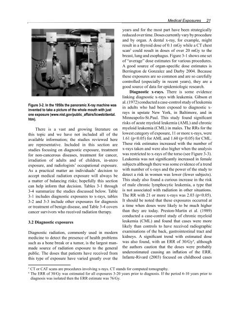
Figure 3-2. In the 1950s the panoramic X-ray machine was<br />
invented to take a picture <strong>of</strong> the whole mouth with just<br />
one exposure (www.nist.gov/public_affairs/licweb/dental.<br />
htm).<br />
There is a vast and growing literature on<br />
this topic and we have not included all <strong>of</strong> the<br />
available information; the studies reviewed here<br />
are representative. Included in this section are<br />
studies focusing on diagnostic exposure, treatment<br />
for non-cancerous diseases, treatment for cancer,<br />
irradiation <strong>of</strong> adults and <strong>of</strong> children, in-utero<br />
exposure, and radiologists’ occupational exposure.<br />
As a practical matter an individuals’ decision to<br />
accept medical radiation exposure will always be<br />
a matter <strong>of</strong> balancing risks; hopefully this section<br />
can help inform that decision. Tables 3-1 through<br />
3-4 summarize the studies discussed below. Table<br />
3-1 includes diagnostic exposures to x-rays, tables<br />
3-2 and 3-3 include other exposures for diagnosis<br />
or treatment <strong>of</strong> benign disease, and Table 3-4 covers<br />
cancer survivors who received radiation therapy.<br />
3.2 Diagnostic exposures<br />
Diagnostic radiation, commonly used in modern<br />
medicine to detect the presence <strong>of</strong> health problems<br />
such as a bone break or a tumor, is the largest manmade<br />
source <strong>of</strong> radiation exposure to the general<br />
public. The doses that patients have received from<br />
this type <strong>of</strong> exposure have varied greatly over the<br />
Medical Exposures 21<br />
years and for the most part have been strategically<br />
reduced over time. Doses currently vary by procedure<br />
and by organ. A dental x-ray, for example, might<br />
result in a thyroid dose <strong>of</strong> 0.1 mGy while a CT chest<br />
scan 3 could result in doses <strong>of</strong> over 20 mGy to the<br />
breast, lung and esophagus. Figure 3-1 shows one set<br />
<strong>of</strong> “average” dose estimates for various procedures.<br />
A good source <strong>of</strong> organ-specific dose estimates is<br />
Berrington de Gonzalez and Darby 2004. Because<br />
these exposures are so common and are so carefully<br />
controlled (especially in recent years), they are a<br />
good source <strong>of</strong> data for epidemiologic research.<br />
Diagnostic x-rays. There is some evidence<br />
linking diagnostic x-rays with leukemia. Gibson et<br />
al. (1972) conducted a case-control study <strong>of</strong> leukemia<br />
in adults who had been exposed to diagnostic xrays<br />
in upstate New York, in Baltimore, and in<br />
Minneapolis-St.Paul. This study found significant<br />
risks <strong>of</strong> acute myeloid leukemia (AML) and chronic<br />
myeloid leukemia (CML) in males. The RRs for the<br />
lowest category <strong>of</strong> exposure, 11 or more x-rays, were<br />
1.61 (p