Health Risks of Ionizing Radiation: - Clark University
Health Risks of Ionizing Radiation: - Clark University
Health Risks of Ionizing Radiation: - Clark University
Create successful ePaper yourself
Turn your PDF publications into a flip-book with our unique Google optimized e-Paper software.
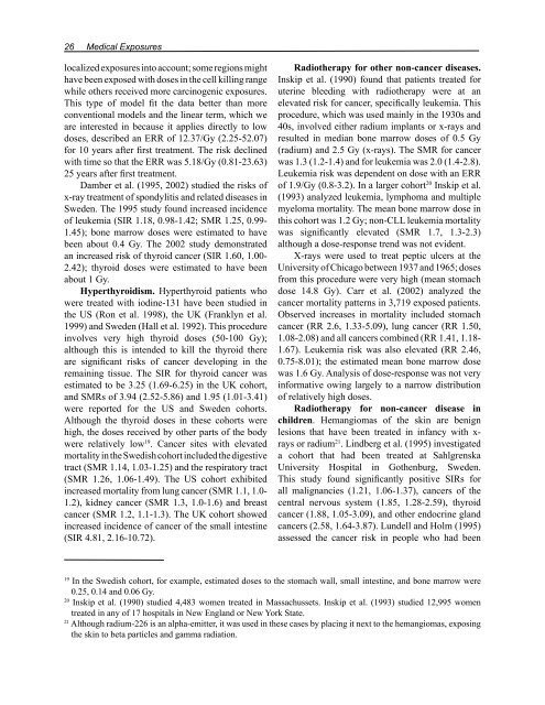
26 Medical Exposures<br />
localized exposures into account; some regions might<br />
have been exposed with doses in the cell killing range<br />
while others received more carcinogenic exposures.<br />
This type <strong>of</strong> model fit the data better than more<br />
conventional models and the linear term, which we<br />
are interested in because it applies directly to low<br />
doses, described an ERR <strong>of</strong> 12.37/Gy (2.25-52.07)<br />
for 10 years after first treatment. The risk declined<br />
with time so that the ERR was 5.18/Gy (0.81-23.63)<br />
25 years after first treatment.<br />
Damber et al. (1995, 2002) studied the risks <strong>of</strong><br />
x-ray treatment <strong>of</strong> spondylitis and related diseases in<br />
Sweden. The 1995 study found increased incidence<br />
<strong>of</strong> leukemia (SIR 1.18, 0.98-1.42; SMR 1.25, 0.99-<br />
1.45); bone marrow doses were estimated to have<br />
been about 0.4 Gy. The 2002 study demonstrated<br />
an increased risk <strong>of</strong> thyroid cancer (SIR 1.60, 1.00-<br />
2.42); thyroid doses were estimated to have been<br />
about 1 Gy.<br />
Hyperthyroidism. Hyperthyroid patients who<br />
were treated with iodine-131 have been studied in<br />
the US (Ron et al. 1998), the UK (Franklyn et al.<br />
1999) and Sweden (Hall et al. 1992). This procedure<br />
involves very high thyroid doses (50-100 Gy);<br />
although this is intended to kill the thyroid there<br />
are significant risks <strong>of</strong> cancer developing in the<br />
remaining tissue. The SIR for thyroid cancer was<br />
estimated to be 3.25 (1.69-6.25) in the UK cohort,<br />
and SMRs <strong>of</strong> 3.94 (2.52-5.86) and 1.95 (1.01-3.41)<br />
were reported for the US and Sweden cohorts.<br />
Although the thyroid doses in these cohorts were<br />
high, the doses received by other parts <strong>of</strong> the body<br />
were relatively low 19 . Cancer sites with elevated<br />
mortality in the Swedish cohort included the digestive<br />
tract (SMR 1.14, 1.03-1.25) and the respiratory tract<br />
(SMR 1.26, 1.06-1.49). The US cohort exhibited<br />
increased mortality from lung cancer (SMR 1.1, 1.0-<br />
1.2), kidney cancer (SMR 1.3, 1.0-1.6) and breast<br />
cancer (SMR 1.2, 1.1-1.3). The UK cohort showed<br />
increased incidence <strong>of</strong> cancer <strong>of</strong> the small intestine<br />
(SIR 4.81, 2.16-10.72).<br />
Radiotherapy for other non-cancer diseases.<br />
Inskip et al. (1990) found that patients treated for<br />
uterine bleeding with radiotherapy were at an<br />
elevated risk for cancer, specifically leukemia. This<br />
procedure, which was used mainly in the 1930s and<br />
40s, involved either radium implants or x-rays and<br />
resulted in median bone marrow doses <strong>of</strong> 0.5 Gy<br />
(radium) and 2.5 Gy (x-rays). The SMR for cancer<br />
was 1.3 (1.2-1.4) and for leukemia was 2.0 (1.4-2.8).<br />
Leukemia risk was dependent on dose with an ERR<br />
<strong>of</strong> 1.9/Gy (0.8-3.2). In a larger cohort 20 Inskip et al.<br />
(1993) analyzed leukemia, lymphoma and multiple<br />
myeloma mortality. The mean bone marrow dose in<br />
this cohort was 1.2 Gy; non-CLL leukemia mortality<br />
was significantly elevated (SMR 1.7, 1.3-2.3)<br />
although a dose-response trend was not evident.<br />
X-rays were used to treat peptic ulcers at the<br />
<strong>University</strong> <strong>of</strong> Chicago between 1937 and 1965; doses<br />
from this procedure were very high (mean stomach<br />
dose 14.8 Gy). Carr et al. (2002) analyzed the<br />
cancer mortality patterns in 3,719 exposed patients.<br />
Observed increases in mortality included stomach<br />
cancer (RR 2.6, 1.33-5.09), lung cancer (RR 1.50,<br />
1.08-2.08) and all cancers combined (RR 1.41, 1.18-<br />
1.67). Leukemia risk was also elevated (RR 2.46,<br />
0.75-8.01); the estimated mean bone marrow dose<br />
was 1.6 Gy. Analysis <strong>of</strong> dose-response was not very<br />
informative owing largely to a narrow distribution<br />
<strong>of</strong> relatively high doses.<br />
Radiotherapy for non-cancer disease in<br />
children. Hemangiomas <strong>of</strong> the skin are benign<br />
lesions that have been treated in infancy with xrays<br />
or radium 21 . Lindberg et al. (1995) investigated<br />
a cohort that had been treated at Sahlgrenska<br />
<strong>University</strong> Hospital in Gothenburg, Sweden.<br />
This study found significantly positive SIRs for<br />
all malignancies (1.21, 1.06-1.37), cancers <strong>of</strong> the<br />
central nervous system (1.85, 1.28-2.59), thyroid<br />
cancer (1.88, 1.05-3.09), and other endocrine gland<br />
cancers (2.58, 1.64-3.87). Lundell and Holm (1995)<br />
assessed the cancer risk in people who had been<br />
19 In the Swedish cohort, for example, estimated doses to the stomach wall, small intestine, and bone marrow were<br />
0.25, 0.14 and 0.06 Gy.<br />
20 Inskip et al. (1990) studied 4,483 women treated in Massachussets. Inskip et al. (1993) studied 12,995 women<br />
treated in any <strong>of</strong> 17 hospitals in New England or New York State.<br />
21 Although radium-226 is an alpha-emitter, it was used in these cases by placing it next to the hemangiomas, exposing<br />
the skin to beta particles and gamma radiation.


