

Download - Journal of Cell and Molecular Biology - Haliç Üniversitesi
Download - Journal of Cell and Molecular Biology - Haliç Üniversitesi
Download - Journal of Cell and Molecular Biology - Haliç Üniversitesi

Create successful ePaper yourself
Turn your PDF publications into a flip-book with our unique Google optimized e-Paper software.
200 µl L-Glutamin, 20 µl penicillin- streptomycin<br />
<strong>and</strong> 200 µl phytohaemagglutinin. After incubation<br />
at 37ºC for 72 hours, 200 µl Colchicine was added<br />
to arrest the cells at metaphase. Following an<br />
additional incubation at 37ºC for 30 minutes <strong>and</strong><br />
centrifugation at 20ºC for 8 min. at 1500 rpm the<br />
supernatant was removed. The pellet was resuspended<br />
with up to 10 ml hypotonic solution<br />
(0.4% KCl solution) vortexed immediately. All the<br />
samples were kept at 37ºC for 20 minutes <strong>and</strong><br />
again centrifuged at the same condition. After<br />
removing supernatant from the samples, the pellet<br />
which contains cells at metaphase, was<br />
homogenised. Fixative solution (methanol <strong>and</strong><br />
acetic acid mixed with 3:1 ratio) was added <strong>and</strong> the<br />
tubes were vortexed for the fixation <strong>of</strong><br />
chromosomes. Then samples were centrifuged after<br />
adding up to 5 ml <strong>of</strong> fixative solution. Supernatant<br />
was discarded from the samples <strong>and</strong> fresh fixative<br />
solution was added to the tubes. This procedure<br />
was repeated until the samples were clarified.<br />
According to the cell density, up to 0.5 ml fixative<br />
solution was added to the samples. Then samples<br />
were homogenized <strong>and</strong> cells were lied onto slide<br />
glasses, which were kept at 4ºC in distilled water<br />
till they are used. After spreading the cells on the<br />
slides, the samples were dried at room temperature<br />
<strong>and</strong> kept overnight at 60ºC.<br />
Y chromosome microdeletions in spontaneous abortions<br />
Karyotyping<br />
GTG (Giemsa-Trypsin) b<strong>and</strong>ing technique was<br />
performed. When the b<strong>and</strong>ing <strong>of</strong> the chromosomes<br />
was not successful, the protocol was repeated.<br />
After staining, at least 20 metaphase plaques were<br />
analysed for each sample (Figure 1).<br />
Detection <strong>of</strong> Y chromosome microdeletions<br />
DNA isolation from blood<br />
DNA was extracted from 200 µl peripheral blood by<br />
using High Pure PCR Template Preparation Kit<br />
(Roche-Germany) according to the manufacturer’s<br />
protocol.<br />
Multiplex polymerase chain reaction<br />
(multiplex PCR)<br />
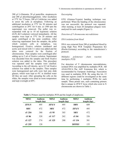
Table 1. Primers used for multiplex PCR <strong>and</strong> the length <strong>of</strong> amplicons.<br />
MIX1 Amlicon<br />
length (bp)<br />
MIX2 Amlicon<br />
length (bp)<br />
For detection <strong>of</strong> Y chromosome microdeletions,<br />
isolated DNA was amplified by multiplex PCR. AB<br />
ANALITICA–The AZF Extension Kit, which is<br />
recommended by European Andrology Association<br />
was used in multiplex PCR. By using this kit, 13<br />
different regions could be investigated at the same<br />
time by performing 3 multiplex PCRs for each<br />
sample. Three primer sets, each containing primers<br />
that is unique to ZFX/Y locus which also exist in X<br />
chromosome are shown in Table 1.<br />
MIX3 Amlicon<br />
length (bp)<br />
ZFX/Y 495 ZFX/Y 495 DBY 689<br />
SRY 472 SRY 472 ZFX/Y 495<br />
sY 254 380 sY 95 303 SRY 472<br />
sY 86 320 sY 117 262 sY 84 326<br />
sY 127 274 sY 125 200 sY 134 301<br />
sY 255 120 DFFRY 155<br />
47


