
You also want an ePaper? Increase the reach of your titles
YUMPU automatically turns print PDFs into web optimized ePapers that Google loves.
<strong>EMBL</strong> Research at a Glance 2009<br />
Claude Antony<br />
PhD 198, Université Paris VI.<br />
Postdoctoral research at<br />
<strong>EMBL</strong> 1987-1989.<br />
Group Leader at CNRS 199-<br />
2003.<br />
Facility head and team<br />
leader at <strong>EMBL</strong> since 2003.<br />
Cell cytoskeleton: an electron tomography analysis<br />
Previous and current research<br />
In January 2008 we established a new electron tomography set-up, which will be invaluable for future<br />
innovative projects at <strong>EMBL</strong>. Our focus of interest is the organisation of the fission yeast and<br />
budding yeast microtubular cytoskeleton using electron tomography, which not only allows the<br />
reconstruction, modelling and quantifying of subcellular elements, but also enables the visualisation<br />
of a number of structural features in very fine detail.<br />
Having performed a detailed analysis of the wild type fission yeast cytoskeleton array (Höög et al.,<br />
2007), we have carried out investigations on EB1/mal3 and CLIP170/tip1 MAPs (microtubule associated<br />
proteins) mutants which regulate microtubule dynamics, and also on microtubule<br />
bundling factors.<br />
Plus end tracking protein mutants: Mal3 deletion mutant displays disordered and shorter microtubule<br />
bundles in the cytoplasm, although the number of microtubules within these bundles<br />
is similar to that in wt cells. There is no apparent antiparallel overlapping bundle and this deletion<br />
also causes alteration to the spindle pole body (SPB). The deletion of Tip1 causes severe alterations<br />
as the total microtubule polymer length is considerably reduced and a loss of SPB-microtubule<br />
connection is observed. Our data suggest that Tip1p may play a role in microtubule<br />
nucleation which is supported by abnormally thin filaments, numerous in the deletion strain.<br />
We also studied microtubule regrowth after depolymerisation of the whole microtubular array using the carbendazim microtubule depolymerising<br />
drug (MBC). Upon drug wash-out we captured the early stages of microtubule bundle reassembly and observed detailed features of<br />
the growing microtubules. This analysis was carried out in collaboration with the Brunner group (opposite), and will be published soon.<br />
Microtubule bundling factors: To understand inter-microtubule bonds, we reconstructed microtubule arrays in deletion strains which affect<br />
the bundling function. Ase1 (encoding a non-motor homodimer protein) deletion strain shows a loss of parallel organisation of the bundles<br />
despite some overlapping area where microtubules remain associated but with a reduced inter-microtubule spacing. The deletion of Klp2 (a kinesin-like<br />
protein which slides microtubules along pre-existing ones in a minus end directed manner) shows mostly bundles composed of two<br />
microtubules. In the double deletion ase1Δ/klp2Δ a microtubule overlap region still persists but in a parallel orientation. SPB-associated bundles<br />
are missing. In all these mutants strains inter-microtubule bridges can be detected, suggesting that other bundling factors still prevail.<br />
Future projects and goals<br />
A new project has been started in collaboration with the Knop (page 16) and Kaksonen (page 15) groups concerning the reconstruction of the<br />
microtubule bundles involved in the karyogamy process in the budding yeast mating pathway by using tomography<br />
and microtubule modelling. We will also investigate the cytoskeleton architecture in shmooing cells<br />
(polarisation process of mating cells). This will involve the analysis of both microtubule and actin bundles in<br />
shmooing cells and their connection with the plasma membrane and the SPB.<br />
In conjunction with in-house and external research groups we are now starting a major project to reconstruct<br />
the Xenopus mitotic spindle. Cryofixation and preparation of samples for tomography acquisition are just the<br />
first steps. The large-scale reconstruction of such a huge structure, and detailed parts of it, will be performed<br />
using extensive montaging and joining of tomograms. In the course of this project we hope to elucidate the spindle<br />
microtubule architecture at fine resolution and in doing so, derive information about microtubule polarity<br />
in the midzone of the spindle. A precise analysis of the organisation of the spindle poles should also be carried<br />
out. A student and a postdoc are currently working on this long-term project.<br />
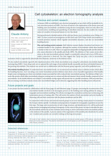
3D model of Ase1 deletion strain showing the disorganised microtubule bundles (green), SPB (yellow), mitochondria<br />
(blue) and nucleus (turquoise). The plasma membrane and cell wall appear as semi-transparent in grey. (Picture by<br />
Helio Roque, postdoc).<br />
Selected references<br />
Colombelli, J., Tangemo, C., Haselman, U., Antony, C., Stelzer, E.H.,<br />
Pepperkok, R. & Reynaud, E.G. (2008). A correlative light and<br />
electron microscopy method based on laser micropatterning and<br />
etching. Methods Mol. Biol., 57, 203-13<br />
Höög, J.L. & Antony, C. (2007). Whole-cell investigation of<br />
microtubule cytoskeleton architecture by electron tomography.<br />
Methods Cell Biol., 79, 15-167<br />
10<br />
Höög, J.L., Schwartz, C., Noon, A.T., O’Toole, E.T., Mastronarde,<br />
D.N., McIntosh, J.R. & Antony, C. (2007). Organization of interphase<br />
microtubules in fission yeast analyzed by electron tomography. Dev.<br />
Cell, 12, 39-361<br />
Toya, M., Sato, M., Haselmann, U., Asakawa, K., Brunner, D.,<br />
Antony, C. & Toda, T. (2007). Gamma-tubulin complex-mediated<br />
anchoring of spindle microtubules to spindle-pole bodies requires<br />
Msd1 in fission yeast. Nat. Cell Biol., 9, 66-653