
You also want an ePaper? Increase the reach of your titles
YUMPU automatically turns print PDFs into web optimized ePapers that Google loves.
<strong>EMBL</strong> Research at a Glance 2009<br />
Rainer<br />
Pepperkok<br />
PhD 1992, University of<br />
Kaiserslautern.<br />
Postdoctoral work at<br />
University of Geneva.<br />
Lab head at the Imperial<br />
Cancer Research Fund,<br />
London.<br />
Team leader at <strong>EMBL</strong> since<br />
1998.<br />
Membrane traffic in the early secretory pathway<br />
Previous and current research<br />
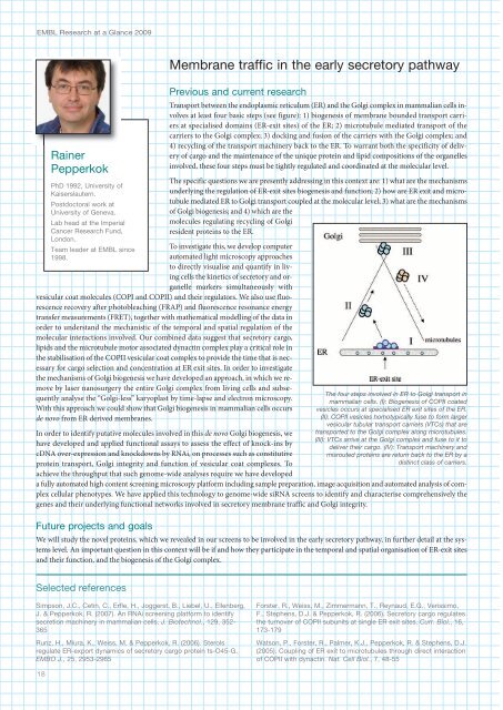
Transport between the endoplasmic reticulum (ER) and the Golgi complex in mammalian cells involves<br />
at least four basic steps (see figure): 1) biogenesis of membrane bounded transport carriers<br />
at specialised domains (ER-exit sites) of the ER; 2) microtubule mediated transport of the<br />
carriers to the Golgi complex; 3) docking and fusion of the carriers with the Golgi complex; and<br />
4) recycling of the transport machinery back to the ER. To warrant both the specificity of delivery<br />
of cargo and the maintenance of the unique protein and lipid compositions of the organelles<br />
involved, these four steps must be tightly regulated and coordinated at the molecular level.<br />
The specific questions we are presently addressing in this context are: 1) what are the mechanisms<br />
underlying the regulation of ER-exit sites biogenesis and function; 2) how are ER exit and microtubule<br />
mediated ER to Golgi transport coupled at the molecular level; 3) what are the mechanisms<br />
of Golgi biogenesis; and 4) which are the<br />
molecules regulating recycling of Golgi<br />
resident proteins to the ER.<br />
To investigate this, we develop computer<br />
automated light microscopy approaches<br />
to directly visualise and quantify in living<br />
cells the kinetics of secretory and organelle<br />
markers simultaneously with<br />
vesicular coat molecules (COPI and COPII) and their regulators. We also use fluorescence<br />
recovery after photobleaching (FRAP) and fluorescence resonance energy<br />
transfer measurements (FRET), together with mathematical modelling of the data in<br />
order to understand the mechanistic of the temporal and spatial regulation of the<br />
molecular interactions involved. Our combined data suggest that secretory cargo,<br />
lipids and the microtubule motor associated dynactin complex play a critical role in<br />
the stabilisation of the COPII vesicular coat complex to provide the time that is necessary<br />
for cargo selection and concentration at ER exit sites. In order to investigate<br />
the mechanisms of Golgi biogenesis we have developed an approach, in which we remove<br />
by laser nanosurgery the entire Golgi complex from living cells and subsequently<br />
analyse the “Golgi-less” karyoplast by time-lapse and electron microscopy.<br />
With this approach we could show that Golgi biogenesis in mammalian cells occurs<br />
de novo from ER derived membranes.<br />
In order to identify putative molecules involved in this de novo Golgi biogenesis, we<br />
have developed and applied functional assays to assess the effect of knock-ins by<br />
cDNA over-expression and knockdowns by RNAi, on processes such as constitutive<br />
protein transport, Golgi integrity and function of vesicular coat complexes. To<br />
achieve the throughput that such genome-wide analyses require we have developed<br />
a fully automated high content screening microscopy platform including sample preparation, image acquisition and automated analysis of complex<br />
cellular phenotypes. We have applied this technology to genome-wide siRNA screens to identify and characterise comprehensively the<br />
genes and their underlying functional networks involved in secretory membrane traffic and Golgi integrity.<br />
Future projects and goals<br />
The four steps involved in ER to Golgi transport in<br />
mammalian cells. (I): Biogenesis of COPII coated<br />
vesicles occurs at specialised ER exit sites of the ER.<br />
(II): COPII vesicles homotypically fuse to form larger<br />
vesicular tubular transport carriers (VTCs) that are<br />
transported to the Golgi complex along microtubules.<br />
(III): VTCs arrive at the Golgi complex and fuse to it to<br />
deliver their cargo. (IV): Transport machinery and<br />
misrouted proteins are return back to the ER by a<br />
distinct class of carriers.<br />
We will study the novel proteins, which we revealed in our screens to be involved in the early secretory pathway, in further detail at the systems<br />
level. An important question in this context will be if and how they participate in the temporal and spatial organisation of ER-exit sites<br />
and their function, and the biogenesis of the Golgi complex.<br />
Selected references<br />
Simpson, J.C., Cetin, C., Erfle, H., Joggerst, B., Liebel, U., Ellenberg,<br />
J. & Pepperkok, R. (2007). An RNAi screening platform to identify<br />
secretion machinery in mammalian cells. J. Biotechnol., 129, 352-<br />
365<br />
Runz, H., Miura, K., Weiss, M. & Pepperkok, R. (2006). Sterols<br />
regulate ER-export dynamics of secretory cargo protein ts-O5-G.<br />
EMBO J., 25, 2953-2965<br />
Forster, R., Weiss, M., Zimmermann, T., Reynaud, E.G., Verissimo,<br />
F., Stephens, D.J. & Pepperkok, R. (2006). Secretory cargo regulates<br />
the turnover of COPII subunits at single ER exit sites. Curr. Biol., 16,<br />
173-179<br />
Watson, P., Forster, R., Palmer, K.J., Pepperkok, R. & Stephens, D.J.<br />
(2005). Coupling of ER exit to microtubules through direct interaction<br />
of COPII with dynactin. Nat. Cell Biol., 7, 8-55<br />
18