
You also want an ePaper? Increase the reach of your titles
YUMPU automatically turns print PDFs into web optimized ePapers that Google loves.
Cell Biology and Biophysics Unit<br />
Cell morphogenesis and spatial microtubule<br />
organisation<br />
Previous and current research<br />
To create a defined morphology, cells need to polarise and correctly orient their polarity axis. Both<br />
processes require a defined intracellular order based on the specific sub-cellular arrangement of<br />
actin and microtubule filaments. Our investigations focus on the contribution of microtubules,<br />
whose organisation varies tremendously between different cell types and also in individual cells<br />
during different developmental or cell cycle stages. Little is known about how this variability is<br />
achieved and how cells switch from one organisational state to another. We address these questions<br />
in two model organisms, the unicellular fission yeast Schizosaccharomyces pombe and the fruit fly<br />
Drosophila melanogaster.<br />
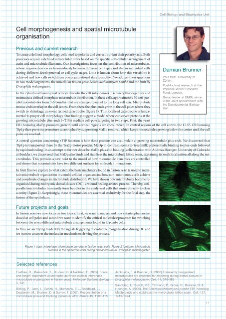
In the cylindrical fission yeast cells we describe the cell autonomous machinery that organises and<br />
maintains a defined interphase microtubule distribution. In these cells, approximately 30 anti-parallel<br />
microtubules form 3-6 bundles that are arranged parallel to the long cell axis. Microtubule<br />
minus-ends overlap in the cell centre. From there the plus-ends grow to the cell poles where they<br />
switch to shrinkage, an event termed catastrophe (figure 1). This localised catastrophe is fundamental<br />
to proper cell morphology. Our findings suggest a model where conserved proteins at the<br />
growing microtubule plus-ends (+TIPs) mediate cell-pole targeting in two steps. First, the yeast<br />
EB1 homolog Mal3p promotes growth until cortical regions are encountered. In central regions of the cell cortex, the CLIP-170 homolog<br />
Tip1p then prevents premature catastrophes by suppressing Mal3p removal, which keeps microtubules growing below the cortex until the cell<br />
poles are reached.<br />
A central question concerning +TIP function is how these proteins can accumulate at growing microtubule plus-ends. We discovered that<br />
Tip1p is transported there by the Tea2p motor protein. Mal3p in contrast, seems to ‘treadmill’, preferentially binding to plus-ends followed<br />
by rapid unbinding. In an attempt to further describe Mal3p plus-end binding (collaboration with Andreas Hoenger, University of Colorado<br />
at Boulder), we discovered that Mal3p also binds and stabilises the microtubule lattice seam, explaining its weak localisation all along the microtubules.<br />
This provides a new twist to the model of how microtubule dynamics are controlled<br />
and shows that microtubules have two different surfaces for molecular interactions.<br />
In fruit flies we explore to what extent the basic machinery found in fission yeast is used to maintain<br />
microtubule organisation in a multi-cellular organism and how non-autonomous cells achieve<br />
and coordinate changes in microtubule distribution. We have shown how microtubules become reorganised<br />
during embryonic dorsal closure (DC), a wound healing-related process. Thereby, antiparallel<br />
microtubules transiently form bundles in the epidermal cells that move dorsally to close<br />
a cavity (figure 2). Surprisingly, these microtubules are essential exclusively for the final step, the<br />
fusion of the epithelium.<br />
Future projects and goals<br />
In fission yeast we now focus on two topics. First, we want to understand how catastrophes are induced<br />
at cell poles and second we want to identify the critical molecules/processes for switching<br />
between the seven different microtubule arrangements found in S. pombe cells.<br />
In flies, we are trying to identify the signals triggering microtubule reorganisation during DC and<br />
we want to uncover the molecular mechanisms driving the process.<br />
Damian Brunner<br />
PhD 1995, University of<br />
Zürich.<br />
Postdoctoral research at the<br />
Imperial Cancer Research<br />
Fund, London.<br />
Group leader at <strong>EMBL</strong> since<br />
2000. Joint appointment with<br />
the Developmental Biology<br />
Unit.<br />
Figure 1 (top): Interphase microtubule bundles in fission yeast cells. Figure 2 (bottom): Microtubule<br />
bundles in the epidermal cells during dorsal closure in Drosophila melanogaster.<br />
Selected references<br />
Foethke, D., Makushok, T., Brunner, D. & Nédélec, F. (2009). Force<br />
and length-dependent catastrophe activities explain interphase<br />
microtubule organization in fission yeast. Molecular Systems Biology,<br />
5, 21<br />
Bieling, P., Laan, L., Schek, H., Munteanu, E.L., Sandblad, L.,<br />
Dogterom, M., Brunner, D. & Surrey, T. (2007). Reconstitution of a<br />
microtubule plus-end tracking system in vitro. Nature 5, 1100-115<br />
Jankovics, F. & Brunner, D. (2006) Transiently reorganised<br />
microtubules are essential for zippering during dorsal closure in<br />
Drosophila melanogaster. Cell, 11, 375-385<br />
Sandblad, L., Busch, K.E., Tittmann, P., Gross, H., Brunner, D. &<br />
Hoenger, A. (2006). The Schizosaccharomyces pombe EB1 homolog<br />
Mal3p binds and stabilizes the microtubule lattice seam. Cell, 127,<br />
115-12<br />
11