

Biophysical studies of membrane proteins/peptides. Interaction with ...
Biophysical studies of membrane proteins/peptides. Interaction with ...
Biophysical studies of membrane proteins/peptides. Interaction with ...

You also want an ePaper? Increase the reach of your titles
YUMPU automatically turns print PDFs into web optimized ePapers that Google loves.
F. Fernandes et al. / Biochimica et Biophysica Acta 1758 (2006) 1777–1786<br />
1783<br />
qffiffiffiffiffiffiffiffiffiffiffiffiffiffiffiffiffiffiffiffiffiffiffiffiffiffiffiffiffiffiffiffiffiffiffiffiffiffiffiffiffiffiffiffiffiffiffiffiffiffiffiffiffiffiffiffiffiffiffiffiffiffiffiffiffiffiffiffiffiffiffiffiffiffiffiffiffiffiffiffiffiffiffiffiffiffiffiffiffiffiffiffiffiffi<br />
½H4 Baf Š<br />
¼ 1 K bð½Baf Š T<br />
þ½H4Š T<br />
Þþ ð1 þ K b ð½Baf Š T<br />
þ½H4Š T<br />
ÞÞ 2 4ðKb 2½Baf Š T ½H4Š T Þ<br />
ð7Þ<br />
½H4Š T<br />
ð 2K b Þ½H4Š T<br />
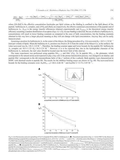
where [H4-Baf] is the effective concentration (molecules per lipid volume as the binding is confined to the lipid phase) <strong>of</strong> the<br />
peptide–bafilomycin A 1 complex, and, [H4] T and [Baf] T are respectively, the effective analytical concentrations <strong>of</strong> the peptide and <strong>of</strong><br />
bafilomycin A 1 . E EXP is the energy transfer efficiencies obtained experimentally and E random is the theoretical energy transfer<br />
efficiency assuming a random distribution <strong>of</strong> acceptors (Eqs. (1)–(3)). In case binding is detected, the use <strong>of</strong> effective bafilomycin A 1<br />
concentrations will result in lower binding constants as compared to the ones <strong>of</strong> bulk concentrations, but the binding constants<br />
obtained in this way have a larger physical meaning as they will not change <strong>with</strong> lipid concentration. Anyway they can be easily<br />
interconverted.<br />
Assuming a position for bafilomycin A 1 in the center <strong>of</strong> the bilayer, the fitting procedure (Fig. 6A) recovered K b =10.5±1.32 M − 1<br />
(mol per volume <strong>of</strong> lipid). When the bafilomycin A 1 position was fixed to 20 Å from the center <strong>of</strong> the bilayer (i.e. at the surface), the<br />
value recovered was K b =36.2±2.8 M − 1 . Therefore, the binding constant upper and lower bounds for the peptide H4–bafilomycin<br />
A 1 complex are 10.5±1.32 13.72±3.05 M − 1 .<br />
Fig. 6. (A) Fluorescence emission quenching <strong>of</strong> Tyr15 <strong>of</strong> peptide H4 by FRET to bafilomycin A 1 (●). Theoretical expectations (Eqs. (1–3)) for FRET in the absence <strong>of</strong><br />
inhibitorbindingtopeptideH4assumingpositionsforbafilomycinA 1 <strong>of</strong>12.5Å(⋯),8.5Å(–––),and4.5Å(—) fromthecenter<strong>of</strong>thebilayer.( )Fitting<strong>of</strong>equations6 and<br />
7 to FRET data assuming a bafilomycin A 1 position <strong>of</strong> 4.5 Å from the center <strong>of</strong> the bilayer. The interval 10.5±1.34


