

April Journal-2009.p65 - Association of Biotechnology and Pharmacy
April Journal-2009.p65 - Association of Biotechnology and Pharmacy
April Journal-2009.p65 - Association of Biotechnology and Pharmacy

Create successful ePaper yourself
Turn your PDF publications into a flip-book with our unique Google optimized e-Paper software.
Current Trends in <strong>Biotechnology</strong> <strong>and</strong> <strong>Pharmacy</strong><br />
Vol. 3 (2) 138-148, <strong>April</strong> 2009. ISSN 0973-8916<br />
in vitro. The HUVECs were plated on the<br />
Matrigel. The HUVECs in the basal medium could<br />
not form tubes <strong>and</strong> VEGF was used to induce<br />
the tube formation. In the positive control group<br />
stimulated with VEGF (10ng), HUVECs rapidly<br />
aligned with one another <strong>and</strong> formed tube-like<br />
structures resembling a capillary plexus within 8<br />
hours, after VEGF treatment. However,<br />
treatment with withaferin A prevented VEGF -<br />
stimulated tube formation <strong>of</strong> HUVECs in a<br />
concentration (3.5-14µg) - dependent manner<br />
(Fig. 1). Meanwhile, no cytotoxicity was observed<br />
under this concentration range <strong>of</strong> withaferin A<br />
used in the assay. Withaferin A was shown to<br />
interfere with the ability <strong>of</strong> HUVECs to form the<br />
in vitro vessel-like tubes, one <strong>of</strong> the important<br />
traits <strong>of</strong> the endothelial cells.<br />
142<br />
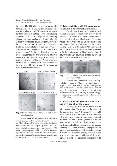
Withaferin A inhibits VEGF induced neovascularization<br />
on chick chorioallantoic membrane<br />
CAM assay is one <strong>of</strong> the widely used<br />
validation assays for formation <strong>of</strong> new blood<br />
vessels. In order to further verify if withaferin A<br />
is an inhibitor <strong>of</strong> new blood vessel formation,<br />
withaferin A was applied on chorioallantoic<br />
membrane <strong>of</strong> chick embryo to test its in vivo<br />
antiangiogenic activity. In the CAM assay model<br />
withaferin A induced avasculature zone formation<br />
in the developing embryos. Notably newly formed<br />
microvessels were regressed around the area <strong>of</strong><br />
withaferin A treated CAM (Fig. 2).<br />
Fig. 2: Effect <strong>of</strong> withaferin A on neovascularization<br />
in the chick CAM<br />
Withaferin A was applied on CAM <strong>of</strong> 11-dayold<br />
chicken embryo. After 48h <strong>of</strong> incubation, the<br />
applied area was inspected for changes in<br />
neovascularization. The arrows indicate the applied<br />
area. The data shown represents the result <strong>of</strong> an<br />
experiment, which was done using maximum six eggs<br />
in each group. All photographs were taken at same<br />
magnification.<br />
Fig. 1: Effect <strong>of</strong> withaferin A on VEGF induced HUVECs<br />
tube formation<br />
HUVECs (5X10 3 cells) cultured in EGM medium<br />
with 3.5µg, 7µg <strong>and</strong> 14 µg withaferin A was added to<br />
the Matrigel coated 96 wells plate. After incubation<br />
for 8 hours at 37 0 C, capillary networks were<br />
photographed <strong>and</strong> quantified (Magnification: X40).<br />
Concentration dependent inhibition <strong>of</strong> tube formation<br />
by withaferin A was recorded. All datas are presented<br />
as mean from three different experiments with triplicates<br />
<strong>and</strong> means <strong>of</strong> ± S.E.M. P


