2008 Summer Meeting - Leeds - The Pathological Society of Great ...
2008 Summer Meeting - Leeds - The Pathological Society of Great ...
2008 Summer Meeting - Leeds - The Pathological Society of Great ...
You also want an ePaper? Increase the reach of your titles
YUMPU automatically turns print PDFs into web optimized ePapers that Google loves.
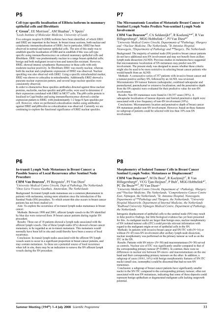
P5Cell type specific localisation <strong>of</strong> ERbeta is<strong>of</strong>orms in mammaryepithelial cells and fibroblastsCGreen 1 , EE Morrison 1 , AM Shaaban 1 ,VSpeirs 11 <strong>Leeds</strong> Institute <strong>of</strong> Molecular Medicine, University <strong>of</strong> <strong>Leeds</strong>Five estrogen receptor ß (ERß) is<strong>of</strong>orms have been identified, <strong>of</strong> which ERß1and ERß2 are important in the breast. In breast tissue sections, both nuclear andcytoplasmic immunolocalisation <strong>of</strong> ERß1, but in particular, ERß2 has beenobserved in normal and tumour epithelial cells. <strong>The</strong> aim <strong>of</strong> this study was toestablish specific localisation <strong>of</strong> ERß and in establish if this was cell typespecific using immun<strong>of</strong>luorescence on cultured mammary epithelial cells andfibroblasts. ERß1 was predominantly nuclear in a range breast epithelial cells;benign and both malignant invasive/non-and tamoxifen-resistant. However,ERß2, showed intense cytoplasmic fluorescence in these cells with onlymoderate nuclear positivity. In fibroblasts ERß1 was mostly nuclear, whereasboth the nuclear and cytoplasmic expression <strong>of</strong> ERß2 was observed. Nuclearspeckling was also observed with ERß2. Using a specific mitochondrial marker,ERß2 was shown to colocalise in mitochondria. Additionally ERß2 showed apunctate nuclear expression pattern, and several large nuclear speckles wereconsistently observed.In order to characterise these speckles antibodies directed against three nuclearproteins, nucleolin, nuclear speckle and p80 coilin, were used to determine iftheir expression correlated with ERß2 in MCF7 cells. <strong>The</strong> p80 coilin protein islocalised in Cajal bodies and using immun<strong>of</strong>luorescence showed a similarexpression pattern to ERß2, with predominantly 1-2 large 0.5µm speckles percell. However, when we performed colocalisation studies using antibodiesagainst ERß2 and p80coilin no colocalisation was observed. Currently we arecontinuing to explore the functional significance <strong>of</strong> ERß2 nuclear speckles.P7<strong>The</strong> Microanatomic Location <strong>of</strong> Metastatic Breast Cancer inSentinel Lymph Nodes Predicts Non-sentinel Lymph NodeInvolvementCHM Van Deurzen* 1 , CA Seldenrijk† 2 , R Koelemij** 2 ,RVanHillegersberg# 1 , MGG Hobbelink+ 1 , PJ Van Diest* 11 University Medical Centre Utrecht, Departments <strong>of</strong> *Pathology, #Surgeryand +Nuclear Medicine, <strong>The</strong> Netherlands, 2 St Antonius Hospital,Nieuwegein, †Departments <strong>of</strong> Pathology and **Surgery, <strong>The</strong> NetherlandsBackground: <strong>The</strong> majority <strong>of</strong> sentinel node (SN) positive breast cancer patientsdo not have additional non-SN involvement and may not benefit from axillarylymph node dissection (ALND). Previous studies in melanoma have suggestedthat microanatomic localization <strong>of</strong> SN metastases may predict non-SNinvolvement. <strong>The</strong> present study was designed to assess whether these criteriamight also be used to be more restrictive in selecting breast cancer patients whowould benefit from an ALND.Methods: A consecutive series <strong>of</strong> 357 patients with invasive breast cancer anda tumour positive axillary SN, followed by an ALND, was reviewed.Microanatomic SN tumour features (subcapsular, combined subcapsular andparenchymal, parenchymal or extensive localization, and the penetrative depthfrom the SN capsule) were evaluated for their predictive value for non-SNinvolvement.Results: Non-SN metastases were found in 136/357 cases (38%). Asubcapsular localisation <strong>of</strong> tumour deposits and limited penetrative depth wereassociated with a low frequency <strong>of</strong> non-SN involvement (10%).Conclusions: Microanatomic location and penetrative depth <strong>of</strong> breast cancerSN metastases predict non-SN involvement. However, based on these featuresno subgroup <strong>of</strong> patients could be selected with less than 10% non-SNinvolvement.P6In-transit Lymph Node Metastases in Breast Cancer: aPossible Source <strong>of</strong> Local Recurrence after Sentinel NodeProcedureCHM Van Deurzen 1 , PJ Borgstein 2 , PJ Van Diest 11 University Medical Centre Utrecht, Dept <strong>of</strong> Pathology,<strong>The</strong> Netherlands2 Onze Lieve Vrouwe Gasthuis, Amsterdam, <strong>The</strong> NetherlandsBackground: In-transit lymph node metastases are a common phenomenon inpatients with melanoma, raising more attention since the introduction <strong>of</strong> theSentinel Node (SN) procedure. To which extent this also occurs in breast cancerpatients has not been studied yet.Aim: To explore the occurrence <strong>of</strong> in-transit lymph nodes metastases in breastcancer.Methods: Between 1998 and 2001, afferent lymph vessels to the SN identifiedby blue dye were removed from 18 breast cancer patients during regular SNprocedure.Results: Three out <strong>of</strong> 18 patients showed a lymph node associated with theafferent lymph vessels. One <strong>of</strong> these lymph nodes (6%) showed a breast cancermetastasis, to be regarded as an in-transit metastasis. This metastasis wouldnormally have been left in situ and could thereby have been a source <strong>of</strong> localrecurrence.Conclusion: In-transit lymph nodes associated with the afferent SN lymphvessels seem to occur in a significant proportion in breast cancer patients, andmay contain metastases. As these are a potential source <strong>of</strong> local recurrencewhen left in situ, there may be an indication to remove the SN afferent lymphvessels during the SN procedure.P8Morphometry <strong>of</strong> Isolated Tumour Cells in Breast CancerSentinel Lymph Nodes: Metastases or Displacement?CHM Van Deurzen+ 1 ,MDe Boer 2 , R Koelemij† 3 ,RVanHillegersberg# 1 , VCG Tjan-Heijnen 4 ,PBult 5 , MGG Hobbelink* 1 ,PC De Bruin** 3 , PJ Van Diest+ 11 University Medical Centre Utrecht, Department <strong>of</strong> +Pathology, #Surgeryand *Nuclear Medicine, <strong>The</strong> Netherlands, 2 Comprehensive Cancer CentreEast, Nijmegen, the Netherlands, 3 St. Antonius Hospital, Nieuwegein,Departments <strong>of</strong> **Pathology and †Surgery, the Netherlands, 4 UniversityHospital Maastricht, Department <strong>of</strong> Internal Medicine, the Netherlands5 Radboud University Nijmegen Medical Centre, Department <strong>of</strong> Pathology,the NetherlandsIatrogenic displacement <strong>of</strong> epithelial cells to the sentinel node (SN) may resultin false positive findings, but little biological evidence has yet been presentedfor this. As malignant nuclei are larger than benign ones, nuclear morphometry<strong>of</strong> SN isolated tumour cells (ITC) could provide relevant information withregard to the malignant origin-or-not <strong>of</strong> epithelial cells in the SN.Methods: In patients with invasive breast cancer and SN ITC with (N=16) orwithout (N=45) non-SN involvement after axillary lymph node dissection,nuclear morphometry was performed on the primary tumour as well as on theITC in the SN.Results: Patients with SN micro- (N=30) and macrometastases (N=30) servedas controls. Nuclear size <strong>of</strong> ITC was significantly smaller compared to that <strong>of</strong>the corresponding primary tumour (P


