2008 Summer Meeting - Leeds - The Pathological Society of Great ...
2008 Summer Meeting - Leeds - The Pathological Society of Great ...
2008 Summer Meeting - Leeds - The Pathological Society of Great ...
You also want an ePaper? Increase the reach of your titles
YUMPU automatically turns print PDFs into web optimized ePapers that Google loves.
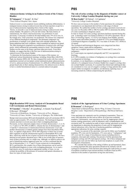
P83Adenocarcinoma Arising in an Endocervicosis <strong>of</strong> the UrinaryBladderM Nakaguro 1 ,YSuzuki 1 ,KOno 11 Tosei General Hospital, Seto, JapanEndocervicosis is a non-neoplastic lesion exhibiting mullerian differentiation. Amalignant tumour arising in endocervicosis has not been reported, except for acase <strong>of</strong> adenocarcinoma originating from a lesion <strong>of</strong> the vagina.Herein, we describe a case <strong>of</strong> adenocarcinoma arising in endocervicosis <strong>of</strong> theurinary bladder. <strong>The</strong> patient is a 58-year-old woman. She had a history <strong>of</strong>endometriosis, for which a total hysterectomy was performed 16 yearspreviously. Cystoscopy revealed an elevated nodular mass, 4*4 cm in size, inthe triangle area. Total cystectomy was performed. <strong>The</strong> tumour was composed<strong>of</strong> two different histological components. <strong>The</strong> dominant component wasproliferation <strong>of</strong> multiple cystic glands lined with cuboidal epithelium. <strong>The</strong>lining epithelium had clear cytoplasm, which resembled an endocervical gland.<strong>The</strong> other histological component was proliferation <strong>of</strong> atypical cells with largenuclei, which infiltrated the surrounding connective tissue. This histologicalfinding was the same as the biopsy specimen taken previously. From thesefindings, we suggest that this is the first case <strong>of</strong> adenocarcinoma arising inendocervicosis <strong>of</strong> the urinary bladder.To determine the histochemical nature <strong>of</strong> the mucus <strong>of</strong> this tumour, weperformed various histochemical stainings, including Alcian Blue (AB)-PAS,High iron diamine (HID)-AB. We then compared the results with four controlpopulations (normal cervical gland, normal endometrial gland, adenocarcinoma<strong>of</strong> cervix, adenocarcinoma <strong>of</strong> endometrium). It is difficult to differentiate thesegroups based only on their staining characteristics, but endocervicosis is shownto have a similar histochemical nature to a normal endocervical gland.P85<strong>The</strong> role <strong>of</strong> urine cytology in the diagnosis <strong>of</strong> bladder cancer atUniversity College London Hospitals during one yearM Ben-Gashir 1 ,MFalzon 1 , A Capitanio 11 University College London HospitalsWe have seen an increase in the number <strong>of</strong> urine specimens for cytologicalexamination in our department derived from a wide range <strong>of</strong> clinicalspecialities. We audited the number <strong>of</strong> urine cytology specimens received overone year (1st December 2006 to 30th November 2007) to determine the ability<strong>of</strong> a urine examination to diagnosis cancer.In total we found 1522 urine cytology specimens had been reported during thisyear. A histology report was found in relation to 216 (24%) specimens. Out <strong>of</strong>these 216 histology reports, 113 (52%) were biopsies from bladder, prostate,kidney, ureter and urethra (urology) <strong>of</strong> which 82 (73%) were bladder biopsies.A histological diagnosis <strong>of</strong> papillary TCC was made in 50 and a TCC (NOS) in10 reports.<strong>The</strong> cytological and histological diagnoses were categorized into threecategories; cancer, atypia and no malignancy.<strong>The</strong>re was a complete agreement in 8 cases <strong>of</strong> cancer and 22 cases <strong>of</strong> nomalignancy.In 11 cases atypia was reported cytologically and TCC was reported inhistology.In 41 (50%) samples, no evidence <strong>of</strong> malignancy on cytology but a carcinomawas diagnosed on histology.In summary, in our department, a cytological diagnosis <strong>of</strong> malignancy isindicative <strong>of</strong> a malignant lesion <strong>of</strong> urinary bladder but a negative cytology doesnot exclude bladder carcinoma. It is essential that clinical and radiological dataare considered in every case <strong>of</strong> a negative cytology to triage patients that wouldrequire further investigation. In addition, our laboratory exploring ways <strong>of</strong>increasing sensitivity without compromising the specificity.P84High-Resolution SNP-Array Analysis <strong>of</strong> Chromophobe RenalCell Carcinomas and Renal OncocytomasM Yusenko 1 ,TBoethe 2 , B Ljundberg 3 , A Geurts Van Kessel 4 ,GKovacs 1 ,RKuiper 41 University <strong>of</strong> Heidelberg, Germany, 2 University <strong>of</strong> Pecs, Hungary,3 University <strong>of</strong> Umea, Sweden, 4 University <strong>of</strong> Nijmegen, <strong>The</strong> NetherlandsBenign renal oncocytomas (RO) and chromophobe renal cell carcinomas (RCC)make up approximately 10% <strong>of</strong> renal cell tumours. <strong>The</strong> histological diagnosisremains uncertain in several cases due to their overlapping phenotype.Previously, we have detected complex losses <strong>of</strong> whole chromosomes 1, 2, 6, 10,13, 17 and 21 in 70%-90% <strong>of</strong> chRCCs, whereas the loss <strong>of</strong> chromosomes 1 and14 and the Y chromosome or translocation between chromosome 11q13 andanother chromosomes or random genetic changes have been described in ROs.Now, we applied a high density 250 K SNP oligoarray to detect loss <strong>of</strong> smallDNA segments at specific chromosomal regions in chromophobe RCCs andROs, which may mark the loci <strong>of</strong> genes involved in the tumorigenesis. We havedetected several genetic alterations up to 1Mb in size in both types <strong>of</strong> tumours,most <strong>of</strong> them occurring only in a single case. Although, we failed to achieve ourgoal <strong>of</strong> identifying tumour suppressor gene loci, we have confirmed that theloss <strong>of</strong> chromosomes 2, 13, 17 and 21 occurs exclusively in chromophobeRCCs and therefore, the complex loss <strong>of</strong> these chromosomes can be used forthe differential diagnosis in cases with overlapping phenotype. As we used ahigh density array for analysing an appropriate number <strong>of</strong> cases, we canexclude the posibility <strong>of</strong> the presence <strong>of</strong> recurrent snall genetic alterations inthese two types <strong>of</strong> renal cancers.P86Analysis <strong>of</strong> the Appropriateness <strong>of</strong> Urine Cytology SpecimensR Brannan 1 , G Rodrigues 21 Department <strong>of</strong> Histopathology, Bexley Wing, St James' UniversityHospital, <strong>Leeds</strong>, 2 Department <strong>of</strong> Histopathology, Hull Royal Infirmary,HullUrine specimens are commonly sent for cytological examination. <strong>The</strong>re aresome valid indications for this such as follow up <strong>of</strong> previous urothelialneoplasia. Unfortunately, some specimens are sent for inappropriate clinicalreasons creating unnecessary work for laboratory staff, cytopathologists andsecretarial staff. We conducted an audit <strong>of</strong> urine cytology specimens received inour department to determine the proportion <strong>of</strong> cases sent for inappropriateclinical indications with a final aim <strong>of</strong> producing guidelines outlining theappropriate indications for urine cytology requests.Twenty two percent <strong>of</strong> the specimens were sent for follow up <strong>of</strong> urothelialneoplasia, 39% were sent for haematuria, 15% were sent as “? malignancy” and24% were sent for a range <strong>of</strong> miscellaneous reasons including infectivesymptoms, chest pain and vomiting. One percent <strong>of</strong> all the specimens hadmalignant cytology and 10% had atypical cytology. All <strong>of</strong> these malignant andatypical cases were sent for follow up <strong>of</strong> previous neoplasia or as part <strong>of</strong> theinvestigation <strong>of</strong> haematuria.We conclude that outside <strong>of</strong> the setting <strong>of</strong> follow up and haematuria, thereis a strong argument that urine specimens should not be routinely sent forcytological examination. If this had been done during this audit period, no cases<strong>of</strong> neoplasia would have been missed and there would have been an 40%reduction in the number <strong>of</strong> specimens saving time and money.<strong>Summer</strong> <strong>Meeting</strong> (194 th ) 1–4 July <strong>2008</strong> Scientific Programme53


