2008 Summer Meeting - Leeds - The Pathological Society of Great ...
2008 Summer Meeting - Leeds - The Pathological Society of Great ...
2008 Summer Meeting - Leeds - The Pathological Society of Great ...
You also want an ePaper? Increase the reach of your titles
YUMPU automatically turns print PDFs into web optimized ePapers that Google loves.
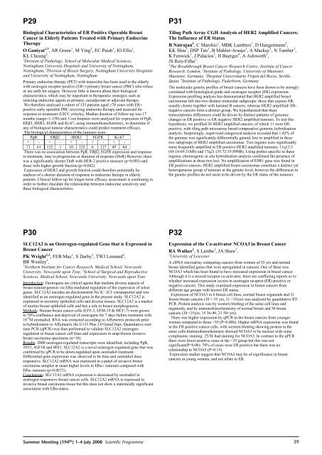
P29Biological Characteristics <strong>of</strong> ER Positive Operable BreastCancer in Elderly Patients Treated with Primary Endocrine<strong>The</strong>rapyOGaniyat 1,2 , AR Green 1 ,MYing 2 , EC Paish 1 , IO Ellis 1 ,KL Cheung 2 .1 Division <strong>of</strong> Pathology, School <strong>of</strong> Molecular Medical Sciences,Nottingham University Hospitals and University <strong>of</strong> Nottingham,Nottingham, 2 Division <strong>of</strong> Breast Surgery, Nottingham University Hospitalsand University <strong>of</strong> Nottingham, NottinghamPrimary endocrine therapy (PET) with tamoxifen has been used in the elderlywith oestrogen receptor positive (ER+) primary breast cancer (PBC) who refuseor are unfit for surgery. However little is known about their biologicalcharacteristics, which may be important in therapeutic strategies such asselecting endocrine agents as primary, neoadjuvant or adjuvant therapy.We therefore analysed a cohort <strong>of</strong> 133 patients aged 70 years with ER+positive early operable PBC receiving endocrine therapy and assessed forresponse to treatment (UICC criteria). Median duration <strong>of</strong> follow-up was 17months (range=1-150) and. Core biopsies were analysed for expression <strong>of</strong> PgR,ER2, HER2, EGFR and Ki-67, using immunohistochemistry, to determine ifany <strong>of</strong> biological tumour characteristics could predict treatment efficacy.<strong>The</strong> biological characteristics <strong>of</strong> the tumours were:PgR ER2 HER2 EGFR Ki-67+ - + - + - + - + -71 61 132 1 10 123 6 127 49 84<strong>The</strong>re was no association between PgR, ER2, EGFR expression and responseto treatment, time to progression or duration <strong>of</strong> response (DoR) However, therewas a significantly shorter DoR with HER-2 positive tumours (p=0.003) andthose with higher growth fraction (p=0.042).Expression <strong>of</strong> HER2 and growth fraction could therefore potentially bemarkers <strong>of</strong> a shorter duration <strong>of</strong> response to endocrine therapy in elderlypatients. Clinical follow-up for longer-term efficacy assessment is continuing inorder to further elucidate the relationship between endocrine sensitivity andthese biological characteristics.P31Tiling Path Array CGH Analysis <strong>of</strong> HER2 Amplified Cancers:<strong>The</strong> Influence <strong>of</strong> ER StatusR Natrajan 1 , C Marchio 1 , MBK Lambros 1 , D Hungermann 2 ,KK Shiu 1 ,DSP Tan 1 , B Mahler-Araujo 1 , A Mackay 1 ,NTamber 1 ,K Fenwick 1 , J Palacios 3 , H Buerger 4 , A Ashworth 1 ,JS Reis-Filho 1 .1 <strong>The</strong> Breakthrough Breast Cancer Research Centre, Institute <strong>of</strong> CancerResearch, London, 2 Institute <strong>of</strong> Pathology, University <strong>of</strong> Muenster,Muenster, Germany, 3 Hospital Universitario Virgen del Rocío, Seville,Spain, 4 Institute <strong>of</strong> Pathology, Paderborn, Germany<strong>The</strong> molecular genetic pr<strong>of</strong>iles <strong>of</strong> breast cancers have been shown to be stronglycorrelated with histological grade and oestrogen receptor (ER) expression.Expression pr<strong>of</strong>iling analysis has demonstrated that HER2 amplified breastcarcinomas fall into two distinct molecular subgroups: those that express ER,usually cluster together with luminal B cancers, whereas HER2 amplified/ ERnegativecancers form a distinct group. We hypothesised that thesetranscriptomic differences could be driven by distinct patterns <strong>of</strong> genomicchanges in ER positive vs ER negative HER2 amplified tumours. To test thishypothesis, we pr<strong>of</strong>iled 26 HER2 amplified cancers, <strong>of</strong> which 11 were ERpositive,with tiling path microarray based comparative genomic hybridisationanalysis. Surprisingly, supervised categorical analysis revealed that 1.42% <strong>of</strong>the genome was significantly differentially gained, lost or amplified in thesetwo subgroups <strong>of</strong> HER2 amplified carcinomas. Two regions were significantlymore frequently amplified in ER positive HER2 amplified tumours: 11q13.3(69.18-69.31Mb) and 17q21 (35.72-35.89Mb). Using probes specific to theseregions, chromogenic in situ hybridisation analysis confirmed the presence <strong>of</strong>amplifications at these two loci. No amplification <strong>of</strong> ESR1 gene was found inER positive cancers. HER2 amplified breast carcinomas constitute a distinct yetheterogeneous group <strong>of</strong> tumours at the genetic level, however the differences inthe genetic pr<strong>of</strong>iles do not seem to be driven by the ER status <strong>of</strong> the tumours.P30SLC12A2 is an Oestrogen-regulated Gene that is Expressed inBreast CancerPK Wright 1,2 , FEB May 1 ,SDarby 1 , TWJ Lennard 2 ,BR Westley 1 .1 Northern Institute for Cancer Research, Medical School, NewcastleUniversity, Newcastle upon Tyne, 2 School <strong>of</strong> Surgical and ReproductiveSciences, Medical School, Newcastle University, Newcastle upon TyneIntroduction: Oestrogens are critical agents that mediate diverse aspects <strong>of</strong>breast tumourigenesis via ER-mediated regulation <strong>of</strong> the expression <strong>of</strong> selectgenes. SLC12A2 encodes an electroneutral Na + K + -2Cl - cotransporter and wasidentified as an oestrogen-regulated gene in the present study. SLC12A2 isexpressed in secretory epithelial cells and diverse tissues. SLC12A2 is a marker<strong>of</strong> murine breast epithelial cells and has a role in breast morphogenesis.Methods: Human breast cancer cells (EFF-3, EFM-19 & MCF-7) were grownto 70% confluence and deprived <strong>of</strong> oestrogens for 7 days before treatment with10 -9 M oestradiol. RNA was extracted according to Affymetrix protocols priorto hybridization to Affymetrix Hu U133 Plus 2.0 GeneChips. Quantitative realtimePCR (qPCR) was then performed to validate SLC12A2 oestrogenregulationin breast cancer cell lines and expression in snap-frozen invasivebreast carcinoma specimens (n=18).Results: 2800 oestrogen-regulated transcripts were identified, including PgR,TFF1, IGF1R and IRS1. SLC12A2 is a novel oestrogen-regulated gene that wasconfirmed by qPCR to be down-regulated upon oestradiol treatment.Differential gene expression was observed to be time and oestradiol doseresponsive. SLC12A2 mRNA was expressed in a panel <strong>of</strong> invasive breastcarcinoma samples at mean higher levels in ER+ tumours compared withER- tumours (p=0.0872).Conclusions: SLC12A2 mRNA expression is decreased by oestradiol inoestrogen-responsive breast cancer cells. SLC12A2 mRNA is expressed ininvasive breast carcinoma tissue but this does not show a statistically significantassociation with ER-status.P32Expression <strong>of</strong> the Co-activator NCOA3 in Breast CancerRA Walker 1 ,SLambe 1 ,JA Shaw 1 .1 University <strong>of</strong> LeicesterA cDNA microarray comparing cancers from women <strong>of</strong> 35 yrs and normalbreast identified genes that were upregulated in cancers. One <strong>of</strong> these wasNCOA3 which has been found to have increased expression in breast cancer.Although it is a steroid receptor co-activator, there are conflicting reports as towhether increased expression occurs in oestrogen receptor (ER) positive ornegative cancers. This study examined expression in breast cancers fromdifferent age groups with known ER status.Expression <strong>of</strong> NCOA3 in 6 breast cell lines, normal breast organoids and 21frozen breast cancers (10 < 35 yrs, 11 >35yrs) was analysed by quantitative RT-PCR. Protein analysis was by western blotting <strong>of</strong> the same cell lines andorganoids, and by immunohistochemistry <strong>of</strong> normal breast and 56 breastcancers (20 yrs).<strong>The</strong>re was higher expression by qPCR in the breast cancers from youngerwomen compared to those >35 (P=0.006). Higher mRNA expression was foundin the ER positive cancer cells, with westren blotting showing protein in thesame cells.Immunohistochemistry showed NCOA3 to be nuclear with somecytoplasmic staining. 25/56 had staining for NCOA3. In contrast to the qPCRthere were fewer positive cases in the


