Summary of HIR Equipment (2012-2013) - High Impact Research
Summary of HIR Equipment (2012-2013) - High Impact Research
Summary of HIR Equipment (2012-2013) - High Impact Research
- No tags were found...

You also want an ePaper? Increase the reach of your titles
YUMPU automatically turns print PDFs into web optimized ePapers that Google loves.
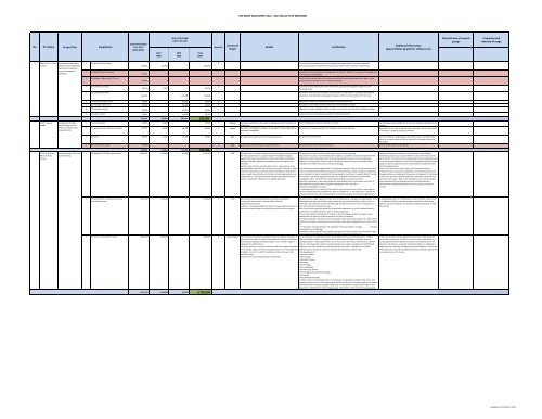
<strong>HIR</strong>-MOHE EQUIPMENT <strong>2012</strong> - <strong>2013</strong> (FACULTY OF MEDICINE)No PI's Name Project Title <strong>Equipment</strong>sEstimated BudgetYear 2 & 3(<strong>2012</strong>-<strong>2013</strong>)<strong>2012</strong>(RM)Approved Budget(After VM Lab)<strong>2013</strong>(RM)Total(RM)QuantityCountry <strong>of</strong>OriginDetailsJustificationAdditional information(space/utilities eg electric, s<strong>of</strong>tware etc)Potential users/researchgroupsFrequency andintensity <strong>of</strong> usage6 Assoc. Pr<strong>of</strong>. Dr. Tunku Improving articular disease 1 E6-Motion capture systemKamarulprediction and management,regenerative therapy andsarcoma management2 E7-MIMICS Visual 3D s<strong>of</strong>tware(NOCERAL)3 E8-S<strong>of</strong>twares (Sigma plot, SPSS, etc.,)250,000 320,000 - 320,00041,000 0 - -35,000 0 - -1 This instrument is required to quantify the baseline movement pattern. Data obtained will beinformative and used to determine the root cause <strong>of</strong> injuries and to develop a treatment plan.1 Required to process and edit the 2 D image data (CT, micro CT, MRI etc.,) to construct 3 D models withatmost accuracy and flexibility.1 Statitical s<strong>of</strong>twares are required to analyse and validate the genereated experimental datas , makecomplex graphs and illustratrions for a better publication.4 E9-Inverted microscope5 E10-3D Surface Scanner20,425 20,500 - 20,500160,000 - 120,000 120,0001 To view live cells by allowing the entire environment upon which the organism resides on to themicroscope stage.1 Required to analyse a real-world object or environment to collect data on its shape and possibly itsappearance. Data obtained can be applied to design orthotecis and prostehetics for clinical use.6 E11-Mechanobiology station 81,000 - 81,000 81,000 1 A platform to conduct all experiments related to mechantransduction.7 E12-Portable cutting unit 30,000 - 30,000 30,000 1 Requried to cut the hard bones for biomechanical and histological analysis.8 E13-Shaking incubator40,000 - 40,000 40,0001 Required for mixing or biological liquids as wellas the incubation and cultivation <strong>of</strong> bilogical samples.9 E21-CO2 Incubator 14,025 - 14,100 14,100 1 To grow the cells in a temperature and Co2 controlled environment.671,450 340,500 285,100 625,6007 Pr<strong>of</strong>. Dr. Awang Development <strong>of</strong> spatiotemporaldengue modelresearchers.1 E2-12 workstations 40,000 34,000 - 34,000 1 Taiwan Computer workstation to be used for modelling by doctoral students and Math modelling and computer simulation <strong>of</strong> models.Bulgiba&infectious disease meta- 2 E7-segmental body composition analyser 120,000 60,000 60,000 120,000 1 Japan Bio-electrical impedance analyser to be used for measuring body fat Measurement <strong>of</strong> body composition for modelling cardiovascular diseases.analysis (JULIUS)and water composition.Space has been made available. We do not have enough workstations at themoment .We need this to carry out surveys on body composition. We do not have anysuch analyser in JCUM at the present moment.3 E9-freezer 90,000 45,000 45,000 90,000 1 USA 80 degree below zero freezer fo storing biospecimens. Storage <strong>of</strong> frozen biomarkers. There is insufficient storage space in the current freezers. We will put thefreezer in a common space and will be used by all investigators involved inthis project.4 E10-LCo2 back up system 30,000 - - - 1 USA Liquid carbon dioxide system as backup for freezer. Backup system for frozen biomarker samples. This is a backup system for the freezers.280,000 139,000 105,000 244,0008 Dr. Puteri Shafinaz Shared <strong>Equipment</strong> for Faculty 1 E1-Quadrupole-TOF Mass spectrometer 2,850,000 2,750,000 100,000 2,850,000 1 USA The equipment is a hybrid <strong>of</strong> liquid/chip separation system and quadrupole The equipment is very crucial for the <strong>HIR</strong> projects that it will be supporting.A designated lab space is available in the Proteomics Facility, MedicalAkmar Bt Abdul (central Facility)TOF mass spectrometer. It is capable <strong>of</strong> performing high throughput Acquisition <strong>of</strong> accurate mass for identification <strong>of</strong> proteins, metabolites and characterization <strong>of</strong> PTM Biotechnology Laboratory, Faculty <strong>of</strong> Medicine which currently houses theRahmantandem MS analysis (scan MS/MS sensitivity and full MS3 capabilities). modification requires high quality data from mass spectrometry analysis and a comprehensive MALDI-TOF MS. The room has been equipped with two air-conditioning unitsDetection <strong>of</strong> protein, peptide and metabolites at low femtogram-level database is an integral part <strong>of</strong> these projects. This level <strong>of</strong> analysis can only be achieved with the for round the clock temperature control for MS equipment. Electrical voltagesensitivityavailability <strong>of</strong> the QTOF instrument and s<strong>of</strong>tware package.supply to meet the requirement <strong>of</strong> the new MS will be arranged with UM’sDynamic range and mass sensitivity performance : 2ppm mass accuracy.electrical engineering unit.<strong>High</strong> speed data acquisition rate: 50 spectra/second for The Chip LC system The instrument proposed integrates HPLC Chip separation directly on-line to the high resolution QTOF Protein mass identification search engine, post-translational patternis ideal to study protein at post-transcriptional modification level where it system. This allows high speed spectral acquisition rates permitting in depth coverage <strong>of</strong> samples that prediction s<strong>of</strong>tware and proprietary database for known glycoproteins andallows in-depth characterization <strong>of</strong> alteration <strong>of</strong> protein glycosylation and can be analyzed in a shorter experimental time compared to conventional LC spotter-MALDI. The chip- phosphoproteins are included with the purchase <strong>of</strong> equipment. In the tenderphosphorylation. The system also allows relative quantitative analysis <strong>of</strong> MS interface is specifically designed to enhance the LC-MS/MS workflow where it allows PTMwe will ensure that future s<strong>of</strong>tware/database update will be provided at noproteins using isotope- labeled and non-labeled approaches.investigations which is presently the cutting-edge technology in proteomics research.cost for the next 3 years. Without this equipment, many research projectsWithout this equipment, many research projects will be delayed and this will impede the process <strong>of</strong> will be delayed and this will impede the process <strong>of</strong> high quality datahigh quality data acquisition that is needed for publication in Tier 1 journals inacquisition that is needed for publication in Tier 1 journals in proteomicsproteomics/metabolomics research.research.The system proposed is not specific to the needs <strong>of</strong> one group and can be used for a wide range <strong>of</strong>application being proteomics/metabolomics/glycomics/lipidomics. It will also be able to support thegrowing number <strong>of</strong> users that are requesting for an alternative approach to move from gel-based toliquid-based proteomics platform.2 E2-<strong>High</strong> Definition Low Vacuum Scanning1,100,000 1,100,000 - 1,100,000 1 USA Scanning electron microscopy (SEM) is an important equipmentPresently, there is a high request from FOM users for SEM services : average 3-4 students/week. All the An allocated space (20' x 25', temparature 15-20°C, Humidity - 60% or less)Electron Microscopyin many multi-facet analysis involving cellular and extraSEM experiments and research done by the students have to use the equipment in other faculty or for installation <strong>of</strong> the unit is already available at the Electron Microscopycellular matrix interactionuniversities. The researcher now use the SEM at ISB and IMR- however there is very long queue, it is (EM) Unit, Faculty <strong>of</strong> Medicine. The equipment will be operated by trainedresponses. The proposed equipment does not require negative pressure or time consuming and not practical to users.EM unit staff.vacuum to produce images nor processing or gold for enhance imaging. EM unit has the pre-requisites needed for sample/slide preparation prior to the viewing procedureunder SEM. Each student will need 2 days for sample preparationThere are also specific requirements <strong>of</strong> samples for live cell imaging whereby the sample must beviewed at the same place as sample preparation for optimum condition.This system proposed is intended as shared equipment under Electron Microscopy Unit, FOM. It is notspecific to the needs to one group and can be used for a wide range <strong>of</strong> applications such as in the fields<strong>of</strong>:• Parasitology • Oncology <strong>Research</strong> • Microbiology • Infectious Disease • Virology• Anatomy•Neurology• Stem Cell Biology•Availability <strong>of</strong> SEM at FOM will help expedite the progress <strong>of</strong> research projects and acquisition <strong>of</strong> highquality images needed for Tier 1 publication in the above mentioned research areas3 E5- Micro-CT scanner/PET scanner 2,800,0002,800,000 -2,800,000 1 United Kingdom This instrument is required to study the small animal models <strong>of</strong> diseases or In vivo evaluation in a longitudinal study is only possible with the use <strong>of</strong> this equipment. It utilizes In terms <strong>of</strong> space wise, we have allocated a space in the newly renovatedinjuries seen in humans. It will also allow anatomical, structural, functional different physical principles or fundamentals to provide unique biological information from the Electron Microscopy lab in the FOM. For the first 2 years, we'll make sureand molecular imaging <strong>of</strong> underlying subject in vivo. It allows images <strong>of</strong> underlying subject. A large range <strong>of</strong> users can use this system, with interest in either live or cadaveric that the appointed vendor absorbs the costs for full maintenance into theradiograph <strong>of</strong> calcified tissuesmodels. It also allows other users to incorporate their in vivo studies to include more sophisticated initial price. We will hire a researcher with medical physics or radiographye.g. bones, teeth etc. or contrast injected models to be imaged and studied imaging and live cell tracking when implemented within the animal. Availability <strong>of</strong> such system background to run the day to day operations on the equipment. Besides that,in close details. Images can later be reconstructed in 2-D or 3-D images with definitely leads to increase in research productivity in the following research areas:in the tender later, it will clearly stated that the future s<strong>of</strong>tware upgrades willor without contrast. It is safe from radiation and does not require any • Oncology <strong>Research</strong>be provided at no cost for 3 years.specific licensing.• Infectious DiseaseFurthermore it can also include whole animal studies.• Inflammation• Metabolic Diseases• Neurology• Gene Therapy• Stem cell Biology• Cardiovascular Disease• Immunology & Transplantation Biology• Toxicology• Drug Metabolism Studies.Currently, from our own findings, there is no similar piece <strong>of</strong> equipment available in UM. Hence, thisequipment will be part <strong>of</strong> a core animal imaging facility and will serve the imaging needs <strong>of</strong> investigatorsfrom UM and other institutes in greater Klang valley. Turning down the request will introduce somedelay in the establishment <strong>of</strong> animal imaging facility. Some projects which could only be leveraged bythe requested scanner will be affected.6,750,000 6,650,000 100,000 6,750,000- updated on 2nd March <strong>2012</strong> -